chemistry and biology of the initial steps in vision: the...
TRANSCRIPT

Lecture
Chemistry and Biology of the Initial Steps in Vision:The Friedenwald Lecture
Krzysztof Palczewski
Department of Pharmacology, Cleveland Center for Membrane and Structural Biology, School of Medicine, Case Western ReserveUniversity, Cleveland, Ohio, United States
Correspondence: Krzysztof Palczew-ski, Department of Pharmacology,School of Medicine, Case WesternReserve University, 10900 EuclidAvenue, Cleveland, OH 44106, USA;[email protected].
Submitted: August 19, 2014Accepted: August 19, 2014
Citation: Palczewski K. Chemistry andbiology of the initial steps in vision:the Friedenwald lecture. Invest
Ophthalmol Vis Sci. 2014;55:6651–6672. DOI:10.1167/iovs.14-15502
Visual transduction is the process in the eye whereby absorption of light in the retina istranslated into electrical signals that ultimately reach the brain. The first challenge presentedby visual transduction is to understand its molecular basis. We know that maintenance ofvision is a continuous process requiring the activation and subsequent restoration of a vitaminA–derived chromophore through a series of chemical reactions catalyzed by enzymes in theretina and retinal pigment epithelium (RPE). Diverse biochemical approaches that identifiedkey proteins and reactions were essential to achieve a mechanistic understanding of thesevisual processes. The three-dimensional arrangements of these enzymes’ polypeptide chainsprovide invaluable insights into their mechanisms of action. A wealth of information hasalready been obtained by solving high-resolution crystal structures of both rhodopsin and theretinoid isomerase from pigment RPE (RPE65). Rhodopsin, which is activated byphotoisomerization of its 11-cis-retinylidene chromophore, is a prototypical member of alarge family of membrane-bound proteins called G protein–coupled receptors (GPCRs).RPE65 is a retinoid isomerase critical for regeneration of the chromophore. Electronmicroscopy (EM) and atomic force microscopy have provided insights into how certainproteins are assembled to form much larger structures such as rod photoreceptor cell outersegment membranes. A second challenge of visual transduction is to use this knowledge todevise therapeutic approaches that can prevent or reverse conditions leading to blindness.Imaging modalities like optical coherence tomography (OCT) and scanning laser ophthal-moscopy (SLO) applied to appropriate animal models as well as human retinal imaging havebeen employed to characterize blinding diseases, monitor their progression, and evaluate thesuccess of therapeutic agents. Lately two-photon (2-PO) imaging, together with biochemicalassays, are revealing functional aspects of vision at a new molecular level. Thesemultidisciplinary approaches combined with suitable animal models and inbred mutantspecies can be especially helpful in translating provocative cell and tissue culture findings intotherapeutic options for further development in animals and eventually in humans. A host ofdifferent approaches and techniques is required for substantial progress in understandingfundamental properties of the visual system.
Keywords: rhodopsin, photoreceptors, phototransduction, G protein–coupled receptor(s),receptor phosphorylation, membrane proteins, signal transduction, protein structure, vision
The last 30 years of visual system research have resulted in agreater understanding of cellular signal transduction
processes in the eye than in any other organ of human body.Signaling pathways, initiated by activation of rod and conevisual pigments, orchestrate precise changes in cGMP and Ca2þ
that act as second messengers to generate electrical signals.1–8
In 1994, when at the University of Washington, I asked: ‘‘Isvertebrate phototransduction solved? Although the generalmechanism of photoactivation is now known in great detail,key steps in the sequence of reactions associated with thequenching, light adaptation, channel function, and restorationof the dark state of photoreceptors remain to be elucidated.Among many questions, a few seem to be critical forunderstanding phototransduction and related hormonal-trans-duction systems: How is the rhodopsin structure related to theactivated and quiescent states of the receptor? . . ..’’9 Detailedprogress has been made in elucidating G protein inactivation,10
rhodopsin kinase and arrestin structure/function,11–13 and
guanylate cyclase-activating proteins’ (GCAPs) regulation ofguanylate cycles,14,15 among other processes. Although farfrom complete and quantitative (Fig. 1), today we have a solidconceptual framework for the processes of visual pigmentphotoactivation, signal amplification, and quenching of thephototransduction cascade.1–8
Tremendous progress has been made at the molecular level,specifically in five areas of phototransduction research. Theseinclude: molecular and atomic resolution structural studies ofmembrane and soluble proteins that contribute to this signalingcascade2; groundbreaking improvements in understanding ofthe visual (retinoid) cycle16; advancement in understanding thecell biology of the visual system17–20 with multiple advancedimaging modalities21–27; identification of disease-causing genesfor a vast number of retinal/eye disorders28; and pursuit ofnovel concepts, along with animal and human trials oftherapeutics, to prevent or stabilize vision loss, or restorevision in blind individuals.
Copyright 2014 The Association for Research in Vision and Ophthalmology, Inc.
www.iovs.org j ISSN: 1552-5783 6651

We have been pioneers in applying novel technologies toobtain detailed molecular insights and, more recently, thera-peutics. My presentation is not intended to be fully compre-hensive or to upstage original discoveries, but rather to providean overview of the recent progress from the perspective of myown laboratory’s research. It is also important to point out thatwithout fundamental molecular-level research into the secretsof life, progress in translational research would be markedlydiminished. Understanding the richness and complexity of theunderlying basic science is essential when addressing chal-lenges posed by the complexity of visual perception and eyediseases such as AMD, glaucoma, or diabetic retinopathy. Acomprehensive and integrated understanding of the visual
system is indeed crucial for the discovery of mechanism-basednovel treatments for blinding diseases.
RHODOPSIN AT THE CENTER OF VISION
Rhodopsin’s function as a light receptor in the eye wasrecognized over a century ago.29–31 This transmembraneprotein has been the major focus of research, perhaps morethan any other protein of the visual system. Without rhodopsinand its homologous cone pigments, there would be no image-forming vision.32–35 Rhodopsin also has a special place inbiological research because it is a prototypical member of theG protein–coupled receptor (GPCR) family, a vast class of cell
FIGURE 1. Phototransduction in a rod outer segment. Phototransduction can be described in three stages shown from top to bottom in this cartoon.When light strikes rhodopsin (red), it causes isomerization of the 11-cis-retinylidene chromophore to an all-trans configuration and a conformationalchange in the opsin protein. This, in turn, leads to formation of a complex with the heterotrimeric G protein, transducin. Nucleotide exchange inthe transducin a-subunit from guanosine diphosphate to GTP causes dissociation of transducin with formation of the transducin a-subunit. Thissubunit interacts with tetrameric cGMP–specific PDE6, whereas the transducin bc-subunit complexes with phosducin. One activated rhodopsinmolecule can activate dozens of transducin molecules in this first amplification stage of phototransduction. Displacement of the inhibitory c-subunitactivates PDE in the second amplification step of phototransduction. The resulting decrease in the concentration of cGMP is associated with adecrease in intradiscal Ca2þ concentration because cGMP is a ligand for cGMP-gated cation channels (shown in blue in the plasma membrane),nonselective channels that also allow passage of Ca2þ in their cGMP-bound state. The low Ca2þ-level is maintained by the light-insensitive Naþ/Ca2þ-Kþ exchanger, which extrudes Ca2þ ions out against a gradient in exchange for Naþ and Kþ ions. Each of the above-activated molecules needs toreturn to its inactive state before absorption of the next photon. Thus, rhodopsin is phosphorylated at its C-terminus by GRK1 (or rhodopsin kinase[RK]), followed by binding of arrestin, a capping protein. Guanosine triphosphate is hydrolyzed by the a-subunit of transducin with the help of aGTPase-activating protein. Guanylate cyclase 1 and GC2 (GC, light/dark-brown box) are activated by Ca2þ-binding proteins (GCAP1 and GCAP2,black ball) in their Ca2þ-free forms to restore cGMP levels and open the cyclic nucleotide–gated cation channels in the plasma membrane.Guanylate cyclase-activating proteins are inactivated and GC activities return to their dark condition. Once GTP is hydrolyzed by the a-subunit oftransducin along with phosphorylation of phosducin, the heterotrimeric G protein is restored. Opsin recombines with 11-cis-retinal and therhodopsin thus formed is ready to be photoactivated. Note that all these processes take place on the cytoplasmic surfaces of disc and plasmamembranes.
The Friedenwald Lecture IOVS j October 2014 j Vol. 55 j No. 10 j 6652

surface proteins that transmit signals across cellular plasmamembranes via transmembrane domains in response tobinding by hormones, neurotransmitters, odorant and tastemolecules.
Rhodopsin is composed of an opsin protein covalentlyattached to a light-sensitive chromophore, 11-cis-retinal. Thepeptide sequence of opsin, the first among GPCRs, wasdetermined by Ovchinnikov36,37 in the former Soviet Union(Shemyakin Institute of Bioorganic Chemistry, USSR Academy ofSciences) and Hargrave38 (University of Illinois) in the UnitedStates, followed shortly by the cloning of its gene by Nathans andHogness39,40 (Johns Hopkins University). Based on severalbiochemical techniques, a topology including seven membrane-spanning a-helical domains was proposed for rhodopsin thatcompares favorably with the one that was determined almost 2decades later by x-ray crystallography.41,42 Mutations in the opsingene are major causes of inherited blinding disorders.43 Themutation of P23H that causes autosomal-dominant retinitispigmentosa (RP), was the first that linked a human blindingdisease to a specific gene.44 To date, over 100 mutations in theopsin gene have been identified that are linked to this disease withjust a few that cause autosomal recessive RP.43,45 The signalingproperties of rhodopsin were also at the forefront of GPCRresearch. Fung et al.46 (Stanford University) demonstrated that thevisual signal is amplified in the retina when one photoactivatedreceptor activates many molecules of the rod-specific G proteincalled transducin. Photoactivated rhodopsin is also a substrate forrhodopsin kinase (or G protein–coupled receptor kinase[GRK1]).47 In the early 1970s, it was found that rhodopsin binds,when activated and phosphorylated, to a capping protein knownas S-antigen (soluble antigen; today, this protein is called arrestin),which is implicated in ocular uveitis.48 Thereafter, all-trans-
retinylidene is hydrolyzed and released from the active site.49
Phosphorhodopsin is dephosphorylated by protein phosphatase2A50,51 and returns to the ground state. The biochemicalcharacterization of rhodopsin signaling was instrumental for thestudies of other GPCRs, as the majority of them use similarmechanisms for propagation and termination of hormonal signals.
Rhodopsin was the first GPCR whose electron densityprojection maps were determined from two-dimensional (2D)crystals analyzed by EM.52,53 However, no high-resolutionstructure was available, making it difficult to understand theactivation mechanism and effect of disease-causing mutations.It was difficult to envision the possibility of crystallizingrhodopsin because the mass of the hydrophilic domain of thisprotein was small, and thus the contact area betweenmolecules within the crystal limited. Obviously rhodopsinpurification and crystallization required detergent, anothermajor obstacle to its crystallization. Many groups were tryingto crystallize this receptor, but we were the one that got therefirst. Research assistant Van Hooser supplied high quality rodouter segment preparations from freshly isolated bovineretinas to a postdoctoral fellow in my laboratory, Okada, whoa few years later obtained the first three-dimensional (3D)diffracting crystals suitable for x-ray crystallography.54 It was aniterative process that only few could stick to and clearly Tetsujihad motivation, talent, and support to optimize the initial hit incrystallization. Perhaps the single-most important break-through was understanding the properties of rhodopsin andhow it behaves under different experimental conditions. Acollaboration with R Stenkamp, D Taylor (both from Universityof Washington), and M Miyano (Structural Biophysics Labora-tory, RIKEN Harima Institute, Japan) then led to a majorbreakthrough: the three-dimensional atomic structure ofrhodopsin, the first of any GPCR (Fig. 2).42 Even today, therhodopsin structure remains the only structure of a nativeGPCR with a covalently bound ligand that has not beenmodified in terms of its amino acid sequence; and it also
contains posttranslational modifications that are native to themammalian retina, including palmitoylation, N-terminal acety-lation, a disulfide bridge, and Asn2 and Asn15 glycosylation(each with [Man]3[GlcNAc]3 groups). The structure ofrhodopsin contains seven transmembrane helices with thechromophore bound via the protonated Schiff base approxi-mately two-thirds of the way from the cytoplasmic surface, aspredicted by biophysical methods.55 The cytoplasmic siderevealed an unexpected helix 8 that runs parallel to the surfaceof the membrane. Another unexpected feature was a ‘‘plug’’formed by intradiscal loops that create a b-sheet structurebeneath the chromophore. Among the three cytoplasmicloops, the C3-loop has displayed the most diverse conforma-tions among the different crystal structures of rhodopsin. Aspecific retinoid-binding pocket allows only some cis-retinoids(11-cis-, 9-cis-, 7-cis- and several double cis-retinals) to bind toopsin.32 The structure explained several dozen biochemicalreports on the structure/function relationships of rhodopsin,mostly proposed via mutagenesis studies (for reviews see Refs.2–4, 16, 56–68).
Although initial efforts were focused on ground-staterhodopsin, our attention then turned toward solving thestructure of activated rhodopsin which occurs only transiently.A renewed effort by Salom et al.69 led to generation of crystalsthat could be activated by light while retaining theircrystallographic integrity (Fig. 2, right). With the help ofanother postdoctoral fellow, Lodowski,70 the structure of thesecrystals was solved. The major breakthrough that Salom et al.69
employed was the use of ammonium sulfate for proteinconcentration, eliminating the excess of detergent. Thisprecipitation technique was not known for membraneproteins. Moreover, crystals with a high content of deter-gent/lipids appeared to be unstable when exposed to light.Discovery came as a result of hard and persistent work. To oursurprise, upon activation, the conformational changes inrhodopsin were not as complex as proposed from earlierEPR studies.71,72 After solving the opsin structures with andwithout a fragment of transducin, other intermediates, andconstitutively active mutants of rhodopsin, a more completepicture of the activation process emerged.73–85 Rhodopsinphotoactivation causes the isomerization of 11-cis- to all-trans-retinylidene and, subsequently, a cascade of irreversibleconformational changes until rhodopsin reaches its meta Istate.67 This transition leads to a change in the counterion ofthe protonated Schiff base, and a new network of hydrogenbonds is established within the chromophore-binding site.61,86
Meta I remains in an equilibrium with the meta II state. Duringtransition from the meta I to meta II state, glutamate (Glu)113is neutralized. This reaction can be readily monitored bychanges in the absorption of rhodopsin from approximately500 nm (in meta I) to 380 nm (in meta II).87 Meanwhile, asmany as 30 water molecules, which fill the transmembranesegment of rhodopsin, become reorganized and unlock the ‘‘E/DRY’’ motif on the cytoplasmic side of the protein along withprotonation of Glu134, a key residue in this microdomain.68
Recent computational studies revealed that a hydrophobiclayer of amino acid residues next to the characteristic NPxxYmotif forms a gate, opening a continuous water channel uponreceptor activation.88 In addition, the end region of helix VImoves toward the cytoplasmic side.67,89,90 Finally, anotherchange occurs in the NPxxY motif of helix VII and helix VIIIconserved in GPCRs.91 These changes are considered criticalduring the activation process for all GPCRs.
To summarize, the rhodopsin x-ray structure: dispelled themyth that GPCRs cannot be crystalized; provided the three-dimensional positions of all amino acid residues including newunanticipated structural elements; set the field on the righttrack toward understanding rhodopsin photoactivation; out-
The Friedenwald Lecture IOVS j October 2014 j Vol. 55 j No. 10 j 6653

lined the binding pocket of visual chromophore; and firmlyestablished a key role for water molecules in a hydrogenbonding network within the hydrophobic core of this protein.
Today, our structural questions are answered in large part by x-ray crystallography. However, x-ray crystallography only capturesproteins in their most thermodynamically favorable conforma-tions under the particular crystallization conditions. Nuclearmagnetic resonance (NMR) techniques are also well suited toreveal the structural dynamics of proteins.92–95 Along with theabove-mentioned changes in photoactivated rhodopsin, relaxa-tion (i.e., increased flexibility) of this receptor allows thefunctional docking of transducin and activation of this G protein.Key remaining unsolved issues are: more detailed structuralorganization of rhodopsin in native membranes and its functionalconsequences; the molecular mechanisms of photoisomerizedchromophore release and rebinding of newly made 11-cis-retinalto unliganded opsin; the biochemical cycle of rhodopsin in thecell, including its synthesis, membrane insertion, intracellulartransport, disc formation and shedding through phagocytosis bythe RPE, and degradation; how pathogenic mutations change thestructure/function of rhodopsin; and a new classification of thesemutants based of their function rather than older classificationsbased on cellular localization in experimental cell lines or thecapability to bind 11-cis-retinal.
RHODOPSIN IS CRITICAL FOR THE ORGANIZATION OF
ROD OUTER SEGMENTS
Rhodopsin is an essential component of the elongated retinalciliary compartment of the rod cell called the ‘‘outer
segment.’’5,32,96 In mice, rod outer segments have a length of23.6 6 0.4 lm and a diameter of 1.22 6 0.12 lm.99 Eachmammalian rod outer segment consists of 600 to 1000 or more(810 6 10 in mice) distinct flattened discs enclosed by theplasma membrane.97–99 Rhodopsin resides in disc membranesas their major component (>90% of total protein), and is lessdensely packed in the plasma membrane.100 Approximatelyhalf of the surface of each disc is occupied by rhodopsin, withthe remainder filled mostly with lipids, cholesterol, and lessabundant proteins.100–102 Moreover, rhodopsin constitutes~50% of the protein mass of rod outer segment plasmamembranes. Functional differences between the two popula-tions of rhodopsin in the plasma and disc membranes remainunknown. Higher levels of cholesterol in the plasma mem-brane compared with disc membranes could decrease rhodop-sin’s activity. Absence of rhodopsin leads to rudimentary ciliarystructures and eventually to death of rod cells.103,104 Diminu-tion of rhodopsin expression proportionally decreases the sizeof rod outer segment structures, while maintaining the samedensity of rhodopsin as in native discs,98,105 whereas overex-pression of rhodopsin results in enlargement of thesestructures that ultimately causes their instability.105–107
Early work using biophysical approaches by Chabre108
(Laboratoire de Biophysique Moleculaire et Cellulaire, Greno-ble, France), Saibil109 (Department of Biological Sciences andISMB Birkbeck College, London, UK), and Cone,110 and Poo111
(Johns Hopkins University) suggested that rhodopsin ismonomeric and moves rapidly within the discs. Such mobilitywas arguably required for high speed phototransduction. Forexample, Poo111 and Cone110 thought that they measured themobility of rhodopsin in frog disc membranes,111 but full
FIGURE 2. Rhodopsin structure and crystals. Left top: ribbon drawings of rhodopsin (PDB accession code: 1F88)42 in the plane of a disc membrane(two views rotated by 1808). Bottom: the intradiscal side (left) and cytoplasmic side (right) of this receptor. Right: a photo-stable crystal form of thedark inactive state (left) and light-exposed activated state (right) of the receptor. Crystal pictures are reproduced from Salom D, Le Trong I, Pohl E, etal. Improvements in G protein-coupled receptor purification yield light stable rhodopsin crystals. J Struct Biol. 2006;156:497–504. Copyright �2006 Elsevier, Inc.; Salom D, Lodowski DT, Stenkamp RE, et al. Crystal structure of a photoactivated deprotonated intermediate of rhodopsin. Proc
Natl Acad Sci U S A. 2006;103:16123–16128. Copyright � 2006 The National Academy of Sciences of the USA; and Salom D, Padayatti PS,Palczewski K. Crystallization of G protein–coupled receptors. Methods Cell Biol. 2013;117:451–468.299 Copyright � 2013 Elsevier, Inc., withpermission from Elsevier, Inc. and the National Academy of Sciences.
The Friedenwald Lecture IOVS j October 2014 j Vol. 55 j No. 10 j 6654

recovery of rhodopsin absorption was never observed in thebleached area. Recent studies found an explanation,112 namelythat rhodopsin in such membranes undergoes transitions fromrhodopsin (~500 nm) � meta II (~380 nm) � meta III (~480nm). Thus, what was measured by Poo111 and Cone110 was apartial return to absorption in the meta III state. More recentwork appears to add more evidence for ‘‘fast diffusingmonomers of rhodopsin’’ within the disc113–115 while anothercatalytic aspect adds to the confusion. Rhodopsin, in itsmonomeric state, can activate transducin either in detergent orin size-controlled membranes called nanodiscs.116,117 Thus,unlike family C GPCRs (neurotransmitter receptors),118 oligo-merization is not required for signal transduction by rhodopsin,at least in model systems.
Hence, classical diffusion studies implicating monomericrhodopsin provided only indirect evidence and more directimaging methods were needed. With this background, a veryactive and productive collaboration developed between mylaboratory and a world expert in atomic force microscopy(AFM) and membrane biology, Andreas Engel from Biozentrum(Basel, Switzerland). Atomic force microscopy is a high-resolution method that can image electronic orbitals of heavymetals or individual atoms (see Ref. 119 for review). Forbiological systems immersed in water, this technique alsoprovides impressive resolution. Years of work produced high-resolution images of rhodopsin in native membranes thatrevealed a densely packed track of dimeric rhodopsin undervarious experimental conditions (Fig. 3).120,121 This result wasvalidated by a number of complementary biochemical andbiophysical techniques.122–124 Indeed, postdoctoral fellowJastrzebska125 demonstrated that rhodopsin’s oligomeric ar-rangement is sensitive to the concentration and nature ofdetergents, but that under mild conditions a row of rhodopsindimers can be isolated. This finding is consistent with theoligomeric structures of many, if not all GPCRs.126 A theoreticalmodel has been generated but remains to be verifiedexperimentally (Fig. 3).99 As shown by cross-linking studies,one interface can be formed through an asymmetric dimer-dimer interaction mediated by helix I and helix 8 contacts thatis present in native membranes.127 Periole and colleagues128
(Rockefeller University) also employed coarse-grain moleculardynamics to provide evidence for self-assembly of rhodopsin inmembranes. The affinity between two opsin molecules asmeasured by Kd is ~10�5 M.129 Thus, considering the 5 mMconcentration of this receptor in disc membranes, most ofrhodopsin should exist in an oligomeric state. This oligomericstructure of rhodopsin is likely to be essential for themaintenance of a stable disc structure. Another postdoctoralfellow, Park,130–132 took this project further by working withDaniel Muller’s group (ETH Zurich, Switzerland) to discern theenergetics of protein unfolding by using single molecule forcemicroscopy. This technique is a variant of AFM, which canidentify stable structural segments that join together to form anative protein. Experimental evidence for the functionalrelationships between rhodopsin dimerization and photo-transduction, or rhodopsin transport and disc formation is stilla goal of active research.
In retrospect, it would make sense that rhodopsin wouldarrange into higher order oligomers because it exists at highconcentrations in discs. The density of rhodopsin in native roddiscs can be estimated on the basis of the amount of rhodopsinin the retina and, more specifically, in the rod outer segments,in conjunction with the size of the rod outer segment and thenumber of lipid molecules per one molecule of rhodopsin(approximately 60; see Ref. 133 for review). Given that themouse retina contains ~6.4 3 106 rods, the number of discsper retina is ~5 3 109. Consequently, the total amount ofrhodopsin per eye is ~650 pmoles or 650 3 10�12 moles 3
6.022 3 1023 molecules/mole ¼ 3.96 3 1014 rhodopsinmolecules. Thus, there are ~8 3 104 rhodopsin moleculespacked per disc. This high density is only somewhat lowerthan that in 2D crystals of rhodopsin. Such a density leaves twooptions: rhodopsin is either organized in a highly orderedarrangement or there is a well-developed repulsion system thatallows rhodopsin to remain monomeric while surrounded byless than two full belts of phospholipids. The existence of sucha hypothetical repulsion system is questionable because thehydrophobic transmembrane segment of rhodopsin lackscharged residues. Charged amino acids are located exclusivelyat the interface of the membranes and water.
Another push in the imaging technology was our workspearheaded by Nickel from the laboratory of Baumeister and
FIGURE 3. Organization of rhodopsin in disc membranes. An AFMtomograph (tilted by 58) shows the paracrystalline arrangement ofrhodopsin dimers in a native disc membrane. The rhodopsin moleculesprotrude from the lipid bilayer by 1.4 6 0.2 nm or one-fourth of theirmass. Inset shows a model for the packing arrangement of rhodopsinmolecules within the paracrystalline arrays in native disc membranes(PDB accession code: 1N3M).99 Helices of rhodopsin are colored asfollows: helix I in blue, helix II in light blue, helix III in green, helix IVin light green, helix V in yellow, helix VI in orange, and helix VII andcytoplasmic helix 8 in red. The red box shows the close contactsbetween four molecules of rhodopsin. The dashed-line rectangle
shows the longer contacts with neighboring dimers of rhodopsin. Thenative membrane structure and the molecular model of rhodopsin arereproduced from Liang Y, Fotiadis D, Filipek S, Saperstein DA,Palczewski K, Engel A. Organization of the G protein-coupled receptorsrhodopsin and opsin in native membranes. J Biol Chem.2003;278:21655–21662. Copyright � 2003 The American Society forBiochemistry and Molecular Biology, Inc.; and Fotiadis D, Liang Y,Filipek S, Saperstein DA, Engel A, Palczewski K. Atomic-forcemicroscopy: rhodopsin dimers in native disc membranes. Nature.2003;421:127–128, with permission from the American Society forBiochemistry and Molecular Biology and Nature Publishing Group.
The Friedenwald Lecture IOVS j October 2014 j Vol. 55 j No. 10 j 6655
Paul Park97 once again. We used cryoelectron tomography tovisualize the detailed morphology of the rod outer segmentfrom the retina of mice in three dimensions. This newtechnique for vision research allowed us to preserve thestructure of rod outer segments because the native tissue wasquickly frozen in liquid ethane without any ice formation, thuspreserving the sample under the most native conditions.Vitrified rod outer segments were among the largest structuresimaged by this tomographic technique. We demonstrated theexistence of spacer structures connecting adjacent discs thatprovided an accurate framework for the space within whichphototransduction occurs. Several years later, the sametechnique was employed to image the connecting cilium.134
PROTEINS INTERACTING WITH RHODOPSIN
In rod outer segments, rhodopsin interacts with four proteinsinvolved in phototransduction.135 Rhodopsin couples withtransducin following photoactivation. With a molar ratio torhodopsin in rod outer segments of approximately 1:10,transducin is the second-most abundant membrane-associatedprotein of this structure. Although it is possible that rhodopsinand transducin are precoupled, this nonproductive complexwould not permit nucleotide exchange on the a-subunit oftransducin.133 Transducin was the first phototransductionprotein to be crystalized in different subunit states.136–139 Afew dozen transducin molecules are activated by a singlephotoactivated rhodopsin.1,140
Interaction between rhodopsin and transducin has beenstudied by a number of biophysical techniques.2,133,141,142
However, isolation of the rhodopsin-G protein complex was amajor accomplishment. Jastrzebska143 was the first to isolatethe rhodopsin-transducin complex in solution and determineits structure by negative-stain EM. Both her biochemical andstructural data were consistent with a pentameric assembly ofthe photoactivated rhodopsin-transducin complex.144–146 Eachrhodopsin monomer within this complex plays a differentstructural role, as revealed biochemically.147 This complex wasdetergent-sensitive, and upon increasing the concentration ofn-dodecyl-b-D-maltoside from 1 to 5 mM,145 one of the tworhodopsin monomers dissociated suggesting that they cooper-ate to form a dimer by hydrophobic interactions. Understand-ing the molecular changes that occur within transducin duringthis nucleotide exchange was also advanced by recentstudies.148–150 Another protein that recognizes photoactivatedrhodopsin is rhodopsin kinase or GRK1.12,47 In mice, GRK1 isexpressed in both rod and cone photoreceptor cells.12 Lack ofthis protein causes Oguchi disease, congenital stationary nightblindness, associated with fundus discoloration and a profounddecrease in dark adaptation.151,152 As a postdoctoral fellow (inHargrave’s laboratory University of Florida), I purified thisenzyme from bovine retina and extensively characterized itsproperties.153–155 This was a challenging project becauserhodopsin kinase is farnesylated and methylated at a Cysresidue in the C-terminus causing the protein to stick tomembranes and hydrophobic surfaces.156 The enzyme wassequenced and cloned in collaboration with Lorenz157 from theLefkowitz group (Duke University). Many years later, it wascrystallized by (reviewed by Homan and Tesmer158) Singh etal.159 (University of Michigan; Fig. 4A), revealing that its mainstructural domain is related to those of several other GRKs inthe human genome. A cone variant of rhodopsin kinase, Gprotein–coupled receptor kinase 7, is expressed in severalvertebrate species, including humans.160–162
We have concurrently studied rhodopsin phosphorylationin vitro and in vivo with a relatively novel application, namelymass spectrometry (MS).163,164 Photoactivated rhodopsin can
be phosphorylated at multiple sites because the recognitiondomain of GRK, which is essential for binding to the core ofthe helical bundle of rhodopsin, is separated from the activesite that binds the C-terminal peptide of rhodopsin (Fig. 4B).165
The C-terminal peptide of rhodopsin that contains all itsphosphorylation sites binds to the kinase active site with lowaffinity165 and, because of their proximity, it is phosphorylatedat multiple different sites, though Ser336, Ser338, and Ser343are preferred.166,167 Kennedy et al.167 expanded this work bycorrelating rhodopsin phosphorylation with visual cycleprocesses and dark adaptation. By regulating the life-time ofindividual rhodopsin molecules, these multiple phosphoryla-tion sites are essential for reproducible and accurate single-photon responses of rod photoreceptors.168,169
Once rhodopsin is photoactivated and phosphorylated, itbinds arrestin.170 During purification, we also noted thatbovine retina has a shorter form of arrestin, called p44
, thatdiffers by replacement of the C-terminal tail of arrestin with asingle amino acid.171–173 This form arises from alternativesplicing of the gene.173 Importantly, p44 binds to photoactivat-ed rhodopsin without phosphorylation.171 From this and otherbiochemical assays, it was proposed that the C-terminal part ofarrestin blocks access to photoactivated rhodopsin unless it isdislodged by the phosphorylated C-terminal domain ofrhodopsin.174,175 This mechanism and the identified bipartitestructure of arrestin appear to be consistent with subsequentx-ray crystallographic studies.176–178 Arrestin interacts withclathrin adaptor protein 2, regulating survival of photorecep-tors in mice.179 Similar to rhodopsin kinase mutations,mutations in arrestin are associated with Oguchi disease.Interestingly, cone cells express a different variant of arrestincalled cone arrestin or arrestin 4.152
Finally, phosphorylated rhodopsin needs to be dephosphor-ylated. The phosphatase involved in the visual system isprotein phosphatase 2A, identified by biochemical isolationand characterized with a specific inhibitor.50,51 For effectivedeactivation of rhodopsin, phosphatase 2A should not act onphosphorylated active rhodopsin. Arrestin prevents suchreactivation by blocking the dephosphorylation of photoacti-vated and phosphorylated rhodopsin until the spent all-trans-retinal chromophore is released or until rhodopsin isregenerated. Although phosphatase 2A is a ubiquitous phos-phatase, it remains to be determined which one of its twocatalytic subunits encoded by two different genes is involved incatalyzing rhodopsin dephosphorylation. This problem couldpossibly be resolved by genetic approaches.
GUANYLATE CYCLASES AND THEIR REGULATORS
Two major soluble messengers in photoreceptor cells are Ca2þ
and cGMP. With the discovery of cGMP as the secondmessenger of phototransduction, there remained the questionof how Ca2þ affects this process, even though electrophysio-logical measurements clearly indicated its role.180 Koch andStryer181 (Stanford University) made the fundamental discoverythat Ca2þ affected guanylate cyclase (GC) activity in biochem-ical assays (Fig. 5A). Moreover, it appeared that this regulationwas mediated by soluble Ca2þ-binding proteins (CaBPs). Thefact that higher GC activity was observed at low Ca2þ
concentrations (as with light-exposed photoreceptor cells)was especially unusual for biological signaling. This behaviorcontrasts with that of the typical and ubiquitous Ca2þ-bindingprotein calmodulin, which affects its targeted effector mole-cules only when loaded with Ca2þ.182 Initially, a Ca2þ-bindingprotein called recoverin was proposed to fulfill this GCregulatory function,183 but neither recombinant recoverinnor highly purified preparations of native recoverin displayed
The Friedenwald Lecture IOVS j October 2014 j Vol. 55 j No. 10 j 6656

FIGURE 4. Structure of rhodopsin kinase (GRK1) and phosphorylation of rhodopsin. (A) Overview of GRK1 with bound ATP (PDB accession code:3C4W).159 The structural domain of regulators of G protein signaling is colored blue, the protein kinase domain violet, and the AGC-kinase C-terminal domain yellow. Atoms of the substrate ATP are shown as spheres. (B) Conceptual model of GRK1 docked to monomeric activatedrhodopsin. Rhodopsin (PDB accession code: 1U19)300 is activated by light (PDB accession code: 3PQR)74 and only then does GRK1 bind to thecytoplasmic surface of the receptor. G protein–coupled receptor kinase 1 is rendered as molecular surface, rhodopsin as a bundle of transmembranehelices with connecting cytoplasmic and intradiscal loops. The active site of GRK1 could have access to the C-tail of a neighboring unactivatedrhodopsin (not shown) in dimeric or oligomeric forms, allowing phosphorylation of an additional nonactivated rhodopsin in rod outer segments(high-gain phosphorylation).301
FIGURE 5. Regulation of GC in photoreceptors. (A) Sensitivity of GC to physiological concentrations of free Ca2þ in rods and cones. (B) Structure ofmyristoylated GCAP1 in three orientations (PDB accession code: 2R2I).201 The N-terminal domain is colored blue (EF-hand 1 and EF-hand 2) and theC-terminal domain is red (EF-3 and EF-4). Ca2þ ions and the myristoyl groups are shown as dark-green spheres and green space–filling shapes,respectively. The surface is made semitransparent to reveal the buried myristoyl group. Red arrows point toward the myristoyl group. SAS, solventaccessibility surface.
The Friedenwald Lecture IOVS j October 2014 j Vol. 55 j No. 10 j 6657

any activity.184,185 Instead, recoverin was later proposed toplay a role in regulating rhodopsin phosphorylation or synaptictransmission.186 The putative activator of GC was leftunidentified. Isolation of the putative GC regulator wasextremely challenging. First, it was difficult to accuratelymeasure GC activity. This problem occurs because GC activityis lower by an order of magnitude than that of cyclic guanosinemonophosphate (cGMP) phosphodiesterase (PDE), whichquickly hydrolyzes cGMP. This difficulty is exacerbated furtherbecause bovine retina obtained from a slaughterhouse hasalready had a significant fraction of rhodopsin bleached (up to10%), which further activates PDE. The second major problemis that soluble factor(s) in rod outer segment extracts areunstable. Additionally, it appeared that the protein(s) wasinactivated by aggregation or irreversible binding to virtuallyany surface. These major obstacles made purification of the GCactivator difficult if not impossible. We solved the first problemby using thio-a–guanosine triphosphate (GTP) as a substratefor GC.187 The resulting product was cyclic-thio-GMP, which isresistant to hydrolysis by PDE. The second problem wasovercome by a herculean effort, assaying thousands of samplesunder different conditions in a shoulder-to-shoulder collabora-tion with my postdoctoral fellow colleague, Gorczyca.188 Theprotein we isolated had the expected properties, and thereforewe called it guanylate cyclase-activating protein (GCAP). It wasquickly cloned in collaboration with W Baehr (Baylor Collegeof Medicine and later University of Utah).189 A bacteriallyexpressed fragment of GCAP was also obtained as a source fordeveloping a monoclonal antibody that recognized both GCAPand a second factor called GCAP2 in the retina.190 Dizhoor etal.,191 in the J Hurley laboratory (University of Washington),independently purified GCAP2 by an alternative method.Molecular cloning and genomic analysis revealed GCAP3 inthe human genome and many other GCAPs in lowervertebrates.192,193 Such GCAPs also could play roles in eitherrod or cone functions depending on their localization.
At the end of the 20th century, advances in genomesequencing of mammalian species allowed us to identify manyother Ca2þ-binding proteins. Subsequent cloning efforts yieldeda family of CaBPs, eight in total, that are related to GCAPs andcalmodulin.194–196 The most studied, CaBP4, is specificallyexpressed in photoreceptors where it localizes to synapticterminals and regulates L-type Cav1.4 channels, specifically theCav1.4 a1-subunit, shifting the activation of Cav1.4 tohyperpolarized voltages in transfected cells.197 The phenotypeof mice lacking CaBP4 is similar to incomplete congenitalstationary night blindness in humans.198 Nuclear magneticresonance structures of CaBP1 were determined for both Mg2þ-bound and Ca2þ-bound states and their structural interactionswith the inositol 1,4,5-triphosphate receptor fragment.199,200
Today, we have the crystal structures of GCAP1 with itshydrophobic myristoylated tail (Fig. 5B),201 GCAP2,202 andGCAP3,203 all in their Ca2þ-bound forms. Thus, it would bedesirable to solve the structure of Ca2þ free GCAP, which is theactive state. A vast number of reports have described thebiochemical properties of these and other neuronal Ca2þ-binding proteins.196 Mouse models have revealed the criticalfunction of both GCAP1 and GCAP2 in phototransduction.15
Human cone-rod dystrophy can be caused by mutations in theGCAP1 gene.204 Along with these studies, key advances werealso made in the molecular characterization of membrane-bound GC1 and GC2.205–207 However, no structural data onretinal GCs are yet available, thus we can make onlyrudimentary speculations about their interactions with GCAPs.It remains unknown how Ca2þ dissociation changes theconformation of GC-GCAP complexes, and how GCs arespecifically activated by GCAPs.
RESTORATION OF PHOTOACTIVE VISUAL PIGMENTS:THE RETINOID (VISUAL) CYCLE
Equally important to phototransduction is the mechanism thatallows resetting to dark conditions after light stimulation.Rhodopsin and other visual pigments are first inactivated bythe action of rhodopsin kinase and arrestin (its differentspliced forms), but eventually photoactivated rhodopsin needsto be returned to its inactive dark state (Fig. 1). The all-trans-
retinal released from photoactivated rhodopsin must beconverted back to 11-cis-retinal. This metabolic transformation,termed the retinoid (visual) cycle, occurs in the photoreceptorcells and the adjacent RPE (Fig. 6). Philip Kiser et al.,16 recentlyreviewed this process in detail and here I will emphasize just afew key points.
Our general contribution to this area of vision research ismultifold. We have applied rigorous chemical analyses togetherwith novel animal models that serve as in vivo tests whereinboth the retina and RPE are naturally kept together, eliminatingmany artifacts associated with isolated retina or the RPE fromcell culture. This work has been supported by analytical andspectroscopic analyses as well as modern electrophysiologicaland imaging techniques. Moreover, we have used both high-and low-resolution methods of structural biology to gaininsights into pivotal steps of isomerization, retinol esterifica-tion, and properties of retinoid-binding proteins.24,208–233
The photoisomerized all-trans-retinal chromophore is re-leased into the cytoplasm, where it is reduced to thecorresponding alcohol by retinol dehydrogenases (RDHs).16
However, a fraction of all-trans-retinal in rods escapes into theintradiscal space. Within the rims of discs resides the ATP-binding cassette (ABC) transporter member 4 (ABCA4)transporter from the ABC transporter superfamily.234,235 Thisprotein transports all-trans-retinal and the excess of 11-cis-retinal that is unbound to opsin in the form of lipid conjugates(N-retinylidene-phosphatidylethanolamine).234,235 A postdoc-toral fellow, Tsybovsky,236 provided the first glimpse into themolecular organization and functional rearrangements of thisprotein by using EM and single-particle reconstruction toobtain an 18-A resolution structure. Notably, significantconformational changes in the cytoplasmic and transmem-brane regions of ABCA4 were observed upon binding of anonhydrolyzable ATP analog. Lack of functional ABCA4 causestoxicity due to accumulation of free retinals, which leads toproduction of retinal adducts, including N-retinylidene-N-retinylethanolamine (A2E). In humans, this abnormality ischaracteristic of a juvenile form of macular degeneration calledStargardt disease.237
Retinoids also undergo redox reactions catalyzed by anumber of enzymes such as short-chain retinol dehydrogenases(SDR) and alcohol dehydrogenases.238,239 Postdoctoral fellowMaeda, used genetic approaches to identify specific enzymesinvolved in the redox reaction process.239 This work continuestoday because of the number of overlapping and redundantactivities found. Several key enzymes were identified: RDH8, aphotoreceptor outer segment enzyme; retSDR, an abundantcone protein; RDH12, an inner segment enzyme; RDH5, whichis a specific 11-cis-retinol RDH; RDH11 in the RPE; and severalothers.239,240 Based on genetic work in mice, it appears thatother dehydrogenases are also involved in the full retinoidcycle.223 Perhaps another extremely important candidate isRDH10; further advancement in elucidating the role of thisenzyme in the retinoid cycle is expected.
Retinoids are delivered from the circulation either inchylomicrons or bound to the protein RBP4. The maintransport mechanism into cells involves stimulated by retinoicacid 6 (STRA6) receptors identified by Kawaguchi andcolleagues (University of California Los Angeles).241 This
The Friedenwald Lecture IOVS j October 2014 j Vol. 55 j No. 10 j 6658

transport does not require ATP, but rather an intracellularenzyme called lecithin:retinol acyl transferase or LRAT242,243;LRAT is highly expressed in stellate cells of the liver and in theRPE, the main reservoir for esterified retinol. In the RPE, LRATfunctions to trap retinol from the circulation and fromphotoreceptors after bleaching; LRAT reduces the intracellularretinol concentration, which then drives the uptake of retinolby STRA6. Once esterified, fatty acid retinyl esters have a highpropensity to form intracellular lipid droplets called retino-somes. First identified by a postdoctoral fellow in mylaboratory, Imanishi, these structures were further character-ized in subsequent studies.244–248 The first insights into themechanism of esterification on a structural level came from thework of Golczak (Fig. 7).249,250 Another former fellow, Moise,
also solved the structure of an LRAT-related protein in anindependent study.251 Lack of LRAT or STRA6 causes severeabnormalities in humans. Inactivating mutations in the LRATgene lead to Leber congenital amaurosis (LCA).252 Moreover,mice lacking this enzyme cannot store retinoids and areblind.253,254 Mutations in STRA6 also cause developmentalabnormalities including microophthalmia (known as Matthew-Wood syndrome255–258). Importantly, treatment with pharma-cological doses of vitamin A can restore vitamin A transportacross these barriers and rescue the vision of Stra6�/�mice.259
The heart of the chromophore regeneration pathways liesin the isomerization of all-trans-retinyl esters to 11-cis-retinol.260–262 My laboratory first observed that only a subsetretinyl esters (or another speculated intermediate) participate
FIGURE 6. The visual (retinoid) cycle. This metabolic renewal of 11-cis-retinal takes place in photoreceptor outer segments and the RPE. First, all-trans-retinylidene is hydrolyzed from opsin and all-trans-retinal diffuses to the cytoplasmic side where it is reduced to an alcohol by membraneassociated all-trans-retinol RDH. Lack of adequate RDH activity can lead to LCA (or RP); all diseases are depicted in red letters. A fraction of all-trans-
retinal is released into the intradiscal side. There all-trans-retinal and phosphatidylethanolamine form a Schiff base and together are transported intothe cytoplasmic side via ABCA4. Lack of ABCA4 transport activity is associated with Stargardt disease, whereas polymorphisms in this gene areassociated with AMD. Retinol diffuses from the cytoplasm to the RPE where it becomes esterified by LRAT to form fatty acid retinyl esters. Suchesters have a propensity to coalescence, thermodynamically driving the transfer from photoreceptor to RPE cells. The esters then serve as substratesfor the isomerization reaction catalyzed by the 65-kDa protein, RPE65. The resulting product, 11-cis-retinol, is oxidized to 11-cis-retinal by the 11-cis-retinol specific RDH5 and dual specificity (cis and trans-retinols) RDHs, including RDH10; 11-cis-retinal diffuses back into the photoreceptor outersegments, a process thermodynamically driven by the formation of stable visual pigments. Essential for transporting and protecting these retinoidsare intracellular and extracellular retinoid-binding proteins such as CRALBP, CRBP1, IRBP and retinol-binding protein 4 (RBP4). Inactivatingmutations in the LRAT and RPE65 genes are causes of childhood blindness because these genes are nonredundant, whereas mutations in RDHs andretinoid-binding proteins have less severe effects but can be associated with RP, cone-rod dystrophy, fundus albipunctatus, fundus albescens orBothnia dystrophy. Retinoids are retained in the eye as a result of LRAT activity. In the bloodstream, retinoids are bound to RBP4 and then enter theeye by passive transport with the help of STRA6. Mutations in the STRA6 gene cause Matthew-Wood Syndrome, a severe disease that includesobesity and mental retardation, as well as faulty eye development and blindness, indicating the importance of STRA6 for retinol transport into thebrain and eye.
The Friedenwald Lecture IOVS j October 2014 j Vol. 55 j No. 10 j 6659

in this reaction, but there was also a large storage fraction.263
The process has yet to be fully elucidated, but it appears thatretinyl esters within the endoplasmic reticulum (ER), notretinosomes, participate directly in the reaction.264 However,retinosomes could constitute dynamic storage vesicles for theesters. A previously well-known protein without an assignedbiochemical function, RPE65, was later identified as the visualcycle retinoid isomerase by three independent groups.260–262
Retinoid isomerization and enzymatic oxidative cleavage ofcarotenoids are catalyzed by a family of evolutionarilyconserved, nonheme iron-containing enzymes named caroten-oid cleavage oxygenases. Even though the reaction catalyzedby RPE65 does not represent oxygenase activity, RPE65belongs to this family.16,265 Kiser,218,266 a former graduatestudent and now a junior faculty member in the Department ofPharmacology at Case Western Reserve University, took up thechallenge of crystalizing this protein from native bovine RPE(Fig. 8), an immense feat that revealed the mechanism ofisomerization catalyzed by this protein. The basic RPE65structural motif is a seven-bladed b-propeller, with the catalyticFe2þ located in the center of the protein. Two structures ofRPE65 were determined in its lipidated and delipidatedforms.216 Our data strongly support retinoid isomerization
due to the Lewis acidity of iron that promotes ester cleavagevia alkyl-oxygen bond scission to release the acid and generatea resonance-stabilized carbocation intermediate, with eventualproduction of the 11-cis-retinol product.264 Previous biochem-ical studies of RPE65, combined with a proper structuralcontext helped to reveal the mechanism of isomerization morethan any of the studies separately.16,264 This area of research isstill very active because it concerns the fundamentals ofcatalytic isomerization. Mutations in RPE65 are known to beresponsible for LCA.267 Mice lacking this enzyme recapitulatethe human condition.268 Moreover, animal studies of thismutation were critical in developing several promisingtherapies.214,269
Retinoids need to be shuttled between different organellesand protected from isomerization, oxidation, and condensa-tion.228,270 Thus, key retinoid-binding proteins are critical formaintaining proper retinoid isomeric and oxidation states.Cellular retinaldehyde–binding protein (CRALBP) in the RPEand Muller cells, and extracellular interphotoreceptor retinoid–binding protein (IRBP) are two major carriers involved.16 Thestructure of CRALBP—with its unanticipated isomerase activ-ity—has been elucidated,271–273 whereas the structure of IRBPhas only been partially characterized.274 Inactivating mutations
FIGURE 7. Enzymatic mechanism of LRAT and the architecture of its active site. Transfer of an acyl moiety from phospholipid onto vitamin A occursin two catalytic steps. The acyl moiety is first transferred onto a catalytic Cys residue forming a thioester. This thioester is then cleaved by anucleophilic attack from retinol’s activated hydroxyl group causing subsequent formation of the final retinyl ester product and restoration of theLRAT active site. A model of the human LRAT catalytic domain based on a LRAT/HRASLS3 chimeric protein reveals that the overall structure of thecatalytic domain resembles the archetypical a/b fold of a NlpC/P60 protein. The architecture of the LRAT active site (blue) shows the positions ofkey residues involved in catalysis in relation to the native HRASLS3 protein depicted in gray (PDB accession code: 4DOT).250 The catalytic Cys161residue is located at the N-terminus of a helix packed against a core of b-sheets containing the conserved His60 and His72 residues.
The Friedenwald Lecture IOVS j October 2014 j Vol. 55 j No. 10 j 6660

in either one of these binding proteins can cause retinaldegenerative disease.275,276
PHOTOTRANSDUCTION AND RETINOID CYCLE:SEPARATE OR TOGETHER?
Experimentally, phototransduction and the retinoid cycle havetypically been investigated in separate contexts. However,these processes are functionally linked and should not beconsidered as separate signaling and metabolic transformationentities (Supplementary Fig. S1). For example, when measur-ing cellular electrophysiological responses of rods and cones,photoreceptors typically are removed from the surroundingRPE. Additionally, little attention is paid to the fact thatalthough retinol and retinal coexist in a redox equilibrium,only the aldehyde causes activation of opsin and is cytotoxic.Conversely, just a few minutes after death, the retinoid cyclelargely ceases to function in dissected eyes. Thus, even thoughthe structures of both the retina and RPE are intact, the cellscannot produce 11-cis-retinal. Indeed, this two-cell system thatevolved from primordial photoreceptor and helper cells, still
poses many experimental challenges. Therefore, it is criticallyimportant to consider the two processes, phototransductionand the retinoid cycle, as biologically fully integrated(Supplementary Fig. S1).
All of our molecular studies have led to a better mechanisticunderstanding of the system, which in turn has providedinsights into targeted therapeutic strategies that we are nowtesting and developing to enter the clinical sphere.
MULTIPHOTON IMAGING OF THE RETINOID CYCLE IN
VISION
It is hard to believe that just a few years ago researchers andophthalmologists did not have imaging techniques such asoptical coherence tomography and scanning laser ophthalmos-copy (SLO) available at their practice. Even more recently, newtechniques for imaging retinoid cycle intermediates weredeveloped based on the nonlinear optical process, two-photon(2PO) excitation. The low energy of light (i.e., red to infrared)used in such techniques is less damaging, produces less noise,and results in improved penetration into the eye comparedwith conventional imaging modalities. These novel technolog-ical breakthroughs can be applied for clinical purposes as well.
In previous sections, I described the technological advancesused to reveal fundamental molecular properties of the visualsystem. In this section I will link these new technologies totranslational research. For in imaging the retina using 2PO, wehad three important advantages: (1) the prior use of 2POexcitation microscopy for the discovery of retinosomes 244,245;trans-scleral imaging allowing high-resolution images of RPEand other cell types; (2) the simultaneous visualization ofmultiple fluorescent molecules; and (3) no need for contrastenhancing agents as the natural fluorescence of retinoidderivatives allows retinal imaging.16 But scientists continuedto make progress, and I was lucky here because of theengineering contributions of Palczewska24,277 (Polgenix, Inc.,Cleveland, OH, USA); and productive collaborations with manyinvestigators including J. Hunter and D. Williams (University ofRochester) and David Piston (Vanderbilt University)24,277 nowallows imaging of fluorophores in living mammals includingprimate eyes. Although the fluorescence quantum yield ofretinols and retinyl esters is low, they can easily be visualizeddue to their high localized tissue concentrations (Fig. 9).Combining imaging with genetics and spectrally sensitivedetectors allowed us to further characterize the structures ofretinosomes. Mice without the Lrat gene cannot synthesizeretinyl esters, and consequently they lack retinosomes. Micewithout the Rpe65 gene cannot transform retinyl esters to 11-cis-retinal; thus they have inflated retinosome-like structuresfull of retinoids.24 Retinosome fluorescence increases ordecreases as a function of rhodopsin activation and retinoidcycle enzyme activities, and thus this provides a measure ofphotoreceptor-RPE function. The enlarged retinosomes caneven be imaged by 3-photon excitation, providing a way tovisualize retinoid dynamics at longer wavelengths of light (~1lm).24 Mice carrying the double gene deletions Abca4�/
�Rdh8�/� feature a massive accumulation of retinal condensa-tion products in the retina, including A2E. These condensationproducts are readily visible because their fluorescencequantum yield is extremely high compared with that of retinol.Moreover, the ratio between condensation products andretinosomes increases with age in mice, suggesting that thiscould be a good indicator of aging processes, Stargardt diseaseor AMD.24
Two-photon imaging allows noninvasive in vivo tracking ofphotoreceptor degeneration events in the presence and
FIGURE 8. Crystal structure of RPE65. (A) Cartoon representationshowing the beta-propeller fold of RPE65. The catalytic iron, located onthe propeller axis, is coordinated by four conserved histidine (His)residues. Three second sphere Glu residues are also important activesite components. Residues of Glu and His are shown in stickrepresentations. (B) Dimeric structure of RPE65. The hydrophobicpatches that mediate RPE65 membrane association (brown) areoriented in parallel. The entrance to the active site cavity (dashed
black lines) is surrounded by residues comprising the hydrophobicpatch (PDB accession code: 4F2Z I).266
The Friedenwald Lecture IOVS j October 2014 j Vol. 55 j No. 10 j 6661

absence of pharmacological intervention. We also have used2PO microscopy with 3D reconstruction methodology for thefirst time to observe damage to photoreceptors subjected tointense light in mice (Fig. 9).21 After such exposure, we foundthat retinoid-derived fluorescence increased strongly in rodouter segments along with a 3-fold expansion of thesestructures in Abca4�/� Rdh8�/� mice. In addition, infiltratedmacrophages cleared the disrupted rod outer segmentmembranes. Transfer of both the fluorophores and membra-nous material required active phagocytosis. These excitingfindings demonstrate that with respect to light-induced retinaldegeneration in mice, the outer segments of rod photoreceptorcells are the primary sites of degeneration. Changes inphotoreceptor cells were followed by secondary changes inthe RPE.21 The use of a noninvasive 2PO imaging approach toassess the effectiveness of novel treatments for retinaldegeneration is exemplified by recent reports. As discussedin more detail below, these advances have included thesequestration of toxic free all-trans-retinal,278 application of asystems pharmacology approach to treat retinal degenera-tion,279 rescue of vision by 9-cis-retinoids,212 and implantationof human induced pluripotent stem cells in mice with adefective RPE.211 We expected that as 2PO imaging matures,this technology will be extremely useful in drug discovery anddevelopment in small animal models (see Ref. 209 for review).
Recently we reported that multiple fluorophores can berepetitively and safely visualized by 2PO fluorescence throughthe front of the eye in live mice.280 Improvement ininstrumentation, inclusion of dispersion compensation, imagemetrics feedback-based adaptive optics (AO) and developmentof a data acquisition algorithm led to identification ofretinosomes and condensation products in the RPE by theircharacteristic localization, fluorescent spectral properties, andtheir absence in certain genetically modified or drug-treatedmice.280 Sharma et al.281 imaged retinal ganglion cells taggedwith an extrinsic label in the living mouse eye using a 2POfluorescence AO-SLO, providing further evidence that measur-ing a functional response from various cell types is feasible.Next, using a chemical analysis that combined high-perfor-mance liquid chromatography (HPLC) and MS, we measuredthe content of different fluorescent retinoids in monkey andhuman eyes to validate the potential of 2PO imaging formonitoring retinoid changes in human eyes.277 Moreover,based on emission at 480 nm, ex vivo images of inner and outersegments of rods and cones were obtained in primate eyes atdifferent eccentricities. These results clearly indicate thathuman retina contains sufficient amounts of fluorescentretinoids for 2PO excitation imaging.277 Hunter and col-leagues26 used a 2PO fluorescence AO-SLO to obtain fluores-cent images of the macaque cone mosaic. Here the strongestsignal was derived from cone inner segments. It also was notedthat the fluorescence response increased following lightstimulation. The current challenge is to connect the changesin multiple fluorescent molecules to retinal function, namely toenable 2PO imaging not only to display the structures, but alsoto quantify the dynamics of biochemical and electrophysiolog-ical photoreceptor responses in human retina.
A noninvasive 2PO imaging approach also has the potentialto detect and monitor early molecular changes triggered byAMD and other human blinding diseases related to defects inretinoid metabolism. So how do we transition from the ex vivoimaging of mouse eye and the in vivo noninvasive imaging ofmouse and monkey eyes to an acceptable instrument forhuman retinal imaging? Most importantly, we anticipate thatfully developed, 2PO excitation–based imaging could providefunctional information about human vision so that prophylac-tic therapies could be developed that prevent visual deterio-ration and blindness. The future challenge will be to create anext generation instrument that can be safely used in humans.Improvements in software, use of better lasers, highersensitivity detectors, and improved optics along with otherelements, such as identification of fluorophores based onlifetimes and other spectral properties, should be implementedin the new device.
PHARMACOLOGICAL THERAPY FOR RETINAL DISEASES
Along with elucidating the structures of retinal proteins andtheir macromolecular coassembly by various structural meth-ods, we also have used animal models to test and advance ourhypotheses. Many useful mouse models were either generatedin my laboratory and/or imported from our colleagues atdifferent institutions. Experiments with these mice not onlyallowed us to test and verify ideas about physiologicalprocesses and pathological chains of events, but also toprovide proof-of-principle that some of our experimentaltherapies will produce useful drugs to combat human blindingdiseases.58,214,282
Van Hooser,283,284 using mice obtained from Redmond268
(National Eye Institute), discovered that 9-cis-retinal can restorevisual pigments in Rpe65�/� mice. Later another researchassistant, Batten,253,254 obtained the same result with newly
FIGURE 9. Three-dimensional 2PO microscopic image of a retina andRPE in the intact eye of a 5-month-old C57BL/6J-Tyrc–2J mouse. Theretinal pigmented epithelium is at the top of the image at z¼ 0 lm. Asection through the retinal inner segments outlined by the light-green
rectangle is shown at a location 30 lm away from the RPE. Anothersection through the outer nuclear layer outlined in blue is shown at alocation 40 lm away from the RPE. Images were obtained with a 730-nm excitation wavelength.
The Friedenwald Lecture IOVS j October 2014 j Vol. 55 j No. 10 j 6662
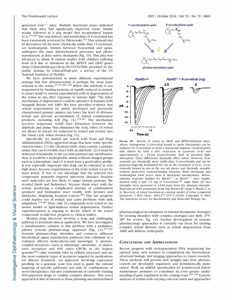
generated Lrat�/� mice. Multiple functional assays indicatedthat these mice had significantly improved vision. Similarresults followed in a dog model that recapitulated humanLCA.215,285 The oral delivery and metabolism of 9-cis-retinal hasbeen extensively reviewed by Palczewski.214 This retinoid andits derivatives are far more chemically stable than 11-cis-retinal;yet isorhodopsin, formed between 9-cis-retinal and opsin,undergoes the same photochemical processes and photo-transduction as does native rhodopsin (Fig. 10). This idea wasadvanced to phase II clinical studies with children sufferingfrom LCA due to mutations in the RPE65 and LRAT genes(http://clinicaltrials.gov/show/NCT01543906 provided in thepublic domain by ClinicalTrials.gov, a service of the USNational Institutes of Health).
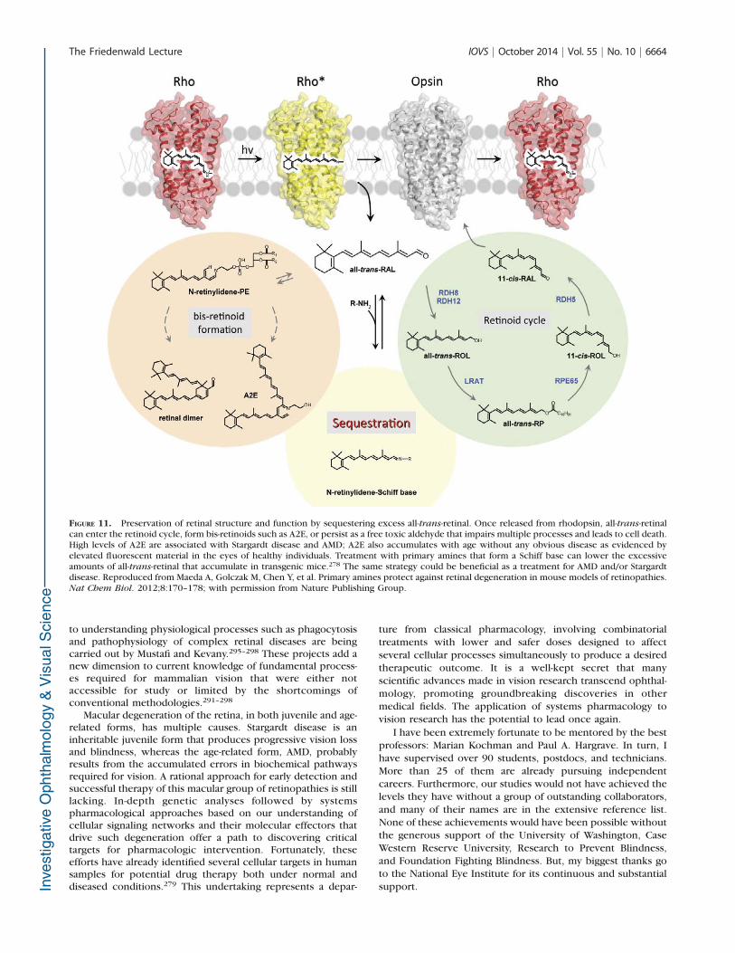
We have demonstrated in many different experimentalsettings that free all-trans-retinal is perhaps the most toxicretinoid in the retina.24,278,286–289 When this aldehyde is notsequestered by binding proteins or rapidly reduced to retinol,it causes death to various experimental cells or degeneration ofthe retina in situ after exposure to intense light. The lattermechanism of degeneration could be operative in humans withStargardt disease and AMD. We have provided evidence thatretinal sequestration by newly developed and previouslycharacterized primary amines can lower the level of all-trans-retinal and prevent accumulation of retinal condensationproducts, including A2E (Fig. 11).278,287 The mechanisminvolves temporary Schiff base formation between thealdehyde and amine that eliminates the toxic effect of retinalyet allows its release for reduction to retinol and reentry intothe visual cycle when needed (Fig. 11).
Specifically, we started our search with Food and DrugAdministration (FDA)–approved drugs that have some specificcharacteristics: (1) the chemical entity must contain a primaryamine that can reversibly react with an aldehyde; (2) it must bedelivered systemically to the eye and partition into membranes;thus, it would be a hydrophobic amine without charged groupssuch as a phosphate; and (3) it must have a good safety profile.It was especially important that drug can be tolerated at veryhigh doses, because sequestration of the toxic retinal occurs bymass action. It was to our advantage that the selected testcompounds primarily targeted infectious diseases, becausesuch molecules are less likely to target human proteins. Weavoided thiols and hydrazines, because thiols react with theretinal, producing a complicated mixture of condensationproducts and hydrazines react readily with ketones andaldehydes, displaying frequent toxicity.290 This last propertycould deprive eye of retinal, and cause problems with darkadaptation.278,287 Thus, only 24 compounds were tested in ourmouse model of light-induced retinal degeneration. Furtherexperimentation is ongoing to decide which of the testedcompounds would best progress to clinical trial(s).
Modern drug discovery involves a long and challengingpathway to potential clinical application. We have investigateda transformative solution to this problem with a multidisci-plinary systems pharmacology approach (Fig. 12).279,287
Systems pharmacology identifies and connects differentbiochemical signal transduction pathways that culminate in acommon effector molecule/second messenger. G protein–coupled receptors—such as adrenergic, adenosine, or musca-rinic receptors and 600 other GPCRs in the humangenome282—play essential roles in cellular processes and arethe most common types of receptors targeted by medicationsfor disease treatment. An approach involving expressionprofiling on a genomic scale was used to guide the rationaltargeting of specific pathways, not only by combinations ofnovel therapeutics, but also combinations of currently existingFDA-approved drugs to combat complex diseases. This novelapproach is also of interest to those pursuing mechanism-based
pharmacological development of rational therapeutic strategiesfor treating disorders with complex etiologies (see Refs. 279,287 for review; Fig. 12). Further development of systemspharmacology approaches is currently our focus for treatingcomplex retinal diseases such as retinal degeneration fromAMD and diabetic retinopathy.
CONCLUSIONS AND APPRECIATIONS
Recent progress with next-generation DNA sequencing hasopened many new avenues to complement the biochemical,structural biology, and imaging approaches to vision research.These methods will provide new insights into how photore-ceptors are developed, organized, and postmitotically main-tained. Work on miRNA spearheaded by postdoctoral fellowSundermeier promises to contribute an even greater under-standing of gene regulation in the coming years.291–294 Geneticanalyses of retinas with varying cone-rod ratios and approaches
FIGURE 10. Rescue of vision in LRAT- and RPE65-deficient mice.Above: Endogenous 11-cis-retinal bound to opsin (rhodopsin) can bereplaced by 9-cis-retinal to form a functional pigment (isorhodopsin)that differs by only a 60% reduction in quantum yield andapproximately a ~15-nm hypsochromic shift in maximal lightabsorption. These differences minimally affect vision. However, 9-cis-retinoids are chemically more stable than 11-cis-retinoids and can bepharmacologically formulated for use in the treatment of LCA; 11-cis-retinoids formed in situ in the eye are photo- and thermally unstablewithout protective retinoid-binding proteins. Both rhodopsin andisorhodopsin form active meta II rhodopsin intermediates. Below:stimulus response families for Rpe65þ/þ or Rpe65�/� mice supple-mented with 0 and 2.5 mg of 9-cis-retinal.283 Light flash (10 ms)strengths were increased in 2-fold steps from the dimmest intensity.Reproduced with permission from Van Hooser JP, Liang Y, Maeda T, etal. Recovery of visual functions in a mouse model of Leber congenitalamaurosis. J Biol Chem. 2002;277:19173–19182. Copyright � 2002The American Society for Biochemistry and Molecular Biology, Inc.
The Friedenwald Lecture IOVS j October 2014 j Vol. 55 j No. 10 j 6663
to understanding physiological processes such as phagocytosisand pathophysiology of complex retinal diseases are beingcarried out by Mustafi and Kevany.295–298 These projects add anew dimension to current knowledge of fundamental process-es required for mammalian vision that were either notaccessible for study or limited by the shortcomings ofconventional methodologies.291–298
Macular degeneration of the retina, in both juvenile and age-related forms, has multiple causes. Stargardt disease is aninheritable juvenile form that produces progressive vision lossand blindness, whereas the age-related form, AMD, probablyresults from the accumulated errors in biochemical pathwaysrequired for vision. A rational approach for early detection andsuccessful therapy of this macular group of retinopathies is stilllacking. In-depth genetic analyses followed by systemspharmacological approaches based on our understanding ofcellular signaling networks and their molecular effectors thatdrive such degeneration offer a path to discovering criticaltargets for pharmacologic intervention. Fortunately, theseefforts have already identified several cellular targets in humansamples for potential drug therapy both under normal anddiseased conditions.279 This undertaking represents a depar-
ture from classical pharmacology, involving combinatorialtreatments with lower and safer doses designed to affectseveral cellular processes simultaneously to produce a desiredtherapeutic outcome. It is a well-kept secret that manyscientific advances made in vision research transcend ophthal-mology, promoting groundbreaking discoveries in othermedical fields. The application of systems pharmacology tovision research has the potential to lead once again.
I have been extremely fortunate to be mentored by the bestprofessors: Marian Kochman and Paul A. Hargrave. In turn, Ihave supervised over 90 students, postdocs, and technicians.More than 25 of them are already pursuing independentcareers. Furthermore, our studies would not have achieved thelevels they have without a group of outstanding collaborators,and many of their names are in the extensive reference list.None of these achievements would have been possible withoutthe generous support of the University of Washington, CaseWestern Reserve University, Research to Prevent Blindness,and Foundation Fighting Blindness. But, my biggest thanks goto the National Eye Institute for its continuous and substantialsupport.
FIGURE 11. Preservation of retinal structure and function by sequestering excess all-trans-retinal. Once released from rhodopsin, all-trans-retinalcan enter the retinoid cycle, form bis-retinoids such as A2E, or persist as a free toxic aldehyde that impairs multiple processes and leads to cell death.High levels of A2E are associated with Stargardt disease and AMD; A2E also accumulates with age without any obvious disease as evidenced byelevated fluorescent material in the eyes of healthy individuals. Treatment with primary amines that form a Schiff base can lower the excessiveamounts of all-trans-retinal that accumulate in transgenic mice.278 The same strategy could be beneficial as a treatment for AMD and/or Stargardtdisease. Reproduced from Maeda A, Golczak M, Chen Y, et al. Primary amines protect against retinal degeneration in mouse models of retinopathies.Nat Chem Biol. 2012;8:170–178; with permission from Nature Publishing Group.
The Friedenwald Lecture IOVS j October 2014 j Vol. 55 j No. 10 j 6664

Acknowledgments
We thank Leslie T. Webster Jr, Akiko Maeda, Paul Park, MarcinGolczak, John J. Mieyal, Yoshikazu Imanishi, Andreas Engel, ArthurS. Polans, and the members of Palczewski’s laboratory for theircomments on this manuscript. We thank Lukas Hofmann, MarcinGolczak, Grazyna Palczewska, David Salom, Andreas Engel, PhilipKiser, and Yu Chen for figure preparation.
Supported by funding from the National Eye Institute, NationalInstitutes of Health Grant R01EY008061 (KP), and the Arnold andMabel Beckman Foundation.
KP is the John H. Hord Professor of Pharmacology.
Disclosure: K. Palczewski, None
References
1. Arshavsky VY, Lamb TD, Pugh EN Jr. G proteins and photo-transduction. Annu Rev Physiol. 2002;64:153–187.
2. Orban T, Jastrzebska B, Palczewski K. Structural approaches tounderstanding retinal proteins needed for vision. Curr Opin
Cell Biol. 2014;27:32–43.
3. Palczewski K, Orban T. From atomic structures to neuronalfunctions of G protein-coupled receptors. Annu Rev Neurosci.2013;36:139–164.
4. Palczewski K. G protein-coupled receptor rhodopsin. Annu Rev
Biochem. 2006;75:743–767.
5. Palczewski K. Chemistry and biology of vision. J Biol Chem.2012;287:1612–1619.
6. Palczewski K. Thematic minireview series on focus on vision. J
Biol Chem. 2012;287:1610–1611.
7. Yau KW, Hardie RC. Phototransduction motifs and variations.Cell. 2009;139:246–264.
8. Luo DG, Xue T, Yau KW. How vision begins: an odyssey. Proc
Natl Acad Sci U S A. 2008;105:9855–9862.
9. Palczewski K. Is vertebrate phototransduction solved? Newinsights into the molecular mechanism of phototransduction.Invest Ophthalmol Vis Sci. 1994;35:3577–3581.
10. Arshavsky VY, Wensel TG. Timing is everything: GTPaseregulation in phototransduction. Invest Ophthalmol Vis Sci.2013;54:7725–7733.
11. Cideciyan AV, Zhao X, Nielsen L, Khani SC, Jacobson SG,Palczewski K. Null mutation in the rhodopsin kinase geneslows recovery kinetics of rod and cone phototransduction inman. Proc Natl Acad Sci U S A. 1998;95:328–333.
12. Chen CK, Burns ME, Spencer M, et al. Abnormal photo-responses and light-induced apoptosis in rods lackingrhodopsin kinase. Proc Natl Acad Sci U S A. 1999;96:3718–3722.
13. Chen J, Simon MI, Matthes MT, Yasumura D, LaVail MM.Increased susceptibility to light damage in an arrestinknockout mouse model of Oguchi disease (stationary nightblindness). Invest Ophthalmol Vis Sci. 1999;40:2978–2982.
14. Howes KA, Pennesi ME, Sokal I, et al. GCAP1 rescues rodphotoreceptor response in GCAP1/GCAP2 knockout mice.EMBO J. 2002;21:1545–1554.
15. Mendez A, Burns ME, Sokal I, et al. Role of guanylate cyclase-activating proteins (GCAPs) in setting the flash sensitivity ofrod photoreceptors. Proc Natl Acad Sci U S A. 2001;98:9948–9953.
16. Kiser PD, Golczak M, Palczewski K. Chemistry of the retinoid(visual) cycle. Chem Rev. 2014;114:194–232.
17. Nemet I, Tian G, Imanishi Y. Submembrane assembly andrenewal of rod photoreceptor cGMP-gated channel: insightinto the actin-dependent process of outer segment morpho-genesis. J Neurosci. 2014;34:8164–8174.
18. Tian G, Ropelewski P, Nemet I, Lee R, Lodowski KH, ImanishiY. An unconventional secretory pathway mediates the ciliatargeting of peripherin/rds. J Neurosci. 2014;34:992–1006.
FIGURE 12. Systems pharmacological strategies prevent the development of light-induced photoreceptor degeneration. In appropriate mousemodels of human retinopathies, antagonists of multiple Gs-coupled GPCRs prevented photoreceptor cell death (red bar, top left). Moreover,pharmacological activation of Gi-coupled GPCRs (black arrow, top middle) that suppress adenylate cyclase activity (red bar, middle) preservedretinal structure and function against an environmental insult (prolonged exposure to intense illumination). Direct inhibition of AC was alsobeneficial (red line). Additionally, Gq-coupled GPCRs can participate in photoreceptor degeneration, and inhibition of these GPCRs also provedeffective in protecting photoreceptors from light-induced degeneration (red bar, top right). Reproduced with permission from Chen Y, PalczewskaG, Mustafi D, et al. Systems pharmacology identifies drug targets for Stargardt disease-associated retinal degeneration. J Clin Invest. 2013;123:5119–5134. Copyright � 2013 American Society for Clinical Investigation.
The Friedenwald Lecture IOVS j October 2014 j Vol. 55 j No. 10 j 6665

19. Sakami S, Kolesnikov AV, Kefalov VJ, Palczewski K. P23Hopsin knock-in mice reveal a novel step in retinal rod discmorphogenesis. Hum Mol Genet. 2014;23:1723–1741.
20. Sokolov M, Lyubarsky AL, Strissel KJ, et al. Massive light-driven translocation of transducin between the two majorcompartments of rod cells: a novel mechanism of lightadaptation. Neuron. 2002;34:95–106.
21. Maeda A, Palczewska G, Golczak M, et al. Two-photonmicroscopy reveals early rod photoreceptor cell damage inlight-exposed mutant mice. Proc Natl Acad Sci U S A. 2014;111:E1428–E1437.
22. Carroll J, Kay DB, Scoles D, Dubra A, Lombardo M. Adaptiveoptics retinal imaging—clinical opportunities and challenges.Curr Eye Res. 2013;38:709–721.
23. Zipfel WR, Williams RM, Webb WW. Nonlinear magic:multiphoton microscopy in the biosciences. Nat Biotechnol.2003;21:1369–1377.
24. Palczewska G, Maeda T, Imanishi Y, et al. Noninvasivemultiphoton fluorescence microscopy resolves retinol andretinal condensation products in mouse eyes. Nat Med. 2010;16:1444–1449.
25. Hofer H, Carroll J, Neitz J, Neitz M, Williams DR. Organizationof the human trichromatic cone mosaic. J Neurosci. 2005;25:9669–9679.
26. Hunter JJ, Masella B, Dubra A, et al. Images of photoreceptorsin living primate eyes using adaptive optics two-photonophthalmoscopy. Biomed Opt Exp. 2011;2:139–148.
27. Geng Y, Dubra A, Yin L, et al. Adaptive optics retinal imagingin the living mouse eye. Biomed Opt Express. 2012;3:715–734.
28. Fritsche LG, Fariss RN, Stambolian D, Abecasis GR, CurcioCA, Swaroop A. Age-related macular degeneration: geneticsand biology coming together [published online ahead ofprint April 16, 2014]. Annu Rev Genomics Hum Genet. doi:10.1146/annurev-genom-090413-025610.
29. Kuhne W. Chemical processes in the retina. Vision Res. 1977;17:1269–1316.
30. Ripps H. Light to sight: milestones in phototransduction.FASEB J. 2010;24:970–975.
31. Ripps H. The color purple: milestones in photochemistry.FASEB J. 2008;22:4038–4043.
32. Filipek S, Stenkamp RE, Teller DC, Palczewski K. G protein-coupled receptor rhodopsin: a prospectus. Annu Rev
Physiol. 2003;65:851–879.
33. Wald G. Molecular basis of visual excitation. Science. 1968;162:230–239.
34. Jacobs GH, Nathans J. The evolution of primate color vision.Sci Am. 2009;300:56–63.
35. Nathans J. The genes for color vision. Sci Am. 1989;260:42–49.
36. Ovchinnikov A, Abdulaev NG, Feigina M, Artamonov ID,Bogachuk AS. Visual rhodopsin. III. Complete amino acidsequence and topography in a membrane [in Russian].Bioorg Khim. 1983;9:1331–1340.
37. Ovchinnikov Yu A. Rhodopsin and bacteriorhodopsin: struc-ture-function relationships. FEBS Lett. 1982;148:179–191.
38. Hargrave PA, McDowell JH, Curtis DR, et al. The structure ofbovine rhodopsin. Biophys Struct Mech. 1983;9:235–244.
39. Nathans J, Hogness DS. Isolation, sequence analysis, andintron-exon arrangement of the gene encoding bovinerhodopsin. Cell. 1983;34:807–814.
40. Nathans J, Hogness DS. Isolation and nucleotide sequence ofthe gene encoding human rhodopsin. Proc Natl Acad Sci U S
A. 1984;81:4851–4855.
41. Argos P, Rao JK, Hargrave PA. Structural prediction ofmembrane-bound proteins. Eur J Biochem. 1982;128:565–575.
42. Palczewski K, Kumasaka T, Hori T, et al. Crystal structure ofrhodopsin: a G protein-coupled receptor. Science. 2000;289:739–745.
43. Nathans J. Rhodopsin: structure, function, and genetics.Biochemistry. 1992;31:4923–4931.
44. Dryja TP, McGee TL, Reichel E, et al. A point mutation of therhodopsin gene in one form of retinitis pigmentosa. Nature.1990;343:364–366.
45. Hartong DT, Berson EL, Dryja TP. Retinitis pigmentosa.Lancet. 2006;368:1795–1809.
46. Fung BK, Hurley JB, Stryer L. Flow of information in the light-triggered cyclic nucleotide cascade of vision. Proc Natl Acad
Sci U S A. 1981;78:152–156.
47. Maeda T, Imanishi Y, Palczewski K. Rhodopsin phosphoryla-tion: 30 years later. Prog Retin Eye Res. 2003;22:417–434.
48. Pfister C, Chabre M, Plouet J, et al. Retinal S antigen identifiedas the 48K protein regulating light-dependent phosphodies-terase in rods. Science. 1985;228:891–893.
49. Jastrzebska B, Palczewski K, Golczak M. Role of bulk water inhydrolysis of the rhodopsin chromophore. J Biol Chem.2011;286:18930–18937.
50. Palczewski K, Hargrave PA, McDowell JH, Ingebritsen TS.The catalytic subunit of phosphatase 2A dephosphorylatesphosphoopsin. Biochemistry. 1989;28:415–419.
51. Palczewski K, McDowell JH, Jakes S, Ingebritsen TS, HargravePA. Regulation of rhodopsin dephosphorylation by arrestin. J
Biol Chem. 1989;264:15770–15773.
52. Schertler GF, Villa C, Henderson R. Projection structure ofrhodopsin. Nature. 1993;362:770–772.
53. Schertler GF, Hargrave PA. Projection structure of frogrhodopsin in two crystal forms. Proc Natl Acad Sci U S A.1995;92:11578–11582.
54. Okada T, Le Trong I, Fox BA, Behnke CA, Stenkamp RE,Palczewski K. X-ray diffraction analysis of three-dimensionalcrystals of bovine rhodopsin obtained from mixed micelles. J
Struct Biol. 2000;130:73–80.
55. Thomas DD, Stryer L. Transverse location of the retinalchromophore of rhodopsin in rod outer segment discmembranes. J Mol Biol. 1982;154:145–157.
56. Okada T, Palczewski K. Crystal structure of rhodopsin:implications for vision and beyond. Curr Opin Struct Biol.2001;11:420–426.
57. Filipek S, Teller DC, Palczewski K, Stenkamp R. Thecrystallographic model of rhodopsin and its use in studiesof other G protein-coupled receptors. Annu Rev Biophys
Biomol Struct. 2003;32:375–397.
58. Park PS, Lodowski DT, Palczewski K. Activation of G protein-coupled receptors: beyond two-state models and tertiaryconformational changes. Annu Rev Pharmacol Toxicol.2008;48:107–141.
59. Menon ST, Han M, Sakmar TP. Rhodopsin: structural basis ofmolecular physiology. Physiol Rev. 2001;81:1659–1688.
60. Sakmar TP, Menon ST, Marin EP, Awad ES. Rhodopsin: insightsfrom recent structural studies. Annu Rev Biophys Biomol
Struct. 2002;31:443–484.
61. Huber T, Sakmar TP. Escaping the flatlands: new approaches forstudying the dynamic assembly and activation of GPCRsignaling complexes. Trends Pharmacol Sci. 2011;32:410–419.
62. Oprian DD. Molecular determinants of spectral propertiesand signal transduction in the visual pigments. Curr Opin
Neurobiol. 1992;2:428–432.
63. Rao VR, Oprian DD. Activating mutations of rhodopsin andother G protein-coupled receptors. Annu Rev Biophys
Biomol Struct. 1996;25:287–314.
64. Hubbell WL, Altenbach C, Hubbell CM, Khorana HG.Rhodopsin structure, dynamics, and activation: a perspectivefrom crystallography, site-directed spin labeling, sulfhydryl
The Friedenwald Lecture IOVS j October 2014 j Vol. 55 j No. 10 j 6666

reactivity, and disulfide cross-linking. Adv Protein Chem.2003;63:243–290.
65. Park PS. Constitutively active rhodopsin and retinal disease.Adv Pharmacol. 2014;70:1–36.
66. Hofmann KP, Scheerer P, Hildebrand PW, et al. A G protein-coupled receptor at work: the rhodopsin model. Trends
Biochem Sci. 2009;34:540–552.
67. Okada T, Ernst OP, Palczewski K, Hofmann KP. Activation ofrhodopsin: new insights from structural and biochemicalstudies. Trends Biochem Sci. 2001;26:318–324.
68. Ernst OP, Lodowski DT, Elstner M, Hegemann P, Brown LS,Kandori H. Microbial and animal rhodopsins: structures,functions, and molecular mechanisms. Chem Rev. 2014;114:126–163.
69. Salom D, Le Trong I, Pohl E, et al. Improvements in G protein-coupled receptor purification yield light stable rhodopsincrystals. J Struct Biol. 2006;156:497–504.
70. Salom D, Lodowski DT, Stenkamp RE, et al. Crystal structureof a photoactivated deprotonated intermediate of rhodopsin.Proc Natl Acad Sci U S A. 2006;103:16123–16128.
71. Farahbakhsh ZT, Hideg K, Hubbell WL. Photoactivatedconformational changes in rhodopsin: a time-resolved spinlabel study. Science. 1993;262:1416–1419.
72. Farrens DL, Altenbach C, Yang K, Hubbell WL, Khorana HG.Requirement of rigid-body motion of transmembrane helicesfor light activation of rhodopsin. Science. 1996;274:768–770.
73. Park JH, Morizumi T, Li Y, et al. Opsin, a structural model forolfactory receptors? Angew Chem Int Ed Engl. 2013;52:11021–11024.
74. Choe HW, Kim YJ, Park JH, et al. Crystal structure ofmetarhodopsin II. Nature. 2011;471:651–655.
75. Scheerer P, Park JH, Hildebrand PW, et al. Crystal structure ofopsin in its G-protein-interacting conformation. Nature.2008;455:497–502.
76. Park JH, Scheerer P, Hofmann KP, Choe HW, Ernst OP. Crystalstructure of the ligand-free G-protein-coupled receptor opsin.Nature. 2008;454:183–187.
77. Sekharan S, Sugihara M, Weingart O, Okada T, Buss V. Proteinassistance in the photoisomerization of rhodopsin and 9-cis-rhodopsin—insights from experiment and theory. J Am
Chem Soc. 2007;129:1052–1054.
78. Nakamichi H, Okada T. X-ray crystallographic analysis of 9-cis-rhodopsin, a model analogue visual pigment. Photochem
Photobiol. 2007;83:232–235.
79. Schreiber M, Sugihara M, Okada T, Buss V. Quantummechanical studies on the crystallographic model of bath-orhodopsin. Angew Chem Int Ed Engl. 2006;45:4274–4277.
80. Nakamichi H, Okada T. Local peptide movement in thephotoreaction intermediate of rhodopsin. Proc Natl Acad Sci
U S A. 2006;103:12729–12734.
81. Nakamichi H, Okada T. Crystallographic analysis of primaryvisual photochemistry. Angew Chem Int Ed Engl. 2006;45:4270–4273.
82. Singhal A, Ostermaier MK, Vishnivetskiy SA, et al. Insightsinto congenital stationary night blindness based on thestructure of G90D rhodopsin. EMBO Rep. 2013;14:520–526.
83. Deupi X, Edwards P, Singhal A, et al. Stabilized G proteinbinding site in the structure of constitutively active metarho-dopsin-II. Proc Natl Acad Sci U S A. 2012;109:119–124.
84. Schertler GF. Signal transduction: the rhodopsin storycontinued. Nature. 2008;453:292–293.
85. Li J, Edwards PC, Burghammer M, Villa C, Schertler GF.Structure of bovine rhodopsin in a trigonal crystal form. J Mol
Biol. 2004;343:1409–1438.
86. Ludeke S, Beck M, Yan EC, Sakmar TP, Siebert F, Vogel R. Therole of Glu181 in the photoactivation of rhodopsin. J Mol
Biol. 2005;353:345–356.
87. Matthews RG, Hubbard R, Brown PK, Wald G. Tautomericforms of metarhodopsin. J Gen Physiol. 1963;47:215–240.
88. Yuan S, Filipek S, Palczewski K, Vogel H. Activation of Gprotein-coupled receptors correlates with the formation of acontinuous internal water pathway. Nat Commun. 2014;5:4733. doi:10.1038/ncomms5733.
89. Angel TE, Chance MR, Palczewski K. Conserved watersmediate structural and functional activation of family A(rhodopsin-like) G protein-coupled receptors. Proc Natl
Acad Sci U S A. 2009;106:8555–8560.
90. Angel TE, Gupta S, Jastrzebska B, Palczewski K, Chance MR.Structural waters define a functional channel mediatingactivation of the GPCR, rhodopsin. Proc Natl Acad Sci U S
A. 2009;106:14367–14372.
91. Fritze O, Filipek S, Kuksa V, Palczewski K, Hofmann KP, ErnstOP. Role of the conserved NPxxY(x)5,6F motif in therhodopsin ground state and during activation. Proc Natl
Acad Sci U S A. 2003;100:2290–2295.
92. Struts AV, Salgado GF, Martinez-Mayorga K, Brown MF. Retinaldynamics underlie its switch from inverse agonist to agonistduring rhodopsin activation. Nat Struct Mol Biol. 2011;18:392–394.
93. Struts AV, Salgado GF, Brown MF. Solid-state 2H NMRrelaxation illuminates functional dynamics of retinal cofactorin membrane activation of rhodopsin. Proc Natl Acad Sci U S
A. 2011;108:8263–8268.
94. Smith SO. Insights into the activation mechanism of the visualreceptor rhodopsin. Biochem Soc Trans. 2012;40:389–393.
95. Smith SO. Structure and activation of the visual pigmentrhodopsin. Annu Rev Biophys. 2010;39:309–328.
96. Hargrave PA. Rhodopsin structure, function, and topographythe Friedenwald lecture. Invest Ophthalmol Vis Sci. 2001;42:3–9.
97. Nickell S, Park PS, Baumeister W, Palczewski K. Three-dimensional architecture of murine rod outer segmentsdetermined by cryoelectron tomography. J Cell Biol. 2007;177:917–925.
98. Liang Y, Fotiadis D, Maeda T, et al. Rhodopsin signaling andorganization in heterozygote rhodopsin knockout mice. J
Biol Chem. 2004;279:48189–48196.
99. Liang Y, Fotiadis D, Filipek S, Saperstein DA, Palczewski K,Engel A. Organization of the G protein-coupled receptorsrhodopsin and opsin in native membranes. J Biol Chem.2003;278:21655–21662.
100. Molday RS. Photoreceptor membrane proteins, phototrans-duction, and retinal degenerative diseases. The FriedenwaldLecture. Invest Ophthalmol Vis Sci. 1998;39:2491–2513.
101. Anderson RE, Maude MB. Phospholipids of bovine outersegments. Biochemistry. 1970;9:3624–3628.
102. Fliesler SJ, Anderson RE. Chemistry and metabolism of lipidsin the vertebrate retina. Prog Lipid Res. 1983;22:79–131.
103. Humphries MM, Rancourt D, Farrar GJ, et al. Retinopathyinduced in mice by targeted disruption of the rhodopsingene. Nat Genet. 1997;15:216–219.
104. Lem J, Krasnoperova NV, Calvert PD, et al. Morphological,physiological, and biochemical changes in rhodopsin knock-out mice. Proc Natl Acad Sci U S A. 1999;96:736–741.
105. Wen XH, Shen L, Brush RS, et al. Overexpression ofrhodopsin alters the structure and photoresponse of rodphotoreceptors. Biophys J. 2009;96:939–950.
106. Olsson JE, Gordon JW, Pawlyk BS, et al. Transgenic mice witha rhodopsin mutation (Pro23His): a mouse model ofautosomal dominant retinitis pigmentosa. Neuron. 1992;9:815–830.
107. Price BA, Sandoval IM, Chan F, et al. Rhodopsin geneexpression determines rod outer segment size and rod cell
The Friedenwald Lecture IOVS j October 2014 j Vol. 55 j No. 10 j 6667
resistance to a dominant-negative neurodegeneration mutant.PLoS One. 2012;7:e49889.
108. Chabre M. X-ray diffraction studies of retinal rods. I. Structureof the disc membrane, effect of illumination. Biochim
Biophys Acta. 1975;382:322–335.
109. Langmack K, Saibil H. Signal transduction in photoreceptors.Biochem Soc Trans. 1991;19:858–860.
110. Cone RA. Rotational diffusion of rhodopsin in the visualreceptor membrane. Nat New Biol. 1972;236:39–43.
111. Poo M, Cone RA. Lateral diffusion of rhodopsin in thephotoreceptor membrane. Nature. 1974;247:438–441.
112. Govardovskii VI, Korenyak DA, Shukolyukov SA, Zueva LV.Lateral diffusion of rhodopsin in photoreceptor membrane: areappraisal. Mol Vis. 2009;15:1717–1729.
113. Hanson SM, Gurevich EV, Vishnivetskiy SA, Ahmed MR, SongX, Gurevich VV. Each rhodopsin molecule binds its ownarrestin. Proc Natl Acad Sci U S A. 2007;104:3125–3128.
114. Ernst OP, Gramse V, Kolbe M, Hofmann KP, Heck M.Monomeric G protein-coupled receptor rhodopsin in solu-tion activates its G protein transducin at the diffusion limit.Proc Natl Acad Sci U S A. 2007;104:10859–10864.
115. Maeda S, Sun D, Singhal A, et al. Crystallization scalepreparation of a stable GPCR signaling complex betweenconstitutively active rhodopsin and G-protein. PLoS One.2014;9:e98714.
116. Bayburt TH, Leitz AJ, Xie G, Oprian DD, Sligar SG. Transducinactivation by nanoscale lipid bilayers containing one and tworhodopsins. J Biol Chem. 2007;282:14875–14881.
117. Whorton MR, Jastrzebska B, Park PS, et al. Efficient couplingof transducin to monomeric rhodopsin in a phospholipidbilayer. J Biol Chem. 2008;283:4387–4394.
118. Niswender CM, Conn PJ. Metabotropic glutamate receptors:physiology, pharmacology, and disease. Annu Rev Pharma-
col Toxicol. 2010;50:295–322.
119. Gross L, Mohn F, Moll N, et al. Bond-order discrimination byatomic force microscopy. Science. 2012;337:1326–1329.
120. Fotiadis D, Liang Y, Filipek S, Saperstein DA, Engel A,Palczewski K. Atomic-force microscopy: rhodopsin dimers innative disc membranes. Nature. 2003;421:127–128.
121. Fotiadis D, Liang Y, Filipek S, Saperstein DA, Engel A,Palczewski K. The G protein-coupled receptor rhodopsin inthe native membrane. FEBS Lett. 2004;564:281–288.
122. Suda K, Filipek S, Palczewski K, Engel A, Fotiadis D. Thesupramolecular structure of the GPCR rhodopsin in solutionand native disc membranes. Mol Membr Biol. 2004;21:435–446.
123. Jastrzebska B, Maeda T, Zhu L, et al. Functional characteriza-tion of rhodopsin monomers and dimers in detergents. J Biol
Chem. 2004;279:54663–54675.
124. Fotiadis D, Jastrzebska B, Philippsen A, Muller DJ, PalczewskiK, Engel A. Structure of the rhodopsin dimer: a workingmodel for G-protein-coupled receptors. Curr Opin Struct
Biol. 2006;16:252–259.
125. Jastrzebska B, Fotiadis D, Jang GF, Stenkamp RE, Engel A,Palczewski K. Functional and structural characterization ofrhodopsin oligomers. J Biol Chem. 2006;281:11917–11922.
126. Palczewski K. Oligomeric forms of G protein-coupledreceptors (GPCRs). Trends Biochem Sci. 2010;35:595–600.
127. Knepp AM, Periole X, Marrink SJ, Sakmar TP, Huber T.Rhodopsin forms a dimer with cytoplasmic helix 8 contactsin native membranes. Biochemistry. 2012;51:1819–1821.
128. Periole X, Huber T, Marrink SJ, Sakmar TP. G protein-coupledreceptors self-assemble in dynamics simulations of modelbilayers. J Am Chem Soc. 2007;129:10126–10132.
129. Comar WD, Schubert SM, Jastrzebska B, Palczewski K, SmithAW. Time-resolved fluorescence spectroscopy measures clus-
tering and mobility of a g protein-coupled receptor opsin in livecell membranes. J Am Chem Soc. 2014;136:8342–8349.
130. Tanuj Sapra K, Park PS, Filipek S, Engel A, Muller DJ,Palczewski K. Detecting molecular interactions that stabilizenative bovine rhodopsin. J Mol Biol. 2006;358:255–269.
131. Park PS, Sapra KT, Kolinski M, Filipek S, Palczewski K, MullerDJ. Stabilizing effect of Zn2þ in native bovine rhodopsin. J
Biol Chem. 2007;282:11377–11385.
132. Sapra KT, Park PS, Palczewski K, Muller DJ. Mechanicalproperties of bovine rhodopsin and bacteriorhodopsin:possible roles in folding and function. Langmuir. 2008;24:1330–1337.
133. Jastrzebska B, Tsybovsky Y, Palczewski K. Complexesbetween photoactivated rhodopsin and transducin: progressand questions. Biochem J. 2010;428:1–10.
134. Gilliam JC, Chang JT, Sandoval IM, et al. Three-dimensionalarchitecture of the rod sensory cilium and its disruption inretinal neurodegeneration. Cell. 2012;151:1029–1041.
135. Ridge KD, Abdulaev NG, Sousa M, Palczewski K. Photo-transduction: crystal clear. Trends Biochem Sci. 2003;28:479–487.
136. Noel JP, Hamm HE, Sigler PB. The 2.2 A crystal structure oftransducin-alpha complexed with GTP gamma S. Nature.1993;366:654–663.
137. Sondek J, Lambright DG, Noel JP, Hamm HE, Sigler PB.GTPase mechanism of Gproteins from the 1.7-A crystalstructure of transducin alpha-GDP-AIF-4. Nature. 1994;372:276–279.
138. Lambright DG, Sondek J, Bohm A, Skiba NP, Hamm HE, SiglerPB. The 2.0 A crystal structure of a heterotrimeric G protein.Nature. 1996;379:311–319.
139. Sondek J, Bohm A, Lambright DG, Hamm HE, Sigler PB.Crystal structure of a G-protein beta gamma dimer at 2.1Aresolution. Nature. 1996;379:369–374.
140. Heck M, Hofmann KP. Maximal rate and nucleotidedependence of rhodopsin-catalyzed transducin activation:initial rate analysis based on a double displacementmechanism. J Biol Chem. 2001;276:10000–10009.
141. Oldham WM, Hamm HE. How do receptors activate Gproteins? Adv Protein Chem. 2007;74:67–93.
142. Oldham WM, Hamm HE. Heterotrimeric G protein activationby G-protein-coupled receptors. Nat Rev Mol Cell Biol. 2008;9:60–71.
143. Jastrzebska B, Golczak M, Fotiadis D, Engel A, Palczewski K.Isolation and functional characterization of a stable complexbetween photoactivated rhodopsin and the G protein,transducin. FASEB J. 2009;23:371–381.
144. Vahedi-Faridi A, Jastrzebska B, Palczewski K, Engel A. 3Dimaging and quantitative analysis of small solubilizedmembrane proteins and their complexes by transmissionelectron microscopy. Microscopy (Oxf). 2013;62:95–107.
145. Jastrzebska B, Ringler P, Palczewski K, Engel A. Therhodopsin-transducin complex houses two distinct rhodop-sin molecules. J Struct Biol. 2013;182:164–172.
146. Jastrzebska B, Ringler P, Lodowski DT, et al. Rhodopsin-transducin heteropentamer: three-dimensional structure andbiochemical characterization. J Struct Biol. 2011;176:387–394.
147. Jastrzebska B, Orban T, Golczak M, Engel A, Palczewski K.Asymmetry of the rhodopsin dimer in complex with trans-ducin. FASEB J. 2013;27:1572–1584.
148. Preininger AM, Meiler J, Hamm HE. Conformational flexibilityand structural dynamics in GPCR-mediated G proteinactivation: a perspective. J Mol Biol. 2013;425:2288–2298.
149. Thaker TM, Sarwar M, Preininger AM, Hamm HE, Iverson TM.A transient interaction between the phosphate binding loopand switch I contributes to the allosteric network between
The Friedenwald Lecture IOVS j October 2014 j Vol. 55 j No. 10 j 6668

receptor and nucleotide in Galphai1. J Biol Chem. 2014;289:11331–11341.
150. Alexander NS, Preininger AM, Kaya AI, Stein RA, Hamm HE,Meiler J. Energetic analysis of the rhodopsin-G-proteincomplex links the alpha5 helix to GDP release. Nat Struct
Mol Biol. 2014;21:56–63.
151. Yamamoto S, Sippel KC, Berson EL, Dryja TP. Defects in therhodopsin kinase gene in the Oguchi form of stationary nightblindness. Nat Genet. 1997;15:175–178.
152. Dryja TP. Molecular genetics of Oguchi disease, fundusalbipunctatus, and other forms of stationary night blindness:LVII Edward Jackson Memorial Lecture. Am J Ophthalmol.2000;130:547–563.
153. Palczewski K, McDowell JH, Hargrave PA. Rhodopsin kinase:substrate specificity and factors that influence activity.Biochemistry. 1988;27:2306–2313.
154. Palczewski K, Arendt A, McDowell JH, Hargrave PA. Substraterecognition determinants for rhodopsin kinase: studies withsynthetic peptides, polyanions, and polycations. Biochemis-
try. 1989;28:8764–8770.
155. Palczewski K, Kahn N, Hargrave PA. Nucleoside inhibitors ofrhodopsin kinase. Biochemistry. 1990;29:6276–6282.
156. Inglese J, Koch WJ, Caron MG, Lefkowitz RJ. Isoprenylation inregulation of signal transduction by G-protein-coupledreceptor kinases. Nature. 1992;359:147–150.
157. Lorenz W, Inglese J, Palczewski K, Onorato JJ, Caron MG,Lefkowitz RJ. The receptor kinase family: primary structureof rhodopsin kinase reveals similarities to the beta-adrenergicreceptor kinase. Proc Natl Acad Sci U S A. 1991;88:8715–8719.
158. Homan KT, Tesmer JJ. Structural insights into G protein-coupled receptor kinase function. Curr Opin Cell Biol. 2014;27:25–31.
159. Singh P, Wang B, Maeda T, Palczewski K, Tesmer JJ. Structuresof rhodopsin kinase in different ligand states reveal keyelements involved in G protein-coupled receptor kinaseactivation. J Biol Chem. 2008;283:14053–14062.
160. Zhao X, Yokoyama K, Whitten ME, Huang J, Gelb MH,Palczewski K. A novel form of rhodopsin kinase from chickenretina and pineal gland. FEBS Lett. 1999;454:115–121.
161. Rinner O, Makhankov YV, Biehlmaier O, Neuhauss SC.Knockdown of cone-specific kinase GRK7 in larval zebrafishleads to impaired cone response recovery and delayed darkadaptation. Neuron. 2005;47:231–242.
162. Weiss ER, Ducceschi MH, Horner TJ, Li A, Craft CM, Osawa S.Species-specific differences in expression of G-protein-coupled receptor kinase (GRK) 7 and GRK1 in mammaliancone photoreceptor cells: implications for cone cell photo-transduction. J Neurosci. 2001;21:9175–9184.
163. Ohguro H, Palczewski K, Ericsson LH, Walsh KA, Johnson RS.Sequential phosphorylation of rhodopsin at multiple sites.Biochemistry. 1993;32:5718–5724.
164. Ohguro H, Johnson RS, Ericsson LH, Walsh KA, Palczewski K.Control of rhodopsin multiple phosphorylation. Biochemis-
try. 1994;33:1023–1028.
165. Palczewski K, Buczylko J, Kaplan MW, Polans AS, Crabb JW.Mechanism of rhodopsin kinase activation. J Biol Chem.1991;266:12949–12955.
166. Ohguro H, Van Hooser JP, Milam AH, Palczewski K.Rhodopsin phosphorylation and dephosphorylation in vivo.J Biol Chem. 1995;270:14259–14262.
167. Kennedy MJ, Lee KA, Niemi GA, et al. Multiple phosphory-lation of rhodopsin and the in vivo chemistry underlying rodphotoreceptor dark adaptation. Neuron. 2001;31:87–101.
168. Doan T, Mendez A, Detwiler PB, Chen J, Rieke F. Multiplephosphorylation sites confer reproducibility of the rod’ssingle-photon responses. Science. 2006;313:530–533.
169. Mendez A, Burns ME, Roca A, et al. Rapid and reproducibledeactivation of rhodopsin requires multiple phosphorylationsites. Neuron. 2000;28:153–164.
170. Kuhn H, Wilden U. Deactivation of photoactivated rhodopsin byrhodopsin-kinase and arrestin. J Recept Res. 1987;7:283–298.
171. Palczewski K, Buczylko J, Ohguro H, et al. Characterization ofa truncated form of arrestin isolated from bovine rod outersegments. Protein Sci. 1994;3:314–324.
172. Pulvermuller A, Maretzki D, Rudnicka-Nawrot M, Smith WC,Palczewski K, Hofmann KP. Functional differences in theinteraction of arrestin and its splice variant, p44, withrhodopsin. Biochemistry. 1997;36:9253–9260.
173. Smith WC, Milam AH, Dugger D, Arendt A, Hargrave PA,Palczewski K. A splice variant of arrestin. Molecular cloningand localization in bovine retina. J Biol Chem. 1994;269:15407–15410.
174. Palczewski K, Pulvermuller A, Buczylko J, Hofmann KP.Phosphorylated rhodopsin and heparin induce similarconformational changes in arrestin. J Biol Chem. 1991;266:18649–18654.
175. Ohguro H, Palczewski K, Walsh KA, Johnson RS. Topographicstudy of arrestin using differential chemical modifications andhydrogen/deuterium exchange. Protein Sci. 1994;3:2428–2434.
176. Palczewski K, Hargrave PA. Studies of ligand binding toarrestin. J Biol Chem. 1991;266:4201–4206.
177. Granzin J, Wilden U, Choe HW, Labahn J, Krafft B, Buldt G. X-ray crystal structure of arrestin from bovine rod outersegments. Nature. 1998;391:918–921.
178. Hirsch JA, Schubert C, Gurevich VV, Sigler PB. The 2.8 Acrystal structure of visual arrestin: a model for arrestin’sregulation. Cell. 1999;97:257–269.
179. Moaven H, Koike Y, Jao CC, Gurevich VV, Langen R, Chen J.Visual arrestin interaction with clathrin adaptor AP-2regulates photoreceptor survival in the vertebrate retina.Proc Natl Acad Sci U S A. 2013;110:9463–9468.
180. Korenbrot JI. Signal mechanisms of phototransduction inretinal rod. CRC Crit Rev Biochem. 1985;17:223–256.
181. Koch KW, Stryer L. Highly cooperative feedback control ofretinal rod guanylate cyclase by calcium ions. Nature. 1988;334:64–66.
182. Polans A, Baehr W, Palczewski K. Turned on by Ca2þ! Thephysiology and pathology of Ca(2þ)-binding proteins in theretina. Trends Neurosci. 1996;19:547–554.
183. Dizhoor AM, Chen CK, Olshevskaya E, Sinelnikova VV,Phillipov P, Hurley JB. Role of the acylated amino terminusof recoverin in Ca(2þ)-dependent membrane interaction.Science. 1993;259:829–832.
184. Hurley JB, Dizhoor AM, Ray S, Stryer L. Recoverin’s role:conclusion withdrawn. Science. 1993;260:740.
185. Gray-Keller MP, Polans AS, Palczewski K, Detwiler PB. Theeffect of recoverin-like calcium-binding proteins on thephotoresponse of retinal rods. Neuron. 1993;10:523–531.
186. Sampath AP, Strissel KJ, Elias R, et al. Recoverin improves rod-mediated vision by enhancing signal transmission in themouse retina. Neuron. 2005;46:413–420.
187. Gorczyca WA, Van Hooser JP, Palczewski K. Nucleotideinhibitors and activators of retinal guanylyl cyclase. Bio-
chemistry. 1994;33:3217–3222.
188. Gorczyca WA, Gray-Keller MP, Detwiler PB, Palczewski K.Purification and physiological evaluation of a guanylatecyclase activating protein from retinal rods. Proc Natl Acad
Sci U S A. 1994;91:4014–4018.
189. Palczewski K, Subbaraya I, Gorczyca WA, et al. Molecularcloning and characterization of retinal photoreceptor gua-nylyl cyclase-activating protein. Neuron. 1994;13:395–404.
The Friedenwald Lecture IOVS j October 2014 j Vol. 55 j No. 10 j 6669
190. Gorczyca WA, Polans AS, Surgucheva IG, Subbaraya I, BaehrW, Palczewski K. Guanylyl cyclase activating protein. Acalcium-sensitive regulator of phototransduction. J Biol
Chem. 1995;270:22029–22036.
191. Dizhoor AM, Olshevskaya EV, Henzel WJ, et al. Cloning,sequencing, and expression of a 24-kDa Ca(2þ)-bindingprotein activating photoreceptor guanylyl cyclase. J Biol
Chem. 1995;270:25200–25206.
192. Imanishi Y, Li N, Sokal I, et al. Characterization of retinalguanylate cyclase-activating protein 3 (GCAP3) from zebra-fish to man. Eur J Neurosci. 2002;15:63–78.
193. Imanishi Y, Yang L, Sokal I, Filipek S, Palczewski K, Baehr W.Diversity of guanylate cyclase-activating proteins (GCAPs) inteleost fish: characterization of three novel GCAPs (GCAP4,GCAP5, GCAP7) from zebrafish (Danio rerio) and predictionof eight GCAPs (GCAP1-8) in pufferfish (Fugu rubripes). J
Mol Evol. 2004;59:204–217.
194. Haeseleer F, Sokal I, Verlinde CL, et al. Five members of anovel Ca(2þ)-binding protein (CABP) subfamily with similar-ity to calmodulin. J Biol Chem. 2000;275:1247–1260.
195. Haeseleer F, Palczewski K. Calmodulin and Ca2þ-bindingproteins (CaBPs): variations on a theme. Adv Exp Med Biol.2002;514:303–317.
196. Haeseleer F, Imanishi Y, Sokal I, Filipek S, Palczewski K.Calcium-binding proteins: intracellular sensors from thecalmodulin superfamily. Biochem Biophys Res Commun.2002;290:615–623.
197. Haeseleer F, Imanishi Y, Maeda T, et al. Essential role of Ca2þ-binding protein 4, a Cav1.4 channel regulator, in photore-ceptor synaptic function. Nat Neurosci. 2004;7:1079–1087.
198. Zeitz C, Kloeckener-Gruissem B, Forster U, et al. Mutations inCABP4, the gene encoding the Ca2þ-binding protein 4, causeautosomal recessive night blindness. Am J Hum Genet. 2006;79:657–667.
199. Wingard JN, Chan J, Bosanac I, et al. Structural analysis ofMg2þ and Ca2þ binding to CaBP1, a neuron-specificregulator of calcium channels. J Biol Chem. 2005;280:37461–37470.
200. Li C, Chan J, Haeseleer F, et al. Structural insights into Ca2þ-dependent regulation of inositol 1,4,5-trisphosphate recep-tors by CaBP1. J Biol Chem. 2009;284:2472–2481.
201. Stephen R, Bereta G, Golczak M, Palczewski K, Sousa MC.Stabilizing function for myristoyl group revealed by thecrystal structure of a neuronal calcium sensor, guanylatecyclase-activating protein 1. Structure. 2007;15:1392–1402.
202. Ames JB, Dizhoor AM, Ikura M, Palczewski K, Stryer L. Three-dimensional structure of guanylyl cyclase activating protein-2, a calcium-sensitive modulator of photoreceptor guanylylcyclases. J Biol Chem. 1999;274:19329–19337.
203. Stephen R, Palczewski K, Sousa MC. The crystal structure ofGCAP3 suggests molecular mechanism of GCAP-linked conedystrophies. J Mol Biol. 2006;359:266–275.
204. Payne AM, Downes SM, Bessant DA, et al. A mutation inguanylate cyclase activator 1A (GUCA1A) in an autosomaldominant cone dystrophy pedigree mapping to a new locuson chromosome 6p21.1. Hum Mol Genet. 1998;7:273–277.
205. Lowe DG, Dizhoor AM, Liu K, et al. Cloning and expressionof a second photoreceptor-specific membrane retina guanylylcyclase (RetGC), RetGC-2. Proc Natl Acad Sci U S A. 1995;92:5535–5539.
206. Dizhoor AM, Lowe DG, Olshevskaya EV, Laura RP, Hurley JB.The human photoreceptor membrane guanylyl cyclase,RetGC, is present in outer segments and is regulated bycalcium and a soluble activator. Neuron. 1994;12:1345–1352.
207. Shyjan AW, de Sauvage FJ, Gillett NA, Goeddel DV, Lowe DG.Molecular cloning of a retina-specific membrane guanylylcyclase. Neuron. 1992;9:727–737.
208. Tsybovsky Y, Palczewski K. Expression, purification andstructural properties of ABC transporter ABCA4 and itsindividual domains. Protein Expr Purif. 2014;97:50–60.
209. Maeda A, Palczewski K. Retinal degeneration in animalmodels with a defective visual cycle. Drug Discov Today Dis
Models. 2014;10:e163–e172.
210. Zhang N, Kolesnikov AV, Jastrzebska B, et al. Autosomalrecessive retinitis pigmentosa E150K opsin mice exhibitphotoreceptor disorganization. J Clin Invest. 2013;123:121–137.
211. Maeda T, Lee MJ, Palczewska G, et al. Retinal pigmentedepithelial cells obtained from human induced pluripotentstem cells possess functional visual cycle enzymes in vitroand in vivo. J Biol Chem. 2013;288:34484–34493.
212. Maeda T, Dong Z, Jin H, et al. QLT091001, a 9-cis-retinalanalog, is well-tolerated by retinas of mice with impairedvisual cycles. Invest Ophthalmol Vis Sci. 2013;54:455–466.
213. Tsybovsky Y, Molday RS, Palczewski K. The ATP-bindingcassette transporter ABCA4: structural and functional prop-erties and role in retinal disease. Adv Exp Med Biol. 2010;703:105–125.
214. Palczewski K. Retinoids for treatment of retinal diseases.Trends Pharmacol Sci. 2010;31:284–295.
215. Palczewski K. Blind dogs that can see: pharmacologicaltreatment of Leber congenital amaurosis caused by a defectivevisual cycle. Arch Ophthalmol. 2010;128:1483–1485.
216. Golczak M, Kiser PD, Lodowski DT, Maeda A, Palczewski K.Importance of membrane structural integrity for RPE65 retinoidisomerization activity. J Biol Chem. 2010;285:9667–9682.
217. Maeda T, Cideciyan AV, Maeda A, et al. Loss of conephotoreceptors caused by chromophore depletion is partial-ly prevented by the artificial chromophore pro-drug, 9-cis-retinyl acetate. Hum Mol Genet. 2009;18:2277–2287.
218. Kiser PD, Golczak M, Lodowski DT, Chance MR, PalczewskiK. Crystal structure of native RPE65, the retinoid isomeraseof the visual cycle. Proc Natl Acad Sci U S A. 2009;106:17325–17330.
219. Maeda A, Maeda T, Golczak M, Palczewski K. Retinopathy inmice induced by disrupted all-trans-retinal clearance. J Biol
Chem. 2008;283:26684–26693.
220. Golczak M, Maeda A, Bereta G, et al. Metabolic basis of visualcycle inhibition by retinoid and nonretinoid compounds inthe vertebrate retina. J Biol Chem. 2008;283:9543–9554.
221. Travis GH, Golczak M, Moise AR, Palczewski K. Diseasescaused by defects in the visual cycle: retinoids as potentialtherapeutic agents. Annu Rev Pharmacol Toxicol. 2007;47:469–512.
222. Schonthaler HB, Lampert JM, Isken A, et al. Evidence forRPE65-independent vision in the cone-dominated zebrafishretina. Eur J Neurosci. 2007;26:1940–1949.
223. Maeda A, Maeda T, Sun W, Zhang H, Baehr W, Palczewski K.Redundant and unique roles of retinol dehydrogenases in themouse retina. Proc Natl Acad Sci U S A. 2007;104:19565–19570.
224. Jacobson SG, Aleman TS, Cideciyan AV, et al. Human conephotoreceptor dependence on RPE65 isomerase. Proc Natl
Acad Sci U S A. 2007;104:15123–15128.
225. Tu DC, Owens LA, Anderson L, et al. Inner retinalphotoreception independent of the visual retinoid cycle.Proc Natl Acad Sci U S A. 2006;103:10426–10431.
226. Maeda A, Maeda T, Imanishi Y, et al. Retinol dehydrogenase(RDH12) protects photoreceptors from light-induced degen-eration in mice. J Biol Chem. 2006;281:37697–37704.
227. Dominguez M, Alvarez R, Perez M, Palczewski K, de Lera AR.The role of the 11-cis-retinal ring methyl substituents invisual pigment formation. Chembiochem. 2006;7:1815–1825.
The Friedenwald Lecture IOVS j October 2014 j Vol. 55 j No. 10 j 6670

228. McBee JK, Palczewski K, Baehr W, Pepperberg DR. Con-fronting complexity: the interlink of phototransduction andretinoid metabolism in the vertebrate retina. Prog Retin Eye
Res. 2001;20:469–529.
229. Stecher H, Palczewski K. Multienzyme analysis of visualcycle. Methods Enzymol. 2000;316:330–344.
230. Driessen CA, Winkens HJ, Hoffmann K, et al. Disruption ofthe 11-cis-retinol dehydrogenase gene leads to accumulationof cis-retinols and cis-retinyl esters. Mol Cell Biol. 2000;20:4275–4287.
231. Cideciyan AV, Haeseleer F, Fariss RN, et al. Rod and conevisual cycle consequences of a null mutation in the 11-cis-retinol dehydrogenase gene in man. Vis Neurosci. 2000;17:667–678.
232. Palczewski K, Van Hooser JP, Garwin GG, Chen J, Liou GI,Saari JC. Kinetics of visual pigment regeneration in excisedmouse eyes and in mice with a targeted disruption of thegene encoding interphotoreceptor retinoid-binding proteinor arrestin. Biochemistry. 1999;38:12012–12019.
233. Saari JC, Garwin GG, Van Hooser JP, Palczewski K. Reductionof all-trans-retinal limits regeneration of visual pigment inmice. Vision Res. 1998;38:1325–1333.
234. Quazi F, Lenevich S, Molday RS. ABCA4 is an N-retinylidene-phosphatidylethanolamine and phosphatidylethanolamineimporter. Nat Commun. 2012;3:925.
235. Boyer NP, Higbee D, Currin MB, et al. Lipofuscin and N-retinylidene-N-retinylethanolamine (A2E) accumulate in ret-inal pigment epithelium in absence of light exposure: theirorigin is 11-cis-retinal. J Biol Chem. 2012;287:22276–22286.
236. Tsybovsky Y, Orban T, Molday RS, Taylor D, Palczewski K.Molecular organization and ATP-induced conformationalchanges of ABCA4, the photoreceptor-specific ABC trans-porter. Structure. 2013;21:854–860.
237. Allikmets R, Singh N, Sun H, et al. A photoreceptor cell-specific ATP-binding transporter gene (ABCR) is mutated inrecessive Stargardt macular dystrophy. Nat Genet. 1997;15:236–246.
238. Tang PH, Kono M, Koutalos Y, Ablonczy Z, Crouch RK. Newinsights into retinoid metabolism and cycling within theretina. Prog Retin Eye Res. 2013;32:48–63.
239. Kiser PD, Golczak M, Maeda A, Palczewski K. Key enzymes ofthe retinoid (visual) cycle in vertebrate retina. Biochim
Biophys Acta. 2012;1821:137–151.
240. Parker RO, Crouch RK. Retinol dehydrogenases (RDHs) inthe visual cycle. Exp Eye Res. 2010;91:788–792.
241. Kawaguchi R, Yu J, Honda J, et al. A membrane receptor forretinol binding protein mediates cellular uptake of vitamin A.Science. 2007;315:820–825.
242. Isken A, Golczak M, Oberhauser V, et al. RBP4 disruptsvitamin A uptake homeostasis in a STRA6-deficient animalmodel for Matthew-Wood syndrome. Cell Metab. 2008;7:258–268.
243. Ruiz A, Winston A, Lim YH, Gilbert BA, Rando RR, Bok D.Molecular and biochemical characterization of lecithinretinol acyltransferase. J Biol Chem. 1999;274:3834–3841.
244. Imanishi Y, Batten ML, Piston DW, Baehr W, Palczewski K.Noninvasive two-photon imaging reveals retinyl ester storagestructures in the eye. J Cell Biol. 2004;164:373–383.
245. Imanishi Y, Gerke V, Palczewski K. Retinosomes: new insightsinto intracellular managing of hydrophobic substances inlipid bodies. J Cell Biol. 2004;166:447–453.
246. Orban T, Palczewska G, Palczewski K. Retinyl ester storageparticles (retinosomes) from the retinal pigmented epitheli-um resemble lipid droplets in other tissues. J Biol Chem.2011;286:17248–17258.
247. Golczak M, Imanishi Y, Kuksa V, Maeda T, Kubota R,Palczewski K. Lecithin:retinol acyltransferase is responsible
for amidation of retinylamine, a potent inhibitor of theretinoid cycle. J Biol Chem. 2005;280:42263–42273.
248. Imanishi Y, Sun W, Maeda T, Maeda A, Palczewski K. Retinylester homeostasis in the adipose differentiation-relatedprotein-deficient retina. J Biol Chem. 2008;283:25091–25102.
249. Golczak M, Palczewski K. An acyl-covalent enzyme interme-diate of lecithin: retinol acyltransferase. J Biol Chem. 2010;285: 29217–29222.
250. Golczak M, Kiser PD, Sears AE, Lodowski DT, Blaner WS,Palczewski K. Structural basis for the acyltransferase activityof lecithin:retinol acyltransferase-like proteins. J Biol Chem.2012;287:23790–23807.
251. Pang XY, Cao J, Addington L, et al. Structure/functionrelationships of adipose phospholipase A2 containing a cys-his-his catalytic triad. J Biol Chem. 2012;287:35260–35274.
252. Thompson DA, Li Y, McHenry CL, et al. Mutations in the geneencoding lecithin retinol acyltransferase are associated withearly-onset severe retinal dystrophy. Nat Genet. 2001;28:123–124.
253. Batten ML, Imanishi Y, Maeda T, et al. Lecithin-retinolacyltransferase is essential for accumulation of all-trans-retinyl esters in the eye and in the liver. J Biol Chem.2004;279:10422–10432.
254. Batten ML, Imanishi Y, Tu DC, et al. Pharmacological andrAAV gene therapy rescue of visual functions in a blindmouse model of Leber congenital amaurosis. PLoS Med.2005;2:e333.
255. Casey J, Kawaguchi R, Morrissey M, et al. First implication ofSTRA6 mutations in isolated anophthalmia, microphthalmia,and coloboma: a new dimension to the STRA6 phenotype.Hum Mutat. 2011;32:1417–1426.
256. Golzio C, Martinovic-Bouriel J, Thomas S, et al. Matthew-Wood syndrome is caused by truncating mutations in theretinol-binding protein receptor gene STRA6. Am J Hum
Genet. 2007;80:1179–1187.
257. Martinovic-Bouriel J, Bernabe-Dupont C, Golzio C, et al.Matthew-Wood syndrome: report of two new cases support-ing autosomal recessive inheritance and exclusion of FGF10and FGFR2. Am J Med Genet A. 2007;143:219–228.
258. Li L, Wei J. A newborn with anophthalmia and pulmonaryhypoplasia (the Matthew-Wood syndrome). Am J Med Genet
A. 2006;140:1564–1566.
259. Amengual J, Zhang N, Kemerer M, Maeda T, Palczewski K,von Lintig J. STRA6 is critical for cellular vitamin A uptakeand homeostasis (published online ahead of print May 22,2014). Hum Mol Genet. doi:10.1093/hmg/ddu258.
260. Jin M, Li S, Moghrabi WN, Sun H, Travis GH. RPE65 is theretinoid isomerase in bovine retinal pigment epithelium. Cell.2005;122:449–459.
261. Redmond TM, Poliakov E, Yu S, Tsai JY, Lu Z, Gentleman S.Mutation of key residues of RPE65 abolishes its enzymaticrole as isomerohydrolase in the visual cycle. Proc Natl Acad
Sci U S A. 2005;102:13658–13663.
262. Moiseyev G, Chen Y, Takahashi Y, Wu BX, Ma JX. RPE65 is theisomerohydrolase in the retinoid visual cycle. Proc Natl Acad
Sci U S A. 2005;102:12413–12418.
263. Stecher H, Gelb MH, Saari JC, Palczewski K. Preferentialrelease of 11-cis-retinol from retinal pigment epithelial cellsin the presence of cellular retinaldehyde-binding protein. J
Biol Chem. 1999;274:8577–8585.
264. Kiser PD, Palczewski K. Membrane-binding and enzymaticproperties of RPE65. Prog Retin Eye Res. 2010;29:428–442.
265. Sui X, Kiser PD, Lintig J, Palczewski K. Structural basis ofcarotenoid cleavage: from bacteria to mammals. Arch
Biochem Biophys. 2013;539:203–213.
The Friedenwald Lecture IOVS j October 2014 j Vol. 55 j No. 10 j 6671

266. Kiser PD, Farquhar ER, Shi W, Sui X, Chance MR, PalczewskiK. Structure of RPE65 isomerase in a lipidic matrix revealsroles for phospholipids and iron in catalysis. Proc Natl Acad
Sci U S A. 2012;109:E2747–E2756.
267. Gu SM, Thompson DA, Srikumari CR, et al. Mutations inRPE65 cause autosomal recessive childhood-onset severeretinal dystrophy. Nat Genet. 1997;17:194–197.
268. Redmond TM, Yu S, Lee E, et al. RPE65 is necessary forproduction of 11-cis-vitamin A in the retinal visual cycle. Nat
Genet. 1998;20:344–351.
269. Cideciyan AV. Leber congenital amaurosis due to RPE65mutations and its treatment with gene therapy. Prog Retin
Eye Res. 2010;29:398–427.
270. McBee JK, Van Hooser JP, Jang GF, Palczewski K. Isomeriza-tion of 11-cis-retinoids to all-trans-retinoids in vitro and invivo. J Biol Chem. 2001;276:48483–48493.
271. Bolze CS, Helbling RE, Owen RL, et al. Human cellularretinaldehyde-binding protein has secondary thermal 9-cis-retinal isomerase activity. J Am Chem Soc. 2014;136:137–146.
272. Helbling RE, Bolze CS, Golczak M, Palczewski K, Stocker A,Cascella M. Cellular retinaldehyde binding protein-differentbinding modes and micro-solvation patterns for high-affinity9-cis- and 11-cis-retinal substrates. J Phys Chem B. 2013;117:10719–10729.
273. He X, Lobsiger J, Stocker A. Bothnia dystrophy is caused bydomino-like rearrangements in cellular retinaldehyde-bindingprotein mutant R234W. Proc Natl Acad Sci U S A. 2009;106:18545–18550.
274. Loew A, Gonzalez-Fernandez F. Crystal structure of thefunctional unit of interphotoreceptor retinoid bindingprotein. Structure. 2002;10:43–49.
275. den Hollander AI, McGee TL, Ziviello C, et al. A homozygousmissense mutation in the IRBP gene (RBP3) associated withautosomal recessive retinitis pigmentosa. Invest Ophthalmol
Vis Sci. 2009;50:1864–1872.
276. Maw MA, Kennedy B, Knight A, et al. Mutation of the geneencoding cellular retinaldehyde-binding protein in autosomalrecessive retinitis pigmentosa. Nat Genet. 1997;17:198–200.
277. Palczewska G, Golczak M, Williams DR, Hunter JJ, PalczewskiK. Endogenous fluorophores enable two-photon imaging of theprimate eye. Invest Ophthalmol Vis Sci. 2014;55: 4438–4447.
278. Maeda A, Golczak M, Chen Y, et al. Primary amines protectagainst retinal degeneration in mouse models of retinopa-thies. Nat Chem Biol. 2012;8:170–178.
279. Chen Y, Palczewska G, Mustafi D, et al. Systems pharmacol-ogy identifies drug targets for Stargardt disease-associatedretinal degeneration. J Clin Invest. 2013;123:5119–5134.
280. Palczewska G, Dong Z, Golczak M, et al. Noninvasive two-photon microscopy imaging of mouse retina and retinalpigment epithelium through the pupil of the eye. Nat Med.2014;20:785–789.
281. Sharma R, Yin L, Geng Y, et al. In vivo two-photon imaging ofthe mouse retina. Biomed Opt Express. 2013;4:1285–1293.
282. Salon JA, Lodowski DT, Palczewski K. The significance of Gprotein-coupled receptor crystallography for drug discovery.Pharmacol Rev. 2011;63:901–937.
283. Van Hooser JP, Liang Y, Maeda T, et al. Recovery of visualfunctions in a mouse model of Leber congenital amaurosis. J
Biol Chem. 2002;277:19173–19182.
284. Van Hooser JP, Aleman TS, He YG, et al. Rapid restoration ofvisual pigment and function with oral retinoid in a mouse
model of childhood blindness. Proc Natl Acad Sci U S A.2000;97:8623–8628.
285. Gearhart PM, Gearhart C, Thompson DA, Petersen-Jones SM.Improvement of visual performance with intravitreal admin-istration of 9-cis-retinal in RPE65-mutant dogs. Arch Oph-
thalmol. 2010;128:1442–1448.
286. Maeda T, Golczak M, Maeda A. Retinal photodamagemediated by all-trans-retinal. Photochem Photobiol. 2012;88:1309–1319.
287. Chen Y, Okano K, Maeda T, et al. Mechanism of all-trans-retinal toxicity with implications for Stargardt disease andage-related macular degeneration. J Biol Chem. 2012;287:5059–5069.
288. Maeda A, Maeda T, Golczak M, et al. Involvement of all-trans-retinal in acute light-induced retinopathy of mice. J Biol
Chem. 2009;284:15173–15183.
289. Okano K, Maeda A, Chen Y, et al. Retinal cone and rodphotoreceptor cells exhibit differential susceptibility to light-induced damage. J Neurochem. 2012;121:146–156.
290. Kalia J, Raines RT. Hydrolytic stability of hydrazones andoximes. Angew Chem Int Ed Engl. 2008;47:7523–7526.
291. Sundermeier TR, Jin H, Kleinjan ML, Mustafi D, Licatalosi DD,Palczewski K. Argonaute high-throughput sequencing ofRNAs isolated by cross-linking immunoprecipitation reveals asnapshot of miRNA gene regulation in the mammalian retina(published online ahead of print September 15, 2014).Biochemistry. doi:10.1021/bi500966b.
292. Sundermeier TR, Zhang N, Vinberg F, et al. DICER1 isessential for survival of postmitotic rod photoreceptor cellsin mice. FASEB J. 2014;8:3780–3791.
293. Sundermeier TR, Vinberg F, Mustafi D, Bai X, Kefalov VJ,Palczewski K. R9AP overexpression alters phototransductionkinetics in iCre75 mice. Invest Ophthalmol Vis Sci. 2014;55:1339–1347.
294. Sundermeier TR, Palczewski K. The physiological impact ofmicroRNA gene regulation in the retina. Cell Mol Life Sci.2012;69:2739–2750.
295. Mustafi D, Kevany BM, Genoud C, Bai X, Palczewski K.Photoreceptor phagocytosis is mediated by phosphoinositidesignaling. FASEB J. 2013;11:4585–4595.
296. Mustafi D, Kevany BM, Bai X, et al. Evolutionarily conservedlong intergenic non-coding RNAs in the eye. Hum Mol Genet.2013;22:2992–3002.
297. Mustafi D, Maeda T, Kohno H, Nadeau JH, Palczewski K.Inflammatory priming predisposes mice to age-related retinaldegeneration. J Clin Invest. 2012;122:2989–3001.
298. Mustafi D, Kevany BM, Genoud C, et al. Defective photore-ceptor phagocytosis in a mouse model of enhanced S-conesyndrome causes progressive retinal degeneration. FASEB J.2011;9:3157–3176.
299. Salom D, Padayatti PS, Palczewski K. Crystallization of Gprotein-coupled receptors. Methods Cell Biol. 2013;117:451–468.
300. Okada T, Sugihara M, Bondar AN, Elstner M, Entel P, Buss V.The retinal conformation and its environment in rhodopsinin light of a new 2.2 A crystal structure. J Mol Biol. 2004;342:571–583.
301. Binder BM, Biernbaum MS, Bownds MD. Light activation ofone rhodopsin molecule causes the phosphorylation ofhundreds of others. A reaction observed in electropermea-bilized frog rod outer segments exposed to dim illumination.J Biol Chem. 1990;265:15333–15340.
The Friedenwald Lecture IOVS j October 2014 j Vol. 55 j No. 10 j 6672