chemistry research journal, 2017, 2 ... - literatur.thuenen.de · standard lipid extracts were...
TRANSCRIPT

Chemistry Research Journal, 2017, 2(2):1-12
Chemistry Research Journal
1
Available online www.chemrj.org
Research Article
ISSN: 2455-8990
CODEN(USA): CRJHA5
Lipid composition of the water hyacinth Eichhornia crassipes (Mart.) Solms
Gerd Liebezeit*, Ralf Wöstmann, Daniel Ziehe
Institute for Chemistry and Biology of the Marine Environment, University of Oldenburg,
Schleusenstrasse 1, 26382 Wilhelmshaven, Germany
Abstract The water hyacinth Eichhornia crassipes was collected in 2004 in the Siak River system, eastern Sumatra.
Standard lipid extracts were analysed for n-alkanes, n-fatty acids, hydroxy fatty acids, n-alcohols, steroids and
terpenoids. n-Alkanes in all plant parts were dominated by C27 to C33 compounds while the n16:0 fatty acid was
dominant in the acid fraction. Here, longer chain compounds had only minor contributions. In addition to steroids
typical for higher plants (C28 and C29 compounds) significant contributions from cholest-5en-3β-ol were observed.
Four unknown pentacyclic triterpenoid alcohols were also present. These could not be detected in river and estuarine
sediments thus suggesting either export of Eichhornia organic matter to the coastal ocean or rapid degradation.
Keywords Lipid composition, water, Eichhornia crassipes (Mart.) Solms
Introduction
The water hyacinth Eichhornia crassipes (Pontederiacae) is notorious for its rapid spread in freshwater systems
under conditions of eutrophication [e.g. 1-2]. It has become a nuisance for fisheries, navigation, water intake to
hydropower plants, irrigation, and recreation in many tropical and subtropical lakes and rivers. Through increased
sedimentation and by shading the water column it affects photosynthesis of phytoplankton and benthic macrophytes,
thereby leading to deoxygenation of underlying waters with a detrimental impact on aquatic organisms, especially
fish. In addition, it facilitates the spread of diseases such as schistosomiasis and malaria [e.g. 1, 3]. Furthermore a
possible relation with cholera outbreaks has been established by Feikin et al. for East Africa [4].
Due to the enormous production of the plant attempts have been made to utilise this biomass for various purposes,
a.o. for water purification [e.g. 5-10], as fuel source [e.g. 11, 12] or for biogas production [e.g. 13, 14].
Surprisingly, despite its environmental and potential economic importance little is known about the chemical
composition of the species and the activities of its constituents.
Allelopathic effects on Chlorella sp. and Scenedesmus obliquus of root extracts have been described by Jin et al.
[15] with the highest activities found for a β-D dehydrated pyranose and pelargonic acid. Sun et al. [16] indicated
that the root exudates of E. crassipes could significantly inhibit the growth of Scenedesmus sp. and Chlamydomonas
reinhardtii. N-phenyl-1-naphthylamine and N-phenyl-2-naphthylamine acid were found to have good inhibition
effects and considered as the main allelochemicals of Eichhornia crassipes. Della Greca [17] also conducted a
similar study and reported that Eichhornia crassipes obviously inhibited Porphyridium aerugineum and Anabaena
azollae, for which phenalene-like allelochemicals such as benzoindenone, dimeric phenalene and phenalene played
the major roles.

Liebezeit G et al Chemistry Research Journal, 2017, 2(2):1-12
Chemistry Research Journal
2
Antibacterial and antifungal activities of aqueous, ethanolic and methanolic root and leaf extracts against a large
number of test organisms have been reported by Fareed et al. [18]. Shanab et al. [19] found similar effects for
several bacteria and fungi. A methanolic whole plant extract showed moderate activity against Pseudomonas
syringae [20], Agrobacterium tumefaciens [21] and five pathogenic fungi and bacteria [22] while an ethanolic
extract did not show any activity against Candida albicans and nine bacterial species [23].
Aqueous leaf extracts provided 13%-65 % protection against lipid peroxidation in rat liver, kidney, and brain tissue
homogenates and exhibited a % cytotoxicity reduction against a lung cancer (NCI-H322) cell line [24]. The extract
also demonstrated considerable antibacterial activity against Proteus vulgaris, Salmonella typhi, and Bordetella
bronchiseptica [24].
Saxena et al. [25] noted that whole plant extracts showed growth inhibitory and juvenile hormone mimicking
activities on Culex quinquefasciatus larvae without, however, further attempts at structural identification. An
aqueous extract had marked and significant histopathological effects on on midgut, integument, fats and muscles of
the 2nd
instar larvae of C. pipiens and the resulting pupae and adults [26].
Devanand and Usha Rani [27] found moderate antifeedant and toxic effects of an acetone leaf extract on third instar
larvae of the tobacco cut worm and the castor semilooper, two lepitopderan pest species of Ricinus communis.
Lalitha et al. [28] presented a review of several compound classes present in Eichhornia crassipes. Matai and
Bagchi [29] report on the composition of amino acids in hydrolysates of the crude water hyacinth protein. This
compound group was also found by class reactions in the stigmatic exudate of E. crassipes besides soluble sugars,
phenols, hydroxy phenols, free amino acids, and free fatty acids [30]. Quantitative data on carotenoids,
carbohydrates, lipids, phenols and proteins have been given by Vasu et al. [31].
Various phenols have been found to be present by Lata et al. [32] who relate the antimelanoma activity of
methanolic extracts of E. crassipes [33] to the presence of these compounds as reactive oxygen scavenging
compounds.
Sanseverino et al. [34] concentrated on C16 and C18 fatty acids with different degrees of unsaturation. While
Arayana et al. [35] provide information on the relative contributions of a number of lipid classes (non-polar lipids,
glycolipids, phospholipids) including a.o. pigments, ester waxes or sterols the only group for which detailed
information is given are the fatty acids, i.e. free ones, methyl esters as well as di- and triacylglycerols in roots,
stalks, leave4s and flowers. All four groups are dominated by the C16 acid followed by C18:2 and C18:1
compounds.
The presence of alkaloids was established by Lata and co-workers [36, 37] and Vasu et al. [31]. Yohimban
derivatives have been isolated by Aboul-Enein et al. [38, 39] together with various phthalic acid esters and fluorine
and chlorine bearing compounds.
Alkaloids, phenols, steroids, tannins, triterpenoids, saponins and ellagic acid have been determined by class
reactions with flavonoids being shown to be absent [31]. On the other hand, flavonoids have been identified by
Thamaraiselvi et al. [40] besides alkaloids, phenols, sterols, terpenoids, anthraquinones and protein, again by class
reactions.
The presence of phytoalexins of the phenylphenalone type and related compounds has been reported by Della Greca
et al. [17, 41-44] and Wang et al. [45]. Five of the initially suggested structures were corrected later based on 2D-
NMR analyses [44]. Hölscher and Schneider [46] reported on the presence of phenylphenalenones in E. crassipes
which are considered to be precursors of phenylphenalones. The compounds found are unusual as they carry the
phenyl moitie in the 8 position rather than at C4, C6, C7 or C9 as is common in previously described
phenylphenalenones from other plant species.
Anthocyanins with a common delphinidine chromophor have been isolated from the flowers [47, 48].
Little is also known about E. crassipes from an ethnobotanical point of view. Perry [49] summarising older literature
mentions that in Kedah, NW Malaysia, the flowers are used in skin treatment of horses. According to Grenand et al.
[50] inflated petioles are used in Guayana in a decoction for febrifugous baths while the petioles themselves are
eaten as diarrhoea treatment. Kunkel [51] reports that young leaves and petioles are occasionally consumed in

Liebezeit G et al Chemistry Research Journal, 2017, 2(2):1-12
Chemistry Research Journal
3
cooked form although they are virtually tasteless. Oudhia [52] notes that in India E. crassipes is used to treat goiter
and that its fresh juice also used as styptic agent. The use of E. crassipes against gonorrhoea has been reported for
Bangladesh [53]. Use of the dried biomass in handicraft has also been reported [54].
Within the framework of a programme investigating the potential of plant lipids as indicators for the origin of
sedimentary organic matter in the Siak River, E Sumatra, we determined the lipid composition of E. crassipes in
terms of hydrocarbons, fatty acids, steroid and triterpenoid compounds.
Material and Methods
Plants were collected in March and September 2004 in the Siak River, Province Riau, Sumatra, Indonesia, upstream
of the province capital Pekanbaru and the Mandau River, a major tributary to the Siak. Field observations in the
Mandau showed extensive coverage of the water surface estimated at about 60 to 70 % in October 2003, being
considerably less during the actual collections in 2004. In the Siak only floating mats of the plant were encountered.
They originated largely from the Tapung kanan, one of the two major Siak sources.
Plants were separated into leaf, stem and roots immediately after sampling and air-dried. After additional freeze
drying the plant material was ground in an agate ball mill at 200 rpm for 30 minutes. Extracts were prepared by
ultrasonic extraction using solvent systems of sequentially increasing polarity: 1st fraction n-hexane, 2
nd fraction - n-
hexane/dichloromethane (50:50 v/v), 3rd
- 5th
fractions - dichloromethane/methanol (90:10 v/v) corresponding in
polarity to hydrocarbons, alcohols and polar N,S,O compounds, respectively. The combined lipid extracts were
rotary-evaporated to dryness and a mixture of squalane, 5α-androstanol, 5α-androstanone and erucic acid was added
as internal standards. The n-alkanes were separated from the total extracts using a 1.0 * 20 cm glass chromatography
column packed with activated silica gel (100-200 mesh). On top of the silica gel, about 10 mm anhydrous Na2SO4
was added to retain remaining water. After adding an aliquot of the total lipid extract redissolved in dichloromethane
to the column, n-alkanes were eluted with 15 ml of hexane while polar compounds were eluted with a mixture of 40
ml dichloromethane/methanol (9:1 v/v).
Long chain n-alkanes (n-C16-39) were analysed with a Hewlett Packard 5890 series II gas chromatograph equipped
with a cold injection system (KAS 3, Gerstel), a FID detector and a J&W DB 5 capillary column (30 m length, 0.25
mm inner diameter, 0.25 µm film thickness) programmed from 60 °C to 300 °C at a rate of 6 °C/min and held at 300
°C for 30 min. Helium was used as carrier gas with a flow rate of 1.2 ml/min. Individual n-alkanes were identified
based on the retention times of authentic standards. Concentrations were calculated by comparison to the response of
the corresponding internal standard (squalane).
Polar compounds were analysed using a Agilent 5973 GC-MS System operating at 70 eV with a mass range of m/z
50-650 in the scan modus. The GC was equipped with a fused silica capillary column of the same specifications as
described above. The carrier gas was helium. The same temperature program as above was used. Before
measurement the polar compounds were derivatised to trimethylsilyl ethers by adding 50 µl of N-methyl-N-
(trimethylsilyl)-trifluoroacetamide (MSTFA) to each sample. Components were identified by comparison of their
mass spectra and retention times with synthetic standards or published data. The different internal standards added
prior to the sample extraction were used for quantification.
A voucher specimen has been deposited at the Herbarium Universitas Andalas, Padang, Sumatra under number 97.
Results and Discussion
Hydrocarbons
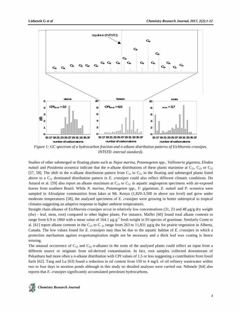
The n-alkane distributions of the different plant parts of E. crassipes are typical of higher terrestrial plant patterns
showing high CPI values (Carbon Preference Index, [55]), reflecting an odd-numbered carbon dominance [56]. In
contrast to other submerged and floating freshwater aquatic macrophytes the n-alkane distribution patterns maximise
at n-C31 in leaf and root samples, while the distribution pattern of stems maximises at n-C27 (Fig. 1). Odd carbon-
numbered dominant distributions of long chain n-alkanes, typically in the range of C25 to C35, are characteristic
components of the epicuticular leaf waxes of higher plants [56].
Liebezeit G et al Chemistry Research Journal, 2017, 2(2):1-12
Chemistry Research Journal
4
Figure 1: GC spectrum of a hydrocarbon fraction and n-alkane distribution patterns of Eichhornia crassipes.
INTSTD–internal standard).
Studies of other submerged or floating plants such as Najas marina, Potamogeton spp., Vallisneria gigantea, Elodea
nuttali and Posidonia oceanica indicate that the n-alkane distributions of these plants maximise at C21, C23 or C25
[57, 58]. The shift in the n-alkane distribution pattern from C21 to C25 in the floating and submerged plants listed
above to a C31 dominated distribution pattern in E. crassipes could also reflect different climatic conditions. Do
Amaral et al. [59] also report an alkane maximum at C29 or C31 in aquatic angiosperm specimens with air-exposed
leaves from southern Brazil. While N. marina, Potamogeton spp., V. gigantean, E. nuttali and P. oceanica were
sampled in Afroalpine communities from lakes at Mt. Kenya (1,820-3,500 m above sea level) and grow under
moderate temperatures [58], the analysed specimens of E. crassipes were growing in hotter subtropical to tropical
climates suggesting an adaptive response to higher ambient temperatures.
Straight chain alkanes of Eichhornia crassipes occur in relatively low concentrations (31, 23 and 48 µg/g dry weight
(dw) - leaf, stem, root) compared to other higher plants. For instance, Maffei [60] found total alkane contents to
range from 6.9 to 1860 with a mean value of 164.1 µg g-1
fresh weight in 93 species of graminae. Similarly Conte et
al. [61] report alkane contents in the C25 to C 35 range from 263 to 11,831 µg/g dw for prairie vegetation in Alberta,
Canada. The low values found for E. crassipes may thus be due to the aquatic habitat of E. crassipes in which a
protection mechanism against evapotranspiration might not be necessary and a thick leaf wax coating is hence
missing.
The unusual occurrence of C16 and C18 n-alkanes in the roots of the analysed plants could reflect an input from a
different source or originate from oil-derived contamination. In fact, root samples collected downstream of
Pekanbaru had more often a n-alkane distribution with CPI values of 1,5 or less suggesting a contribution from fossil
fuels [62]. Tang and Lu [63] found a reduction in oil content from 150 to 4 mg/L of oil refinery wastewater within
two to four days in aeration ponds although in this study no detailed analyses were carried out. Ndimele [64] also
reports that E. crassipes significantly accumulated petroleum hydrocarbons.
C27
C28
C29
C30
C31
C32
C33
C34
C35C37C20
C26C25C24
C23C22C21C19
squalane (INTSTD)
C3
6
C18
leaves
number of carbon atoms
151719212325272931333537
0
2
4
6
8
10stems
number of carbon atoms
151719212325272931333537
µg/g
0
1
2
3
4
5roots
number of carbon atoms
151719212325272931333537
0
2
4
6
8
10
CPI25-31 = 2,8 CPI25-31 = 6,5C P I
25-31 = 3,7
Liebezeit G et al Chemistry Research Journal, 2017, 2(2):1-12
Chemistry Research Journal
5
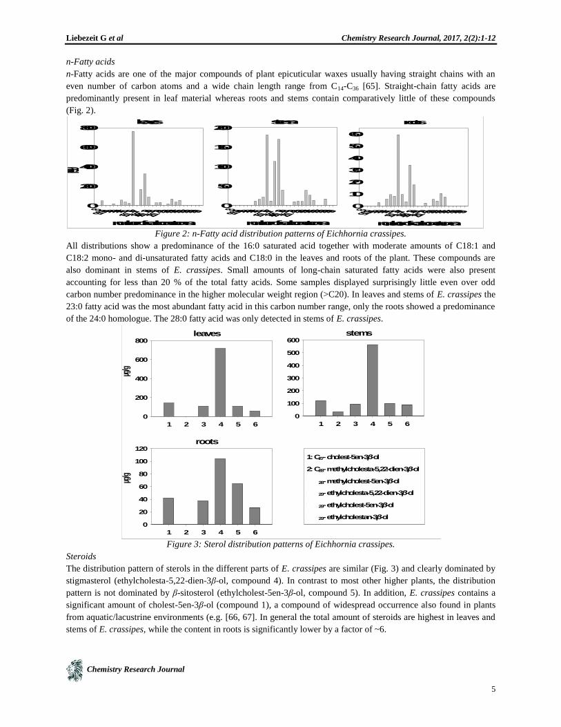
n-Fatty acids
n-Fatty acids are one of the major compounds of plant epicuticular waxes usually having straight chains with an
even number of carbon atoms and a wide chain length range from C14-C36 [65]. Straight-chain fatty acids are
predominantly present in leaf material whereas roots and stems contain comparatively little of these compounds
(Fig. 2).
Figure 2: n-Fatty acid distribution patterns of Eichhornia crassipes.
All distributions show a predominance of the 16:0 saturated acid together with moderate amounts of C18:1 and
C18:2 mono- and di-unsaturated fatty acids and C18:0 in the leaves and roots of the plant. These compounds are
also dominant in stems of E. crassipes. Small amounts of long-chain saturated fatty acids were also present
accounting for less than 20 % of the total fatty acids. Some samples displayed surprisingly little even over odd
carbon number predominance in the higher molecular weight region (>C20). In leaves and stems of E. crassipes the
23:0 fatty acid was the most abundant fatty acid in this carbon number range, only the roots showed a predominance
of the 24:0 homologue. The 28:0 fatty acid was only detected in stems of E. crassipes.
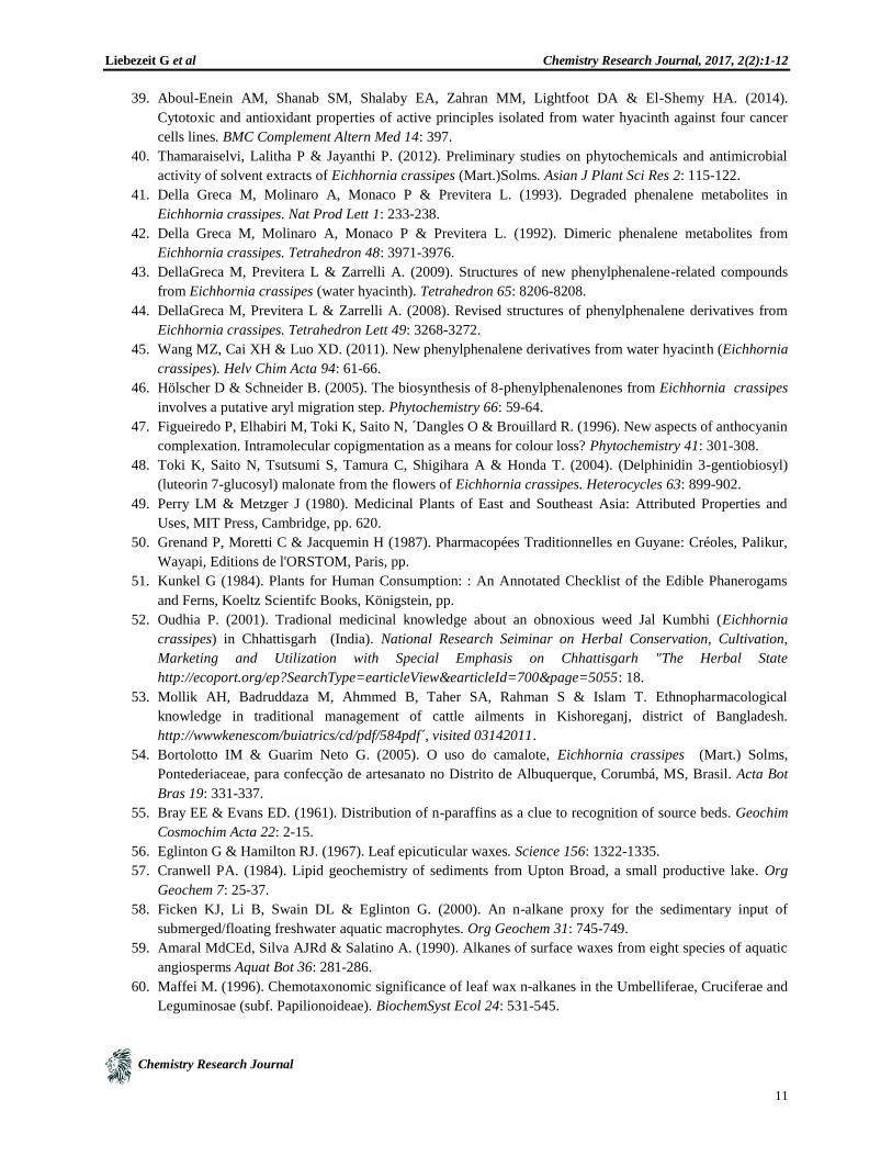
Figure 3: Sterol distribution patterns of Eichhornia crassipes.
Steroids
The distribution pattern of sterols in the different parts of E. crassipes are similar (Fig. 3) and clearly dominated by
stigmasterol (ethylcholesta-5,22-dien-3β-ol, compound 4). In contrast to most other higher plants, the distribution
pattern is not dominated by β-sitosterol (ethylcholest-5en-3β-ol, compound 5). In addition, E. crassipes contains a
significant amount of cholest-5en-3β-ol (compound 1), a compound of widespread occurrence also found in plants
from aquatic/lacustrine environments (e.g. [66, 67]. In general the total amount of steroids are highest in leaves and
stems of E. crassipes, while the content in roots is significantly lower by a factor of ~6.
leaves
number of carbon atoms
89 :110111213141516 :1161718 :218 :1181920 :12021222324252627282930
µg/g
0
200
400
600
800stems
number of carbon atoms
89 :110111213141516 :1161718 :218 :1181920 :120212223242526272829300
50
100
150
200roots
number of carbon atoms
89 :110111213141516 :1161718 :218 :1181920 :120212223242526272829300
10
20
30
40
50
60
roots
1 2 3 4 5 6
0
20
40
60
80
100
120
stems
1 2 3 4 5 6
0
100
200
300
400
500
600leaves
1 2 3 4 5 6
µg/g
0
200
400
600
800
µg/g
1: C27- cholest-5en-3β-ol
2: C28- methylcholesta-5,22-dien-3β-ol
3 : C
28- methylcholest-5en-3β-ol
4 : C
29- ethylcholesta-5,22-dien-3β-ol
5 : C
29- ethylcholest-5en-3β-ol
6 : C
29- ethylcholestan-3β-ol

Liebezeit G et al Chemistry Research Journal, 2017, 2(2):1-12
Chemistry Research Journal
6
Goswami et al. [68] also reported on the dominance of stigmasterol accounting for 55.6 % of total sterols followed
by β-sitosterol (33.4 %9 and campesterol (11.0 %). Cholesterol was not reported to be present.
Sterols with 27 to 29 carbon atoms per molecule are present in all plants and animals. Although a multitude of
combinations of structural and optical isomers of C-27 to C-29 alcohols is theoretically possible, less than ten
distinctively structured C-27 to C-29 sterols comprise more than 90 % (by weight) of biological sterols. C-27 and C-
28 sterols are the most abundant sterols in plankton and marine invertebrates whereas C-29 sterols are the
predominant sterols in higher plants and animals.
The finding of a significant relative contribution from a C27 compound in a higher plant might be taken as an
indication that this is an adaptation to the particular mode of life. However, other floating macrophytes such as e.g.
Potomageton or Lemna have to be investigated to confirm this.
α-Hydroxy-fatty acids
A significant amount of α-hydroxy-fatty acids was only detectable in leaves of E. crassipes (Fig. 4).
Figure 4: α-Hydroxy-fatty acid distribution pattern in Eichhornia crassipes.
The distribution pattern is dominated by even carbon numbered homologues with 20 to 28 carbon atoms and a high
amount of the C24 α-hydroxy-fatty acid in leaves of E. crassipes (943 µg/g). The roots contain only a minor amount
of the C20 α-hydroxy-fatty acid (6 µg/g), while α-hydroxy-fatty acids are completely absent in the stems of the plant.
n-Alcohols
n-Alcohols occur only in leaves and roots of E. crassipes in the range C22-C28 and are predominantly composed of
even-carbon numbered components. Only four compounds were found, i.e. the C22 and C24 in leaf and C22, C24, C26
and C28 in the root (Fig. 5). The distributions are dominated by the C24 homologue in leaves and the C26 one in roots
of E. crassipes.
Figure 5: n-Alcohol distribution in Eichhornia crassipes
leaves
number of carbon atoms
18 20 22 24 26 28 30
µg/g
0
200
400
600
800
1000
roots
number of carbon atoms
18 20 22 24 26 28 30
0
1
2
3
4
5
6
7

Liebezeit G et al Chemistry Research Journal, 2017, 2(2):1-12
Chemistry Research Journal
7
Triterpenoids
Triterpenoids are a widespread class of compounds in the plant kingdom where they occur free or as
glycoconjugates [69]. Unconjugated triterpenes are often found in the epicuticular waxes of plants [70, 71] where
their main function is to prevent water loss and to constitute the first defensive barrier against bacteria, fungi or
insects [71]. The presence of triterpenoids has been reported previously with, however, no structural characterisation
[31]. All plant parts of the analysed E. crassipes specimens contain significant amounts of four triterpenes with
similarly high contents in the leaves and stems of the plant (Fig. 6).
Figure 6: Distribution patterns of unknown pentacyclic triterpenoids of Eichhornia crassipes
The unidentified compound U2 is the only one which occurs in all parts of the plant, while U1 occurs only in leaves
and stems, U3 in stems and roots and U4 in roots of E. crassipes (for mass spectral data see Fig. 7).
Mass spectra of a large number of triterpenoids are available (e.g. [72, 73]. The base peak and the molecular ion in
mass spectra of triterpenoid classes have been found to be characteristic of the basic stereoskeleton [74]. The mass
spectral fragmentation patterns of the unknown compounds U1 and U3 suggest a pentacyclic triterpenoid alcohol
with 30 carbon atoms (M+
498). The fragmentation patterns of compounds U2 and U4 show significant similarities
only distinguishable by a mass shift of two mass units, indicating a double bond in compound U2. Both compounds
contain also 30 carbon atoms, again typical for a pentacyclic triterpenoid alcohol. The mass spectral data of all
unknown compounds show an unusual fragmentation pattern which does not fit analytical reference standards and
literature data of the most common triterpenoids of the oleanan, ursan, lupan or friedelan groups. Thus, the mass
spectral data alone do not provide sufficient information for an unambiguous identification of the triterpenoid
alcohols present in E. crassipes.
Nevertheless, compounds U1 to U4 appear to be unique to E. crassipes and have not been found in any other of the
43 plant species analysed from the river bank vegetation of the Siak (own unpubl. results). They may thus be used to
elucidate whether organic matter from this floating macrophyte is incorporated into sedimentary organic matter
either of the river and estuary or the adjacent coastal waters. Field observations show that water hyacinths may reach
an extensive coverage (up to 70 % of the water surface in the Mandau, a tributary to the Siak) in the freshwater
realms. As soon as the plant comes into contact with saltwater, however, it rapidly disappears and is no longer
observed floating. According to Duke [75] the leaves show epinasty and chlorosis and eventual death in brackish
waters. According to Casabianca and Laugier [76] plants are cankered at salt contents >6 g/L. Similar salinity effects
were also observed in other subtropical and tropical areas such as the Bay of Paranaguá, southern Brazil, or the
Ciénaga Grande de Santa Marta in the Columbian Caribbean (own field observations).

Liebezeit G et al Chemistry Research Journal, 2017, 2(2):1-12
Chemistry Research Journal
8
Figure 7: Mass spectra of the unknown pentacyclic triterpenoids U1–U4.
Due to the large amounts of biomass produced decaying Eichhornia might be a significant contributor to organic
matter in sediments and might also fuel macro- and microheterotrophic metabolism both in the water column and in
the sediment. Analysis of ten sediment samples – two from the Siak tributary Mandau, five from the Siak estuary,
two from the Selat Panjang in front of the Siak estuary and one from the Malacca Strait – did not provide any
evidence for the presence of the terpenoid compounds U1 to U4. This suggests that the Eichhornia biomass, i.e.
particularly leaves and stems, becomes either rapidly disintegrated, is consumed or exported further into the Malacca
Strait.
Conclusions
The distribution patterns of extractable lipid compounds in E. crassipes are typical for higher terrestrial plants with
some noticeable exceptions. The low absolute content of hydrocarbons is possibly a result of the particular mode of
life of the species which does not necessitate the production of organic leaf coatings protecting against desiccation.
In contrast to other submerged and floating plants the n-alkane distribution is dominated by C-29 to C-33
compounds. In leaf material the range is even extended to C37. This suggests that the shift to higher carbon numbers
might be an adaptation to the tropical environment.
The presence of significant amounts of C-27-cholest-5en-3β-ol allows a differentiation from higher land plants,
where C27-steroids are generally absent and the distribution patterns are clearly dominated by C29 compounds.
However, to confirm whether these special traits are typical for floating macrophytes requires additional analyses of
other species, both from temperate and tropical regions.
U1 U2
U4U3
50
100
150
200
250
300
350
400
450
500
0
2000
400
0
600
0
8000
10000
1200
0
14000
16000
18000
20000
2200
0
24000
26000
28000
3000
0
3200
0
34000
36000
38000
40000
m/z--
>
Abundance
leaves123
95
57
27
8
48314
7442
23
9179 20
8365
408
32
450
938
6344
298
498
50
100
150
200
250
300
350
400
450
500
550
600
0
500
0
10000
1500
0
20000
25000
3000
0
35000
4000
0
45000
50000
55000
60000
6500
0
7000
0
75000
80000
8500
0
90000
m/z--
>
Abundance
leaves357
496
2477
3
315
397
147
109 45
3219
191
275 57
0425
528
611
48
1
50 100 150 200 250 300 350 400 450 5000
5000
10000
15000
20000
25000
30000
35000
40000
45000
50000
m/z-->
Abundance
roots 442
483
73
173147
95269
365211
119241
408314 343
38652550329151
5
0
10
0
15
0
20
0
25
0
30
0
35
0
40
0
45
0
50
0
55
0
0
1000
0
20000
30000
40000
5000
0
6000
0
7000
0
80000
9000
0
10000
0
110000
120000
130000
14000
0
15000
0
160000
170000
180000
190000
200000
m/z-->
Abundance
roots498
247
73
357
31
544
2224
275
107
14
7470
191
383
40
8572
541
49
8
48
3

Liebezeit G et al Chemistry Research Journal, 2017, 2(2):1-12
Chemistry Research Journal
9
Acknowledgements
This work has been financially supported by the German Federal Ministry for Education and Research under grant
03F0392B. We are indebted to Joko Samiaji and his students of the University of Riau, Pekanbaru, Sumatra, for
support during the field collections. Cornelia Keuler, Eva Witter and Silke Meyer-Saoudi expertly helped with
sample preparation.
References
1. Mehra A, Farago ME, Banerjee DK & Cordes KB. (1999). The water hyacinth: An environmental friend or
pest? A review. Resour Environ Biotechnol 2: 255-281.
2. Gopal B (1987). Water Hyacinth. Aquatic Plant Studies 1. Elsevier, Amsterdam, pp. 471.
3. Navarro L & Phiri G. (2000). Water hyacinth in Africa and the Middle East. A survey of problems and
solutions. International Development Research Centre, Canada: 1-120.
4. Feikin DR, Tabu CW & Gichuki J. (2010). Does water hyacinth on East African lakes promote cholera
outbreaks? Am J Trop Med Hyg 83: 370-373.
5. Sudhakar Y, Mitra A & Bandyopadhyay M. (2002). Purification of paper and pulp industrial effluent using
aquatic weed Eichornia crassipes. Environ Technol 23: 453-465.
6. So LM, Chu LM & Wong PK. (2003). Microbial enhancement of Cu2+
removal capacity of Eichhornia
crassipes (Mart.). Chemosphere 52: 1499-15023.
7. Sinha S, Pandey K, Mohan D & Singh KP. (2003). Removal of fluoride from aqueous solutions by
Eichhornia crassipes biomass and its carbonized form. Ind Eng Chem Res 42: 6911 -6918.
8. Zimmels Y, Kirzhner F & Schreiber J. (2008). Removal of high organic loads from winery wastewater by
aquatic plants. Water Environ Res 80: 806-822.
9. Cossu R, Haarstad K, Lavagnolo MC & Littarru P. (2001). Removal of municipal solid waste COD and
NH4-N by phyto-reduction: A laboratory-scale comparison of terrestrial and aquatic species at different
organic loads. Ecol Engng 16: 459-470.
10. Chen X, Chen X, Wan X, Weng B & Huang Q. (2010). Water hyacinth (Eichhornia crassipes) waste as an
adsorbent for phosphorus removal from swine wastewater. Bioresour Technol 101: 9025-9030.
11. Mishima D, Kuniki M, Sei K, Soda S, Ike M & Fujita M. (2008). Ethanol production from candidate
energy crops: Water hyacinth (Eichhornia crassipes) and water lettuce (Pistia stratiotes L.). Bioresour
Technol 99: 2495-2500.
12. Nigam JN. (2002). Bioconversion of water-hyacinth (Eichhornia crassipes) hemicellulose acid hydrolysate
to motor fuel ethanol by xylose-fermenting yeast. J Biotechnol 97: 107-116.
13. Singhal V & Rai JPN. (2003). Biogas production from water hyacinth and channel grass used for
phytoremediation of industrial effluents. Bioresource Technol 86: 221-225.
14. Verma VK, Singh YP & Rai JPN. (2007). Biogas production from plant biomass used for phytoremediation
of industrial wastes. Bioresour Technol 98: 1664-1669.
15. Jin ZH, Zhuang YY, Dai SG & Li TL. (2003). Isolation and identification of extracts of Eichhornia
crassipes and their allelopathic effects on algae. Bull Environ Contam Toxicol 71: 1048-1052.
16. Sun WH, Yu SW, Yang SY, Zhao PW, Yu ZW, Wu H, Huang SY & Tang CS. (1993). Allelochemicals
from root exudates of water hyacinth (Eichhornia crassipes). Acta Phyto-physiol Sin 19: 92-96.
17. Della Greca M, Lanzetta R, Molinaro A, Monaco P & Previtera L. (1992). Phenalene metabolites from
Eichhornia crassipes. Bioorg Med Chem Lett 2: 311-314.
18. Fareed MF, Haroon AM & Rabeh SA. (2008). Antimicrobial activity of some macrophytes from Lake
Manzalah (Egypt). Pakistan J Biol Sci 11: 2454-2463.
19. Shanab SMM, Shalaby EA, Lightfoot DA & El-Shemy HA. (2010). Allelopathic effects of water hyacinth
Eichhornia crassipes.. PLoS ONE 5: doi:10.1371/journal.pone.0013200.

Liebezeit G et al Chemistry Research Journal, 2017, 2(2):1-12
Chemistry Research Journal
10
20. Bobbarala V, Rao GS, Aryamithra D, Naidu KC & Rao GS. (2009). Bactericidal activities of fifty
medicinal plants methanolic extracts against Pseudomonas syringae pv.syringae. Biomed Pharmacol J 2:
61-66.
21. Bobbrala B, Rao GS, manduri DB & Naidu KCA. (2009). Biocide potentialities of different plant
methanolic extracts against crown gall bacteria viz Agrobacterium tumefaciens. Biomed Pharmacol J 2: 79-
84.
22. Vadlapudi V. (2010). In vitro antimocrobial activitiy of methanolic extract of selected Indian medicinal
plants. Pharmacophore 1: 214-219.
23. Cochrane BC (1999). Antibacterial and antifungal screening of Florida's exotoc invansive plant species, in
Florida's Garden of Good and Evil, Jones D T and Gamble, B W, Editors., Everglades National Park,
South Florida Natural Resources Center: Homestead, FL. pp. 205-216,
24. Kumar S, Kumar R, Dwivedi A & Pandey AK. (2014). In vitro antioxidant, antibacterial, and cytotoxic
activity and in vivo effect of Syngonium podophyllum and Eichhornia crassipes leaf extracts on isoniazid
induced oxidative stress and hepatic markers. Biomed Res Int: 459452.
25. Saxena RC, Dixit OP & Sukumaran P. (1992). Laboratory assessment of indigenous plant extracts for anti-
juvenile hormone activity in Culex quinquefasciatus. Ind J Med Res 95A: 204-206.
26. Assar AA & El-Sobky MM. (2003). Biological and histopathological studies of some plant extracts on
larvae of Culex pipiens (Diptera: Culicidae). J Egypt Soc Parasitol 33: 189-200.
27. Devanand P & Rani PU. (2008). Biological potency of certain plant extracts in management of two
lepidopteran pests of Ricinus communis L. . J Biopesticides 1: 170-176.
28. Lalitha P, Sripathi SK & Jayanthi P. (2012). Secondary metabolites of Eichhornia crassipes
(Waterhyacinth): a review (1949 to 2011). Nat Prod Commun 7: 1249-1256.
29. Matai S & Bagchi DK (1980). Water hyacinth: a plant with prolific bioproductivity and photosynthesis, in
Proc Internat Symp on Biol Applications of Solar Energy, Gnanam A, Krishnaswamy, S and Kahn, J S,
Editors., MacMillan Co. of India: Madras. pp. 144-148,
30. Kandasamy MK & Vivekanandan M. (1983). Biochemical composition of stigmatic exudate of Eichhornia
crassipes (Mart.) solms. Aquat Bot 16: 41-47.
31. Vasu K, Goud JV, Suryam A & Charya MAS. (2009). Biomolecular and phytochemical analyses of three
aquatic angiosperms. Afr J Microbiol Res 3: 418-421.
32. Lata N, Ali H, Das S & Dubey V. (2010). Antioxidants of Eichhornia crassipes: The world’s worst aquatic
plant. J Pharmacy Res 3: 2105-2106.
33. Huma A, Patel M, N. G & Ahi J. (2009). The world’s worst aquatic plant as a safe cancer medicine
“Antitumor activity on melanoma induced mouse by Eichhornia crassipes: Vivo studies. J Pharmacy Res
2: 1365-1366.
34. Sanseverino AM, Bastviken D, Sundh I, Pickova J & Enrich-Prast A. (2012). Methane carbon supports
aquatic food webs to the fish level. PLoS ONE 7: 8.
35. Arayana GL, Rao KS, Pantulu AJ & Thyagarajan G. (1984). Composition of lipids in roots, stalks, leaves
and flowers of Eichhornia crassipes (Mart.) Solms. Aquat Bot 20: 219-227.
36. Lata N & Dubey V. (2010). Preliminary phytochemical screening of Eichhornia crassipes: the world’s
worst aquatic weed. J Pharmacy Res 3: 1240-1242.
37. Lata N. (2010). Quantification and identification of alkaloids of Eichhornia crassipes: the world's worst
aquatic plant. J Pharm Res 3: 1229.
38. Aboul-Enein AM, Al-Abd AM, Shalaby E, Abul-Ela F, Nasr-Allah AA, Mahmoud AM & El-Shemy HA.
(2011). Eichhornia crassipes (Mart) solms: from water parasite to potential medicinal remedy. Plant Signal
Behav 6: 834-836.
Liebezeit G et al Chemistry Research Journal, 2017, 2(2):1-12
Chemistry Research Journal
11
39. Aboul-Enein AM, Shanab SM, Shalaby EA, Zahran MM, Lightfoot DA & El-Shemy HA. (2014).
Cytotoxic and antioxidant properties of active principles isolated from water hyacinth against four cancer
cells lines. BMC Complement Altern Med 14: 397.
40. Thamaraiselvi, Lalitha P & Jayanthi P. (2012). Preliminary studies on phytochemicals and antimicrobial
activity of solvent extracts of Eichhornia crassipes (Mart.)Solms. Asian J Plant Sci Res 2: 115-122.
41. Della Greca M, Molinaro A, Monaco P & Previtera L. (1993). Degraded phenalene metabolites in
Eichhornia crassipes. Nat Prod Lett 1: 233-238.
42. Della Greca M, Molinaro A, Monaco P & Previtera L. (1992). Dimeric phenalene metabolites from
Eichhornia crassipes. Tetrahedron 48: 3971-3976.
43. DellaGreca M, Previtera L & Zarrelli A. (2009). Structures of new phenylphenalene-related compounds
from Eichhornia crassipes (water hyacinth). Tetrahedron 65: 8206-8208.
44. DellaGreca M, Previtera L & Zarrelli A. (2008). Revised structures of phenylphenalene derivatives from
Eichhornia crassipes. Tetrahedron Lett 49: 3268-3272.
45. Wang MZ, Cai XH & Luo XD. (2011). New phenylphenalene derivatives from water hyacinth (Eichhornia
crassipes). Helv Chim Acta 94: 61-66.
46. Hölscher D & Schneider B. (2005). The biosynthesis of 8-phenylphenalenones from Eichhornia crassipes
involves a putative aryl migration step. Phytochemistry 66: 59-64.
47. Figueiredo P, Elhabiri M, Toki K, Saito N, ´Dangles O & Brouillard R. (1996). New aspects of anthocyanin
complexation. Intramolecular copigmentation as a means for colour loss? Phytochemistry 41: 301-308.
48. Toki K, Saito N, Tsutsumi S, Tamura C, Shigihara A & Honda T. (2004). (Delphinidin 3-gentiobiosyl)
(luteorin 7-glucosyl) malonate from the flowers of Eichhornia crassipes. Heterocycles 63: 899-902.
49. Perry LM & Metzger J (1980). Medicinal Plants of East and Southeast Asia: Attributed Properties and
Uses, MIT Press, Cambridge, pp. 620.
50. Grenand P, Moretti C & Jacquemin H (1987). Pharmacopées Traditionnelles en Guyane: Créoles, Palikur,
Wayapi, Editions de l'ORSTOM, Paris, pp.
51. Kunkel G (1984). Plants for Human Consumption: : An Annotated Checklist of the Edible Phanerogams
and Ferns, Koeltz Scientifc Books, Königstein, pp.
52. Oudhia P. (2001). Tradional medicinal knowledge about an obnoxious weed Jal Kumbhi (Eichhornia
crassipes) in Chhattisgarh (India). National Research Seiminar on Herbal Conservation, Cultivation,
Marketing and Utilization with Special Emphasis on Chhattisgarh "The Herbal State
http://ecoport.org/ep?SearchType=earticleView&earticleId=700&page=5055: 18.
53. Mollik AH, Badruddaza M, Ahmmed B, Taher SA, Rahman S & Islam T. Ethnopharmacological
knowledge in traditional management of cattle ailments in Kishoreganj, district of Bangladesh.
http://wwwkenescom/buiatrics/cd/pdf/584pdf´, visited 03142011.
54. Bortolotto IM & Guarim Neto G. (2005). O uso do camalote, Eichhornia crassipes (Mart.) Solms,
Pontederiaceae, para confecção de artesanato no Distrito de Albuquerque, Corumbá, MS, Brasil. Acta Bot
Bras 19: 331-337.
55. Bray EE & Evans ED. (1961). Distribution of n-paraffins as a clue to recognition of source beds. Geochim
Cosmochim Acta 22: 2-15.
56. Eglinton G & Hamilton RJ. (1967). Leaf epicuticular waxes. Science 156: 1322-1335.
57. Cranwell PA. (1984). Lipid geochemistry of sediments from Upton Broad, a small productive lake. Org
Geochem 7: 25-37.
58. Ficken KJ, Li B, Swain DL & Eglinton G. (2000). An n-alkane proxy for the sedimentary input of
submerged/floating freshwater aquatic macrophytes. Org Geochem 31: 745-749.
59. Amaral MdCEd, Silva AJRd & Salatino A. (1990). Alkanes of surface waxes from eight species of aquatic
angiosperms Aquat Bot 36: 281-286.
60. Maffei M. (1996). Chemotaxonomic significance of leaf wax n-alkanes in the Umbelliferae, Cruciferae and
Leguminosae (subf. Papilionoideae). BiochemSyst Ecol 24: 531-545.

Liebezeit G et al Chemistry Research Journal, 2017, 2(2):1-12
Chemistry Research Journal
12
61. Conte MH, Weber JC, Carlson P & Flanagan LB. (2003). Molecular and carbon isotopic composition of
leaf wax in vegetation and aerosols in a northern prairie ecosystem. Oecologia 135: 67-77.
62. Barakat AO, Mostafa AR, Rullkötter J & Hegazi AR. (1999). Application of multimolecular marker
approach to fingerprint petroleum pollution in the marine environment. Mar Poll Bull 38: 535-544.
63. Tang S-y & Lu X-w. (1993). The use of Eichhornia crassipes to cleanse oil-refinery wastewater in China.
Ecol Engng 2: 243-251.
64. Ndimele PE. (2003). The prospect of phytoremediation of polluted natural wetlands by inhabiting aquatic
macrophytes (Water hyacinth). MSc Thesis, University of Ibadan, Nigeria.
65. Kolattukudy PE (1976). Chemistry and Biochemistry of Natural Waxes, Elsevier, Amsterdam, pp. 290.
66. Grunwald C. (1975). Plant sterols. Ann Rev Plant Physiol 26: 209-236.
67. Gordon MH & Miller LAD. (1997). Development of steryl ester analysis for the detection of admixtures of
vegetable oils. J Am Oil Chem Soc 74: 505-510.
68. Goswami PC, Nag B, Sharma AK, Borthakur A, Singh HD & Baruah JN. (1983). Water hyacinth as a
prospective source of stigmasterol. Curr Sci 52: 806-808.
69. Harborne JB. (1989). Recent advances in chemical ecology. Nat Prod Reports 6: 85-112.
70. Garcia S, Heinzen H, Hubbach C, Martinez R, De Vries JX & Moyna P. (1995). Triterpene methyl ethers
from Palmae epicuticular waxes. Phytochemistry 39: 1381-1382.
71. Tulloch AP (1976). Chemistry of plant waxes, in Chemistry and Biochemistry of Natural Waxes,
Kolattukudy A, Editor, Elsevier: Amsterdam. pp. 174-195.
72. Ogunkoya L. (1981). Application of mass spectrometry in structural problems in triterpenes.
Phytochemistry 20: 121-126.
73. Das MC & Mahato SB. (1983). Triterpenoids. Phytochemistry 22: 1071-1095.
74. Budzikiewicz H, Wilson M & Djerassi C. (1963). Mass spectrometry in structural and stereochemical
problems. XXXII. Pentacyclic triterpenes. 85: 3688-3699.
75. Duke JA (1983). Eichhornia crassipes (Mart.) Solms,
76. Casabianca MLd & Laugier T. (1995). Eichhornia crassipes production on petroliferous wastewaters:
Effects of salinity. Bioresource Technol: 39-43.