chemodiversity of calophyllum inophyllum l. oil bioactive ... · swati & shubhada (2011),...
TRANSCRIPT

Chemodiversity of Calophylluminophyllum L. oil bioactive componentsrelated to their specific geographicaldistribution in the South Pacific regionJoape Ginigini1, Gaël J. Lecellier2, Mael Nicolas3, Mohammed Nour3,Edouard Hnawia3, Nicolas Lebouvier3, Gaëtan Herbette4,Peter Lockhart5 and Phila Raharivelomanana6
1 Pacific Natural Products Research Centre, Institute of Applied Sciences, University of theSouth Pacific, Suva, Fiji
2 Départment de Biologie, Université de Versailles Saint-Quentin-en-Yvelines, Versailles, France3 ISEA EA7484, University of Caledonia, Noumea, New Caledonia4 Spectropole, Campus de St Jérôme, Aix-Marseille Univ, CNRS, Centrale Marseille, FSCM,Marseille, France
5 School of Fundamental Sciences, Massey University, Palmerston North, New Zealand6 EIO UMR241, Université de la Polynésie française, Faa’a, Tahiti, French Polynesia
ABSTRACTBackground: Different parts of the tree Calophyllum inophyllum L. (nuts, leaves,roots, bark, fruits, nut oil and resin) are used as traditional medicines and cosmeticsin most of the Pacific Islands. The oil efficiency as a natural cure and in traditionalcosmetics has been largely described throughout the South Pacific, which led usto investigate C. inophyllum’s chemical and genetic diversity. A correlative study ofthe nut resin and leaf DNA from three distinct archipelagos in the South Pacific wascarried out in order to identify diversity patterns in C. inophyllum across theSouth Pacific.Methods: Calophyllum inophyllum plants were sampled from French Polynesia,New Caledonia and Fiji. We extracted tamanu oil (nut oil) resin for chemo-diversitystudies and sampled leaf tissues for genetic studies. We applied an analysis methoddesigned for small quantities (at a microscale level), and used High PerformanceLiquid Chromatography (HPLC) to establish the chemo-diversity of tamanu oilresin. In-house standards were co-eluted for qualitative determination. Geneticdiversity was assessed using chloroplast barcoding markers (the Acetyl-CoAcarboxylase (accD) gene and the psaA-ycf3 intergenic spacer region).Results: Our HPLC analysis revealed 11 previously known tamanu oil constituents,with variability among plant samples. We also isolated and characterized twonew neoflavonoids from tamanu oil resin namely, tamanolide E1 and E2 which arediastereoisomers. Although genetic analysis revealed low genetic variation, ourmultivariate analysis (PCA) of the tamanu oil resin chemical profiles revealeddifferentiation among geographic regions.Conclusion: We showed here that chromatographic analysis using formalizedin-house standards of oil resin compounds for co-elution studies against oil resinsamples could identify patterns of variation among samples of C. inophyllum, anddiscriminate samples from different geographical origins.
How to cite this article Ginigini J, Lecellier GJ, Nicolas M, Nour M, Hnawia E, Lebouvier N, Herbette G, Lockhart P, Raharivelomanana P.2019. Chemodiversity of Calophyllum inophyllum L. oil bioactive components related to their specific geographical distribution in the SouthPacific region. PeerJ 7:e6896 DOI 10.7717/peerj.6896
Submitted 18 October 2018Accepted 2 April 2019Published 22 May 2019
Corresponding authorGaël J. Lecellier,[email protected]
Academic editorCurtis Daehler
Additional Information andDeclarations can be found onpage 17
DOI 10.7717/peerj.6896
Copyright2019 Ginigini et al.
Distributed underCreative Commons CC-BY 4.0

Subjects Biogeography, Plant Science, BiogeochemistryKeywords Calophyllum inophyllum, Chemodiversity, South Pacific, Neoflavonoids, Oil,Biodiversity
INTRODUCTIONChemical and medicinal properties of Alexandrian laurel (Calophyllum inophyllum),commonly known as beach mahogany, have been extensively studied throughout theworld and even more in the Asia-Pacific region (Léguillier et al., 2015; Pawar, Swati &Shubhada, 2011; Patil et al., 1993). The plant has a myriad of medicinal uses, most of theminvolving topical applications. Recent studies such as Léguillier et al. (2015) and Ansel et al.(2016) have focused more attention on the cosmetic aspects of the plant in the Pacificregion. Other common names for the plant in some Pacific island countries are dilo (Fiji),fetau (Samoa), tamanou de bord de mer (New Caledonia) and tamanu in the Cook Islandsand French Polynesia (Friday & Okano, 2006). Historically, before the conversion ofPolynesians to Christianity, the tamanu trees were considered sacred. They were plantedinside the royal Marae (sacred areas) (Dweck & Meadows, 2002).
Calophyllum inophyllum belongs to the flowering plant family Calophyllaceae(Angiosperm Phylogeny Group, 2009; Prabakaran & Britto, 2012) and is native to theIndo-Pacific region (Africa, India, South East Asia, Australia and the Pacific islands).It grows to a height of 8–15 m and has a large canopy. The wood is widely used for makingcabinet and other furniture, for carving, and for boat and canoe building. Furthermore,different parts of the plant have been used in traditional medicine and as excellent rawmaterial for cosmetics (Dweck &Meadows, 2002). For instance, the nut oil has been used formedicine against skin infections, as a scar remover as well as for other cosmetic uses(Friday & Okano, 2006). In Fiji, the oil is used to cure arthritis and joint pain, as an eye washfor conjunctivitis (Cambie & Ash, 1994) and also to prevent infantile rash. The resin mixedwith strips of bark and leaves is used as a treatment for sore eyes. The green fruit isused against tuberculosis (Cambie & Ash, 1994). In some islands of Polynesia, the oil hasbeen used as an alternative for candle nut oil in lamps and also massaged into hair andused as a common topical application for skin diseases and burns (Prabakaran & Britto,2012). A number of studies have revealed interesting oil biological activities such asantibacterial (Yimdjo et al., 2004), antifungal (Saravanan et al., 2011), anti-inflammatoryagainst skin infections (Bhalla et al., 1980). Most recently it has proven useful for woundhealing (Léguillier et al., 2015). Such is the uniqueness of its properties that “tamanu oil”has been recognized as an active cosmetic ingredient and recorded as C. inophyllumseed oil by the International nomenclature of cosmetic ingredients (Assouvie, 2013;Ansel et al., 2016).
The bioactive components (belonging to neoflavonoid, xanthone and triterpene secondarymetabolite groups) in this plant are highly rich and recognized as having medicinalproperties as shown in Table 1. The composition of the main neoflavonoid compounds inthe oil are as follows: calophyllolide, inophyllums (C, D, E and P), tamanolides D and P,calanolide Gut 70, and finally the calanolides A, B and D (Laure, 2005; Leu et al., 2009;Assouvie, 2013). Also of great interest is the anti-HIV efficiency of pyranocoumarins from
Ginigini et al. (2019), PeerJ, DOI 10.7717/peerj.6896 2/21

the Calophyllum genus (Wang et al., 2006). Recent studies have also examined their usein cicatrization, and as an anti-aging agent (Léguillier et al., 2015; Ansel et al., 2016).The calanolide A compound, a minor constituent of C. inophyllum resin extract (Ansel et al.,2016) has also attracted recent interest because it is the only natural product that hasprogressed into human clinical trials with positive results against HIV-1 (Wang et al., 2006).
The chemical composition analysis of C. inophyllum leaves from various sites inFrench Polynesia, and that of fruits originating from various sites in India, has revealedregional differentiation (Laure, 2005; Pawar, Swati & Shubhada, 2011) betweenC. inophyllum plants found inland and closer to the coast. In order to evaluate the chemicalqualities of the different tamanu oils of the South Pacific, phytochemical analyses aretherefore necessary. Given that secondary metabolites play a role in the adaptation ofplants to biotic and abiotic factors, it would be interesting to know if, as revealed by Pawar,Swati & Shubhada (2011), abiotic factors such as those associated with the geographyof harvesting sites have an influence on the chemical profile of tamanu oil.
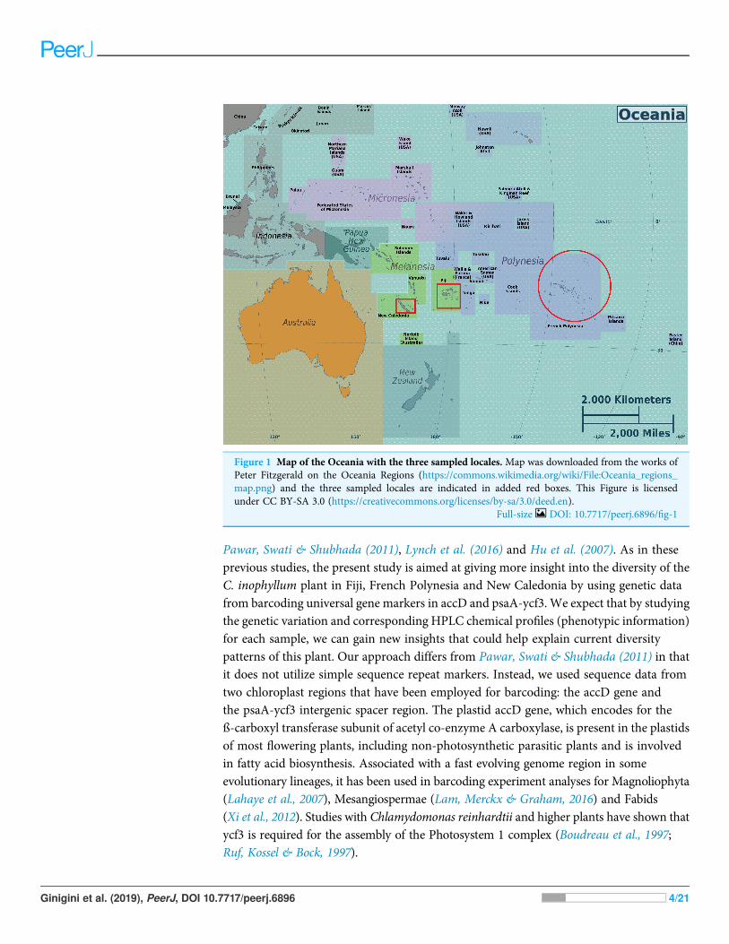
In addition to the general question of regional influence on the chemical profilesof tamanu oil, we were also interested in whether dipyranocoumrins and moreparticularly inophyllums can be used, as described by Pawar, Swati & Shubhada (2011),as chemotaxonomic markers for the identification of C. inophyllum from variousgeographical areas of the South Pacific. To address these questions, from three SouthPacific countries (French Polynesia, New-Caledonia and Fiji) shown on Oceania’s mapin Fig. 1. we characterized genetic diversity together with the phytochemical diversity,following a polyphasic approach, as already applied to some microbial research.
Polyphasic taxonomy is a consensus method of taxonomy developed to incorporatephenotypic, genotypic and phylogenetic data for micro-organisms (Vandamme et al.,1996). A simplified version of this method for plant research, utilizing only genotypeand phenotype variation for elucidation of diversity has been successfully used by
Table 1 Summary of major bioactive compounds found in C. inophyllum resin oil with some barkand leaf constituents and their anti-cancer, anti-microbial, anti-HIV and anti-inflammatory effects.
Compounds Anti-cancer Anti-microbial Anti-HIV Anti-inflammatory
Calophyllolide ⧾b ⧾c ⧾g
Inophyllum P ⧾a,d
Inophyllum C ⧾b ⧾c ⧾a
Inophyllum D ⧾b ⧾a
Inophyllum E ⧾b
Inophyllum B ⧾a,d
Inophyllum A ⧾b ⧾a
Calanolide Gut 70 ⧾h
Calanolide A/Calanolide B ⧾d
Calanolide/Pseudocalanolide D ⧾f
12-Oxocalanolide ⧾d,e
Note:Compound References articles in parenthesis aPatil et al. (1993), bItoigawa et al. (2001), cYimdjo et al. (2004), dKostova &Mojzis (2007), eWang et al. (2006), fIshikawa (2000), gSaxena et al. (1982), hJin et al. (2011).
Ginigini et al. (2019), PeerJ, DOI 10.7717/peerj.6896 3/21
Pawar, Swati & Shubhada (2011), Lynch et al. (2016) and Hu et al. (2007). As in theseprevious studies, the present study is aimed at giving more insight into the diversity of theC. inophyllum plant in Fiji, French Polynesia and New Caledonia by using genetic datafrom barcoding universal gene markers in accD and psaA-ycf3. We expect that by studyingthe genetic variation and corresponding HPLC chemical profiles (phenotypic information)for each sample, we can gain new insights that could help explain current diversitypatterns of this plant. Our approach differs from Pawar, Swati & Shubhada (2011) in thatit does not utilize simple sequence repeat markers. Instead, we used sequence data fromtwo chloroplast regions that have been employed for barcoding: the accD gene andthe psaA-ycf3 intergenic spacer region. The plastid accD gene, which encodes for theß-carboxyl transferase subunit of acetyl co-enzyme A carboxylase, is present in the plastidsof most flowering plants, including non-photosynthetic parasitic plants and is involvedin fatty acid biosynthesis. Associated with a fast evolving genome region in someevolutionary lineages, it has been used in barcoding experiment analyses for Magnoliophyta(Lahaye et al., 2007), Mesangiospermae (Lam, Merckx & Graham, 2016) and Fabids(Xi et al., 2012). Studies with Chlamydomonas reinhardtii and higher plants have shown thatycf3 is required for the assembly of the Photosystem 1 complex (Boudreau et al., 1997;Ruf, Kossel & Bock, 1997).
Figure 1 Map of the Oceania with the three sampled locales. Map was downloaded from the works ofPeter Fitzgerald on the Oceania Regions (https://commons.wikimedia.org/wiki/File:Oceania_regions_map.png) and the three sampled locales are indicated in added red boxes. This Figure is licensedunder CC BY-SA 3.0 (https://creativecommons.org/licenses/by-sa/3.0/deed.en).
Full-size DOI: 10.7717/peerj.6896/fig-1
Ginigini et al. (2019), PeerJ, DOI 10.7717/peerj.6896 4/21
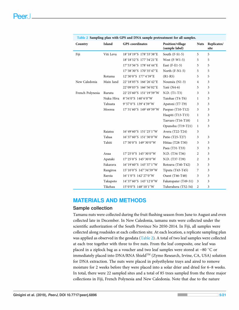
MATERIALS AND METHODSSample collectionTamanu nuts were collected during the fruit flushing season from June to August and evencollected late in December. In New Caledonia, tamanu nuts were collected under thescientific authorization of the South Province No 2050-2014. In Fiji, all samples werecollected along roadsides at each collection site. At each location, a replicate sampling planwas applied as observed in the geodata (Table 2). A total of two leaf samples were collectedat each tree together with three to five nuts. From the leaf composite, one leaf wasplaced in a ziplock bag as a voucher and two leaf samples were stored at -80 �C orimmediately placed into DNA/RNA ShieldTM (Zymo Research, Irvine, CA, USA) solutionfor DNA extraction. The nuts were placed in polyethylene trays and aired to removemoisture for 2 weeks before they were placed into a solar drier and dried for 6–8 weeks.In total, there were 22 sampled sites and a total of 85 trees sampled from the three majorcollections in Fiji, French Polynesia and New Caledonia. Note that due to the nature
Table 2 Sampling plan with GPS and DNA sample pretreatment for all samples.
Country Island GPS coordinates Position/village(sample label)
Nuts Replicates/site
Fiji Viti Levu 18�18′19″S 178�33′38″E South (F-S1-5) 5 5
18�18′52″S 177�34′21″E West (F-W1-5) 5 5
17�53′56″S 178�44′44″E East (F-E1-5) 5 5
17�38′30″S 170�35′47″E North (F-N1-5) 5 5
Rotuma 12�30′0″S 177�4′59″E (R1-R5) 5 5
New Caledonia Main Iand 22�18′05″S 166�26′42″E Nouméa (N1-3) 4 3
22�09′03″S 166�56′02″E Yaté (N4-6) 5 3
French Polynesia Rurutu 22�25′60″S 151�19′59″W N.D. (T1-T3) 3 3
Nuku Hiva 8�54′0″S 140�6′0″W Taiohae (T4-T6) 1 3
Tahuata 9�57′0″S 139�4′59″W Apatoni (T7-T9) 3 3
Moorea 17�31′60″S 149�49′59″W Paopao (T10-T12) 3 3
Haapiti (T13-T15) 1 3
Taevaro (T16-T18) 1 3
Opunohu (T19-T21) 1 3
Raiatea 16�49′60″S 151�25′1″W Avera (T22-T24) 3
Tahaa 16�37′60″S 151�30′0″W Patio (T25-T27) 3 3
Tahiti 17�30′0″S 149�30′0″W Hitiaa (T28-T30) 3 3
Paea (T31-T33) 3 3
Anaa 17�25′0″S 145�30′0″W N.D. (T34-T36) 2 3
Apataki 17�25′0″S 145�30′0″W N.D. (T37-T39) 2 3
Fakarava 16�19′60″S 145�37′1″W Rotoava (T40-T42) 3 3
Rangiroa 15�10′0″S 147�34′59″W Tiputa (T43-T45) 7 3
Raroia 16�1′0″S 142�27′0″W Ouest (T46-T48) 3 3
Takapoto 14�37′60″S 145�12′0″W Fakatopater (T49-51) 3 3
Tikehau 15�0′0″S 148�10′1″W Tuherahera (T52-54) 2 3
Ginigini et al. (2019), PeerJ, DOI 10.7717/peerj.6896 5/21
of collections being conducted in isolated locations from both French Polynesia andNew Caledonia, limited amount of material was collected and thus vouchers specimenscould not be deposited for these two research groups. Only the samples collected in Fijihad vouchers deposited at the South Pacific Regional Herbarium (Table S1).
Microscale extraction, purification and HPLC analysisFrom each of the 85 collected trees, a set of three to five nuts weighing 7–10 g were pickedup and subjected to small scale cold extraction consisting of maceration and nut grindingin a cheese cloth followed by crushing in a mortar and pestle to allow oil expressionthrough the cloth. Samples were washed with EtOAc (provided by VWR Chemicals,Radnor, PA, USA) and sonicated for 5 min to extract all nut components. These were driedin vacuo to yield a crude extract containing classical fatty acids components and de-fattedcompounds. This last crude extract (oil) was then partitioned with EtOH (provided byFisher chemical, Hampton, NH, USA) at a 1:1 ratio v/v to extract only EtOH soluble nonfatty compounds. The EtOH layer was removed and dried under vacuum yielding aresinous extract (called resin) containing neoflavonoids compounds, which will besubmitted to further chemical analysis. Any remaining oil was de-fatted twice with40 mL hexane in total. Resinous extracts were dissolved in EtOAc: ¼cyclohexane(provided by VWR Chemicals) at a 1:1 ratio v/v at a concentration of 10 mg/mL for finalinjection. Chromatographic analyses were performed using an Agilent 1100 seriesgradient HPLC fitted with a UV/DAD system. The HPLC column used was anInterchrome Modula-Cart QS Uptisphere five mm Si column, and the data were viewedon Agilent Chemstation software (Santa Clara, CA, USA). Optimized step gradientelution was applied utilizing Cyclohexane (HPLC grade from Fisher chemical) andEtOAc (HPLC grade from Fisher chemical) as solvent and the analytical conditions wereas follows: flow rate one mL/min, pressure 2,500 psi max, injection volume of four mL.The solvent system gradient conditions are shown in Table S2 in the supportinginformation.
HPLC analysis of the samples from all three locations (Fiji: RawDataS1, New Caledonia:RawDataS2 and French Polynesia: RawDataS3) involved numerous trials to obtain themaximum amount of compounds in the shortest runtime. On completion of the analysis,80 resin samples were chosen from the total of 85 samples and five were discardeddue to poor alignment and high signal to noise. Two more samples were discarded due topoor alignment. In total, 78 samples were analyzed by HPLC. Sample chromatogramprofiles were recorded at 280 nm, wavelength at which most peaks were observed in theoptimized analytical conditions for neoflavonoid standards. The removal of noise,redefinition and alignment of all data was necessary to identify and assign each samplepeak before comparing these to standard compounds which were isolated and identifiedin previous studies (Leu et al., 2009; Leu, 2009; Laure, 2005; Laure et al., 2008).Background fitting and identification of major peaks of the raw HPLC data wereperformed using the R package align DE v2.0.1. Chromatograms were aligned using aprocedure in Scilab version 5.5.1 (Scilab Enterprises, 2012) derived from chromaligner(Wang et al., 2010).
Ginigini et al. (2019), PeerJ, DOI 10.7717/peerj.6896 6/21
Purification of known compounds from commercial tamanu oil resinand isolation of new compoundsA batch of oil resin extract (157 g) from commercial tamanu oil (provided by “Laboratoirede Cosmétologie du Pacifique Sud” manufacture) from French Polynesia was firstpartitioned with EtOH (provided by Fisher chemical) and an aqueous alkaline solutionof Na2CO3 (from VWR chemicals) (10%, v/v). Its organic fraction was washed withdistilled water and then dried with MgSO4 (provided by VWR chemicals) to give a neutralfraction (53 g) after solvent evaporation. This fraction was submitted to flash liquidchromatography on an open column with a silica gel (240–300 mesh) using a stepwisegradient from cyclohexane (provided by VWR chemicals) to EtOAc (provided byVWR chemicals), yielding 12 fractions. Fractions having similar Rf values on silica gel TLC(cyclohexane-acetone, both provided by VWR chemicals, 60:40, v/v) were combined.Fractions 7, 9 and 11 were submitted to repeated preparative HPLC using a VarianDynamax Si column (250 � 21.4 mm id, five mm with cyclohexane-EtOAc (10:90) inisocratic eluent conditions. This chromatographic purification network led to the isolationof new compounds tamanolides E1 and E2 as a mixture (two mg) besides standard knowncompounds namely, calophyllolide, inophyllums (C, D, E, P), calanolides (Gut 70 andA, 12-oxo-calanolide) and tamanolides (D, P).
Analysis of new compoundsNew compounds were identified by NMR and were further analyzed with a Bruker AvanceDRX500 spectrometer (1H—500.13 MHz) equipped with a five mm triple resonanceinverse Cryoprobe TXI (1H/13C/15N) in CDCl3-99.8%, (d1H ¼ 7.26 pm, d13C ¼ 77.16ppm). The HR-ESI-MS data were collected using a QStar Elite mass spectrometer (AppliedBiosystems SCIEX, Concord, ON, Canada) equipped with an ESI source operated inpositive ion mode. Optical rotations were measured with a Perkin-Elmer 241 polarimeterequipped with a sodium (589 nm) lamp and a one dm cell. The FTIR spectra wereestablished with a Thermo-Nicolet IR 200 spectrometer (Waltham, MA, USA) on a KBrcell and four cm-1 resolution.
DNA extraction and amplificationPlant DNA was extracted from young leaf tissue using the Plant Nucleospin© II kit(Macherey-Nagel) according to the manufacturer’s instruction. The leaf samples were firstlyophilized in liquid nitrogen and crushed before being subjected to the manufacturer’sprotocols. Two plant universal chloroplast regions were targeted for amplificationnamely; the Acetyl-CoA carboxylase (accD) gene and the psaA-ycf3 intergenic spacerregion. The amplifications were performed in a Thermo Scientific Arktik Thermo Cycler.The PCR was performed using a Qiagen Taq polymerase kit and primers were diluted to aconcentration of 0.6 mM. All sample amplifications were eluted in distilled milliQ waterat 50 mL volumes and tested for yield with 1% agarose gel electrophoresis. The PCRprogram consisted of 92 �C of incubation during 2 min for one cycle and eight cycles ofPCR (denaturation at 92 �C for 30 s followed by specific annealing temperatures for accD(44 �C) and psaA-ycf3 (43 �C) all at 30 s, and 72 �C for 1 min), followed by 40 cycles
Ginigini et al. (2019), PeerJ, DOI 10.7717/peerj.6896 7/21
of (92 �C for 30 s, extension at primer specific temperature’s accD (46 �C) and psaA-ycf3 at30 s, 72 �C for 1 min). A final 5 min extension was then carried out at 72 �C and foronly one cycle.
Sequencing alignment and data analysisThe samples were sequenced by GATC Biotech sequencing services, Germany. Datareceived in AB1 format were viewed under the Applied Biosystem’s sequencing scannerand corrected for contiguous read lengths and miscalled sequence data were removedespecially in the beginning and at the ends of the raw data files. Files were then alignedusing MEGA6 (Tamura et al., 2013) before they were matched to nucleotide sequencesusing the BLASTn tool available at https://blast.ncbi.nlm.nih.gov/Blast.cgi. Only matcheswith 96–100% similarity were included in the construction of a multiple sequencealignment. Sequences were aligned using ClustalW (Thompson, Higgins & Gibson, 1994)and trees were generated using the MEGA6 phylogeny tools. New haplotypes weredeposited into the NCBI Genbank database using the Bankit tool. Accession numbers aregiven in Table S3.
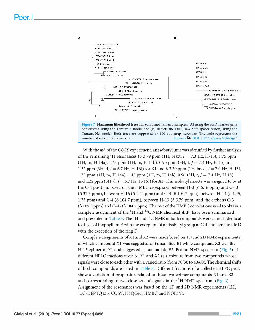
Phylogenetic constructionAll newly determined sequences were checked using the sequencer software for peakintensity and also contiguous length. All sequences with contiguous lengths greater than200 bp were considered for tree construction using MEGA6. Maximum likelihood (ML)trees were constructed for all accD sequences which satisfied our contiguous lengthcriterion. Initial tree(s) for a heuristic search were first obtained by applying Neighbor-Joining (Saitou & Nei, 1987) and BioNJ (Gascuel, 1997) algorithms to a matrix of pairwisedistances estimated using the maximum composite likelihood approach implemented inMEGA, and tree searches were conducted assuming a Tamura-Nei model and a discretegamma distribution to model evolutionary rate differences among sites (five categories(+G, alpha parameter ¼ 0.1523)). The analysis involved 18 nucleotide sequences and allpositions containing gaps and missing data were eliminated before tree building. Therewas a total of 234 positions in the final dataset. The optimal tree obtained has been drawnto scale, with branch lengths indicating the number of substitutions per site. A ML treewas also constructed for the psaA-ycf3 Fiji sequence data only, as the Tahiti and NewCaledonia DNA for this chloroplast region could not be amplified, possibly due to DNAdeterioration. A Tamura-Nei model (Tamura & Nei, 1993) was assumed. The optimal treehas been shown, drawn to scale, with branch lengths determined by the number ofsubstitutions per site. Non parametric bootstrap values are shown for internal branches. Theanalysis of this second data set involved 11 nucleotide sequences. Positions containing gapsand missing data were eliminated. There was a total of 55 positions in this dataset.
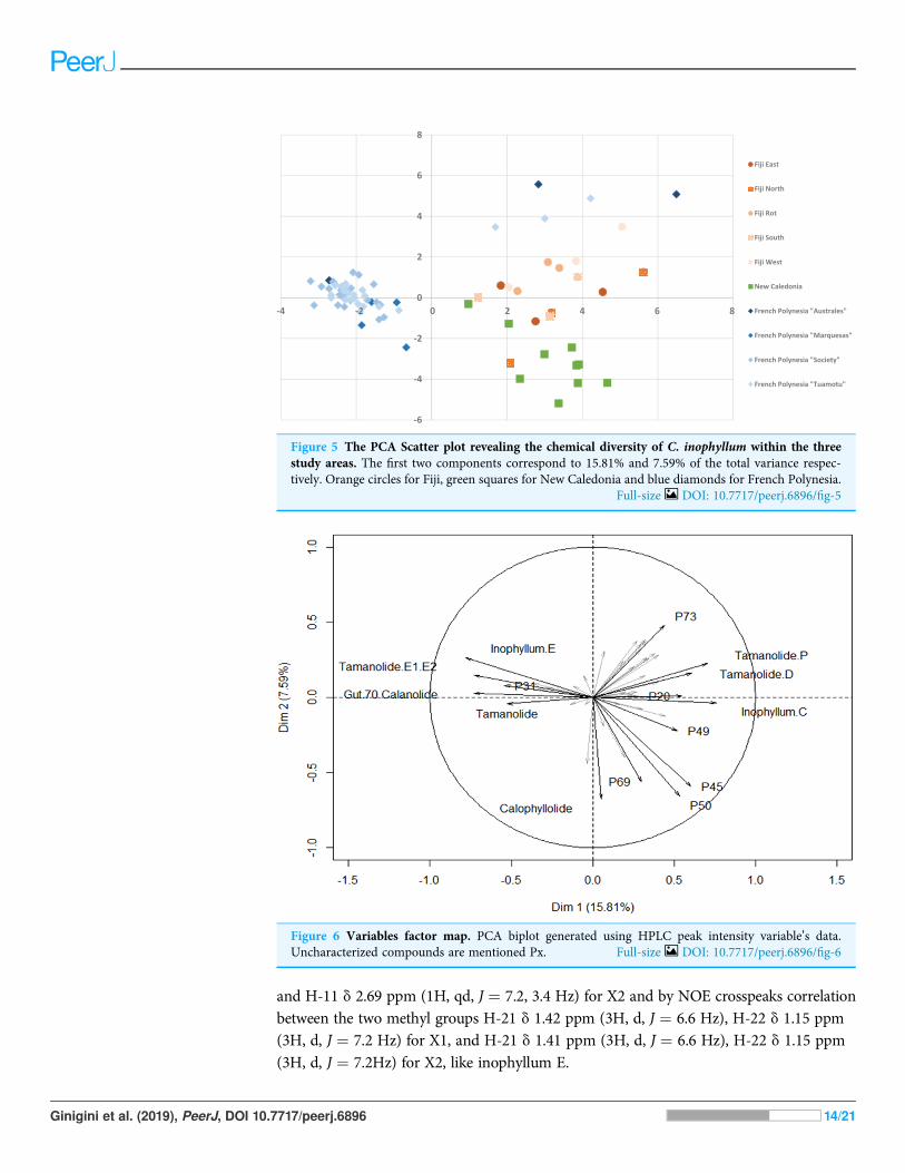
Statistical analysisMultivariate statistical analysis was applied to all the chemical data in order to reveal theextent to which the 12 major compounds making up the chemical compositions of the47 C. inophyllum oil resin samples were geographically distributed. Data were normalized
Ginigini et al. (2019), PeerJ, DOI 10.7717/peerj.6896 8/21
by log transformation prior to principle component analysis (PCA). PCA was performedwith the package Ade4 using R version 3.1 software (R Development Core Team, 2018)and a PCA biplot was drawn using Microsoft Excel software. The diversity of chemicalcompounds that contributed to the highest discrimination at a geospatial level werevisualized and a scatter plot was generated from Microsoft Excel.
RESULTSChemical compositionThe fractionation, purification and identification of resin extracts (by spectroscopicmethods) yielded 11 compounds from commercial tamanu oil (Figs. 2A–2I), whichwere identified as constituents previously reported (Laure et al., 2008; Leu et al., 2009).In addition to calophyllolide, inophyllums (C, D, E, P), calanolides (Gut 70 and A,12-oxo-calanolide) and tamanolides (D, P), these analyses also led to the isolation of twonew compounds X1 and X2 as an epimeric mixture. The presence of these two compoundsX1 and X2 from the same peak in two fractions was clearly shown in 1H theoretical(Fig. 3A) and experimental spectra (Fig. 3B). As the chemical shifts shown in Table 3 ofthese compounds were quite similar, evidence of their epimeric existence was indicated by1H NMR revealing more signals than expected from one compound. These most likelycorrespond to two sets of signals for two very close compounds that we propose here to beX1: tamanolide E1 and X2: tamanolide E2 (Fig. 2J). The chemical characteristics oftamanolide E1 and E2 are: amorphous yellowish powder; ½a�D25 ¼ �19:6 (c 0.003, CHCl3).FTIR (CCl4): 3,075, 2,979, 2,926, 2,908, 2,877, 2,832, 1,740, 1,695, 1,643, 1,606, 1,575,1,461, 1,382, 1,209, 1,152, 1,121 cm-1. 1H NMR (CDCl3, 500 MHz) and 13C (CDCl3, 125MHz) (see Table 3); HR-ESI-MS m/z 383.1850 [M + H]+ (calcd. for C23H27O5, 383.1853).
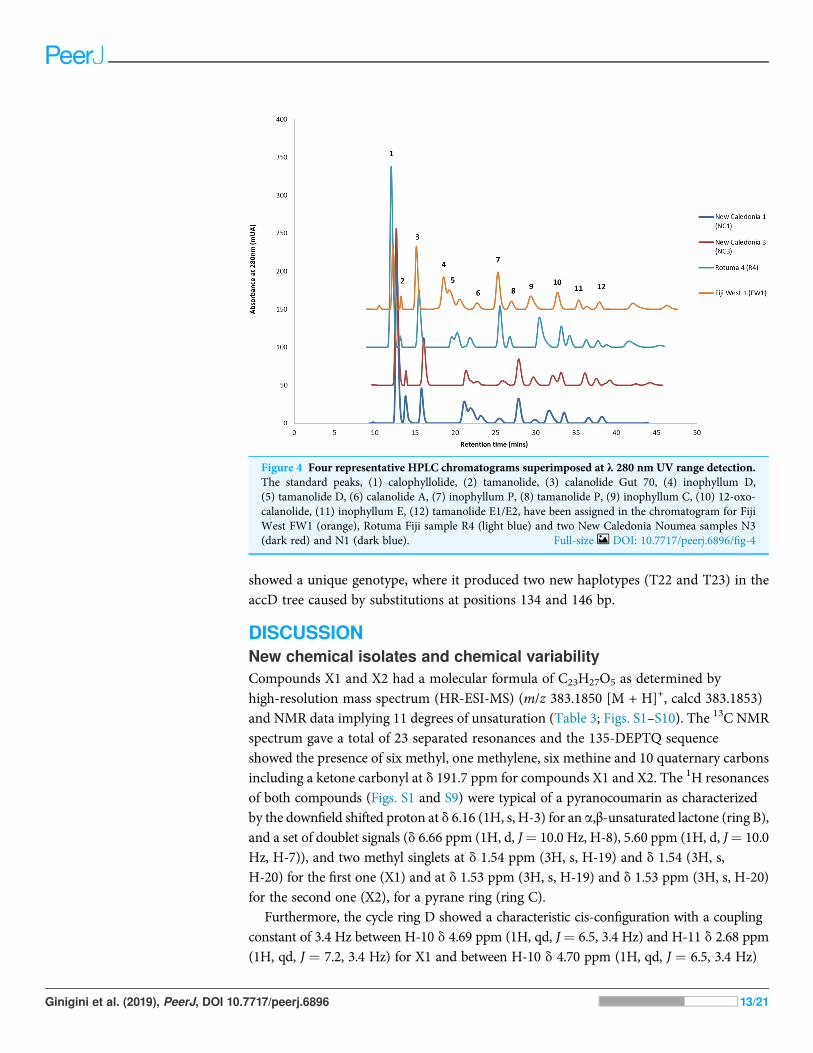
Chemical diversity of C. inophyllum within the three regionsHPLC analysis of C. inophyllum was performed to investigate the phytochemicalcomponents of tamanu oil in different samples and their geographical distribution. A totalof 11 isolated compounds (Fig. 4: peaks 1–11), along with two new compounds (Fig. 4:peak 12), were used as external standards to qualitatively analyze four sample ofC. inophyllum from three different locations, Fiji West (FW1), New Caledonia Noumea(N1 and N3) and Rotuma Fiji (R4). The phytochemical components of each sample wereidentified by comparing the retention time of the external standards (Fig. 4).
The alignment of all samples through R and Scilab provided a matrix containing theintegrated peak area of the identified features. Data were normalized by log transformationprior to PCA analysis. The samples can be discriminated into three main regions (Fig. 5)with the French Polynesian population on the left of the graph, the New Caledoniapopulation on the bottom right and the Fiji population on the upper right. Within FrenchPolynesia and Fiji, no discrimination between the sites or archipelagos was observed withthese first two components.
The different proportions of the compounds leading to biogeographic discrimination ofthe three regions is shown in the variables factor map (Fig. 6). The first component,representing 15.8% of the total variance, discriminated notably tamanolide E1/E2 and Gut
Ginigini et al. (2019), PeerJ, DOI 10.7717/peerj.6896 9/21

70 calanolide from inophyllum C and tamanolides D and P. The proportion of tamanolideE1/E2, Gut 70 calanolide, tamanolide and inophyllum E were the main and significantcompounds prominent in the composition of the oil resin from French Polynesia,while tamanolide P, D and inophyllum C were more prominently represented inthe Fiji samples. The second component (7.6% of the total variance) discriminated mainlyunknown compounds and calophyllolide, compound P73 positively and calophyllolide,P45, P50 and P69 negatively. Characteristic of the oil resin from New Caledonia were
Figure 2 Structures of isolated compounds. (A) Calophyllolide, (B) Calanolide GUT 70, (C) Tamanolide,(D) Inophyllums P, D and C, (E) Tamanolide P, (F) 12-Oxo-calanolide, (G) Calanolide A, (H) Inophyllum E,(I) Tamanolide D and (J) Tamanolide E1,E2. Full-size DOI: 10.7717/peerj.6896/fig-2
Ginigini et al. (2019), PeerJ, DOI 10.7717/peerj.6896 10/21
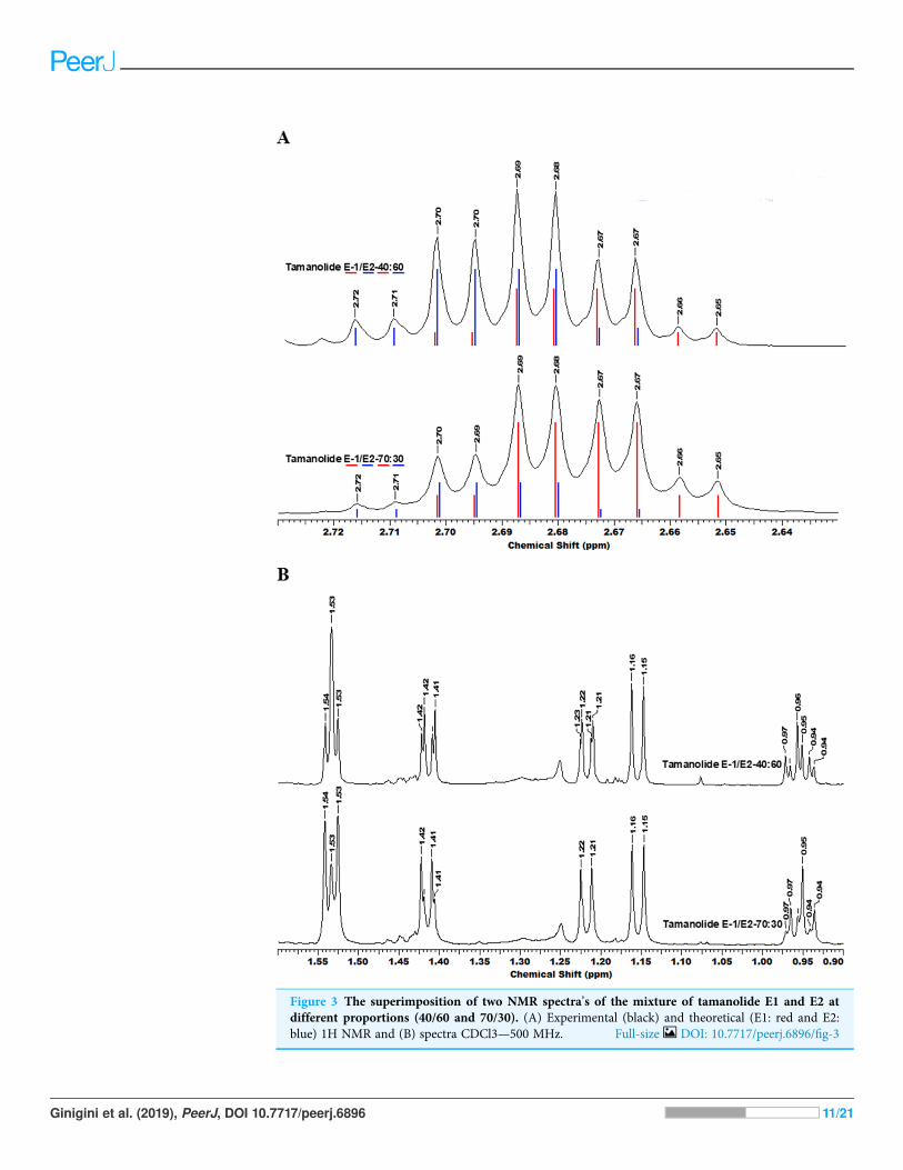
Figure 3 The superimposition of two NMR spectra’s of the mixture of tamanolide E1 and E2 atdifferent proportions (40/60 and 70/30). (A) Experimental (black) and theoretical (E1: red and E2:blue) 1H NMR and (B) spectra CDCl3—500 MHz. Full-size DOI: 10.7717/peerj.6896/fig-3
Ginigini et al. (2019), PeerJ, DOI 10.7717/peerj.6896 11/21
constituents such as calophyllolide and compound P69, which although unknown andunidentified, appeared to be unique in its occurrence as suggested by the PCA analysis.The chemical diversity of the Tahiti samples namely from the Tuamotu’s andAustrales as seen in Fig. 5, were also unique with the presence of the compound P73.
Genetic discriminationThe best fitting models of evolution determined by the MEGA6 option were respectivelyTamura 3 model and Tamura-Nei model as substitution models for accD marker geneand psaA-Ycf3 spacer region. Phylogenetic relationships assuming this model ofsubstitution for C. inophyllum are shown in Figs. 7A and 7B for the accD gene and for thepsaA-ycf3 spacer region, respectively. Interestingly, only Raiatea from French Polynesia
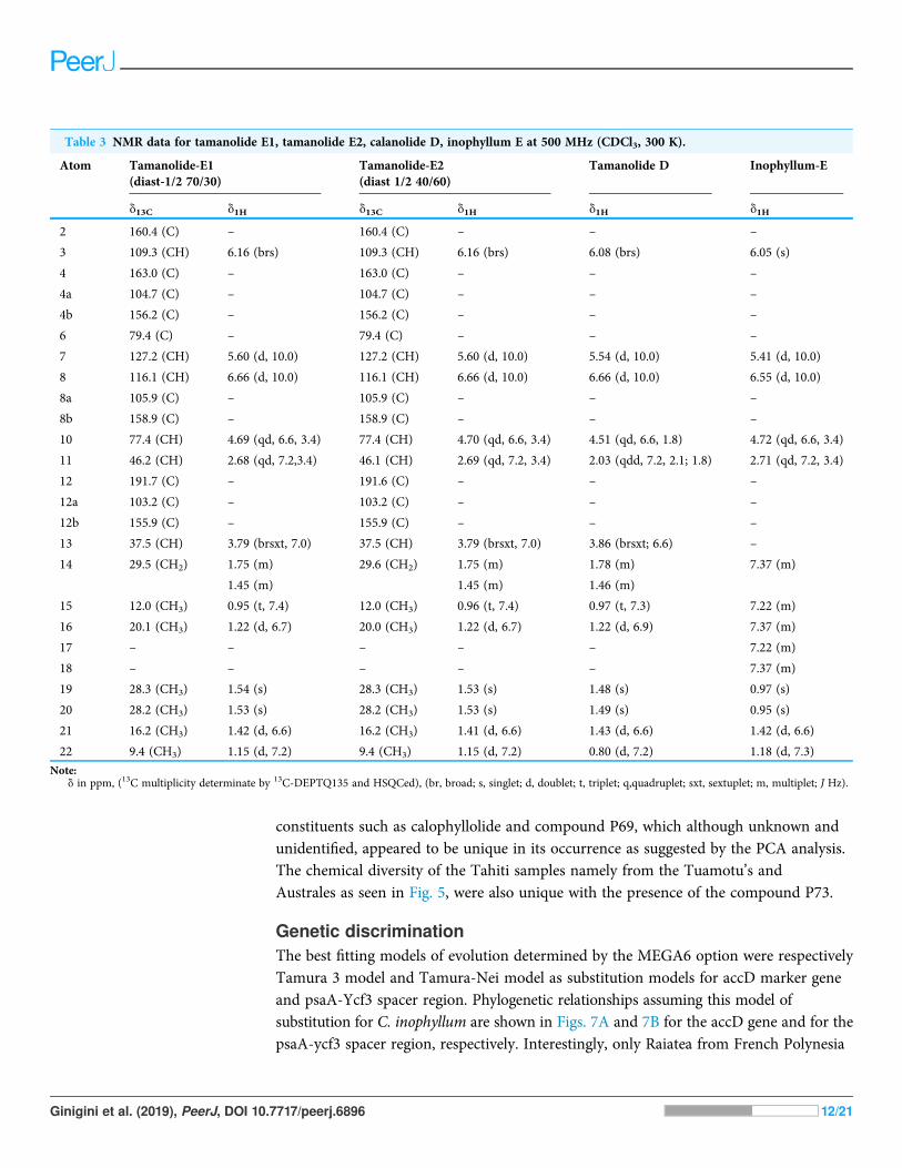
Table 3 NMR data for tamanolide E1, tamanolide E2, calanolide D, inophyllum E at 500 MHz (CDCl3, 300 K).
Atom Tamanolide-E1(diast-1/2 70/30)
Tamanolide-E2(diast 1/2 40/60)
Tamanolide D Inophyllum-E
d13C d1H d13C d1H d1H d1H
2 160.4 (C) – 160.4 (C) – – –
3 109.3 (CH) 6.16 (brs) 109.3 (CH) 6.16 (brs) 6.08 (brs) 6.05 (s)
4 163.0 (C) – 163.0 (C) – – –
4a 104.7 (C) – 104.7 (C) – – –
4b 156.2 (C) – 156.2 (C) – – –
6 79.4 (C) – 79.4 (C) – – –
7 127.2 (CH) 5.60 (d, 10.0) 127.2 (CH) 5.60 (d, 10.0) 5.54 (d, 10.0) 5.41 (d, 10.0)
8 116.1 (CH) 6.66 (d, 10.0) 116.1 (CH) 6.66 (d, 10.0) 6.66 (d, 10.0) 6.55 (d, 10.0)
8a 105.9 (C) – 105.9 (C) – – –
8b 158.9 (C) – 158.9 (C) – – –
10 77.4 (CH) 4.69 (qd, 6.6, 3.4) 77.4 (CH) 4.70 (qd, 6.6, 3.4) 4.51 (qd, 6.6, 1.8) 4.72 (qd, 6.6, 3.4)
11 46.2 (CH) 2.68 (qd, 7.2,3.4) 46.1 (CH) 2.69 (qd, 7.2, 3.4) 2.03 (qdd, 7.2, 2.1; 1.8) 2.71 (qd, 7.2, 3.4)
12 191.7 (C) – 191.6 (C) – – –
12a 103.2 (C) – 103.2 (C) – – –
12b 155.9 (C) – 155.9 (C) – – –
13 37.5 (CH) 3.79 (brsxt, 7.0) 37.5 (CH) 3.79 (brsxt, 7.0) 3.86 (brsxt; 6.6) –
14 29.5 (CH2) 1.75 (m) 29.6 (CH2) 1.75 (m) 1.78 (m) 7.37 (m)
1.45 (m) 1.45 (m) 1.46 (m)
15 12.0 (CH3) 0.95 (t, 7.4) 12.0 (CH3) 0.96 (t, 7.4) 0.97 (t, 7.3) 7.22 (m)
16 20.1 (CH3) 1.22 (d, 6.7) 20.0 (CH3) 1.22 (d, 6.7) 1.22 (d, 6.9) 7.37 (m)
17 – – – – – 7.22 (m)
18 – – – – – 7.37 (m)
19 28.3 (CH3) 1.54 (s) 28.3 (CH3) 1.53 (s) 1.48 (s) 0.97 (s)
20 28.2 (CH3) 1.53 (s) 28.2 (CH3) 1.53 (s) 1.49 (s) 0.95 (s)
21 16.2 (CH3) 1.42 (d, 6.6) 16.2 (CH3) 1.41 (d, 6.6) 1.43 (d, 6.6) 1.42 (d, 6.6)
22 9.4 (CH3) 1.15 (d, 7.2) 9.4 (CH3) 1.15 (d, 7.2) 0.80 (d, 7.2) 1.18 (d, 7.3)
Note:d in ppm, (13C multiplicity determinate by 13C-DEPTQ135 and HSQCed), (br, broad; s, singlet; d, doublet; t, triplet; q,quadruplet; sxt, sextuplet; m, multiplet; J Hz).
Ginigini et al. (2019), PeerJ, DOI 10.7717/peerj.6896 12/21
showed a unique genotype, where it produced two new haplotypes (T22 and T23) in theaccD tree caused by substitutions at positions 134 and 146 bp.
DISCUSSIONNew chemical isolates and chemical variabilityCompounds X1 and X2 had a molecular formula of C23H27O5 as determined byhigh-resolution mass spectrum (HR-ESI-MS) (m/z 383.1850 [M + H]+, calcd 383.1853)and NMR data implying 11 degrees of unsaturation (Table 3; Figs. S1–S10). The 13C NMRspectrum gave a total of 23 separated resonances and the 135-DEPTQ sequenceshowed the presence of six methyl, one methylene, six methine and 10 quaternary carbonsincluding a ketone carbonyl at d 191.7 ppm for compounds X1 and X2. The 1H resonancesof both compounds (Figs. S1 and S9) were typical of a pyranocoumarin as characterizedby the downfield shifted proton at d 6.16 (1H, s, H-3) for an a,β-unsaturated lactone (ring B),and a set of doublet signals (d 6.66 ppm (1H, d, J¼ 10.0 Hz, H-8), 5.60 ppm (1H, d, J¼ 10.0Hz, H-7)), and two methyl singlets at d 1.54 ppm (3H, s, H-19) and d 1.54 (3H, s,H-20) for the first one (X1) and at d 1.53 ppm (3H, s, H-19) and d 1.53 ppm (3H, s, H-20)for the second one (X2), for a pyrane ring (ring C).
Furthermore, the cycle ring D showed a characteristic cis-configuration with a couplingconstant of 3.4 Hz between H-10 d 4.69 ppm (1H, qd, J ¼ 6.5, 3.4 Hz) and H-11 d 2.68 ppm(1H, qd, J ¼ 7.2, 3.4 Hz) for X1 and between H-10 d 4.70 ppm (1H, qd, J ¼ 6.5, 3.4 Hz)
Figure 4 Four representative HPLC chromatograms superimposed at λ 280 nm UV range detection.The standard peaks, (1) calophyllolide, (2) tamanolide, (3) calanolide Gut 70, (4) inophyllum D,(5) tamanolide D, (6) calanolide A, (7) inophyllum P, (8) tamanolide P, (9) inophyllum C, (10) 12-oxo-calanolide, (11) inophyllum E, (12) tamanolide E1/E2, have been assigned in the chromatogram for FijiWest FW1 (orange), Rotuma Fiji sample R4 (light blue) and two New Caledonia Noumea samples N3(dark red) and N1 (dark blue). Full-size DOI: 10.7717/peerj.6896/fig-4
Ginigini et al. (2019), PeerJ, DOI 10.7717/peerj.6896 13/21
and H-11 d 2.69 ppm (1H, qd, J ¼ 7.2, 3.4 Hz) for X2 and by NOE crosspeaks correlationbetween the two methyl groups H-21 d 1.42 ppm (3H, d, J ¼ 6.6 Hz), H-22 d 1.15 ppm(3H, d, J ¼ 7.2 Hz) for X1, and H-21 d 1.41 ppm (3H, d, J ¼ 6.6 Hz), H-22 d 1.15 ppm(3H, d, J ¼ 7.2Hz) for X2, like inophyllum E.
-6
-4
-2
0
2
4
6
8
-4 -2 0 2 4 6 8
Fiji East
Fiji North
Fiji Rot
Fiji South
Fiji West
New Caledonia
French Polynesia "Australes"
French Polynesia "Marquesas"
French Polynesia "Society"
French Polynesia "Tuamotu"
Figure 5 The PCA Scatter plot revealing the chemical diversity of C. inophyllum within the threestudy areas. The first two components correspond to 15.81% and 7.59% of the total variance respec-tively. Orange circles for Fiji, green squares for New Caledonia and blue diamonds for French Polynesia.
Full-size DOI: 10.7717/peerj.6896/fig-5
Figure 6 Variables factor map. PCA biplot generated using HPLC peak intensity variable’s data.Uncharacterized compounds are mentioned Px. Full-size DOI: 10.7717/peerj.6896/fig-6
Ginigini et al. (2019), PeerJ, DOI 10.7717/peerj.6896 14/21
With the aid of the COSY experiment, an isobutyl unit was identified by further analysisof the remaining 1H resonances (d 3.79 ppm (1H, brsxt, J ¼ 7.0 Hz, H-13), 1.75 ppm(1H, m, H-14a), 1.45 ppm (1H, m, H-14b), 0.95 ppm (3H, t, J ¼ 7.4 Hz, H-15) and1.22 ppm (3H, d, J ¼ 6.7 Hz, H-16)) for X1 and d 3.79 ppm (1H, brsxt, J ¼ 7.0 Hz, H-13),1.75 ppm (1H, m, H-14a), 1.45 ppm (1H, m, H-14b), 0.96 (3H, t, J ¼ 7.4 Hz, H-15)and 1.22 ppm (3H, d, J¼ 6.7 Hz, H-16)) for X2. This isobutyl moiety was assigned to be atthe C-4 position, based on the HMBC crosspeaks between H-3 (d 6.16 ppm) and C-13(d 37.5 ppm), between H-16 (d 1.22 ppm) and C-4 (d 104.7 ppm), between H-14 (d 1.45,1.75 ppm) and C-4 (d 104.7 ppm), between H-13 (d 3.79 ppm) and the carbons C-3(d 109.3 ppm) and C-4a (d 104.7 ppm). The rest of the HMBC correlations used to obtain acomplete assignment of the 1H and 13C NMR chemical shift, have been summarizedand presented in Table 3. The 1H and 13C NMR of both compounds were almost identicalto those of inophyllum E with the exception of an isobutyl group at C-4 and tamanolide Dwith the exception of the ring D.
Complete assignments of X1 and X2 were made based on 1D and 2DNMR experiments,of which compound X1 was suggested as tamanolide E1 while compound X2 was theH-13 epimer of X1 and suggested as tamanolide E2. Proton NMR spectrum (Fig. 3) ofdifferent HPLC fractions revealed X1 and X2 as a mixture from two compounds whosesignals were close to each other with a varied ratio (from 70/30 to 40/60). The chemical shiftsof both compounds are listed in Table 3. Different fractions of a collected HLPC peakshow a variation of proportion related to these two epimer compounds X1 and X2and corresponding to two close sets of signals in the 1H NMR spectrum (Fig. 3).Assignment of the resonances was based on the 1D and 2D NMR experiments (1H,13C-DEPTQ135, COSY, HSQCed, HMBC and NOESY).
Figure 7 Maximum likelihood trees for combined tamanu samples. (A) using the accD marker geneconstructed using the Tamura 3 model and (B) depicts the Fiji (PsaA-Ycf3 spacer region) using theTamura-Nei model. Both trees are supported by 500 bootstrap iterations. The scale represents thenumber of substitutions per site. Full-size DOI: 10.7717/peerj.6896/fig-7
Ginigini et al. (2019), PeerJ, DOI 10.7717/peerj.6896 15/21
Finally, the structures of 11 known compounds are shown (Fig. 2) including the twonew compounds assigned as tamanolide E1 and E2 after elucidation from exhaustiveNMR analysis (1H, 13C-DEPTQ135, COSY, HSQCed, HMBC, NOESY), including thecomparison of experimental and theoretical results (Figs. 3A and 3B) and by comparisonwith NMR data of the phenylcoumarin back bone from inophyllum E and tamanolide D.
Consistent with earlier findings (Prabakaran & Britto, 2012; Laure et al., 2008),calophyllolide remains as the dominant major compound in tamanu resin for all isolatedchemotypes. Calanolide A which is the most potent of all C. inophyllum compoundswith anti-HIV-1 activity (Wang et al., 2006) was found as a minor component inC. inophyllum oil. The pyranocoumarines inophyllum B and P (also active against HIV-1)were also isolated. The former is not only the most active but is also the only naturalproduct undergoing clinical trials against HIV-1 (Wang et al., 2006). As seen in Fig. 4, the280 nm UV spectrum revealed the maximum number of peaks and therefore standardsand samples were targeted for visualization at this wavelength.
All the standards isolated here as seen in Fig. 4 have been isolated in previous studiesfrom French Polynesian C. inophyllum oil (Leu, 2009; Leu et al., 2009; Laure, 2005). Aunique characteristic of C. inophyllum oil is the presence of a resin which has been shownto contain neoflavonoid biologically active constituents (Ansel et al., 2016). Previousstudies byMcKee et al. (1998) and Li et al. (2007) have indicated the use of HPLC analysesto investigate differences in chemical profiles from different populations. In our case, wehave looked at samples from three localities. The population from New Caledonia wascharacterized by peaks not found in earlier studied populations and these novel peakssuggest new and specific chemomarkers. Unique to this study as shown in the biplot onFig. 6 is that we were able to show the chemodiversity of tamanu resin oil across the threestudy sites based on chemical compounds that contribute to the highest discrimination at ageographical level from the PCA analysis. This point is important and rather unique in thestudy as this reveals the chemical specificity of compounds and their proportions intamanu resin by geographical locations across the South Pacific.
DNA variabilityThis is the first reported work on C. inophyllum using the universal chloroplast barcodingregions the gene accD and the intergenic region psaA-ycf3. Low levels of geneticvariation were found with these markers, and this variation was not functionally linked todifferences in chemical composition. The absence of genetic variation among Fiji psaA-ycf3regions suggests low genetic variation in C. inophyllum. However, analyses of additionalchloroplast gene regions or nuclear ISSR markers may yet reveal more discrimination withinthe Fiji archipelago and between populations from different archipelagos.
CONCLUSIONSOur results are informative in revealing that chemical differences in tamanu resin canbe a tool for the discrimination of samples and geographic regions. In our case,chromatographic data proved to be more informative and highly discriminative than DNAbarcoding data, possibly owing to low genetic variation in the used chloroplast regions.
Ginigini et al. (2019), PeerJ, DOI 10.7717/peerj.6896 16/21
Additional chloroplast barcoding regions or utilization of nuclear microsatellitesmay give a better perspective on patterns of genetic diversity. Of interest were haplotypes(T22 and T23) which were distinct from the more commonly found C. inophyllumgenotype. Polyphasic taxonomy that considers both chemical diversity and genetic diversitypresents the best approach to delineate biological variation across geographical boundaries.However, higher levels of genetic resolution are required to characterize variation in thedisjunct distribution of C. inophyllum across the South Pacific and to bring insight intothe diversification processes that have occurred following geographic isolation. Thisdiversification includes the evolution of novel pyranocoumarins compounds characterizedby this study: tamanolide E1 and E2 (C-13 epimers as a mixture).
ACKNOWLEDGEMENTSThis paper is dedicated to the memory of our wonderful colleague, supervisor and friendProf William Aalbersberg who has passed away. We thank and greatly appreciate himfor his wisdom and inspiring research leadership. We are grateful to the people of FrenchPolynesia, Fiji and New Caledonia for giving us access to sampling areas to collect ournuts and leaf samples. We thank Cloe Check, Juliette Prevost, Nicolas Martin, and RaniteaLy for performing HPLC analysis on all the samples. We also thank Mr. John Bennettfor supplying oil extracts and assisting in the processing of samples in Fiji as well asMr. Olivier Touboul (LCPS) for tamanu sample collection in French Polynesia. Specialthanks to Mr. Marika Tuiwawa and Mr. Alivereti Naikatini for their assistance in thesample collection and deposition of samples in Fiji.
ADDITIONAL INFORMATION AND DECLARATIONS
FundingThe research was supported by grant AFD CPF 137901A project no 1317 (No SPP46-1-2013).The funders had no role in study design, data collection and analysis, decision to publish,or preparation of the manuscript.
Grant DisclosuresThe following grant information was disclosed by the authors:AFD CPF 137901A project no 1317: No SPP46-1-2013.
Competing InterestsThe authors declare that they have no competing interests.
Author Contributions� Joape Ginigini performed the experiments, analyzed the data, contributed reagents/materials/analysis tools, prepared figures and/or tables, authored or reviewed drafts ofthe paper, approved the final draft.
� Gaël J. Lecellier conceived and designed the experiments, performed the experiments,analyzed the data, contributed reagents/materials/analysis tools, prepared figures and/ortables, authored or reviewed drafts of the paper, approved the final draft.
Ginigini et al. (2019), PeerJ, DOI 10.7717/peerj.6896 17/21
� Mael Nicolas performed the experiments, analyzed the data, contributed reagents/materials/analysis tools, authored or reviewed drafts of the paper, approved the final draft.
� Mohammed Nour conceived and designed the experiments, authored or reviewed draftsof the paper, approved the final draft.
� Edouard Hnawia contributed reagents/materials/analysis tools, authored or revieweddrafts of the paper, approved the final draft.
� Nicolas Lebouvier conceived and designed the experiments, contributed reagents/materials/analysis tools, authored or reviewed drafts of the paper, approved the final draft.
� Gaëtan Herbette performed the experiments, analyzed the data, contributed reagents/materials/analysis tools, prepared figures and/or tables, authored or reviewed drafts ofthe paper, approved the final draft.
� Peter Lockhart analyzed the data, contributed reagents/materials/analysis tools,authored or reviewed drafts of the paper, approved the final draft.
� Phila Raharivelomanana conceived and designed the experiments, performed theexperiments, analyzed the data, contributed reagents/materials/analysis tools, authoredor reviewed drafts of the paper, approved the final draft.
Field Study PermissionsThe following information was supplied relating to field study approvals (i.e., approvingbody and any reference numbers):
In New Caledonia, tamanu nuts were collected under the scientific authorization of theSouth Province No 2050-2014
DNA DepositionThe following information was supplied regarding the deposition of DNA sequences:
The accD data is available at NCBI (accession number 983209430) and psaA-ycf3(accession number 983209444).
Data AvailabilityThe following information was supplied regarding data availability:
The raw measurements are available in Datasets S1–S3. Each file provides the data forone sample from one geographic site. These data were used for the chemodiversity analysisto compare the profiles between the different regions.
Supplemental InformationSupplemental information for this article can be found online at http://dx.doi.org/10.7717/peerj.6896#supplemental-information.
REFERENCESAngiosperm Phylogeny Group. 2009. An update of the angiosperm phylogeny group classification
for the orders and families of flowering plants: APG III. Botanical Journal of the Linnean Society161(2):105–121 DOI 10.1111/j.1095-8339.2009.00996.x.
Ansel J-L, Lupo E, Mijouin L, Guillot S, Butaud J, Ho R, Lecellier G, Raharivelomanana P,Pichon C. 2016. Biological activity of Polynesian Calophyllum inophyllum oil extract onhuman skin cells. Planta Medica 82(11/12):961–966 DOI 10.1055/s-0042-108205.
Ginigini et al. (2019), PeerJ, DOI 10.7717/peerj.6896 18/21
Assouvie N. 2013. Le Tamanu (Calophyllum inophyllum L.) en Polynésie Française et autre espècesdu genre Calophyllum: De l’usage en médecine traditionnelle à l’émergence d’un médicamentanti-VIH. Thèse de doctorat en pharmacie, Université de Bordeaux Ségalen, p. 122, no2013BOR2P019.
Bhalla TN, Saxena RC, Nigam SK, Misra G, Bhargava KP. 1980. Calophyllolide—a new non-steroidal anti-inflammatory agent. Indian Journal of Medicinal Research 72:762–765.
Boudreau E, Takahashi Y, Lemieux M, Rochaix J-D. 1997. The chloroplast ycf3 and ycf4 openreading frames of Chlamydomonas reinhardtii are required for the accumulation of thephotosystem I complex. EMBO Journal 16(20):6095–6104 DOI 10.1093/emboj/16.20.6095.
Cambie RC, Ash J. 1994. Dicotyledons in Fijian medicinal plants. Australia: CSIRO, 119–120.
Dweck AC, Meadows T. 2002. Tamanu (Calophyllum inophyllum)—the African, Asian,Polynesian and Pacific Panacea. International Journal of Cosmetic Sciences 24(6):341–348DOI 10.1046/j.1467-2494.2002.00160.x.
Friday JB, Okano D. 2006. Calophylum inophyllum (Kamani). Species profiles for Pacific Islandagroforestry. Ver 2.1. Hōlualoa, Hawai’i: Permanent Agriculture Resources (PAR).Available at http://www.traditionaltree.org.
Gascuel O. 1997. BIONJ: an improved version of the NJ algorithm based on a simple model ofsequence data. Molecular Biology and Evolution 14(7):685–695DOI 10.1093/oxfordjournals.molbev.a025808.
Hu Y, Zhang Q, Xin H, Qin L-P, Lu B-R, Rehman K, Zheng H. 2007. Association betweenchemical and genetic variation of Vitex roduntifolia population from different locations inChina: its implication for quality control of medicinal plants. Biomedical Chromatography21(9):967–975 DOI 10.1002/bmc.841.
Ishikawa T. 2000. Anti HIV-1 active Calophyllum Coumarins: distribution, chemistry and activity.Heterocycles 53(2):453–474 DOI 10.3987/REV-99-526.
Itoigawa M, Ito C, Hugh T, Tan W, Kuchide M, Tokuda H, Nishino H, Furukawa H. 2001.Cancer chemo preventive agents, 4-phenylcoumarins from Calophyllum inophyllum.Cancer Letters 169(1):15–19 DOI 10.1016/S0304-3835(01)00521-3.
Jin L, Tabe Y, Kimura S, Zhou Y, Kuroda J, Asou H, Inaba T, Konopleva M, Andreeff M,Miida T. 2011. Antiproliferative and proapoptotic activity of GUT-70 mediated through potent,inhibition of Hsp90 in mantle cell lymphoma. British Journal of Cancer 104(1):91–100DOI 10.1038/sj.bjc.6606007.
Kostova I, Mojzis J. 2007. Biologically active coumarins as inhibitors of HIV-1. Future HIVTherapy 1(3):315–329 DOI 10.2217/17469600.1.3.315.
Lahaye R, Van Der Bank M, Bogarin D, Warner J, Pupulin F, Gigot G, Maurin O, Duthoit S,Barraclough T, Savolainen V. 2007. DNA barcoding the floras of biodiversity hotspots.Proceedings of the National Academy of Sciences of the United States of America105(8):2923–2928 DOI 10.1073/pnas.0709936105.
Lam VKY, Merckx VSFT, Graham SW. 2016. A few-gene plastid phylogenetic frameworkfor mycoheterotrophic monocots. American Journal of Botany 103(4):692–708DOI 10.3732/ajb.1500412.
Laure F. 2005. Etude de la composition chimique et de la biodiversité du Calophyllum inophyllumde Polynésie Française. PhD thesis, Université de la Polynésie Française, p. 357,no 2005POLF0001.
Laure F, Raharivelomanana P, Butaud J-F, Bianchini J-P, Gaydou EM. 2008. Screening forAnti-HIV-1 inophyllums by HPLC-DAD of Calophyllum inophyllum leaf extracts from FrenchPolynesia Islands. Analytica Chimica Acta 624(1):147–153 DOI 10.1016/j.aca.2008.06.046.
Ginigini et al. (2019), PeerJ, DOI 10.7717/peerj.6896 19/21
Léguillier T, Lecsö-Bornet M, Lémus C, Rousseau-Ralliard D, Lebouvier N, Hnawia E, Nour M,Aalbersberg W, Ghazi K, Raharivelomanana P, Rat P. 2015. The wound healing andantibacterial activity of five ethnomedical Calophyllum inophyllum oils: an alternativetherapeutic strategy to treat infected wounds. PLOS ONE 10(9):e0138602DOI 10.1371/journal.pone.0138602.
Leu T. 2009. Contribution à la connaissance de la flore polynésienne: évaluation de l’intérêtpharmacologique de quelques plantes médicinales et étude phytochimique du Tamanu(Calophyllum inophyllum L. – Clusiaceae). Doctoral thesis, Université de la Polynésie Française,Papeete, Tahiti.
Leu T, Raharivelomanana P, Soulet S, Bianchini J-P, Herbette G, Faure R. 2009. New tricyclicand tetracyclic pyranocoumarins with an unprecedented C-4 substituent. Structure elucidationof Tamanolide, Tamanolide D and Tamanolide P from Calophyllum inophyllum of FrenchPolynesia. Magnetic Resonance in Chemistry 47(11):989–993 DOI 10.1002/mrc.2482.
Li HL, Zhang WD, Zhang C, Han T, Liu R-H, Hu J, Chen H-S. 2007. Comparative analysis ofchemical profile of wild and cultivated populations of Corydalis saxicola by high performanceliquid chromatography. Phytochemical Analysis 18(5):393–400 DOI 10.1002/pca.994.
Lynch RC, Vergara D, Tittes S, White K, Schwartz CJ, Gibbs MJ, Ruthenburg TC, DeCesare K,Land DP, Kane NC. 2016. Genomic and chemical diversity in Cannabis. Critical Reviews inPlant Sciences 35(5–6):349–363 DOI 10.1080/07352689.2016.1265363.
McKee TC, Covington CD, Fuller RW, Bokesch HR, Young S, Cardellina JH II, Kadushin MR,Soejarto DD, Stevens PF, Cragg GM, Boyd MR. 1998. Pyranocoumarins from tropical speciesof genus Calophyllum: a taxonomic study of extracts in national cancer institute collection.Journal of Natural Products 61(10):1252–1256 DOI 10.1021/np980140a.
Patil AD, Freyer AJ, Eggleston DS, Haltiwanger RC, Bean MF, Taylor PB, Caranfa MJ,Breen AL, Bartus HR, Johnson RK, Hertzberg RP, Westley JW. 1993. The Inophyllums, novelinhibitors of HIV-1 reverse transcriptase isolated from Malaysian tree, Calophyllum inophyllumLinn. Journal of Medicinal Chemistry 36(26):4131–4138 DOI 10.1021/jm00078a001.
Pawar KD, Swati JP, Shubhada RT. 2011. Association between chemical and genetic variationin Calophyllum inophyllum, a medicinally important tree of the Western Ghats of India.Plant Systematics and Evolution 292(3–4):257–265 DOI 10.1007/s00606-010-0409-8.
Prabakaran K, Britto SJ. 2012. Biology, agroforestry and medical value of Calophyllum inophyllumL. (Clusiacea): a review. International Journal of Natural Products Research 1(2):24–33.
R Development Core Team. 2018. R: a language and environment for statistical computing.Vienna: R Foundation for Statistical Computing. Available at https://www.R-project.org/.
Ruf S, Kossel H, Bock R. 1997. Targeted inactivation of a tobacco intron-containing open readingframe reveals a novel chloroplast-encoded photosystem I-related gene. Journal of Cell Biology139(1):95–102 DOI 10.1083/jcb.139.1.95.
Saitou N, Nei M. 1987. The neighbor-joining method: a new method for reconstructingphylogenetic trees. Molecular Biology and Evolution 4(4):406–425DOI 10.1093/oxfordjournals.molbev.a040454.
Saravanan R, Dhachinamoorthi D, Senthilkumar K, Thamizhvanan K. 2011. Antimicrobialactivity of various extracts from various parts of Calophyllum inophyllum L. Journal ofApplied Pharmaceutical Science 1(3):102–106.
Saxena RC, Nath R, Palit G, Nigam SK, Bhargava KP. 1982. Effect of Calophyllolide, a non-steroidal anti-inflammatory agent, on capillary permeability. Planta Medica 44(4):246–248DOI 10.1055/s-2007-971459.
Ginigini et al. (2019), PeerJ, DOI 10.7717/peerj.6896 20/21
Scilab Enterprises. 2012. Scilab: free and open source software for numerical computation(Windows 8, Version 5.5.1) (Scilab). Available at http://www.scilab.org.
Tamura K, Nei M. 1993. Estimation of the number of nucleotide substitutions in the controlregion of mitochondrial DNA in humans and chimpanzees. Molecular Biology and Evolution10(3):512–526 DOI 10.1093/oxfordjournals.molbev.a040023.
Tamura K, Stecher G, Peterson D, Filipski A, Kumar S. 2013. MEGA6: molecularevolutionary genetics analysis version 6.0. Molecular Biology and Evolution 30(12):2725–2729DOI 10.1093/molbev/mst197.
Thompson JD, Higgins DG, Gibson TJ. 1994. CLUSTAL W: improving the sensitivity ofprogressive multiple sequence alignment through sequence weighting, position-specific gappenalties and weight matrix choice. Nucleic Acids Research 22(22):4673–4680DOI 10.1093/nar/22.22.4673.
Vandamme P, Pot B, Gillis M, De Vos P, Kersters K, Swings J. 1996. Polyphasic taxonomy,a consensus approach to bacterial systematics. Microbiological Reviews 60(2):407–438.
Wang S-Y, Ho T-J, Kuo C-H, Tseng YJ. 2010. Chromaligner: a web server for chromatogramalignment. Bioinformatics 26(18):2338–2339 DOI 10.1093/bioinformatics/btq337.
Wang L, Ma T, Liu G, Liang X, Fang W. 2006. Recent progress in Calophyllum Coumarins aspotent anti-HIV agents. In: Liang X, Fang W, eds. Medicinal Chemistry of Medicinal NaturalProducts. China: Wiley-Interscience, 325–350.
Xi Z, Ruhfel RB, Schaefera H, Amorimd AM, Sugumarane M, Wurdack KJ, Endress PK,Matthews ML, Stevens PF, Mathews S, Davis CC. 2012. Phylogenomics and a posterioridata partitioning resolve the Cretaceous angiosperm radiation Malpighiales. Proceedings of theNational Academy of Sciences of the United States of America 109(43):17519–17524DOI 10.1073/pnas.1205818109.
Yimdjo MC, Azebaze GA, Nkengfack AE, Meyer AM, Bodo B, Fomum ZT. 2004. Antimicrobialand cytotoxic agents from Calophyllum inophyllum. Phytochemistry 65(20):2789–2795DOI 10.1016/j.phytochem.2004.08.024.
Ginigini et al. (2019), PeerJ, DOI 10.7717/peerj.6896 21/21