chemomechanical caries removal (cmcr) agents: review and ... · patients and preserving healthy...
TRANSCRIPT
Journal of Dentistry and Oral Hygiene Vol. 3(3), pp.34-45, March 2011 Available online at http://www.academicjournals.org/JDOH ISSN 2141-2472 ©2011 Academic Journals
Full Length Research Paper
Chemomechanical caries removal (CMCR) agents: Review and clinical application in primary teeth
M. Ganesh and Dhaval Parikh*
Department of Pedodontics and Preventive Dentistry, Ahmedabad Dental College and Hospital, Gandhinagar,
Gujarat, India.
Accepted 8 October, 2010
Chemomechanical caries removal (CMCR) is a non-invasive technique eliminating infected dentine via a chemical agent. This is a method of caries removal based on dissolution. Instead of drilling, this method uses a chemical agent assisted by an atraumatic mechanical force to remove soft carious structure. It was introduced to dentistry as an alternative method of caries removal and is mainly indicated to overcome the inconvenience of using burs and local anesthesia, causing less discomfort to patients and preserving healthy dental structure, there by complying the concept of the minimal invasive dentistry (MID). Various agents with their methods have been used in the past for CMCR, but only a few have got into a stable clinical practice. Among them we have the Carisolv, which is the most successful and commonly used agent while Papacarie gives the promising result as CMCR agent of the future. Carisolv came into use at the end of 20
th century. It consists of two component mixtures (mainly
amino acid and hypochlorite), forming an active gel. Papacarie is an emerging CMCR agent of the 21st
century. Papacarie is composed basically of papain, chloramines and toluidine blue. Papain interacts with exposed collagen by the dissolution of dentine minerals through bacteria, making the infected dentine softer, and allowing its removal with non cutting instruments without local anesthesia and burs. This paper attempts to look into this method in detail with various agents of CMCR and also present case reports regarding the two most commonly used agents. Key words: Chemomechanical caries removal (CMCR), carisolv, papacarie, infected dentine.
INTRODUCTION Restorative dental treatment of carious teeth in children, which involves removal of caries with conventional drill, is considered traumatic mainly due to fear and anxiety of children and their parents (Scott et al., 1984). The aversion to the rotational instrument noise and anesthesia are the main factors for this situation (Ayer et al., 1983). Such factors most of the time not only delay the dental treatment but also leads to avoidance of dental treatment by children, leading to the advancement of the caries process to emergency situations. Unfortunately in these situations, the treatments are more complicated, making the use of anesthesia mandatory (Michelle et al., 2005).
On the other hand, in every field of dentistry, *Corresponding author. E-mail: [email protected].
awareness towards the importance of preserving tooth tissue combined with a patient-friendly approach is becoming self-evident. It has been shown that operative dental treatment often leads to an increasing scale to further operative and more invasive treatment. Wherever possible, tissue should be preserved, and invasive treatment should be kept to a minimum. The best way to ensure a maximum life for the natural tooth is to respect the sound tissue and protect it from damage by using minimally-invasive techniques in restorative dentistry (Banerjee et al., 2000).
With the development of new dental restorative materials, advances in adhesive dentistry, a better under-standing of the caries process and the tooth’s potential for remineralization, the management of dental caries has drastically evolved from G.V. Black’s “Extension for prevention” to “Construction with conservation” (Tandon, 2008). This concept includes the early detection of lesions, individual caries risk assessment, non-surgical
interventions and modified surgical approach that includes, smaller tooth preparations with modified cavity designs and adhesive dental materials and repair rather than replacement of failing restorations. The goal is to preserve the natural tooth structure.
Minimally invasive dentistry adopts a philosophy that integrates prevention, remineralization and minimal intervention for the placement and replacement of restorations. Minimally invasive dentistry reaches the treatment objective using the least invasive surgical approach, with the removal of the minimal amount of healthy tissues. It includes the following different techniques: (1) Air abrasion (Myers, 1954). (2) Atraumatic restorative technique (Frencken et al., 1996). (3) Sono abrasion (Banerjee et al., 2000). (4) Laser (Keller et al., 1998). (5) Chemomechanical caries removal (CMCR) (Ericson et al., 1999). Among all these techniques, atraumatic restorative tech-nique is the most documented alternative to traditional drilling for dentine caries removal but Chemomechanical caries removal (CMCR) holds a lot of promise as an effective alternative to the traditional method. It involves the application of a chemical solution to the carious dentine followed by gentle removal with hand instru-ments. Chemomechanical caries removal is a method for minimally-invasive, gentle dentine caries removal based on biological principles (Zinc et al., 1988). Evolution of chemomechanical caries removal agents: The background
The earliest attempts to remove caries involved the use of a hand drill which was soon surpassed in 1871 by James Morrison’s treadle instrument, developed from the mechanism of Isaac Singer’s sewing machine. Modern high speed drills are the latest development of this more than a century old technique (Ring, 1985). Conventional caries removal and cavity preparation entail the use of the burs. Disadvantages of this system include: (1) The perception by patients that drilling is unpleasant. (2) Local anesthesia is frequently required. (3) Drilling can cause deleterious thermal effect combined with the use of pressure for caries removal, causing pulpal effects. (4) The use of a hand piece may result in removal of softened, but affected dentine, resulting in an excessive loss of sound tooth tissue. As a result of these disadvantages, there is a growing demand for procedures or material that facilitates caries management without the above mentioned is
Ganesh and Parikh 35 advantages.
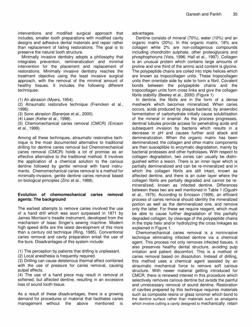
Dentine consists of mineral (70%), water (10%) and an organic matrix (20%). In this organic matrix, 18% are collagen while 2% are non-collagenous compounds including chondroitin sulphate, other proteoglycans and phosphophoryns (Veis, 1996; Hall et al., 1997). Collagen is an unusual protein which contains large amounts of proline and one third of the amino acid content is glycine. The polypeptide chains are coiled into triple helices which are known as tropocollagen units. These tropocollagen units then orientate side by side to form a fibril. Covalent bonds between the polypeptide chains and the tropocollagen units form cross links and give the collagen fibrils stability (Beeley et al., 2000) (Figure 1)
In dentine, the fibrils are in the form of a dense meshwork which becomes mineralized. When caries occurs, acids produced by plaque bacteria; by anaerobic fermentation of carbohydrate initially cause solubilisation of the mineral in enamel. As the process progresses, dentinal tubules provide access for penetrating acids and subsequent invasion by bacteria which results in a decrease in pH and causes further acid attack and demineralization. When the organic matrix has been demineralized, the collagen and other matrix components are then susceptible to enzymatic degradation, mainly by bacterial proteases and other hydrolases. With respect to collagen degradation, two zones can usually be distin-guished within a lesion. There is an inner layer which is partially demineralized and can be re-mineralized and in which the collagen fibrils are still intact, known as affected dentine, and there is an outer layer where the collagen fibrils are partially degraded and cannot be re-mineralized, known as infected dentine. Differences between these two are well mentioned in Table 1 (Ogushi et al., 1975). According to Ericsson (1999), an efficient process of caries removal should identify the mineralized portion as well as the demineralized one, and remove only the latter. For these we require reagent, which must be able to cause further degradation of this partially degraded collagen, by cleavage of the polypeptide chains in the triple helix and/or hydrolyzing the cross linkages as explained in Figure 1.
Chemomechanical caries removal is a noninvasive technique eliminating infected dentine via a chemical agent. This process not only removes infected tissues, it also preserves healthy dental structure, avoiding pulp irritation and patient discomfort. This is a method of caries removal based on dissolution. Instead of drilling, this method uses a chemical agent assisted by an atraumatic mechanical force to remove soft carious structure. With newer material getting introduced for CMCR, there is renewed interest in this procedure which selectively removes carious dentine but avoids the painful and unnecessary removal of sound dentine. Restoration of cavities prepared by this technique requires materials such as composite resins or glass ionomer which bond to the dentine surface rather than materials such as amalgams which involve cutting a cavity designed to mechanically retain
36 J. Dent. Oral Hyg.
Figure 1. The structure of collagen, (a) Polypeptide chain. Possible sites of cleavage by chemomechanical carries removal reagents by degradation of glycine or hydroxyproline are indicated by red arrows, (b) Triple helix. Sites of cleavage by degradation of intra-molecular cross links are shown by red arrows, (c) Tropocollagen units assembled to form a collagen fibril. Sites of cleavage by degradation of intra-molecular cross links are shown by red arrows (Modified from Dow et al., 1996).
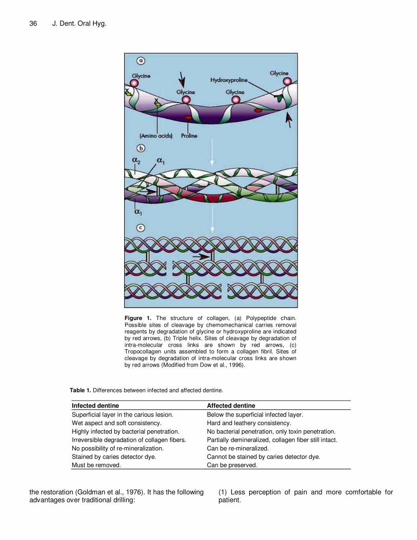
Table 1. Differences between infected and affected dentine.
Infected dentine Affected dentine
Superficial layer in the carious lesion. Below the superficial infected layer. Wet aspect and soft consistency. Hard and leathery consistency. Highly infected by bacterial penetration. No bacterial penetration, only toxin penetration. Irreversible degradation of collagen fibers. Partially demineralized, collagen fiber still intact. No possibility of re-mineralization. Can be re-mineralized. Stained by caries detector dye. Cannot be stained by caries detector dye. Must be removed. Can be preserved.
the restoration (Goldman et al., 1976). It has the following advantages over traditional drilling:
(1) Less perception of pain and more comfortable for patient.
(2) Less fear and anxiety to method, leads to less discomfort to patients especially in children. (3) Removes only infected layer and leads to more tissue preservation. (4) No pulpal irritation. (5) Well suited to the treatment of deciduous teeth, dental phobic’s and medically compromised patients. (6) Better removal of caries in uncooperative patients. (7) Useful in physically handicapped patients. (8) Useful in patients with T.B like infectious diseases (prevent droplet infection). Importance of chemomechanical caries removal agents in pediatric dentistry Fear and anxiety are known barriers to the receptivity of dental treatment and in detriment to oral health. The conventional drilling techniques are associated with discomfort, especially among children (Stewart et al., 1994). Normally, the triggering factors are local anesthesia, low and high speed rotary instruments, and previous dental treatment. In children, it is difficult to differentiate between fear and anxiety-originated behavior problems. The most anxiety-provoking procedure for children, however, is the local anesthetic injection (Kuscu et al., 2006). Thus, changes in dentistry routines such as the chemomechanical caries removal, sedation with nitrous oxide, and general anesthesia are becoming necessary. The chemomechanical caries removal method was developed specifically to overcome these barriers and to preserve the healthy dentine tissue. This method is characterized by the use of a material that acts on the pre-degraded collagen of the lesion, promotes its softening, doesn’t affect the adjacent healthy tissues, and avoids pain stimuli (chemical action). This method is further characterized by removing the softened carious tissue via gentle excavation (mechanical action), which makes this technique an efficacious alternative method to treat carious lesions since it allies no traumatic characteristics with bactericide and bacteriostatic action.
Papacarie and Carisolv can be successfully used in special health care needs (SHCN) patients and phobic adults in pediatric dentistry and public health sectors (Carrillo et al., 2005). Currently, research in dentistry has concentrated its efforts on the quality of treatment given to SHCN patients, those who present some deviation from the normal standards (identifiable or not), and those who for this reason, require special attention and approaches for a given length of time or indefinitely (Figueredo et al., 2003). Therefore, the chemome-chanical technique for removing caries is an efficient option when approaching and supplying oral care for these patients.
Other factors also influence the child and their res-ponse to dental treatment. Children who were submitted to prolonged treatment or hospitalized are usually more
Ganesh and Parikh 37 fearful and afraid of hospitalization regarding dental treat-ment. Therefore, the chemomechanical caries removal technique is an efficient therapeutic alternative to prevent fear and anxiety among these patients. Children who were submitted to local anesthesia during dental treatment demonstrated more fear (66.8%) than those who were not submitted to anesthesia (50.8%) (Singh et al., 2000). Another positive factor regarding this tech-nique is that it does not require local anesthesia during the procedure since the carious tissue is softened by the gel and its removal by gentle hand instruments does not promote any stimulus or pressure that would lead to discomfort and/or pain ( Pereira et al., 2004).
Burke et al. (1995) did a study on permanent and deciduous teeth using NMAB solution similar to the com-position to Caridex. They found that CMCR was more effective on deciduous teeth than on permanent teeth. Kotb et al. (2009) did a study on primary teeth using conventional drilling method and Papacarie. They found that Papacarie could be an effective caries removal method to treat children, particularly those present with early childhood caries or management problems. This method may be desirable in pediatric dentistry since it allows minimally invasive techniques to be applied, considered to be less painful, noise and vibration free, and patients were more comfortable than with the conventional technique. Journey from GK 101 -- GK 101 E -- caridex to carisolv Around 1970, the need for an alternative to conventional rotary led to a research by Habib et al. (1976) who studied the effect of sodium hypochlorite, which is a non-specific proteolytic agent on the removal of carious material from dentine. Sodium hypochlorite itself however was too corrosive for use on healthy tissue and very un-stable. So they decided to incorporate it into Sorensen’s buffer (which contains glycine, sodium chloride and sodium hydroxide) in an attempt to minimize this problem. Quite fortuitously, a reaction occurred which resulted in a product which was more effective in removal of carious dentine than a saline placebo. This involved the chlorination of glycine to form N-Monochloroglycine (NMG) and the reagent subsequently became known as GK 101 and marketed in 1972 as first CMCR agent (Goldman et al., 1976). According to Kurosaki et al. (1977), GK 101 would soften only the first layer of carious dentine, and would not affect the second layer, and it has a very slow action. It was the major disadvantage and limitation of the GK 101.
In subsequent studies, they found that the system was more effective if glycine was replaced by amino butyric acid; the product then being N-monochloroaminobutyric acid (NMAB) also designated GK-101E in 1975. The mechanism of action of NMG and NMAB on collagen is
38 J. Dent. Oral Hyg. still unclear and knowledge of the chemistry of chlorination of amino acids and their effects is still very limited. Originally it was thought that the procedure involved chlorination of the partially degraded collagen in the carious lesion and the conversion of hydroxyproline to pyrrole-2-carboxylic acid (Habib et al., 1975). More recent work suggests that cleavage by oxidation of glycine residues could also be involved (Yip et al., 1989). This causes disruption of the collagen fibrils which become more friable and can then be removed.
The NMAB system was patented in the US in 1975, and a further patent taken out by the National Patent Dental Corporation, New York in 1987. It received FDA approval for use in the USA in 1984 and was marketed in the 1980’s as caridex. It consisted of two solutions; solution 1 containing sodium hypochlorite and solution 2 containing glycine, aminobutyric acid, sodium chloride and sodium hydroxide. The two solutions were mixed immediately before use to give the working reagent [pH 12 (Gulcin et al., 2004)] which was stable for 1 h. A delivery system was also available for caridex that consists of: (1) Reservoir for the solution, a heater and a pump which passed the liquid, warmed to body temperature through a tube to a hand piece and an applicator tip which came in various shapes and sizes. (2) The solution was applied to the carious lesion by means of this application which was used to loosen the carious dentine by a gentle scraping action; the debris together with the spent solution being removed by aspiration. (3) Application was continued until the dentine remaining was deemed sound by normal clinical tactile criteria. With suitable accessible soft lesions, after 15 to 20 min treatment, only clinically sound dentine remained. (4) The reagent selectively removed carious dentine leaving a surface with many overhangs and undercuts. The procedure avoids the painful removal of sound dentine but is ineffective in the removal of hard eburnated parts of the lesion; removal of eburnated caries however may not be necessary. (5) Recently it has been shown that discoloration in carious dentine results from the Maillard reaction which modifies amino acids in collagen thereby making them more resistant to proteolytic attack and inhibiting lesion progression in discolored dentine (Kleter et al., 1998). Limitations of caridex system (1) Rotary and/or hand instruments may still be needed for the removal of tissue or material other than degraded dentine collagen. This includes access to small or interproximal carious lesions, removal of enamel overlying the caries, removal of existing restorations, and for cavity design when non-adhesive restorative materials are used.
(2) Large volumes of solution were needed (200 to 500 ml) and the procedure was slow and also costly. (3) Only certain cavities were suitable for treatment by the technique and because of the time involved (10 to 15 min) and limited use, its popularity waned. (4) Although there were studies on the efficacy of caries removal by the procedure, studies on the long term success of cavities restored after CMCR treatment were lacking. Because of the time required for CMCR treatment, the large volumes of solution needed and the fact that the delivery system was no longer commercially available, the use of CMCR despite its potential became minimal. In the early 1990’s caridex ceased to be marketed and the manufacturer’s patent lapsed.
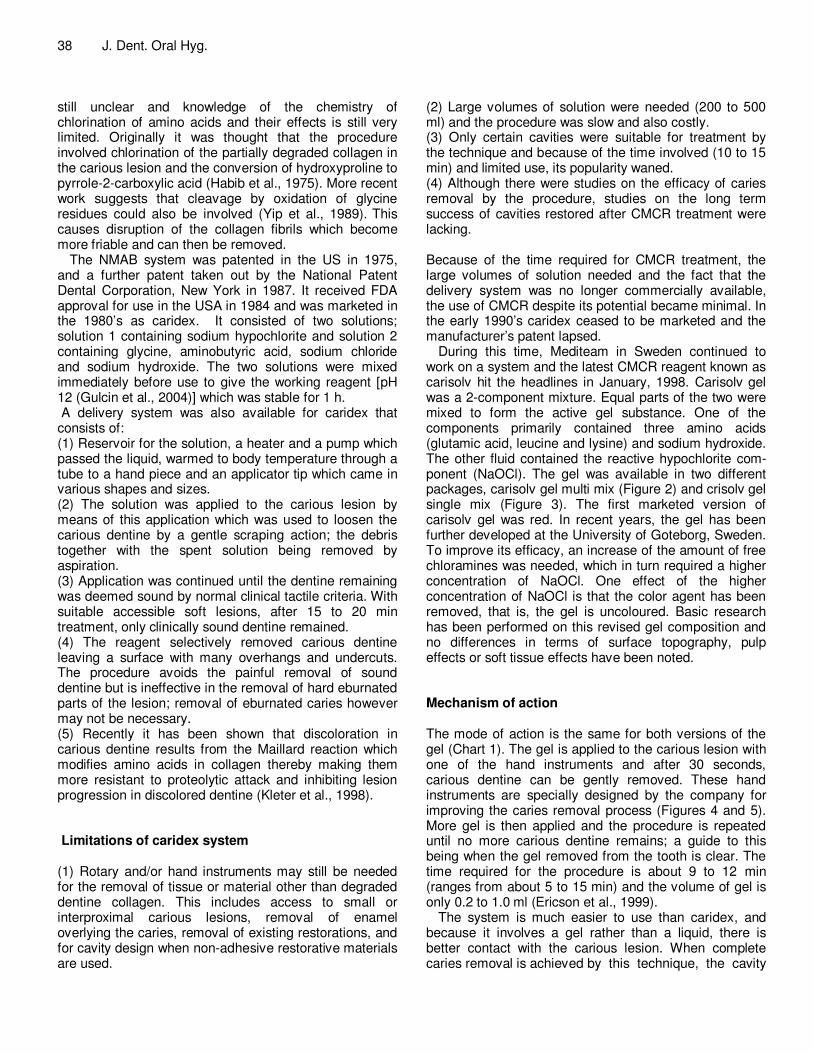
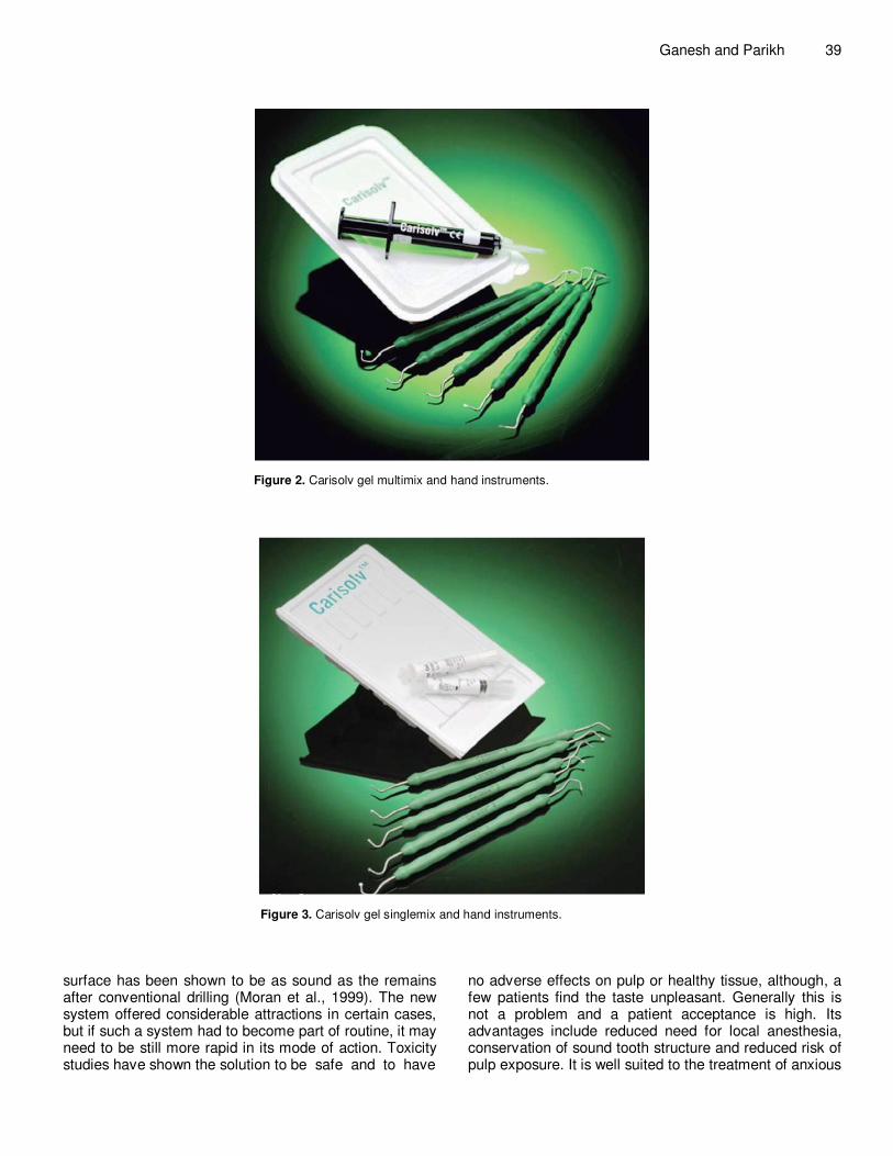
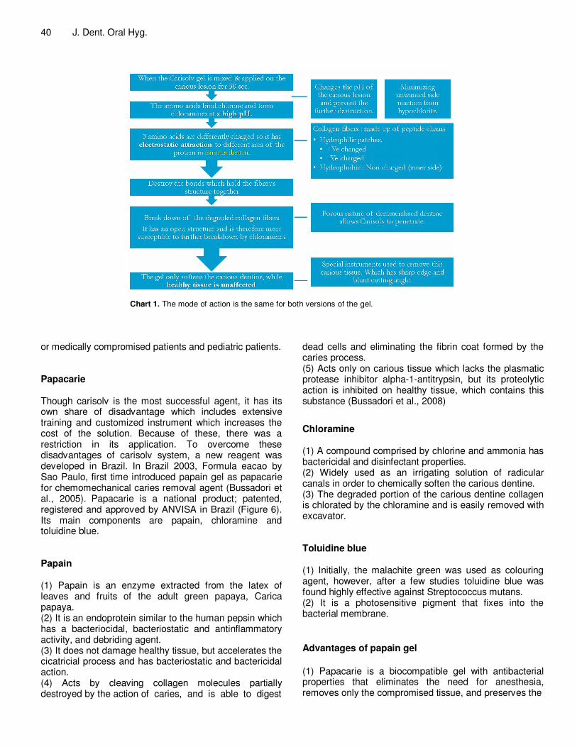
During this time, Mediteam in Sweden continued to work on a system and the latest CMCR reagent known as carisolv hit the headlines in January, 1998. Carisolv gel was a 2-component mixture. Equal parts of the two were mixed to form the active gel substance. One of the components primarily contained three amino acids (glutamic acid, leucine and lysine) and sodium hydroxide. The other fluid contained the reactive hypochlorite com-ponent (NaOCl). The gel was available in two different packages, carisolv gel multi mix (Figure 2) and crisolv gel single mix (Figure 3). The first marketed version of carisolv gel was red. In recent years, the gel has been further developed at the University of Goteborg, Sweden. To improve its efficacy, an increase of the amount of free chloramines was needed, which in turn required a higher concentration of NaOCl. One effect of the higher concentration of NaOCl is that the color agent has been removed, that is, the gel is uncoloured. Basic research has been performed on this revised gel composition and no differences in terms of surface topography, pulp effects or soft tissue effects have been noted. Mechanism of action The mode of action is the same for both versions of the gel (Chart 1). The gel is applied to the carious lesion with one of the hand instruments and after 30 seconds, carious dentine can be gently removed. These hand instruments are specially designed by the company for improving the caries removal process (Figures 4 and 5). More gel is then applied and the procedure is repeated until no more carious dentine remains; a guide to this being when the gel removed from the tooth is clear. The time required for the procedure is about 9 to 12 min (ranges from about 5 to 15 min) and the volume of gel is only 0.2 to 1.0 ml (Ericson et al., 1999).
The system is much easier to use than caridex, and because it involves a gel rather than a liquid, there is better contact with the carious lesion. When complete caries removal is achieved by this technique, the cavity
Ganesh and Parikh 39
Figure 2. Carisolv gel multimix and hand instruments.
Figure 3. Carisolv gel singlemix and hand instruments.
surface has been shown to be as sound as the remains after conventional drilling (Moran et al., 1999). The new system offered considerable attractions in certain cases, but if such a system had to become part of routine, it may need to be still more rapid in its mode of action. Toxicity studies have shown the solution to be safe and to have
no adverse effects on pulp or healthy tissue, although, a few patients find the taste unpleasant. Generally this is not a problem and a patient acceptance is high. Its advantages include reduced need for local anesthesia, conservation of sound tooth structure and reduced risk of pulp exposure. It is well suited to the treatment of anxious
40 J. Dent. Oral Hyg.
Chart 1. The mode of action is the same for both versions of the gel.
or medically compromised patients and pediatric patients. Papacarie Though carisolv is the most successful agent, it has its own share of disadvantage which includes extensive training and customized instrument which increases the cost of the solution. Because of these, there was a restriction in its application. To overcome these disadvantages of carisolv system, a new reagent was developed in Brazil. In Brazil 2003, Formula eacao by Sao Paulo, first time introduced papain gel as papacarie for chemomechanical caries removal agent (Bussadori et al., 2005). Papacarie is a national product; patented, registered and approved by ANVISA in Brazil (Figure 6). Its main components are papain, chloramine and toluidine blue. Papain (1) Papain is an enzyme extracted from the latex of leaves and fruits of the adult green papaya, Carica papaya. (2) It is an endoprotein similar to the human pepsin which has a bacteriocidal, bacteriostatic and antinflammatory activity, and debriding agent. (3) It does not damage healthy tissue, but accelerates the cicatricial process and has bacteriostatic and bactericidal action. (4) Acts by cleaving collagen molecules partially destroyed by the action of caries, and is able to digest
dead cells and eliminating the fibrin coat formed by the caries process. (5) Acts only on carious tissue which lacks the plasmatic protease inhibitor alpha-1-antitrypsin, but its proteolytic action is inhibited on healthy tissue, which contains this substance (Bussadori et al., 2008) Chloramine (1) A compound comprised by chlorine and ammonia has bactericidal and disinfectant properties. (2) Widely used as an irrigating solution of radicular canals in order to chemically soften the carious dentine. (3) The degraded portion of the carious dentine collagen is chlorated by the chloramine and is easily removed with excavator. Toluidine blue (1) Initially, the malachite green was used as colouring agent, however, after a few studies toluidine blue was found highly effective against Streptococcus mutans. (2) It is a photosensitive pigment that fixes into the bacterial membrane. Advantages of papain gel (1) Papacarie is a biocompatible gel with antibacterial properties that eliminates the need for anesthesia, removes only the compromised tissue, and preserves the
Ganesh and Parikh 41
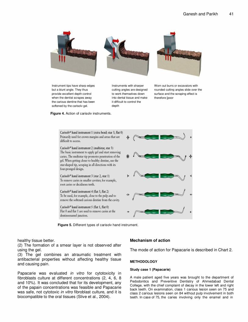
Instrument tips have sharp edges but a blunt angle. They thus provide excellent depth control when the dentist scrapes away the carious dentine that has been softened by the carisolv gel.
Instruments with sharper cutting angles are designed to work themselves down into dental tissue and make it difficult to control the depth
Worn out burrs or excavators with rounded cutting angles slide over the surface and the scraping effect is therefore [poor
Figure 4. Action of carisolv instruments.
Figure 5. Different types of carisolv hand instrument.
healthy tissue better. (2) The formation of a smear layer is not observed after using the gel. (3) The gel combines an atraumatic treatment with antibacterial properties without affecting healthy tissue and causing pain. Papacarie was evaluated in vitro for cytotoxicity in fibroblasts culture at different concentrations (2, 4, 6, 8 and 10%). It was concluded that for its development, any of the papain concentrations was feasible and Papacarie was safe, not cytotoxic in vitro fibroblast culture, and it is biocompatible to the oral tissues (Slive et al., 2004).
Mechanism of action The mode of action for Papacarie is described in Chart 2. METHODOLOGY Study case 1 (Papacarie) A male patient aged five years was brought to the department of Pedodontics and Preventive Dentistry of Ahmedabad Dental College, with the chief complaint of decay in the lower left and right back teeth. On examination, class 1 carious lesion seen on 75 and class 2 carious lesions seen on 84 without pulp involvement in both teeth. In case of 75, the caries involving only the enamel and in
42 J. Dent. Oral Hyg.
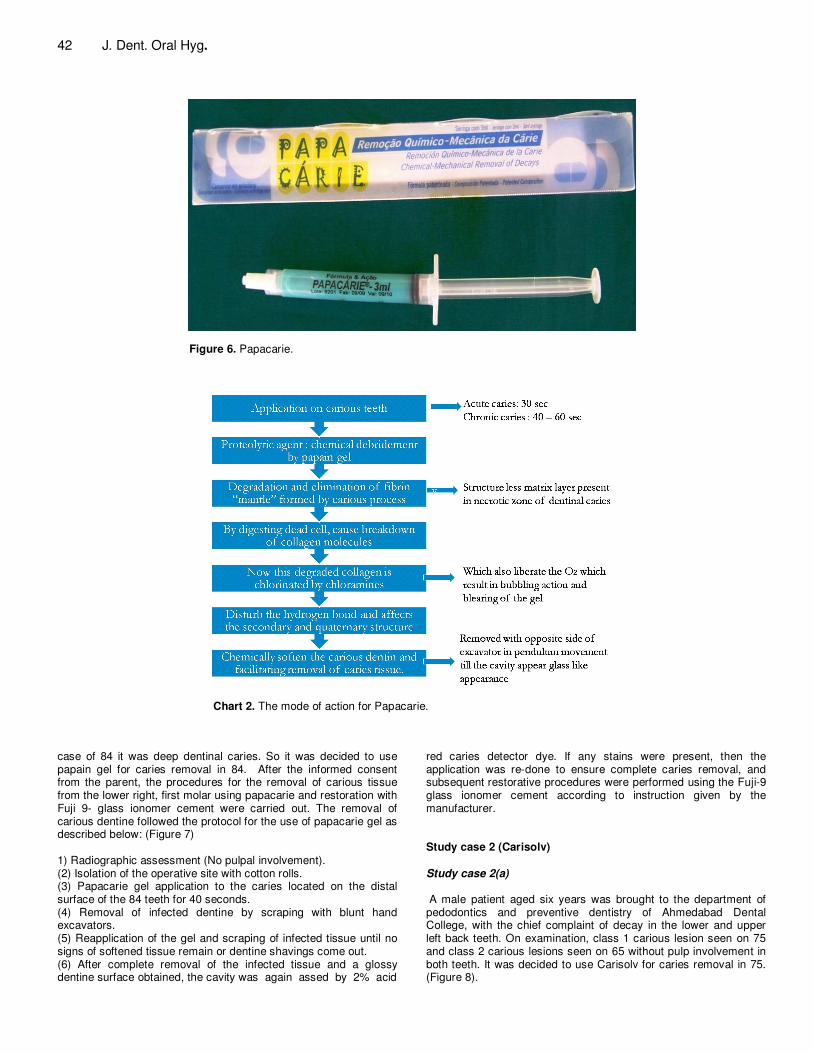
Figure 6. Papacarie.
Chart 2. The mode of action for Papacarie.
case of 84 it was deep dentinal caries. So it was decided to use papain gel for caries removal in 84. After the informed consent from the parent, the procedures for the removal of carious tissue from the lower right, first molar using papacarie and restoration with Fuji 9- glass ionomer cement were carried out. The removal of carious dentine followed the protocol for the use of papacarie gel as described below: (Figure 7) 1) Radiographic assessment (No pulpal involvement). (2) Isolation of the operative site with cotton rolls. (3) Papacarie gel application to the caries located on the distal surface of the 84 teeth for 40 seconds. (4) Removal of infected dentine by scraping with blunt hand excavators. (5) Reapplication of the gel and scraping of infected tissue until no signs of softened tissue remain or dentine shavings come out. (6) After complete removal of the infected tissue and a glossy dentine surface obtained, the cavity was again assed by 2% acid
red caries detector dye. If any stains were present, then the application was re-done to ensure complete caries removal, and subsequent restorative procedures were performed using the Fuji-9 glass ionomer cement according to instruction given by the manufacturer. Study case 2 (Carisolv)
Study case 2(a)
A male patient aged six years was brought to the department of pedodontics and preventive dentistry of Ahmedabad Dental College, with the chief complaint of decay in the lower and upper left back teeth. On examination, class 1 carious lesion seen on 75 and class 2 carious lesions seen on 65 without pulp involvement in both teeth. It was decided to use Carisolv for caries removal in 75. (Figure 8).
Ganesh and Parikh 43
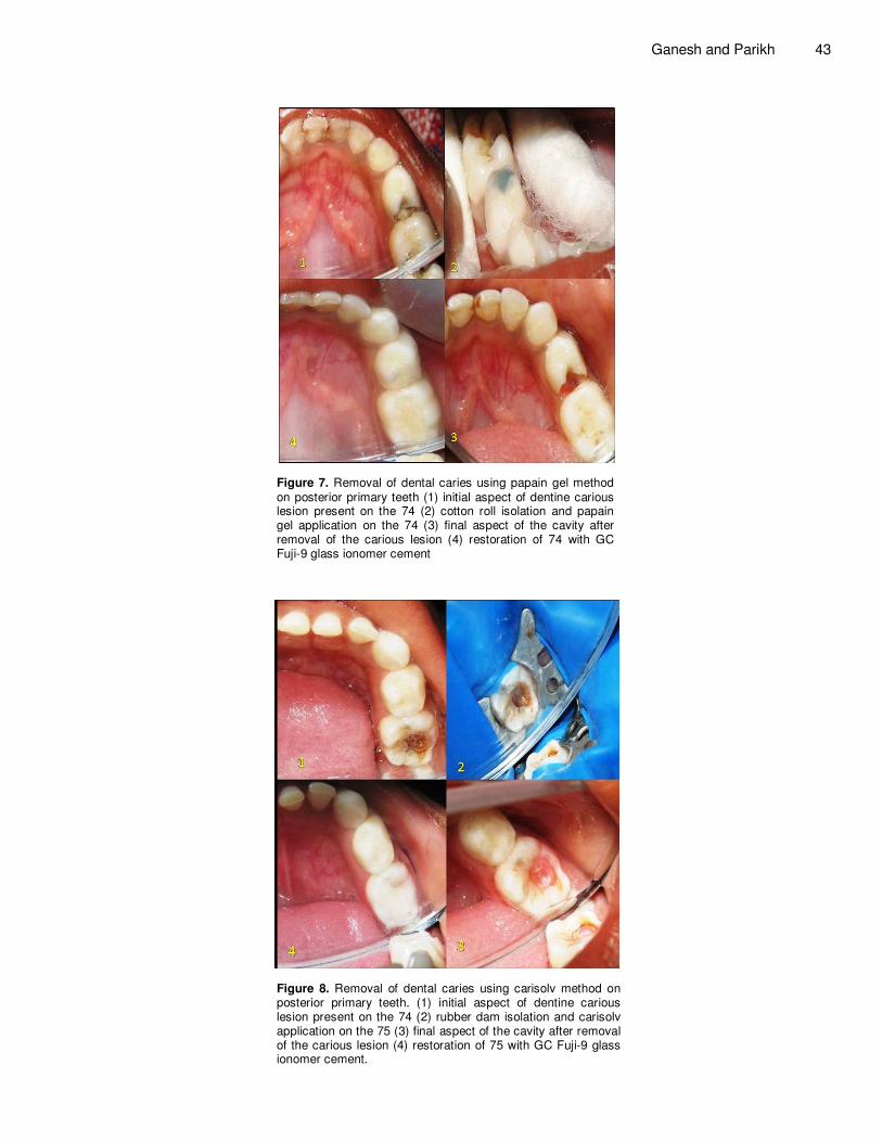
Figure 7. Removal of dental caries using papain gel method on posterior primary teeth (1) initial aspect of dentine carious lesion present on the 74 (2) cotton roll isolation and papain gel application on the 74 (3) final aspect of the cavity after removal of the carious lesion (4) restoration of 74 with GC Fuji-9 glass ionomer cement
Figure 8. Removal of dental caries using carisolv method on posterior primary teeth. (1) initial aspect of dentine carious lesion present on the 74 (2) rubber dam isolation and carisolv application on the 75 (3) final aspect of the cavity after removal of the carious lesion (4) restoration of 75 with GC Fuji-9 glass ionomer cement.
44 J. Dent. Oral Hyg.
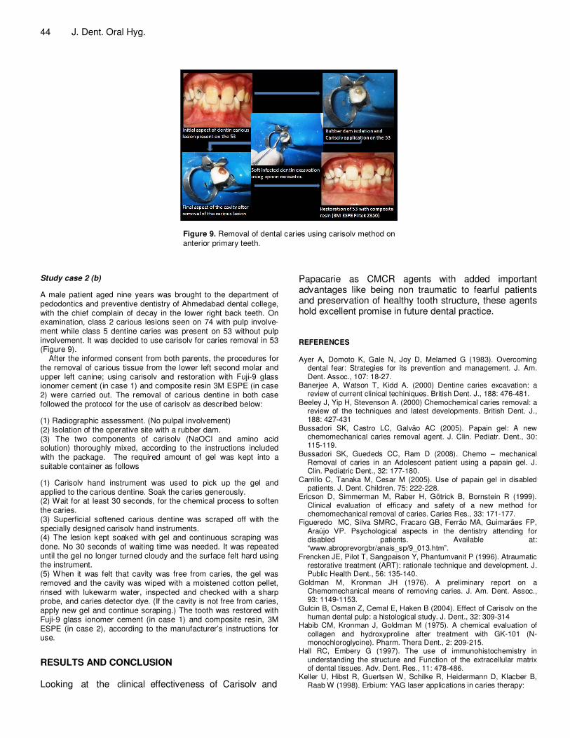
Figure 9. Removal of dental caries using carisolv method on anterior primary teeth.
Study case 2 (b) A male patient aged nine years was brought to the department of pedodontics and preventive dentistry of Ahmedabad dental college, with the chief complain of decay in the lower right back teeth. On examination, class 2 carious lesions seen on 74 with pulp involve-ment while class 5 dentine caries was present on 53 without pulp involvement. It was decided to use carisolv for caries removal in 53 (Figure 9).
After the informed consent from both parents, the procedures for the removal of carious tissue from the lower left second molar and upper left canine; using carisolv and restoration with Fuji-9 glass ionomer cement (in case 1) and composite resin 3M ESPE (in case 2) were carried out. The removal of carious dentine in both case followed the protocol for the use of carisolv as described below: (1) Radiographic assessment. (No pulpal involvement) (2) Isolation of the operative site with a rubber dam. (3) The two components of carisolv (NaOCl and amino acid solution) thoroughly mixed, according to the instructions included with the package. The required amount of gel was kept into a suitable container as follows (1) Carisolv hand instrument was used to pick up the gel and applied to the carious dentine. Soak the caries generously. (2) Wait for at least 30 seconds, for the chemical process to soften the caries. (3) Superficial softened carious dentine was scraped off with the specially designed carisolv hand instruments. (4) The lesion kept soaked with gel and continuous scraping was done. No 30 seconds of waiting time was needed. It was repeated until the gel no longer turned cloudy and the surface felt hard using the instrument. (5) When it was felt that cavity was free from caries, the gel was removed and the cavity was wiped with a moistened cotton pellet, rinsed with lukewarm water, inspected and checked with a sharp probe, and caries detector dye. (If the cavity is not free from caries, apply new gel and continue scraping.) The tooth was restored with Fuji-9 glass ionomer cement (in case 1) and composite resin, 3M ESPE (in case 2), according to the manufacturer’s instructions for use.
RESULTS AND CONCLUSION Looking at the clinical effectiveness of Carisolv and
Papacarie as CMCR agents with added important advantages like being non traumatic to fearful patients and preservation of healthy tooth structure, these agents hold excellent promise in future dental practice. REFERENCES Ayer A, Domoto K, Gale N, Joy D, Melamed G (1983). Overcoming
dental fear: Strategies for its prevention and management. J. Am. Dent. Assoc., 107: 18-27.
Banerjee A, Watson T, Kidd A. (2000) Dentine caries excavation: a review of current clinical techiniques. British Dent. J., 188: 476-481.
Beeley J, Yip H, Stevenson A. (2000) Chemochemical caries removal: a review of the techniques and latest developments. British Dent. J., 188: 427-431
Bussadori SK, Castro LC, Galvão AC (2005). Papain gel: A new chemomechanical caries removal agent. J. Clin. Pediatr. Dent., 30: 115-119.
Bussadori SK, Guededs CC, Ram D (2008). Chemo – mechanical Removal of caries in an Adolescent patient using a papain gel. J. Clin. Pediatric Dent., 32: 177-180.
Carrillo C, Tanaka M, Cesar M (2005). Use of papain gel in disabled patients. J. Dent. Children. 75: 222-228.
Ericson D, Simmerman M, Raber H, Götrick B, Bornstein R (1999). Clinical evaluation of efficacy and safety of a new method for chemomechanical removal of caries. Caries Res., 33: 171-177.
Figueredo MC, Silva SMRC, Fracaro GB, Ferrão MA, Guimarães FP, Araújo VP. Psychological aspects in the dentistry attending for disabled patients. Available at: “www.abroprevorgbr/anais_sp/9_013.htm”.
Frencken JE, Pilot T, Sangpaison Y, Phantumvanit P (1996). Atraumatic restorative treatment (ART): rationale technique and development. J. Public Health Dent., 56: 135-140.
Goldman M, Kronman JH (1976). A preliminary report on a Chemomechanical means of removing caries. J. Am. Dent. Assoc., 93: 1149-1153.
Gulcin B, Osman Z, Cemal E, Haken B (2004). Effect of Carisolv on the human dental pulp: a histological study. J. Dent., 32: 309-314
Habib CM, Kronman J, Goldman M (1975). A chemical evaluation of collagen and hydroxyproline after treatment with GK-101 (N-monochloroglycine). Pharm. Thera Dent., 2: 209-215.
Hall RC, Embery G (1997). The use of immunohistochemistry in understanding the structure and Function of the extracellular matrix of dental tissues. Adv. Dent. Res., 11: 478-486.
Keller U, Hibst R, Guertsen W, Schilke R, Heidermann D, Klacber B, Raab W (1998). Erbium: YAG laser applications in caries therapy:

evaluation of patient’s perception and Acceptance. J. Dent., 26: 649- 656. Kleter GA, Damen JJM, Buigus MJ (1998). Modification of amino acid
residues in carious dentine matrix. J. Dent. Res., 77: 488-495. Kotb RM, Abdella A, Kateb MA, Ahmed A (2009). Clinical evaluation of
Papacarie in primary teeth. J. Clin. Pediatric Dent., 34: 117-123. Kuscu OO, Akyuz S (2006). Children’s preferences concerning the
physical appearance of dental injectors. J Dent Child. 73:116-21. Michelle M, Luis F (2005). Chemomechanical caries removal: current
evidences. RBO, 62(4): 125-129 Moran C, Lynch E, Petersson L, Borshboom P (1999). Comparison of
caries removal using Carisolv or a conventional slow speed rotary instrument. Caries Res., 33: 313.
Myers GE (1954). The air abrasive technique. Br. Dent. J., 1954; 97: 291-295.
Ogushi K, Fusayama T (1975). Electron microscopic structures of two layers of carious dentine. J. Dent. Res., 54: 1019-1026.
Pereira SA, Silva LR, Piccinini DPF, Santos EM, Bussadori SK (2004). Comparison antimicrobial potential in vitro between two materials for the Chemomechanical caries removal. In: 21ª Annual Reunion SBPqO, 2004, Águas de Lindóia. Pesqui Odont Bras, 18: 78.
Ring ME (1985). Dentistry; an Illustrated History. pp. 250-251.New York: Abrams.
Ganesh and Parikh 45 Scott S, Hirschman R, Schroder K (1984). Historical antecedents of
dental anxiety. J. Am. Dent. Assoc., 108: 42-45. Silva LR (2004). Papacárie: A new system for the Chemomechanical
caries removal—case report. Rev. Paul Odontol., 16: 4-8. Singh KA, Morares ABA, Ambrosano GMB (2000). Fear, anxiety, and
control related to dental treatment. Pesqui Odont Bras. 14:131-6. Stewart JM, Marcus M, Christesen P, Lin W (1994). Comprehensive
treatment among dental school patients with high and low anxiety. J. Dent. Educ., 58: 697-700.
Tandon S (2008). Chapter: Principles and concepts of Cavity Preparation,Text book of Pedodontics. 2nd edi Paras Medical Publisher, New Delhi, pp. 308-314.
Veis A (1996). In Comper W.D. (Ed) Extracellular Matrix, Volume 1 Tissue Function. 41-76 Amsterdam: Harwood Academic Press,
Yip H K, Beeley JA (1989). Studies on the reaction of NaHcl and NMAB with collagen. J. Dent. Res., 68: 982.
Zinc JH, McInnes-Ledoux P, Capdeboscq C, Weinberg R (1988). Chemomechanical caries removal - a clinical evaluation. J. Oral Rehab., 15: 23-33.