chemotaxis of eucaryotic cells. video cell aggregation
Post on 21-Dec-2015
218 views
TRANSCRIPT
Chemotaxis of Eucaryotic Cells

Video Cell Aggregation
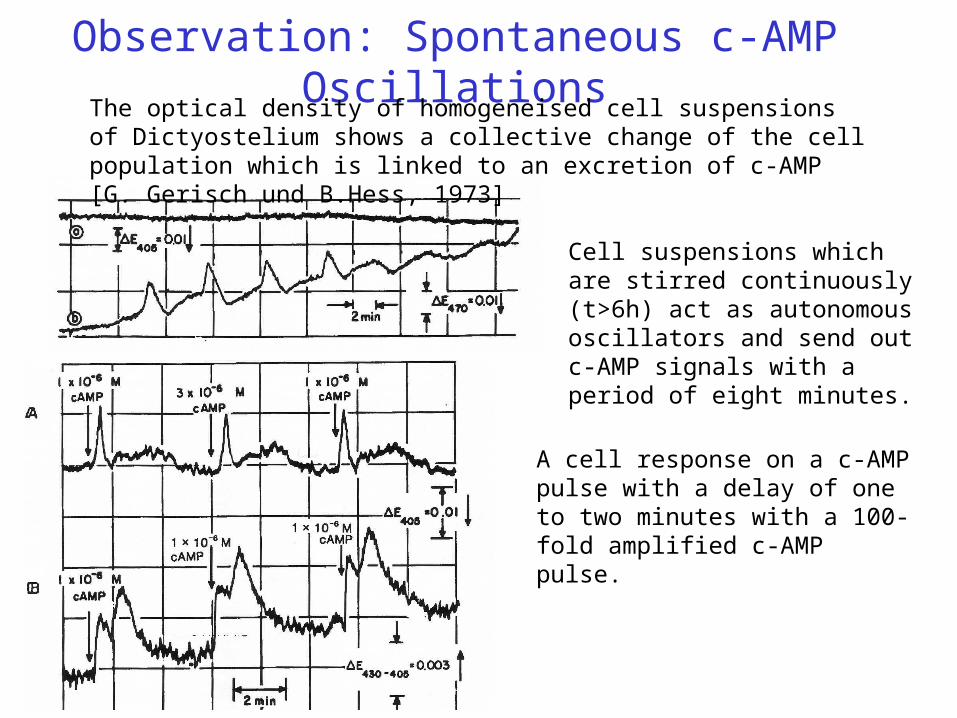
Observation: Spontaneous c-AMP OscillationsThe optical density of homogeneised cell suspensions of Dictyostelium shows a collective change of the cell population which is linked to an excretion of c-AMP [G. Gerisch und B.Hess, 1973]
Cell suspensions which are stirred continuously (t>6h) act as autonomous oscillators and send out c-AMP signals with a period of eight minutes.
A cell response on a c-AMP pulse with a delay of one to two minutes with a 100-fold amplified c-AMP pulse.
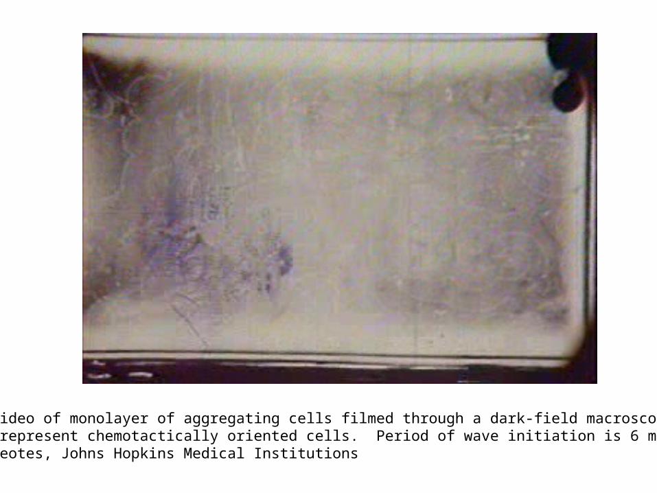
Time-lapse video of monolayer of aggregating cells filmed through a dark-field macroscope. White bands represent chemotactically oriented cells. Period of wave initiation is 6 minutes. From P. Devreotes, Johns Hopkins Medical Institutions
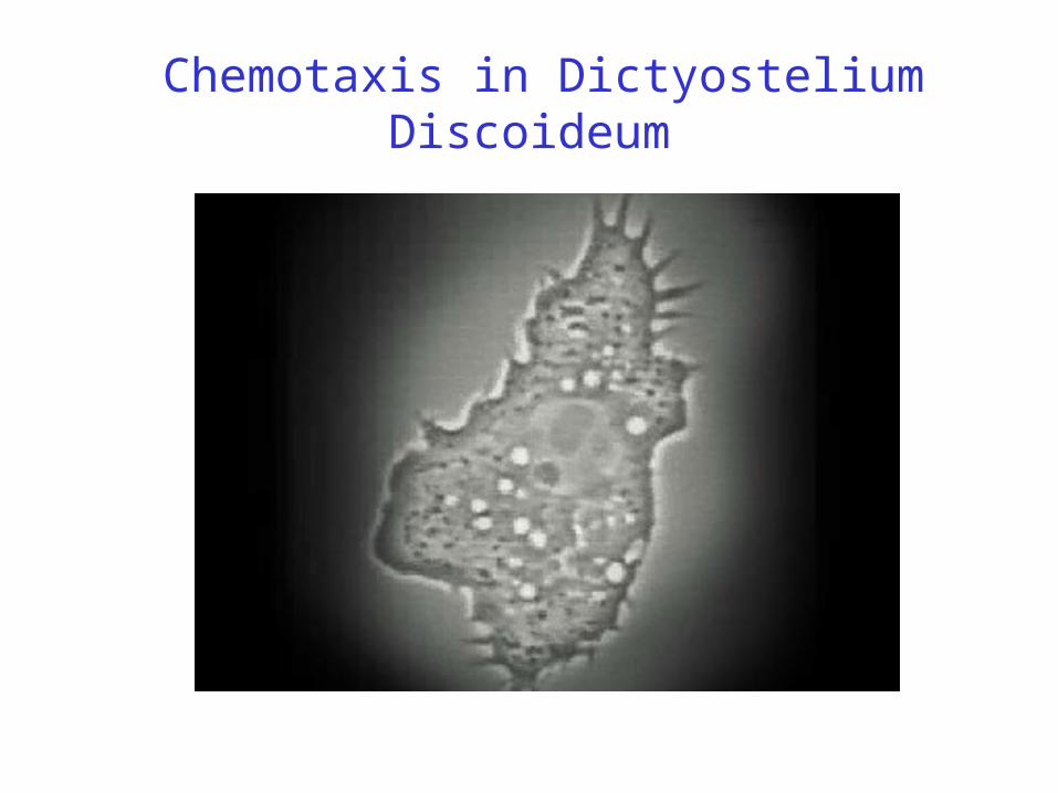
Chemotaxis in Dictyostelium Discoideum
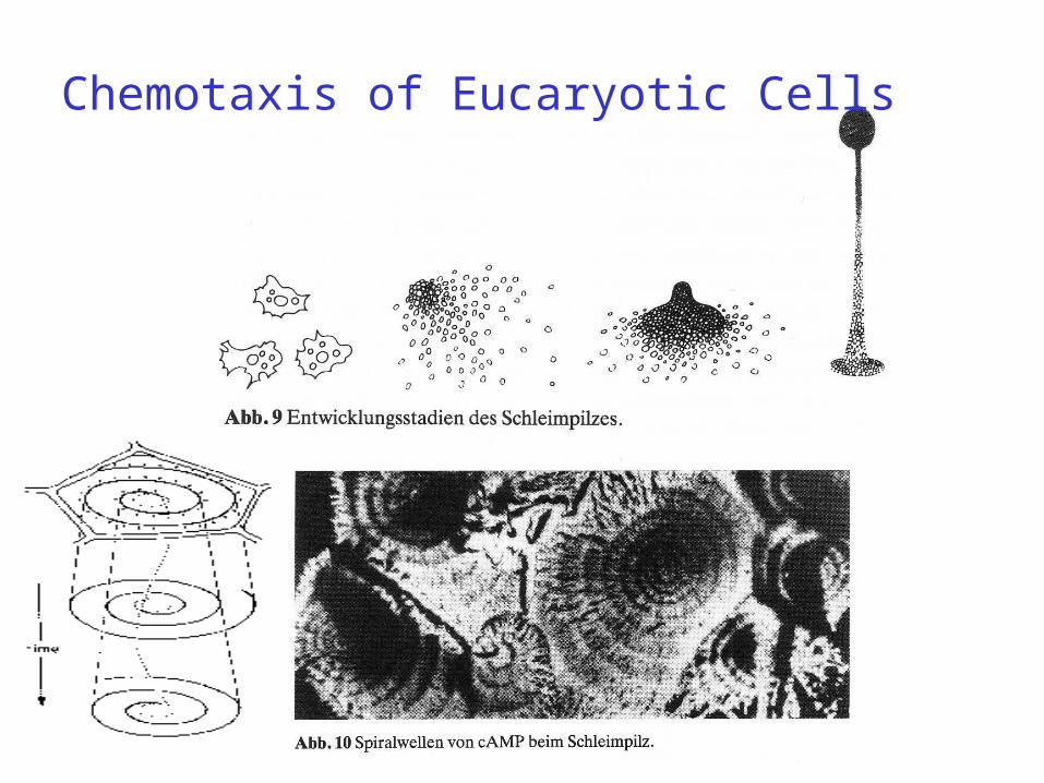
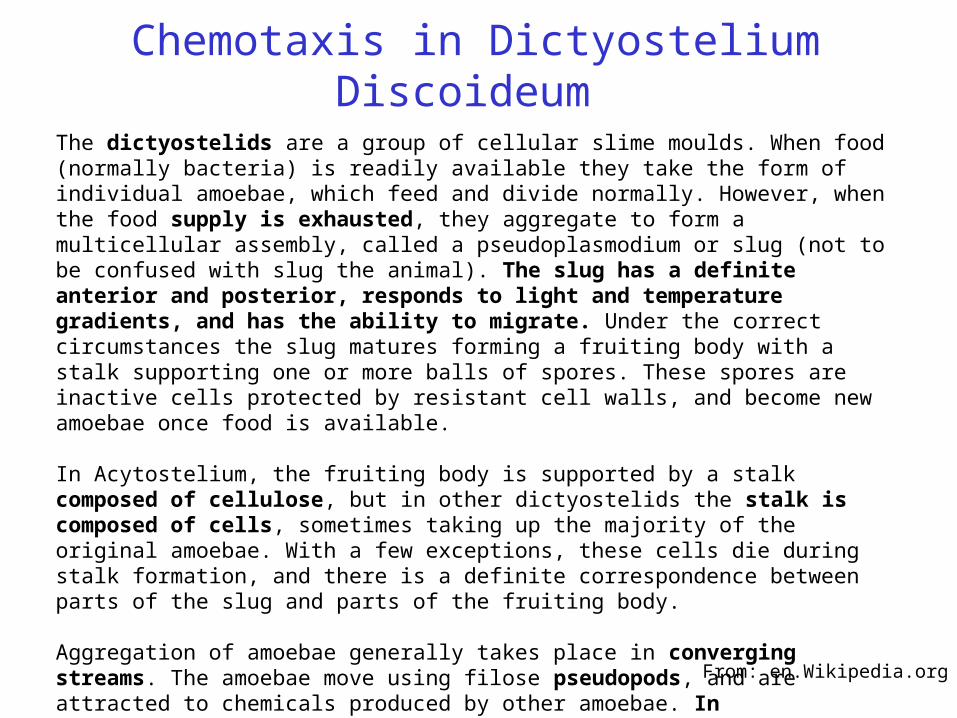
The dictyostelids are a group of cellular slime moulds. When food (normally bacteria) is readily available they take the form of individual amoebae, which feed and divide normally. However, when the food supply is exhausted, they aggregate to form a multicellular assembly, called a pseudoplasmodium or slug (not to be confused with slug the animal). The slug has a definite anterior and posterior, responds to light and temperature gradients, and has the ability to migrate. Under the correct circumstances the slug matures forming a fruiting body with a stalk supporting one or more balls of spores. These spores are inactive cells protected by resistant cell walls, and become new amoebae once food is available.
In Acytostelium, the fruiting body is supported by a stalk composed of cellulose, but in other dictyostelids the stalk is composed of cells, sometimes taking up the majority of the original amoebae. With a few exceptions, these cells die during stalk formation, and there is a definite correspondence between parts of the slug and parts of the fruiting body.
Aggregation of amoebae generally takes place in converging streams. The amoebae move using filose pseudopods, and are attracted to chemicals produced by other amoebae. In Dictyostelium, aggregation is signalled by cAMP, but others use different chemicals.
Dictyostelium has been used as a model organism in molecular biology and genetics, and is studied as an example of cell communication, differentiation, and programmed cell death.
From: en.Wikipedia.org

Der slime mouldDictyostelium Discoideum
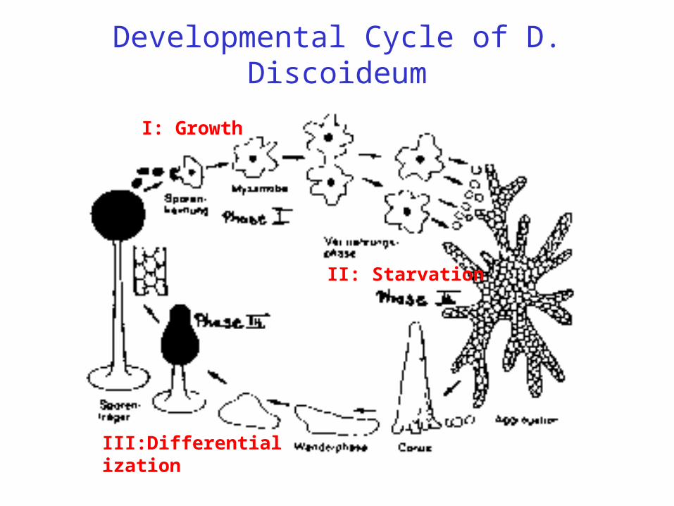
Developmental Cycle of D. Discoideum
II: Starvation
I: Growth
III:Differentialization
The Life-Cycle of Didi
Phase I:The amobae live (on agar plates) under paradise-like conditions (good relation
between nutricients and population density) as single cellular organisms and grow by cell division. This phase consists of exponential growth and a stationary state of constant cell number.Phase II:
The scarcity of nutricients leads after eight hours to a collective motion of the cells to statistically distributed centers, guided by collective chemotaxis. We will see that this is the result of cellular signalling using the messenger c-AMP.
Phase III:Differentialisation of the cells and growth of the slime mould. Three cell types
develop: spores which are capable of subsequent cell division, stabilizing cells of the stalk and cells forming the skin of the spore head.
Chemotaxis in Dictyostelium Discoideum
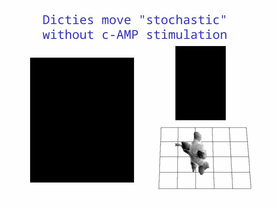
Dicties move "stochastic" without c-AMP stimulation
Eukaryotic Chemotaxis
• Directional sensing - Local excitation - global inhibition
• Polarization - Lipid-based signalling
• Locomotion - Actin polymerization
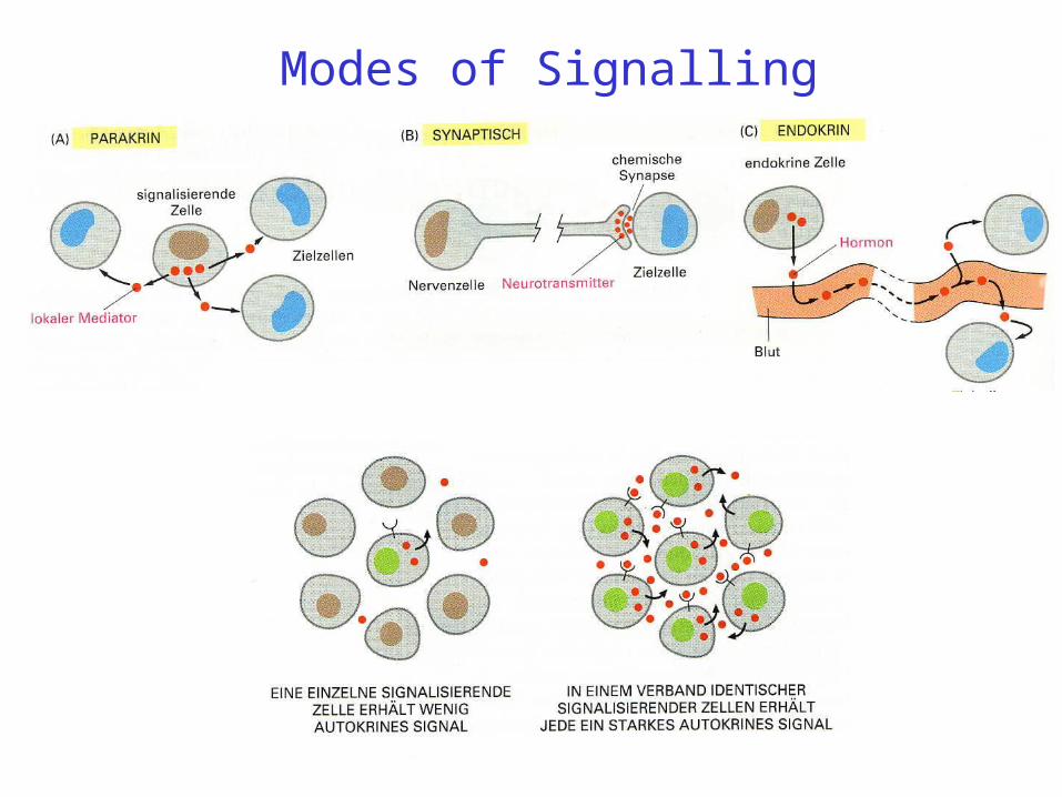
Modes of Signalling
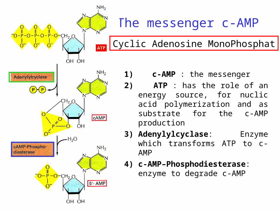
The messenger c-AMP
1) c-AMP : the messenger
2) ATP : has the role of an energy source, for nuclic acid polymerization and as substrate for the c-AMP production
3) Adenylylcyclase: Enzyme which transforms ATP to c-AMP
4) c-AMP–Phosphodiesterase: enzyme to degrade c-AMP
Cyclic Adenosine MonoPhosphat
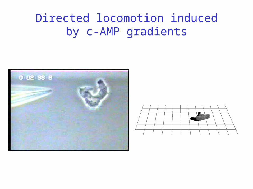
Directed locomotion inducedby c-AMP gradients
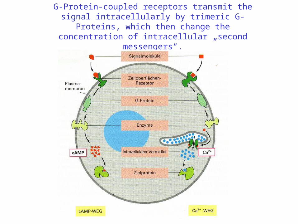
G-Protein-coupled receptors transmit the signal intracellularly by trimeric G-Proteins, which then change the concentration of intracellular „second messengers“.
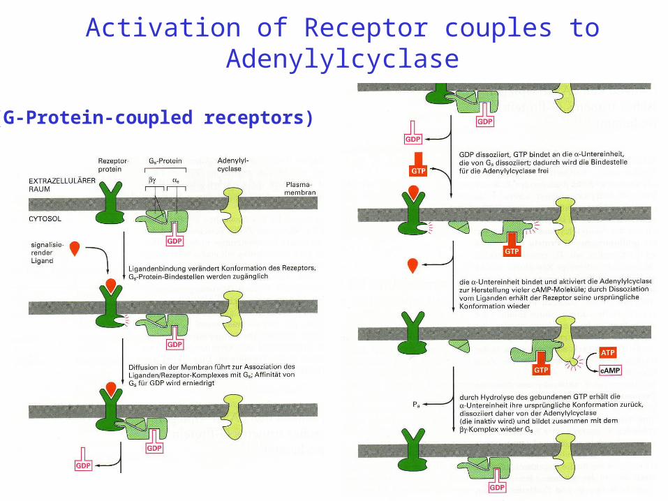
Activation of Receptor couples to Adenylylcyclase
Bild
(G-Protein-coupled receptors)
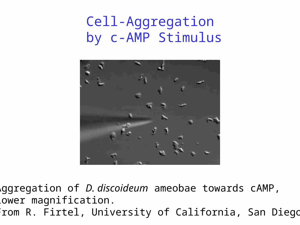
Cell-Aggregation by c-AMP Stimulus
Aggregation of D. discoideum ameobae towards cAMP, lower magnification. From R. Firtel, University of California, San Diego
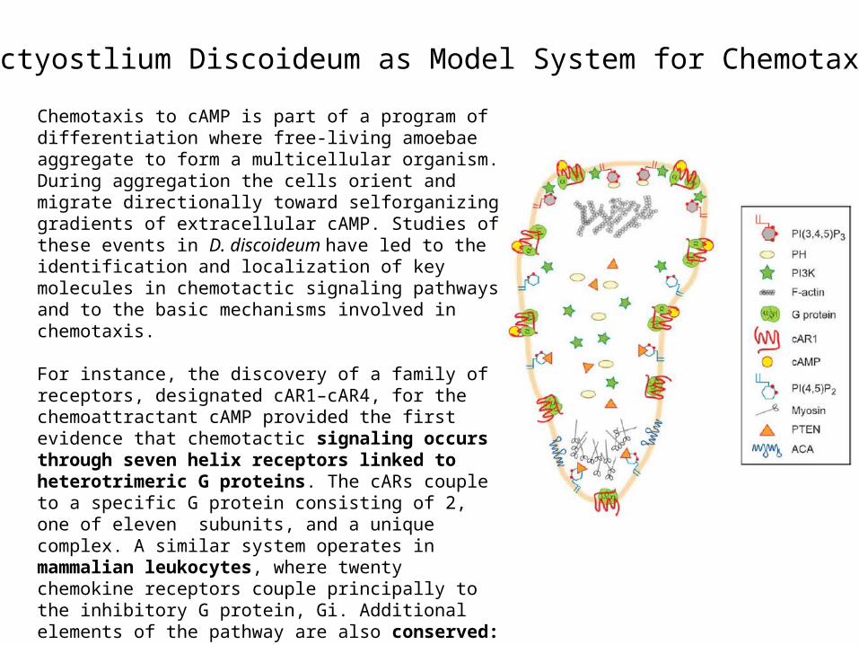
Chemotaxis to cAMP is part of a program of differentiation where free-living amoebae aggregate to form a multicellular organism. During aggregation the cells orient and migrate directionally toward selforganizing gradients of extracellular cAMP. Studies of these events in D. discoideum have led to the identification and localization of key molecules in chemotactic signaling pathways and to the basic mechanisms involved in chemotaxis.
For instance, the discovery of a family of receptors, designated cAR1–cAR4, for the chemoattractant cAMP provided the first evidence that chemotactic signaling occurs through seven helix receptors linked to heterotrimeric G proteins. The cARs couple to a specific G protein consisting of 2, one of eleven subunits, and a unique complex. A similar system operates in mammalian leukocytes, where twenty chemokine receptors couple principally to the inhibitory G protein, Gi. Additional elements of the pathway are also conserved: exposure of amoebae or leukocytes to chemoattractants results in increases in multiple second messengers, including PIs (phosphoinositides), cAMP, cGMP, IP3, and Ca2, and subsequent rearrangements in the cytoskeleton.
Dictyostlium Discoideum as Model System for Chemotaxis
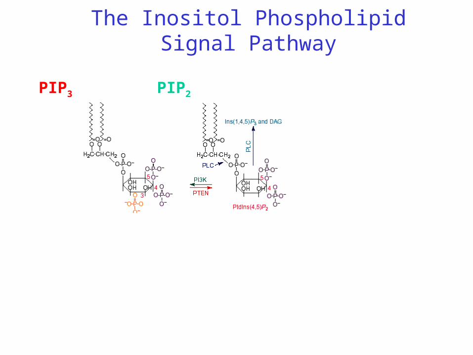
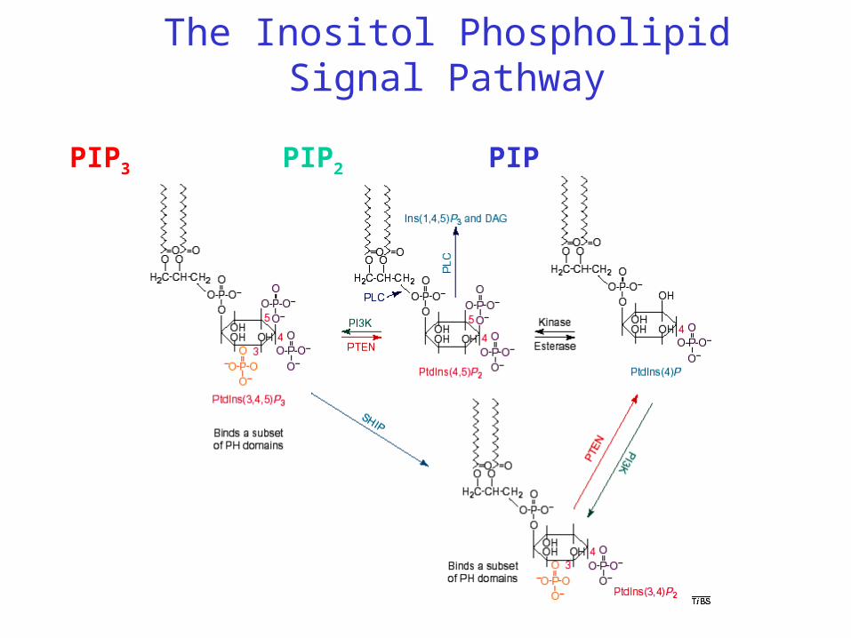
The Inositol Phospholipid Signal Pathway
PIPPIP2PIP3
G
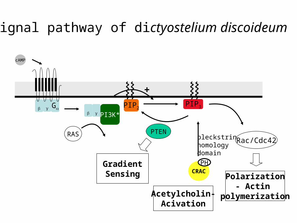
Signal pathway of dictyostelium discoideum
PIP2 PIP3
CRAC
cAMP
PI3K*
PH
PTENRac/Cdc42
Polarization- Actin
polymerization
RAS
GradientSensing
pleckstrinhomologydomain
+
Acetylcholin-Acivation
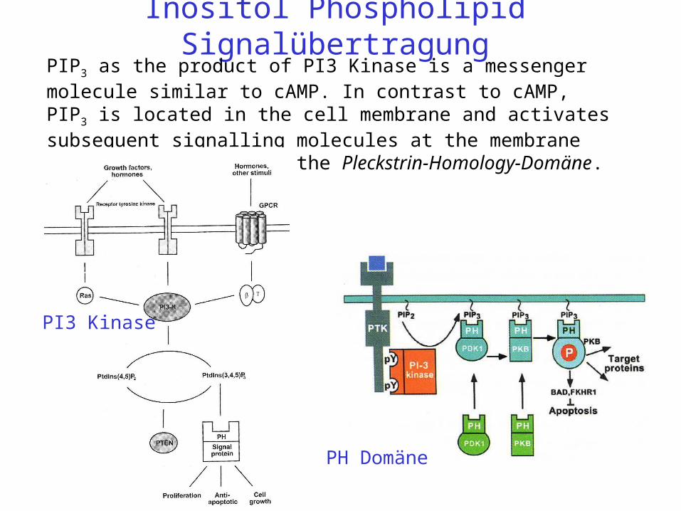
Inositol Phospholipid SignalübertragungPIP3 as the product of PI3 Kinase is a messenger molecule similar to cAMP. In contrast to cAMP, PIP3 is located in the cell membrane and activates subsequent signalling molecules at the membrane surface by binding to the Pleckstrin-Homology-Domäne.
PH Domäne
PI3 Kinase
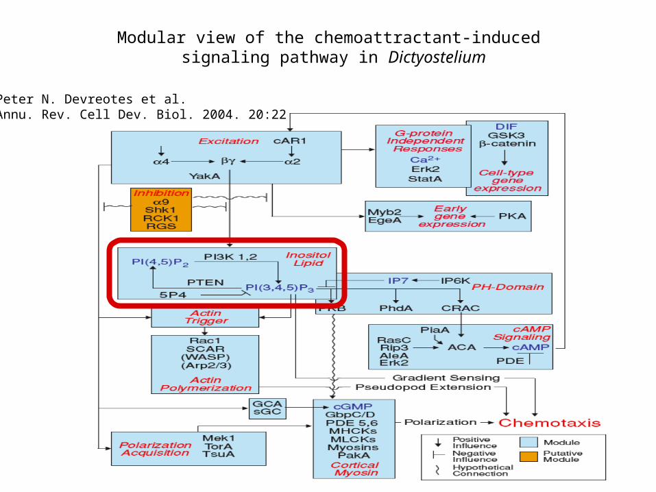
Modular view of the chemoattractant-induced signaling pathway in Dictyostelium
Peter N. Devreotes et al.Annu. Rev. Cell Dev. Biol. 2004. 20:22
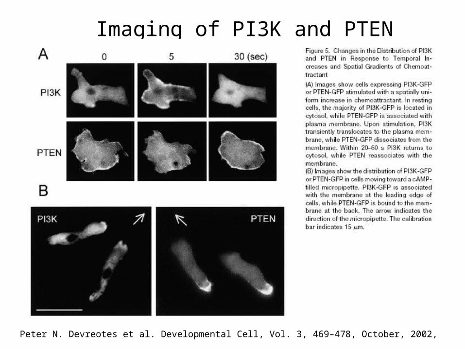
Peter N. Devreotes et al. Developmental Cell, Vol. 3, 469–478, October, 2002,
Imaging of PI3K and PTEN
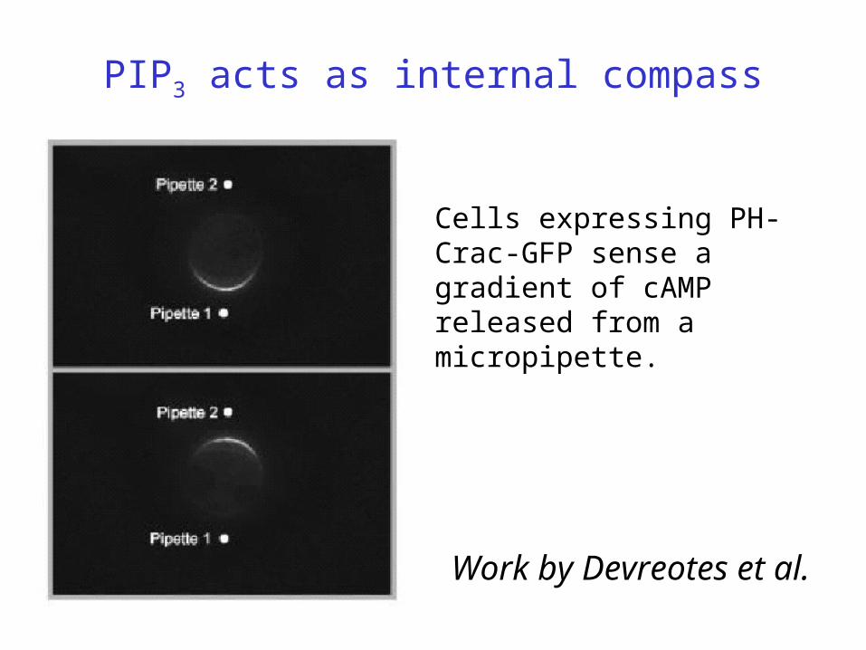
PIP3 acts as internal compass
Cells expressing PH-Crac-GFP sense a gradient of cAMP released from a micropipette.
Work by Devreotes et al.
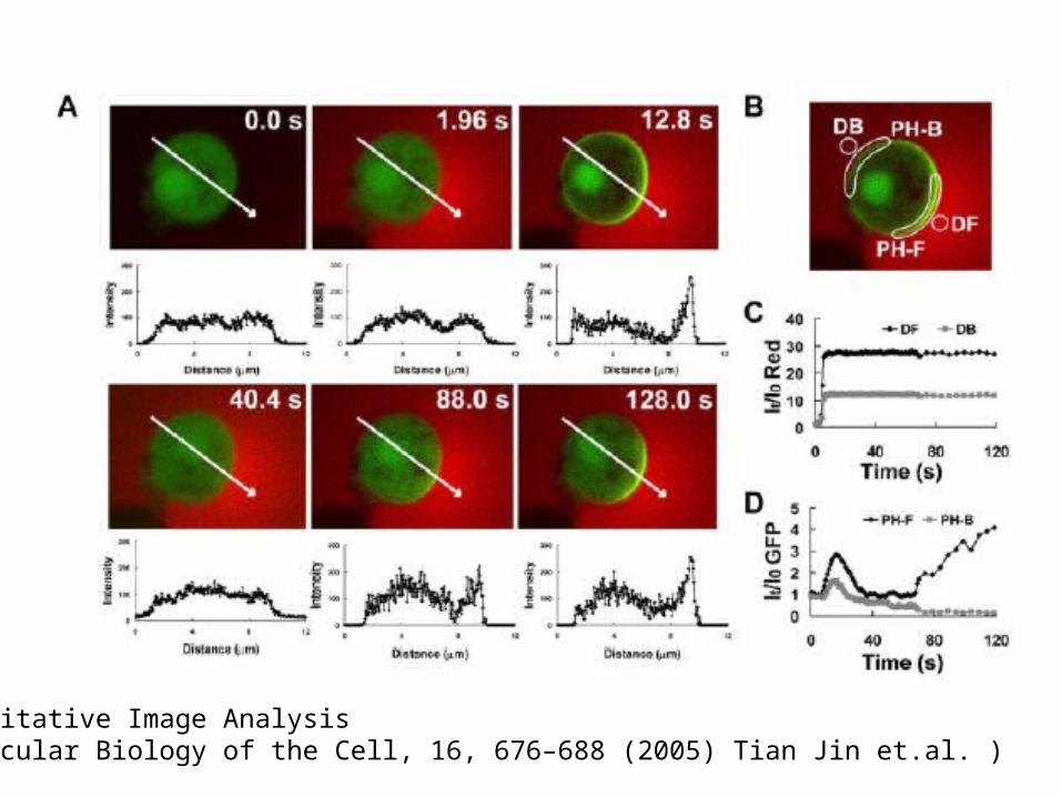
Quantitative Image Analysis (Molecular Biology of the Cell, 16, 676–688 (2005) Tian Jin et.al. )
The Inositol Phospholipid Signal Pathway
PIPPIP2PIP3
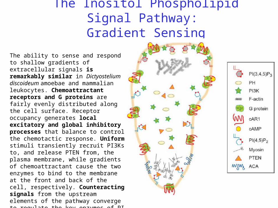
The Inositol Phospholipid Signal Pathway: Gradient Sensing
The ability to sense and respond to shallow gradients of extracellular signals is remarkably similar in Dictyostelium discoideum amoebae and mammalian leukocytes. Chemoattractant receptors and G proteins are fairly evenly distributed along the cell surface. Receptor occupancy generates local excitatory and global inhibitory processes that balance to control the chemotactic response. Uniform stimuli transiently recruit PI3Ks to, and release PTEN from, the plasma membrane, while gradients of chemoattractant cause the two enzymes to bind to the membrane at the front and back of the cell, respectively. Counteracting signals from the upstream elements of the pathway converge to regulate the key enzymes of PI metabolism, localize these lipids, and direct pseudopod formation.
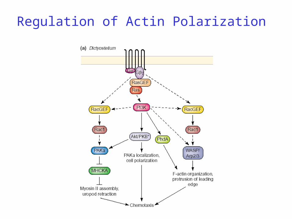
Regulation of Actin Polarization
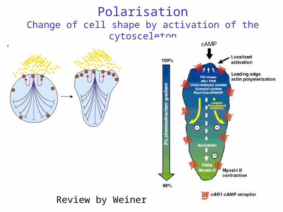
PolarisationChange of cell shape by activation of the cytosceleton
Review by Weiner
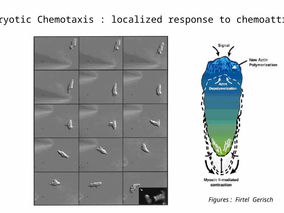
Eucaryotic Chemotaxis : localized response to chemoattractant
Figures : Firtel Gerisch
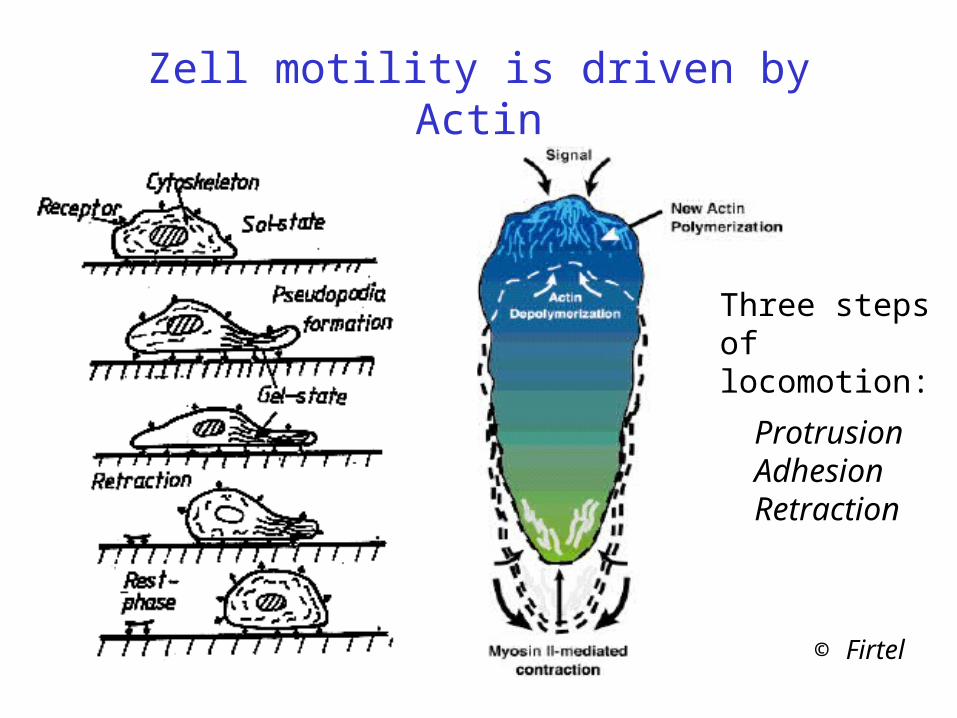
Zell motility is driven by Actin
© Firtel
ProtrusionAdhesionRetraction
Three steps of locomotion:
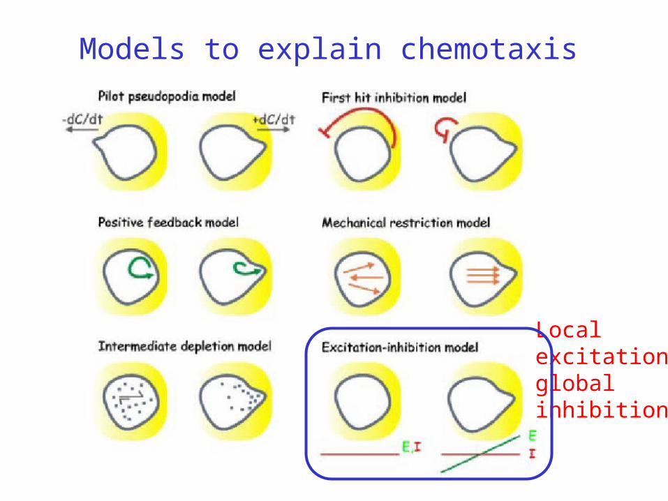
Models to explain chemotaxis
Local excitationglobal inhibition
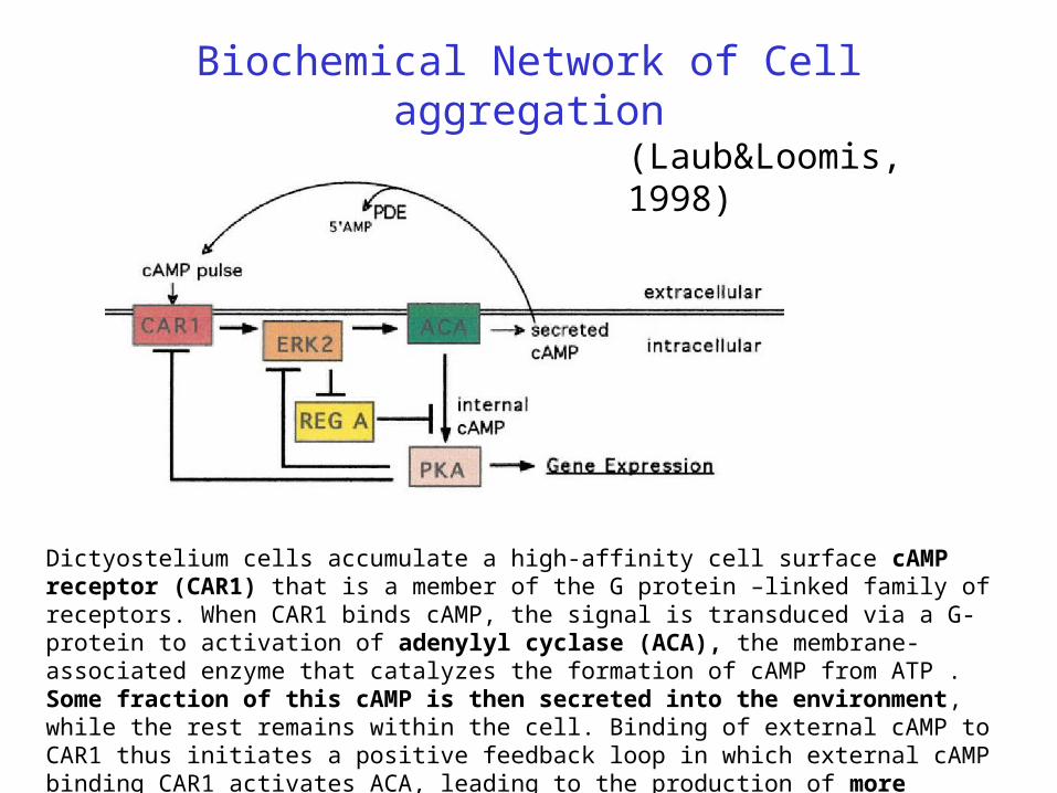
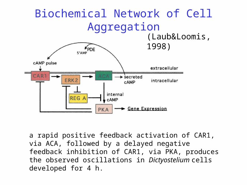
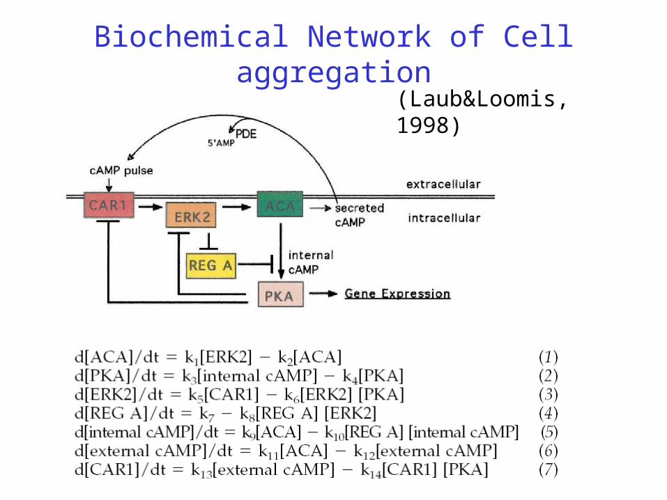
Biochemical Network of Cell aggregation
(Laub&Loomis, 1998)
Dictyostelium cells accumulate a high-affinity cell surface cAMP receptor (CAR1) that is a member of the G protein –linked family of receptors. When CAR1 binds cAMP, the signal is transduced via a G-protein to activation of adenylyl cyclase (ACA), the membrane-associated enzyme that catalyzes the formation of cAMP from ATP . Some fraction of this cAMP is then secreted into the environment, while the rest remains within the cell. Binding of external cAMP to CAR1 thus initiates a positive feedback loop in which external cAMP binding CAR1 activates ACA, leading to the production of more external cAMP.
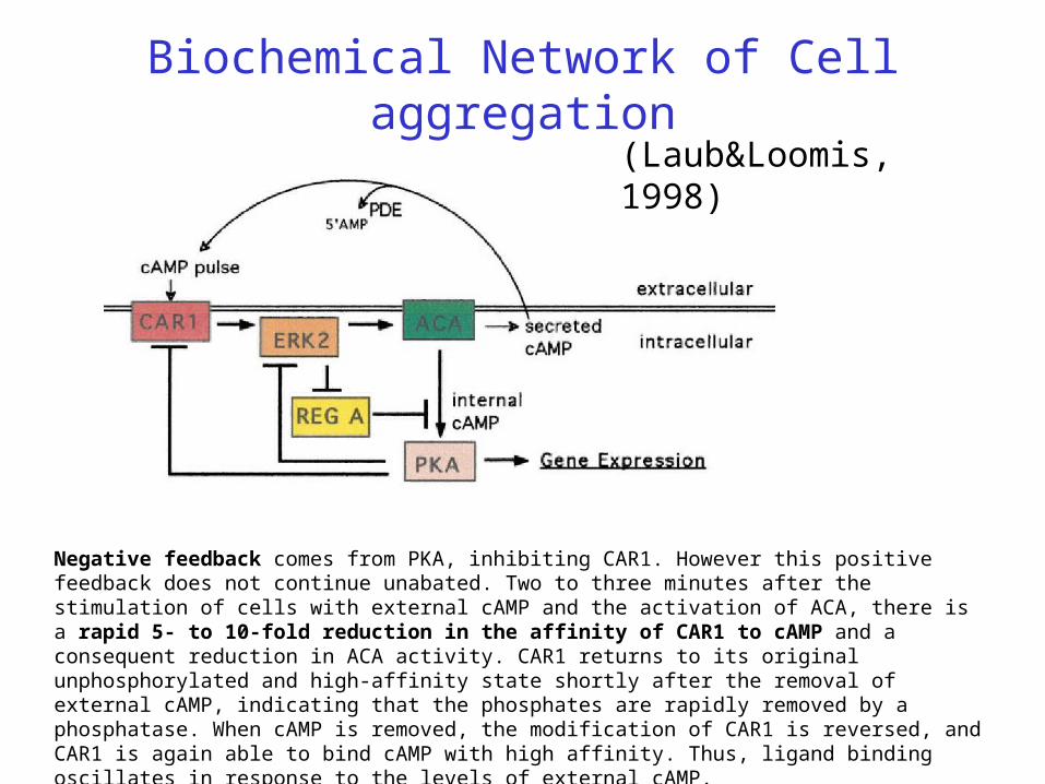
(Laub&Loomis, 1998)
Negative feedback comes from PKA, inhibiting CAR1. However this positive feedback does not continue unabated. Two to three minutes after the stimulation of cells with external cAMP and the activation of ACA, there is a rapid 5- to 10-fold reduction in the affinity of CAR1 to cAMP and a consequent reduction in ACA activity. CAR1 returns to its original unphosphorylated and high-affinity state shortly after the removal of external cAMP, indicating that the phosphates are rapidly removed by a phosphatase. When cAMP is removed, the modification of CAR1 is reversed, and CAR1 is again able to bind cAMP with high affinity. Thus, ligand binding oscillates in response to the levels of external cAMP.
Biochemical Network of Cell aggregation
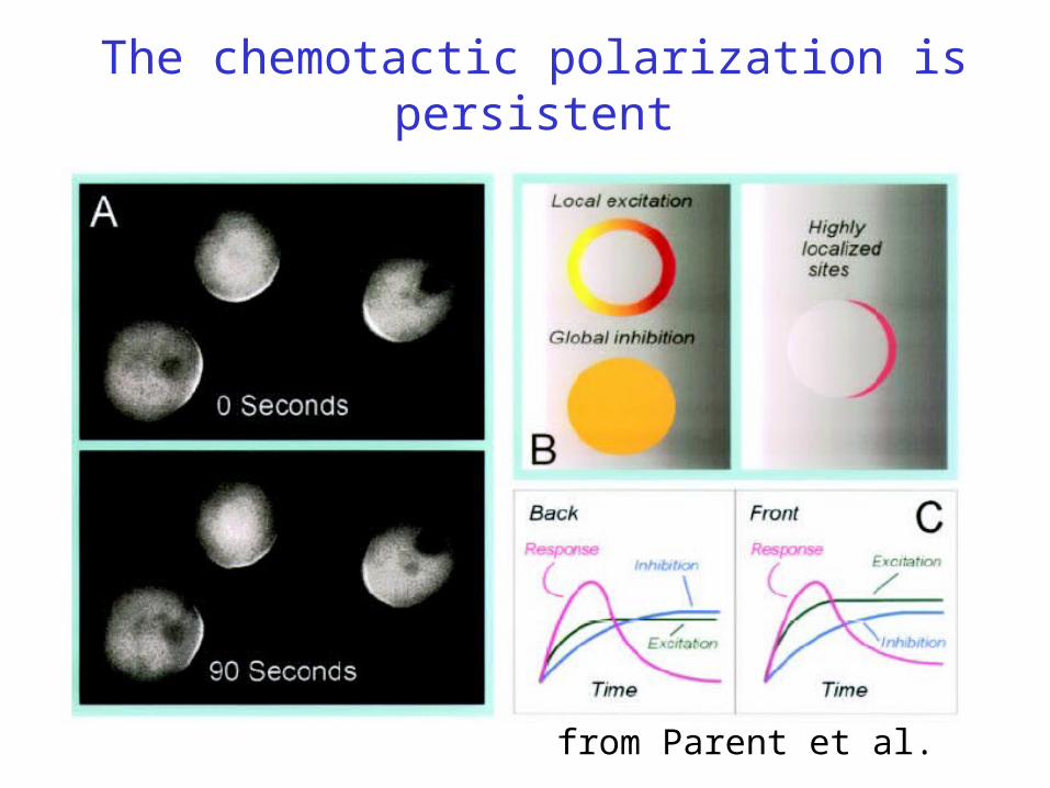
The chemotactic polarization is persistent
from Parent et al.
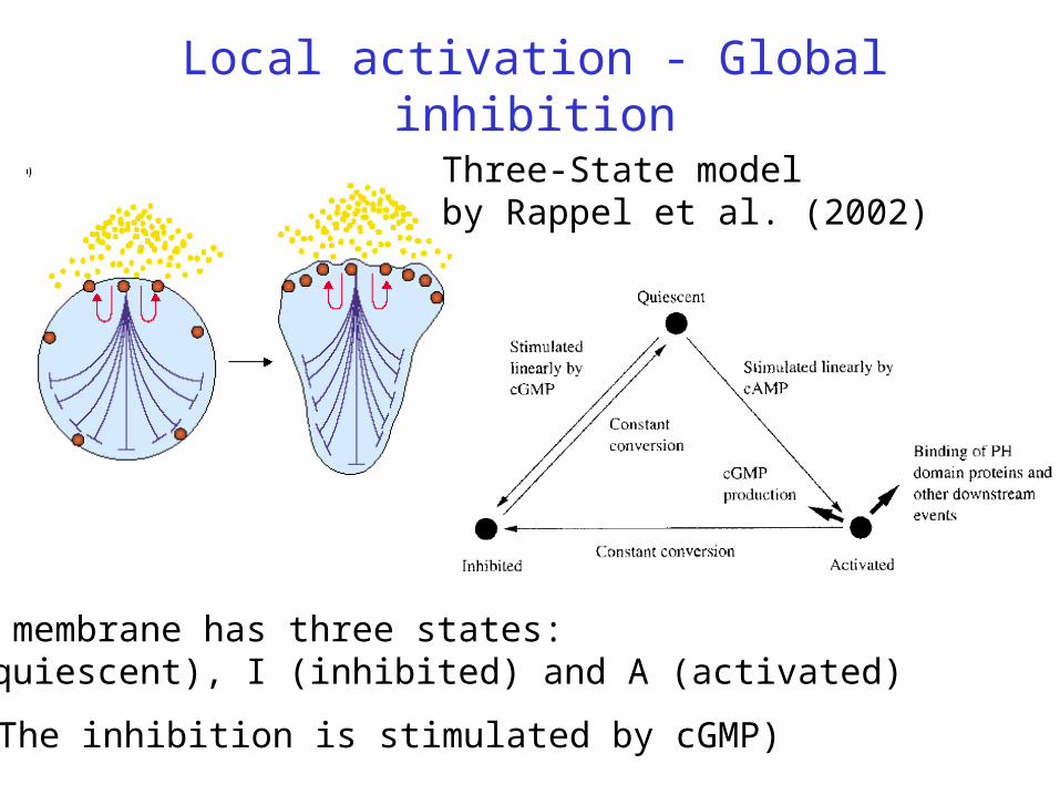
Local activation - Global inhibition
Three-State modelby Rappel et al. (2002)
(The inhibition is stimulated by cGMP)
The membrane has three states: Q (quiescent), I (inhibited) and A (activated)
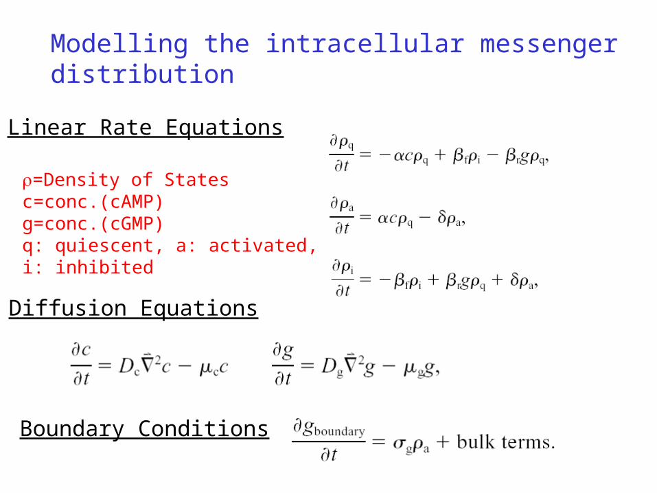
Diffusion Equations
Modelling the intracellular messenger distribution
Linear Rate Equations
Boundary Conditions
=Density of Statesc=conc.(cAMP)g=conc.(cGMP)q: quiescent, a: activated,i: inhibited
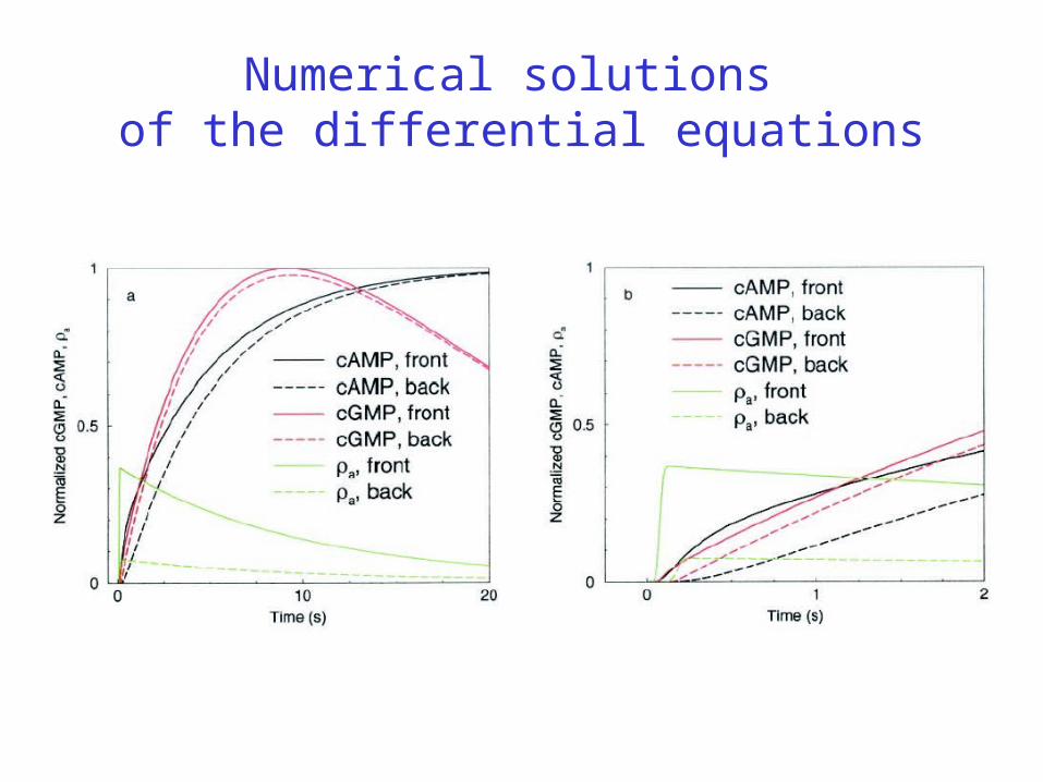
Numerical solutions of the differential equations
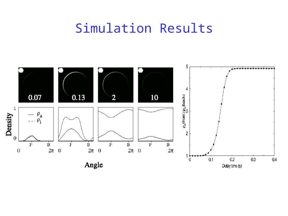
Simulation Results
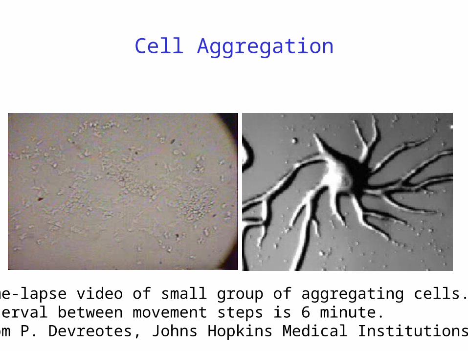
Cell Aggregation
Time-lapse video of small group of aggregating cells. Interval between movement steps is 6 minute. From P. Devreotes, Johns Hopkins Medical Institutions.
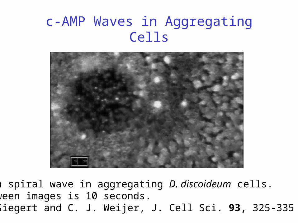
c-AMP Waves in Aggregating Cells
Core of a spiral wave in aggregating D. discoideum cells. Time between images is 10 seconds. From F. Siegert and C. J. Weijer, J. Cell Sci. 93, 325-335 (1989).
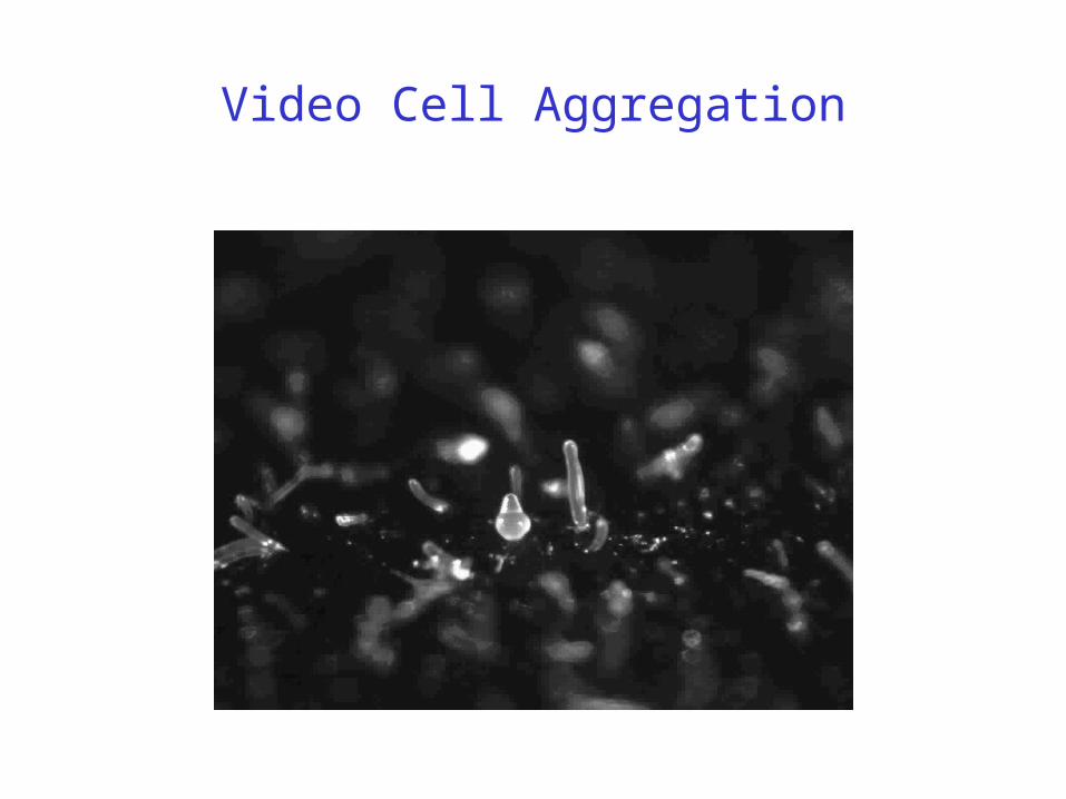
Video Cell Aggregation
Biochemical Network of Cell Aggregation
(Laub&Loomis, 1998)
a rapid positive feedback activation of CAR1, via ACA, followed by a delayed negative feedback inhibition of CAR1, via PKA, produces the observed oscillations in Dictyostelium cells developed for 4 h.

Dark field waves of D. discoideum cells on caffeine agar. Time between images is 36 seconds. From F. Siegert and C. J. Weijer, J. Cell Sci. 93, 325-335 (1989).
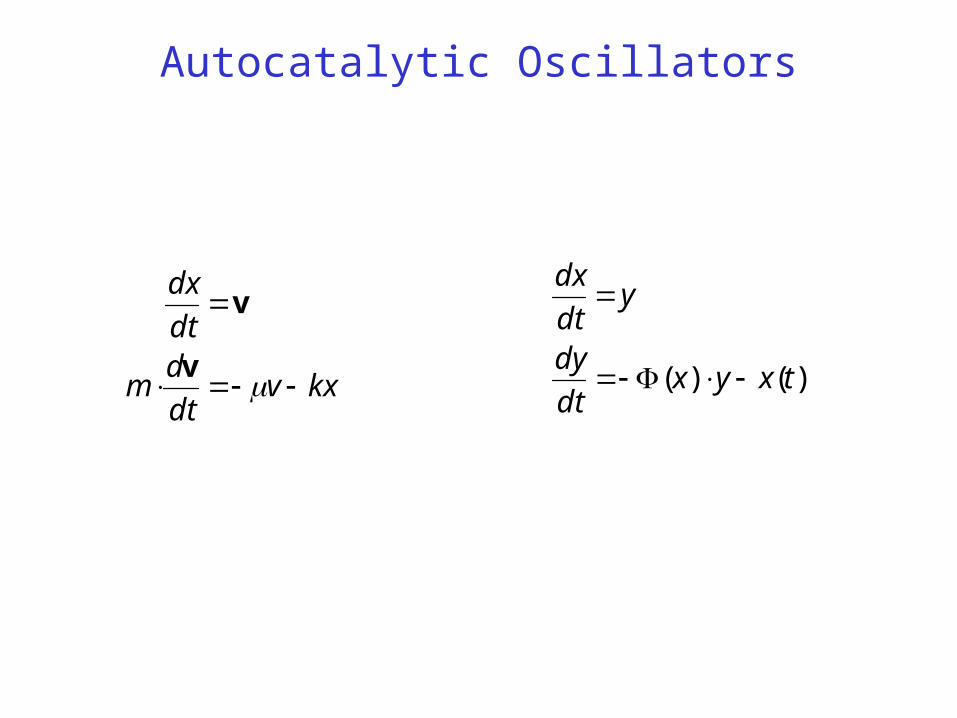
Autocatalytic Oscillators
)()( txyxdt
dy
ydt
dx
kxvdt
dm
dt
dx
v
v
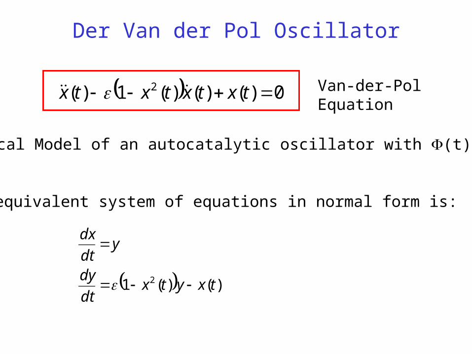
Der Van der Pol Oscillator
0)()()(1)( 2 txtxtxtx Van-der-PolEquation
)()(1 2 txytxdt
dy
ydt
dx
An equivalent system of equations in normal form is:
Classical Model of an autocatalytic oscillator with (t)=(x2-1).
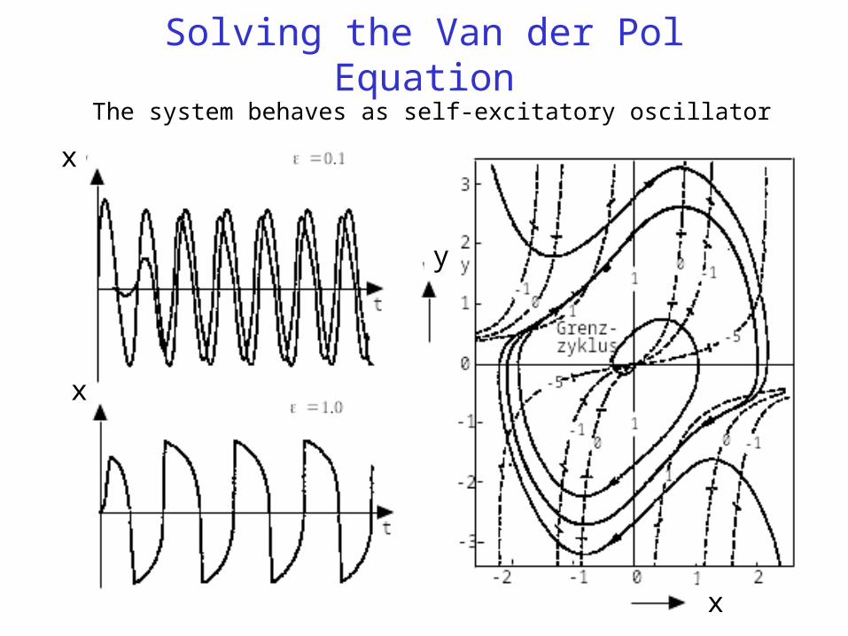
Solving the Van der Pol EquationThe system behaves as self-excitatory oscillator
x
x
x
y
The allosteric control of enzyme activity
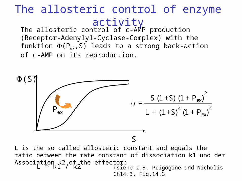
=S (1 +S) (1 + Pex)
2
L + (1 +S)2
(1 + Pex)2
(S)
S
The allosteric control of c-AMP production (Receptor-Adenylyl-Cyclase-Complex) with the funktion (Pex,S) leads to a strong back-action of c-AMP on its reproduction.
(siehe z.B. Prigogine and Nicholis Ch14.3, Fig.14.3
Pex
L = k1 / k2
L is the so called allosteric constant and equals the ratio between the rate constant of dissociation k1 und der Association k2 of the effector:
Reprise
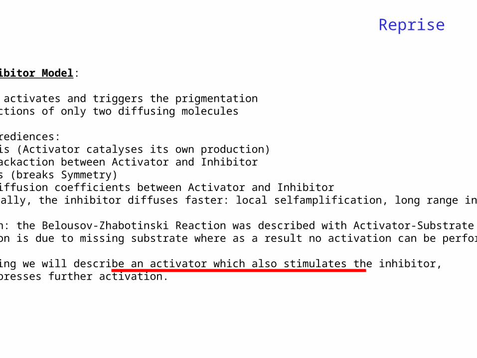
Aktivator-Inhibitor Model:
The Activator activates and triggers the prigmentationNonlinear reactions of only two diffusing molecules
Importent ingrediences:- Autocatalysis (Activator catalyses its own production)- Nonlinear backaction between Activator and Inhibitor- Fluctuations (breaks Symmetry)- Different Diffusion coefficients between Activator and Inhibitor
(Typically, the inhibitor diffuses faster: local selfamplification, long range inhibition)
For comparison: the Belousov-Zhabotinski Reaction was described with Activator-Substrate model. The back action is due to missing substrate where as a result no activation can be performed any more.
In the following we will describe an activator which also stimulates the inhibitor, but which suppresses further activation.
Model of Goldbeter&Segel
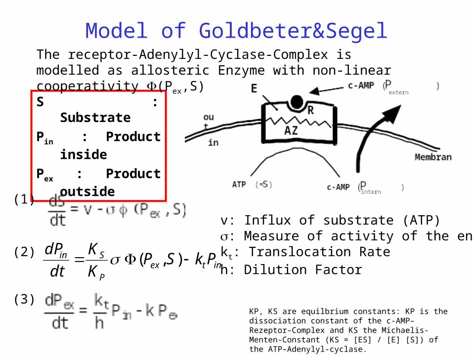
S : Substrate
Pin : Product inside
Pex : Product outside
(1)
(2)
(3)
The receptor-Adenylyl-Cyclase-Complex is modelled as allosteric Enzyme with non-linear cooperativity (Pex,S)
v: Influx of substrate (ATP): Measure of activity of the enzymekt: Translocation Rateh: Dilution Factorintex
P
Sin PkSPK
K
dt
dP ),(
KP, KS are equilbrium constants: KP is the dissociation constant of the c-AMP–Rezeptor–Complex and KS the Michaelis-Menten-Constant (KS = [ES] / [E] [S]) of the ATP–Adenylyl-cyclase.
Approximations of the Goldbeter Equation
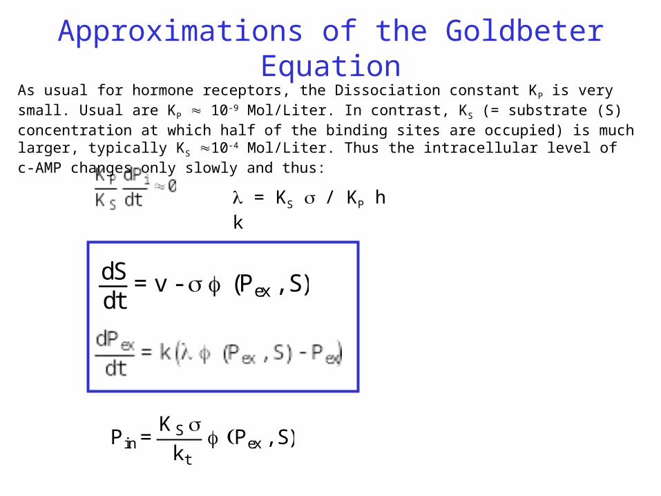
dSdt
= v - (Pex , S)
P in =KS
k t
Pex , S)
= KS / KP h k
As usual for hormone receptors, the Dissociation constant KP is very small. Usual are KP 10-9 Mol/Liter. In contrast, KS (= substrate (S) concentration at which half of the binding sites are occupied) is much larger, typically KS 10-4 Mol/Liter. Thus the intracellular level of c-AMP changes only slowly and thus:
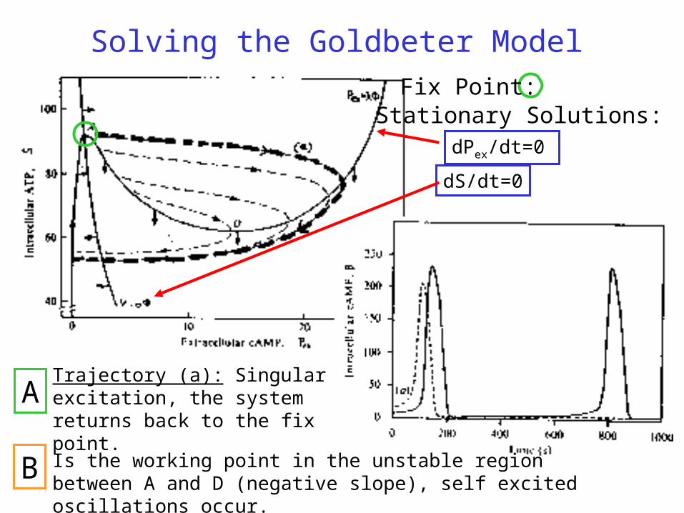
Solving the Goldbeter Model
Stationary Solutions:
dS/dt=0
dPex/dt=0
Fix Point:
Trajectory (a): Singular excitation, the system returns back to the fix point.
Is the working point in the unstable region between A and D (negative slope), self excited oscillations occur.
A
B

Observation: Spontaneous c-AMP OscillationsThe optical density of homogeneised cell suspensions of Dictyostelium shows a collective change of the cell population which is linked to an excretion of c-AMP [G. Gerisch und B.Hess, 1973]
Cell suspensions which are stirred continuously (t>6h) act as autonomous oscillators and send out c-AMP signals with a period of eight minutes.
A cell response on a c-AMP pulse with a delay of one to two minutes with a 100-fold amplified c-AMP pulse.

Time-lapse video of monolayer of aggregating cells filmed through a dark-field macroscope. White bands represent chemotactically oriented cells. Period of wave initiation is 6 minutes. From P. Devreotes, Johns Hopkins Medical Institutions
Biochemical Network of Cell aggregation
(Laub&Loomis, 1998)
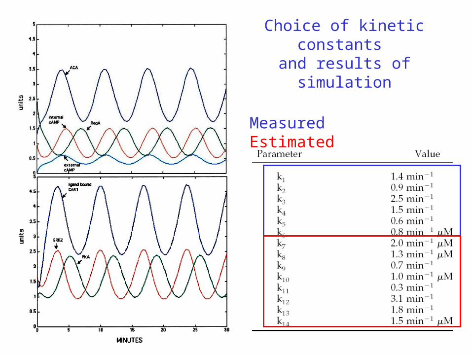
Choice of kinetic constants and results of simulation
MeasuredEstimated
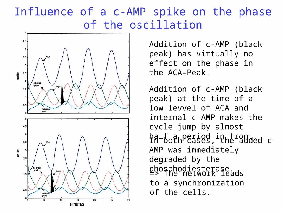
Influence of a c-AMP spike on the phase of the oscillation
Addition of c-AMP (black peak) has virtually no effect on the phase in the ACA-Peak.
In both cases, the added c-AMP was immediately degraded by the phosphodiesterase.
Addition of c-AMP (black peak) at the time of a low levvel of ACA and internal c-AMP makes the cycle jump by almost half a period in front.
=> The network leads to a synchronization of the cells.
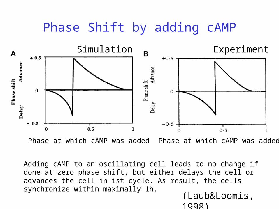
Phase Shift by adding cAMP
Simulation Experiment
(Laub&Loomis, 1998)
Phase at which cAMP was added
Adding cAMP to an oscillating cell leads to no change if done at zero phase shift, but either delays the cell or advances the cell in ist cycle. As result, the cells synchronize within maximally 1h.
Phase at which cAMP was added
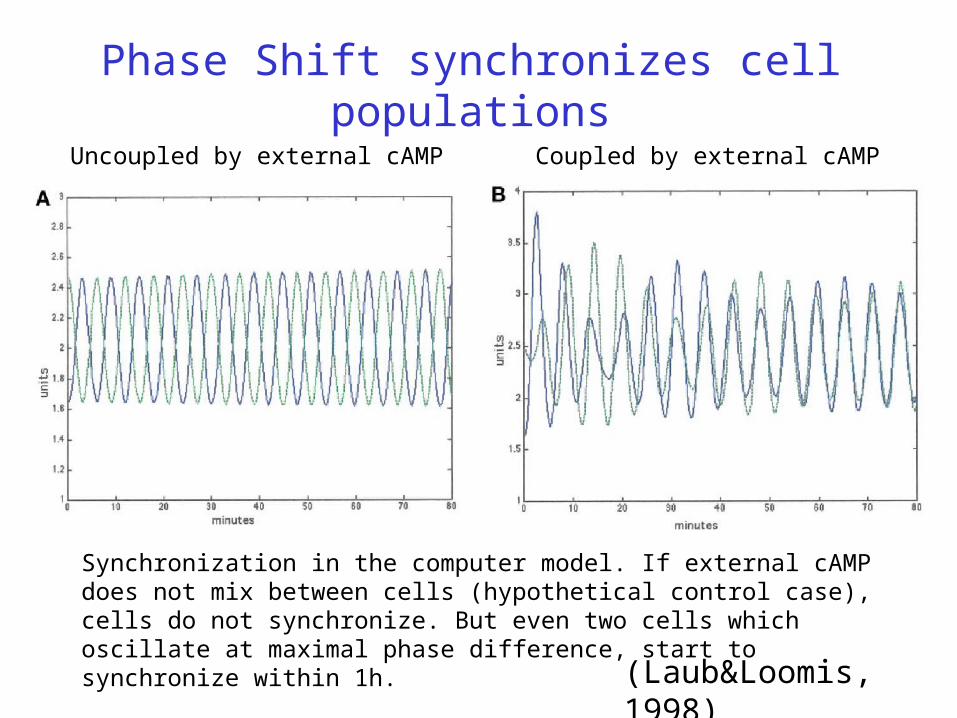
Phase Shift synchronizes cell populations
Uncoupled by external cAMP Coupled by external cAMP
(Laub&Loomis, 1998)
Synchronization in the computer model. If external cAMP does not mix between cells (hypothetical control case), cells do not synchronize. But even two cells which oscillate at maximal phase difference, start to synchronize within 1h.
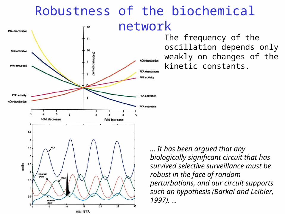
Robustness of the biochemical networkThe frequency of the oscillation depends only weakly on changes of the kinetic constants.
... It has been argued that any biologically significant circuit that has survived selective surveillance must be robust in the face of random perturbations, and our circuit supports such an hypothesis (Barkai and Leibler, 1997). ...
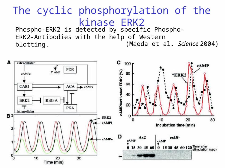
The cyclic phosphorylation of the kinase ERK2Phospho-ERK2 is detected by specific Phospho-ERK2-Antibodies with the help of Western blotting.
(Maeda et al. Science 2004)
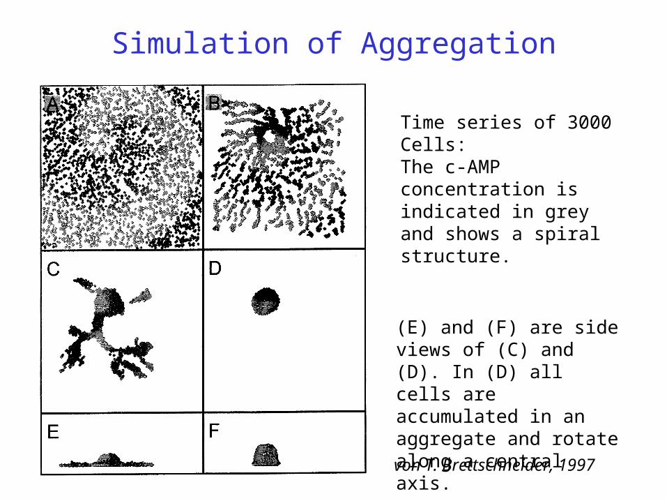
Simulation of Aggregation
Time series of 3000 Cells:The c-AMP concentration is indicated in grey and shows a spiral structure.
(E) and (F) are side views of (C) and (D). In (D) all cells are accumulated in an aggregate and rotate along a central axis.
von T. Brettschneider, 1997
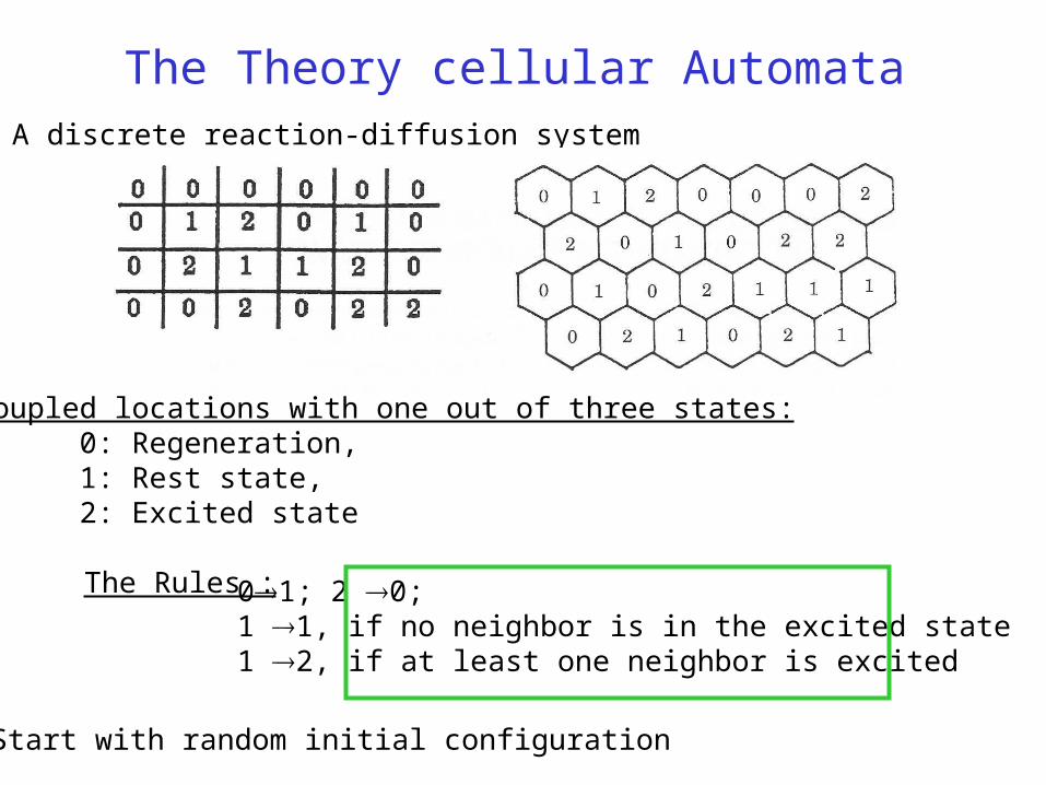
The Theory cellular Automata
Coupled locations with one out of three states:0: Regeneration,1: Rest state,2: Excited state
A discrete reaction-diffusion system
The Rules :
Start with random initial configuration
01; 2 0; 1 1, if no neighbor is in the excited state1 2, if at least one neighbor is excited
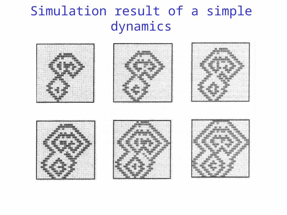
Simulation result of a simple dynamics