chest injuries remain a significant cause of morbidity and mortality
TRANSCRIPT
Chest injuries remain a significant cause of morbidity and mortality
1STN E-Library 2012
9_Thoracic Injuries

9_Thoracic Injuries
STN E-Library 2012 2
There are many vital structures located within the chest cavity, too numerous to review in detail.
The purpose of this presentation is to provide an overview of major injuries in the thorax and some of the early interventions associated with their management.
3STN E-Library 2012
9_Thoracic Injuries
Many cases of traumatic deaths due to chest injury may be prevented by prompt diagnosis and a standardized therapeutic approach in the trauma resuscitation room. A high index of suspicion for lethal injury patterns, based on the mechanism of trauma and the clinical presentation, is a crucial to prompt diagnosis and intervention.
These injuries may be quite subtle in their presentation or they manifest through a very dramatic presentation with profound physiologic derangements leading to death within minutes if not corrected.
• Immediate deaths from blunt chest trauma usually related to heart or thoracic aorta disruption
• Death within 3 hours of injury related to airway obstruction, cardiac tamponade, aortic disruption, or continued hemorrhage
• Late deaths from blunt chest trauma usually due to missed injuries, multiorgan system failure, systemic inflammatory response syndrome, respiratory complications, and infections
• Many injuries are managed nonoperatively
• About 85% of patients with thoracic injuries only require a chest tube, observation, and pain management
Trauma, 6th Edition, Indications for and Techniques of Thoracotomy, pg 512.
4STN E-Library 2012
9_Thoracic Injuries
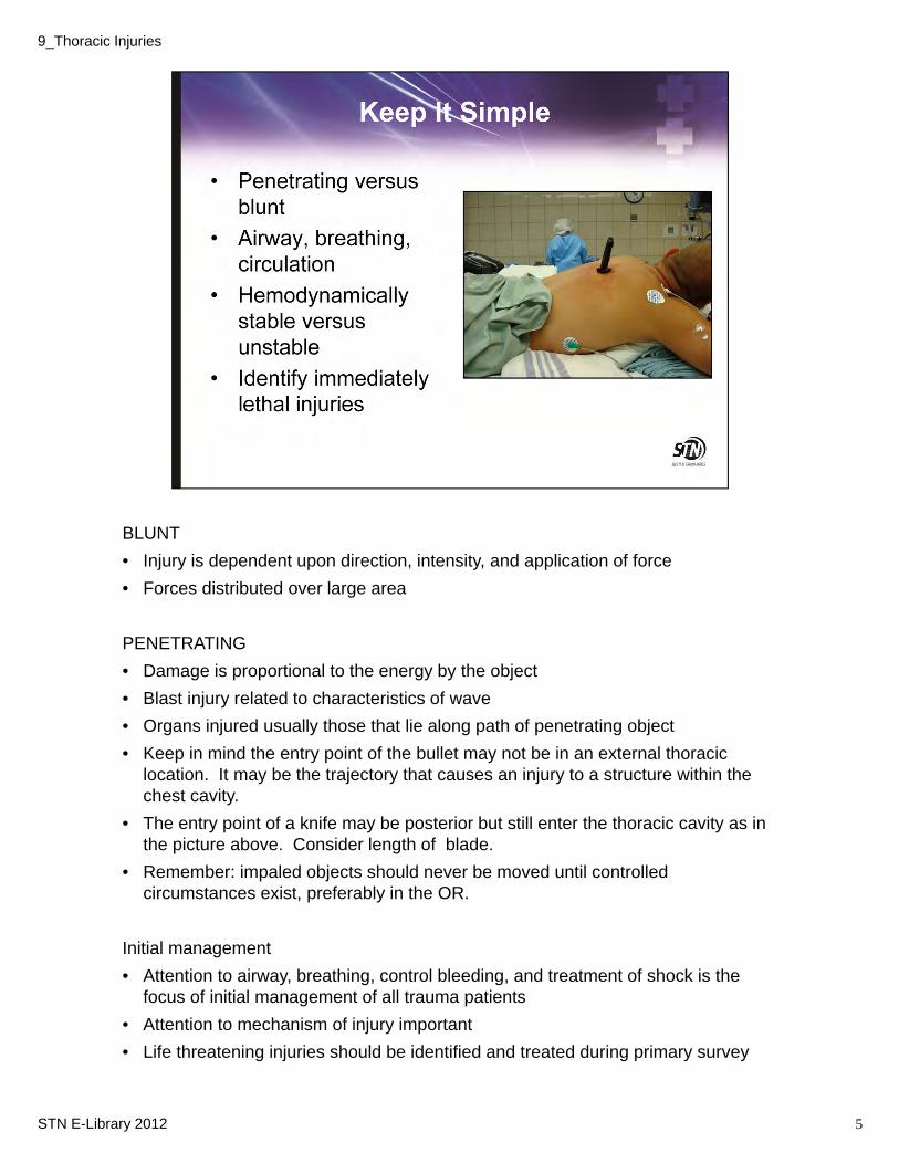
BLUNT
• Injury is dependent upon direction, intensity, and application of force
• Forces distributed over large area
PENETRATING
• Damage is proportional to the energy by the object
• Blast injury related to characteristics of wave
• Organs injured usually those that lie along path of penetrating object
• Keep in mind the entry point of the bullet may not be in an external thoracic location. It may be the trajectory that causes an injury to a structure within the chest cavity.
• The entry point of a knife may be posterior but still enter the thoracic cavity as in the picture above. Consider length of blade.
• Remember: impaled objects should never be moved until controlled circumstances exist, preferably in the OR.
Initial management
• Attention to airway, breathing, control bleeding, and treatment of shock is the focus of initial management of all trauma patients
• Attention to mechanism of injury important
• Life threatening injuries should be identified and treated during primary survey
5STN E-Library 2012
9_Thoracic Injuries
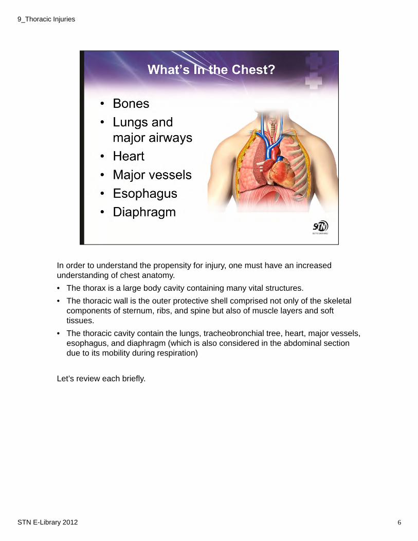
In order to understand the propensity for injury, one must have an increased understanding of chest anatomy.
• The thorax is a large body cavity containing many vital structures.
• The thoracic wall is the outer protective shell comprised not only of the skeletal components of sternum, ribs, and spine but also of muscle layers and soft tissues.
• The thoracic cavity contain the lungs, tracheobronchial tree, heart, major vessels, esophagus, and diaphragm (which is also considered in the abdominal section due to its mobility during respiration)
Let’s review each briefly.
6STN E-Library 2012
9_Thoracic Injuries
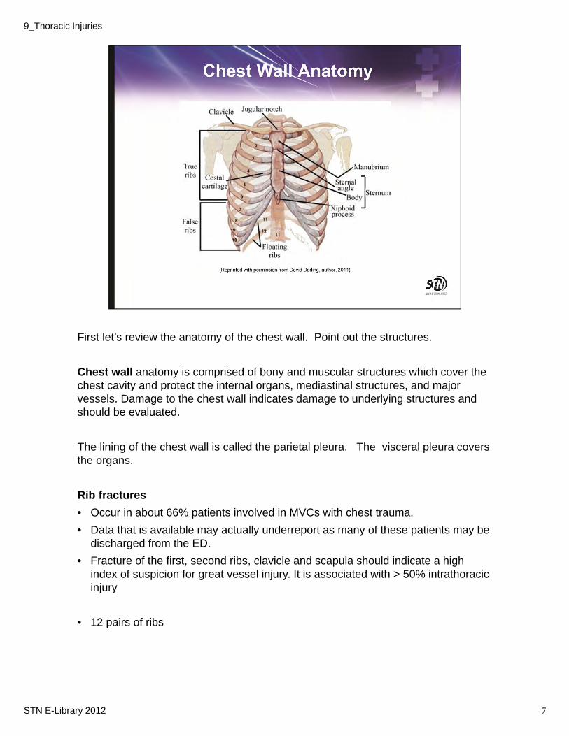
First let’s review the anatomy of the chest wall. Point out the structures.
Chest wall anatomy is comprised of bony and muscular structures which cover the chest cavity and protect the internal organs, mediastinal structures, and major vessels. Damage to the chest wall indicates damage to underlying structures and should be evaluated.
The lining of the chest wall is called the parietal pleura. The visceral pleura covers the organs.
Rib fractures
• Occur in about 66% patients involved in MVCs with chest trauma.
• Data that is available may actually underreport as many of these patients may be discharged from the ED.
• Fracture of the first, second ribs, clavicle and scapula should indicate a high index of suspicion for great vessel injury. It is associated with > 50% intrathoracic injury
• 12 pairs of ribs
7STN E-Library 2012
9_Thoracic Injuries
• 1st 7 attached anteriorly via costal cartilages to sternum
• 8-10 connected anteriorly to each other via costal cartilages and synovial joints
• 11 & 12 ribs not connected anteriorly, floating ribs.
• Intercostal spaces: spaces between ribs; number to correlate with rib superior to space.
• Intercostal spaces contain muscles that are instrumental to work of breathing, arteries, veins, and nerves that run immediately under rib.
• Intercostal space named for rib above it.
• Landmark lines
►1st rib lies posterior to clavicles
►Scapula ends at 7th intercostal space
►Nipple on male at 4th intercostal space
• The bony structures also include the clavicle and scapula.
The Encyclopedia of Science
http://www.daviddarling.info/encyclopedia/R/rib-cage.html
9_Thoracic Injuries
STN E-Library 2012 7
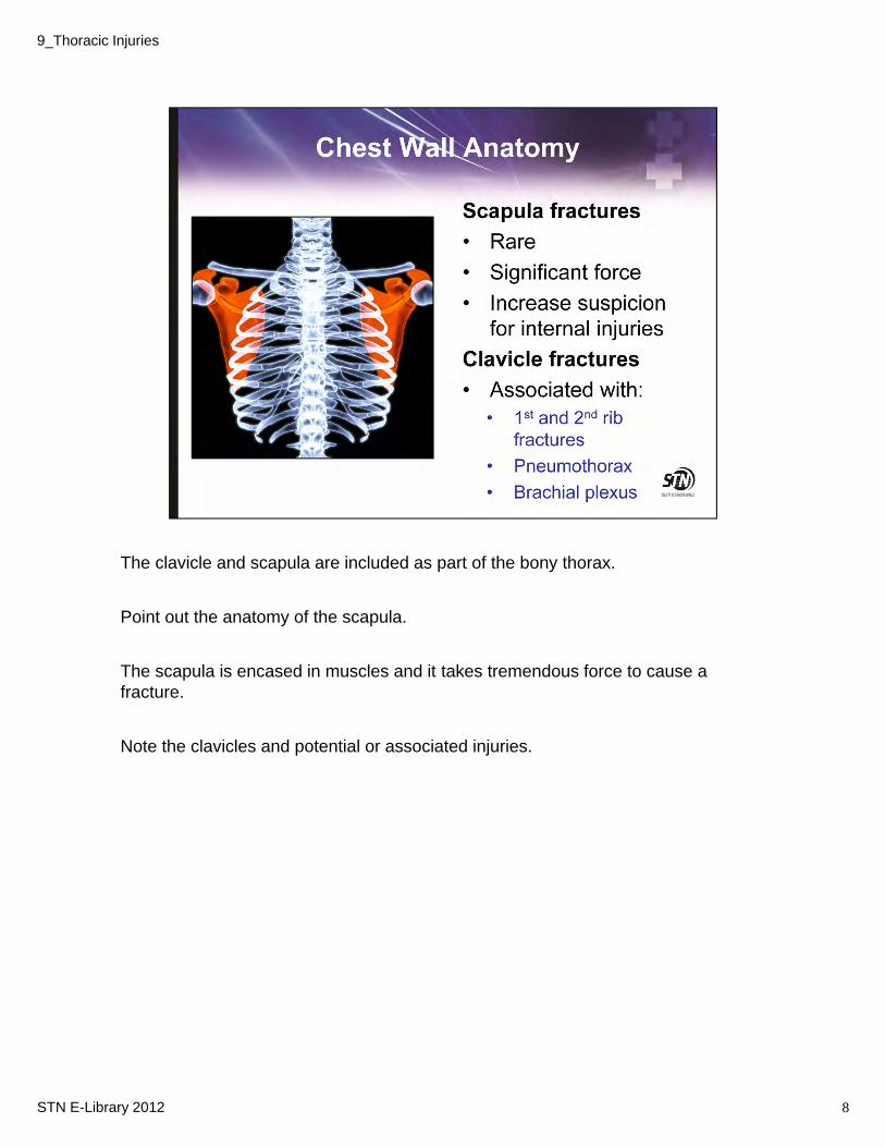
The clavicle and scapula are included as part of the bony thorax.
Point out the anatomy of the scapula.
The scapula is encased in muscles and it takes tremendous force to cause a fracture.
Note the clavicles and potential or associated injuries.
8STN E-Library 2012
9_Thoracic Injuries
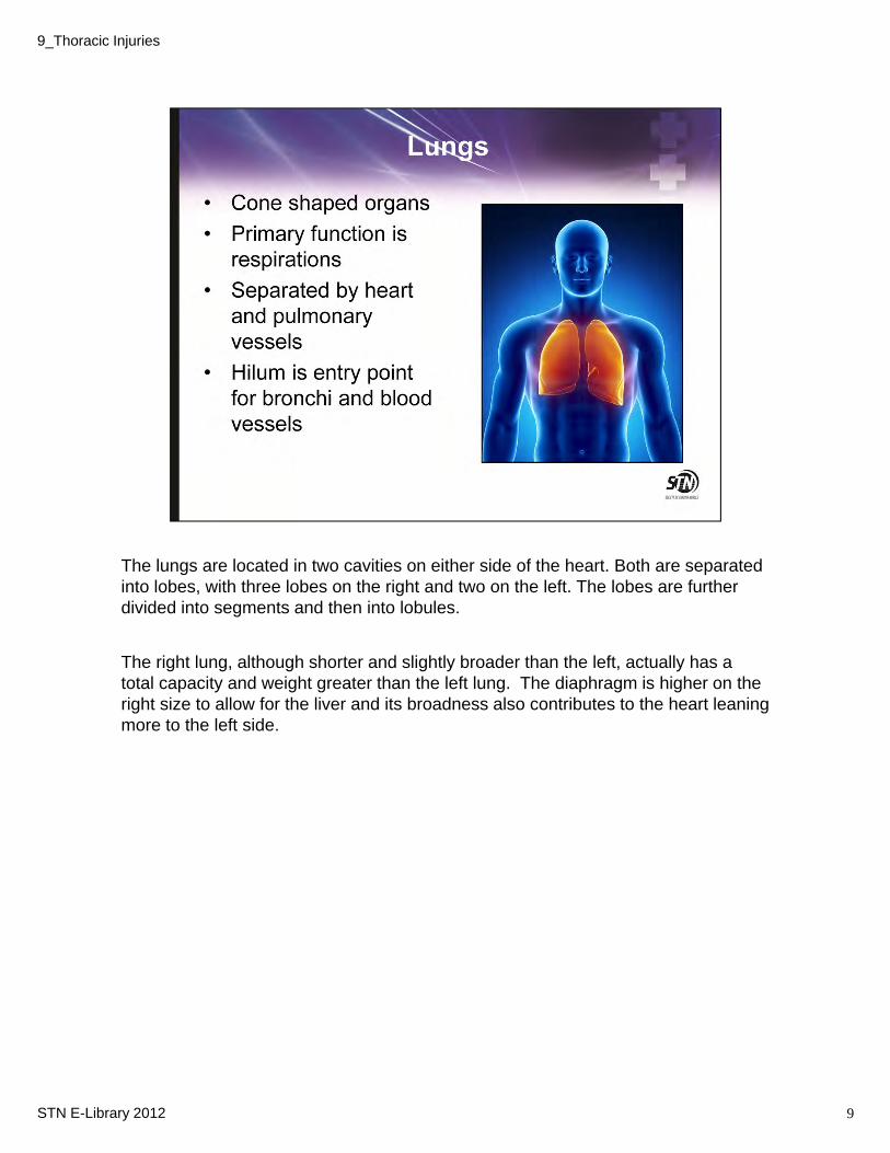
The lungs are located in two cavities on either side of the heart. Both are separated into lobes, with three lobes on the right and two on the left. The lobes are further divided into segments and then into lobules.
The right lung, although shorter and slightly broader than the left, actually has a total capacity and weight greater than the left lung. The diaphragm is higher on the right size to allow for the liver and its broadness also contributes to the heart leaning more to the left side.
9STN E-Library 2012
9_Thoracic Injuries
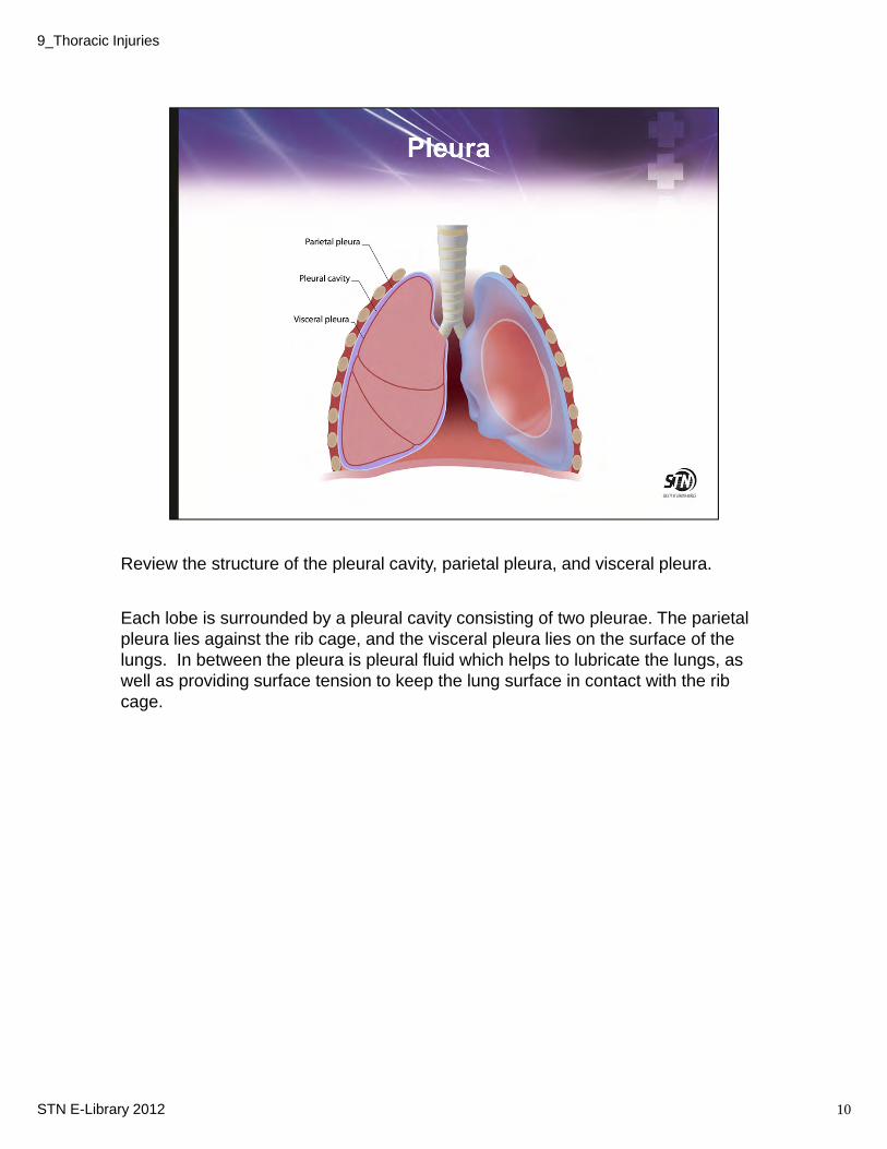
Review the structure of the pleural cavity, parietal pleura, and visceral pleura.
Each lobe is surrounded by a pleural cavity consisting of two pleurae. The parietal pleura lies against the rib cage, and the visceral pleura lies on the surface of the lungs. In between the pleura is pleural fluid which helps to lubricate the lungs, as well as providing surface tension to keep the lung surface in contact with the rib cage.
10STN E-Library 2012
9_Thoracic Injuries
11
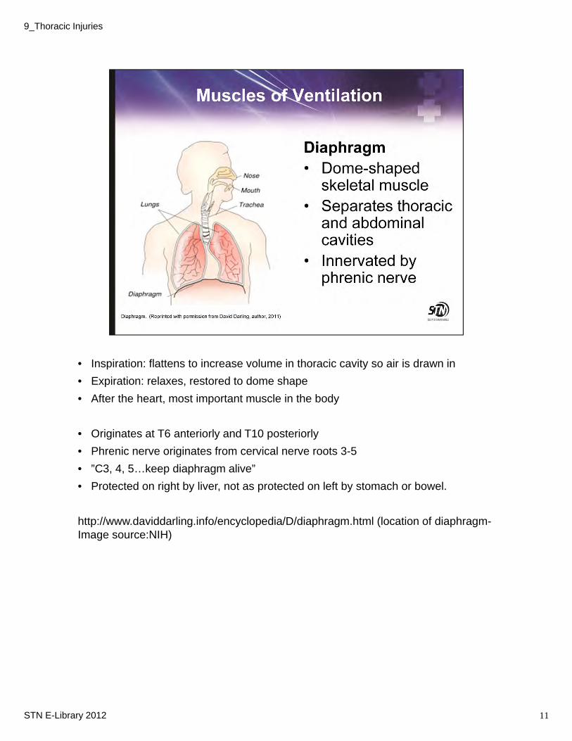
• Inspiration: flattens to increase volume in thoracic cavity so air is drawn in
• Expiration: relaxes, restored to dome shape
• After the heart, most important muscle in the body
• Originates at T6 anteriorly and T10 posteriorly
• Phrenic nerve originates from cervical nerve roots 3-5
• ”C3, 4, 5…keep diaphragm alive”
• Protected on right by liver, not as protected on left by stomach or bowel.
http://www.daviddarling.info/encyclopedia/D/diaphragm.html (location of diaphragm-Image source:NIH)
STN E-Library 2012
9_Thoracic Injuries

• Pressure within lungs must be less than atmospheric pressure for inspiration to occur.
• Diaphragm and intercostal muscles work to increase size of thorax, intrathoracic pressure decreases below atmospheric pressure and air moves into lung
• Muscles relax during expiration and a rise in intrathoracic pressure causes air to move out of the lungs
Intercostal Muscles:
External: extend from ribs and vertebrae to origins of costal cartilage
Internal: extend from sternum to angles of ribs
Thoracic spinal nerves branch out to form intercostal nerves
Accessory Muscles:
Neck: Sternocleidomastoids (raise sternum)
Scalenes (elevate 1st and 2nd ribs)
Abdominal: Augment rate and depth of breathing; generally implies an increase in work of breathing.
12STN E-Library 2012
9_Thoracic Injuries
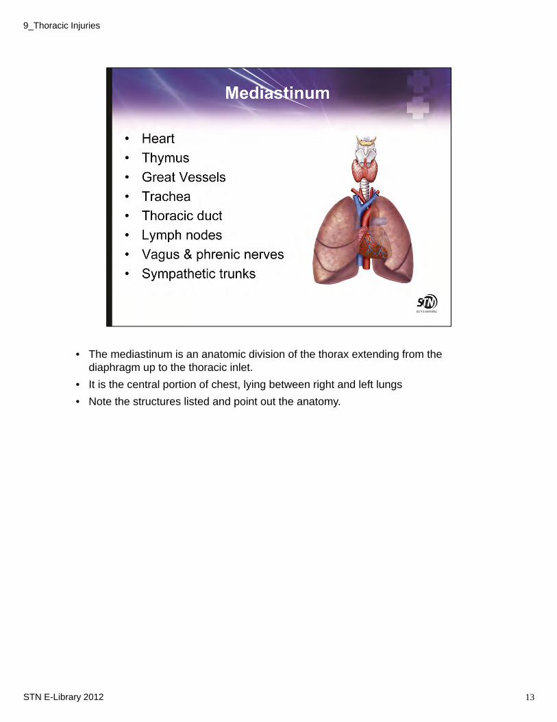
• The mediastinum is an anatomic division of the thorax extending from the diaphragm up to the thoracic inlet.
• It is the central portion of chest, lying between right and left lungs
• Note the structures listed and point out the anatomy.
13STN E-Library 2012
9_Thoracic Injuries
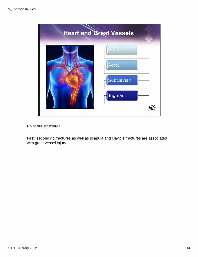
Point out structures.
First, second rib fractures as well as scapula and clavicle fractures are associated with great vessel injury.
14STN E-Library 2012
9_Thoracic Injuries
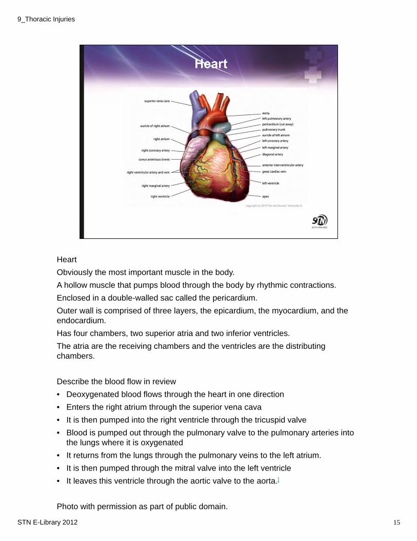
Heart
Obviously the most important muscle in the body.
A hollow muscle that pumps blood through the body by rhythmic contractions.
Enclosed in a double-walled sac called the pericardium.
Outer wall is comprised of three layers, the epicardium, the myocardium, and the endocardium.
Has four chambers, two superior atria and two inferior ventricles.
The atria are the receiving chambers and the ventricles are the distributing chambers.
Describe the blood flow in review
• Deoxygenated blood flows through the heart in one direction
• Enters the right atrium through the superior vena cava
• It is then pumped into the right ventricle through the tricuspid valve
• Blood is pumped out through the pulmonary valve to the pulmonary arteries into the lungs where it is oxygenated
• It returns from the lungs through the pulmonary veins to the left atrium.
• It is then pumped through the mitral valve into the left ventricle
• It leaves this ventricle through the aortic valve to the aorta.]
Photo with permission as part of public domain.
15STN E-Library 2012
9_Thoracic Injuries
16
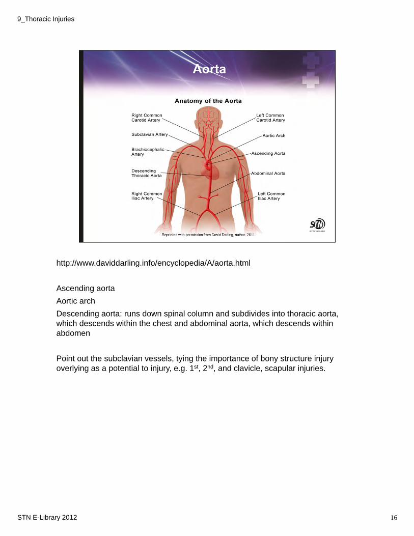
http://www.daviddarling.info/encyclopedia/A/aorta.html
Ascending aorta
Aortic arch
Descending aorta: runs down spinal column and subdivides into thoracic aorta, which descends within the chest and abdominal aorta, which descends within abdomen
Point out the subclavian vessels, tying the importance of bony structure injury overlying as a potential to injury, e.g. 1st, 2nd, and clavicle, scapular injuries.
STN E-Library 2012
9_Thoracic Injuries
17
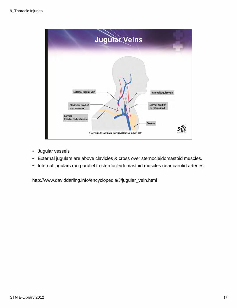
• Jugular vessels
• External jugulars are above clavicles & cross over sternocleidomastoid muscles.
• Internal jugulars run parallel to sternocleidomastoid muscles near carotid arteries
http://www.daviddarling.info/encyclopedia/J/jugular_vein.html
STN E-Library 2012
9_Thoracic Injuries
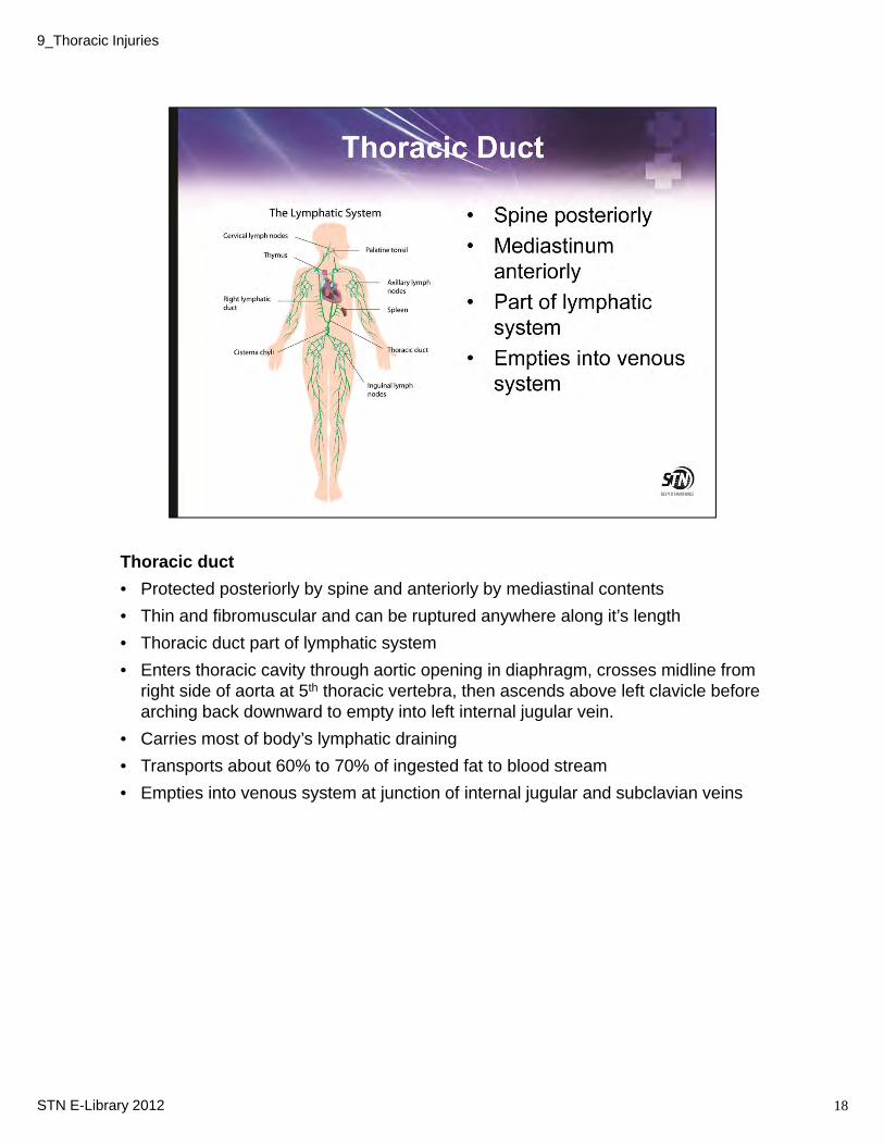
Thoracic duct
• Protected posteriorly by spine and anteriorly by mediastinal contents
• Thin and fibromuscular and can be ruptured anywhere along it’s length
• Thoracic duct part of lymphatic system
• Enters thoracic cavity through aortic opening in diaphragm, crosses midline from right side of aorta at 5th thoracic vertebra, then ascends above left clavicle before arching back downward to empty into left internal jugular vein.
• Carries most of body’s lymphatic draining
• Transports about 60% to 70% of ingested fat to blood stream
• Empties into venous system at junction of internal jugular and subclavian veins
18STN E-Library 2012
9_Thoracic Injuries
9_Thoracic Injuries
STN E‐Library 2012 19
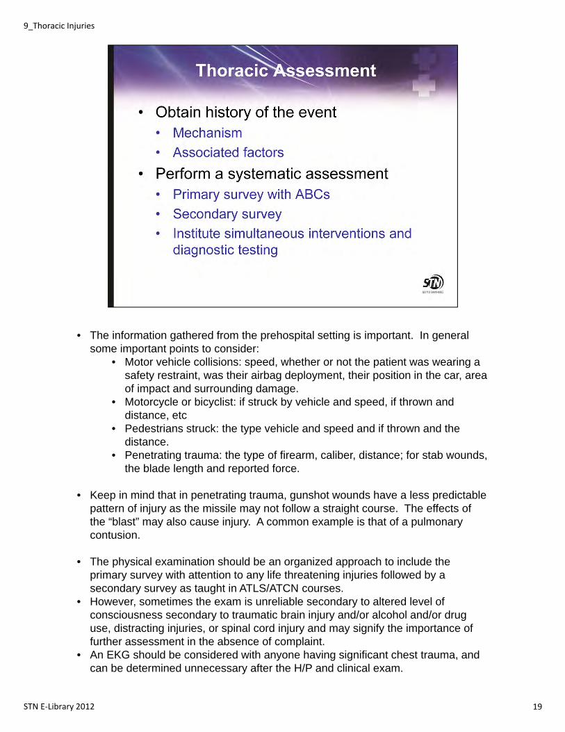
• The information gathered from the prehospital setting is important. In general some important points to consider:
• Motor vehicle collisions: speed, whether or not the patient was wearing a safety restraint, was their airbag deployment, their position in the car, area of impact and surrounding damage.
• Motorcycle or bicyclist: if struck by vehicle and speed, if thrown and distance, etc
• Pedestrians struck: the type vehicle and speed and if thrown and the distance.
• Penetrating trauma: the type of firearm, caliber, distance; for stab wounds, the blade length and reported force.
• Keep in mind that in penetrating trauma, gunshot wounds have a less predictable pattern of injury as the missile may not follow a straight course. The effects of the “blast” may also cause injury. A common example is that of a pulmonary contusion.
• The physical examination should be an organized approach to include the primary survey with attention to any life threatening injuries followed by a secondary survey as taught in ATLS/ATCN courses.
• However, sometimes the exam is unreliable secondary to altered level of consciousness secondary to traumatic brain injury and/or alcohol and/or drug use, distracting injuries, or spinal cord injury and may signify the importance of further assessment in the absence of complaint.
• An EKG should be considered with anyone having significant chest trauma, and can be determined unnecessary after the H/P and clinical exam.

Findings will vary according to the injured structures or their affect on the body’s physiology. There are some common findings in patient with chest trauma.
It is still important that one rely on physical exam.
20STN E-Library 2012
9_Thoracic Injuries
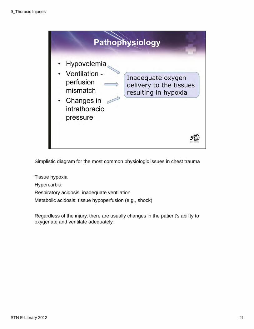
Simplistic diagram for the most common physiologic issues in chest trauma
Tissue hypoxia
Hypercarbia
Respiratory acidosis: inadequate ventilation
Metabolic acidosis: tissue hypoperfusion (e.g., shock)
Regardless of the injury, there are usually changes in the patient’s ability to oxygenate and ventilate adequately.
21STN E-Library 2012
9_Thoracic Injuries
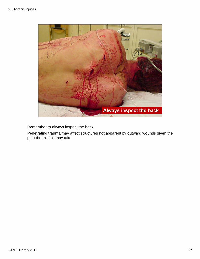
Remember to always inspect the back.
Penetrating trauma may affect structures not apparent by outward wounds given the path the missile may take.
22STN E-Library 2012
9_Thoracic Injuries
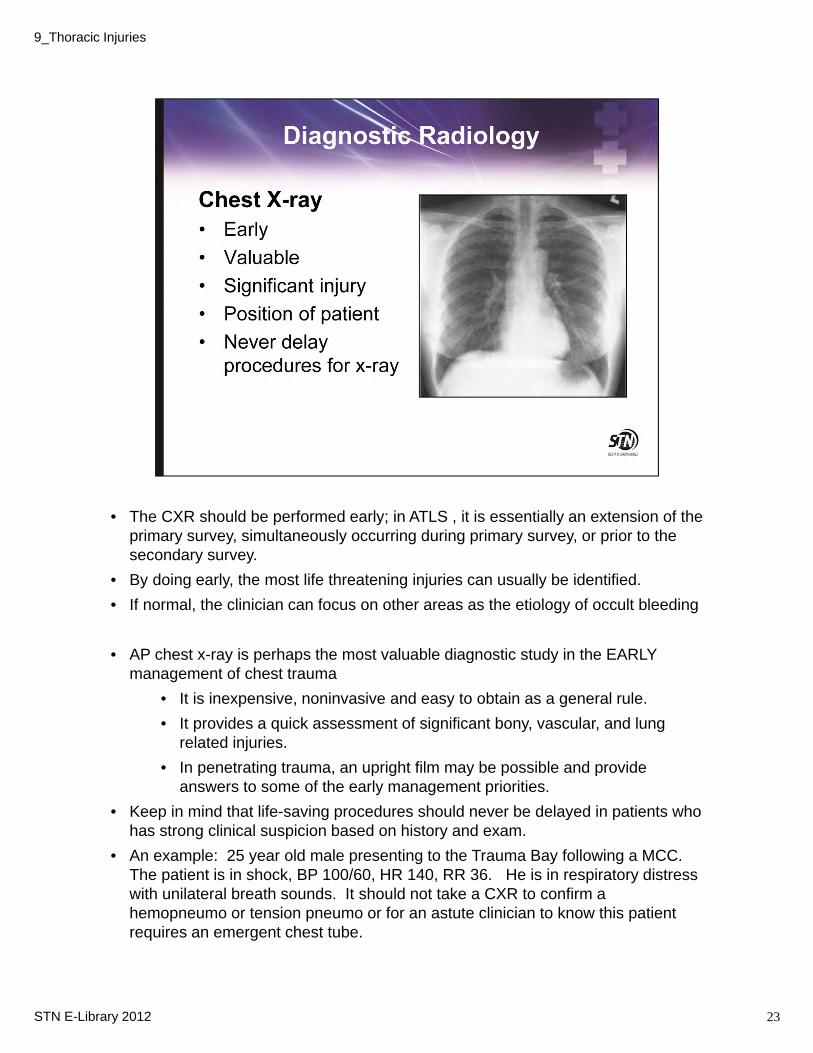
• The CXR should be performed early; in ATLS , it is essentially an extension of the primary survey, simultaneously occurring during primary survey, or prior to the secondary survey.
• By doing early, the most life threatening injuries can usually be identified.
• If normal, the clinician can focus on other areas as the etiology of occult bleeding
• AP chest x-ray is perhaps the most valuable diagnostic study in the EARLY management of chest trauma
• It is inexpensive, noninvasive and easy to obtain as a general rule.
• It provides a quick assessment of significant bony, vascular, and lung related injuries.
• In penetrating trauma, an upright film may be possible and provide answers to some of the early management priorities.
• Keep in mind that life-saving procedures should never be delayed in patients who has strong clinical suspicion based on history and exam.
• An example: 25 year old male presenting to the Trauma Bay following a MCC. The patient is in shock, BP 100/60, HR 140, RR 36. He is in respiratory distress with unilateral breath sounds. It should not take a CXR to confirm a hemopneumo or tension pneumo or for an astute clinician to know this patient requires an emergent chest tube.
23STN E-Library 2012
9_Thoracic Injuries
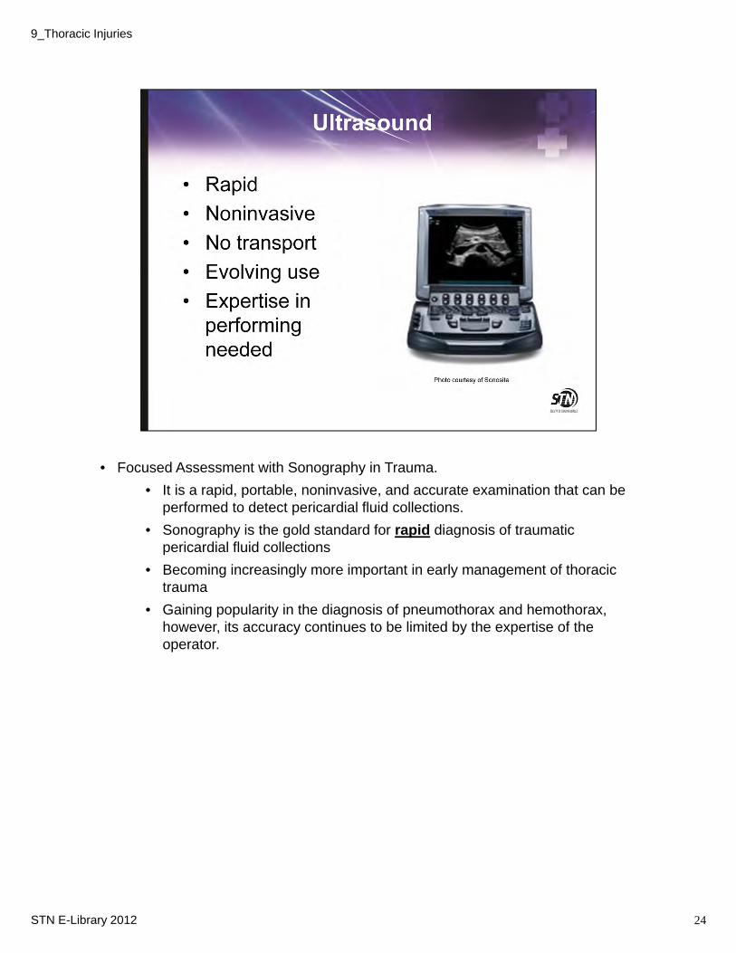
• Focused Assessment with Sonography in Trauma.
• It is a rapid, portable, noninvasive, and accurate examination that can be performed to detect pericardial fluid collections.
• Sonography is the gold standard for rapid diagnosis of traumatic pericardial fluid collections
• Becoming increasingly more important in early management of thoracic trauma
• Gaining popularity in the diagnosis of pneumothorax and hemothorax, however, its accuracy continues to be limited by the expertise of the operator.
24STN E-Library 2012
9_Thoracic Injuries
9_Thoracic Injuries
STN E‐Library 2012 25
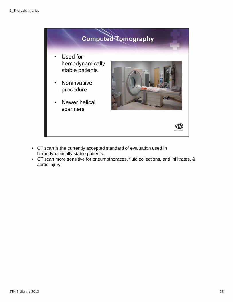
• CT scan is the currently accepted standard of evaluation used in hemodynamically stable patients.
• CT scan more sensitive for pneumothoraces, fluid collections, and infiltrates, & aortic injury
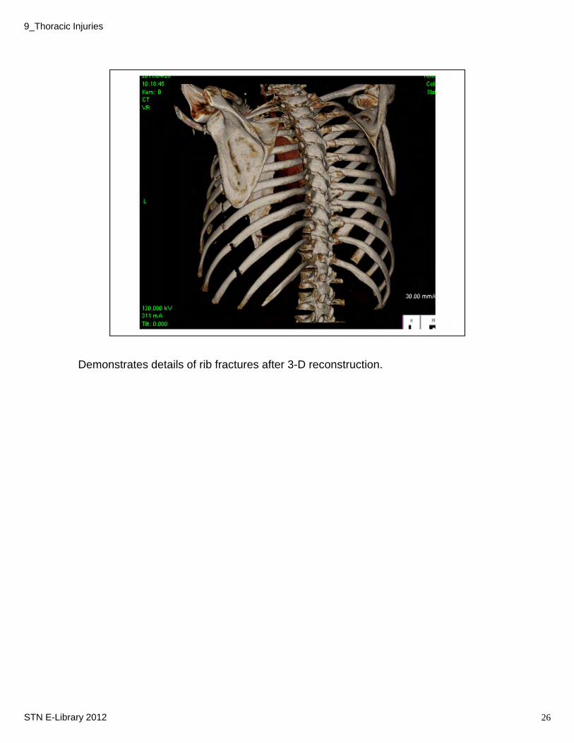
Demonstrates details of rib fractures after 3-D reconstruction.
26STN E-Library 2012
9_Thoracic Injuries
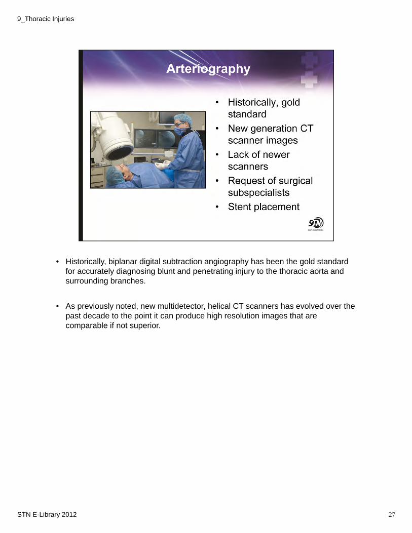
• Historically, biplanar digital subtraction angiography has been the gold standard for accurately diagnosing blunt and penetrating injury to the thoracic aorta and surrounding branches.
• As previously noted, new multidetector, helical CT scanners has evolved over the past decade to the point it can produce high resolution images that are comparable if not superior.
27STN E-Library 2012
9_Thoracic Injuries
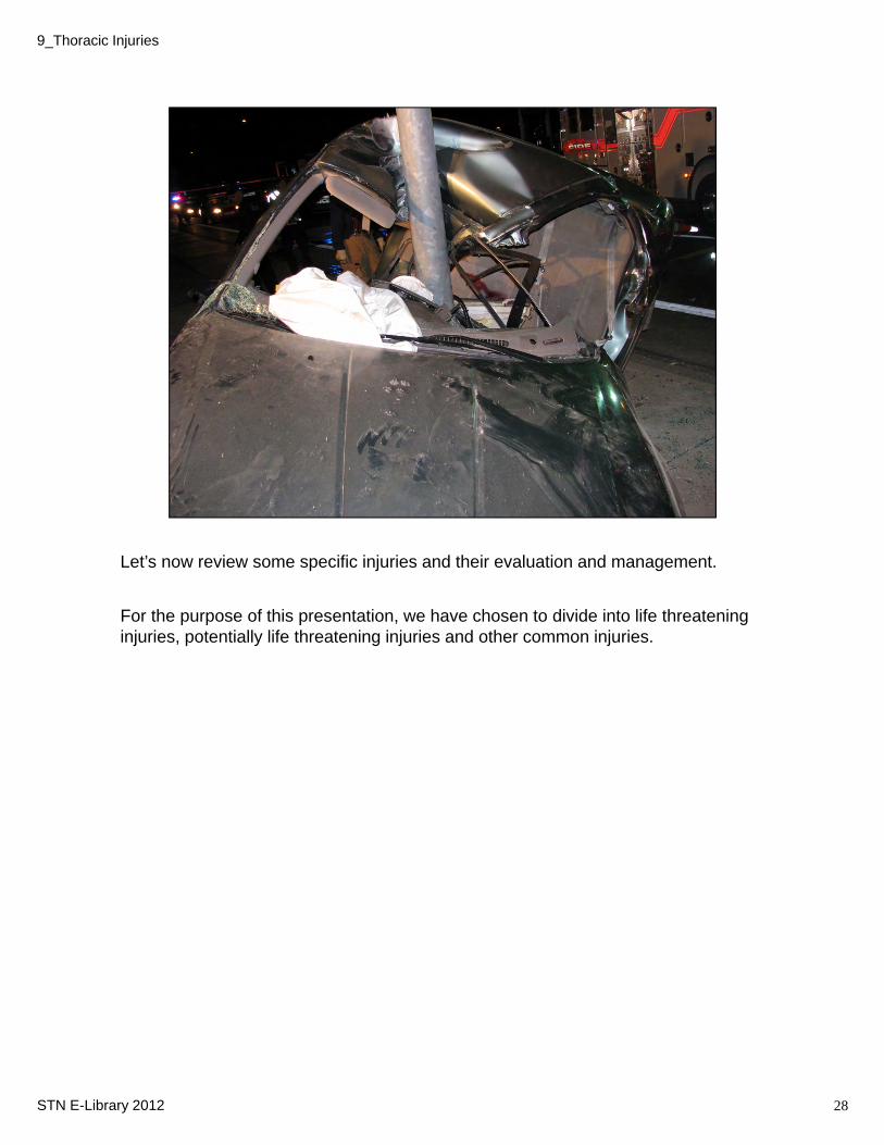
Let’s now review some specific injuries and their evaluation and management.
For the purpose of this presentation, we have chosen to divide into life threatening injuries, potentially life threatening injuries and other common injuries.
28STN E-Library 2012
9_Thoracic Injuries
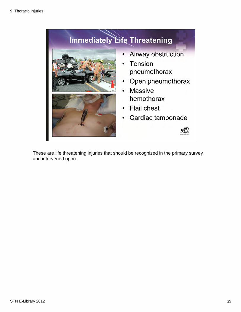
These are life threatening injuries that should be recognized in the primary survey and intervened upon.
29STN E-Library 2012
9_Thoracic Injuries
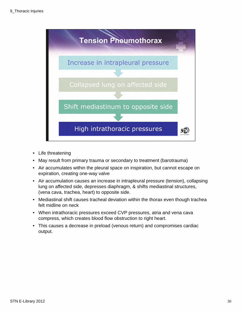
• Life threatening
• May result from primary trauma or secondary to treatment (barotrauma)
• Air accumulates within the pleural space on inspiration, but cannot escape on expiration, creating one-way valve
• Air accumulation causes an increase in intrapleural pressure (tension), collapsing lung on affected side, depresses diaphragm, & shifts mediastinal structures, (vena cava, trachea, heart) to opposite side.
• Mediastinal shift causes tracheal deviation within the thorax even though trachea felt midline on neck
• When intrathoracic pressures exceed CVP pressures, atria and vena cava compress, which creates blood flow obstruction to right heart.
• This causes a decrease in preload (venous return) and compromises cardiac output.
30STN E-Library 2012
9_Thoracic Injuries
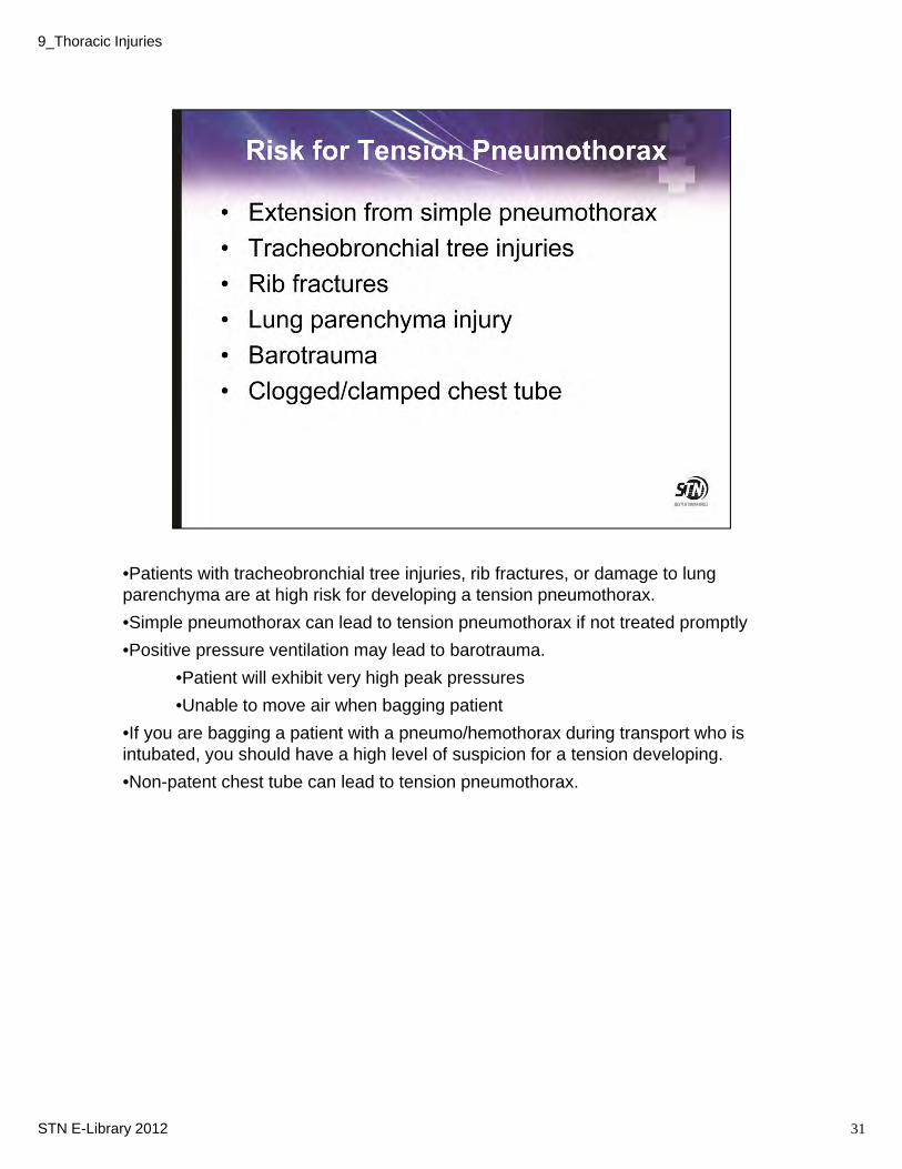
•Patients with tracheobronchial tree injuries, rib fractures, or damage to lung parenchyma are at high risk for developing a tension pneumothorax.
•Simple pneumothorax can lead to tension pneumothorax if not treated promptly
•Positive pressure ventilation may lead to barotrauma.
•Patient will exhibit very high peak pressures
•Unable to move air when bagging patient
•If you are bagging a patient with a pneumo/hemothorax during transport who is intubated, you should have a high level of suspicion for a tension developing.
•Non-patent chest tube can lead to tension pneumothorax.
31STN E-Library 2012
9_Thoracic Injuries
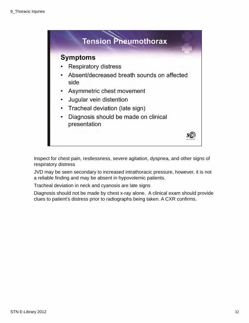
Inspect for chest pain, restlessness, severe agitation, dyspnea, and other signs of respiratory distress
JVD may be seen secondary to increased intrathoracic pressure, however, it is not a reliable finding and may be absent in hypovolemic patients.
Tracheal deviation in neck and cyanosis are late signs
Diagnosis should not be made by chest x-ray alone. A clinical exam should provide clues to patient’s distress prior to radiographs being taken. A CXR confirms.
32STN E-Library 2012
9_Thoracic Injuries
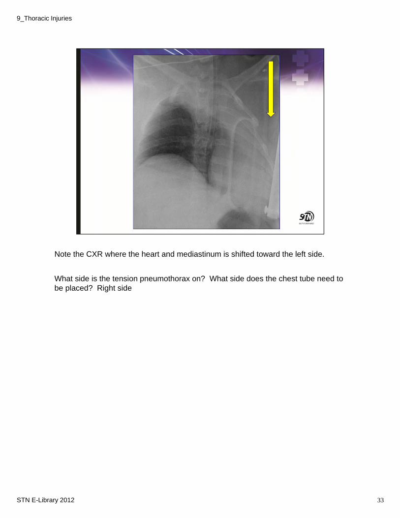
Note the CXR where the heart and mediastinum is shifted toward the left side.
What side is the tension pneumothorax on? What side does the chest tube need to be placed? Right side
33STN E-Library 2012
9_Thoracic Injuries
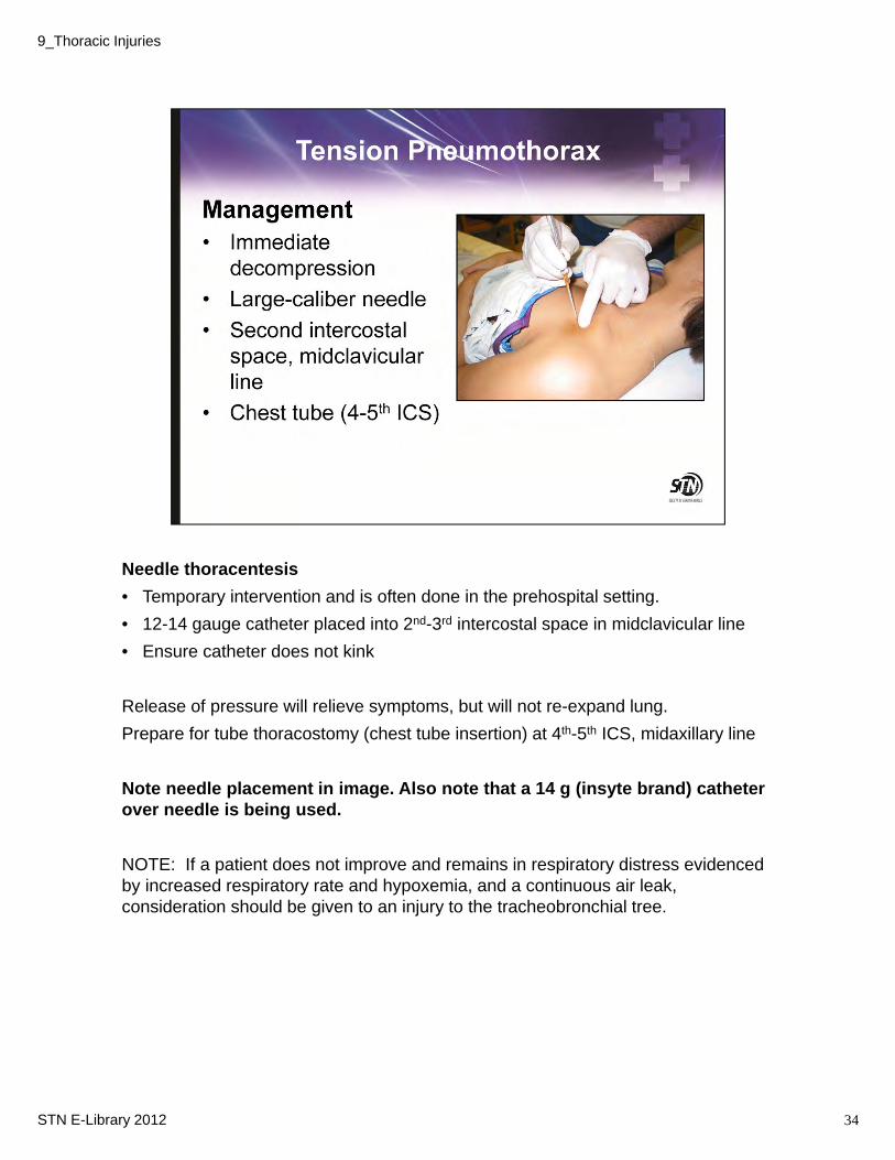
Needle thoracentesis
• Temporary intervention and is often done in the prehospital setting.
• 12-14 gauge catheter placed into 2nd-3rd intercostal space in midclavicular line
• Ensure catheter does not kink
Release of pressure will relieve symptoms, but will not re-expand lung.
Prepare for tube thoracostomy (chest tube insertion) at 4th-5th ICS, midaxillary line
Note needle placement in image. Also note that a 14 g (insyte brand) catheter over needle is being used.
NOTE: If a patient does not improve and remains in respiratory distress evidenced by increased respiratory rate and hypoxemia, and a continuous air leak, consideration should be given to an injury to the tracheobronchial tree.
34STN E-Library 2012
9_Thoracic Injuries
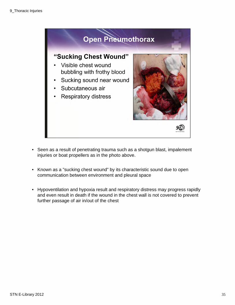
• Seen as a result of penetrating trauma such as a shotgun blast, impalement injuries or boat propellers as in the photo above.
• Known as a ”sucking chest wound” by its characteristic sound due to open communication between environment and pleural space
• Hypoventilation and hypoxia result and respiratory distress may progress rapidly and even result in death if the wound in the chest wall is not covered to prevent further passage of air in/out of the chest
35STN E-Library 2012
9_Thoracic Injuries
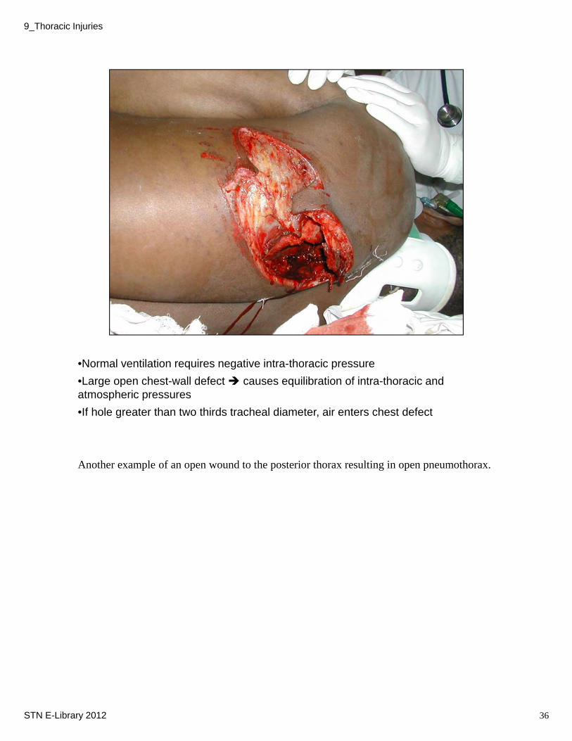
•Normal ventilation requires negative intra-thoracic pressure
•Large open chest-wall defect causes equilibration of intra-thoracic and atmospheric pressures
•If hole greater than two thirds tracheal diameter, air enters chest defect
Another example of an open wound to the posterior thorax resulting in open pneumothorax.
36STN E-Library 2012
9_Thoracic Injuries
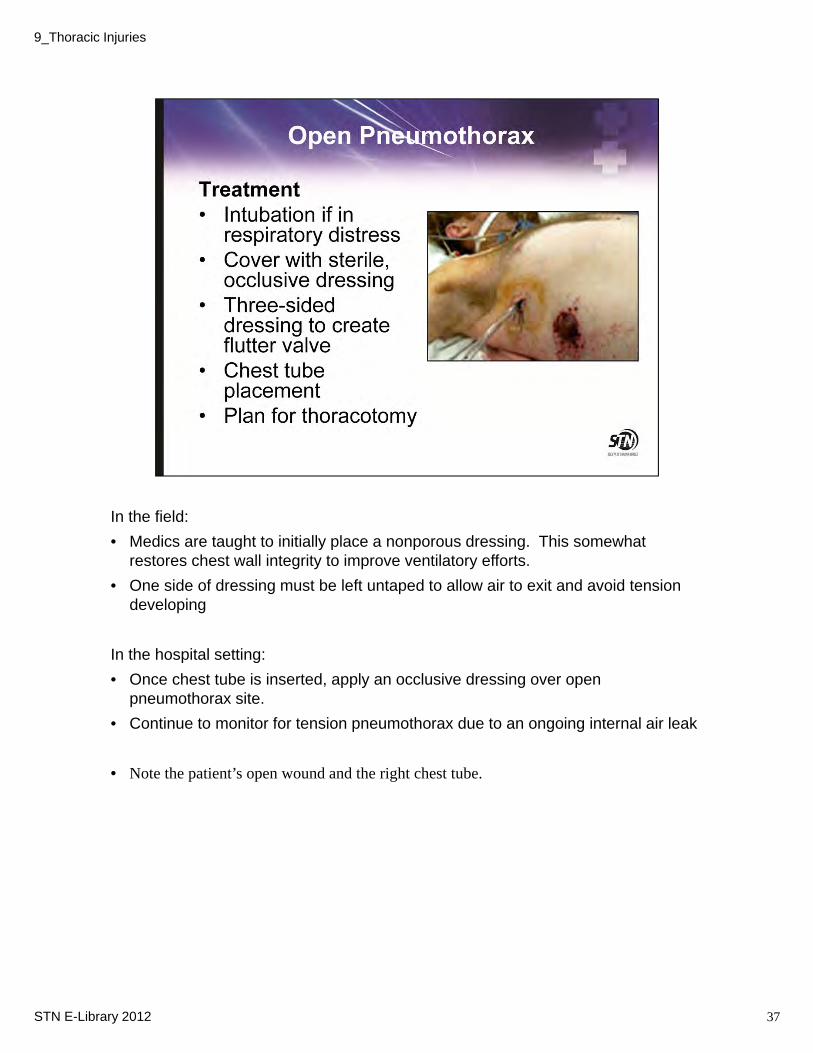
In the field:
• Medics are taught to initially place a nonporous dressing. This somewhat restores chest wall integrity to improve ventilatory efforts.
• One side of dressing must be left untaped to allow air to exit and avoid tension developing
In the hospital setting:
• Once chest tube is inserted, apply an occlusive dressing over open pneumothorax site.
• Continue to monitor for tension pneumothorax due to an ongoing internal air leak
• Note the patient’s open wound and the right chest tube.
37STN E-Library 2012
9_Thoracic Injuries
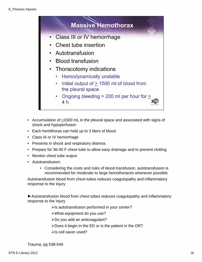
• Accumulation of >1500 mL in the pleural space and associated with signs of shock and hypoperfusion
• Each hemithorax can hold up to 3 liters of blood
• Class III or IV hemorrhage
• Presents in shock and respiratory distress
• Prepare for 36-40 F chest tube to allow easy drainage and to prevent clotting
• Monitor chest tube output
• Autotransfusion:
• Considering the costs and risks of blood transfusion, autotransfusion is recommended for moderate to large hemothoraces whenever possible
Autotransfusion blood from chest tubes reduces coagulopathy and inflammatory response to the injury
►Autotransfusion blood from chest tubes reduces coagulopathy and inflammatory response to the injury
Is autotransfusion performed in your center?
What equipment do you use?
Do you add an anticoagulant?
Does it begin in the ED or is the patient in the OR?
Is cell saver used?
Trauma, pg 538-540
38STN E-Library 2012
9_Thoracic Injuries
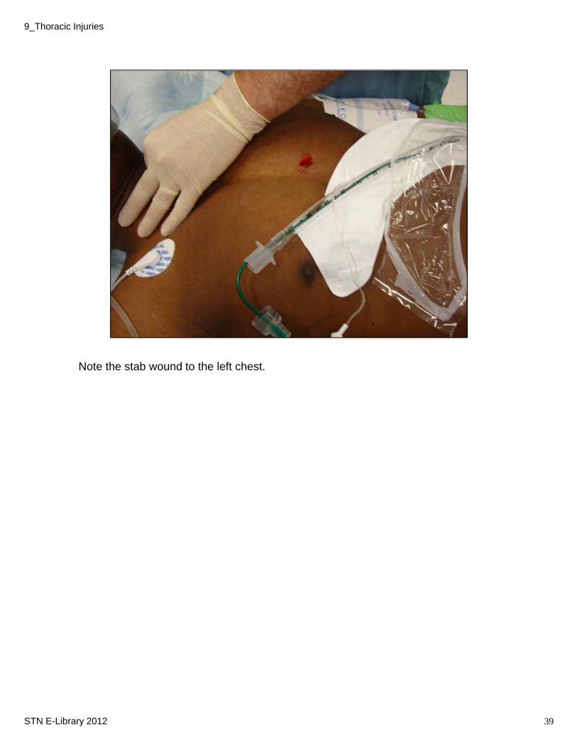
Note the stab wound to the left chest.
39STN E-Library 2012
9_Thoracic Injuries
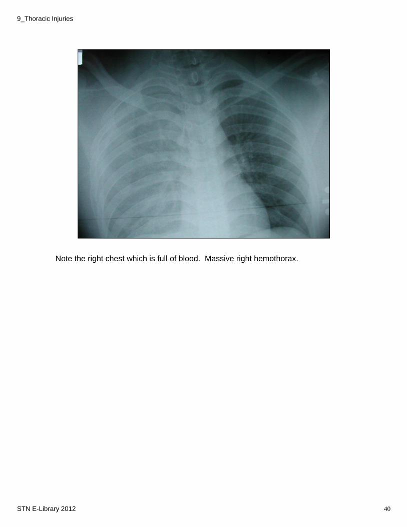
Note the right chest which is full of blood. Massive right hemothorax.
40STN E-Library 2012
9_Thoracic Injuries
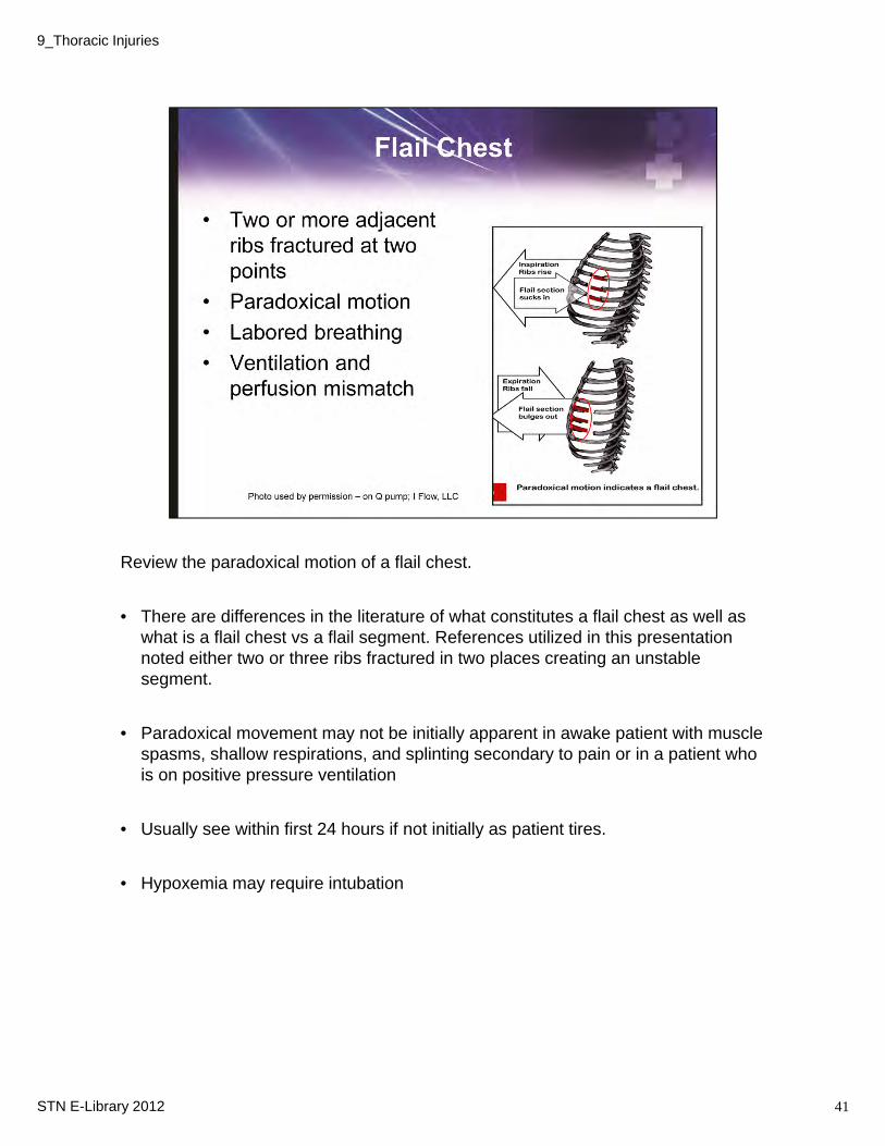
Review the paradoxical motion of a flail chest.
• There are differences in the literature of what constitutes a flail chest as well as what is a flail chest vs a flail segment. References utilized in this presentation noted either two or three ribs fractured in two places creating an unstable segment.
• Paradoxical movement may not be initially apparent in awake patient with muscle spasms, shallow respirations, and splinting secondary to pain or in a patient who is on positive pressure ventilation
• Usually see within first 24 hours if not initially as patient tires.
• Hypoxemia may require intubation
41STN E-Library 2012
9_Thoracic Injuries
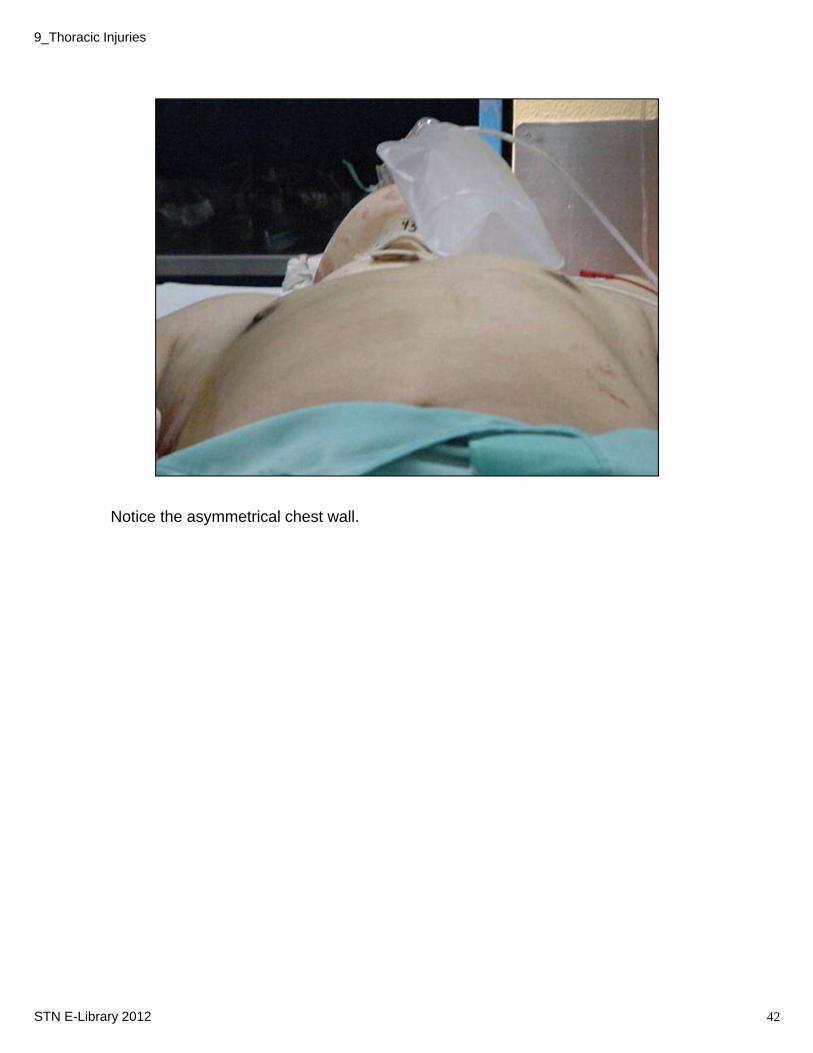
Notice the asymmetrical chest wall.
42STN E-Library 2012
9_Thoracic Injuries
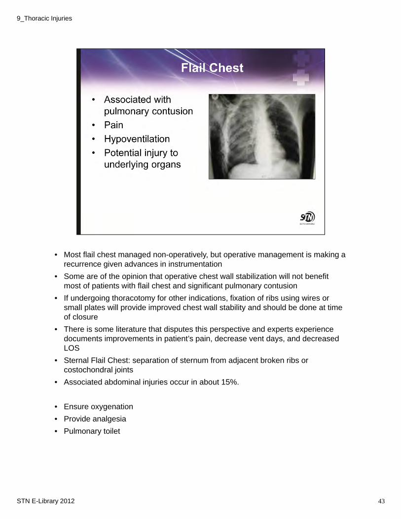
• Most flail chest managed non-operatively, but operative management is making a recurrence given advances in instrumentation
• Some are of the opinion that operative chest wall stabilization will not benefit most of patients with flail chest and significant pulmonary contusion
• If undergoing thoracotomy for other indications, fixation of ribs using wires or small plates will provide improved chest wall stability and should be done at time of closure
• There is some literature that disputes this perspective and experts experience documents improvements in patient’s pain, decrease vent days, and decreased LOS
• Sternal Flail Chest: separation of sternum from adjacent broken ribs or costochondral joints
• Associated abdominal injuries occur in about 15%.
• Ensure oxygenation
• Provide analgesia
• Pulmonary toilet
43STN E-Library 2012
9_Thoracic Injuries
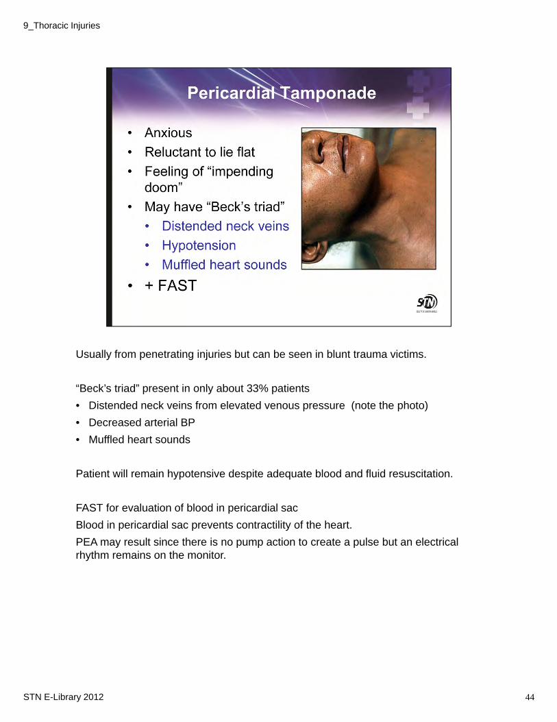
Usually from penetrating injuries but can be seen in blunt trauma victims.
“Beck’s triad” present in only about 33% patients
• Distended neck veins from elevated venous pressure (note the photo)
• Decreased arterial BP
• Muffled heart sounds
Patient will remain hypotensive despite adequate blood and fluid resuscitation.
FAST for evaluation of blood in pericardial sac
Blood in pericardial sac prevents contractility of the heart.
PEA may result since there is no pump action to create a pulse but an electrical rhythm remains on the monitor.
44STN E-Library 2012
9_Thoracic Injuries

• Pericardial sac normally contains 20-30 ml of serous fluid and will not distend so a small amount (75-100 ml) may actually compress the heart and restrict its ability to contract.
• Normal compensatory mechanism is increase in heart rate which initially sustains BP, but decreases peripheral perfusion
• Fall of systolic blood pressure is result of excessive right ventricle filling which then causes interventricular septum to shift to the left
• This septal shift impairs left ventricular filling causing fall in cardiac output• Increasing pericardial pressure decreases coronary blood flow resulting in myocardial ischemia
and cardiac decompensation• Inadequate peripheral perfusion intensifies metabolic acidosis, which also weakens myocardial
contractility• Distended superficial neck veins is late sign and is more prominent during inspiration (Kussmaul’s
sign)
• Pericardiocentesis• Currently, is minimally used in Level I trauma center • Can be done as a temporizing measure until pericardiotomy can be performed• Place patient supine position with head and torso elevated 45 degrees• 16- or 18-gauge needle attached to 60-ml syringe• Insert needle left side of xiphoid, pointing toward posterior aspect of left shoulder• Popping senstation usually felt when pericardium entered• Withdrawal of small amount can be life saving• Repeat as frequently as needed to maintain SBP > 90 mm Hg• Can inadvertently cause iatrogenic injury to coronary artery, myocardium, etc.
• Pericardial window is preferred in hemodynamically stable patients. Procedure can be done in the OR under more controlled circumstances and the ability to further intervene with additional procedures is readily available.
45STN E-Library 2012
9_Thoracic Injuries
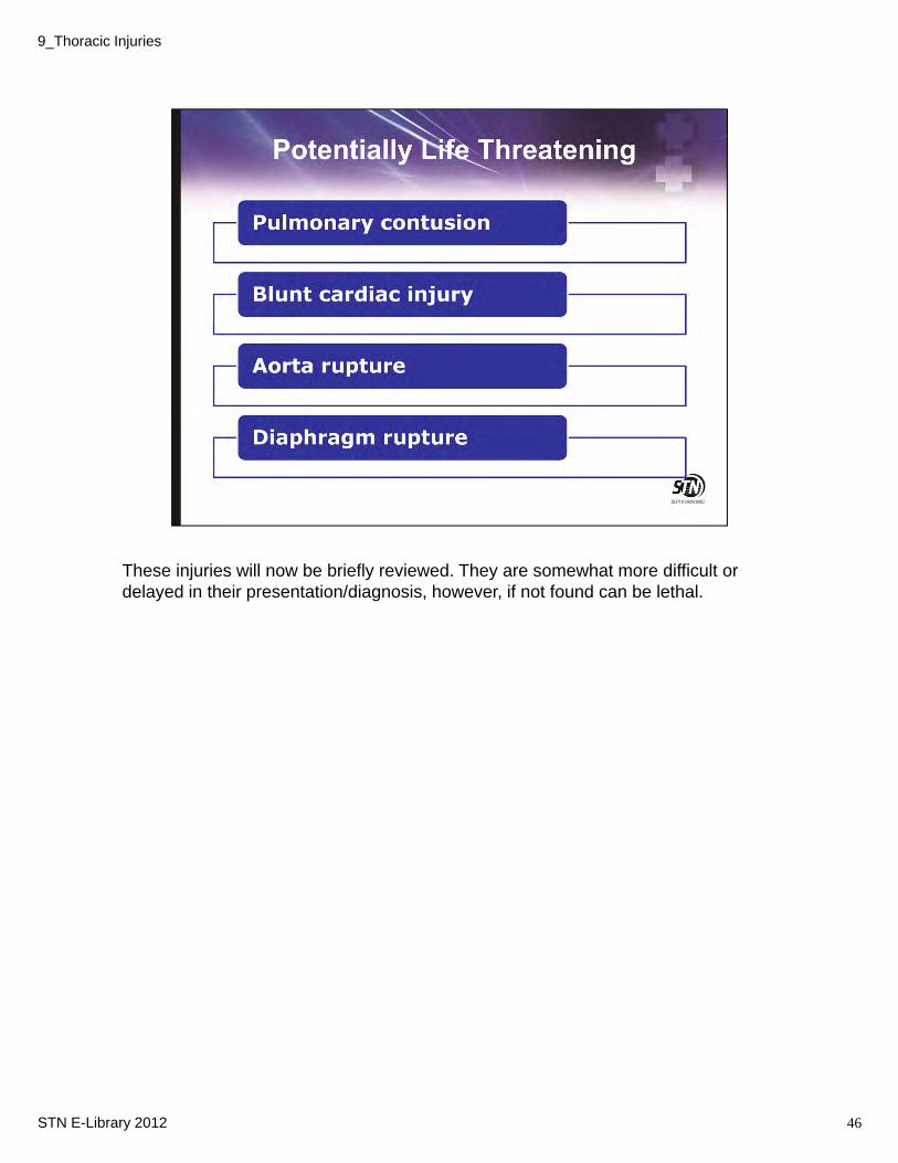
These injuries will now be briefly reviewed. They are somewhat more difficult or delayed in their presentation/diagnosis, however, if not found can be lethal.
46STN E-Library 2012
9_Thoracic Injuries
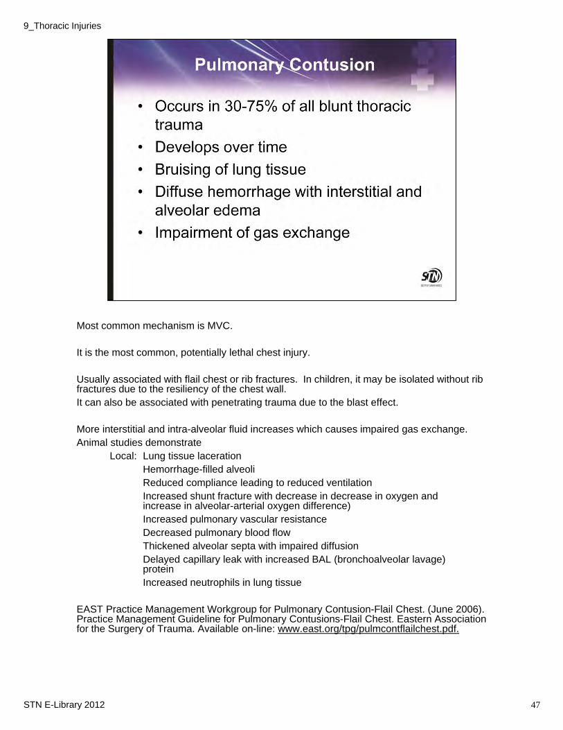
Most common mechanism is MVC.
It is the most common, potentially lethal chest injury.
Usually associated with flail chest or rib fractures. In children, it may be isolated without rib fractures due to the resiliency of the chest wall.It can also be associated with penetrating trauma due to the blast effect.
More interstitial and intra-alveolar fluid increases which causes impaired gas exchange.Animal studies demonstrate
Local: Lung tissue lacerationHemorrhage-filled alveoliReduced compliance leading to reduced ventilationIncreased shunt fracture with decrease in decrease in oxygen and increase in alveolar-arterial oxygen difference)Increased pulmonary vascular resistanceDecreased pulmonary blood flowThickened alveolar septa with impaired diffusionDelayed capillary leak with increased BAL (bronchoalveolar lavage) proteinIncreased neutrophils in lung tissue
EAST Practice Management Workgroup for Pulmonary Contusion-Flail Chest. (June 2006). Practice Management Guideline for Pulmonary Contusions-Flail Chest. Eastern Association for the Surgery of Trauma. Available on-line: www.east.org/tpg/pulmcontflailchest.pdf.
47STN E-Library 2012
9_Thoracic Injuries
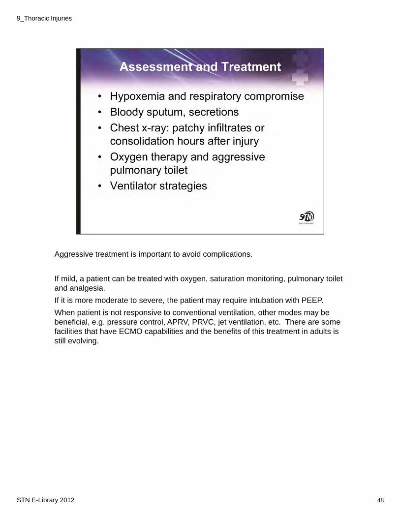
Aggressive treatment is important to avoid complications.
If mild, a patient can be treated with oxygen, saturation monitoring, pulmonary toilet and analgesia.
If it is more moderate to severe, the patient may require intubation with PEEP.
When patient is not responsive to conventional ventilation, other modes may be beneficial, e.g. pressure control, APRV, PRVC, jet ventilation, etc. There are some facilities that have ECMO capabilities and the benefits of this treatment in adults is still evolving.
48STN E-Library 2012
9_Thoracic Injuries
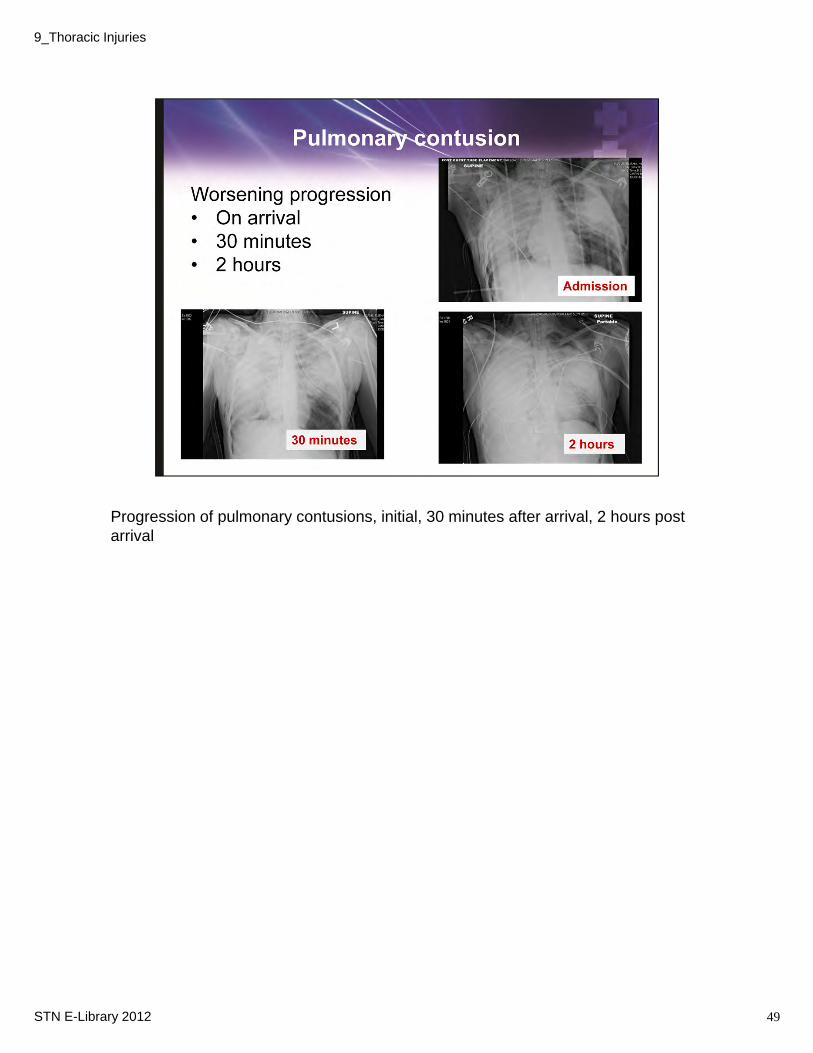
Progression of pulmonary contusions, initial, 30 minutes after arrival, 2 hours post arrival
49STN E-Library 2012
9_Thoracic Injuries
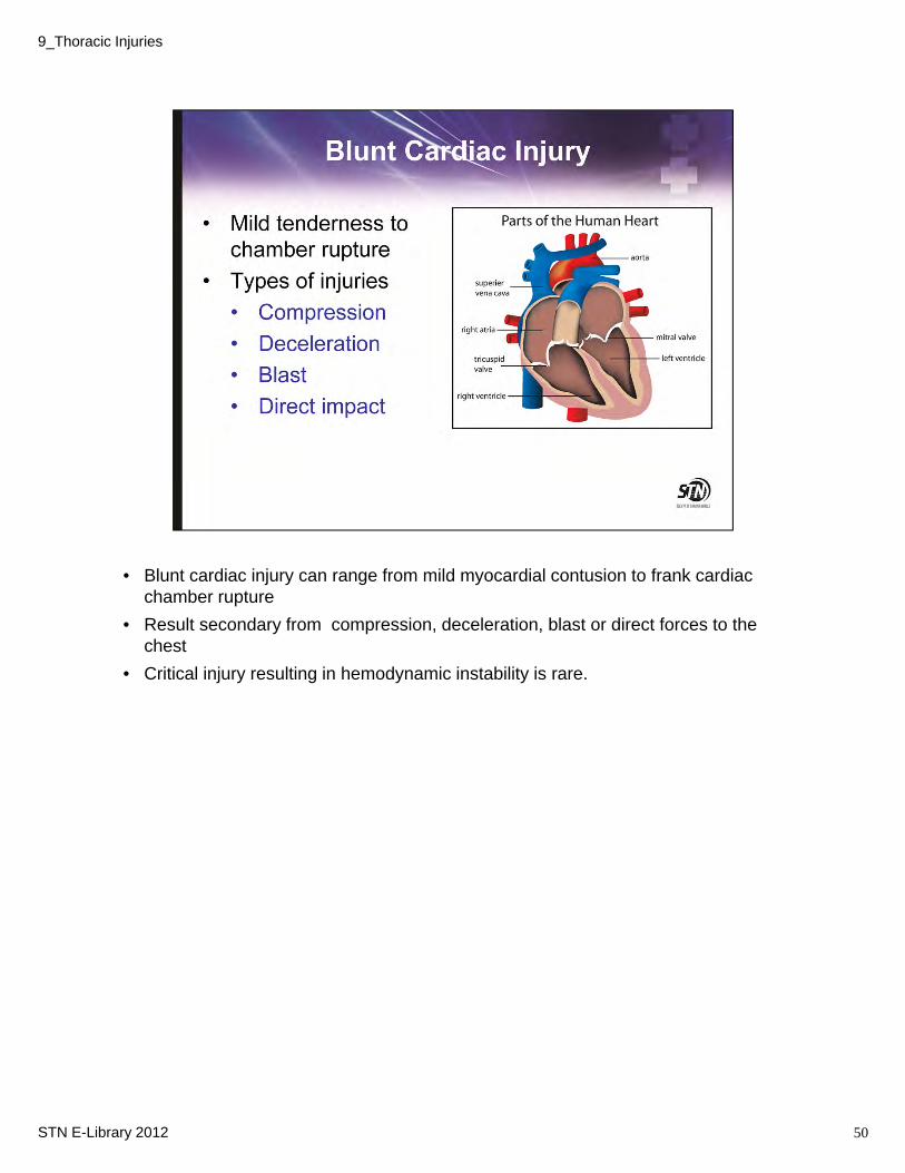
• Blunt cardiac injury can range from mild myocardial contusion to frank cardiac chamber rupture
• Result secondary from compression, deceleration, blast or direct forces to the chest
• Critical injury resulting in hemodynamic instability is rare.
50STN E-Library 2012
9_Thoracic Injuries
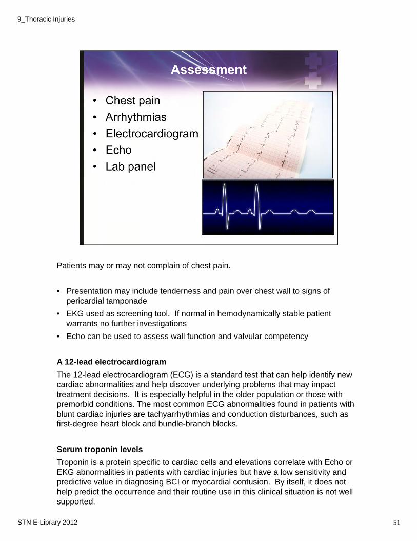
Patients may or may not complain of chest pain.
• Presentation may include tenderness and pain over chest wall to signs of pericardial tamponade
• EKG used as screening tool. If normal in hemodynamically stable patient warrants no further investigations
• Echo can be used to assess wall function and valvular competency
A 12-lead electrocardiogram
The 12-lead electrocardiogram (ECG) is a standard test that can help identify new cardiac abnormalities and help discover underlying problems that may impact treatment decisions. It is especially helpful in the older population or those with premorbid conditions. The most common ECG abnormalities found in patients with blunt cardiac injuries are tachyarrhythmias and conduction disturbances, such as first-degree heart block and bundle-branch blocks.
Serum troponin levels
Troponin is a protein specific to cardiac cells and elevations correlate with Echo or EKG abnormalities in patients with cardiac injuries but have a low sensitivity and predictive value in diagnosing BCI or myocardial contusion. By itself, it does not help predict the occurrence and their routine use in this clinical situation is not well supported.
51STN E-Library 2012
9_Thoracic Injuries

Serum myocardial muscle creatine kinase isoenzyme levels
Measurement of creatine kinase-MB levels is frequently performed in patients with possible blunt myocardial injuries. This diagnostic modality has poor sensitivity, specificity, and positive predictive value in relation to clinically significant blunt myocardial injuries.
9_Thoracic Injuries
STN E-Library 2012 51
• Common cause of sudden death following MVC or fall from height…deceleration injuries.
• More survivors now than historically in some areas of the country due to improved trauma systems, EMS transport, access to care, etc
• Survival depends on incomplete laceration near ligamentum arteriosum or hematoma at site
• Must have high index of suspicion
• May not have specific symptoms
• Delay in recognition may result in early in-hospital rupture
ATLS pg 95
Associated injuries in patients with blunt aortic injuries are found in 81%.
Injuries in drivers may differ from those in passengers.
52STN E-Library 2012
9_Thoracic Injuries
Some patients do not complain of pain while others may have general complaints, nonspecific to aortic injury, such as difficulty breathing, while others should raise suspicion such as intrascapular pain.
Clinical signs may include new onset murmur, upper extremity hypertension or bilateral femoral pulse deficit
CT angio in an appropriate test over formal arteriography in the diagnosis. In fact, with the new helical scanner and improved imaging, it has been shown to be highly sensitive and specific, approaching that of arteriography and one reference citing a study indicating higher.
TEE – may be useful in hemodynamically unstable patients as it can be performed in the ED, the OR, however, does require the expertise of someone trained to use and their availability e.g. cardiologist, anesthesiologist, etc.
53STN E-Library 2012
9_Thoracic Injuries
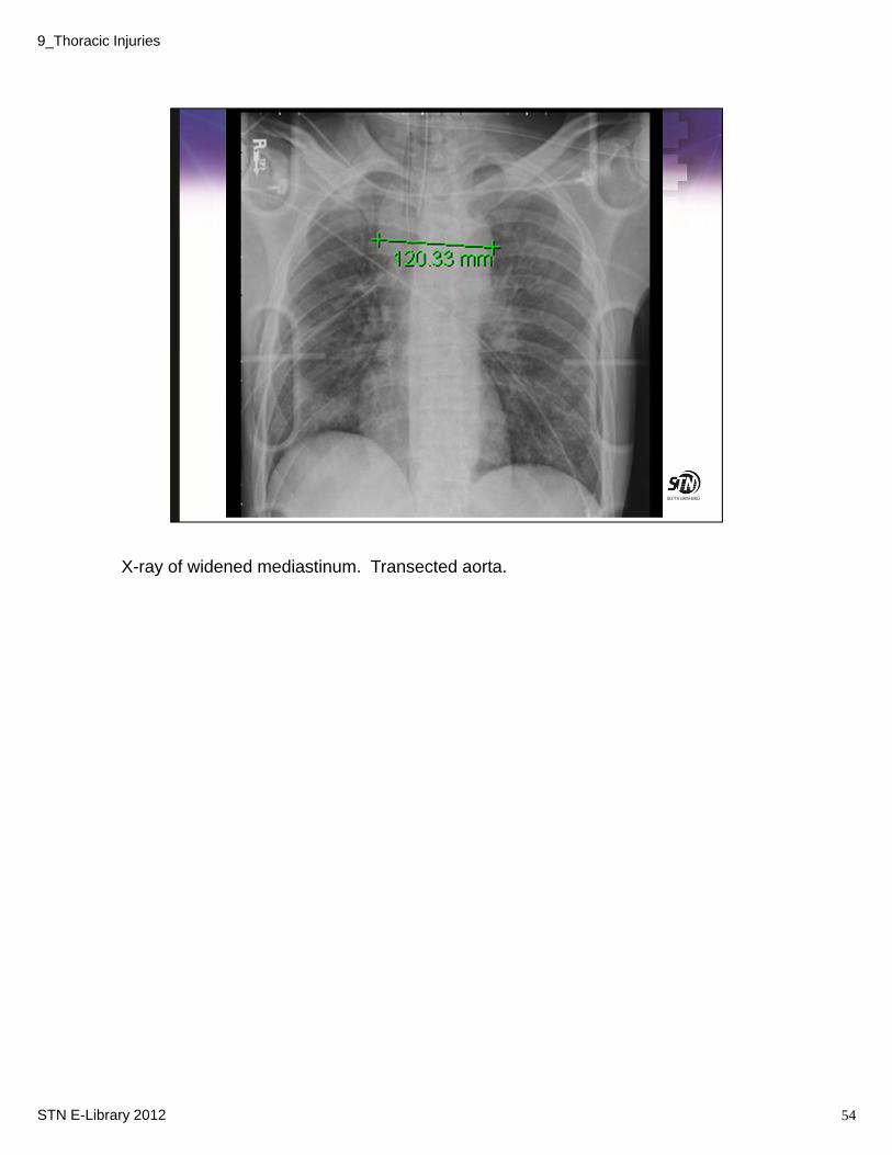
X-ray of widened mediastinum. Transected aorta.
54STN E-Library 2012
9_Thoracic Injuries
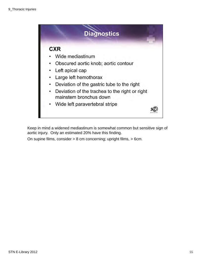
Keep in mind a widened mediastinum is somewhat common but sensitive sign of aortic injury. Only an estimated 20% have this finding.
On supine films, consider > 8 cm concerning; upright films, > 6cm.
55STN E-Library 2012
9_Thoracic Injuries
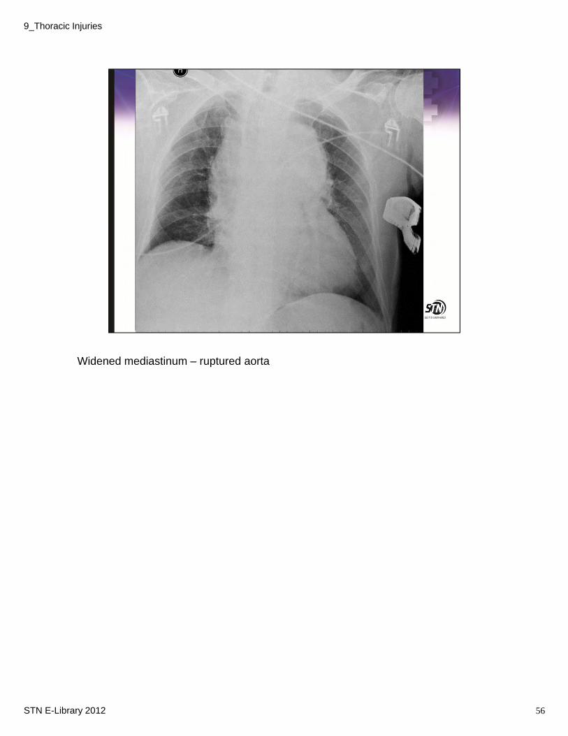
Widened mediastinum – ruptured aorta
56STN E-Library 2012
9_Thoracic Injuries
9_Thoracic Injuries
STN E-Library 2012 57
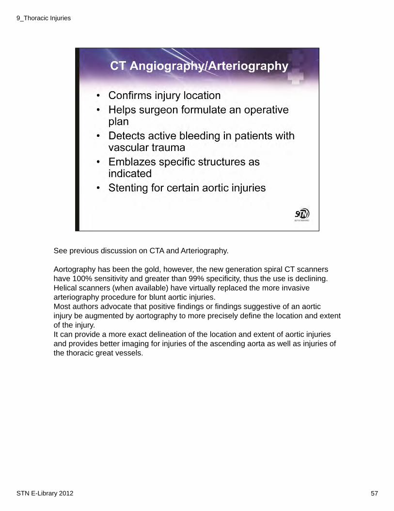
See previous discussion on CTA and Arteriography.
Aortography has been the gold, however, the new generation spiral CT scanners have 100% sensitivity and greater than 99% specificity, thus the use is declining. Helical scanners (when available) have virtually replaced the more invasive arteriography procedure for blunt aortic injuries. Most authors advocate that positive findings or findings suggestive of an aortic injury be augmented by aortography to more precisely define the location and extent of the injury.It can provide a more exact delineation of the location and extent of aortic injuries and provides better imaging for injuries of the ascending aorta as well as injuries of the thoracic great vessels.
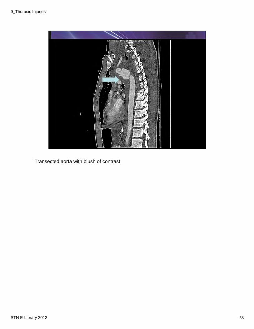
Transected aorta with blush of contrast
58STN E-Library 2012
9_Thoracic Injuries
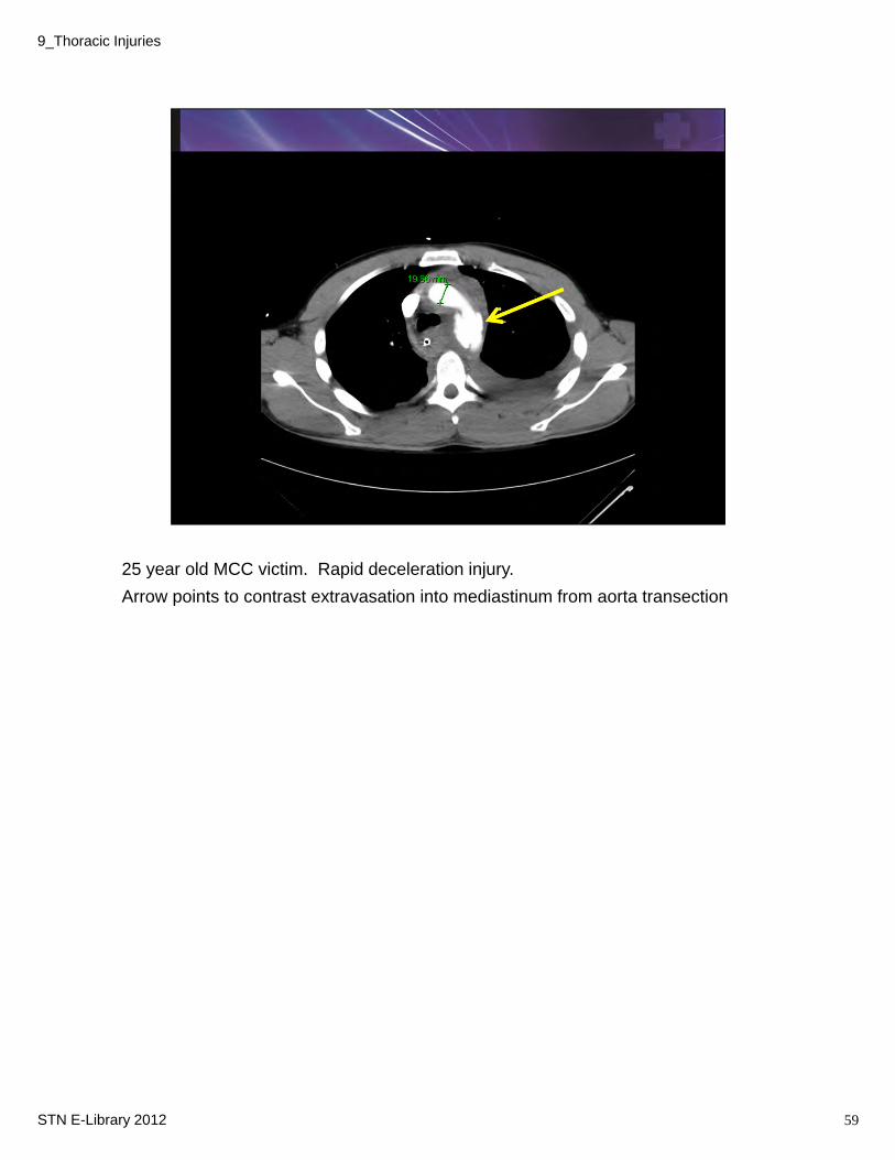
25 year old MCC victim. Rapid deceleration injury.
Arrow points to contrast extravasation into mediastinum from aorta transection
59STN E-Library 2012
9_Thoracic Injuries
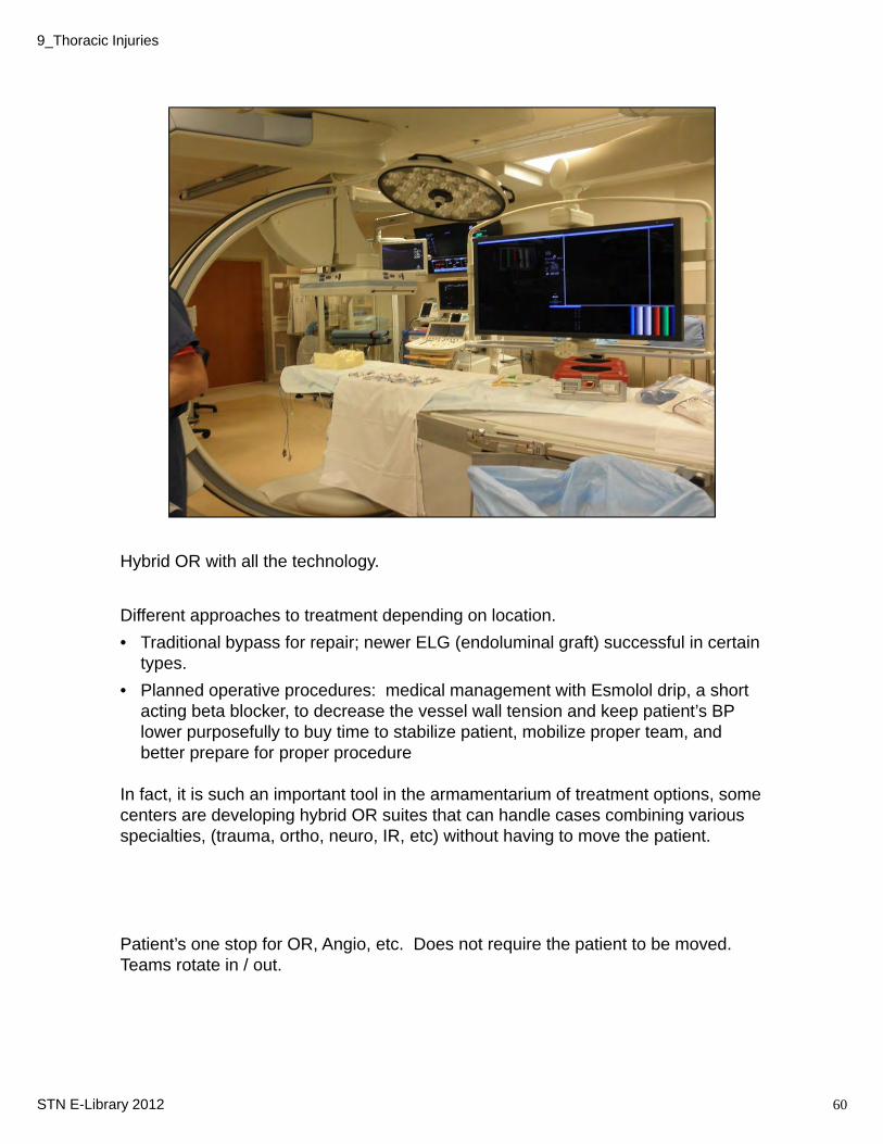
Hybrid OR with all the technology.
Different approaches to treatment depending on location.
• Traditional bypass for repair; newer ELG (endoluminal graft) successful in certain types.
• Planned operative procedures: medical management with Esmolol drip, a short acting beta blocker, to decrease the vessel wall tension and keep patient’s BP lower purposefully to buy time to stabilize patient, mobilize proper team, and better prepare for proper procedure
In fact, it is such an important tool in the armamentarium of treatment options, some centers are developing hybrid OR suites that can handle cases combining various specialties, (trauma, ortho, neuro, IR, etc) without having to move the patient.
Patient’s one stop for OR, Angio, etc. Does not require the patient to be moved. Teams rotate in / out.
60STN E-Library 2012
9_Thoracic Injuries
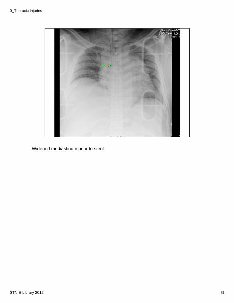
Widened mediastinum prior to stent.
61STN E-Library 2012
9_Thoracic Injuries
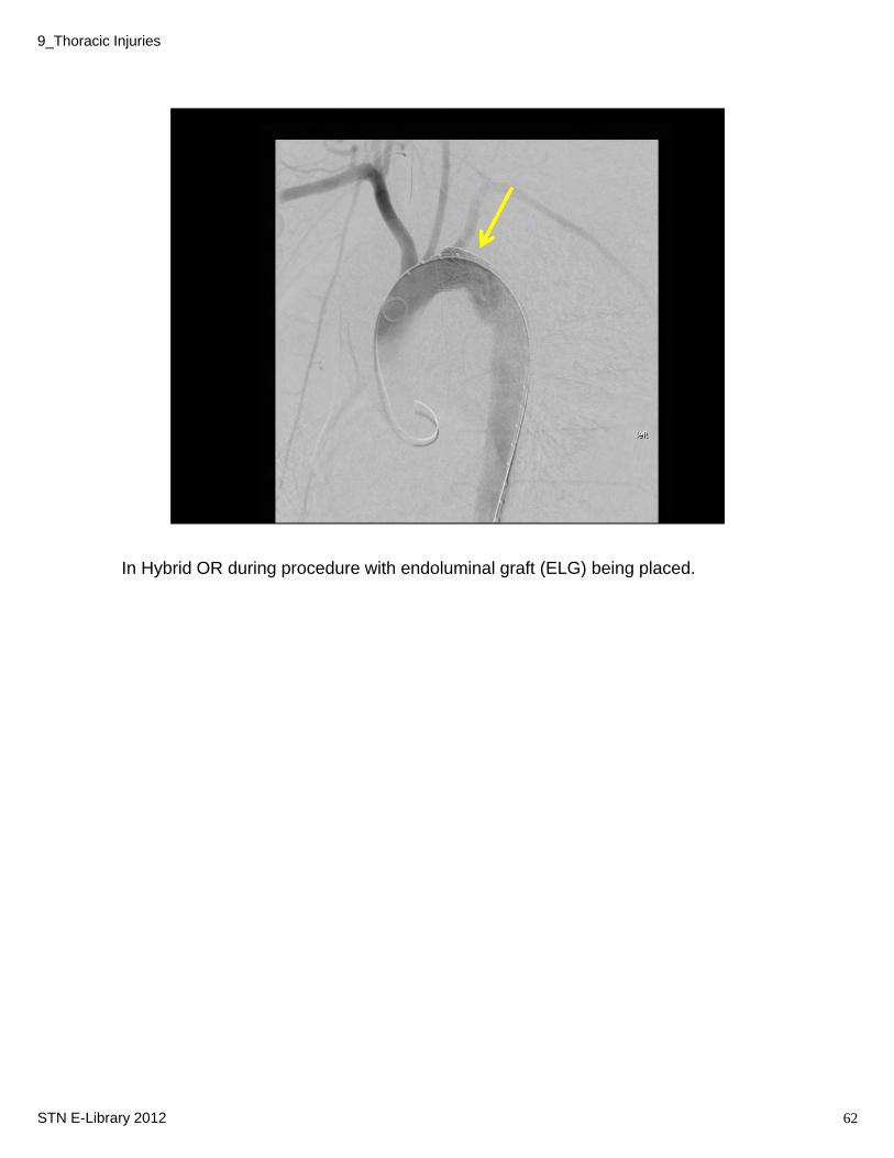
In Hybrid OR during procedure with endoluminal graft (ELG) being placed.
62STN E-Library 2012
9_Thoracic Injuries
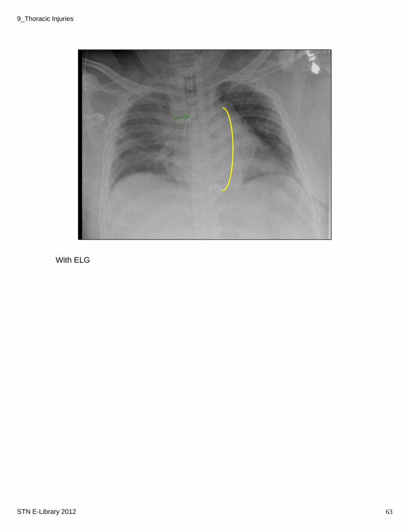
With ELG
63STN E-Library 2012
9_Thoracic Injuries
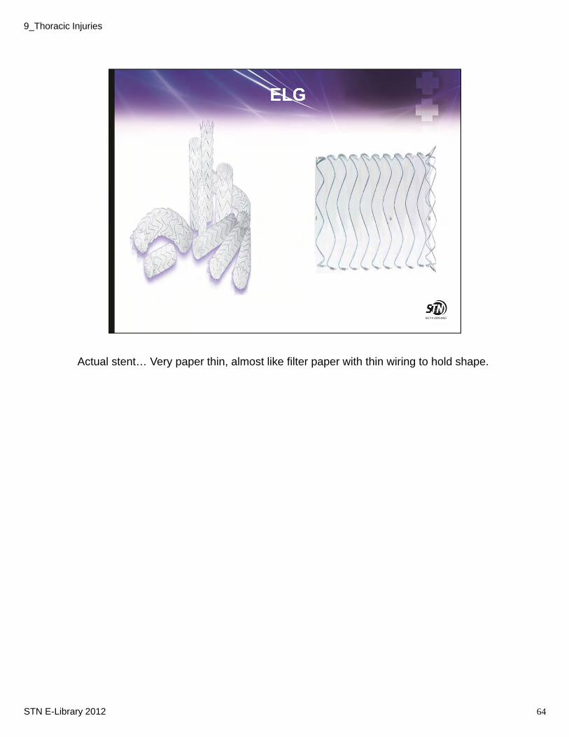
Actual stent… Very paper thin, almost like filter paper with thin wiring to hold shape.
64STN E-Library 2012
9_Thoracic Injuries
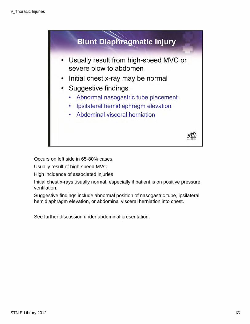
Occurs on left side in 65-80% cases.
Usually result of high-speed MVC
High incidence of associated injuries
Initial chest x-rays usually normal, especially if patient is on positive pressure ventilation.
Suggestive findings include abnormal position of nasogastric tube, ipsilateral hemidiaphragm elevation, or abdominal visceral herniation into chest.
See further discussion under abdominal presentation.
65STN E-Library 2012
9_Thoracic Injuries
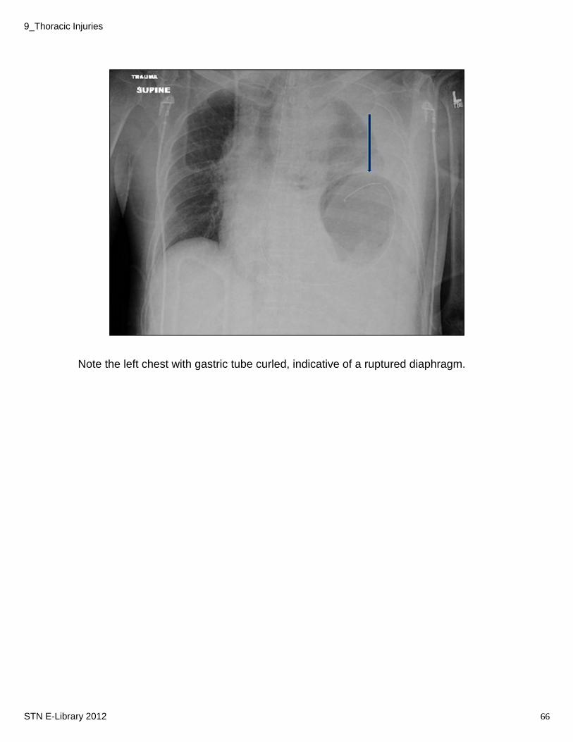
Note the left chest with gastric tube curled, indicative of a ruptured diaphragm.
66STN E-Library 2012
9_Thoracic Injuries
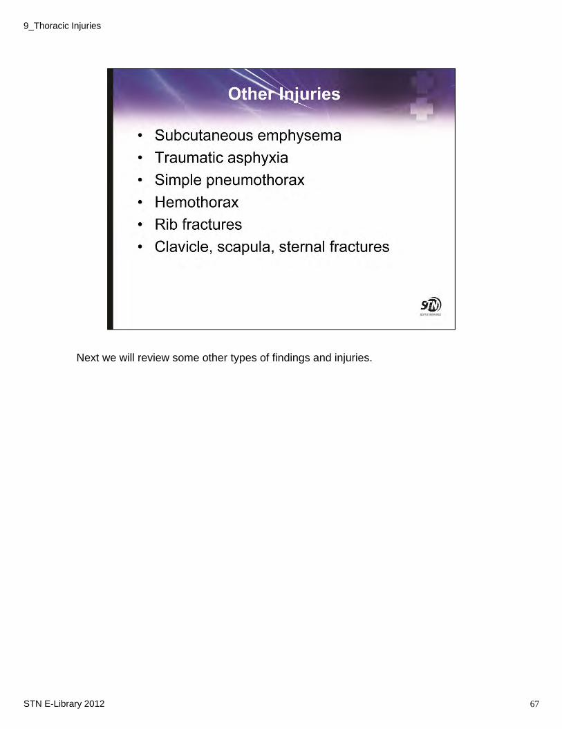
Next we will review some other types of findings and injuries.
67STN E-Library 2012
9_Thoracic Injuries

Confirm cause with CT scan
May feel like “rice krispies” under the skin.
68STN E-Library 2012
9_Thoracic Injuries
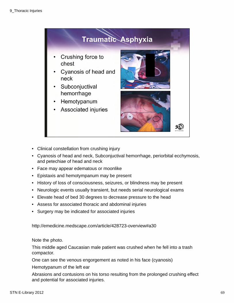
• Clinical constellation from crushing injury
• Cyanosis of head and neck, Subconjuctival hemorrhage, periorbital ecchymosis, and petechiae of head and neck
• Face may appear edematous or moonlike
• Epistaxis and hemotympanum may be present
• History of loss of consciousness, seizures, or blindness may be present
• Neurologic events usually transient, but needs serial neurological exams
• Elevate head of bed 30 degrees to decrease pressure to the head
• Assess for associated thoracic and abdominal injuries
• Surgery may be indicated for associated injuries
http://emedicine.medscape.com/article/428723-overview#a30
Note the photo.
This middle aged Caucasian male patient was crushed when he fell into a trash compactor.
One can see the venous engorgement as noted in his face (cyanosis)
Hemotypanum of the left ear
Abrasions and contusions on his torso resulting from the prolonged crushing effect and potential for associated injuries.
69STN E-Library 2012
9_Thoracic Injuries

Pitfall: Do not underestimate pathophysiology of rib fracture and their effects, especially in the older population
Extent of fractures varies with age, young people ribs more elastic and recoil from impact
National Center for Health Statistics: 350,000 people treated for rib fractures in 2010
Most are not admitted so accuracy in numbers is debatable.
Mechanisms: MVC, Fall from heights
• Rib fractures are the most commonly injured structure in the chest, occurring in 2/3 of patients with thoracic trauma.
• Multiple rib fractures cause significant pain. Patients usually report inspiratory chest pain and discomfort
• Ribs 4-10 are most frequently involved.
• Pain causes respiratory compromise, leading to:
• Reduced pulmonary function
• Sputum retention
• Ineffective cough
• Decreased ventilation leading to hypercapnia and hypoxemia
• Effective analgesia improves gas exchange, pulmonary toilet, LOS, and patient well being.
70STN E-Library 2012
9_Thoracic Injuries
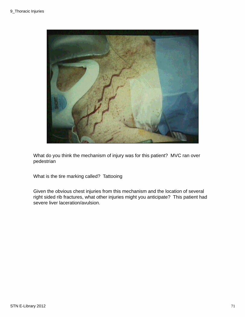
What do you think the mechanism of injury was for this patient? MVC ran over pedestrian
What is the tire marking called? Tattooing
Given the obvious chest injuries from this mechanism and the location of several right sided rib fractures, what other injuries might you anticipate? This patient had severe liver laceration/avulsion.
71STN E-Library 2012
9_Thoracic Injuries
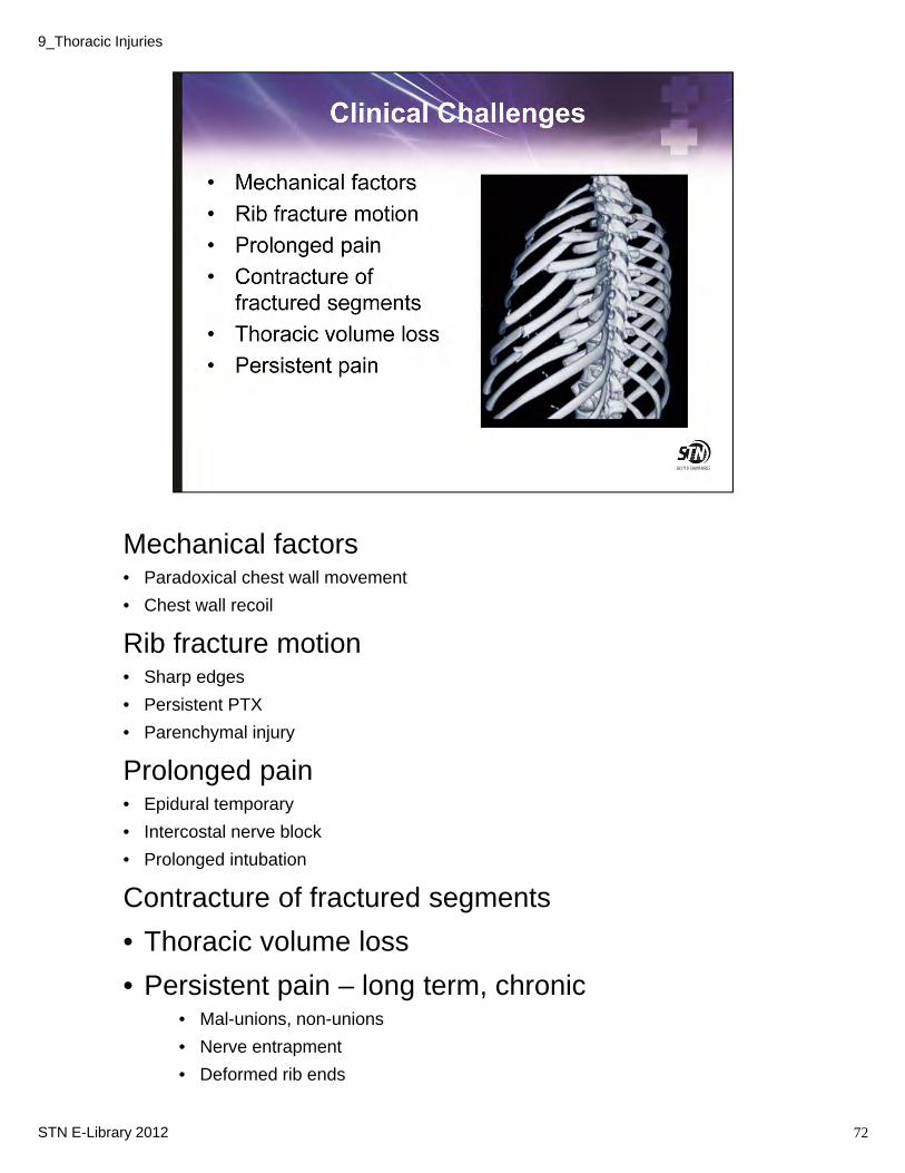
Mechanical factors• Paradoxical chest wall movement
• Chest wall recoil
Rib fracture motion• Sharp edges
• Persistent PTX
• Parenchymal injury
Prolonged pain• Epidural temporary
• Intercostal nerve block
• Prolonged intubation
Contracture of fractured segments
• Thoracic volume loss
• Persistent pain – long term, chronic• Mal-unions, non-unions
• Nerve entrapment
• Deformed rib ends
72STN E-Library 2012
9_Thoracic Injuries
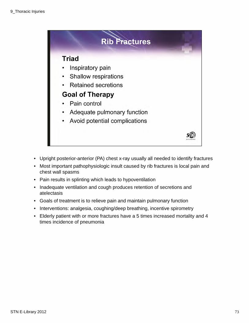
• Upright posterior-anterior (PA) chest x-ray usually all needed to identify fractures
• Most important pathophysiologic insult caused by rib fractures is local pain and chest wall spasms
• Pain results in splinting which leads to hypoventilation
• Inadequate ventilation and cough produces retention of secretions and atelectasis
• Goals of treatment is to relieve pain and maintain pulmonary function
• Interventions: analgesia, coughing/deep breathing, incentive spirometry
• Elderly patient with or more fractures have a 5 times increased mortality and 4 times incidence of pneumonia
73STN E-Library 2012
9_Thoracic Injuries
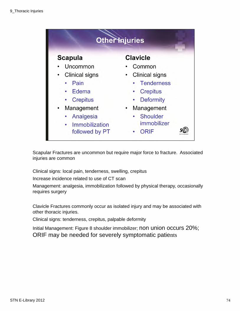
Scapular Fractures are uncommon but require major force to fracture. Associated injuries are common
Clinical signs: local pain, tenderness, swelling, crepitus
Increase incidence related to use of CT scan
Management: analgesia, immobilization followed by physical therapy, occasionally requires surgery
Clavicle Fractures commonly occur as isolated injury and may be associated with other thoracic injuries.
Clinical signs: tenderness, crepitus, palpable deformity
Initial Management: Figure 8 shoulder immobilizer; non union occurs 20%; ORIF may be needed for severely symptomatic patients
74STN E-Library 2012
9_Thoracic Injuries
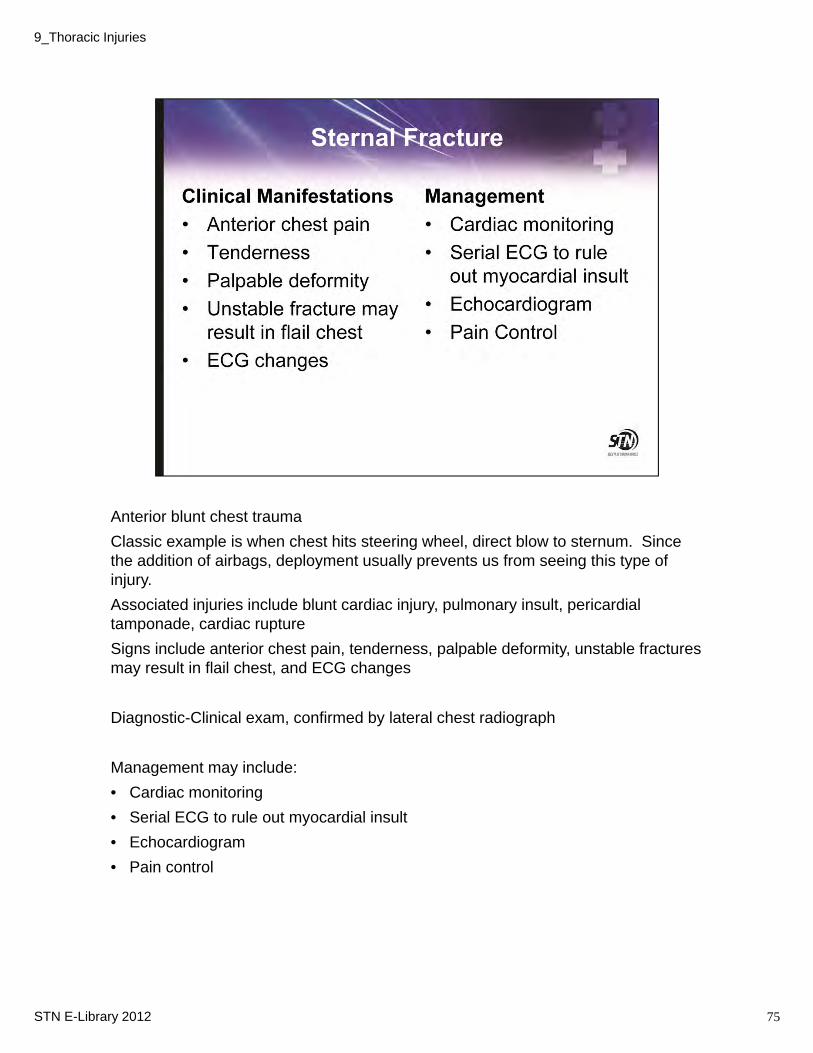
Anterior blunt chest trauma
Classic example is when chest hits steering wheel, direct blow to sternum. Since the addition of airbags, deployment usually prevents us from seeing this type of injury.
Associated injuries include blunt cardiac injury, pulmonary insult, pericardial tamponade, cardiac rupture
Signs include anterior chest pain, tenderness, palpable deformity, unstable fractures may result in flail chest, and ECG changes
Diagnostic-Clinical exam, confirmed by lateral chest radiograph
Management may include:
• Cardiac monitoring
• Serial ECG to rule out myocardial insult
• Echocardiogram
• Pain control
75STN E-Library 2012
9_Thoracic Injuries
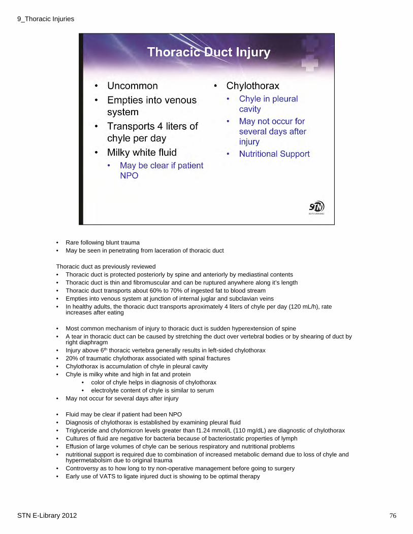
• Rare following blunt trauma• May be seen in penetrating from laceration of thoracic duct
Thoracic duct as previously reviewed• Thoracic duct is protected posteriorly by spine and anteriorly by mediastinal contents• Thoracic duct is thin and fibromuscular and can be ruptured anywhere along it’s length• Thoracic duct transports about 60% to 70% of ingested fat to blood stream• Empties into venous system at junction of internal juglar and subclavian veins• In healthy adults, the thoracic duct transports aproximately 4 liters of chyle per day (120 mL/h), rate
increases after eating
• Most common mechanism of injury to thoracic duct is sudden hyperextension of spine• A tear in thoracic duct can be caused by stretching the duct over vertebral bodies or by shearing of duct by
right diaphragm• Injury above 6th thoracic vertebra generally results in left-sided chylothorax• 20% of traumatic chylothorax associated with spinal fractures• Chylothorax is accumulation of chyle in pleural cavity• Chyle is milky white and high in fat and protein
• color of chyle helps in diagnosis of chylothorax• electrolyte content of chyle is similar to serum
• May not occur for several days after injury
• Fluid may be clear if patient had been NPO• Diagnosis of chylothorax is established by examining pleural fluid• Triglyceride and chylomicron levels greater than f1.24 mmol/L (110 mg/dL) are diagnostic of chylothorax• Cultures of fluid are negative for bacteria because of bacteriostatic properties of lymph• Effusion of large volumes of chyle can be serious respiratory and nutritional problems• nutritional support is required due to combination of increased metabolic demand due to loss of chyle and
hypermetabolsim due to original trauma• Controversy as to how long to try non-operative management before going to surgery• Early use of VATS to ligate injured duct is showing to be optimal therapy
76STN E-Library 2012
9_Thoracic Injuries
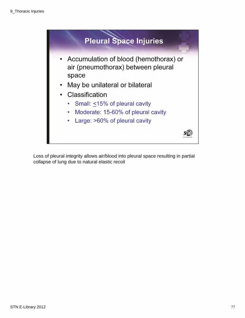
Loss of pleural integrity allows air/blood into pleural space resulting in partial collapse of lung due to natural elastic recoil
77STN E-Library 2012
9_Thoracic Injuries

Review the assessment of someone with suspected pneumothorax
78STN E-Library 2012
9_Thoracic Injuries
79
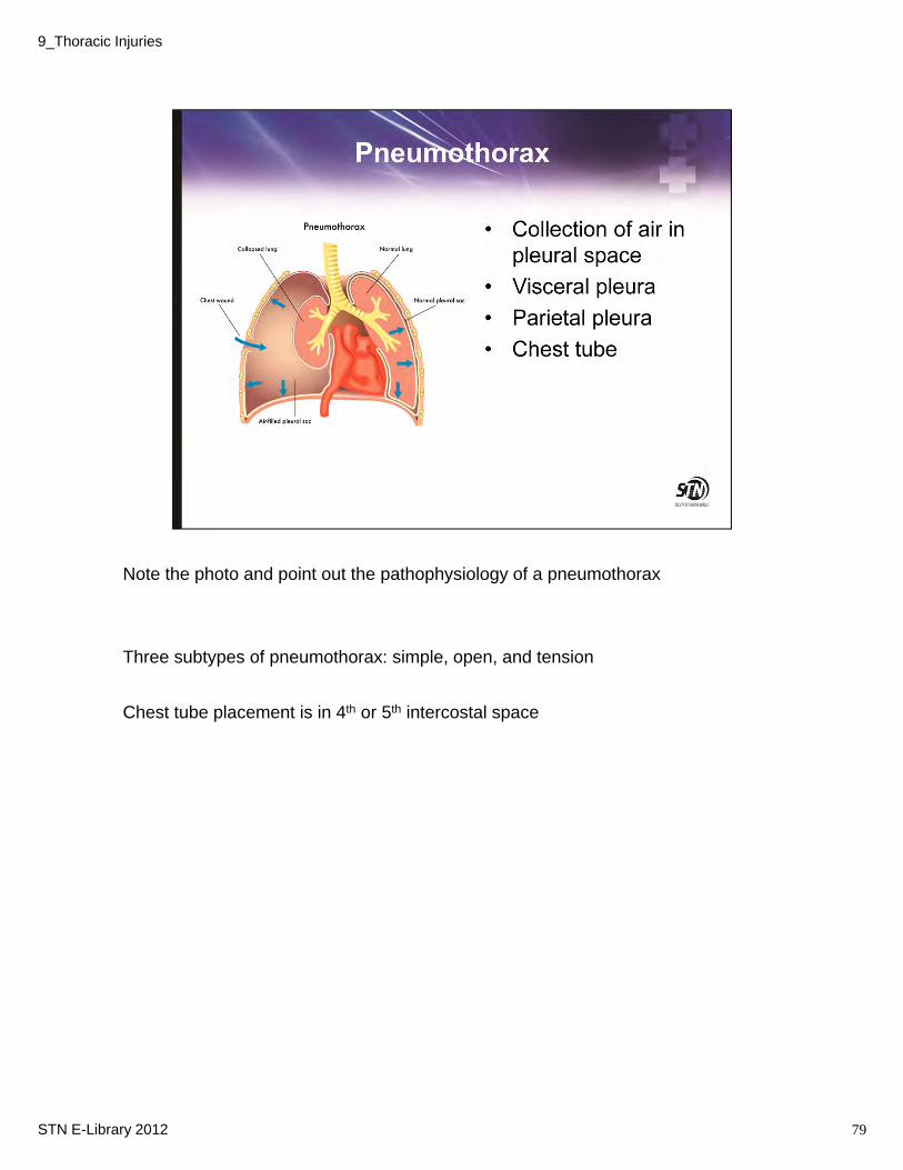
Note the photo and point out the pathophysiology of a pneumothorax
Three subtypes of pneumothorax: simple, open, and tension
Chest tube placement is in 4th or 5th intercostal space
STN E-Library 2012
9_Thoracic Injuries
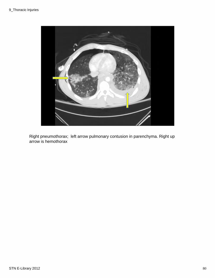
Right pneumothorax; left arrow pulmonary contusion in parenchyma. Right up arrow is hemothorax
80STN E-Library 2012
9_Thoracic Injuries
81
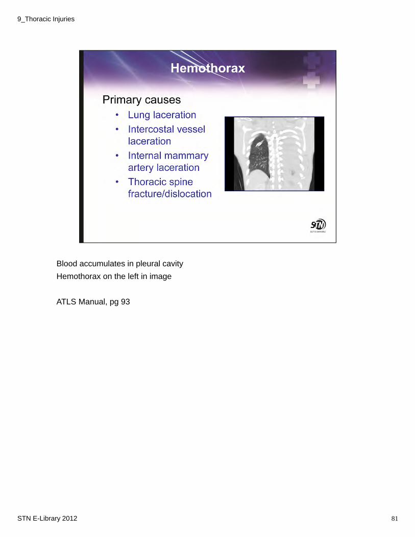
Blood accumulates in pleural cavity
Hemothorax on the left in image
ATLS Manual, pg 93
STN E-Library 2012
9_Thoracic Injuries
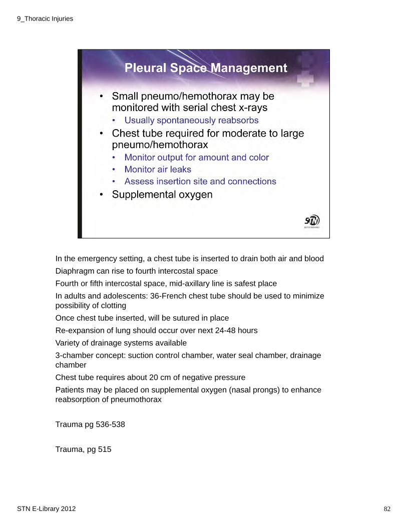
In the emergency setting, a chest tube is inserted to drain both air and blood
Diaphragm can rise to fourth intercostal space
Fourth or fifth intercostal space, mid-axillary line is safest place
In adults and adolescents: 36-French chest tube should be used to minimize possibility of clotting
Once chest tube inserted, will be sutured in place
Re-expansion of lung should occur over next 24-48 hours
Variety of drainage systems available
3-chamber concept: suction control chamber, water seal chamber, drainage chamber
Chest tube requires about 20 cm of negative pressure
Patients may be placed on supplemental oxygen (nasal prongs) to enhance reabsorption of pneumothorax
Trauma pg 536-538
Trauma, pg 515
82STN E-Library 2012
9_Thoracic Injuries

83STN E-Library 2012
9_Thoracic Injuries
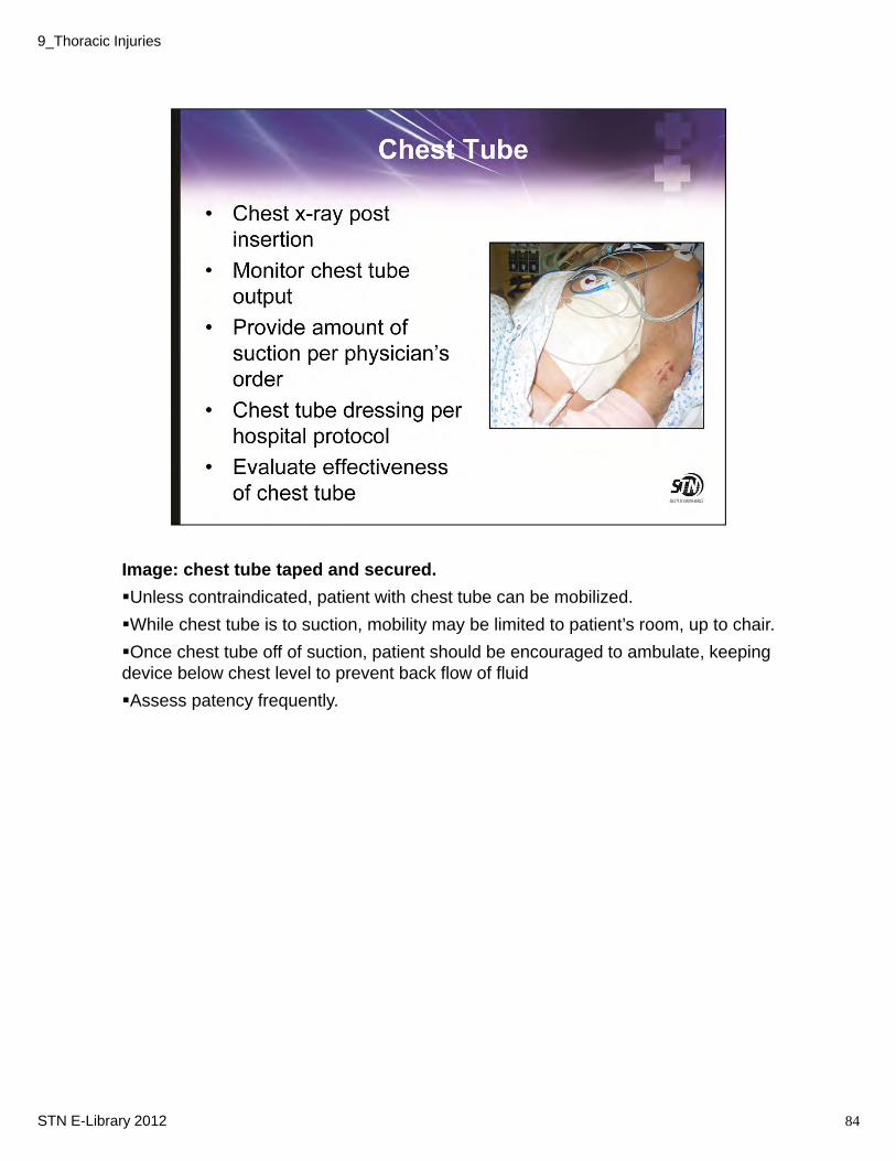
Image: chest tube taped and secured.
Unless contraindicated, patient with chest tube can be mobilized.
While chest tube is to suction, mobility may be limited to patient’s room, up to chair.
Once chest tube off of suction, patient should be encouraged to ambulate, keeping device below chest level to prevent back flow of fluid
Assess patency frequently.
84STN E-Library 2012
9_Thoracic Injuries
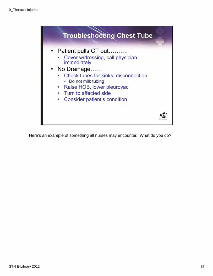
Here’s an example of something all nurses may encounter. What do you do?
85STN E-Library 2012
9_Thoracic Injuries
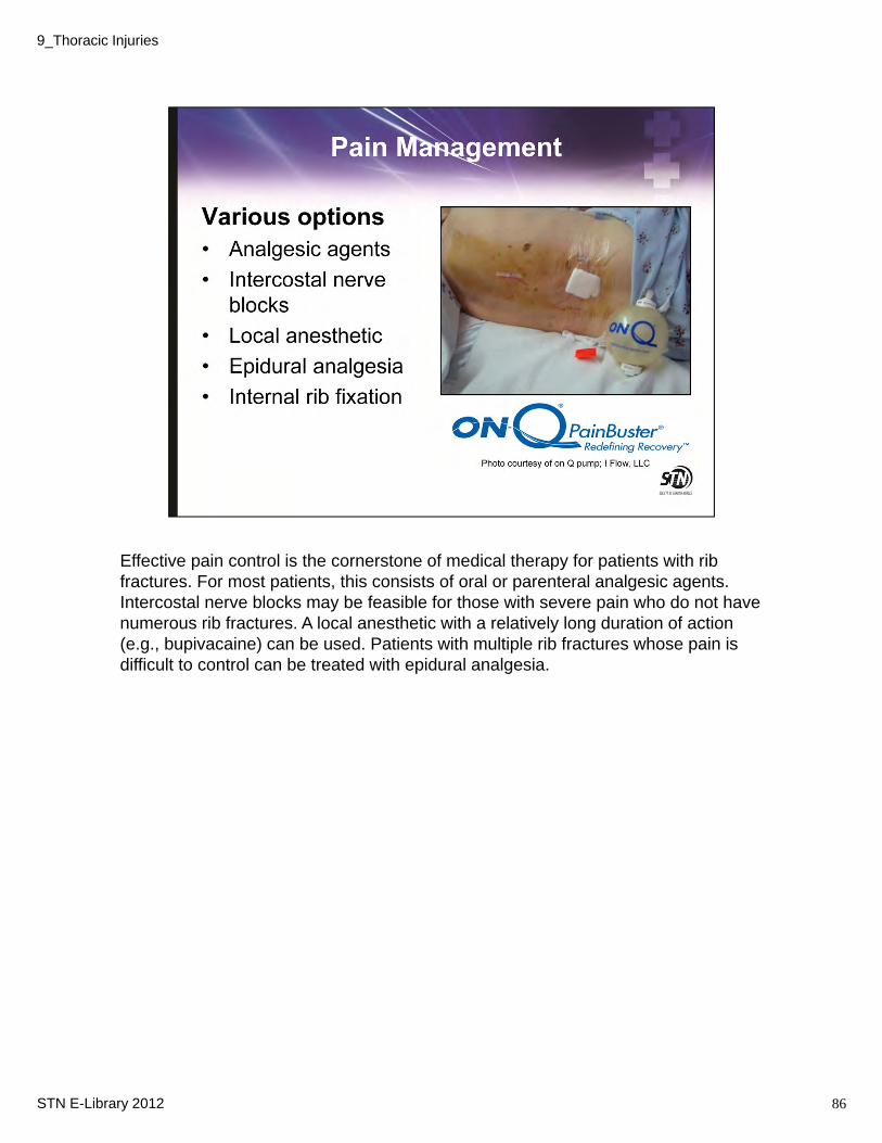
Effective pain control is the cornerstone of medical therapy for patients with rib fractures. For most patients, this consists of oral or parenteral analgesic agents. Intercostal nerve blocks may be feasible for those with severe pain who do not have numerous rib fractures. A local anesthetic with a relatively long duration of action (e.g., bupivacaine) can be used. Patients with multiple rib fractures whose pain is difficult to control can be treated with epidural analgesia.
86STN E-Library 2012
9_Thoracic Injuries
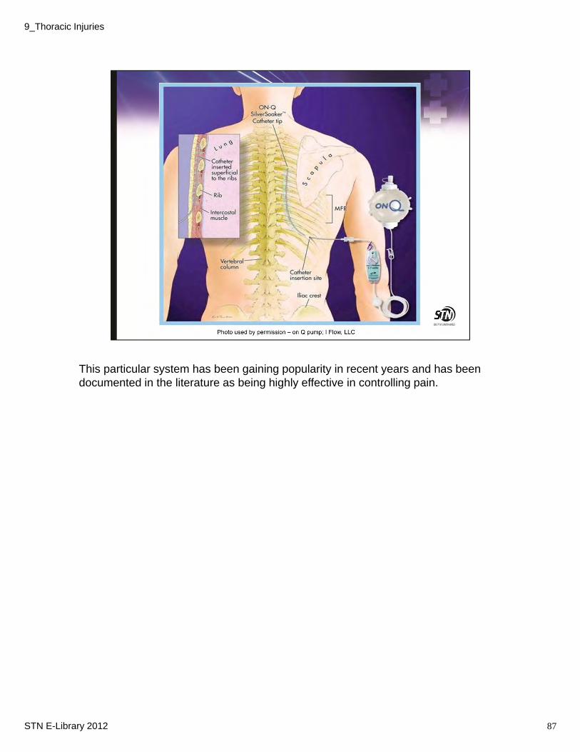
This particular system has been gaining popularity in recent years and has been documented in the literature as being highly effective in controlling pain.
87STN E-Library 2012
9_Thoracic Injuries
As noted. By decreasing a patient’s pain, only positive things will result in many facets of care.
88STN E-Library 2012
9_Thoracic Injuries
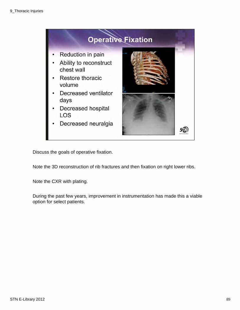
Discuss the goals of operative fixation.
Note the 3D reconstruction of rib fractures and then fixation on right lower ribs.
Note the CXR with plating.
During the past few years, improvement in instrumentation has made this a viable option for select patients.
89STN E-Library 2012
9_Thoracic Injuries
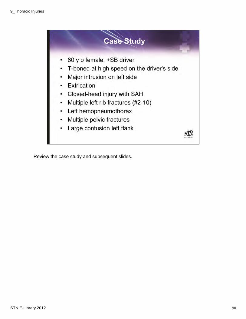
Review the case study and subsequent slides.
90STN E-Library 2012
9_Thoracic Injuries
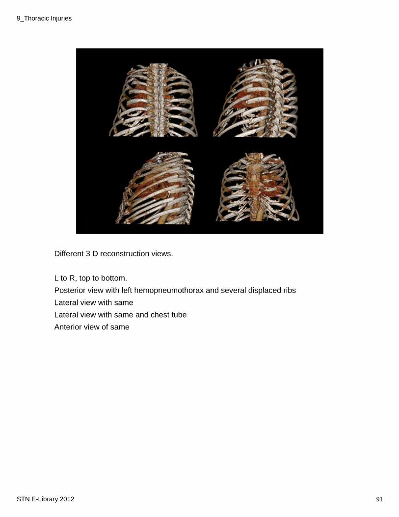
Different 3 D reconstruction views.
L to R, top to bottom.
Posterior view with left hemopneumothorax and several displaced ribs
Lateral view with same
Lateral view with same and chest tube
Anterior view of same
91STN E-Library 2012
9_Thoracic Injuries
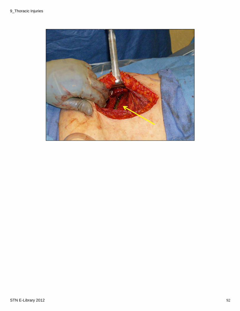
Left ribs 2-10 fractures
Note the intraoperative photo post fixation.
Rib plating left 4-8 ribs (
92STN E-Library 2012
9_Thoracic Injuries
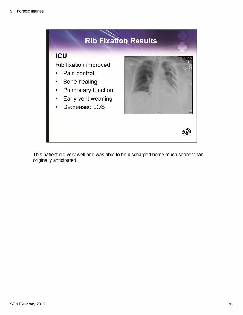
This patient did very well and was able to be discharged home much sooner than originally anticipated.
93STN E-Library 2012
9_Thoracic Injuries
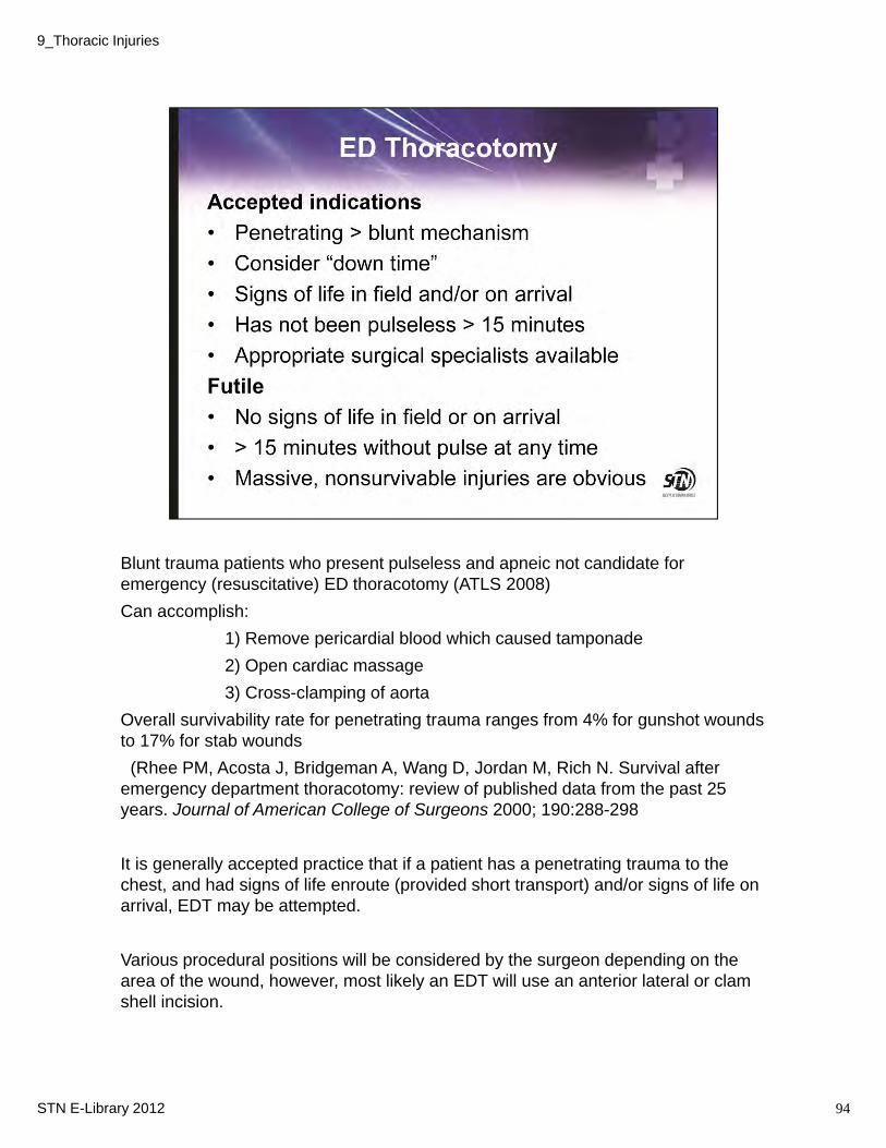
Blunt trauma patients who present pulseless and apneic not candidate for emergency (resuscitative) ED thoracotomy (ATLS 2008)
Can accomplish:
1) Remove pericardial blood which caused tamponade
2) Open cardiac massage
3) Cross-clamping of aorta
Overall survivability rate for penetrating trauma ranges from 4% for gunshot wounds to 17% for stab wounds
(Rhee PM, Acosta J, Bridgeman A, Wang D, Jordan M, Rich N. Survival after emergency department thoracotomy: review of published data from the past 25 years. Journal of American College of Surgeons 2000; 190:288-298
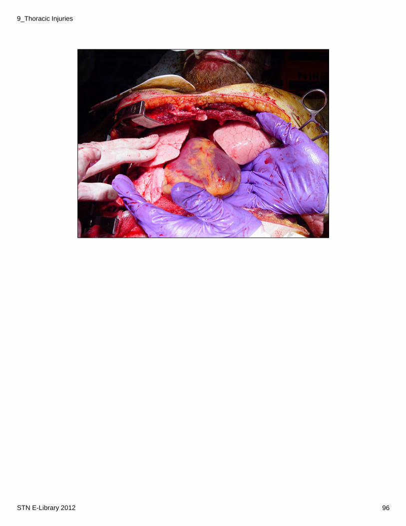
It is generally accepted practice that if a patient has a penetrating trauma to the chest, and had signs of life enroute (provided short transport) and/or signs of life on arrival, EDT may be attempted.
Various procedural positions will be considered by the surgeon depending on the area of the wound, however, most likely an EDT will use an anterior lateral or clam shell incision.
94STN E-Library 2012
9_Thoracic Injuries
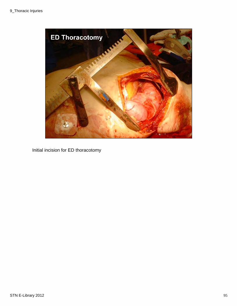
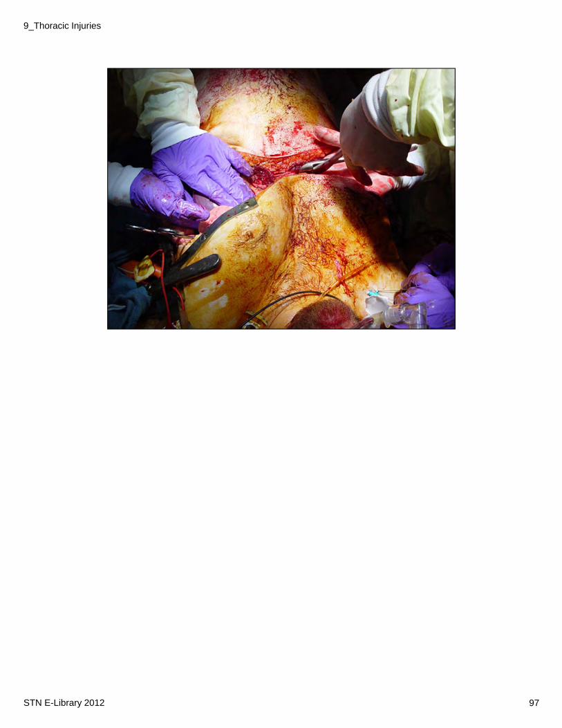
Initial incision for ED thoracotomy
95STN E-Library 2012
9_Thoracic Injuries
9_Thoracic Injuries
STN E-Library 2012 96
9_Thoracic Injuries
STN E-Library 2012 97
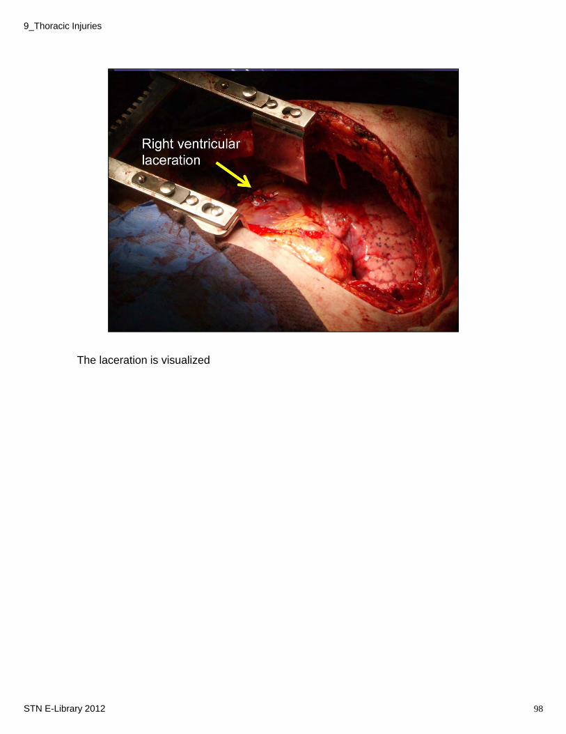
The laceration is visualized
98STN E-Library 2012
9_Thoracic Injuries
99STN E-Library 2012
9_Thoracic Injuries
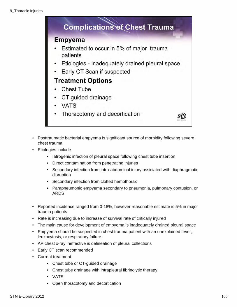
• Posttraumatic bacterial empyema is significant source of morbidity following severe chest trauma
• Etiologies include
• Iatrogenic infection of pleural space following chest tube insertion
• Direct contamination from penetrating injuries
• Secondary infection from intra-abdominal injury assiciated with diaphragmatic disruption
• Secondary infection from clotted hemothorax
• Parapneumonic empyema secondary to pneumonia, pulmonary contusion, or ARDS
• Reported incidence ranged from 0-18%, however reasonable estimate is 5% in major trauma patients
• Rate is increasing due to increase of survival rate of critically injured
• The main cause for development of empyema is inadequately drained pleural space
• Empyema should be suspected in chest trauma patient with an unexplained fever, leukocytosis, or respiratory failure
• AP chest x-ray ineffective is delineation of pleural collections
• Early CT scan recommended
• Current treatment
• Chest tube or CT-guided drainage
• Chest tube drainage with intrapleural fibrinolytic therapy
• VATS
• Open thoracotomy and decortication
100STN E-Library 2012
9_Thoracic Injuries
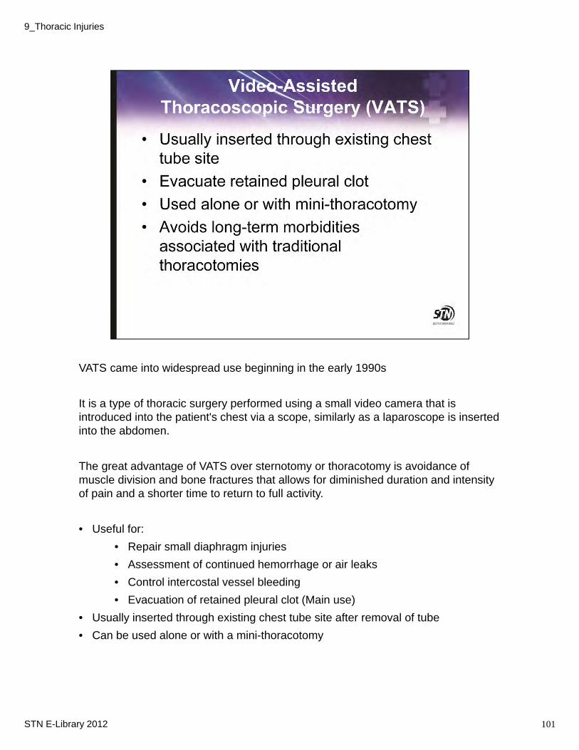
VATS came into widespread use beginning in the early 1990s
It is a type of thoracic surgery performed using a small video camera that is introduced into the patient's chest via a scope, similarly as a laparoscope is inserted into the abdomen.
The great advantage of VATS over sternotomy or thoracotomy is avoidance of muscle division and bone fractures that allows for diminished duration and intensity of pain and a shorter time to return to full activity.
• Useful for:
• Repair small diaphragm injuries
• Assessment of continued hemorrhage or air leaks
• Control intercostal vessel bleeding
• Evacuation of retained pleural clot (Main use)
• Usually inserted through existing chest tube site after removal of tube
• Can be used alone or with a mini-thoracotomy
101STN E-Library 2012
9_Thoracic Injuries

Air Leak
• Air leaks common following chest trauma
• Usually only request chest tube for treatment
• Large air leak mandates urgent bronchoscopy to rule out tracheo-bronchial injury
• May also be observed if chest tube is not positioned correctly or with a large pulmonary laceration
• Complete lung expansion is goal of therapy
Pneumatocele
• Posttraumatic cysts uncommon following chest trauma
• CT scan to diagnose
• Air disruption may lead to air collection within lung parenchyma that does not connect with pleural space
• Seen more frequently with patients who have severe chest trauma and required prolonged ventilatory support
• Most lesion resolve following weaning from ventilator
• If lesions continue to expand or if become infected, may require CT guided drainage
102STN E-Library 2012
9_Thoracic Injuries
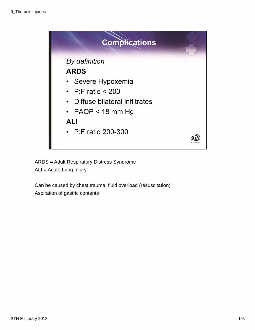
ARDS = Adult Respiratory Distress Syndrome
ALI = Acute Lung Injury
Can be caused by chest trauma, fluid overload (resuscitation)
Aspiration of gastric contents
103STN E-Library 2012
9_Thoracic Injuries
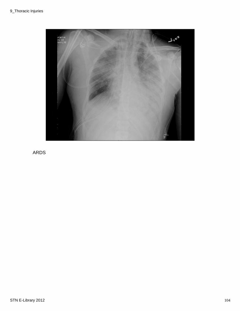
ARDS
104STN E-Library 2012
9_Thoracic Injuries
• In summary, nursing considerations revolve around prompt assessment and reassessment to ensure the patient’s condition does not deteriorate.
• The ABCs are of utmost importance as many injuries are life threatening or have the potential to be life threatening, especially if missed.
• Maintaining a high index of suspicion in consideration of the mechanism of injury and all the thoracic structures that have potential for injury is important.
• Having a heightened awareness will allow an astute care giver to promptly intervene in situations that might otherwise go unnoticed.
• Understanding some of the potential complications / sequelae will also allow for increased monitoring
105STN E-Library 2012
9_Thoracic Injuries
Thoracic trauma continues to produce some of the most challenging and deadly injuries seen. All intrathoracic organs play a vital role in normal physiology and maintaining stability.
Unrecognized injuries (and those untreated) may result in death within minutes from shock, hypoxia, etc.
The mechanism of injury may provide guidance as to potential injuries.
Maintaining an adequate airway with ventilation is key.
Clinical exam is important.
CXR is often the only study required early on.
A chest tube treats the majority of both blunt and penetrating traumatic injuries.
Pain control is essential in preventing complications such as pulmonary atelectasis, pneumonia, and respiratory issues.
106STN E-Library 2012
9_Thoracic Injuries