chest wall injuries wall injuries nordter trauma radiology 9th nordic course may 23 - 26, 2016...
TRANSCRIPT
CHEST WALL INJURIES
NORDTER Trauma Radiology9th Nordic CourseMay 23 - 26, 2016 Aarhus, Denmark
Mari Nummela, MD, Fellow in Musculoskeletal RadiologyTöölö Trauma Center, Helsinki University Hospital, Finland

CHEST WALL INJURIES Incidence of blunt chest trauma
Mechanism of injury
Anatomy of the chest wall
Rib and costochondral fracture types
Underlying intrathoracic injuries and
deceleration injuries (through cases)
Examples of associated injuries (sternum etc.)
Different modalities in chest trauma
Clinical aspects
Key points
Nordter 2016/Chest wall injuries 2
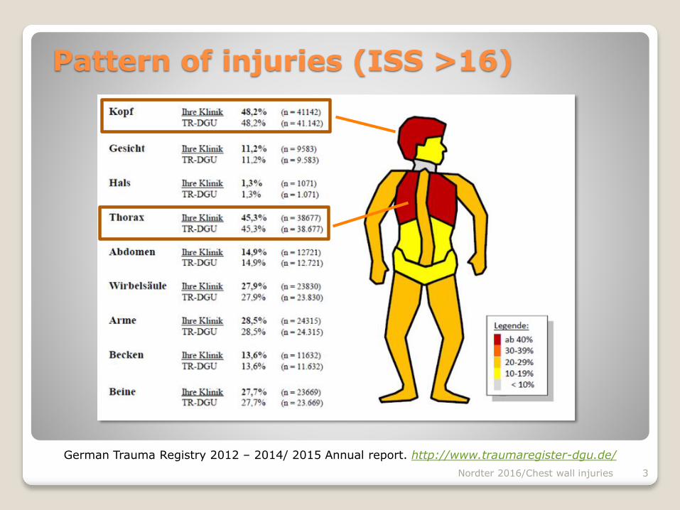
Pattern of injuries (ISS >16)
German Trauma Registry 2012 – 2014/ 2015 Annual report. http://www.traumaregister-dgu.de/
Nordter 2016/Chest wall injuries 3
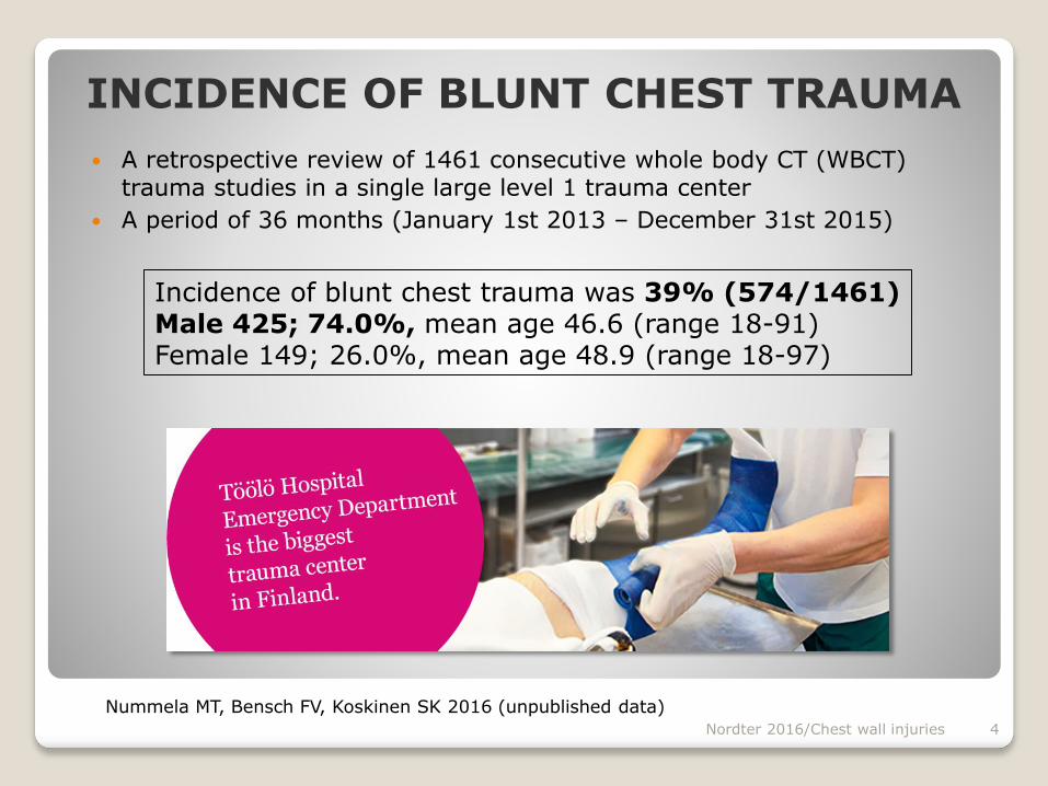
INCIDENCE OF BLUNT CHEST TRAUMA
A retrospective review of 1461 consecutive whole body CT (WBCT) trauma studies in a single large level 1 trauma center
A period of 36 months (January 1st 2013 – December 31st 2015)
Incidence of blunt chest trauma was 39% (574/1461)Male 425; 74.0%, mean age 46.6 (range 18-91)Female 149; 26.0%, mean age 48.9 (range 18-97)
Nummela MT, Bensch FV, Koskinen SK 2016 (unpublished data)
Nordter 2016/Chest wall injuries 4

MOI in chest trauma
(mechanism of injury)
Fall from height 36% MVA 27% Motor cycle and bicycle
accidents 23%
Nummela MT, Bensch FV, Koskinen SK 2016 (unpublished data)
Mechanism of injuryn=574
n %
Fall 207 36 %
MVA 157 27 %
MCA 75 13 %
BCA 58 10 %
Compression 21 4 %
Assault 16 3 %
Pedestrian 29 5 %
Other 11 2 %
Total 574 100 %
Nordter 2016/Chest wall injuries 5
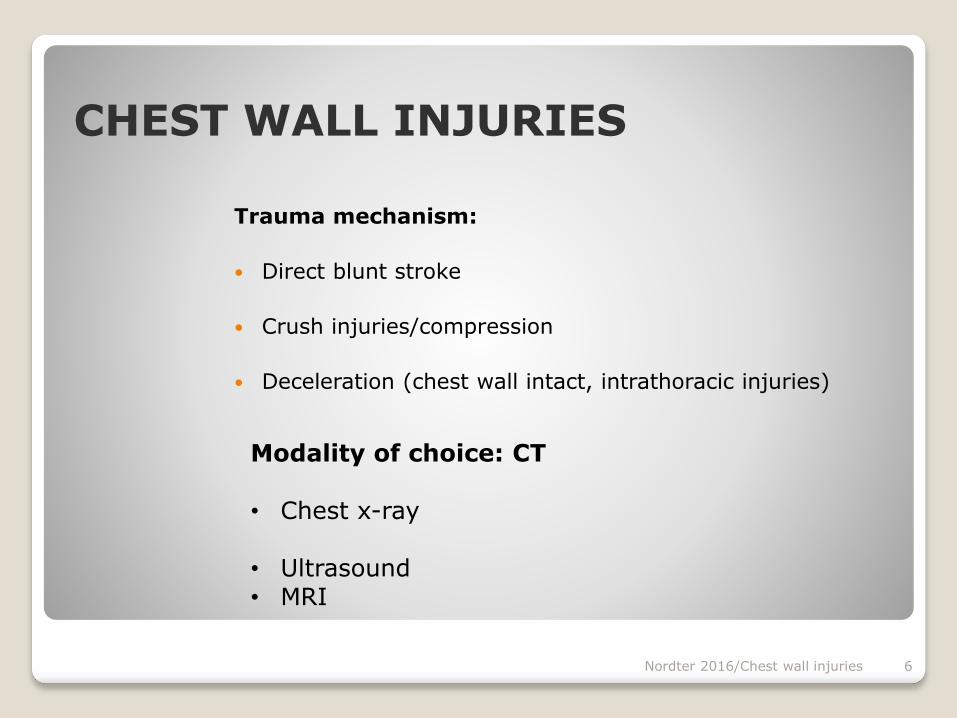
CHEST WALL INJURIES
Trauma mechanism:
Direct blunt stroke
Crush injuries/compression
Deceleration (chest wall intact, intrathoracic injuries)
Nordter 2016/Chest wall injuries 6
Modality of choice: CT
• Chest x-ray
• Ultrasound • MRI
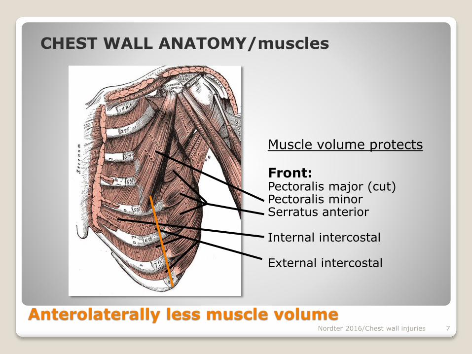
Anterolaterally less muscle volume
Muscle volume protects
Front:Pectoralis major (cut)Pectoralis minorSerratus anterior
Internal intercostal
External intercostal
Nordter 2016/Chest wall injuries 7
CHEST WALL ANATOMY/muscles

Muscle volume protects
Back:• Latissimus dorsi• Rhomboid muscles • Trapezius
Nordter 2016/Chest wall injuries 8
Trapezius
Latissimus dorsi
CHEST WALL ANATOMY/muscles
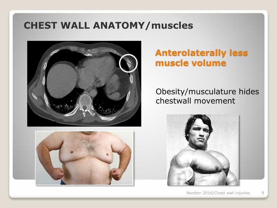
Anterolaterally less muscle volume
Obesity/musculature hides chestwall movement
Nordter 2016/Chest wall injuries 9
CHEST WALL ANATOMY/muscles
Nordter 2016/Chest wall injuries 10
CHEST WALL ANATOMY/muscles
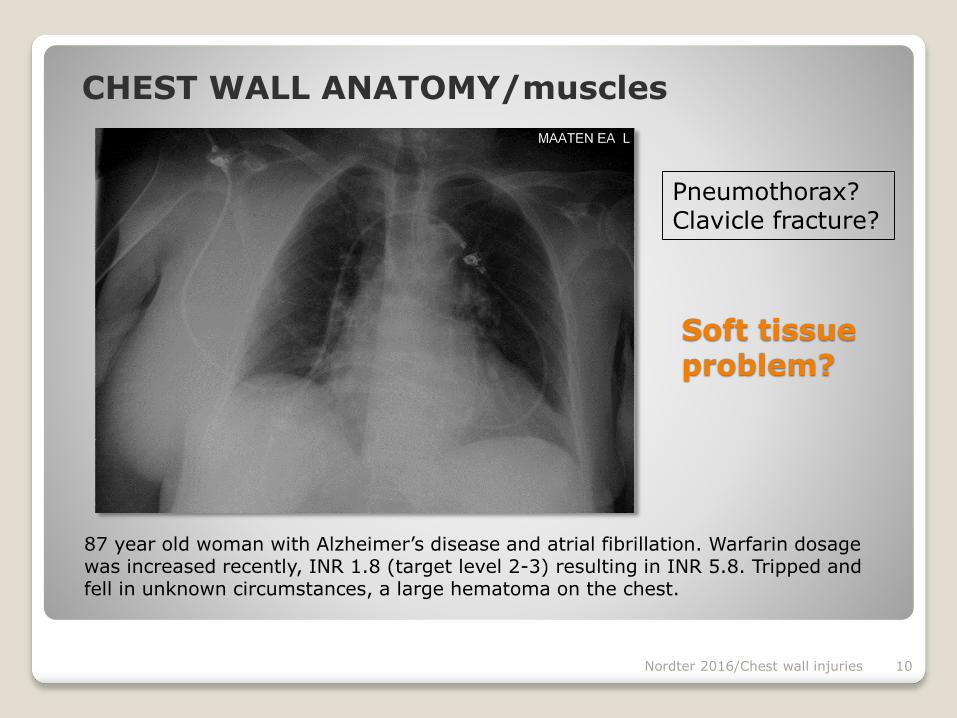
Soft tissue problem?
87 year old woman with Alzheimer’s disease and atrial fibrillation. Warfarin dosage was increased recently, INR 1.8 (target level 2-3) resulting in INR 5.8. Tripped and fell in unknown circumstances, a large hematoma on the chest.
Pneumothorax?Clavicle fracture?

Nordter 2016/Chest wall injuries 11
CHEST WALL ANATOMY/musclesCT ordered:Is there intrathoracic bleeding?
87 year old woman with Alzheimer’s disease and atrial fibrillation. INR 5.8, hemoglobin level dropped down to Hb 74 g/l.

Nordter 2016/Chest wall injuries 12
CHEST WALL ANATOMY/muscles
CT ordered:No intrathoracic bleeding
87 year old woman with Alzheimer’s disease and atrial fibrillation. INR 5.8, hemoglobin level dropped to Hb 74 g/l.
ART COR
VEN SAG
Pectoralis major & minor torn
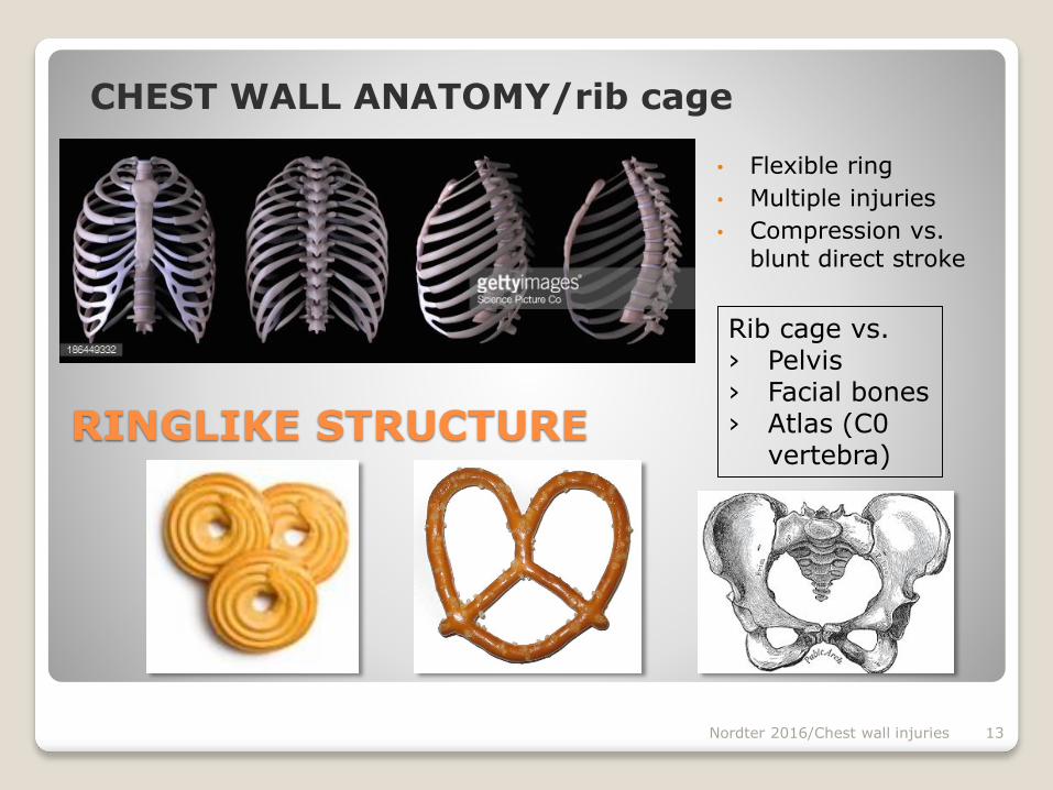
RINGLIKE STRUCTURE
• Flexible ring
• Multiple injuries
• Compression vs. blunt direct stroke
Rib cage vs.› Pelvis› Facial bones› Atlas (C0
vertebra)
Nordter 2016/Chest wall injuries 13
CHEST WALL ANATOMY/rib cage

Ribs also protect the abdominal organs
Nordter 2016/Chest wall injuries 14
Liver
Right kidney
Spleen
Pancreas
Left kidney
It’s not just about the chest!
CHEST WALL ANATOMY/rib cage
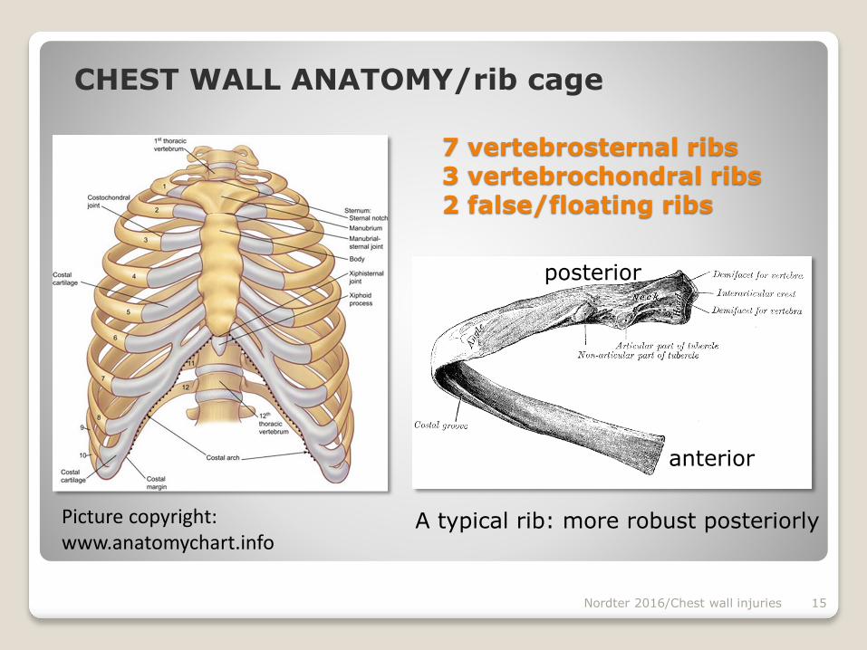
7 vertebrosternal ribs 3 vertebrochondral ribs2 false/floating ribs
Picture copyright: www.anatomychart.info
Nordter 2016/Chest wall injuries 15
anterior
posterior
A typical rib: more robust posteriorly
CHEST WALL ANATOMY/rib cage

Geometry of human ribs pertinent to orthopedic chest-wall reconstruction.M. Mohr et al. / Journal of Biomechanics 40 (2007) 1310–1317
Nordter 2016/Chest wall injuries 16
CHEST WALL ANATOMY/ribs
Outer cortex is thinner laterally and anteriorly
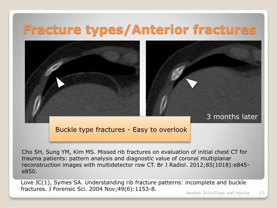
Fracture types/Anterior fractures
Buckle type fractures - Easy to overlook
Cho SH, Sung YM, Kim MS. Missed rib fractures on evaluation of initial chest CT for trauma patients: pattern analysis and diagnostic value of coronal multiplanar reconstruction images with multidetector row CT. Br J Radiol. 2012;85(1018):e845-e850.
Nordter 2016/Chest wall injuries 17
Love JC(1), Symes SA. Understanding rib fracture patterns: incomplete and buckle fractures. J Forensic Sci. 2004 Nov;49(6):1153-8.
3 months later
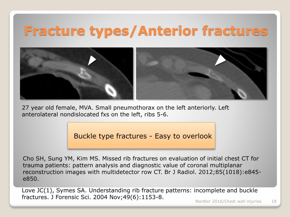
Fracture types/Anterior fractures
Buckle type fractures - Easy to overlook
Cho SH, Sung YM, Kim MS. Missed rib fractures on evaluation of initial chest CT for trauma patients: pattern analysis and diagnostic value of coronal multiplanar reconstruction images with multidetector row CT. Br J Radiol. 2012;85(1018):e845-e850.
Nordter 2016/Chest wall injuries 18
27 year old female, MVA. Small pneumothorax on the left anteriorly. Left anterolateral nondislocated fxs on the left, ribs 5-6.
Love JC(1), Symes SA. Understanding rib fracture patterns: incomplete and buckle fractures. J Forensic Sci. 2004 Nov;49(6):1153-8.
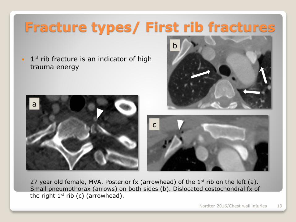
Fracture types/ First rib fractures
1st rib fracture is an indicator of high trauma energy
Nordter 2016/Chest wall injuries 19
27 year old female, MVA. Posterior fx (arrowhead) of the 1st rib on the left (a).Small pneumothorax (arrows) on both sides (b). Dislocated costochondral fx of the right 1st rib (c) (arrowhead).
b
a
c

Anterior nondislocated fractures vs. pseudarthrosis of the first rib
Degree of ossification varies greatly between individuals
Nordter 2016/Chest wall injuries 20
Gossner J. Pseudarthrosis of the cartilaginous part of the first rib is a common incidental finding on chest CT. Diagn Interv Imaging 2015.
Fig 1. Gas bubbles lining a fracture in the costochondral junction (arrowhead). Sternum (S) fracture on the right (star), a retrosternal hematoma.
Fig 2. 42 year old male, MCA. Fracture was suspected. There is air inside the cleft like costochondral junction (arrows), but no fracture line or air outside the junction.
Fracture types/ First rib fractures
S
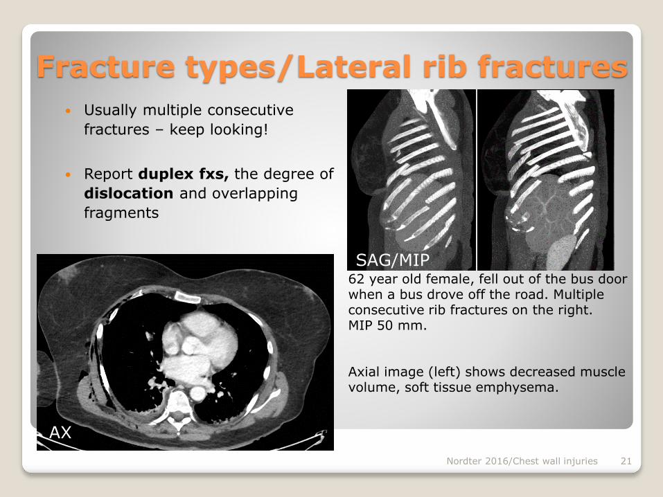
Fracture types/Lateral rib fractures
Usually multiple consecutive
fractures – keep looking!
Report duplex fxs, the degree of
dislocation and overlapping
fragments
Nordter 2016/Chest wall injuries 21
62 year old female, fell out of the bus door when a bus drove off the road. Multiple consecutive rib fractures on the right. MIP 50 mm.
Axial image (left) shows decreased muscle volume, soft tissue emphysema.
AX
SAG/MIP
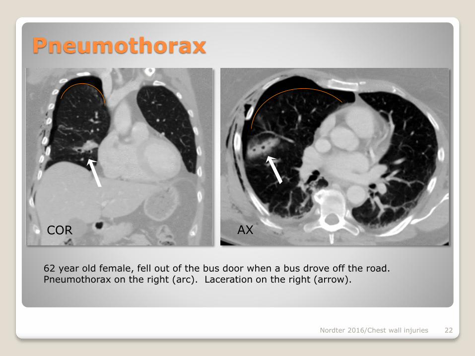
Pneumothorax
Nordter 2016/Chest wall injuries 22
62 year old female, fell out of the bus door when a bus drove off the road. Pneumothorax on the right (arc). Laceration on the right (arrow).
COR AX
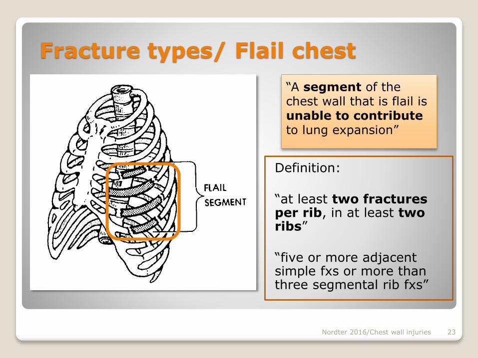
“A segment of the chest wall that is flail is unable to contributeto lung expansion”
Nordter 2016/Chest wall injuries 23
Fracture types/ Flail chest
Definition:
“at least two fractures per rib, in at least two ribs”
“five or more adjacent simple fxs or more than three segmental rib fxs”
Nordter 2016/Chest wall injuries 24
Fracture types/ Flail chest
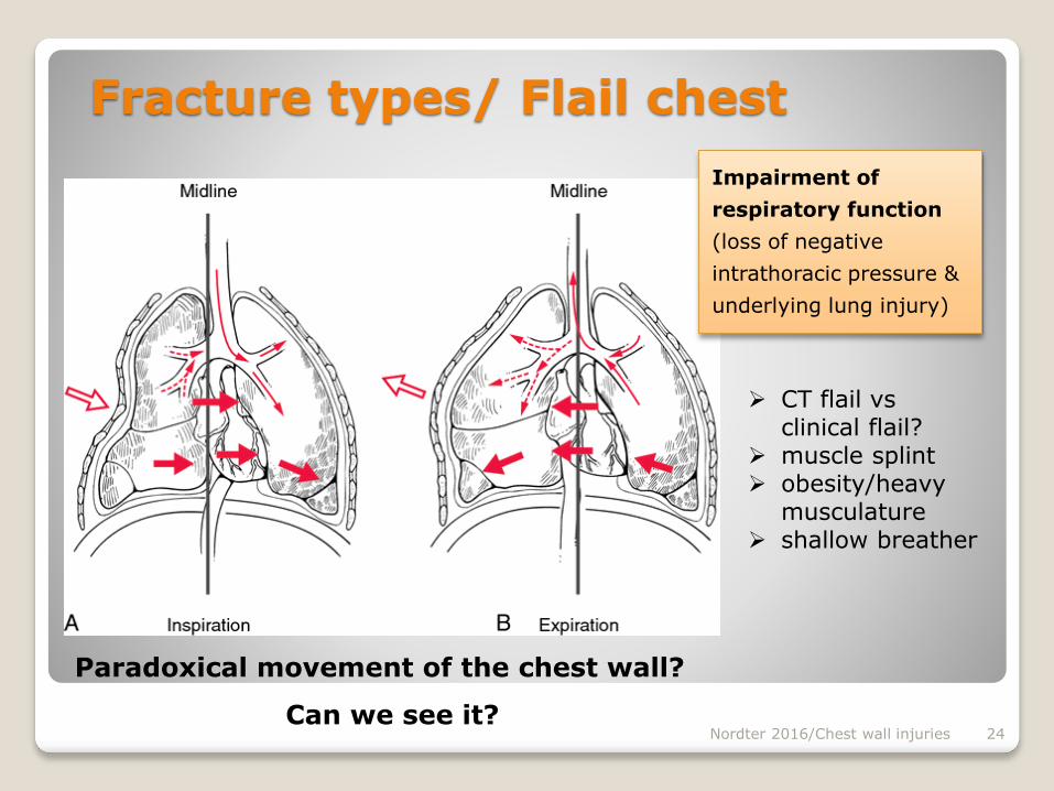
Paradoxical movement of the chest wall?
Can we see it?
Impairment of
respiratory function
(loss of negative
intrathoracic pressure &
underlying lung injury)
CT flail vs clinical flail?
muscle splint obesity/heavy
musculature shallow breather
Fracture types/ Flail chest
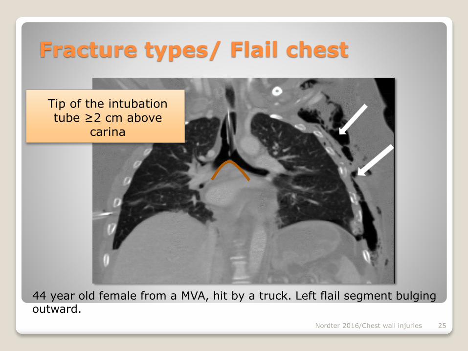
Nordter 2016/Chest wall injuries 25
44 year old female from a MVA, hit by a truck. Left flail segment bulging outward.
Tip of the intubation tube ≥2 cm above
carina
Fracture types/ Flail chest
Nordter 2016/Chest wall injuries 26
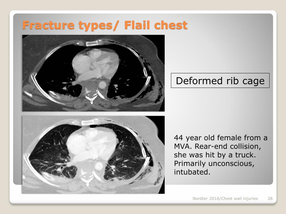
44 year old female from a MVA. Rear-end collision, she was hit by a truck. Primarily unconscious, intubated.
Deformed rib cage
Flail chest/Additional injuries
Nordter 2016/Chest wall injuries 27
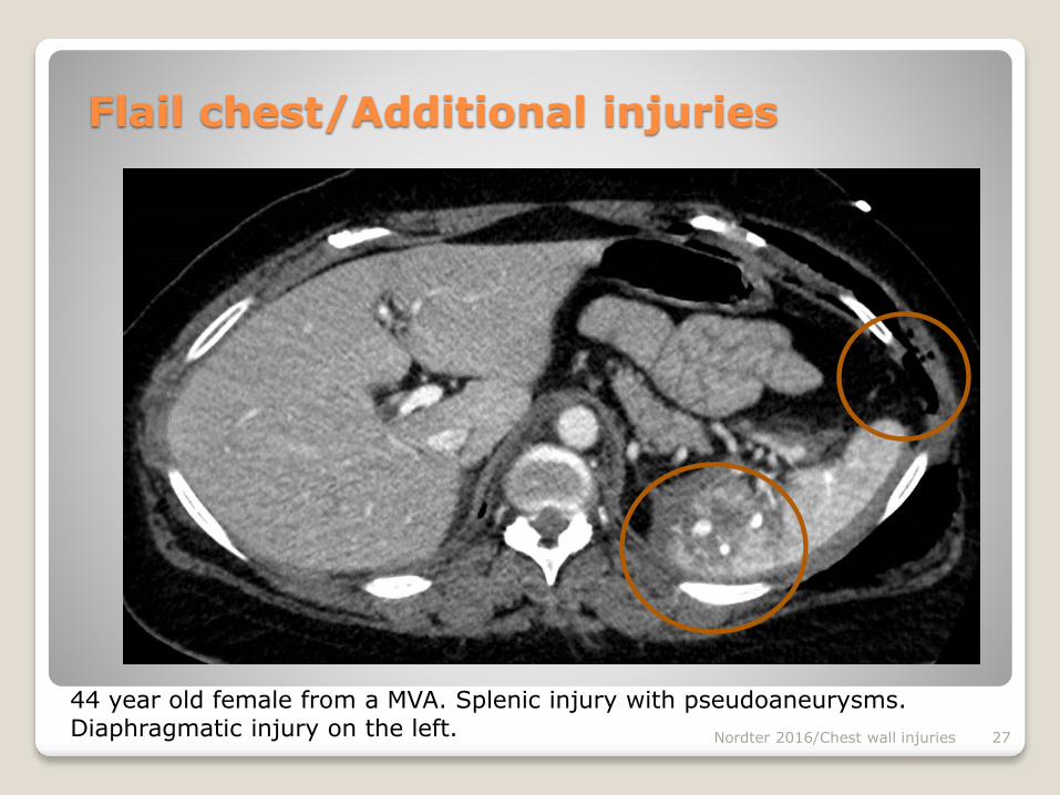
44 year old female from a MVA. Splenic injury with pseudoaneurysms. Diaphragmatic injury on the left.
Flail chest/Additional injuries
Nordter 2016/Chest wall injuries 28
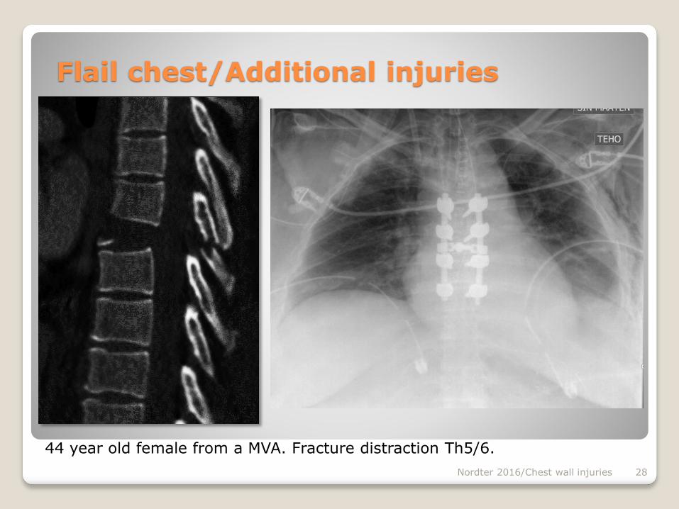
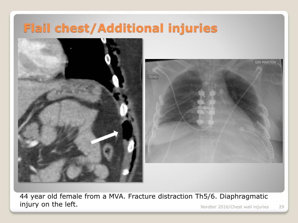
44 year old female from a MVA. Fracture distraction Th5/6.
Flail chest/Additional injuries
Nordter 2016/Chest wall injuries 29
44 year old female from a MVA. Fracture distraction Th5/6. Diaphragmatic injury on the left.
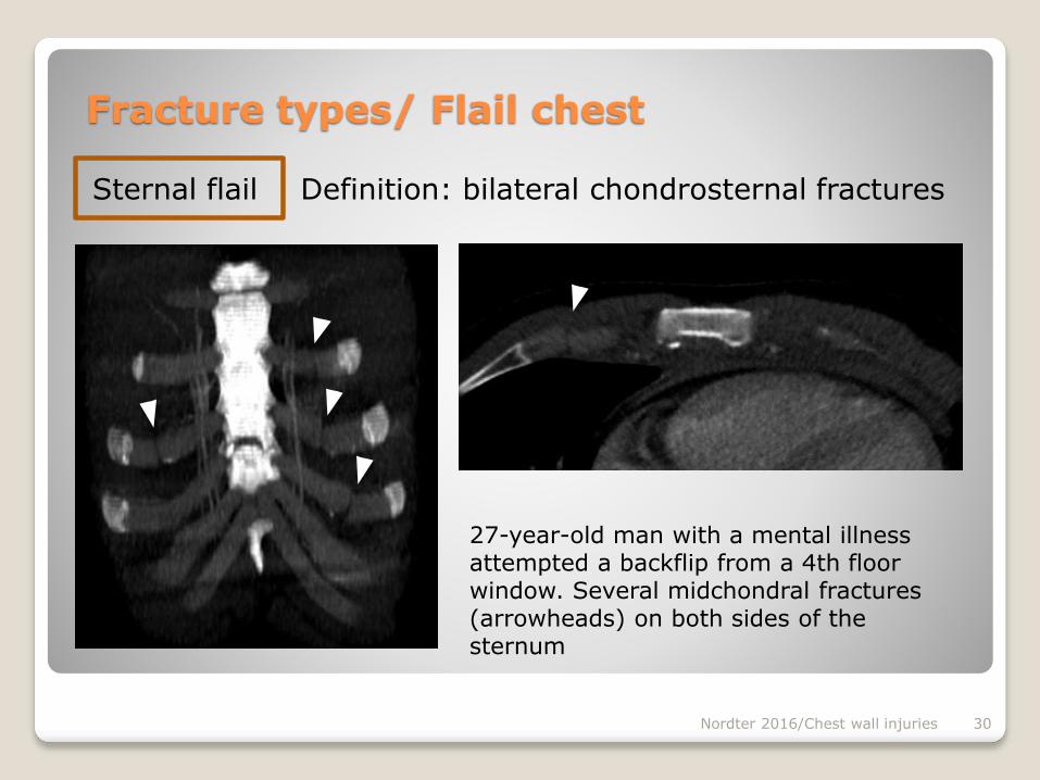
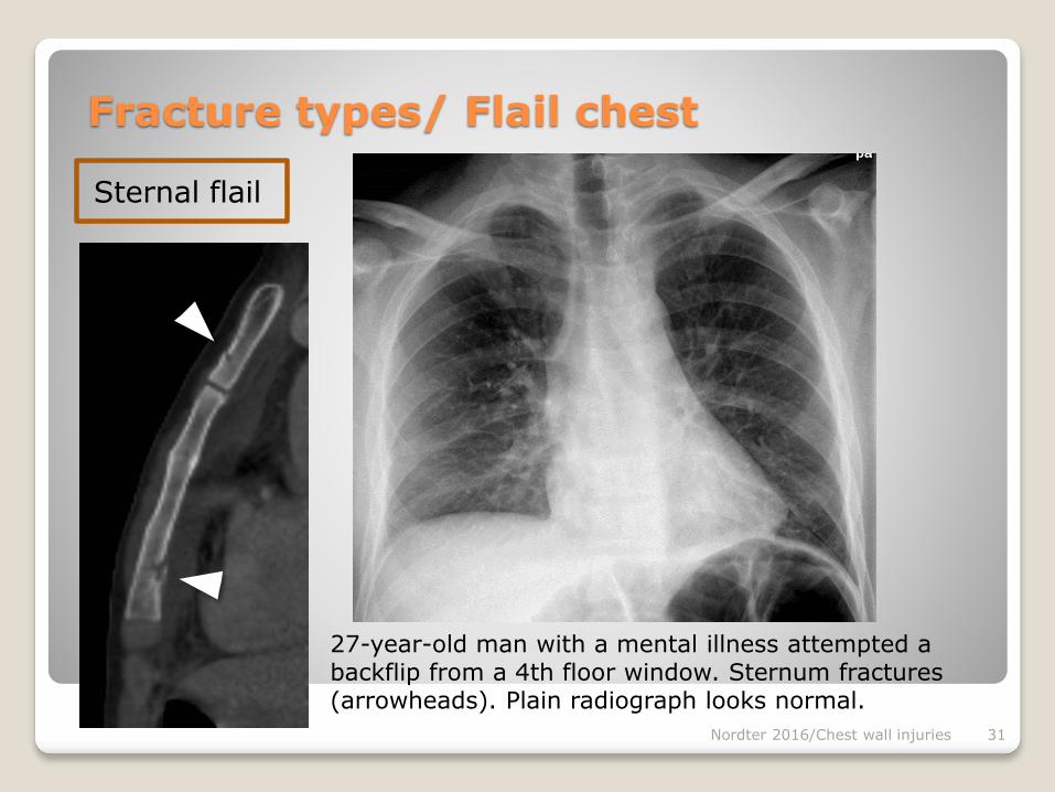
Sternal flail Definition: bilateral chondrosternal fractures
Nordter 2016/Chest wall injuries 30
Fracture types/ Flail chest
27-year-old man with a mental illness attempted a backflip from a 4th floor window. Several midchondral fractures (arrowheads) on both sides of the sternum
Sternal flail
Nordter 2016/Chest wall injuries 31
Fracture types/ Flail chest
27-year-old man with a mental illness attempted a backflip from a 4th floor window. Sternum fractures (arrowheads). Plain radiograph looks normal.
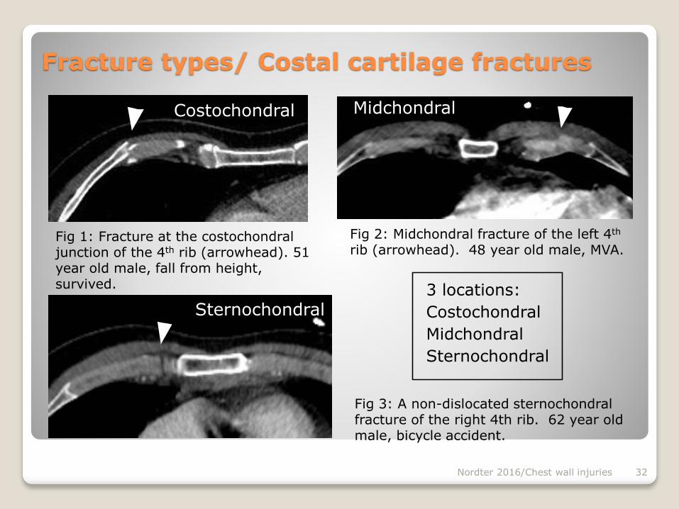
3 locations:
Costochondral
Midchondral
Sternochondral
Nordter 2016/Chest wall injuries 32
Fig 2: Midchondral fracture of the left 4th
rib (arrowhead). 48 year old male, MVA.Fig 1: Fracture at the costochondral junction of the 4th rib (arrowhead). 51 year old male, fall from height, survived.
Costochondral Midchondral
Sternochondral
Fig 3: A non-dislocated sternochondralfracture of the right 4th rib. 62 year old male, bicycle accident.
Fracture types/ Costal cartilage fractures
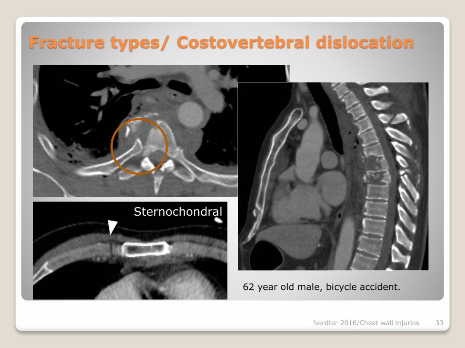
Nordter 2016/Chest wall injuries 33
Sternochondral
62 year old male, bicycle accident.
Fracture types/ Costovertebral dislocation
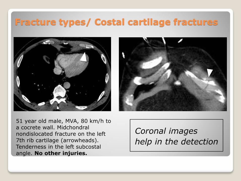
Fracture types/ Costal cartilage fractures
Coronal images
help in the detection
51 year old male, MVA, 80 km/h to a cocrete wall. Midchondral nondislocated fracture on the left 7th rib cartilage (arrowheads). Tenderness in the left subcostal angle. No other injuries.
Nordter 2016/Chest wall injuries 35
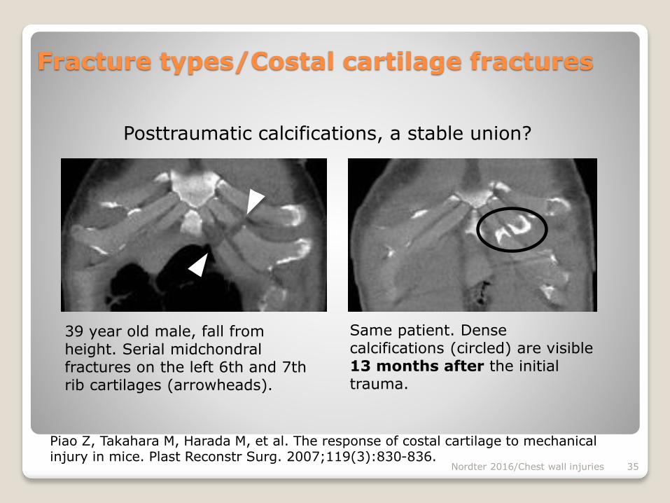
Posttraumatic calcifications, a stable union?
39 year old male, fall from height. Serial midchondral fractures on the left 6th and 7th rib cartilages (arrowheads).
Piao Z, Takahara M, Harada M, et al. The response of costal cartilage to mechanical injury in mice. Plast Reconstr Surg. 2007;119(3):830-836.
Fracture types/Costal cartilage fractures
Same patient. Dense calcifications (circled) are visible 13 months after the initial trauma.
Nordter 2016/Chest wall injuries 36
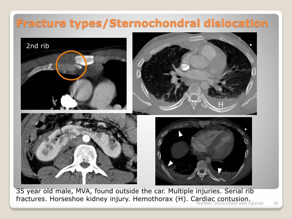
Fracture types/Sternochondral dislocation
35 year old male, MVA, found outside the car. Multiple injuries. Serial rib fractures. Horseshoe kidney injury. Hemothorax (H). Cardiac contusion.
H
2nd rib

Nordter 2016/Chest wall injuries 37
16 year old male, anterior direct stroke to the shoulder in a bandy game. Right sternoclavicular posterior luxation.
Fracture types/Sternoclavicular dislocation
CT angiography to excludevascular injury
Nordter 2016/Chest wall injuries 38
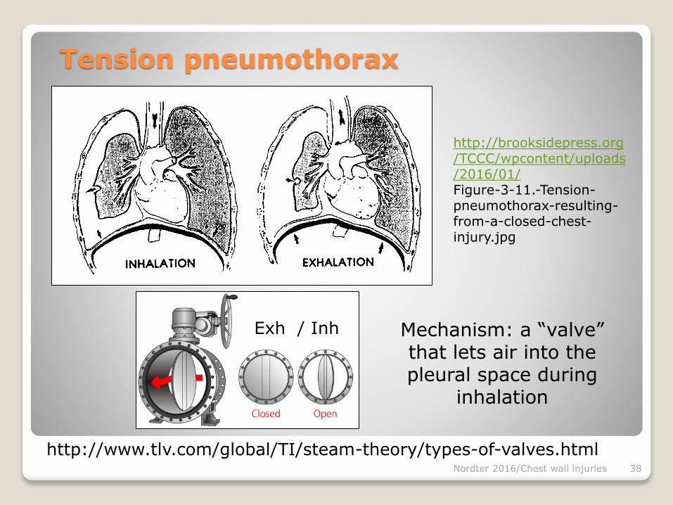
Mechanism: a “valve” that lets air into the pleural space during
inhalation
http://brooksidepress.org/TCCC/wpcontent/uploads/2016/01/Figure-3-11.-Tension-pneumothorax-resulting-from-a-closed-chest-injury.jpg
Tension pneumothorax
Exh / Inh
http://www.tlv.com/global/TI/steam-theory/types-of-valves.html
Nordter 2016/Chest wall injuries 39
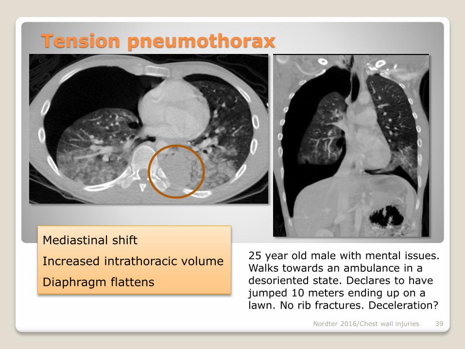
Tension pneumothorax
25 year old male with mental issues. Walks towards an ambulance in a desoriented state. Declares to have jumped 10 meters ending up on a lawn. No rib fractures. Deceleration?
Mediastinal shift
Increased intrathoracic volume
Diaphragm flattens
Nordter 2016/Chest wall injuries 40
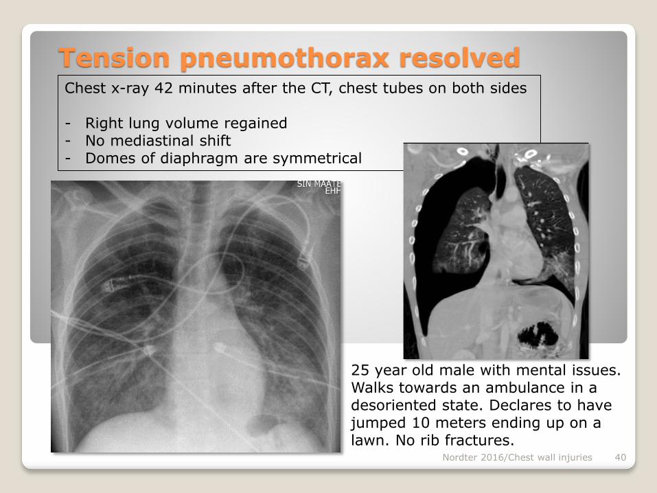
Tension pneumothorax resolved
25 year old male with mental issues. Walks towards an ambulance in a desoriented state. Declares to have jumped 10 meters ending up on a lawn. No rib fractures.
Chest x-ray 42 minutes after the CT, chest tubes on both sides
- Right lung volume regained- No mediastinal shift- Domes of diaphragm are symmetrical
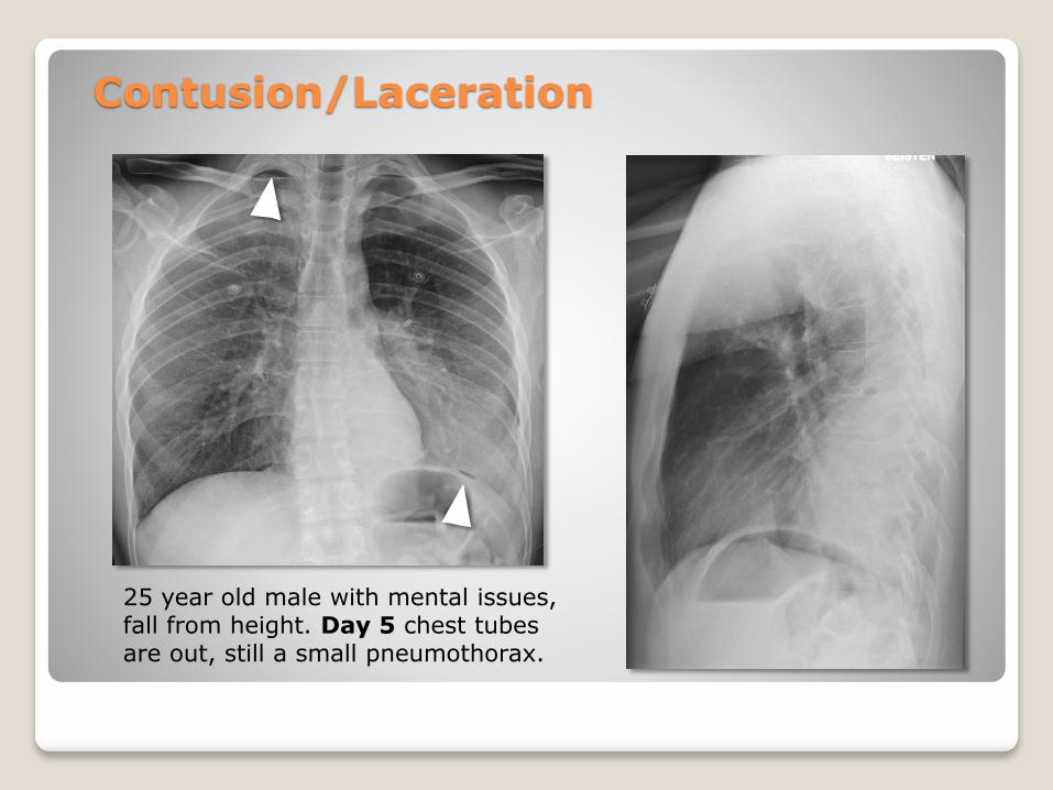
25 year old male with mental issues, fall from height. Day 5 chest tubes are out, still a small pneumothorax.
Contusion/Laceration
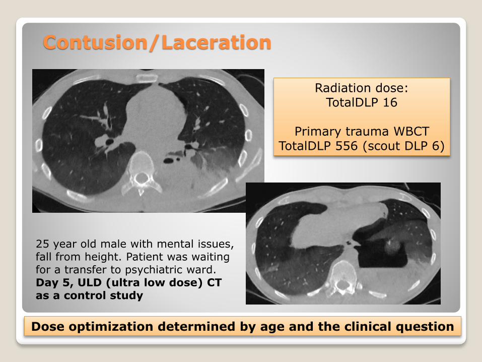
25 year old male with mental issues, fall from height. Patient was waiting for a transfer to psychiatric ward. Day 5, ULD (ultra low dose) CT as a control study
Contusion/Laceration
Radiation dose: TotalDLP 16
Primary trauma WBCTTotalDLP 556 (scout DLP 6)
Dose optimization determined by age and the clinical question

25 year old male with mental issues, fall from height. Patient was waiting for a transfer to psychiatric ward. Day 5, ULD (ultra low dose) CT with MBIR (model based iterative reconstruction (GE)
Contusion/Laceration
ULD CT fit for evaluation ofVolume of pneumothorax
Contusions Tube positioning
Amount and location of fluid
CAVE!Suspicion of active bleeding
or empyemarequires i.v. contrast
and assesment of mediastinal structures a
higher dose!
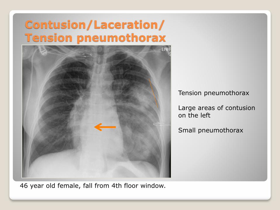
Contusion/Laceration/Tension pneumothorax
46 year old female, fall from 4th floor window.
Tension pneumothorax
Large areas of contusionon the left
Small pneumothorax
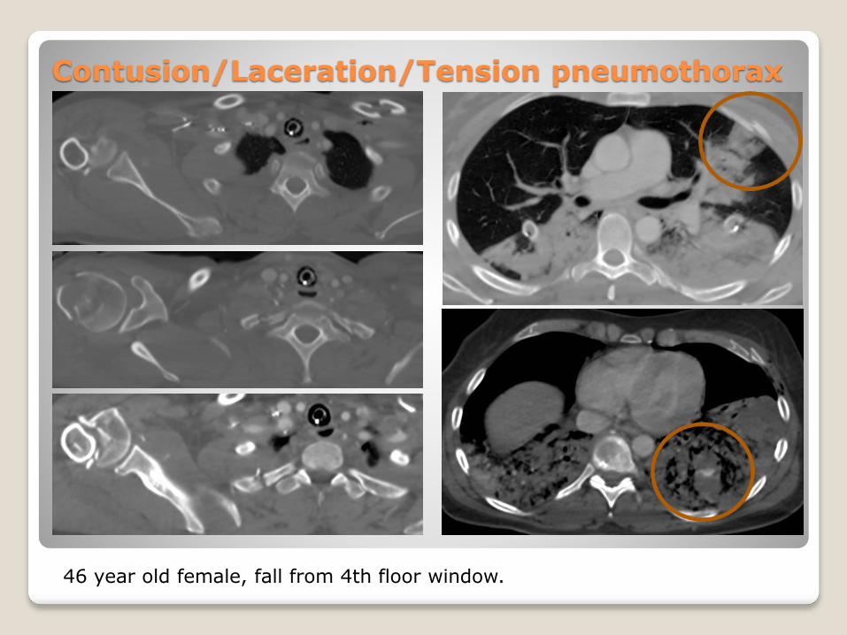
Contusion/Laceration/Tension pneumothorax
46 year old female, fall from 4th floor window.
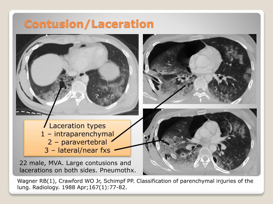
22 male, MVA. Large contusions and lacerations on both sides. Pneumothx.
Contusion/Laceration
Laceration types1 – intraparenchymal
2 – paravertebral3 – lateral/near fxs
Wagner RB(1), Crawford WO Jr, Schimpf PP. Classification of parenchymal injuries of the lung. Radiology. 1988 Apr;167(1):77-82.
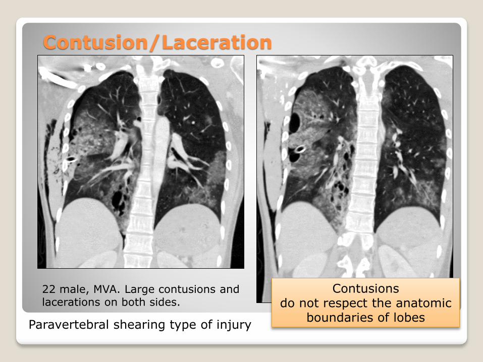
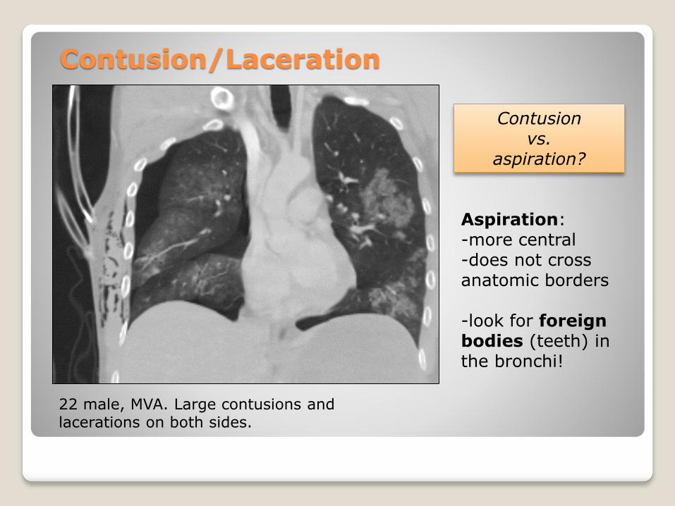
22 male, MVA. Large contusions and lacerations on both sides.
Contusion/Laceration
Contusionsdo not respect the anatomic
boundaries of lobesParavertebral shearing type of injury
22 male, MVA. Large contusions and lacerations on both sides.
Contusion/Laceration
Contusionvs.
aspiration?
Aspiration:-more central-does not cross anatomic borders
-look for foreignbodies (teeth) in the bronchi!
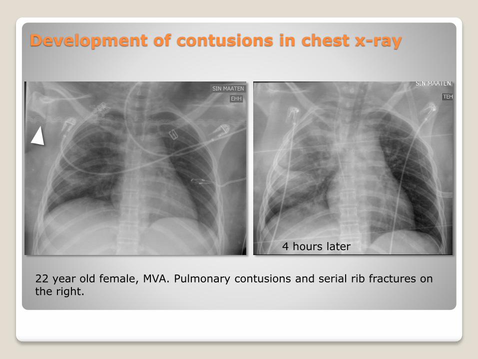
Development of contusions in chest x-ray
22 year old female, MVA. Pulmonary contusions and serial rib fractures on the right.
4 hours later
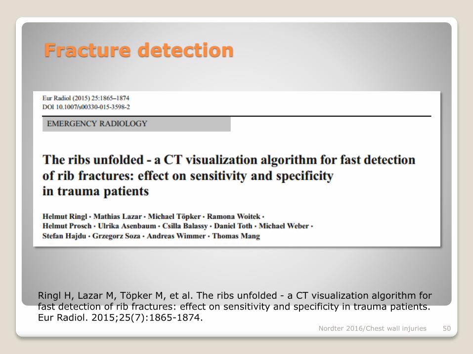
Fracture detection
Nordter 2016/Chest wall injuries 50
Ringl H, Lazar M, Töpker M, et al. The ribs unfolded - a CT visualization algorithm for fast detection of rib fractures: effect on sensitivity and specificity in trauma patients. Eur Radiol. 2015;25(7):1865-1874.
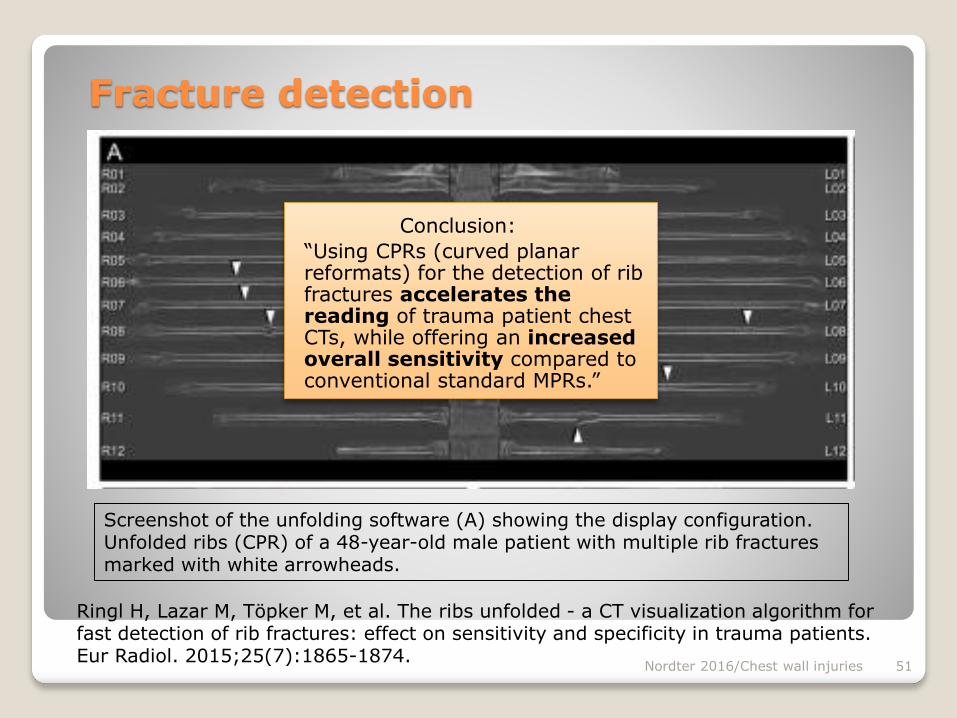
Nordter 2016/Chest wall injuries 51
Screenshot of the unfolding software (A) showing the display configuration. Unfolded ribs (CPR) of a 48-year-old male patient with multiple rib fractures marked with white arrowheads.
Fracture detection
Ringl H, Lazar M, Töpker M, et al. The ribs unfolded - a CT visualization algorithm for fast detection of rib fractures: effect on sensitivity and specificity in trauma patients. Eur Radiol. 2015;25(7):1865-1874.
Conclusion:
“Using CPRs (curved planar reformats) for the detection of rib fractures accelerates the reading of trauma patient chest CTs, while offering an increased overall sensitivity compared to conventional standard MPRs.”
Nordter 2016/Chest wall injuries 52
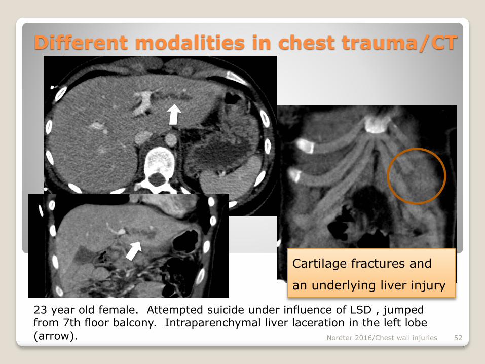
Different modalities in chest trauma/CT
Cartilage fractures and
an underlying liver injury
23 year old female. Attempted suicide under influence of LSD , jumpedfrom 7th floor balcony. Intraparenchymal liver laceration in the left lobe(arrow).
Nordter 2016/Chest wall injuries 53
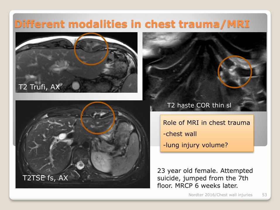
Different modalities in chest trauma/MRI
23 year old female. Attempted suicide, jumped from the 7th floor. MRCP 6 weeks later.
Role of MRI in chest trauma
-chest wall
-lung injury volume?
T2 Trufi, AX
T2 haste COR thin sl
T2TSE fs, AX
Nordter 2016/Chest wall injuries 54
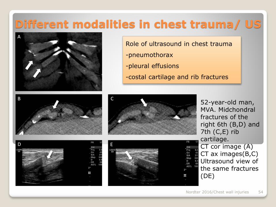
Different modalities in chest trauma/ US
Role of ultrasound in chest trauma
-pneumothorax
-pleural effusions
-costal cartilage and rib fractures
52-year-old man, MVA. Midchondralfractures of the right 6th (B,D) and 7th (C,E) rib cartilage.CT cor image (A)CT ax images(B,C)Ultrasound view of the same fractures(DE)
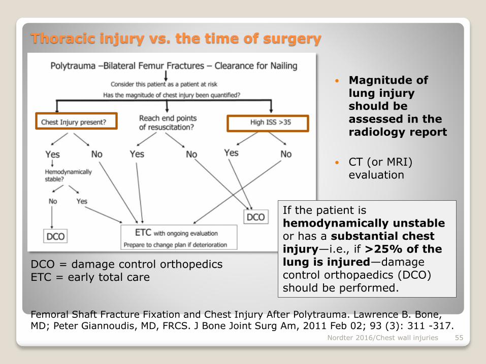
Magnitude of lung injury should be assessed in the radiology report
CT (or MRI) evaluation
Nordter 2016/Chest wall injuries 55
Femoral Shaft Fracture Fixation and Chest Injury After Polytrauma. Lawrence B. Bone, MD; Peter Giannoudis, MD, FRCS. J Bone Joint Surg Am, 2011 Feb 02; 93 (3): 311 -317.
If the patient is hemodynamically unstable or has a substantial chest injury—i.e., if >25% of the lung is injured—damage control orthopaedics (DCO) should be performed.
Thoracic injury vs. the time of surgery
DCO = damage control orthopedicsETC = early total care
Nordter 2016/Chest wall injuries 56
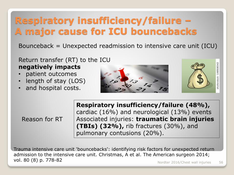
Trauma intensive care unit 'bouncebacks': identifying risk factors for unexpected return admission to the intensive care unit. Christmas, A et al. The American surgeon 2014; vol. 80 (8) p. 778-82
Respiratory insufficiency/failure –A major cause for ICU bouncebacks
Respiratory insufficiency/failure (48%), cardiac (16%) and neurological (13%) events Associated injuries: traumatic brain injuries (TBIs) (32%), rib fractures (30%), and pulmonary contusions (20%).
Bounceback = Unexpected readmission to intensive care unit (ICU)
Return transfer (RT) to the ICUnegatively impacts • patient outcomes • length of stay (LOS)• and hospital costs.
Reason for RT
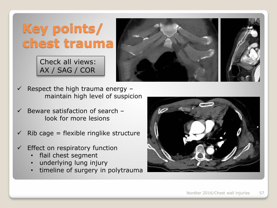
Key points/ chest trauma
Respect the high trauma energy –maintain high level of suspicion
Beware satisfaction of search –look for more lesions
Rib cage = flexible ringlike structure
Effect on respiratory function • flail chest segment• underlying lung injury• timeline of surgery in polytrauma
Nordter 2016/Chest wall injuries 57
Check all views:AX / SAG / COR
Literature
Nordter 2016/Chest wall injuries 58
CHEST WALL INJURIES
Peters S, Nicolas V, Heyer CM. Multidetector computed tomography-spectrum of blunt chest wall and lung
injuries polytraumatized patients. Clin Radiol. 2010;65(4):333-338.
Miller LA. Chest wall, lung, and pleural space trauma. Radiol Clin North Am. 2006;44(2):213-224, viii.
Lomoschitz FM, Eisenhuber E, Linnau KF, Peloschek P, Schoder M, Bankier AA. Imaging of chest trauma:
radiological patterns of injury and diagnostic algorithms. Eur J Radiol. 2003;48(1):61-70.
Mirvis SE, Harris JHJ. Chest: Trauma. In: John H. Harris; Jr., Thomas L Pope Jr., ed. Harris & Harris’ Radiology
of Emergency Medicine. 5th ed. Philadelphia: Lippincott Williams & Wilkins, Wolters Kluwer; 2012:489-572.
Sangster GP, González-Beicos A, Carbo AI, et al. Blunt traumatic injuries of the lung parenchyma, pleura,
thoracic wall, and intrathoracic airways: multidetector computer tomography imaging findings. Emerg
Radiol. 2007;14(5):297-310.
Malghem J, Vande Berg B, Lecouvet F, Maldague B. Costal cartilage fractures as revealed on CT and
sonography. AJR Am J Roentgenol. 2001;176(2):429-432.
Subhas N, Kline MJ, Moskal MJ, White LM, Recht MP. MRI Evaluation of Costal Cartilage Injuries. Am J
Roentgenol. November 2008:191: 129-132.
Bone LB, Giannoudis P. Femoral Shaft Fracture Fixation and Chest Injury After Polytrauma. J Bone Joint Surg
Am, 2011 Feb 02; 93 (3): 311 -317.