chicago -...
TRANSCRIPT

1
CHICAGO October 30 – November 1, 2018
Presented by Retina Consultants, Ltd., University of Chicago, and The Illinois Eye and Ear Infirmary

Meeting Wifi Network: UClub Password: 1234567890
2
Dear Colleagues,
On behalf of Retina Consultants, Ltd., University of Chicago and University of Illinois Eye and Ear Infirmary it is our honor to welcome you ROP HOT TOPICS Chicago!
We are pleased to have you participate in this important program. We are all on the same journey to improve the lives of our patients; however limited data forces us to navigate through difficult, confused clinical challenges at many levels.
The objective of this meeting is to allow an exploration and encourage discussion of key aspects of ROP that inform clinical management, now and in the future.
Many thanks to all the members of the Program Committee, Faculty, and volunteers who have worked hard to create a dynamic program.
A special thank you to our Section Organizers Peter Campbell, Mary Elizabeth Hartnett, Michael Msall and Haresh Kirpalani.
Also, gratitude to our volunteer organizers Mark Solinksi, April Ingram, Kate Lewis and Aneta Firosz. We look forward to joining you as you continue your vital work to improve the diagnosis, management and treatment of ROP. Warmest Regards, ROP HOT TOPICS Organizing Committee
Michael Shapiro, MD Michael Blair, MD Anna Ells, MD, FRCS(C)
Sarah Rodriguez, MD, MPH RV Paul Chan, MD, FACS
WELCOME

Meeting Wifi Network: UClub Password: 1234567890
3
ROP Hot Topics - Abstracts Visual, refractive and anatomical outcomes in clinical practice of 15 years of ROP in the Indian Twin cities ROP study database (ITCROPS). Subhadra Jalali, Priyanaka Kammari, Mahesh Kumar, Mohammed Mansoor, Padmaja K Rani, Divya Balakrishnan. L V Prasad Eye Institute, Hyderabad, India. Objective: Report outcomes in treated and spontaneously regressed retinopathy of prematurity (ROP) in clinical practice. Method: The prospective ROP database (year 2000- 2014) was analysed retrospectively. Of 1474 eyes with ROP, 757 had spontaneous regression and 717 underwent sight preserving treatments (Laser/ Avastin/ Vitrectomy/ belt buckle). Visual and retinal outcomes were categorized into good, fair and poor based on the visual acuity and anatomical status of the retina respectively. Refractive outcomes were categorized as mild, moderate and severe. Birth weight and gestational age were assessed for associations with outcomes. Results: The spontaneously regressed group (486 eyes) showed good, fair and poor visual outcomes in 453, 25 and 8 eyes respectively whereas in the treated group (624 eyes) these outcomes were seen in 503, 82, and 39 eyes respectively. There were 402 mild, 60 moderate and 26 severe refractive outcomes in the regressed group and 266 mild, 116 moderate and 204 severe refractive outcomes in the treated group. Of 752 spontaneously regressed eyes, 738 had good, 12 fair and 2 poor retinal outcomes. Of 689 eyes in the treated group 588 had good, 66 fair and 35 poor retinal outcomes. High refractive errors in spontaneously regressed group were significantly associated with lower gestational age. Conclusion: With timely screening, close follow-up and prompt treatments, most babies who develop ROP can retain the good visual and retinal anatomic outcomes in the treated group having more severe disease as in the spontaneously regressed group that have lesser severity, depicting sight preservation and promotion.
_________________________________________________

Meeting Wifi Network: UClub Password: 1234567890
4
Optical coherent tomography angiography imaging and fine foveal structure in patients with history of prematurity Hiroyuki Kondo and Hirofumi Morita Department of Ophthalmology, University of Occupational and Environmental Health, Japan Objective: To determine the area of the foveal avascular zone (FAZ) and foveal thickness by optical coherent tomography angiography (OCTA) in patients with a history of retinopathy of prematurity (ROP). Study Design: Retrospective cohort study. Methods: Fifty-four (32 female and 19 male) patients with a gestational age of ≤34 weeks and routine ROP were studied. The SS-OCT angiography (OCTA) images with a 3 mm X 3 mm scan were obtained from all patients. The main outcome measures were the FAZ area and the thickness of the inner retinal layer (IRL) and the outer retinal layer (ORL). Results: OCTA images were obtained from 84 eyes that had a mean visual acuity of -0.096 ± 0.1 logMAR units and a mean refractive error (spherical equivalent) of -0.87 ± 3.4 diopters. The mean FAZ area was 0.17 ± 0.1 mm2, and eyes that required treatment had significantly smaller FAZ areas (0.16 ± 0.1 mm2) than eyes not requiring treatment (0.18 ± 0.1 mm2) or eyes without ROP (0.21 ± 0.13 mm2). The mean thickness of the IRL at the foveal center was 15.6 ± 15.9 μm and that of the ORL was 192.7 ± 16.1 μm. Both thicknesses were negatively correlated with the FAZ area (r = -0.67, P<0.0001 and r = -0.48, P<0.0001). Conclusions: The smaller FAZ area and abnormal thicknesses of the IRL and ORL in eyes with a history of ROP are probably due to the abnormal blood vessels growth and spread throughout the retina.
________________________________________________

Meeting Wifi Network: UClub Password: 1234567890
5
Foveal development in infants treated with bevacizumab or laser photocoagulation for retinopathy of prematurity. R Vogel, M. Strampe, O. Fagbemi, A. Vistocky, S. Tarima, J. Carroll, D. Costakos. Published in Ophthalmology. 125(3)444-452, 2018. PMID:29103792 Objective: To characterize and quantify early foveal development in preterm infants and to compare this development between eyes treated with intravitreal bevacizumab or laser photocoagulation (LPC) and untreated eyes. Methods: An observational case series of one hundred thirty-one preterm infants undergoing retinopathy of prematurity (ROP) screenings was performed longitudinally using handheld OCT. Thickness measurements of the inner and outer retinal layers were obtained at the foveal center and the nasal and temporal foveal rims. Comparisons between treated and untreated eyes were adjusted for age and other confounding variables. Main outcome measures included weekly change in inner and outer retinal thickness and presence of inner retinal layers, ellipsoid zone (EZ), and cystoid macular changes (CMCs). Results: Outer retinal thickness at the foveal center increased by 3.1 μm/week in untreated eyes and 7.2 μm/week in bevacizumab-treated eyes (P = 0.038). Eyes treated with LPC had a lower probability of having all inner retinal layers present at the foveal center (odds ratio, 0.04; P = 0.001) and a lower probability of having the EZ present at the foveal center (odds ratio, 0.07; P = 0.024) compared with untreated eyes. Cystoid macular changes were found in 53% of patients and 22% of imaging sessions. The age-adjusted incidence of CMCs was not correlated with bevacizumab or LPC treatment. Conclusions: Intravitreal bevacizumab therapy for ROP is associated with more rapid outer retinal thickening at the foveal center, whereas LPC is associated with earlier extrusion of the inner retinal layers and delayed development of the EZ at the foveal center. Long-term follow-up is needed to determine the visual significance of these findings. _________________________________________________

Meeting Wifi Network: UClub Password: 1234567890
6
Macular Structures, Optical Components, and Visual Acuity in Preschool Children after Intravitreal Bevacizumab or Laser Treatment Wei-Chi Wu, Yung-Sung Lee, Lai-Chu See, Shu-Hao Chang, Nan-Kai Wang, Yih-Shiou Hwang, Chi-Chun Lai, Kuan-Jen Chen Purpose: To investigate the macular structures, optical components, and visual acuity in preschool-aged children with a history of type I retinopathy of prematurity who underwent either intravitreal bevacizumab (IVB), laser, or a combination of treatments. Design: Prospective cross-sectional study. Setting: A referred medical center in Taiwan. Study Population: 80 eyes from 42 patients (33 IVB-treated eyes from 17 children, 24 laser-treated eyes from 13 children, and 23 laser + IVB-treated eyes from 12 children). Observation Procedure: Spectral-domain optical coherence tomography. Main Outcome Measures: The retinal thickness in the foveal area and the associated morphologic changes in foveal depression. Results: Compared with the laser-treated and laser + IVB-treated eyes, the IVB-treated eyes had less myopia and deeper anterior chamber depths but presented similar axial lengths and corneal curvatures (P = .001, .002, .95 and .16, respectively). The IVB-treated eyes had significantly thinner foveal, parafoveal, and perifoveal retinal thicknesses (P < .01 for all) and a higher incidence of foveal depression than the laser- or laser + IVB-treated eyes. The macular and subfoveal choroidal thicknesses did not differ among the groups (P = .21 and .63, respectively). Moreover, compared with the eyes treated with laser or laser + IVB, the IVB-treated eyes had better uncorrected visual acuity, although a significant difference was not observed in best-corrected visual acuity (P = .008 and .29, respectively). Conclusions: Compared with laser therapy, IVB-treated eyes were associated with deeper anterior chamber depths and thinner foveal, parafoveal and perifoveal thicknesses. Moreover, these IVB-treated eyes had less refractive errors and better uncorrected visual acuity. _________________________________________________
Meeting Wifi Network: UClub Password: 1234567890
7
Visual and Refraction Outcomes of Laser Ablation and Bevacizumab in management of ROP Sarah Hilkert Rodriguez, MD, MPH, Sidney Schechet, MD, Nandita Anand, MD, Michael Paul Blair, MD Purpose: To report refractive and visual outcomes with intravitreal bevacizumab (IVB) and laser for type 1 ROP and to correlate structural finding with functional outcomes. Methods: Retrospective chart reviews identified infants treated for type 1 ROP between 2006 – 2016, who received laser before (n = 41) and IVB after (n = 46) the implementation of a change in practice. Among infants who received IVB, 35 completed an exam under anesthesia with fluorescein angiography (FA) and prophylactic peripheral retinal photocoagulation (IVB-PRP). Refractive outcomes compared 34 eyes of 19 infants with laser and 40 eyes of 21 infants with IVB-PRP between 2-4 years. Visual acuity (VA) was available for 24 patients who were older than 4 years of age. Results: Mean SE was -7.4 + 5.2 for laser and -0.16 + 2.2 for IVB-PRP (p < 0.001). This relationship persisted after stratification by zone of ROP and the presence of aggressive posterior ROP. Prior to prophylactic laser, all eyes had peripheral capillary dropout and other vascular abnormalities, such as shunts, tangles, or abnormal branching; 86% had peripheral nonperfusion, 63% had leakage, and 20% had posterior pole abnormalities. Median VA was 20/30, and 37/48 (77%) eyes were 20/40 or better. Discussion: Delayed prophylactic laser, in hopes of reducing the risk of late retinal detachment, does not appear to negate the refractive benefits of IVB. Vascular abnormalities after IVB are not necessarily incompatible with good vision but may indicate residual ischemia, for which laser to peripheral avascular retina may help prevent associated complications. 1. Mintz-Hittner HA, Kennedy KA, Chuang AZ. BEAT- ROP Cooperative Group. Efficacy of intravitreal bevacizumab for stage 3+ retinopathy of prematurity. The New England Journal of Medicine 2011; 364:603-15. 2. Geloneck MM, Chuang AZ, Clark WL, et al. Refractive outcomes following bevacizumab monotherapy compared with conventional laser treatment: a randomized clinical trial. JAMA Ophthalmology 2014;132(11):1327–33. 3. Hwang CK, Hubbard GB, Hutchinson AK, Lambert SR. Outcomes after intravitreal bevacizumab versus laser photocoagulation for retinopathy of prematurity: A 5-year retrospective analysis. Ophthalmology 2015;122(5):1008-15. 4. Garcia Gonzalez JM, Snyder L, Blair M, Rohr A, Shapiro M, Greenwald M. Prophylactic peripheral laser and fluorescein angiography after bevacizumab for retinopathy of prematurity. Retina 2018 Apr;38(4):764-772. 5. Lepore D, Quinn GE, Molle F et al. Follow-up to Age 4 Years of Treatment of Type 1 Retinopathy of Prematurity Intravitreal Bevacizumab Injection versus Laser: Fluorescein Angiographic Findings. Ophthalmology 2018; 125(2):218-226. 6. Early Treatment for Retinopathy of Prematurity Cooperative Gropu, Good WV, Hardy RJ, et al. Final visual acuity results in the early treatment for retinopathy of prematurity study. Arch Ophthalmol 2010; 128(6): 663 – 671.
_________________________________________________

Meeting Wifi Network: UClub Password: 1234567890
8
Mueller cells and VEGF inhibition Mary Elizabeth Hartnett, MD, FACS, FARVO John A. Moran Eye Center, University of Utah Objective(s): To describe 1) a representative model of ROP; and 2) gene therapy to knock down Mueller cell (MC)-VEGF or endothelial cell (EC)-VEGF receptor 2 (VEGFR2) to compare effects between broad VEGF inhibition from MC-VEGF knockdown or selective EC-VEGFR2 knockdown on intravitreal neovascularization, physiologic retinal vascular development and retina. Study Design: Experimental studies with gene therapy in rat model of ROP Methods: Newborn Sprague-Dawley rats were exposed to 24-hour repeated cycles between 50% and 10% oxygen for 14 days followed by room air. At day 8, subretinal delivery of lentiviruses carrying: A) MC-VEGFA shRNA, MC-VEGF164 shRNA or MC-luciferase shRNA (control) or B) EC- VEGFR2 shRNA or EC-luciferase shRNA (control) were performed. Outcomes were peripheral avascular/total retinal area (AVA), intravitreal neovascular/total retina area (IVNV), electroretinograms (ERGs), thicknesses of retinal layers. Results: Compared to respective luciferase controls, MC-VEGFA or MC-VEGF164 knockdown significantly reduced IVNV but not AVA, whereas knockdown of EC-VEGFR2 reduced IVNV and AVA. Compared to respective controls, knockdown of MC-VEGFA thinned the outer nuclear layer, whereas knockdown of MC-VEGF164 or EC-VEGFR2 did not adversely affect neural retinal thicknesses. Compared to respective controls, MC-VEGFA or MC-VEGF164 and EC-VEGFR2 knockdown led to improved b-wave amplitudes on ERsG. Conclusions: MC-VEGFA knockdown is similar to inhibiting VEGF with an intravitreal antibody and has potentially adverse effects on the retina. Knockdown of MC-VEGF164 permits secreted forms of VEGF to access the neural retina and did not show thinning of the retina. EC-VEGFR2 knockdown not only reduces IVNV, but also allows physiologic retinal vascular development. _________________________________________________

Meeting Wifi Network: UClub Password: 1234567890
9
Oxygen Induced Retinopathy Mouse Model Lois Smith, MD, PhD, Harvard Medical School, Boston Children's Hospital Objective: To understand the strengths and weaknesses of the mouse model of oxygen-induced retinopathy for the study of retinopathy of prematurity (ROP) Study Design: The model design with oxygen exposure of 7 day old mouse pups for 5 days followed by room air until P17 is set to evaluate proliferative retinopathy, vaso-regression and revascularization with interventions Methods: The normal mouse retinal vascular development and the effect of intervention of oxygen will be discussed Results: the use of the mouse OIR model with VEGF, IGF-1, and omega 3 LCPUFAS will be discussed Conclusions: the mouse OIR model has been useful to study aspect of ROP 1. Smith LE, Wesolowski E, McLellan A, Kostyk SK, D'Amato R, Sullivan R, D'Amore PA. Oxygen-induced retinopathy in the mouse. Invest Ophthalmol Vis Sci 1994; 35(1):101-11 1. Stahl A, Connor K, Sapieha P, Willett K, Krah N, Dennison R, Chen J, Guerin K, Smith LE. Computer-aided quantification of retinal neovascularization. Angiogenesis Sept 12 p297-301 2009 2. Connor K, Krah N, Dennison R, Willett K, Aderman C, Chen J, Guerin K, Sapieha P, Stahl A, Smith LE. Quantification of oxygen-induced retinopathy in the mouse: a model of vessel loss, vessel regrowth and pathological angiogenesis. Nature Protocols 2009;4(11):1565-73. 3. Stahl A, Connor KM, Sapieha P, Chen J, Dennison RJ, Krah NM, Seaward MR, Willett KL, Aderman CM, Guerin KI, Hua J, Löfqvist C, Hellström A, Smith LE. The mouse retina as an angiogenesis model. Invest Ophthalmol Vis Sci. 2010 Jun;51(6):2813-26. Review
_________________________________________________

Meeting Wifi Network: UClub Password: 1234567890
10
Imaging of Retinal Oxygen Extraction, Vascularization, and Thickness in Oxygen-induced Retinopathy Mahnaz Shahidi1, Olachi Mezu-Ndubuisi2, Justin Wanek3, Pang-yu Teng4, Norman Blair3 1.Department of Ophthalmology, University of Southern California 2.Department of Pediatrics, University of Wisconsin Madison 3.Department of Ophthalmology and Visual Sciences, University of Illinois at Chicago 4.Department of Radiology, University of California Los Angeles Objective(s): The objective of the research study was to determine alterations in retinal oxygen extraction, vascularization, and thickness in experimental oxygen-induced retinopathy (OIR). Study Design: Prospective cross-sectional. Methods: The study was performed in 19 neonatal OIR and 11 control mice. The OIR mice were exposed to 77% oxygen from postnatal day 7 (P7) to P12, while the control mice were exposed to room air. Phosphorescence lifetime imaging, fluorescein angiography, and spectral domain optical coherence tomography were performed between P17 and P19. Image analysis was performed to measure retinal vascular oxygen tension, areas of , and total retinal thickness. Results: Retinal arteriovenous oxygen tension difference was increased in OIR mice compared to control mice. Retinal capillary network between major retinal vessels was dense in control mice. In contrast, hypovascular regions with fewer capillaries or avascular regions devoid of visible capillaries were observed in OIR mice. Retinal thickness in avascular regions was significantly lower than thickness in vascular and hypovascular regions. Conclusions: The findings suggest that sparse retinal vascular network due to OIR results in elevated oxygen extraction to supply a larger tissue volume and in retinal thinning due to either arrested retinal development or ischemia-induced apoptosis. _________________________________________________

Meeting Wifi Network: UClub Password: 1234567890
11
Using Metabolic Retinal Vascular Imaging to Understand ROP Pathology-Visible-light OCT Amani Fawzi, MD, Brian Soetikno, Hao Zhang, and Kathryn Farrow. Objective: to study the retinal metabolic oxygen rates during 50/10 oxygen induced retinopathy (OIR) in the rat Study Design:cross-sectional study of the OIR rat model, compared to room mate controls Methods: we used visible light OCT to study the retinal metabolism at P18 in the OIR model. This technology allowed us to measure vascular oxyhemoglobin, retinal blood flow and therefore the retinal oxygen metabolism, in vivo. We complemented our studies with retinal morphometry and detailed studies of the vasculature ex-vivo Results: In the OIR, we found that retinal metabolic rate was significantly reduced, along with significant reduction in retinal thickness at all layers. The retinal vascular flat mounts showed 10% neovascularization. Conclusions: Imaging retinal vascular morphology does not capture the complexity of retinal metabolism and consequences of ischemia during OIR. These studies become more relevant as they relate to the pathobiology of ROP regression and progression in humans. Applications of the vis-OCT technology to human disease offers an exciting new tool to understand the balance between oxygen supply and demand in ischemic retinopathies, and hopefully soon, ROP. _________________________________________________
Meeting Wifi Network: UClub Password: 1234567890
12
SAFER technique for the treatment of Type 1 ROP Armie Harper M.D. Austin Retina Associates The description of the SAFER technique for the treatment of type 1 ROP. Specifically, S=32 gauge short needle ( n=220), A=antiseptic before and after,F=followup 48-72 hours post injection ( babies cannot tell you if they get an infection ) , E=extra attention to detail - sterile instruments, masks, ora nomagram, presence of conjunctivitis , R=recheck every 1-2 weeks after antivegf injection with fluorescein angiogram and then laser if necessary between 60 and 65 weeks . In addition, the retrospective results of 100 micropremies ( <750 gm) will be presented. _________________________________________________
Meeting Wifi Network: UClub Password: 1234567890
13
How To Successfully Manage a ROP Service in an Anti-VEGF Era Catherin I Negron MBA, Ana J Rodriguez RN, Audina M Berrocal MD. Objectives: Describe the importance of developing a team approach to retinopathy of prematurity care in order to help streamline the process of the longer follow-ups required by use of the off-label anti-VEGF antibody. Study Design: 5-year retrospective review Setting: NICU at Holtz Children's Hospital, and Bascom Palmer Eye Institute, Miami, Florida. Procedures: We reviewed the rate of success of the ROP service managing the incidence of the off-label use of anti-VEGF antibody bevacizumab (Avastin) given by intravitreal injection for children with advanced ROP as primary salvage treatment versus laser treatment performed from 2012 to 2017. Results: In the 5-year review, one thousand seven hundred sixty two (1762) children were screened. Forty seven (47, 2.67%) were treated with off-label use of anti-VEGF antibody bevacizumab (Avastin) who required weekly follow-up for a prolonged period of time, and extended follow up until 82 weeks PMA or treatment recurrence. Only two (2, 4.3%) children out the total were lost to follow due to a coordinator oversight. Conclusion: Even when a well-established ROP service protocol is in place, there is still potential for some children to miss their follow up examination, not only due to non-compliant parents, but also because of staff error. Inappropriate long periods between follow-up examinations could lead to future malpractice claims and patient harm. Since infants who receive an intravitreal injection need to be examined for longer periods, it is essential to ensure a team approach to ROP care between the neonatal, ophthalmic team, and parents to help streamline the process and prevent most of these difficulties. _________________________________________________

Meeting Wifi Network: UClub Password: 1234567890
14
Management of difficult cases in ROP Audina M. Berrocal, MD Bascom Palmer Eye Institute Background: ROP continues to have progression despite treatment. We want to look at the management of difficult cases in ROP. Methods: Retrospective chart review of difficult cases referred to JMH/BPEI over the last five years. Results: We have identified cases of failed treatment that require aggressive management to maintain visual development. Conclusion: ROP continues to be a devastating disease once it fails treatment. We like to showcase management of these difficult cases. _________________________________________________

Meeting Wifi Network: UClub Password: 1234567890
15
Posterior Laser Barrage in Advancing Retinopathy of Prematurity: A prospective randomized study Ruchir Tewari MD,1 Parijat Chandra MD,1 Ramesh Agarwal MD,2 Rajvardhan Azad MD1 1. Dr. Rajendra Prasad Centre for Ophthalmic Sciences, All India Institute of Medical Sciences, New Delhi, India 2. Division of Neonatology, Department of Pediatrics, All India Institute of Medical Sciences, New Delhi, India Purpose: To compare the outcomes of conventional laser photocoagulation versus additional posterior barrage laser in advanced stage 3 ROP. Methods: 20 infants with bilateral symmetric zone 2 stage 3 advancing ROP were treated with conventional laser treatment followed by randomization of one eye to receive additional posterior retinal laser. Disc-fovea and inter-arcade distance were measured. They were followed up prospectively for three months. Structural and functional outcomes and safety profile were analysed. Results: 18/20 (90%) eyes in the study group and 19/20 (95%) eyes in the control group achieved regression of disease. Faster and complete regression was observed at 4 weeks after posterior laser compared to the control group (p=0.024). Disc-fovea and inter-arcade distance were comparable in both groups. Conclusion: Additional posterior barrage laser is a safe technique that led to faster and more complete regression in eyes with advancing ROP. Final regression profile was comparable in both treatment modalities. _________________________________________________

Meeting Wifi Network: UClub Password: 1234567890
16
What we know about ROP and Omega-3 fatty acids Shira L Robbins, MD, FAAO, FAAP, Clinical Professor of Ophthalmology, Director of Neonatal Ophthalmology, University of California San Diego Objective: To explore the concept and usage of Omega-3 fatty acid nutritional supplementation and its impact on Retinopathy of Prematurity (ROP). Study Design: Review of Prospective Human trials. Methods: Literature review including Pubmed, ClinicalTrials.gov, (US) and EU Clinical Trials Registry websites were searched for all prospective human clinical trials using supplemental Omega-3 fatty acids in relationship to ROP. Results: There have been several clinical trials across 3 continents including 1 at the speaker’s home institution, the University of California San Diego. Conclusions: After animal studies of oxygen induced retinopathy demonstrated the association of Omega-3 fatty acids and less neovascularization, there has been clinical interest in using this nutritional supplement as a means of prophylaxis for ROP. While much of ROP research has been focused on diagnosis and treatment, the possibility of prophylaxis is intrinsically appealing. The multiple international clinical trials have shed some light on this potential nutraceutical for ROP. _________________________________________________

Meeting Wifi Network: UClub Password: 1234567890
17
Prethreshold retinopathy of prematurity: VEGF inhibition without VEGF inhibitors Michael W. Gaynon, Ronald J. Wong, David K. Stevenson, Philip Sunshine Objective: To discuss addressable patient-care factors for premature infants, early and late in the post-natal period, that may encourage more physiologic retinal vessel formation, hopefully reducing the rate of progression to Type I ROP. Study Design: Commentary, Literature Review Methods: Discussion of addressable postnatal systemic factors after premature birth. Results: Discussion of:
1. Oxygen management - Optimal settings early and late? Supplemental oxygen for prethreshold ROP? 2. Anemia management - Should we maintain or correct anemia, early and late? Transfusion versus Erythropoietin? 3. Light Adaptation State - Given the effect of light adaptation on rod photoreceptor oxygen consumption, what light levels are best early and late? 4. Nutrition - What do we know about the potential value of supplementing IGF, Arachidonic Acid, PUFA and perhaps other nutritionals early and late?
Conclusions: Laser and VEGF inhibitors are used, alone or together, for Type I ROP. VEGF inhibitors more rapidly inactivate the effects of excessive VEGF and may therefore be better than laser for the initial treatment of Aggressive Posterior ROP, but they have their own risks, including late recurrence of ROP, prolonged systemic exposure and loss of fenestrations in the capillary beds of the choriocapillaris, choroid plexus, renal capillaries and capillaries in the pancreas and thyroid. Attention to a variety of systemic factors earlier on may have the potential to encourage more physiologic retinal vessel growth, thus avoiding the need to make a choice among potentially hazardous interventions necessary for Type I ROP. _________________________________________________

Meeting Wifi Network: UClub Password: 1234567890
18
Prophylactic Peripheral Laser and Fluorescein Angiography after Bevacizumab for Retinopathy of Premtuarity Michael P. Blair, MD, Michael Shapiro, MD, Sarah H. Rodriguez, MD, MPH, Jose Garcia Gonzalez, MD Purpose: To report reactivation rate after bevacizumab treatment for retinopathy of prematurity (ROP) in eyes with classic ROP (CROP) versus aggressive posterior ROP (APROP) and to report peripheral fluorescein angiography findings in these eyes. Methods: Retrospective chart review was conducted on consecutive infants treated with bevacizumab for ROP, followed by fluorescein angiography and prophylactic laser to persistent avascular retina. Results: Sixty-four eyes of 33 patients were included. Mean gestational age was 25 weeks with mean birth weight of 674 g. Mean follow-up was 125 weeks post-menstrual age (PMA). Reactivation requiring treatment after initial bevacizumab was more common in eyes with APROP (8/16) than with CROP (2/48; P < 0.0001). At mean 73 weeks PMA, eyes with APROP had more avascular retina (mean 4.4 disk diameters vs. 2.6 disk diameters; P = 0.0004) and higher percentage of leakage (11/11 eyes vs. 22/38 eyes; P = 0.01) on fluorescein angiography than in eyes with CROP. Unfavorable outcome occurred in 1 of 16 eyes with APROP and in no eyes with CROP. No eye that underwent prophylactic laser after bevacizumab had a poor structural outcome. Conclusion: In our study, bevacizumab-treated eyes with APROP have a higher likelihood of recurrence and larger area of persistent nonperfusion than in eyes with CROP. Treatment of ROP with bevacizumab followed by prophylactic laser has a low rate of unfavorable structural outcome.
_________________________________________________

Meeting Wifi Network: UClub Password: 1234567890
19
Systemic VEGF and Recurrence after Intravitreal Ranibizumab for ROP Shunji Kusaka, Koji Sugioka, Shoko Hiraki, Kazuki Kuniyoshi, Yuhei Konishi, Shunsuke Minakata, Norihisa Wada Departments of Ophthalmology and Pediatrics, Kindai University Faculty of Medicine Objectives: To investigate the efficacy of intravitreal ranibizumab (0.25mg) injection (IVR) and its effect on systemic vascular endothelial growth factor (VEGF) in the treatment of retinopathy of prematurity (ROP). Study Design: Prospective study, and partially retrospective, case control study Methods: Thirty-five eyes of 18 ROP patients (mean birth weight; 698.0g, mean birth; 25 weeks) were enrolled. Serum samples were collected immediately prior to treatment (0D), 1 day (D), 7D, 14D, 28D after IVR, and tested for VEGF using enzyme-linked immunosorbent assay. The patients were monitored at least 4 months (mean 15.3 months). Results: The mean serum VEGF levels (pg/ml) were 1,090.4 (0D), 437.9 (1D), 684.0 (7D), 949.9 (14D), and 1,042.1 (28D). Only the value of 1D was statistically lower than that of 0D (p=0.004, paired t-test). Recurrence was seen in 9 eyes (25.7%) at 51.1 D (mean) after IVR. Conclusion: Suppression of serum VEGF level seems to recover by 7D after IVR. Careful follow-up is recommended as the rate of reactivation was relatively high. _________________________________________________

Meeting Wifi Network: UClub Password: 1234567890
20
Ranibizumab for the treatment of retinopathy of prematurity (ROP): 24-week results from the randomized, multi-center, open-label RAINBOW study Domenico Lepore on behalf of the RAINBOW study group Objectives: The primary objective was to demonstrate superior efficacy of intravitreal ranibizumab (RBZ) 0.2 mg vs laser in pre-term infants based on the treatment success criteria measured at or until 24-week (W). Study design: RAINBOW was a randomized, multi-center, open-label, 3-arm, parallel-group clinical trial. Methods: Pre-term infants weighing <1500 grams at birth and having bilateral ROP with one of the retinal findings in each eye (Zone I, Stages 1+, 2+, 3/3+ disease; Zone II, stage 3+ disease; or AP-ROP ) were included and randomized 1:1:1 to receive intravitreal RBZ-0.2 mg/RBZ-0.1 mg/laser. Treatment success criteria was measured at or until 24W: survival and no intervention until 24W; absence of active ROP at 24W and unfavorable structural outcomes at/before 24W. Results: Of the 225 randomized patients (RBZ-0.2, n=74; RBZ-0.1, n=77; laser, n=74), treatment success was seen in 56/70 (80.0%) patients with RBZ-0.2, 57/76 (75.0%) with RBZ-0.1, and 45/68 (66.2%) following laser. Success rates were clinically relevant (treatment difference: RBZ-0.2/laser [OR]: 2.19 [95% CI: 0.993, 4.824]; one-sided P=0.0254, marginally above the pre-specified P=0.025). Patients received a mean of 2.4/2.5 RBZ injections in the RBZ-0.2/RBZ-0.1 groups. Overall, 12 (5.5%; RBZ-0.2/RBZ-0.1/laser=4/4/4) patients died during the study. Over 24W, the rate of safety events was low. Conclusions: Highest treatment success (80%) was observed in the RBZ-0.2 group. RBZ-0.2 mg-treated patients were twice more likely to achieve treatment success versus laser (considered clinically relevant). Overall, RBZ treatment was found to be well-tolerated with no new safety findings.
_________________________________________________

Meeting Wifi Network: UClub Password: 1234567890
21
Big data, Biased data, and Good data: Evidence in ROP Gil Binenbaum MD MSCE,Richard Shafritz Endowed Chair of Ophthalmology Research at the Children’s Hospital of Philadelphia Most clinicians are familiar with the pyramid of evidence for clinical research, at the top of which sits the randomized controlled trial. While ROP randomized trials help guide much of our practice, RCT’s are as susceptible to study design limitations as are non-randomized studies, and many of the data being published on ROP come from non-randomized and even non-comparative studies. We will review some basic clinical epidemiological principles that are central to evaluation of a research study, in the context of deciding what results to incorporate into ROP management. Topics will include the central importance of the research question, types of bias and confounding, what randomization does and does not do, retrospective versus prospective data collection, comparative versus non-comparative study designs, accounting for inter-eye correlation, “real-life” versus “controlled research conditions” data, and “big data” strengths and limitations. _________________________________________________

Meeting Wifi Network: UClub Password: 1234567890
22
Factors Associated with Retinopathy of Prematurity (ROP) Ophthalmology Exam Workload Robert W. Arnold, MD, FAAP1,3, Jack Jacob, MD² ³, Zinnia Matrix³, Debra Skopec, RN4, Benjamin Ticho, MD4 1. Alaska Children’s Eye and Strabismus 2. Alaska Neonatology Associates / Mednax Medical Group 3. Glacier Medical Software 4. Advocate Christ Medical Center, Oak Lawn, IL Purpose: Report on ROP workload as it relates to severity of ROP disease, gestational age at discharge from ROP care, and practice variation in workload independent of ROP severity and gestational age at discharge. Methods: Data analysis on 1771 patients ≤ 30 weeks gestation at birth from a de-identified dataset of 13 Newborn Intensive Care Units (NICUs). Results: A total of 4183 ROP examinations were conducted. There was a positive relationship between the severity of ROP and the number of exams conducted per patient, and between the severity of ROP and post-gestational age at discharge from acute ROP care. The progression from no ROP to stage 1 ROP added an additional 12% to the exam burden, from stage 1 to stage 2 ROP added and additional 15% to the exam burden, and progression from stage 2 to stage 3 ROP added 7.3% to the exam burden. The addition of pre-plus or plus disease did not add to the exam burden beyond the stage of ROP. The progression of ROP also affected the post conceptual age at discharge from acute ROP care for all stages of ROP and for each gestational age category. After correcting for ROP severity and gestational age at discharge, there was significant practice variation in the number of exams performed and in the rate of ROP progression. Performing fewer examinations for a given stage of ROP did not result in delayed or adverse treatment outcomes. Conclusion: The progression of severity of ROP independent of plus disease, and practice variation in the number of exams conducted for each stage of ROP both contribute to ROP workload. Addressing these factors has the potential to decrease ROP workload without compromising adherence to American Academy of Pediatrics (AAP) guidelines. Financial disclosures: Drs. Jacob, Arnold and Ms. Matrix are board members of Glacier Medical Software and developers of ROP Check© software. No disclosures for Dr. Ticho and Ms. Skopec. _________________________________________________

Meeting Wifi Network: UClub Password: 1234567890
23
Neurodevelopmental Outcomes Following intravitreal Ranibizumab Injection for Retinopathy of Prematurity Anna L. Ells, MD, April D. Ingram, Alex S. Platt, Patrick C. Mitchell, MD, Kamran Yusuf, MD Background: The aim of this study was to compare the neurodevelopmental outcomes infants treated for severe retinopathy of prematurity (ROP) with low dose, anti-VEGF, ranibizumab, or laser photocoagulation. Methods: This was a retrospective case matched study comparing neurodevelopmental outcomes. Infants included in each group were matched for time of hospital admission, Scores for Neonatal Acute Physiology (SNAP II), gestational age (+/- 1 week), birthweight (+/- 100 grams), highest stage and lowest zone of ROP at time of treatment. Measures of neurodevelopmental outcome included the Cognitive, Language, Motor Bayley Scales of Infant and Toddler Development. The Gross Motor Function Classification System (GMFCS) for cerebral palsy was included and presence of significant hearing deficits were noted. Results: Forty infants were enrolled, 20 in each treatment group and matched for the defined criteria. At 21 months chronological age, neurodevelopmental outcomes were available for 35 infants. There was no statistically significant difference in Bayley scores between the two groups. Three patients in each group were diagnosed with cerebral palsy and one patient in the laser treated group has hearing deficit. Conclusions: Twenty-one months after laser photocoagulation or intravitreal injection of ranibizumab for severe ROP, no significant differences in neurodevelopment were found. _________________________________________________
Meeting Wifi Network: UClub Password: 1234567890
24
Neurodevelopmental Outcomes Following Intravitreal Bevacizumab Therapy for Retinopathy of Prematurity: a prospective, case-control study Yuan-Yao Fan, MD,1 Yu-Shu Huang, MD,2,3 Chung-Ying Huang, MD,1,3 Jen-Fu Hsu, MD,3,4 Wei-Chi Wu, MD, PhD1,3 1.Department of Ophthalmology, Chang Gung Memorial Hospital, Taoyuan, Taiwan 2.Department of Child Psychiatry and Sleep Center, Chang Gung Memorial Hospital, Taoyuan, Taiwan 3.College of Medicine, Chang Gung University, Taoyuan, Taiwan 4.Division of Pediatric Neonatology, Department of Pediatrics, Chang Gung Memorial Hospital, Taoyuan, Taiwan Objective: To evaluate the neurodevelopmental and ocular developmental outcomes in premature children who underwent intravitreal bevacizumab injection (IVB) for treatment of type 1 retinopathy of prematurity (ROP) Design: Prospective case-control study Subjects: We enrolled 3 groups of premature patients aged between 1 and 3 years: premature children who had no ROP history (Group 0), premature children with history of ROP without treatment (Group 1), and premature children who had received a single intravitreal bevacizumab (0.625mg) for treatment of ROP (Group 2). Methods: Ocular developmental assessment, including cycloplegic refractometry, axial length and Cardiff visual acuity, and neurodevelopmental assessment by Bayley Scales of Infant and Toddler Development, Third Edition (Bayley-III), were performed in these patients at the age of 1 to 3 years. Analysis of variance (ANOVA) was used to compare the outcomes between groups. Main Outcome Measures: Ocular developmental outcomes and Bayley-III scores at the age of 1 to 3 years Results: In total, 128 patients (71 boys and 57 girls) were included for final analysis. The mean age of assessment was 1.52 ± 0.59 years. Group 0 patients had significantly higher gestational age, birth weight, Apgar scores, and significantly lower systemic risk factors as compared to Group 1 and Group 2. There were no significant differences in demographics of patients and the systemic risk factors between Group 1 and Group 2. The cylindrical power and the spherical equivalent were significantly larger in Group 2 than Group 0. The Cardiff acuity was significantly worse in Group 2 than in Group 0. There were no significant differences between Group 1 and Group 2 in spherical power, cylindrical power, spherical equivalent, axial length and Cardiff acuity. Neurodevelopmental assessment using Bayley-III showed no statistically significant difference among the three groups in most of the aspects except that the Cognitive scaled scores were significantly lower in Group 2 as compared to Group 0. Conclusions: At the mean age of 1.5 years, children with prior history of IVB (Group 2) have similar refractive and visual outcomes and similar neurodevelopmental outcomes as compared to premature patients with ROP without requirement of treatment (Group 1). _________________________________________________

Meeting Wifi Network: UClub Password: 1234567890
25
Neurodevelopmental Outcomes in Infants with Retinopathy of Prematurity treated with Bevacizumab versus Laser Maram Isaac, MBBS, MHA, Kamini Raghuram, MD, FRCPC, Junmin Yang, MSc, Alaa AlAli, MD, FRCSC, Kamiar Mireskandari, FRCOphth PhD, Linh G Ly, MD, MEd, FRCPC Edmond Kelly, MD, FRCPC, Rudaina Banihani, MD, MEd, Prakesh S. Shah, MD, FRCPC, Nasrin Tehrani, MBBCh, FRCSC Purpose: To compare neurodevelopmental and visual outcomes in preterm infants treated with intravitreal bevacizumab (IVB) to conventional laser ablation at 18-24 months corrected age. Study Design: Retrospective cohort study. Methods: We collected data between April 2009-June 2015 at three hospitals in Toronto, Ontario, Canada. The primary outcome was moderate-significant neurodevelopmental impairment (NDI), defined as a composite of neuromotor, neurocognitive and/or neurosensory impairment. The severity of impairment was determined by the most significant impairment in any domain. Secondary neurodevelopmental outcomes were significant NDI (sNDI), cerebral palsy, hearing loss and composite scores of cognitive, language and motor domains of the Bayley Scales of Infant Development, 3rd edition. Secondary visual outcomes included structural, visual and refractive outcomes. Univariate and multivariate logistic regression were performed for the outcomes, adjusting for significant confounders. Results: Sixty-four infants met inclusion criteria. 34 (60 eyes) were treated with IVB and 30 (59 eyes) received laser. There were no baseline neonatal differences between IVB and laser groups. Moderate-significant NDI was present in 76.5% and 66.7%, (p=0.38) of all treated IVB and laser infants, respectively, (OR 1.63, 95% CI 0.54, 4.87, adjusted OR for GA, sex, sepsis and ROP severity was 1.77, 95% CI 0.46,6.73). There were no differences in secondary neurodevelopmental, structural or visual outcomes. Infants in IVB group had significantly lower myopia than laser group, p=0.02. Conclusions: Infants in this cohort had similar neurodevelopmental outcomes after treatment with bevacizumab compared to laser. Larger randomized controlled trials are required to establish long-term safety of IVB in preterm neonates. _________________________________________________

Meeting Wifi Network: UClub Password: 1234567890
26
Effect of Anti-VEGF Treatment for Retinopathy of Prematurity on Major Body Organs: An In-Vivo Study. Kamiar Mireskandari; FRCOphth, PhD Objective(s): To investigated the effect of systemic anti-VEGF in rat pups with equivalent maturity to a 32 week neonate to assess body and organ development. Study Design: In-vivo randomized experiment comparing anti-VEGF to control group. Methods: A single dose of either anti-VEGF antibody (AF-564), or saline was administered to newborn rats intra-peritoneally on the first day of life and the pups’ body weight and overall health was monitored daily. At 2 weeks post treatment, the serum concentration of anti-VEGF antibody was measured and the brain, lung, heart, kidney and liver of the animals were harvested and weighed. The heart was processed to measure the Fulton index (right ventricular assessment as a surrogate for pulmonary hypertension). All other organs were processed for mRNA expression of VEGF and VEGF receptors (R1&R2) using real-time quantitative qPCR. Results: All pups survived and no group differences in body, brain, lung, kidney and liver weights were noted. The VEGF antibody was still detected in serum 14 days after the injection. The AF-564-treated pups exhibited increased total heart weight (p<0.01) and Fulton index (p<0.05). AF-564 exposure resulted in increase in lung (p<0.002) and kidney (p<0.01) VEGF mRNA expressions and the lung (p<0.02) VEGF-R1 and kidney (P<0.001) VEGF-R2 mRNA expression. No changes were seen in the liver and brain. Conclusions: Anti-VEGF antibody exposure in newborn rats was associated with pulmonary hypertension and alterations in lung and kidney VEGF mRNA expressions. Multi-organ safety of anti-VEGF treatment in ROP needs investigation and not just neurodevelopmental outcomes. _________________________________________________

Meeting Wifi Network: UClub Password: 1234567890
27
Neurodevelopmental Outcomes in High Risk Preterm Infants: Clinical Trials, Measures, and Safety Lessons from Clinical Trials in promoting safety and neuroprotection Haresh Kirpalani MD
- discuss safety studies in neonatology including Caffeine for Apnea of Prematurity, - neuroprotection and reducing adverse outcomes after BPD - lessons from titrating oxygen to reduce ROP - What are the best designs for safety? Key Questions:
- What are the Neonatal Research Networks and How might they be collaborators in reducing ROP and its complications? One of the purported complications of anti vegf is lung growth. - How does one demonstrated long term impact on lung function and safety? - What is the best way to count outcomes : either binary (DD not DD) or ordinal (0=normal in functioning; 1= mild at risk in functioning 2= mild disability (SS 70-84), 3= moderate disability (SS 55-70) 4= severe disability (SS <55) - Should we do a non-inferiority trial of laser vs antivegf?
Neonatal morbidities( BPD, NEC, Sepsis, and Sonographic Brain Injury), Zip Code, and biomarkers: Information at discharge that predict Bayley Outcomes Bree Andrews MD, MPH
- how common counts of neonatal morbidities predict death or disability at 2Years - importance of biomarkers, growth, and postnatal environements including Zipcode. Key questions:
- What neonatal morbidities determine outcome?Does length of stay predict who will be disabled.? - Among children with BPD what determines the management of their ROP? - Would you as a neonatologist prefer a conscious sedation procedure or generalized anesthesia. - How do you time elective surgery in bpd for hernias and circumcisions ? - In neonatal followup programs, how soon do you know if an intervention is increasing severity of disability?
Assessing neurodevelopmental trajectories in high risk preterm infants through apps and engagement : lessons from cryorop and neuroprotection Michael Msall MD
- How do we measure development? - What are the tools? - What do we know about assessing children with vision or motor disability. - What items on the Bayley are most sensitive to having functional vision? - What do GMS pick up at 12-16 weeks. - Of a cohort of 500 ELBW and a background risk of 10-15% CP and 20% DD, and 2 % blindness, how many will be detected by GMs as at highest risk for CP - Do GMS pick up those with evolving moderate and severe neuromotor disability - Does motor skill impact on 2 year and 5 year cognitive outcomes?

Meeting Wifi Network: UClub Password: 1234567890
28
The Premature Infant's Developmental Trajectory: What do we know and when do we know it? How do you optimize development for medically complex infants including those with ROP?
Bree Andrews, MD, MPH - The Center for Healthy Families Department of Pediatrics Section of Neonatology Neonatal morbidities(ROP, BPD, NEC, Sepsis, and Sonographic Brain Injury), socio-ecomomic status, and biomarkers: Information at dischargepredicts Bayley III Outcomes. Dr. Andrews will discuss how common counts of neonatal morbidities predict death or disability at 2 years of age including the opportunities and the limitations of using the Bayley III exam . She will also discuss importance of biomarkers, growth, and postnatal environements including socioeconomic status. Key questions: 1.What neonatal morbidities determine outcome? 2. How can we improve the outcomes of NICU graduates? Among children with BPD what determines the management of their ROP? Would you as a neonatologist prefer a conscious sedation procedure or generalized anesthesia? Dr. Andrews will discuss practical management of complex NICU graduates through the lens of ROP. Key questions: 1. Is there a way to pair ROP procedures with other important procedures(neuro-imaging, surgical repairs?) to minimize additional morbidity?
_________________________________________________

Meeting Wifi Network: UClub Password: 1234567890
29
Anesthesia and neurotoxicity: caution and care Ellen Choi, MD Recent years have seen concerns regarding the potential neurotoxicity of commonly used anesthetic medications on the developing brain. Early animal studies showed widespread apoptosis when developing brains were exposed to anesthetic agents, with exposed animals exhibiting abnormal behavior and neurocognitive dysfunction. Early human studies, consisting primarily of limited retrospective population-based cohort reviews, raised concerns about long-term behavioral and cognitive deficits in children exposed to anesthetic agents at a young age, particularly in those with repeated or extended exposures. In response to this public health concern, the Food and Drug Administration and the International Anesthesia Research Society partnered to form SmartTots, a research collaborative investigating anesthetic neurotoxicity and safety in children. Preliminary data from ongoing prospective clinical studies suggests that a single, short exposure to general anesthesia during infancy is not associated with childhood neurocognitive dysfunction. The long-term effect of multiple or extended exposures to general anesthesia in infancy remains unknown. _________________________________________________

Meeting Wifi Network: UClub Password: 1234567890
30
Basics of Deep Learning James Brown, MD Deep learning is an old idea that has risen to prominence in the era of big data and high performance computing. Despite being only a small subfield of machine learning research just a few years ago, it has become synonymous with "artificial intelligence" and has made a significant impact on the healthcare industry. In this talk, I will explain how deep learning works and describe in detail one specific type of deep learning “model”; the convolutional neural network (CNN). CNNs are designed to operate directly on image data to make predictions (classification), identify objects (localization) and delineate structures (segmentation). I will describe each of these applications of CNNs in the context of retinopathy of prematurity, detailing the work we have conducted within the i-ROP consortium to objectively measure plus disease severity from fundus photographs. _________________________________________________
Meeting Wifi Network: UClub Password: 1234567890
31
Deep learning based system for automated diagnosis of plus disease Alay Banker (Health and Care Foundation, Ahmedabad , India), J Peter Campbell, James Brown, Jayashree Kalpathy-Cramer, Robison Chan, Chiang MD Chiang, Sang Jin Kim Purpose: We developed a deep learning based system for automated diagnosis of plus disease. In this analysis, we retrospectively evaluate the performance of the fully automated algorithm or diagnosis of plus disease from retinal photographs in an Indian population. Methods: A dataset of 10,116 images from 384 eye exams were acquired between 2012 and 2017, with multiple fields of view from both eyes. The automated algorithm was used to predict whether each image was ‘normal’, ‘pre-plus’ or ‘plus’. ROC analysis was used to evaluate performance at the exam level. Results: The algorithm can classify plus disease (vs. normal or pre-plus) and normal (vs. pre-plus or plus)’ with AUCs of 0.85 and 0.89, respectively. Conclusions: The algorithm can classify plus disease with reasonable performance without modifications. Retraining the network with annotated data from this population may improve performance. _________________________________________________

Meeting Wifi Network: UClub Password: 1234567890
32
From desk to bedside: translating advances in artificial intelligence into improved care for retinopathy of prematurity J. Peter Campbell, MD, MPH Objective: To evaluate the real-world effectiveness of a deep learning system in ROP telemedicine Study Design: We developed a continuous plus disease severity scale using a DL based plus disease classifier. We are currently evaluating the performance of DL scale on existing telemedicine databases for the detection of moderate (type 2/pre-plus) and severe (type 1) ROP. We are evaluating the clinical utility of this scale for objective evaluation of disease diagnosis, progression, regression, and risk modeling. Finally, we are exploring the research potential of exploring high dimensional feature visualization techniques for identifying unique disease phenotypes, such as aggressive posterior ROP (APROP). Results: In a US-based ROP cohort study, the DL system achieved an area under the receiver operating characteristic curve of 0.96 for detection of severe ROP, and 0.91 for moderate ROP. We will present preliminary results from feature space-based visualization of disease phenotypes, progression, and risk modeling. Conclusions: Deep learning may improve the screening, diagnosis, and management of ROP in the future. _________________________________________________
Meeting Wifi Network: UClub Password: 1234567890
33
Punctate Hyperreflective Vitreous Opacities Visualized by Handheld Spectral Domain Optical Coherence Tomography in Premature Infants Screened for Retinopathy of Prematurity Cabrera MT, Zepeda EM, Gillette TB, Shariff A, Grant L, Ding L, Tarczy-Hornoch K Objectives: Vitreous changes in retinopathy of prematurity (ROP) are poorly understood. The goal of this study is to characterize punctate vitreous opacities seen on handheld spectral domain optical coherence tomography (SD-OCT). Study Design: Prospective observational study. Methods: Infants requiring ROP screening between July 2015 and December 2017 were imaged using handheld SD-OCT at the time of routine examinations. Trained graders masked to the clinical assessment analyzed each OCT scan of the right eye for vitreoretinal findings. Disagreement was mediated by a third trained grader. Punctate hyperreflective vitreous opacities seen on OCT were correlated with clinical ROP severity and other OCT vitreoretinal pathologies. Results: Among 93 infants studied (51% male, mean gestational age 28.3±2.9 weeks, mean birthweight 1008.2 kg±287.8 grams), 22/93 (38%) developed ROP (14/93 (15%) Stage 3). Agreement for OCT graders was 91% (kappa=0.8, p<0.001). Punctate hyperreflective vitreous opacities developed in 31/93 (33%) of infants and were associated with the presence of ROP (p=0.005), maximum ROP stage (p=0.005), and pre-plus or plus disease (p=0.002). Conclusions: Punctate hyperreflective vitreous opacities seen on handheld SD-OCT are a marker for advanced ROP. The opacities may represent cellular proliferation, protein or hemoglobin associated with advanced ROP. Further study should explore handheld SD-OCT as a non-invasive ROP screening tool. _________________________________________________
Meeting Wifi Network: UClub Password: 1234567890
34
OCT/OCTA in ROP J. Peter Campbell, MD, MPH, Gangjun Liu, PhD, Michael Chiang, MD Background: OCT has changed the way adult retina is practiced due to the value added by the technology to complement the clinical exam with quantifiable measures of disease severity and detection of subclinical disease, earlier treatment, and better outcomes. Many of the leading causes of blindness in adults (e.g. diabetic retinopathy and age-related macular degeneration) now rely on OCT to guide diagnosis and management, but this technology has yet to penetrate into the day to day management of retinopathy of prematurity (ROP). Purpose: To evaluate the role of optical coherence tomography angiography (OCTA) and ultra-widefield (UWF) OCT in the clinical diagnosis of ROP. Methods: We designed a 100 kHz swept source OCTA system with a novel prototype handheld probe for use in the neonatal intensive care unit. Results: We will present preliminary results of a prototype handheld OCT system for use in ROP, as well as summarize the available literature on the use of OCT in ROP. Discussion: UWF-OCT and OCTA may play a role in the diagnosis and management of patients with ROP. Larger prospective studies will be needed to clearly demonstrate the value added of this technology to the ophthalmoscopic exam. _________________________________________________

Meeting Wifi Network: UClub Password: 1234567890
35
Posterior segment photography in premature babies using the ultra-widefield camera OPTOS Guillermo Salcedo-Villanueva ,Juan Carlos Romo, Francisco PérezVazquez, María Ana Martínez-Castellanos Purpose: To assess the posterior segment of premature babies using the Optos ultra-wide field laser ophthalmoscope and to evaluate if it is possible to identify the zone and stage in retinopathy of prematurity (ROP) from these photographs. Methods: Observational, cross-sectional, blinded study. We analyzed ultra-wide field photographs of 50 infants who were brought in for ROP screening between April and June 2017. Two ROP experts (MMC and GSV) evaluated 50 sets independently, classifying each image by zone and stage. The inter-rater agreement was determined using the Cohen’s kappa coefficient. Demographic data was described using central tendency measures. Results: 99 photos were examined (50 from right and 49 from left eyes). Babies were born between 25 and 35 weeks of gestation (mean 30.6 weeks), 27 were male (54%). At the time of the exam, patients had an average of 76.7 days (32-244) of extrauterine life. The average birth weight was 1367g (870-2200g). When evaluating by zones, the inter-rater agreement was substantial (κ = 0.65), the stage evaluation was substantial as well (κ = 0.659). Representative photographs of each ROP stage in which both examiners agreed were found. Conclusions: The Optos ultra-widefield camera permitted the acquisition of good definition photographs that let us assess the posterior segment of preterm babies. The inter-rater agreement was consistent reinforcing the potential applicability of the camera for obtaining photographs that can assess and document ROP. _________________________________________________

Meeting Wifi Network: UClub Password: 1234567890
36
Fluorescein angiography (FA) after Anti-VEGF Domenico Lepore, Marco H. Ji, Graham E. Quinn Objective: To study vascular development of eyes treated with intravitreal injection of antiVEGF for type 1 retinopathy of prematurity (ROP). Subjects: All inborn babies with type 1 ROP at Neonatal Intensive Care Unit of the Catholic University in Rome from 1 September 2009. Methods: Digital retinal imaging and fluorescein angiography were performed at treatment time and at follow up for up to 8 years after injection. Results: all injected eyes showed a certain degree of abnormality at either the periphery (avascular area, vessel leakage, shunts, abnormal vessel branching and tangles) and/or the posterior pole (hyperfluorescent lesions, absence of foveal avascular zone). Conclusion: Fluorescein angiography has shown potentially long-term ocular effects after treatment with antiVEFG. Further studies are needed to understand their importance. _________________________________________________

Meeting Wifi Network: UClub Password: 1234567890
37
Describing ROP: Current Limitations & New Challenges Fielder AR,1 Reynolds JD,2 Wallace DK,3 Stahl A,4 Chiang MF,5 Quinn GE 6 1 City University of London, UK 2 Ophthalmology, University at Buffalo, Buffalo, New York 3 Indiana University, Indianaolis, Indiana, USA 4 University Eye Hospital Freiburg, Germany 5 Casey Eye Institute, Oregon Health & Science University, Portland, Oregon 6 The Children's Hospital of Philadelphia, Philadelphia, USA Objective: identify current components of the current ROP classification which require clarification by the ROP community Study design: consensus agreement among a group of ROP experts on International Classification of Retinopathy of Prematurity (ICROP) components Methods: scrutinize ICROP for possible discrepancies regarding zone, stage severity, plus disease and regression. Results: Zone: delineating of zone is open to interpretation and needs clarification. Does the notch define ROP zone? Stage: is aggressive-posterior ROP (AP-ROP) an isolated entity, or part of the stage 3 spectrum? Can AP-ROP and stage 3 can coexist in an eye? Preplus and plus are key ICROP components, yet are not consistently diagnosed. The group asks whether – based on a consensus-agreed image set - vascular changes should be categorised ranging from normal to preplus, to mild, moderate and severe plus? Regression: defining in greater detail the short and longterm features of regression is a new imperative of the anti-VEGF era. Therapeutic failure must be robustly differentiated from recurrence. Failure for the peripheral retina to fully vascularize following spontaneous ROP regression has long been known (ICROP 1987) but it is important to clarify whether failure to vascularize following anti-VEGF treatment carries a greater risk for recurrence or retinal detachment. Conclusions: ICROP has served ophthalmology well, but with increasing understanding of the features of ROP, standard approaches to its clinical descriptors are essential to providing improved care to premature infants. The response to anti-VEGF agents poses new challenges and requires greater understanding of regression in the short and long term. _________________________________________________

Meeting Wifi Network: UClub Password: 1234567890
38
Proposal for a refined classification of zone in ROP B. Lorenz, R. Knobloch, L. Lytvynchuk, M. Andrassi-Darida Dept of Ophthalmology, Justus-Liebig-University Giessen, Universitaetsklinikum Giessen and Marburg GmbH, Giessen Campus, Germany [email protected] Purpose: The actual classification (ICROP revisited1) describes ROP extension by 3 concentric rings centered on the optic disc. This does not reflect the well-known nasotemporal asymmetry2. In order to understand and compare the efficiency of different therapies in acute ROP a more realistic assignment to zones is needed. Methods: We analysed wide-field retinal images taken in clinical routine with different RetCam devices (RetCam II, III and shuttle, Clarity Medical Systems Inc., Pleasanton, CA) in a retrospective case series of ROP stage 3+ in zone I or posterior zone II before treatment with 0.312 mg Bevacizumab, excluding APROP. Zones were determined using an overlay (Matlab, R2017a, MathWorks, Natick, MA) based on the distance between the optic disc and the fovea which had to be available on all images included in the study. The circle was divided in 12 segments of 30 degrees each, and 2 different analyses were used (1) 6 segments for the nasal and temporal half respectively, and (2) 3 segments in the temporal, superior, nasal and inferior quadrant respectively, but rotated by 45 degrees. Results: Images from 31 eyes / 17 premature infants (mean BW 584g, mean GA 24 wks) could be included in the study. All 6 segments could be assessed for zones in 17 eyes for the temporal, and in 7 eyes for the nasal periphery. Assessment by quadrants allowed complete evaluation of segments as follows: temporal 26 eyes, nasal 19 eyes, superior 21 eyes, and inferior 12 eyes. Extension of ROP to posterior zone II in both, the nasal and the temporal half retina, was observed in 12/31 eyes, and to zone I in 8/31 eyes. A nasotemporal asymmetry was present in 10/31 eyes, whereas in 1/31 eyes a “reverse” nasotemporal asymmetry was seen (nasal posterior zone II, temporal zone I). Discrepancies in the assignment to zone I between the temporal half and the temporal quadrant in 5/31 eyes were due to an irregular ROP extension with only single segments assigned to zone I. Conclusions: Our data suggest that classification by zone should take into account the nasotemporal asymmetry. We propose assessment of quadrants rotated by 45 degrees to describe acute ROP (e.g. T1/N1; T2/N1) as this includes uneven borders of ROP extension. Assessment of superior and inferior quadrants though desirable were difficult on images taken in clinical routine. Literature 1.The international classification of retinopathy of prematurity revisited. An International Committee for the Classification of Retinopathy of Prematurity. Arch Ophthalmol 2005, 123: 991-999. 2.Nasotemporal asymmetry of retinopathy of prematuriy. Gallagher K, Moseley MJ, Tandon A, Watson MP, Cocker KD, Fielder AR. Arch Ophthalmol 2003, 121: 1563-1568.
_________________________________________________
Meeting Wifi Network: UClub Password: 1234567890
39
Nomenclature for Recurrence Anna L. Ells, MD, Michael P. Blair, MD Reactivation of ROP happens after antiVEGF treatment but risk of progression to RD is uncertain. Obstacles remain to understanding recurrence and when retreatment is needed acutely or as planned prophylaxis against late detachment. First, reporting and detecting recurrence may be haphazard. There is a publication bias against negative outcomes. Those few publications that report failures may have a referral bias. Recurrence may be undetected as a child grows and resists examination of the periphery, where reactivation is likely to occur. Importantly, late recurrences may occur after study period has ended. Second, publications may differ on terminology. Many publications reports ‘treatment requiring recurrence’. What does this mean? Return of plus? Return of EFP? Any ridge structure? Vascular arrest? Vascular arrest with tortuosity? Do all reactivations require treatment? Does anterior vascular arrest require treatment? Do we call a second anterior growth of EFP, Stage 3. Is this Type 2 ROP? Do we call any recurrent Plus Type 1 ROP? What stage do we call vascular arrest without demarcation? Is this stage 0 ROP? Do some of us routinely treat Stage 0? New terminology is needed. In order to study recurrence in terms of rate and treatment necessity, consistency in terminology is needed.
_________________________________________________

Meeting Wifi Network: UClub Password: 1234567890
40
Variable Retinal vascularity at birth provides insights to ROP vulnerability, classification and effects of Anti-VEGF therapies Subhadra Jalali, Tapas Ranjan Padhi, Padmaja K Rani, Divya Balachandran, Komal Agarwal, Hitesh Agarwal. C. Madhavi, Pramod Reddy, Rishita Nutheti. L V Prasad eye Institute and Fernandez Maternity hospital, Hyderabad, India. Objective(s): To report understanding of variability in retinal vascularization of newborns in the context of ROP and especially anti-VEGF therapies and ROP classification Study Design: Prospective cross sectional evaluation of retinal vascularity of newborns within a week of birth and factors that influence retinal vascularization. Methods: All newborns in a unit, including term and preterms were evaluated within a week of birth to document the retinal vascularity status at birth. Various maternal, neonatal and postnatal factors were evaluated and correlated with extent of retinal vascularization. Literature was evaluated for better understanding and creating a hypothesis of how retinal vascularity at birth has effects on ROP classification and anti-VEGF effects. Main outcome measure was extent and pattern of retinal vessels at birth. Results: At birth, extent of retinal vascularization was unpredictive and highly variable. Out of 110 newborns, 21 had immature retina. Four eyes were born with zone 1 vascularity and 17 were in zone 2. On multivariate analysis,Gestational age was the most important predictor of what a child was born with (OR 43.1). However maternal anaemia (OR 9.2) and poor lung function at birth (OR 21.1) were significantly associated with immature retina. Hypothetical understanding of ROP classification and response to Anti-VEGF therapy in the context of these findings were created. Conclusions: Besides gestational age, maternal and neonatal factors have an influence on what type of retinal vessels a baby is born with. These could have important implications on the classification and vulnerability to type of ROP, besides the anti-VEGF effects. _________________________________________________

Meeting Wifi Network: UClub Password: 1234567890
41
A Literature Review of Telemedicine in ROP P. Lloyd Hildebrand, MD, Anna L. Ells, MD, FRCS(C) Over 100 peer-reviewed publications have documented the various aspects of telemedicine in ROP including models and systems of care, diagnostic accuracy, patterns of disease, health economics, education and training and complications. This review provides a solid foundation for the next phase of research endeavors and identifies needs including advances in imaging technology, big data and imaging analytics, artificial intelligence and health services research.
_________________________________________________
Meeting Wifi Network: UClub Password: 1234567890
42
A Telemedicine System for Evaluation of Acute-phase ROP – e-ROP Graham Quinn, MD, MSCE. Children’s Hospital of Philadelphia The present strategy to identify infants needing treatment for retinopathy of prematurity (ROP) requires repeated examinations of at-risk infants by physicians, though less than 10% ultimately require treatment. Retinal imaging by non-physicians with remote image interpretation by non-physicians may provide a more efficient strategy. We undertook the NIH/NEI-funded “Telemedicine System for the Evaluation of Acute-Phase Retinopathy of Prematurity- e-ROP” study 1 to evaluate the validity of a telemedicine system to identify infants who have sufficiently severe ROP to require evaluation by an ophthalmologist to consider treatment. Starting at about 32 weeks’ postmenstrual age, 1257 infants with BW <1251g underwent examination by an ophthalmologist and imaging by non-physician staff using a wide-field camera in NICUS in 13 North American centers from May 25, 2011, through October 31, 2013. Ophthalmologists documented findings consistent with referral-warranted (RW) ROP (ie, zone I ROP, stage 3 ROP or worse, or plus disease). A standard 6-image set per eye was sent to a central server and graded by 2 trained, masked, non-physician readers. The image gradings were compared to the eye examination results.1,2 The 1257 infants (mean birth weight, 864g; mean gestational age, 27weeks) underwent a median of 3 sessions of examinations and imaging; exams documented characteristics of RW-ROP in 18.2% of eyes (19.4% of infants). When both eyes of an infant were considered for the presence of RW-ROP, sensitivity for imaging was 90.0% (95% CI, 85.4-93.5), specificity 87.0% (95% CI, 84.0-89.5), NPV 97.3%, and PPV 62.5% at the observed RW-ROP rate. Among patients with one or both eyes undergoing treatment, sensitivity was 98.2% (95% CI, 94.4-99.4), with specificity of 80.2% (95% CI, 77.0-83.0), NPV of 99.6%, and PPV of 44.3% at a 13.8% treatment-requiring ROP rate. Only 3 of 162 infants treated by clinical center ophthalmologists did not have RW-ROP detected on the last image grading before treatment. Where did discrepancies arise in e-ROP? Were clinical findings not documented on image grading and did image grading note findings not observed on examination? When we examined discrepant cases in the 5520 image-exam sets, there were 161 false negatives and a masked group of ROP experts determined that in only 46.5% of cases did the review agree with the clinical examination. Among the 854 false positives, the expert evaluation estimated that 70% would agree with the image gradings.3 The e-ROP results provide strong support for the validity of remote evaluation by trained non-physician readers of digital retinal images taken by trained non-physician imagers from infants at risk for RW-ROP. The evolution of our understanding of the utility of telemedicine in ROP is still underway and decisions about whether to implement ROP telemedicine must be made based on best available evidence. The Joint Technical Report on “Telemedicine for Evaluation of Retinopathy of Prematurity“ published by the AAP Section on Ophthalmology, AAO, and AACO4 is a step toward developing a standard approach to ROP telemedicine.4 Further refinements in the ROP imaging and grading are needed. Developing improved grading protocols to determine location of the retinopathy and to describe clearly which image sets are “ungradeable” are essential for establishing performance standards for telemedicine-based ROP evaluation programs. 1.Quinn GE, Ying GS, Pan W, et al. Telemedicine Approaches to Evaluating Acute-Phase Retinopathy of Prematurity Cooperative G. Detection of Potentially Severe Retinopathy of Prematurity by Remote Image Grading. JAMA Ophthalmol. 2017;135(9):982-986. 2.Quinn GE, Ying GS, Daniel E, et al. Validity of a telemedicine system for the evaluation of acute-phase retinopathy of prematurity. JAMA Ophthalmol. 2014;132(10):1178-1184. 3.Quinn GE, Ells A, Capone A, Jr., et al. Analysis of Discrepancy Between Diagnostic Clinical Examination Findings and Corresponding Evaluation of Digital Images in the Telemedicine Approaches to Evaluating Acute-Phase Retinopathy of Prematurity Study. JAMA Ophthalmol. 2016;134(11):1263-1270. 4.Fierson WM, Capone A, Jr. Telemedicine for evaluation of retinopathy of prematurity. Pediatrics. 2015;135(1):e238-254.
_________________________________________________

Meeting Wifi Network: UClub Password: 1234567890
43
The Aravind Hospital Telemedicine ROP Program Venkatapathy Narendran, MD Aravind Eye Hospital, Tamilnadu, India. The Aravind Hospital Telemedicine ROP Program called Retinopathy of Prematurity Eradication – Save Our Sight (ROPE-SOS) started in August 2015. The main objective of this program is to do ROP screening in the underserved and rural areas by a trained technician (non-ophthalmologist) using Retcam Shuttle camera. Fundus images are transmitted to a remote ROP expert (via 4G Network) and babies identified with blinding ROP are immediately referred to the base hospital for treatment or if the child is too sick to travel, prompt onsite laser treatment would be done by the ROP expert visiting the NICU with portable diode laser within 3 days. The turnaround time for each report is around 12-15 minutes. Every week the team covers 52 NICU’s in 12 districts of South Indian states of Tamilnadu and Kerala covering a population of 50 million, over an area of approx. 50,000 km. From August 2015 to September 2018 a total of 15,407 babies have been screened (8462 new and 6945 review). Of these 3498 babies had any stage ROP and 269 babies were treated for Type 1 blinding ROP. Laser was given to 263 eyes of 144 babies, anti VEGF injection in 242 eyes of 121 babies and 3 eyes of 6 babies required viterctomy.
_________________________________________________
Meeting Wifi Network: UClub Password: 1234567890
44
Development of Smartphone-Based ROP Examination Device Alay Banker, Yash Nagarsheth Banker’s Retina Clinic; C3 Prototypes, M. Eng. Univ Michigan C3 Fundus camera is a portable fundus camera which uses a smartphone along with an indirect bio lens for examining the fundus of the patient. It is a portable, affordable fundus camera which has been designed to carry to remote locations where access to high-tech and expensive equipment is absent. It works on the concept of indirect ophthalmoscopy where the flash of the smartphone acts as the source of light. A unique feature of the C3 fundus camera is that it has a forehead support which makes conducting the fundus examination very easy and user friendly compared to using a smartphone and an indirect lens to take images of the fundus. We have developed a special attachment for the C3 fundus camera which enables it to be used for ROP examination. The special attachment includes a unique curvature which sits on the cheek of the baby giving stability for the examination of the fundus along with data acquisition. We have tested the device with pan retinal lens which gives us a wide field of view compared to other indirect lenses. For tele medicine applications, we have developed a patient management system application for the smartphone which will enable the examiner to capture videos and images for referrals, follow ups and patient education. The main advantages of this device over regular indirect ophthalmoscopy will be data recording for different cases and stages of ROP. The application will ease data transfer from a diagnostic point of view as it will help you send and receive data through a medium of your choice (email, drive, whatsapp, etc.)
_________________________________________________

Meeting Wifi Network: UClub Password: 1234567890
45
Phase 1 Dosing Study of Bevacizumab for ROP: 12-month Outcomes David K. Wallace, MD, MPH; Eric R. Crouch, MD; Raymond T. Kraker, MSPH; Jonathan M. Holmes, BM, BCh; Michael X. Repka, MD, MBA; Janine E. Collinge, MD; Michael E. Gray, MD; Heather A. Smith, MD; Paul G. Steinkuller, MD Objective: There are concerns about potential systemic toxicity of intravitreal bevacizumab; therefore, lower doses are being used to treat type 1 retinopathy of prematurity (ROP). Our aim was to assess 12-month outcomes for infants treated with lower doses. Study design: Masked, multi-center (PEDIG), phase 1 dose de-escalation study Methods: Sixty-one study eyes with type 1 ROP were treated initially with doses of 0.25 mg, 0.125 mg, 0.063 mg, or 0.031 mg of bevacizumab, and 57 fellow eyes were treated with a dose one level higher than the study eye. Additional treatment after 4 weeks was at investigator discretion. Results: Forty-six of 61 infants (75%) had 12-month follow-up (46 study eyes and 43 fellow eyes). Of 87 eyes with a cycloplegic refraction, 12 (14%) had myopia >-5.00D spherical equivalent (SE); 2 (2%) had hyperopia >+5.00D SE. Five infants (11%) had anisometropia >1.50D SE. Abnormalities of the cornea, lens, or anterior segment were reported in 1, 3 and 3 eye(s), respectively. Optic nerve atrophy was identified in 11 (13%) eyes; one eye had a total retinal detachment. Strabismus was reported in 13 infants (28%), manifest nystagmus in 7 (15%), and amblyopia in 3 (7%). Overall, 98% of infants had central fixation in each eye (44 of 45 eyes). Total bevacizumab dose was not associated with any ocular finding at the eye or infant level (P>0.10). Conclusions: In this study of low dose bevacizumab, the rates of high myopia, strabismus, nystagmus, and other ocular findings at one year were low and consistent with rates reported for higher doses.
_________________________________________________

Meeting Wifi Network: UClub Password: 1234567890
46
Updates Outcomes of Treatment of Type 1 Retinopathy of Prematurity with a Lower Dose of Intravitreal Bevacizumab Amit Bhatt, MD; Leanne Little, MS; Terry Kang, MD; Radha Ram, MD; Meesha Khatker, MS; Ann Demny, RN; Paul Steinkuller, MD Background: Retinopathy of prematurity (ROP) is a potentially blinding neovascular disease of the retina which can be effectively treated with intravitreal injection of bevacizumab, an anti-vascular endothelial growth factor (VEGF) antibody. Because of concerns over systemic absorption and the potential risk to organodevelopment in premature infants, it is important to determine whether lower doses may be effective. This study retrospectively investigated the efficacy of a low dose of bevacizumab (0.25 mg) in the treatment of ROP. Methods: Eighty-eight eyes with Type 1 ROP were injected with a 0.25mg dose of intravitreal bevacizumab. These eyes were evaluated for initial regression as well as reactivation of Type 1 ROP. Patients were discharged from screening at >55 weeks PMA in patients attaining full retinal vascularization in close proximity to the temporal ora serrata, or at >65 weeks PMA in patients achieving Zone 3 maturity and no concerning features for reactivation. . All eyes were followed until at least 12 months of age, and none demonstrated late reactivation. Results: All 88 eyes demonstrated initial regression of Type 1 ROP, and 81 eyes (92.0%) were discharged from screening without reactivation of Type 1 ROP. Recurrent disease occurred in seven eyes, and was more often associated with patients having younger gestational age (23.9±0.4 weeks vs 25.0±1.6 weeks, p=0.0004), male sex (86% vs 37%, p=0.0173), and multiple co-morbidities (100% vs 37%, p=0.0016). No statistically significant difference in recurrence of disease was found based on birth weight, singleton vsmultiple gestation pregnancies, initial zone of ROP when treated, or mean PMA at first injection. Conclusions: A single intravitreal injection of a 0.25mg dose of bevacizumab is effective in treating ROP in very low birth weight premature infants. Close follow-up should be maintained due to the risk of reactivation, especially in patients with zone I disease and multiple medical comorbidities. Late reactivation before 12 months of age did not occur. _________________________________________________

Meeting Wifi Network: UClub Password: 1234567890
47
Lessons learned from treating APROP: A consecutive case series imaged with fluorescein angiopraphy Birgit Lorenz, Monika Andrassi-Darida Dept of Ophthalmology, Justus-Liebig-University Giessen, Universitaetsklinikum Giessen and Marburg GmbH, Giessen Campus, Germany [email protected] Background: Recurrences of aggressive posterior retinopathy of prematurity APROP may occur with both, intravitreal anti VEGF therapy (e.g. Bevacizumab) and transpupillary laser therapy despite initial successful control of the disease. This requires longterm follow-up of extremely prematurely born infants. Recurrence is defined as reappearance of plus disease as well as neovascularisation anterior and posterior to the new border of vascularisation. Methods: RetCam wide angle imaging including fluorescein angiography (FA) were performed (1) before and after each primary intravitreal injection of 0.312 mg /0.025ml Bevacicumab per eye, and (2) before and after each additional therapy (repeat intravitreal injection, laser, cryo, or pars-plana vitrectomy). Eighteen eyes (8 infants, GA 21-27 wks, BW 430g – 890g) could be included in this retrospective image analysis. Results: One single injection did control the disease in 4 eyes/2 infants. All other infants required additional therapies best visualised with wide-angle fluorescein angiography, and listed in the table. A final unfavorable outcome was observed only in 1/18 eyes. Fundus imaging was performed 13 times on average (range 7-24), FA 5 times on average (range 3-9). Conclusions: Intravitreal Bevacizumab 0.312 mg/eye is an effective treatment for the acute stage of APROP. However, permanent treatment success requires close follow-up examinations including FA to monitor and control recurrences both anterior and posterior to the new border of vascularisation. _________________________________________________
Treatment Eyes 1x IVOM 4 1x IVOM +1x Laser 3 2x IVOM +1x Laser 5 3x IVOM +1x Laser 2
3x IVOM + 2x Laser 2 1x IVOM+2xLaser+2x Cryo 1 1x Laser+4 surgeries 1

Meeting Wifi Network: UClub Password: 1234567890
48
Primary Monotherapy Anti-VEGF treatment of APROP: One year follow-up results of more than 100 eyes. Alay S. Banker, Rita Gangwani, Chintan Sarvaiya, Harita Shah Health and Care Foundation and Banker’s Retina Clinic, Ahmedabad, India. Purpose: To determine outcomes of APROP following anti-vascular endothelial growth factor (anti-VEGF) monotherapy. Methods: Retrospective review of records of infants who received intravitreal anti-VEGF injection for APROP with atleast one year follow-up . Outcome measures included regression of APROP, degree of vascularization to the periphery, recurrence rate and complications. Results: A total of 101 eyes out of the 181 eyes with APROP treated with primary monotherapy anti-VEGF injection were included in the study. Mean birth weight of these infants was 1184.75 grams (range: 530-2000 grams) and mean gestational age was 28.1 + 2.8 weeks (range: 25-32 weeks). Mean age at 1st injection was 35.7 + 3.8 weeks. Mean follow up was 122 weeks (range: 54-302 weeks). There was complete regression in 81 eyes while 20 eyes (19.8%) had a recurrence of threshold disease. Mean time to recurrence was 14.46 weeks (range: 5-37 weeks). After re-treatment, the final anatomic outcomes were: fully/vascularized upto one disc area from ora in 81 eyes, peripheral avascularity in zone 3 in 15 eyes. There was macular dragging in 3 eyes while 2 eyes developed retinal detachment. Conclusion: Recurrence of ROP following primary monotherapy anti-VEGF injections in APROP at initial presentation was variable in time following first injection. Following re-treatment, regular follow up for close monitoring of progression of vascularity is extremely essential for timely diagnosis of complications such as retinal detachment. _________________________________________________
Meeting Wifi Network: UClub Password: 1234567890
49
Anti-VEGF treatment for type 1 ROP: an unsettled issue Graham E Quinn, MD, MSCE, Division of Ophthalmology, The Children's Hospital of Philadelphia, Philadelphia, Pennsylvania Objective: Determine level of evidence supporting the use of anti-VEGF for type 1 ROP Study design: Retrospective Methods: Review of current literature on ocular and systemic outcomes after treatment with anti-VEGF for severe ROP (defined generally as Type 1 ROP) Results: The 2011 results from the BEAT-ROP study1 showed improved outcomes of severe ROP after intravitreal injection of 0.625mg bevacizumab compared eyes of patients treated with laser photocoagulation. Despite sparse data from this and other studies2,3 on long term ocular and systemic complications, treatment with injection of anti-VEGF drugs has become increasingly common around the world - sometimes for more severe appearing posterior disease, sometimes no other options are available, sometimes for convenience. However, there are reports of late recurrence of disease,4 ocular5-7 and neuro-developmental problems,8,9 evidence of prolonged lowering of serum VEGF levels,10 and increased frequency of examinations required in the first year compared to laser-treated eyes.2 This has led to concerns about the dosage of bevacizumab required, consideration of other drugs, and the need for further studies. There is currently a NIH-funded study underway11,12 (D Wallace, PI) designed to determine the least bevacizumab dose required for effective treatment and results thus show effectiveness using 1/20th of the original dosage. This will likely lead to a large, long term randomized trial to determine risk/benefit of such treatment in terms of ocular and systemic effects. Conclusions: There continues to be a need for evaluation of the use of bevacizumab and other anti-VEGF drugs in acute phase ROP to determine ocular and systemic effects. 1. Mintz-Hittner HA, Kennedy KA, Chuang AZ, Group B-RC. Efficacy of intravitreal bevacizumab for stage 3+ retinopathy of prematurity. N Engl J Med. 2011;364(7):603-615. 2. Isaac M, Mireskandari K, Tehrani N. Treatment of type 1 retinopathy of prematurity with bevacizumab versus laser. J AAPOS. 2015;19(2):140-144. 3. Hwang CK, Hubbard GB, Hutchinson AK, Lambert SR. Outcomes after Intravitreal Bevacizumab versus Laser Photocoagulation for Retinopathy of Prematurity: A 5-Year Retrospective Analysis. Ophthalmology. 2015;122(5):1008-1015. 4. Abe Y, Sato T, Takagi M, et al. A case of Rabson-Mendenhall syndrome with a novel mutation in the tyrosine kinase domain of the insulin receptor gene complicated by medullary sponge kidney. J Pediatr Endocrinol Metab. 2012;25(5-6):587-590. 5. Lepore D, Quinn GE, Molle F, et al. Intravitreal bevacizumab versus laser treatment in type 1 retinopathy of prematurity: report on fluorescein angiographic findings. Ophthalmology. 2014;121(11):2212-2219. 6. Lepore D, Quinn GE, Molle F, et al. Follow-up to Age 4 Years of Treatment of Type 1 Retinopathy of Prematurity Intravitreal Bevacizumab Injection versus Laser: Fluorescein Angiographic Findings. Ophthalmology. 2018;125(2):218-226. 7. Geloneck MM, Chuang AZ, Clark WL, et al. Refractive outcomes following bevacizumab monotherapy compared with conventional laser treatment: a randomized clinical trial. JAMA Ophthalmol. 2014;132(11):1327-1333. 8. Morin J, Luu TM, Superstein R, et al. Neurodevelopmental Outcomes Following Bevacizumab Injections for Retinopathy of Prematurity. Pediatrics. 2016;137(4). 9. Lien R, Yu MH, Hsu KH, et al. Neurodevelopmental Outcomes in Infants with Retinopathy of Prematurity and Bevacizumab Treatment. PLoS One. 2016;11(1):e0148019. 10. Sato T, Wada K, Arahori H, et al. Serum concentrations of bevacizumab (avastin) and vascular endothelial growth factor in infants with retinopathy of prematurity. Am J Ophthalmol. 2012;153(2):327-333 e321. 11. Wallace DK, Kraker RT, Freedman SF, et al. Assessment of Lower Doses of Intravitreous Bevacizumab for Retinopathy of Prematurity: A Phase 1 Dosing Study. JAMA Ophthalmol. 2017;135(6):654-656. 12. Wallace DK, Dean TW, Hartnett ME, et al. A Dosing Study of Bevacizumab for Retinopathy of Prematurity: Late Recurrences and Additional Treatments. Ophthalmology. 2018. _________________________________________________
Meeting Wifi Network: UClub Password: 1234567890
50
Anti-VEGF for ROP: A Decade of Confusion. Michael J. Shapiro, MD,1,2 Michael P. Blair, MD,1,3 Sarah Hilkert. Rodriguez, MD3 R.V. Paul Chan, MD, MSc2 Anna Ells, MD, FRCS(C)4 1. Retina Consultants, LTD, Des Plaines, Illinois USA 2. Department of Ophthalmology and Visual Science, University of Illinois at Chicago USA 3. Department of Ophthalmology and Visual Science, University of Chicago, Chicago, Illinois USA 4. Calgary Retina Consultants, Calgary, Alberta, Canada
This paper will review the history of the controversy of anti-VEGF therapy for retinopathy of prematurity (ROP) and its present status.
BEAT-ROP provided prospective evidence that bevacizumab is effective for stage 3+ ROP as defined.
RAINBOW study should provide type 1 evidence, but at this time it was not independently reviewed.
Intravitreal injections in adults have an estimated endophthalmitis risk of 0.028%, cataract risk of 0.02%, retina detachment risk of 0.01%, and vitreous hemorrhage 0.1 %
Although the controversy regarding the use of anti-VEGF therapy for certain adult retina disease is now of historical interest, the controversy regarding its use in ROP continues. Of particular interest are the discussions that involve non-physicians and non-scientific institutions which have contributed to the debate about the use of anti-VEGF in ROP. For example, there have been corporations that have released statements regarding the safety concerns with anti-VEGF for ROP and they have discouraged its use for ROP. And in the medical community, journal editors have given space to opinion pieces expressing warnings about safety.
The controversy has made implementation of evidence-based medicine uncertain. This confusion probably had unintended consequences: 1. Avoidance of anti-VEGF when it could have prevented retinal detachment, 2. Scientific emphasis on avoiding bevacizumab, 3. Retrospective and other studies of anti-VEGF that have built-in selection bias, based on avoidance of bevacizumab in milder disease and healthier cohorts.
To date, the clinical studies regarding neurodevelopment, raise interesting questions and may generate hypotheses. These studies, however, are not evidence for clinical decisions because of small numbers and non-randomization that allows significant selection bias. Moreover, the tools for neurodevelopment were not performed in a validated manner and are not generally constructed for visually impaired children. Anecdotal findings have been interpreted with strong assertions without support.
We will suggest a more productive approach, in which robust features and measurements are sought in order to be better attuned to meaningful and detectable issues. We must collaborate with our neonatal colleagues to find specific observations in the eye and body that might indicate systemic and neurodevelopmental complications after anti-VEGF treatment. These finding will then need to be validated.
Finally, we ask each local unit to critically consider the risk of anti-VEGF: What is a significant developmental complication? What is the likelihood of its detection? What might be its incidence? How does this weigh against the injection’s benefit? Also, we need to have clear discussions with the families of children with treatment-requiring ROP about the risks, benefits, and alternatives of anti-VEGF treatment. This paper will not address the issue of standard of care for ROP. _________________________________________________

Meeting Wifi Network: UClub Password: 1234567890
51
Stage 5 ROP surgery with radiofrequency 3 years follow up Maria A Martínez-Castellanos, Jazmín Baca Moreno, Elsa Hernández Piñamora, Luis Daniel García-Arzate,Vanesa Tirado González,Enrique GArcía Velázquez, Santiago Soberón-Ventura,NicolasCrim. Purpose: To describe our experience with management of eyes with stage 5 retinopathy of prematurity (ROP) with radiofrequency membranectomy. Methods: Radiofrequency membranectomy surgery was done on 110 eyes of patients with stage 5 ROP, closed funnel. Lens was sacrificed in all eyes. Surgery involved an attempt to clear all preretinal tissue and open the membrane in a cross section and external drainage and bimanual surgery under viscoelastic was performed. Results: In a 3 year period, anatomical success (defined as attached posterior pole) was achieved in 30.5% cases. Visual ability to discriminate stationary objects was obtained in 38.2% eyes, 32.1% had motion perception, 28.7% had light stimulus perception , and only 2 infants had no light perception. Significant postoperative problems included reproliferation and secondary glaucoma. Conclusion: Performing surgery in stage 5 retinopathy of prematurity is controversial, partially due to limited anatomical and poor reported visual results. Although visual acuities were relatively low, they were useful to these patients. We believe that radiofrequency membranectomy is an option in the instrumentation for stage V ROP. _________________________________________________

Meeting Wifi Network: UClub Password: 1234567890
52
Late Retinal Detachment on Retinopathy of Prematurity ( ROP ) Following Ablative Retinal Coagulation. Hideyuki Hayashi MD, PhD. Fukuoka, Japan Objective(s): To clarify the clinical characteristics of late RD in regressed ROP following ablative retinal coagulation. Study Design: A retrospective case series. Methods: Sixteen eyes of fourteen patients (eight males) were treated for late RD on regressed ROP following ablative retinal coagulation. Patient background, clinical characteristics and main outcome measures were studied. Results: Average gestational weeks at birth were 26.57, and the weight were 988.2 gr. Eight eyes (50%) were treated by PHC, five eyes (32.2%) by Cryo. and three eyes (18.8%) by both for acute ROP. Following initial Treatment, twelve eyes (75%) showed dragged macula. Unrecognized RD was first found on four cases (28.6%) at the age older than ten years, on eight cases (57.1%) between three to ten years and two eyes (14.2%) at younger than three years. RD was first found since decrease of BCVA on six case (42.9%), or routine fundus examination on eight cases (57.1%). Retinal break(s) were detected on fourteen eyes (87.5%), and the break located at the margin of retinal coagulation scar on ten of fourteen eyes (71.4%). As final surgical outcome following vitrectomy, scleral buckling or both, reattachment of the retina obtained on ten eyes (62.5%). Conclusions: Late RD on ROP treated by retinal coagulation should be tractional-rhegmatogenous nature and highly related with retinal coagulation scar, and intravenous anti-VEGF may have great help in future. _________________________________________________

Meeting Wifi Network: UClub Password: 1234567890
53
Management of Progressive ROP Andres Kychenthal B. MD; Paola Dorta S. MD, MHA KYDOFT Foundation, Santiago, Chile The management of ROP cases with progression of the disease need to consider their primary treatment, zone and stage of disease. There are two main situations depending if the retina is attached or if there is a retinal detachment present. A flow chart taking these and other factors into consideration and the different treatments available, will be discussed to possibly allow to choose the better treatment option for each patient. _________________________________________________
Meeting Wifi Network: UClub Password: 1234567890
54
Findings in Persistent Retinopathy of Prematurity Clinton C. Warren, MD; Jonathon B. Young, BS; Mara R. Goldberg, BS; Thomas B. Connor Jr., MD; Iris S. Kassem, MD, PhD; Deborah M. Costakos, MD, MS Published in Ophthalmic Surg Lasers Imaging Retina. 2018;49:497–503. Objective: To determine whether retinopathy of prematurity (ROP) that persists beyond a postmenstrual age (PMA) of 45 weeks has abnormalities that can be documented by fundus photography or fluorescein angiography (FA). Methods: Fundus photographs and FAs were reviewed for all premature infants who underwent FA for persistent ROP after 45 weeks PMA. Results: Of the 487 infants who were screened for ROP, 16 (3.3%) demonstrated ROP beyond 45 weeks. Seven (43.8%) infants received prior treatment with intravitreal bevacizumab (IVB) for Type 1 ROP. FAs were obtained in eight cases; four subjects were previously treated with IVB. Leakage at the vascular-avascular border was demonstrated in seven subjects (87.5%). Shunt vessels, posterior retinal nonperfusion, and absence of the foveal avascular zone was limited to the IVB group. Conclusions: There are persistent vascular abnormalities among infants with ROP beyond 45 weeks. Findings that may be missed by RetCam fundus photographs were highlighted with FA. _________________________________________________

Meeting Wifi Network: UClub Password: 1234567890
55
ROP in Chile 1995-2018 Andres Kychenthal B. MD; Paola Dorta S. MD, MHA KYDOFT Foundation, Santiago, Chile As in many developing countries ROP has been a leading cause of childhood blindness in Chile This presentation will focus on the key events, including Heath Care Reforms, that allow the management of ROP to go from isolated programs to a countrywide platform for the diagnosis and treatment embracing nearly 100% of preterm babies. The introduction of the main treatment and diagnosis modalities: laser photocoagulation, vitreo-retinal surgery, anti-angiogenic drugs and telemedicine are described. _________________________________________________

Meeting Wifi Network: UClub Password: 1234567890
56
Late fibrovascular contraction or Recurrent ROP post intravitreous Bevacizumab at 75 weeks Atchara Amphornphruet, MD An infant, GA 30 weeks, BW 1550 grams refered to me for non-regressing ROP after intravitreal bevacizumab and LIO in both eyes. At 34 weeks, first screening ROP, she was diagnosed with ROP stage 3 zone 1-2 with plus both eyes then LIO was done in both eyes. At 36 weeks, ROP was not regressed so then IVB was considered in both eyes. At 45 weeks, 4500 g, she was referred to Bangkok due to ROP still has not regressed in right eye. The left eye, ROP had totally regressed. FA was done, the right eye shown continued vasculazied to periphery with a lot of avascular areas. No plus but mild fibrovascular membrane at optic nerve. The left eye shown some of avascular areas with active vasculariazed. Patient still keeps scheduled follow up every 3-4 weeks, at 75 weeks follow up, there was fibrovacular contraction with total retinal detachment in the right eye with active NV in the left eye.
Meeting Wifi Network: UClub Password: 1234567890
57
Histopathological analysis of retinopathy of prematurity after intravitreal bevacizumab. C. Bee, H. Potter, C Burris, D. Reddy, K. Simons, D Costakos. Published in JAAPOS Apr;21(2):159-60, 2017 PMID: 28219740 Objective: We report the case of a premature infant with end-organ failure who developed high-risk retinopathy of prematurity (ROP) bilaterally and was treated with intravitreal bevacizumab (IVB) injection therapy with regression noted on follow-up clinical examination. The infant died 3 weeks after IVB injection therapy. Histopathological analysis was conducted on bilateral globes and revealed persistent preretinal vessels. Methods: Case report with consent. Genetic testing was done with whole exome sequencing trio analysis utilizing two software programs in data analysis, including Carpo Novo (Medical College of Wisconsin, Milwaukee, WI), which annotates genetic variants or mutations, and GapMine (Medical College of Wisconsin), which identifies unsequenced regions of the genome. Autopsy and histological examination of the bilateral globes was also performed. Results: Genetic testing did not identify any clinically significant variants or mutations. Histopathological analysis revealed tufts of preretinal vessels arising at the junction between patent intraretinal vessels and immature proliferating and differentiating endothelial cells in an avascular peripheral retina. There was no evidence of alterations in choroidal structure or architecture. Conclusions: Given that clinical regression of ROP is typically noted only days after IVB, histological evidence of ROP suggests that disease may take longer than 3 weeks to regress. There were no factors that would suggest the future development of abnormal peripheral vasculature, but this may not be appreciated until further maturation has occurred. Careful follow-up is still required post bevacizumab intravitreal injection. _________________________________________________

Meeting Wifi Network: UClub Password: 1234567890
58
Glaucoma in an Infant Receiving Intravitreal Bevacizumab for Management of Retinopathy of Prematurity Molly D. Scripture, BS; Charline S. Boente, MD; Kathryn M. Haider, MD Introduction: Retrospective case study reporting sustained increase in intraocular pressure (IOP) after one dose of bevacizumab in an infant with retinopathy of prematurity (ROP). Results: 23 week-old male weighing 460 g developed type I ROP at 36 weeks gestation. Intravitreal bevacizumab (0.25 mg/0.02 mL) was administered bilaterally. Corneal clouding was noticed at 4 weeks. IOP measured at 56 mm Hg with no improvements after topical and systemic medications. 360° trabeculotomy was performed at 43 and 44 weeks resulting in decreased corneal clouding and subsequently the removal of glaucoma agents. Regular checkups showed IOP measurements from 19-25 mm Hg with a cup-to-disc ratio stable at 0.3. Discussion/Conclusion: Although we cannot prove cause and effect in this case, the timeline of sustained increase in IOP is suspicious. Other possibilities could explain this observation and additional studies are required. Pediatric ophthalmologists using bevacizumab should be aware of the potential complication of sustained IOP1,2. 1. Baek SU, Park IW, Suh W. Long-term intraocular pressure changes after intravitreal 3. injection of bevacizumab. Cutan Ocul Toxicol 2016;35(4):293-5. 2. Sniegowski M, Mandava N, Kahook MY. Sustained Intraocular Pressure Elevation After Intravitreal Injection of Bevacizumab and Ranibizumab Associated with Trabeculitis. Open Ophthalmol J 2010;4:28-29.
_________________________________________________
Meeting Wifi Network: UClub Password: 1234567890
59
Conjunctival Ocular Flora Before and After Povidone Iodine in Neonates Sapna Desai MD, Ryan Vogel MD, Iris S Kassem MD PhD, Deborah M Costakos MD MS All Author Affiliations: Medical College of Wisconsin, Department of Ophthalmology Financial Support: Research in this publication was supported in part by the National Eye Institute of the National Institutes of Health under award K08EY024645. Additional support was provided in part by a Children’s Research Institute grant. The sponsor or funding organization had no role in the design or conduct of this research. Objective: Intravitreal injections are used more frequently for treatment of type 1 retinopathy of prematurity (ROP. There is no direct evidence on the efficacy of 5% PI or topical antibiotics during or after the injection in neonates. This study identifies conjunctival flora, antibiotic susceptibility, and effect of 5% povidone iodine (PI) in neonates in the neonatal intensive care unit (NICU) at Children’s Hospital of Wisconsin (CHW) at risk of treatment-warranted ROP. Methods: Consent was obtained by the parent of 40 neonates in the CHW NICU undergoing ROP exams as clinically indicated. A sample was obtained from the fornix of a randomly selected eye from each participant using an E-swab™. Two drops of 5% PI were instilled in the fornix. After two minutes, another conjunctival sample was obtained. Samples were sent for Gram stain, culture, speciation, quantification of colony-forming units, and susceptibility to five ophthalmic antibiotics (erythromycin, polymyxin, tobramycin, ofloxacin, and moxifloxacin). Results: There was a statistically significant decrease in the number of positive cultures after 5% PI (p=0.022). The most common species isolated before and after PI were coagulase-negative Staphylococcus (58% and 57% of positive cultures, respectively) and Streptococcus viridans (20% and 16%, respectively). The decrease in the number of cultures positive for coagulase-negative Staphylococci was statistically significant (p=0.0073), while the decrease was not statistically significant for Streptococcus viridans (p=0.13). Most coagulase-negative Staphylococci were susceptible to ofloxacin, moxiflocaxin, or tobramycin. Conclusions: PI does significantly decrease the overall bacterial load in neonates, but it is not significantly effective for all of the most common bacterial causes of endophthalmitis. Antibiotic prophylaxis should be considered prior to intravitreal injection and continued afterwards in this population.
_________________________________________________

Meeting Wifi Network: UClub Password: 1234567890
60
Clinical Course and Treatment Rates of Retinopathy of Prematurity in Extremely Premature Infants Born at Under 26 Weeks Gestational Age Gurdeep Jhaj, MD, Deborah M Costakos, MD, MS, Kelly Laurenti, MD, Iris S Kassem MD, PhD Medical College of Wisconsin, Department of Ophthalmology This information was presented at the American Academy of Pediatric Ophthalmology and Strabismus Annual Meeting, 2018 Objective: Advances in neonatal care have increased the survival of infants born prior to 26 weeks corrected gestational age (CGA). These infants are not well represented in prior retinopathy of prematurity (ROP) studies. This study aims to better characterize the incidence and natural history of ROP in the youngest premature infants. Design: A retrospective and prospective case series. Participants: 299 infants born less than 26 weeks CGA at birth. Methods: Medical records of infants born at less than 26 weeks GA were reviewed. Main Outcome Measures: The rates of incidence and course of ROP, including onset and type of treatment for Type 1 ROP. Results: Two infants were born at 22 weeks CGA, 45 born at 23 weeks CGA, 109 born at 24 weeks CGA, and 97 born at 25 weeks CGA. The gestational weight (GW) for these groups were 563.33±25 grams for 22 week infants, 573.29±13.83 grams for 23 weeks, 658.71±10.99 grams for 24 weeks, and 701.32±15.03 grams for 25 weeks. Infants with Type 1 ROP had statistically significant lower GW in GA 24-25 CGA groups (p<0.01), however there was no difference in the weights of infants with or without Type 1 ROP in the younger GA 22-23 CGA groups. The incidence of Type 1 ROP was 50% for 22 week infants, 66.66% at for 23 weeks, 37.61% for 24 weeks, and 30.93% for 25 weeks. The age of initial ROP onset versus the age at Type 1 ROP was: GA 22 weeks, 33.29 weeks versus 34.29 weeks; GA 23 weeks, 33.00±0.53 weeks vs 34.31±0.69 weeks; GA 24 weeks, 33.16±0.34 weeks vs 35.31±0.51 weeks; GA 25 weeks, 33.95±0.57 weeks vs 36±0.41 weeks. Anti-vascular endothelial growth factor (anti-VEGF) intravitreal injections were used to treat Type 1 ROP earlier than laser photocoagulation for all age groups: 34.49±0.31 weeks vs 36.45±0.247 weeks (p<0.01). Younger infants treated with anti-VEGF had lower rates of secondary laser photocoagulation retreatment and had greater time intervals between retreatment. Conclusions: Infants born at less than 25 weeks GA develop Type 1 ROP more frequently and at a younger age than older infants. Ophthalmologists should consider examining these infants more frequently than the current guidelines regardless of zone or stage.
_____________________________________
Meeting Wifi Network: UClub Password: 1234567890
61
Posterior Foveal Development Jose M Garcia, MD Retina Consultants, Ltd, Des Plaines, Illinois Objective: To describe foveal development after intravitreal bevacizumab treatment for aggressive posterior retinopathy of prematurity (APROP). Method: Retrospective single case report. Case: A 1310 g male twin, born at 31 weeks post menstrual age (PMA) as a product of in vitro fertilization, was diagnosed with APROP with undeveloped fovea at 33 weeks PMA. Intravitreal bevacizumab (0.625 mg) was injected in both eyes. Unfortunately, despite this treatment multiple surgical interventions were required to treat retinal detachment OS, at which time foveal development was studied OD by fluorescein angiography (FA). At 37 weeks PMA, foveal vascular development was noted to be present clinically and peri-foveal capillary ring was nearly complete on FA. Laser photocoagulation (LPC) was performed to the peripheral avascular retina, sparing the avascular temporal macula. At 45 weeks PMA, the foveal capillary ring was complete. However, due to recurrence of vascular extra retinal proliferation, additional LPC was applied to the temporal macula and a second dose of intravitreal bevacizumab (0.625 mg) was injected. At 54 weeks PMA, a fully developed foveal capillary ring was observed without disease reactivation. At the last exam, performed at 78 weeks PMA, OCT and FA showed developing inner and outer laminar structures and improved foveal capillary ring. Discussion: Treatment of APROP with undeveloped foveal vascular ring can be challenging. LPC, a standard treatment for ROP, would ablate the fovea. There has been concern that bevacizumab, as an inhibitor of angiogenesis, can disrupt foveal development, particularly given irregular vascular patterns seen in the periphery after treatment. Nonetheless, bevacizumab was injected in this case as it is less destructive to the fovea. Despite multiple doses of bevacizumab, the foveal vascular ring ultimately formed, albeit with irregular macular vessels. Mintz-Hittner et al. proposed that the foveal avascular zone (FAZ) during vasculogenesis is densely vascularized with a fine meshwork of inner retinal vessels that subsequently undergoes regression by apoptosis. They also reported that infants born ≤ 30 weeks’ gestational age developed a small or abnormal FAZ, which was suggested as a mark for prematurity. Similar findings included Henaine-Berra et al., who observed evidence of foveal capillaries with subsequent involution with formation of FAZ in 25 eyes (53%). In their study, they reported development of FAZ in 10 of 16 (56%) patients born ≤ 30 weeks gestational age and no development of FAZ in 4 of 8 of patients born at ≤ 36 weeks (50%). Elsewhere, Lepore et al. reported a higher number of persistent macular abnormalities, such as absence of FAZ or hypofluorescent lesions in the posterior pole, in eyes treated with bevacizumab versus eyes treated with laser (75.0% vs. 36.4%; p < 0.05). In our case, due to the posterior nature of our patient’s disease, a wedge-shaped avascular area was present on initial presentation and no fine vascularization was noted. Despite multiple treatments with bevacizumab, the foveal capillary ring with central FAZ ultimately developed. Previous OCT studies have demonstrated the development of inner and outer retinal layers at the fovea in premature infants ex-utero. At 78 weeks PMA, our patient’s OCT demonstrated fairly normal foveal contour with normal layer development. Conclusion: Treatment of APROP, particularly with undeveloped fovea, can add complexity. Foveal development can proceed despite antiangiogenic treatment with bevacizumab.
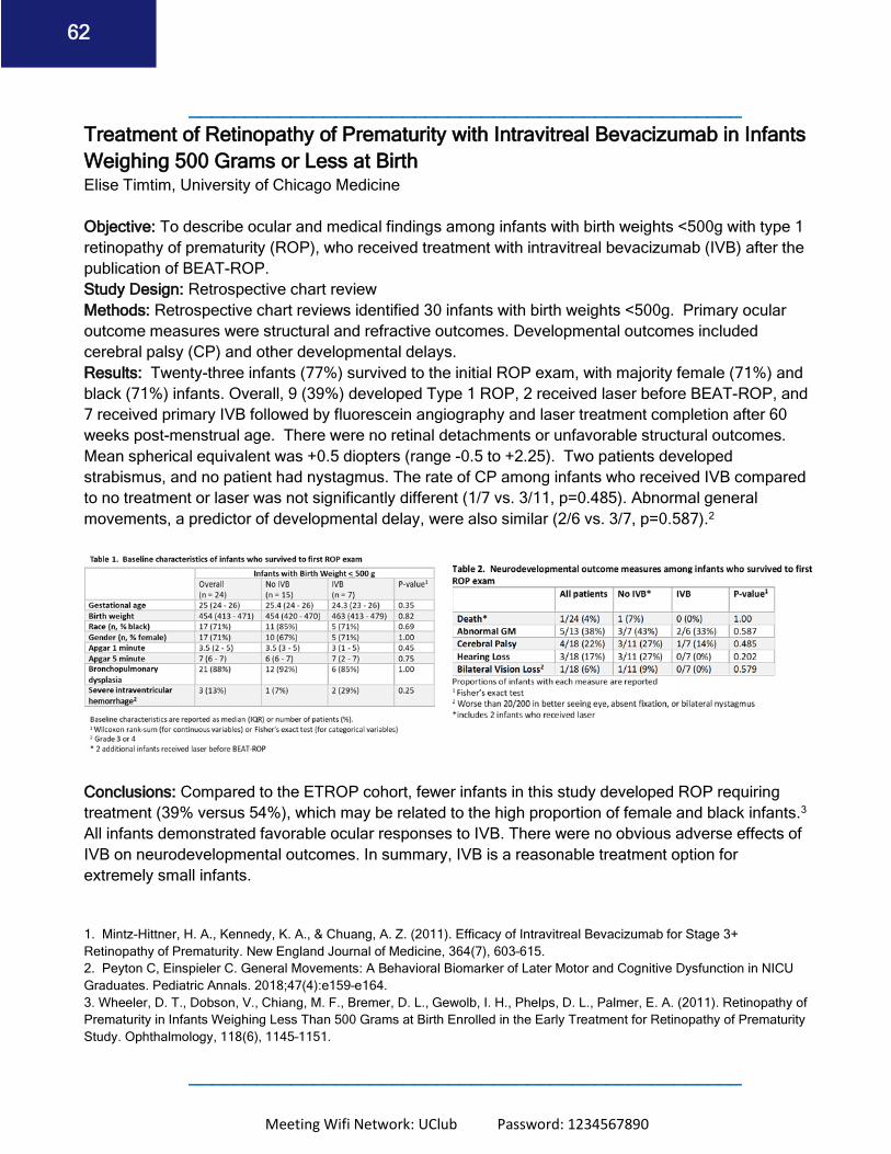
Meeting Wifi Network: UClub Password: 1234567890
62
_________________________________________________ Treatment of Retinopathy of Prematurity with Intravitreal Bevacizumab in Infants Weighing 500 Grams or Less at Birth Elise Timtim, University of Chicago Medicine Objective: To describe ocular and medical findings among infants with birth weights <500g with type 1 retinopathy of prematurity (ROP), who received treatment with intravitreal bevacizumab (IVB) after the publication of BEAT-ROP. Study Design: Retrospective chart review Methods: Retrospective chart reviews identified 30 infants with birth weights <500g. Primary ocular outcome measures were structural and refractive outcomes. Developmental outcomes included cerebral palsy (CP) and other developmental delays. Results: Twenty-three infants (77%) survived to the initial ROP exam, with majority female (71%) and black (71%) infants. Overall, 9 (39%) developed Type 1 ROP, 2 received laser before BEAT-ROP, and 7 received primary IVB followed by fluorescein angiography and laser treatment completion after 60 weeks post-menstrual age. There were no retinal detachments or unfavorable structural outcomes. Mean spherical equivalent was +0.5 diopters (range -0.5 to +2.25). Two patients developed strabismus, and no patient had nystagmus. The rate of CP among infants who received IVB compared to no treatment or laser was not significantly different (1/7 vs. 3/11, p=0.485). Abnormal general movements, a predictor of developmental delay, were also similar (2/6 vs. 3/7, p=0.587).2
Conclusions: Compared to the ETROP cohort, fewer infants in this study developed ROP requiring treatment (39% versus 54%), which may be related to the high proportion of female and black infants.3 All infants demonstrated favorable ocular responses to IVB. There were no obvious adverse effects of IVB on neurodevelopmental outcomes. In summary, IVB is a reasonable treatment option for extremely small infants. 1. Mintz-Hittner, H. A., Kennedy, K. A., & Chuang, A. Z. (2011). Efficacy of Intravitreal Bevacizumab for Stage 3+ Retinopathy of Prematurity. New England Journal of Medicine, 364(7), 603–615. 2. Peyton C, Einspieler C. General Movements: A Behavioral Biomarker of Later Motor and Cognitive Dysfunction in NICU Graduates. Pediatric Annals. 2018;47(4):e159–e164. 3. Wheeler, D. T., Dobson, V., Chiang, M. F., Bremer, D. L., Gewolb, I. H., Phelps, D. L., Palmer, E. A. (2011). Retinopathy of Prematurity in Infants Weighing Less Than 500 Grams at Birth Enrolled in the Early Treatment for Retinopathy of Prematurity Study. Ophthalmology, 118(6), 1145–1151.
_________________________________________________

Meeting Wifi Network: UClub Password: 1234567890
63
Bilateral Vitreous Hemorrhage Following Bilateral Intravitreal Injections of Bevacizumab in an Infant with Retinopathy of Prematurity Christos Theophanous, MD1, Sidney Schechet, MD1, Sarah Hilkert Rodriguez, MD, MPH1, Michael Blair, MD1 1. Department of Ophthalmology and Visual Sciences, University of Chicago Medicine, Chicago, IL Since the publication of BEAT-ROP in 2011, intravitreal bevacizumab has become increasingly common for the treatment of posterior type 1 retinopathy of prematurity (ROP). However, long-term data on the safety and efficacy of IVB for ROP is lacking. Vitreous hemorrhage following bevacizumab injections have been rarely reported in ROP infants, and the need for treatment of these hemorrhages remains in question. Here, the authors report a case of bilateral vitreous hemorrhage in a premature infant within two weeks of bilateral intravitreal injections of bevacizumab. These hemorrhages resolved without intervention with regression of ROP in both eyes.
_________________________________________________

Meeting Wifi Network: UClub Password: 1234567890
64
Ocular and neurodevelopmental outcomes among infants treated for retinopathy of prematurity using a commercial claims database. Michael H Zhang, Michael Blair, Sarah Hilkert Rodriguez Purpose: To describe neurodevelopmental and ocular outcomes among infants treated for ROP using Marketscan, a national insurance claims database. Methods: This study uses Marketscan, a national insurance claims database, to evaluate neurodevelopmental and ocular outcomes among infants treated from 2011-2014 with at least 2 years follow-up. Results: Of 18,384 infants with ROP and 2 years follow-up, 224 received laser and 59 received injections. Overall, 6% treated with injections and 4.5% treated with laser expired (p=0.251). One patient developed endophthalmitis after injection. There was a trend towards less retinal detachment with injections than laser (5% v. 11%, p = 0.190). Infants who received injections were 5 times more likely to have a second procedure (36% v. 9%, p < 0.001). Rates of vitreous hemorrhage, corneal opacities, cataracts, glaucoma, and strabismus were not significantly different. Rates of any developmental delay were 91% with laser and 93% with injections. Comparing injection to laser, other delays were motor (19% v. 22%, p= 0.541), cognitive (37% v. 34%, p=0.676) and language (62% v. 49%, p=0.063). Rates of CP were 37% with injections and 17% with laser, p=0.001. Infants receiving injections were more likely to have severe intraventricular hemorrhage (29% v. 17%, p = 0.05). Conclusion: Ocular outcomes appear similar by treatment group. Although developmental outcomes seem to favor laser treatment, severe intraventricular hemorrhage likely represents a confounding factor. There appears to be a propensity to treat sicker infants with injections. _________________________________________________
Meeting Wifi Network: UClub Password: 1234567890
65
The Rat Fluctuating Oxygen-induced Retinopathy model adapted to clarify disparity in observational studies of premature infants Julia P. Shulman MD, New York Medical College, Turo; M. Elizabeth Hartnett MD, John A. Moran Eye Center, University of Utah Objective(s): To describe 1) the rat oxygen-induced retinopathy (OIR) model of ROP and 2) an adapted model used to address disparity in clinical observational studies regarding risk of maternal preeclampsia on infant ROP by removing the confounder, premature birth. Study Design: Clinical – retrospective study of 290,992 live births over 10 years in Utah Basic – Maternal uteroplacental insufficiency (UPI) vs. control in dams and OIR vs. room air (RA) in pups. Methods: Clinical: Risk of ROP in a full cohort of maternal preeclampsia vs. none and preterm very-low birth weight (P-vLBW) vs. full-term infants compared to a restricted subcohort including only P-vLBW infants analyzed with GEE. Basic: UPI/OIR, UPI/RA, contr/OIR, contr/RA pups analyzed for OIR, retinal and serum erythropoietin (EPO) and other factors analyzed by multivariable mixed effects linear regression with STATA14. Results: ROP risk was increased by maternal preeclampsia in the full cohort but reduced in the P- vLBW subcohort. Compared to cont/OIR, UPI/OIR pups had less severe OIR, catch-up growth, increased EPO and slightly increased serum IGF-1, but not VEGF. Serum IGF-1 in UPI/OIR was reduced compared to UPI/RA. UPI dams had lower serum anti-angiogenic factors at birth than control. Conclusions: Observational studies in human preterm infants are prone to collider bias, because premature birth is associated with preeclampsia and ROP. The rat OIR model lacks the confounder, preterm birth. Our evidence supports the hypothesis that maternal UPI may upregulate hypoxia- induced angiogenic factors in infants able to produce them and reduce the risk of severe ROP. _________________________________________________

Meeting Wifi Network: UClub Password: 1234567890
66
Refractive Outcomes after Anti-VEGF and Laser Nandita Anand, MD Purpose: To report refractive outcomes comparing primary intravitreal bevacizumab (IVB) with delayed peripheral retinal photocoagulation (IVB-PRP) to primary peripheral retinal photocoagulation (PPRP) in infants treated for posterior type 1 ROP. Methods: Retrospective chart reviews identified 87 infants at a tertiary referral center treated for posterior type 1 ROP between 2006-2016. Consecutive infants received PPRP before (n = 41) and IVB after (n = 46) the implementation of change in treatment practice to primary IVB (PIVB). Among infants in the PIVB group, 35 completed an exam under anesthesia (EUA) with fluorescein angiography (IVFA) and prophylactic peripheral retinal photocoagulation after 60 weeks post-menstrual age (PMA). The final analysis included 34 eyes of 19 infants with PPRP and 40 eyes of 21 infants with IVB-PRP. The primary outcome is spherical equivalent (SE) in diopters, determined by cycloplegic refraction between 2-4 years. Results: Mean SE was -7.4 + 5.2 for PPRP and -0.16 + 2.2 for IVB-PRP (p < 0.001). This relationship persisted after stratification by zone of ROP and the presence of aggressive posterior ROP. Prior to delayed laser treatment completion, 70% of eyes treated with PIVB had leakage on IVFA. There was no statistically significant difference in mean SE comparing the IVB-PRP group to a small number of infants who received IVB monotherapy. Conclusions: Infants treated with IVB-PRP are significantly less myopic than those treated with PPRP. Delayed laser after 60 weeks PMA, in hopes of reducing the risk of late retinal detachment, does not negate the refractive benefits of PIVB.
_________________________________________________ The Successful Non-Surgical Treatment for Surgical Retinopathy of Prematurity Cases. Supalert Prakhunhungsit MD*, Audina M. Berrocal MD** *Ophthalmology Department, Faculty of Medicine, Siriraj Hospital, Mahidol University, Thailand, and International Pediatric Retina Scholar, Bascom Palmer Eye Institute. **Retina Department, Bascom Palmer Eye Institute. Objective: to report the successful non-surgical measures for retinopathy of prematurity(ROP) stage 4A cases. The present study aims to evaluate the efficacy of a non-surgical treatment such as laser indirect ophthalmoscopy as well as intravitreal bevacizumab. Study Design: case series Methods: We evaluated premature newborns diagnosed with ROP stage 4A who were referred to Bascom Palmer Eye Institute and neonatal intensive care unit, Holtz Children’s Hospital. The
Meeting Wifi Network: UClub Password: 1234567890
67
treatment given and outcome were detailed. The primary ocular outcomes were the quiescence of ROP activity and the non-progression of the vitreoretinal traction. Results: There was the total of five eyes with ROP stage 4A in three premature patients. The gestation age ranged from 23 to 29.4 weeks (mean=25.4). The birth weight ranged from 425 to 1370 g. (mean=798.3 g). Pre-referral, four eyes were injected with bevacizumab intravitreally and six eyes were treated with indirect laser ophthalmoscopy. Five eyes remained stage 4A whereas one eye had stage 5 prior to referral. The activity of ROP was assessed with fundus photography, fundus autofluorescence, and intravenous fluorescein angiography. Then, all five eyes with stage 4A were treated with diode laser indirect ophthalmoscopy, intravitreal bevacizumab injection (mean numbers of injection=1), and sub Tenon’s triamcinolone acetonide. The quiescence of ROP activity was achieved during the follow up visit and there was no progression of the vitreoretinal traction observed in all treated eyes. Conclusions: Non-surgical management such as intravitreal bevacizumab and laser indirect ophthalmoscopy as well as adjunct treatment with sub-Tenon’s triamcinolone acetonide could be successful alternatives to intraocular surgery for ROP stage 4A patients. _________________________________________________ Prospective clinical study of two different regimens of combined laser photocoagulation and intravitreal bevacizumab treatment for retinopathy of prematurity. Subhadra Jalali, Venkateshnaidu Laveti, Divya Balakrishnan, Padmaja K.Rani, Ashik Mohamed. L V Prasad Eye Institute, Hyderabad, India. Objective: To evaluate, in a combined treatment strategy for treatment warranted retinopathy of prematurity (ROP), which is a better regimen, peripheral laser photocoagulation (LPC) first or intravitreal bevacizumab (IVB) first. Methods: 22 Babies (44 eyes) with ROP were recruited prospectively. All the right eyes received LPC on day one followed by IVB on day four (LPC group). In all left eyes IVB was injected on day one
Meeting Wifi Network: UClub Password: 1234567890
68
followed by LPC on day four (IVB group). The primary outcome measure was the proportion of eyes that had complete ROP regression with no additional treatment within 2 weeks of the onset of therapy. Results: In LPC first group 72.7% (16/22) eyes had complete ROP regression with no additional treatment within 2 weeks of the onset of therapy. In the IVB group 95.5% (21/22 eyes) had complete regression within 2 weeks. Additional laser within a month had complete regression in all eyes in each group. One baby (two eyes, one from each group) had late recurrence at 5 months. Conclusion: The combined therapy strategy was successful for ROP management. Administration of anti- vascular endothelial growth factor injection before the peripheral laser was better than the reverse strategy of laser first. _________________________________________________