children with spastic cerebral palsy: aspects of …
TRANSCRIPT

DEPARTMENT OF WOMAN AND CHILD HEALTH Karolinska Institutet, Stockholm, Sweden
CHILDREN WITH SPASTIC CEREBRAL PALSY: ASPECTS OF
MUSCLE ACTIVITY AND BOTULINUM TOXIN A
TREATMENT
Kristina Tedroff
Stockholm 2009

All previously published papers were reproduced with permission from the publisher.The diagram on page 40 has previously been printed in “The Neurology of the newborn” ed. J.Volpe (2008) and is reprinted with permission from Elsevier books. Published by Karolinska Institutet. Printed by Repro Print AB, Stockholm © Kristina Tedroff, 2009 ISBN 978‐91‐7409‐211‐0

To my family

ABSTRACT Backgound: Cerebral Palsy (CP) is a heterogeneous disorder in which movement and posture are always affected. Spasticity is one of the most common symptoms. A spastic muscle prevents normal motor behaviour and is believed to cause secondary contractures. Other motor symptoms include central dyscoordination causing defects in coordination and excecution of motion and excessive co‐contraction in antagonist muscles. Muscle activity in antagonist and adjacent muscles during voluntary movements such as maximum voluntary isometric contraction (MVIC) is not completely understood in children with CP nor in children with typical development (TD). Despite a lack of strong evidence from randomised controlled trials and little long‐term data, intramuscular injections with botulinum toxin A (BoNT‐A) for treatment of increased muscle tone in children with CP has become increasingly popular over the last decade. Aims: The aims of the thesis were to compare the patterns of muscle activation during MVIC in lower extremity muscles and determine whether children with CP have more co‐activity than TD children. A further aim was to write a comprehensive review on BoNT‐A treatment with recommendations for future research. Further aims were to evaluate the effect of early BoNT‐A treatment in toddlers with CP, and to prospectively evaluate any long‐term effects of BoNT‐A on muscle tone and joint range of motion (ROM) in the lower extremities of children with CP. Methods/Results: Children with diplegic and hemiplegic CP and TD were assessed with surface EMGs. It was found that Children with CP display greater variability in muscle onset order, shorter latencies to onset of other muscles than the intended muscle and twice as much co‐activity in both antagonist and adjacent muscles, during MVIC compared to TD children. Ninety‐four children with CP were prospectively followed for a maximum of 3 years and 7 months during which time they received a maximum of eight injections per muscle of BoNT‐A. Outcome measurements included muscle tone and joint range of motion (ROM). BoNT‐A injections reduced long‐term spasticity in all muscle‐groups examined: the gastrocnemius, hamstring, and adductor muscles. Improvement in ROM, however, was only significant after the first injection; after further injections, joint ROM was reduced. Children with CP, under 2 years of age at study start, participated in a randomized trial which compared the effects of one year of early BoNT‐A treatment in the gastrocnemius muscle combined with a daily stretching program to a stretching program alone. The effects on ankle and knee ROM, muscle tone in ankle and knee flexors, gross motor function measure (GMFM) and pediatric evaluation of disability inventory (PEDI) were evaluated at one year and at 3.5 years after study commencement. Gait was evaluated with 3D‐gait analysis at 5 years of age. Early treatment with BoNT‐A significantly increased knee joint ROM and although not significantly, also increased ankle joint dorsiflexion in the BoNT‐A group after 1 year. Children in the control group experienced significantly reduced joint ROM at both joint levels at 3.5 years after study commencement. No differences in GMFM or PEDI scores or 3D‐gait data were detected comparing the groups. Conclusions: The activation of muscles differs between children with CP and children with TD when performing a voluntary movement and children with CP express twice as much co‐activity. Early BoNT‐A intervention in toddlers with CP seems to influence muscle tone and contracture development also after 3.5 years. The effect on gait development remain inconclusive.When BoNT‐A treatment in older children (mean age 5.5y at treatment start) is evaluated this suggests that BoNT‐A can be effective in reducing muscle tone over a longer period, but not in preventing development of contractures in spastic muscles. The dissociation between the effects on muscle tone and ROM indicates that development of contractures is not coupled to increased muscle tone alone, but might be caused by other mechanisms. Keywords: Cerebral Palsy, children, spasticity, voluntary activity, co‐activity, synergistic muscle activity, botulinum toxin A, long‐term, contracture, gait development ISBN 978‐91‐7409‐211‐0

LIST OF PUBLICATIONS The thesis is based on original articles listed below. They will be referred to in the text by their Roman numerals. I. Kristina Tedroff, Loretta M Knutson,Gary L Soderberg
Synergistic muscle activation during maximum voluntary contractions in children with and without spastic cerebral palsy Developmental Medicine & Child Neurology 2006, 48: 789–796
II. Kristina Tedroff, Loretta M Knutson,Gary L Soderberg Co‐activity during maximum voluntary contraction: a study of four lower‐extremity muscles in children with and without cerebral palsy Developmental Medicine & Child Neurology 2008, 50: 377–381
III. Hans Forssberg, Kristina B Tedroff Botulinum toxin treatment in cerebral palsy: intervention with poor evaluation? Developmental Medicine & Child Neurology 1997,39: 635‐640
IV. Kristina Tedroff, Fredrik Granath, Hans Forssberg, Yvonne Haglund‐Åkerlind Long‐term effects of botulinum toxin A in children with cerebral palsy Developmental Medicine & Child Neurology, Epub 2009 January 6
V. Kristina Tedroff, Kristina Löwing, Elena M. Gutierrez‐Farewik, Yvonne Haglund‐Åkerlind, Hans Forssberg Botulinum toxin A treatment in toddlers with cerebral palsy: Effects on muscle tone, contracture development and gait pattern. A Randomized Controlled Trial. Manuscript

CONTENTS Introduction ......................................................................................................... 1
1.1 Cerebral Palsy ..................................................................................... 1 1.2 Definition and Classification ............................................................... 2 1.3 The Motor Symptoms ......................................................................... 3
1.3.1 Dystonia/Dyskinesia ............................................................... 4 1.3.2 Hyperreflexia .......................................................................... 4 1.3.3 Mirror Movements ................................................................. 4 1.3.4 Retained Neonatal Reflexes ................................................... 5 1.3.5 Paresis ..................................................................................... 5 1.3.6 Spasticity ................................................................................. 7 1.3.7 Musculoskeletal Malformations .......................................... 10 1.3.8 Co‐contraction ...................................................................... 10 1.3.9 Central Dyscoordination ...................................................... 11
1.4 Treatment in Cerebral Palsy ............................................................. 14 1.4.1 Rehabilitation Management ................................................ 14 1.4.2 Medical Management – Oral Medication ........................... 16 1.4.3 Surgical Procedures .............................................................. 17 1.4.4 Botulinum toxin A ................................................................. 17
2 Aim of The Thesis ........................................................................................ 21 3 Materials and Methods .............................................................................. 22
3.1 Study outlines ................................................................................... 22 3.2 Subjects ............................................................................................. 23
3.2.1 Inclusion/Exclusion Criteria .................................................. 23 3.3 Methods ............................................................................................ 25
3.3.1 Time axis ............................................................................... 25 3.3.2 Body Function and Structure Assessments ......................... 25 3.3.3 Assessments at Activity and Participation Level ................. 28 3.3.4 Intervention Techniques ...................................................... 31
3.4 Data Analysis ..................................................................................... 31 3.4.1 Statistics ................................................................................ 34
4 Results and Discussion ............................................................................... 36 5 Conclusions and Clinical implications ........................................................ 59
Clinical implications .................................................................................... 60 6 Appendix ..................................................................................................... 61 7 Acknowledgements .................................................................................... 62 8 References .................................................................................................. 64
LIST OF ABBREVIATIONS AS BoNT‐A BW CI CNS CP CPG DTRs EMG GMFCS GMFM LG MAS MH MVIC OGS PEDI PRS PT RCT ROM SD TA TD VL
Ashworth score Botulinum toxin A Body weight Confidence interval Central nervous system Cerebral Palsy Central pattern generator Deep tendon reflexes Electromyography/ electromyographic Gross Motor Function Classification System Gross Motor Function Measure Lateral gastrocnemius muscle Modified Ashworth Scale Medial hamstring muscle Maximum voluntary isometric contraction Observational gait scale Pediatric Evaluation of Disability Inventory Physician rating scale Physiotherapist Randomized controlled trial (joint) Range of motion Standard deviation Tibialis anterior muscle Typical development Vastus lateralis muscle

1
INTRODUCTION 1.1 CEREBRAL PALSY Cerebral Palsy (CP) is the most common life long disability affecting motor development. For some decades the overall incidence of CP in the Western world countries has remained stable at about 2‐3/1000 births (Himmelmann et al., 2005;Bhasin et al., 2006;Westbom et al., 2007) with a higher prevalence among children born preterm (Himpens et al., 2008). CP has traditionally been referred to as an "umbrella term". As such the term CP involves several different etiologies for the permanent lesion in the developing brain, which by convention occurred before the age of two. But it also illustrates the heterogeneity of clinical symptoms, where the degree of motor and other non‐motor neurological involvement span a wide spectrum, ranging from mild sometimes and barely noticeable, to severe disability. Long term effects of CP on patients and their families often include pain, reduced participation in society, and in many cases, a financial burden (Schwartz et al., 1999;Jahnsen et al., 2003;Breau et al., 2003;Houlihan et al., 2004;Jahnsen et al., 2004;Michelsen et al., 2005;Michelsen et al., 2006;Law et al., 2006;Russo et al., 2008;Majnemer et al., 2008). Similarly, the effect on society in general involves considerable costs for both medical and non‐medical expenses, such as home modification and transportation, as well as indirect costs due to loss of productivity in the adult patient and among parents of children with CP (Ireys et al., 1997;2004;Hoving et al., 2007a). Over the past 15 years, knowledge concerning several aspects of CP has increased considerably. Since this thesis is based in part on long‐term clinical studies from the 1990s, the introduction will be based in part on knowledge acquired at the time these studies were designed. Recent advances in understanding will be put forward in the discussion, along with the interpretation of our results.

2
1.2 DEFINITION AND CLASSIFICATION Motor deficit is the hallmark of CP. Earlier definitions and classifications have focused on this aspect (Bax, 1964;Mutch et al., 1992). Efforts to quantify the level of functional motor ability coupled with expanded knowledge about the impact of non‐motor neurodevelopmental problems encountered in CP have created a need for a new and more comprehensive definition and classification. In 2004 an international workshop was held in Bethesda, MD, USA, in order to revise the definition and classification of CP. "The Definition and Classification of Cerebral Palsy, April 2006" was published in its final form in February 2007 (Rosenbaum et al., 2007) : Cerebral palsy (CP) describes a group of permanent disorders of the development of movement and posture, causing activity limitation, that are attributed to non‐progressive disturbances that occurred in the developing fetal or infant brain. The motor disorders of cerebral palsy are often accompanied by disturbances of sensation, perception, cognition, communication, and behavior; by epilepsy, and by secondary musculoskeletal problems.
In contrast to the historical viewpoint, this new definition illuminates neurological "comorbidities." The role of such problems is becoming increasingly recognized in relation to the growth of a child into an adult with CP (Murphy et al., 2000;Michelsen et al., 2006).
The classification has four components:
1. A) First, the most dominant motor disorder, which may be spastic, dystonic, athetotic, or ataxic, is addressed along with B) the extent to which individuals are limited by their motor function. Gross motor function is most often described in relation to ambulation and activity limitation and the Gross Motor Function Classification System (GMFCS) is internationally validated, accepted, and commonly used for this purpose (Palisano et al., 1997). Hand‐arm function is classified separately using the Manual Ability Classification System (MACS) (Eliasson et al., 2006).
2. Accompanying impairments. In many children with CP, impairments other than motor impairment interfere with activities of daily life and these may be equally or more troublesome for the individual than the characteristic motor symptoms. Examples of such impairments include cognitive and attention deficits, vision and hearing problems, seizure disorders, and emotional and behavioral disorders. The recommendation for such impairments is to classify them as being either present or absent. The Surveillance of Cerebral Palsy in Europe (SCPE) provides a clinical tool for this purpose (Surveillance of Cerebral Palsy in Europe (SCPE), 2000).
3. A) The pattern and extent of the motor disorder should be described. The recommended description is currently also in use by the SCPE and differentiates between unilateral and bilateral involvement. B) Recent advances in imaging techniques and quantitative motor assessments have provided the basis for

3
correlation between neuroimaging findings and the clinical picture. Currently the recommendation is for all children with CP to have a neuroimaging examination and for findings, if any, should be stated.
4. As a future goal the classification could also address causes and timing. However, in a minority of cases‐‐for example, a near drowning incident in a previously healthy child‐‐the cause is already known and thus can be stated.
Moreover, the new definition and classification also more closely follow the WHO International Classification of Functioning, Disability and Health (ICF) framework (2001). In this international model, domains of activity/participation were designed to describe and measure disease or injury for the individual in terms of health and disability.
The ICF framework shifts focus from cause to impact, allowing different health conditions to be measured using a common metric, "the ruler of health and disability" (2001). In addition, the WHO defines health more broadly, which encompasses physical, mental, and social well‐being (1948).
1.3 THE MOTOR SYMPTOMS From a motor control perspective, CP is best described as a set of different motor disorders with motor dysfunction that varies in scope and type. For simplicity, symptoms may be categorized as excess symptoms, which are added onto normal motor behavior, and deficit symptoms, in which the normal motor repertoire fails to develop (Forssberg H., 2003). Table 1 lists the various motor dysfunctions. Table 1
Cerebral Palsy The motor symptoms
Excess symptoms • Spasticity • Dystonia/Dyskinesia • Hyperreflexia • Co‐contraction • Mirror movements • Musculoskeletal Malformations • Retained Developmental Reactions
Deficit symptoms • Paresis • Central Dyscoordination
4
This thesis will mainly focus on spasticity and co‐contraction, as well as on musculoskeletal malformations and central dyscoordination, whereas other motor symptoms will be more cursively addressed. 1.3.1 Dystonia/Dyskinesia Patients with dystonia or hyperkinetic symptoms such as chorea or athetosis, commonly referred to as dyskinesia, suffer from abnormal movements often seen when initiating a movement. Abnormal posture and motor pattern in dyskinesias are secondary to impaired muscle tone and coordination regulation following injury to the extrapyramidal system, usually localized to the basal ganglia (Himmelmann et al., 2007). Dystonia is characterized by abnormal stereotyped shifts in muscle tone and is often induced by movement or external stimuli. Voluntary movements may be distorted or movements may be involuntary. Affected muscles or limbs may "freeze" in abnormal positions or postures, often characterized by rotation, extension, or flexion. Dyskinesias, chorea, and athetosis are abnormal excessive movements also commonly seen when voluntary movement is initiated. Children with hyperkinetic or choreoathetotic CP are severely hampered in motor activity by extensive involuntary movements and overflow of muscle activity to muscle groups other than those intended. Dystonic symptoms commonly affect speech and swallowing. Symptoms of dystonia and dyskinesia often evolve over time and may become apparent later than initial symptoms, such as spasticity (Burke et al., 1980). 1.3.2 Hyperreflexia Deep tendon reflexes (DTRs) are often exaggerated on the side contralateral to an upper motor neuron lesion. Subtle asymmetry may be difficult to clinically discern and in such cases it is often useful to attempt to elicit reflexes that are usually hard to provoke, such as the pectoral, finger flexor, or hamstring reflexes. In bilateral lesions, these reflexes will be bilaterally exaggerated. When such reflexes are clearly hyperactive, the "reflex zone" in which a DTR can be elicited is often enlarged. Hyperactivity of DTRs is often accompanied by clonus. Clonus is a phenomenon in which sustained muscle stretch (elicited in milder cases by testing DTRs) results in repetitive contraction and relaxation of that muscle. In more severe cases clonus may be difficult to extinguish, occurring spontaneously after only minor movement of a limb. 1.3.3 Mirror Movements Mirror movements are unintended movements that occur in corresponding muscles on the side contralateral to voluntary activity. In healthy adults and children the cause

5
is believed to be simultaneous activation of crossed corticospinal pathways from both hemispheres (Mayston et al., 1999). Lesions in the developing brain have a different impact on the sensory‐motor system than lesions acquired in the adult brain. As the nervous system matures, different neural systems compete for synaptic space. Following injury or altered neural activity, circuits and pathways that normally disappear during development may persist or reactivate. Several recent studies applying Transcranial Magnetic Stimulation (TMS) and functional Magnetic Resonance Imaging (fMRI) in children with unilateral CP have demonstrated aberrant cortical circuits and corticospinal pathways from the motor cortex to the hand (Carr et al., 1993;Carr, 1996;Staudt et al., 2002;Vandermeeren et al., 2003a;Vandermeeren et al., 2003b;Eyre et al., 2007). In the developing brain, secondary motor areas in the premotor cortex and supplementary motor cortex that take over the disturbed arm/hand function will usually compensate for small or limited lesions affecting the primary sensory‐motor cortex (Staudt et al., 2002). However, larger lesions may also involve these secondary motor areas and hand motor control will subsequently be transferred to the undamaged or healthy ipsilateral hemisphere. In these cases, motor control could be exercised by exuberant ipsilateral connections from the primary motor cortex area to the hand muscles (Carr et al., 1993). In persons with unilateral CP the presence of ipsilateral corticospinal pathways correlates with strong mirror movements in the healthy hand (Holmström et al., 2009). During bimanual tasks, when each hand is assigned a different concurrent activity, the presence of mirror movements may considerably hamper or even prevent purposeful movement (Kuhtz‐Buschbeck et al., 2000). Many children and adults suffering from mirror movements have developed strategies by which they "lock" all movement in the hemiplegic or affected hand, thus functionally becoming one‐handed. 1.3.4 Retained Neonatal Reflexes Healthy infants possess a set of specific reaction patterns or neonatal reflexes that are present at birth, but generally disappear within the first year of life. Such reactions include the Moro reflex, palmar and plantar grasp, Asymmetrical Tonic Neck Reflex (ATNR), rooting, neonatal stepping, and the crossed extensor reflex. Persistent or even exaggerated neonatal reflexes often hamper children, adolescents, and adults with CP, including those with spastic CP who have a low functional level (e.g., those with a GMFCS level IV‐V) and those with dystonic/dyskinetic CP. For example, an easily elicited ATNR prevents intentional movement in upper extremities; similarly, a persistent Moro or startle reaction effectively disrupts voluntary actions. 1.3.5 Paresis Paresis denotes the inability to generate sufficient or appropriate muscle strength and can be tested by eliciting maximum voluntary contractions. However, this measure is highly subjective, since performing a maximum contraction largely depends on the motivation and endurance of the individual. In neurology, paresis is often used to
6
describe weakness. Weakness is recognized as a pervasive symptom in CP, but knowledge about this is quite recent, resulting from neurophysiological studies, as well as a multitude of strength training studies (Damiano et al., 1995;Wiley and Damiano, 1998;Engsberg et al., 1999;Ross et al., 2001;Rose and McGill, 2005;Stackhouse et al., 2005). Furthermore, high‐functioning children with unilateral or mild bilateral spastic CP have been found to be weaker than typically developed (TD) children in all major muscle groups of the lower extremities (Wiley and Damiano, 1998). In a recent study by Ross and Engsberg, spasticity, strength and functional outcome, as assessed by GMFM‐66 and a range of gait analysis parameters, were tested in 97 children with CP and GMFCS level I‐III. They found that spasticity only accounted for a maximum of 8% of the variance in gait and gross motor function, whereas moderate to high correlations were found between strength and these functions, where strength accounted for up to 69% of the variance (Ross and Engsberg, 2007). The human body contains more than 400 skeletal muscles, each typically comprising thousands of muscle fibers working in parallel and organized into a smaller number of motor units (Walsh EG, 1992a;Loeb GE and Ghez C, 2000). The motor unit consists of a motor neuron and the muscle fibers that it innervates. In muscles controlling fine motor function, such as those of the hand or eye, the number of fibers innervated by a single neuron is least, while large muscle groups controlling gross motor function, such as those of the thighs, have the greatest number of fibers innervated per neuron. The neuromuscular junction is the synapse between the motor neuron and the muscle fiber. The ventral spinal cord contains the nuclei for the motor neurons. The motor nuclei in the spinal cord receive input from 1) the descending ventral corticospinal tract, 2) the descending lateral corticospinal tract, 3) primary afferent neurons from cutaneous and deep peripheral receptors, and 4) inhibitory interneurons. The pathophysiology behind paresis in CP is primarily due to deficient recruitment of motor neurons and consequently incomplete activation of the motor unit. This has been shown in studies comparing children with CP to children with typical motor development (Elder et al., 2003;Rose and McGill, 2005;Stackhouse et al., 2005). Rose and McGill investigated muscle strength, neuromuscular activation, and motor‐unit firing characteristics in subjects with CP and in age‐matched controls (Rose and McGill, 2005). During maximum voluntary contractions of the medial gastrocnemius and anterior tibial muscles they demonstrated that participants with CP produced significantly less torque compared with age‐matched controls, and that neuromuscular activation during maximum voluntary contraction was significantly reduced in participants with CP compared with controls. The two subject groups were also tested for submaximum contraction and when compared for the same submaximum level of neuromuscular activation they found no difference in motor‐unit recruitment and firing rates between the groups. Subjects with CP were probably unable to recruit the higher threshold motor‐units or to induce higher firing rates in lower threshold motor units due to a central deficiency (Rose and McGill, 2005). Stackhouse et al showed that 12 children with CP were significantly weaker and had lower agonist voluntary muscle activation, as assessed by surface EMG during maximum voluntary isometric contractions, than 10 TD children (Stackhouse et al., 2005).

7
Elder et al examined ankle muscle weakness in 28 children with spastic CP and in 14 controls (Elder et al., 2003). Strength of ankle dorsiflexors and plantar flexors was significantly reduced in the group with CP; moreover, these muscles were found to be weak based on measurements showing a significant reduction in specific tension. In addition, muscle EMG during maximum voluntary contractions was reduced in the group with CP (Elder et al., 2003). However, weakness in CP is due not only to a deficiency in motor unit activation; several other factors contribute (Rose and McGill, 1998). The total number of muscle fibers is fixed at birth, but muscle volume and mass increase through the process of hypertrophy as the child grows. The larger the cross‐sectional area of the muscle, the higher the actual force that can be produced (Gage JR, 2004), and a recent study by Tonson et al concluded that isometric muscle strength is proportional to muscle volume, regardless of age (Tonson et al., 2008). At the "One Small Step Gait Laboratory" in London ,UK, several recent studies revealed that children with CP have smaller muscle bellies and muscle volume loss compared to children with typical motor development (Fry et al., 2004;Fry et al., 2007;Malaiya et al., 2007). In addition to the contractile component, known as the sarcomere, muscles fibers also contain noncontractile elements such as connective tissue. Morphologic changes in contractile and noncontractile elements of muscle have been found in spastic CP(Ito et al., 1996;Friden and Lieber, 2003). Ito and coworkers found that muscle specimens obtained from children with spastic CP during surgery contained up to twice the anticipated number of type 1 muscle fibers (Ito et al., 1996). Muscles containing a large number of type 1 fibers (or red fibers) are fatigue‐resistant, but can only produce relatively small amounts of tension over a long period of time (Walsh EG, 1992b) . Voluntary force production also depends on synchronization of muscles working in concert to produce a force, i.e. synergistic motor units. This synchronization results from a common drive (Forssberg H., 2003). Finally, low force production may also result from conflicting or counteracting forces resulting from co‐activity in muscle antagonists and other muscles (subtraction weakness). The effect of co‐activity will be discussed later in this thesis. 1.3.6 Spasticity "Muscle 'tonus' has been described, defined, and measured in a multitude of different ways. Scientifically speaking, however, there is no such single property of muscle as its tonus. Rather tonus is a convenient term, which includes many different properties such as elasticity, viscosity, and muscle reflexes," according to the writings of Fenn and Garvey in 1934 (Fenn WO and Garvey PH, 1934). Similarly, today muscle tone is described as the force with which a muscle resists being lengthened. Muscle force depends on the intrinsic elasticity or stiffness of the muscles. In addition, there is a neural contribution to muscle tone related to the stretch reflex feedback loop that resists lengthening (Pearson K, 2000). The increased muscle tone found in spasticity is involuntary and often aggravated by external stimuli, emotional stress, and emotions, as well as by unrelated and/or concurrent health problems such as infections or constipation.

8
The term spasticity is commonly used to describe several motor symptoms found in CP and other neurological disorders, not merely to describe muscle hypertonicity. In clinical management and research, it is important to identify the various symptoms in order to choose the most effective therapeutic interventions or to design appropriate studies. In recent decades, the definition presented by Lance has often been used: "A motor disorder characterized by a velocity‐dependent increase in the tonic stretch reflexes with exaggerated tendon jerks, resulting from hyperexcitability of the stretch reflex" (Lance JW, 1980). The velocity‐dependent increase in muscle resistance to movement is essential and differentiates spasticity from other forms of increased muscular tone such as dystonia or rigidity. This definition has been challenged to some extent in recent years. One reason is that certain studies have revealed a lack of coordination between clinical spasticity and signs of hyperreflexia (Sinkjaer et al., 1993;Schindler‐Ivens and Shields, 2004;Poon and Hui‐Chan, 2008) and another is when the affected and the healthy limb in unilateral CP display similar stretch reflex patterns (Lin et al., 1994a;Lin et al., 1994b). In a recent study by Poon and Hui‐Chan children with spastic CP demonstrated significantly stronger soleus stretch reflex/M‐response during both voluntary dorsiflexion and plantar flexion of the ankle when compared with children with TD, but no correlation was found with clinical spasticity (Poon and Hui‐Chan, 2008). Lin and coworkers examined the muscles of both lower extremities in children with hemiplegic CP using surface EMG at varying muscle lengths in order to investigate the stretch reflex response at varying frequencies and velocities. They found that both non‐paretic and hemiparetic muscles (proximal and distal antagonist‐agonist pairs) displayed similar velocity‐dependent reflex thresholds in both the healthy and paretic limbs. (Lin et al., 1994a;Lin et al., 1994b). Spasticity is the consequence of an upper motor neuron lesion anywhere from the cortex to the spinal level. Lawrence and Kuypers demonstrated that an isolated lesion to the corticospinal tract above the foramen magnum in monkeys will result in slowness and loss of dexterity, but not spasticity (Lawrence and Kuypers, 1968a;Lawrence and Kuypers, 1968b). In humans a lesion of the upper motor neuron is also likely to damage nearby motor pathways such as the corticoreticulospinal tract. It is thought that combined damage to these tracts gives rise to changes in background activity of alpha motor neurons and interneurons at the segmental level, which is considered essential for development of spasticity (Pearson K, 2000). Reduced inhibition of Ia afferents (presynaptic) utilizing γ‐aminobutyric acid (GABA) as a neurotransmitter has been suggested. The effective reduction in spasticity seen after oral or intrathecal administration of the GABA agonist, baclofen, supports this hypothesis (Hoving et al., 2006). Spasticity in CP mainly hampers voluntary movements, but may sometimes be helpful for the weak child when, for example, spasticity of the lower extremities may be useful for weightbearing or support.

9
Severe spasticity can be both painful and energy‐consuming (Hemingway et al., 2001), which is one of the factors contributing to the growth disturbances often seen in CP. Many assumptions have been made concerning the relationship of spasticity and various aspects of functioning in CP, which will be addressed in this thesis:
• Spasticity is believed to negatively correlate with functioning in CP. • Spasticity causes weakness. • Spasticity is also commonly referred to as the cause of development of
contractures.
Contracture development
When relaxed muscle fibers fail to reach "normal length," or a length appropriate for the bone, we talk about contractures. Consequently contractures will reduce joint range of motion (ROM) for the joint(s) which they span. Monoarticular muscles, such as the tibialis anterior muscle that performs a dorsal flexion of the ankle, span one joint, while biarticular muscles, such as the gastrocnemius muscle, span two joints ‐ in this case, the ankle and knee. Development of contractures is very common in CP, especially in spastic CP. Spasticity is often believed to be the ultimate cause of contracture in CP (Peacock and Staudt, 1990;Hof, 2001;Gage JR and Schwartz M, 2004). A specific chain of events that precede contracture development has been proposed:
1. Abnormal motor control results in muscle hypertonia/spasticity. 2. The antagonist muscles are too weak to counteract the spastic muscle. 3. The affected (spastic) muscle will constantly be in a shortened state. 4. Ultimately, the spastic muscle fails to adapt to new length demands or to
"grow with the bone" (Hof, 2001;Gage JR and Schwartz M, 2004). Several animal studies have shown that skeletal muscle grows in parallel or in apparent response to bone growth (Williams and Goldspink, 1976;Williams and Goldspink, 1978;Ziv et al., 1984). In addition, a study on cats by Tabary et al (1972) showed that muscles restrained in a shortened position lost sarcomeres, while those restrained in a lengthened position actually added sarcomeres in series (Tabary et al., 1972). The conclusion that spasticity is a cause of contracture in CP in fact builds on a case of circumstantial evidence. No human study supports this conclusion, although two animal reports lend some support. In one study, healthy guinea pigs were injected with tetanus toxin. When muscles in this study were allowed to be shortened by the tonus‐increasing toxin, the muscles decreased in length (Huet de la Tour et al., 1979). In another study from 1993, now considered a classic from an interventional perspective, Cosgrove and Graham compared normal and spastic mice (Cosgrove and Graham, 1994). They randomized 52 newborn mice, 10 of which later turned out were spastic, to intramuscular injections of botulinum toxin A (BoNT‐A) or saline injection into the gastrocnemius muscles for their entire growth period of about 2 months. Since the 5 spastic mice that received BoNT‐A injections had normal muscle belly length compared with the 5 spastic mice that received saline injections, the authors concluded that they restored normal muscle growth by reducing the increased muscle
10
tone. Moreover, they proposed that other muscle‐tone reducing interventions might reduce the incidence of contracture. But this study included only a small sample and the relevance of this mouse model to humans has also come into question ‐ mice develop myoclonus involving all four limbs (Gough et al., 2005). The main symptoms seen in the spastic mouse are tremor, episodic spasms, and a disturbed righting response, a clinical picture that differs from spasticity in humans (Gough et al., 2005). However, an author such as Robert Samilson (Samilson, 1981) have pointed to the lack of knowledge about the pathophysiological mechanism underlying contracture development, and more recently Jean‐Pierre Lin proposed that "Contractures are posture‐dependent and arise through disuse and weakness" (Lin, 2003). 1.3.7 Musculoskeletal Malformations An imbalance of muscle strength and tone as found in spastic forms of CP will often increase over time, with or without intervention. This imbalance across the affected joint will contribute to muscle weakness and atrophy. The added effect of motor symptoms and reduced gross motor function will over time result in soft tissue contractures and ultimately in joint malformations (Soo et al., 2006). Joint contracture mainly involves three different structures: the joint capsule, the ligament, and the muscle with tendon are all modified by the pathology. Thus a fixed contracture results in a "true" reduction of joint movement and is not attributable to increased muscle tone. Surgical soft tissue or tendon lengthening may address this peripheral effect of muscle imbalance, but may also add to the problem by weakening the muscles (Moseley CF, 1992). Moreover, the recurrence rate for contractures is high after orthopedic surgery (Dhawlikar et al., 1992;Stout J and Van Heest AE, 2004). When skeletal muscle malformations ultimately present, more radical procedures such as osteotomies and arthrodesis are needed. 1.3.8 Co‐contraction The literature defines co‐contraction and co‐activation in different ways (Ikeda et al., 1998;Chen et al., 2003;Elder et al., 2003). Usually, co‐contraction is used when the analysis or context is related to force or torque data, and co‐activation is used when the analysis is based on electromyographic (EMG) data. Co‐activity refers to electrical activity in, for example, an antagonistic muscle which occurs within a specified time period of agonist muscle activity. The specific time period varies for different tasks. Co‐activity, of course, is a pre requisite for co‐contraction. Co‐contraction is a feature of normal motor behavior and recognized as a common motor control strategy when stability or improved motor accuracy are needed (Gribble et al., 2003;van Roon et al., 2005) Typically, the nervous system increases the level of co‐contraction in the antagonistic/agonistic muscles that surround a joint in order to stabilize limb position and improve movement precision (van Galen GP and de Jong WP, 1995;van Galen and van Huygevoort, 2000;Gribble et al., 2003).

11
However, in cerebral palsy inhibition to antagonistic muscles decreases and corticospinal pathways become altered, resulting in one of the core features of the disorder ‐ increased levels of co‐contraction (Leonard et al., 1990;Brouwer and Ashby, 1991;O'Sullivan et al., 1998). A large number of studies show that children with CP have excessive co‐activity, sometimes expressed as co‐contraction in a variety of different situations where agonist‐antagonist activity has been investigated. This has been demonstrated in regard to automated movements such as postural control (Nashner et al., 1983;Brogren et al., 1998;Burtner et al., 1998) and locomotion (Berger et al., 1984;Leonard et al., 1991) as well as in voluntary movements (Ikeda et al., 1998;Elder et al., 2003;Stackhouse et al., 2005) One descriptive study compared pediatric and adult patients with dystonic or spastic CP The patients with dystonic CP were found to have higher levels of co‐contraction and increased resistance to external motion at slow velocities. Another finding was that muscle strength was more impaired in patients with dystonia and the authors hypothesized that this may be a result of the higher level of muscle co‐contraction (Lebiedowska et al., 2004).Contrary to these findings, normal strength was found in ankle dorsiflexors and plantar flexors when two children suffering from CP with hemidystonia were examined in a study evaluating features associated with equinus gait (in children with hemiplegic CP). The authors hypothesized that strength was preserved in the muscles, but that functional impairment was the result of strong involuntary co‐contractions (subtraction weakness) (Lin and Brown, 1992). In the study by Stackhouse et al previously discussed in section 1.3.5, children with spastic CP showed significantly greater agonist coactivation than did children with typical motor development. This was believed to be one cause of the significantly reduced muscle strength seen in these children. 1.3.9 Central Dyscoordination
In order for a specific movement to be exercised smoothly and in an ordered sequential fashion, muscle contractions must all occur in a precise spatial and temporal pattern. In children with CP these patterns of movement and coordination are impaired (Forssberg, 1999). Human movement can be divided into those relating to spontaneous innate behavior and those that must be learned and trained. Innate behaviors exist in all species and are essential for survival; they include coughing, feeding, blinking, withdrawal, and in many species, even locomotion immediately after birth. Innate movements are controlled by networks of neurons located in phylogenetically old parts of the central nervous system (CNS): the brain stem and the spinal cord. These networks have been called central pattern generators or CPGs (Leonard, 1992;Grillner, 2006) and are defined as a "network of neurons…able to produce a repetitive rhythmic output…that is automatic and independent of necessary sensory feedback" (Delcomyn, 1980). Learned movements are voluntary and improve with practice as the individual learns to anticipate and correct for environmental factors impacting the body. Learned movements are hierarchically organized and become highly automated once fully learned (Ghez C and Krakauer J, 2000) .

12
The spinal cord is the lowest level of the motor organization containing neuronal circuitry for automated movements such as locomotion and reflexes. Parallel reflex pathways for the head and face are located in the brain stem. The simplest reflex pathway is monosynaptic, involving only a primary afferent sensory neuron and efferent motor neuron (Ghez C and Krakauer J, 2000). The brainstem is at the intermediate level of the motor hierarchy containing two systems—the medial and lateral—that receive afferent input from the cerebral cortex and subcortical nuclei. Both project to the spinal cord, where the medial system integrates visual, somatosensory, and vestibular information for postural control and where the lateral system, important for goal‐directed movements of the hand and arm, projects to more distal muscles. The cortex executes the highest level of motor control. The primary motor cortex and premotor areas project through the corticospinal tract directly to the spinal cord. In addition, motor tracts for postural control, originating in the brain stem, are modulated through regulation from the cortex. Gerloff and co workers recently investigated the impact of re‐afferent somatosensory signaling and corresponding neuronal activity in the somatosensory cortex on the primary motor cortex and related muscle activity. For this purpose they used magnetoencephalographic (MEG) and transcranial magnetic stimulation in three adult patients with congenital lesions to the pyramidal tract (Gerloff et al., 2006). In the subjects included in their study there had been a relocation of the primary motor cortex to the contralesional hemisphere ipsilateral to the affected hand. Wheras the somatosensory cortex had remained in the lesioned side. The differentiated location of the primary motor and sensory cortex made an otherwise impossible, unequivocal topographic differentiation possible with MEG. In all three patients there was no relevant contribution from the sensory cortex to the the corticomuscular coherence (corticomuscular coherence =the interaction between primary motor cortex and muscle). The motor actions of an individual also share important characteristics when performed in different ways. For instance, the letters used in writing will have the same basic individual shape and pattern regardless of whether an individual writes with upper case letters, the left hand, the foot, or even the mouth. Canadian neuropsychologist Donald Hebb coined the term motor equivalence in 1949 to describe this phenomenon (Hebb DO, 1949). Motor equivalence suggests that voluntary movements are somehow abstractly mapped in the brain, rather than representing a specific set of ordered muscle contractions and joint positions (Ghez C and Krakauer J, 2000). This concept of movement was further elaborated upon and to an extent supported by the model of motor synergies. By grouping muscles into synergies, less complex demands are made on the CNS when carrying out voluntary motion (Forssberg H., 2003). When we refer to a movement disorder problem with central dyscoordination in CP, we generally mean that for a number of specific tasks, both innate and learned, children with CP display aberrant patterns of motor activation and coactivation, which results in less smooth trajectories, deviant force control, and possibly changed synergies and in some instances immature motor patterns. Examples of central dyscoordination are loss of dexterity and abnormal gait development in CP.
13
Loss of dexterity
One of the salient symptoms found in CP is the reduced ability to use one or both hands to manipulate objects and to perform fine motor movements. For children with unilateral CP, problems related to impaired hand function constitute the major motor problem in about 50% of cases. (Sanner G, 1999). Impaired coordination of fingertip forces during object manipulation is one factor contributing to loss of dexterity (Eliasson et al., 1991;Forssberg et al., 1999). Development of reach and grasp involves control and coordination of several systems, including cognitive, visual/spatial, sensorimotor, and postural. In CP not only do various aspects of movement required for a lifting task change, such as movement initiation, sequencing, and the amount of force required, but the ability to store and purposefully use prior information about the object is also impaired. Gait development
Some CPGs generate locomotor movements in lower vertebrates such as the lamprey. In addition, there is evidence from studies of higher vertebrates such as cats of CPG‐controlled locomotion (Forssberg et al., 1980;Grillner, 2006). In humans, purposeful locomotion functions are mostly controlled by supraspinal circuits, although it appears that even in humans, some aspects of locomotion are controlled by the spinal cord (Dietz et al., 1994). Human gait is bipedal and plantigrade (heel‐toe strike), which is unique in the mammalian world. Primates walk on two legs but not with a plantigrade gait. The plantigrade gait pattern contributes to increased stability and is less energy consuming. Development of the heel strike is of particular importance (Forssberg H., 2003). The first stepping like movements in the fetus occur at 10‐12 weeks of gestation. Referred to as infant stepping, these movements persist in the neonate and can be elicited when the child is held upright over a horizontal surface. Supported gait arises at 7‐9 months of age. These movements are voluntary and probably goal‐oriented, as opposed to infant stepping, which is induced by weight‐bearing or stretching of the hips (Leonard et al., 1991;Forssberg H, 1992). Unsupported walking typically develops between 9‐18 months of age and is probably closely correlated to the development of postural control. Initially, the pattern of locomotion is immature with coactivation of flexor and extensor muscles resulting in synchronized flexion‐extension in all lower extremity joints. At the end of the swing phase the plantar flexors become activated, resulting in a digitigrade (toes first) gait pattern. Contrary to mature gait, a large EMG peak can be elicited in several lower extremity muscles after foot contact, indicating a hypersensitive stretch reflex (Forssberg H, 1992). After some months of unsupported walking, the pattern begins to transform into the mature digitigrade pattern and after 2 years of age most TD children have developed a prominent heel strike that includes active dorsiflexion of the forefoot. The transformation from digitigrade to plantigrade gait is dependent on supraspinal circuits. Experience and activity‐dependent neural plasticity achieved during the first year of walking probably also contribute (Forssberg,

14
1999). Desynchronization of joint movements follows, but up to the age of 12 years energy expenditure for walking is higher in children than in adults, indicating that fine‐tuning and final maturation of locomotion is a lengthy process (Forssberg H, 1992). Gait development in cerebral palsy
Prior to independent or unsupported gait, children with spastic CP exhibit a gait pattern that is similar to that of children with typical motor development (Leonard et al., 1991). However, plantigrade gait fails to develop and instead children with CP typically continue to display a digitigrade gait pattern with higher levels of coactivation and premature activation of the plantar flexors before foot contact. In plantigrade gait, major calf muscle contraction occurs at the end of the supportive phase (stance), acting as a strong forward propulsive force. In digitigrade gait, the "forward energy" of the gait is decreased due to premature calf muscle contraction. Many ambulatory children with CP develop contractures and musculoskeletal malformations; these constraints and other developmental changes eventually alter locomotion from the original gait pattern seen in CP resembling that of infant stepping. However, a variety of different patterns may arise depending on the individual potential of the child (Forssberg H, 1992). 1.4 TREATMENT IN CEREBRAL PALSY
Clinical trials are difficult to conduct in children with CP. Because of large individual differences and heterogeneity of symptoms, results from interventional trials may be difficult to assess. As such, clinical trial results that clearly support a specific therapy are scarce. Generally speaking, therapy can be divided into rehabilitation management, medical management with oral and local therapy, and surgical interventions (either neurosurgical or orthopedic). For many years spasticity has been assumed to be the primary reason for functional limitations and secondary changes such as development of contractures in CP. Consequently, treatment has focused on reducing spasticity. Children diagnosed with (or suspected of having) CP are often referred to a physiotherapist (PT) for initial intervention. In most countries rehabilitation management is the basis of intervention. However, there is no such thing as "standard" physical therapy, nor any "gold standard" treatment. 1.4.1 Rehabilitation Management
Over the past century, rehabilitation management of children with CP has greatly changed. Generally speaking, three different "periods" are recognized, see Figure1. First, for nearly half a century, treatment focused on muscle stretching and strengthening, orthotic applications, and functional skills training. This early period

15
gave way to a middle or transitional period, where the focus was on the nervous system. Emphasis shifted toward reduction of excessive muscle tone and primitive reflex inhibition to allow balance and equilibrium reactions to function, thereby facilitating development of normal movement patterns. A major concern early in the middle period was that strength training might cause undesirable spasticity. Several different methods frequently used in rehabilitation of children with CP emerged during this period, such as Vojta, Bobath, and Petö. Gradually, the limitations or failures of management in the middle period became apparent, and new management concepts emerged. The contemporary period is characterized by a renewed emphasis on strengthening and several recent studies point to the value of such training for gait and overall function in children with CP (Damiano et al., 1995;MacPhail and Kramer, 1995;Dodd et al., 2003). Figure 1. Interventions in cerebral palsy, a= procedures to manage spasticity, b= BWSTT= body weight supported treadmill training. Printed with permission of the figure's author Dr. Loretta M. Knutson Pt, PhD, Springfield, Mo, USA, past president of the Section on Pediatrics, American Physical Therapy Association.

16
In addition, Ross and Engsberg (2002) refuted the belief that strengthening caused excess spasticity in a study that examined these parameters in a group of 60 subjects with CP and 50 with TD. They found no relationship between spasticity and strength, either within the same muscle group or in opposing muscle groups of the knee and ankle joints in the patients with CP (Ross and Engsberg, 2002). A new therapy with promising functional gains, known as "goal‐directed therapy," has been tried in the research setting (Löwing). Goal‐directed therapy emphasizes a wide range of opportunities for play in ecological settings as the child is actively engaged in learning everyday skills (Gibb E and Pick A, 2000). Goal‐directed therapy takes a family‐centered approach aimed at addressing the needs of both child and family. An important aspect of this intervention is the initial selection of individually designed treatment goals (Ahl et al., 2005).In a recently concluded open prospective study conducted at the Neuropediatric Unit, Department of Woman and Child Health, Karolinska Institutet, two groups of preschool children with CP were studied. The impact of goal directed‐functional therapy and activity‐focused therapy on everyday activities and gross motor function was evaluated. Both groups received activity‐focused training in an ecological setting over a 12‐week period. The children who received goal‐directed functional therapy improved significantly more in most aspects of everyday activities as measured by the PEDI when compared with the children with activity‐focused training alone (Löwing K et al., 2008). 1.4.2 Medical Management – Oral Medication
Concomitantly with rehabilitation management, the medical management of children with CP has also evolved. One of the major changes included the creation of specialized treatment or "spasticity teams," multidisciplinary units often including a pediatric neurologist, pediatric orthopedic surgeon, neurosurgeon, PT, and sometimes an orthotics technician. These teams jointly evaluate children with severe problems to determine an optimal treatment plan. In many cases an orthopedic surgeon and/or a pediatric neurologist will see on a regular basis those children who do not require such a specialized team approach. In cases with generalized diffuse muscle tone problems, oral medications are often considered at an early stage, especially in cases where altered muscle tone causes pain or interferes with care. Unfortunately, many of the oral drugs used to treat increased or changing muscle tone have potential or dose‐related side effects, such as sedation and respiratory depression. Benzodiazepines are probably the most commonly used oral medication and have been used for many decades as an effective means of reducing increased muscle tone. Benzodiazepines facilitate GABA‐mediated transmission at GABAA receptors (Svob et al., 2008). GABA is the major fast inhibitory neurotransmitter in the mammalian brain (Korpi et al., 2002) and spinal cord. Diazepam is often used, but it is highly sedating and prone to induce true physiological dependence (Svob et al., 2008). Low‐dose clonazepam has successfully been used without sedation in some cases (Dahlin et al., 1993). The second most common choice after benzodiazepines is the oral baclofen. As mentioned earlier, baclofen is a GABA agonist that acts by binding to GABAB receptors, both pre‐ and postsynaptically, thereby inhibiting mono‐ and polysynaptic reflexes. Sedation, dizziness, weakness, and

17
nausea are the most common side effects of oral baclofen, but with slow dose titration, effective treatment without side effects can be accomplished in many cases. Oral baclofen treatment in children with CP has been studied in two placebo‐controlled blinded trials that found it superior to placebo in reducing spasticity (Gormley ME et al., 2004;Svob et al., 2008). 1.4.3 Surgical Procedures
Orthopedic surgery has long been a major medical intervention for children with CP. However, the focus of surgical planning has shifted from staging multiple and repeated surgical events over the person’s lifespan to a single, multilevel procedure, in part to avoid the need for recovery on multiple occasions (Gage and Novacheck, 2001). Two neurosurgical procedures are also available. Their general approach is similar: to restore descending inhibition, which has been disrupted at the spinal level. Selective dorsal rhizotomy, originally described in the early 1900s (Foerster 1913), was "reinvented" by Peacock and Arens in the 1980s principally to treat ambulatory children with spastic diplegic CP (Peacock and Arens, 1982;Peacock et al., 1987). In this procedure, afferent excitatory nerve fibers emerging from the proprioceptors in the muscle spindles are cut where they enter the posterior root of the spinal cord, thereby reducing afferent excitation. Rhizotomy thereby has a permanent effect that reduces spasticity. A metaanalysis revealed a small positive effect on gross motor function (assessed by GMFM) and a reduction of muscle tone when either rhizotomy alone or together with physical therapy was compared to physical therapy alone in three studies (McLaughlin et al., 2002). In a recent publication evaluating long‐term effects 20 years after rhizotomy, 14 ambulatory patients were assessed with gait analysis. Researchers found improvement in gait speed (when normalized by leg length to compensate for child growth) and knee range of motion postsurgically (Langerak et al., 2008). The second neurosurgical procedure involves placement of an intrathecal baclofen pump (ITB). This non‐permanent and adjustable intervention increases afferent inhibition at the spinal level. The main indication is spastic CP and most patients undergoing ITB suffer from severe problems with general muscle tone. A minority of these patients are ambulatory, but the treatment is also suitable for ambulatory patients with bilateral CP. The tone‐reducing effects of this treatment have been demonstrated in both blinded bolus screening studies and retrospective long‐term studies (Butler and Campbell, 2000;Gilmartin et al., 2000;Hoving et al., 2007b). Unlike rhizotomy, ITB treatment is also effective for the treatment of dystonia (Albright et al., 2001).
1.4.4 Botulinum toxin A
Clostridium botulinum is an anaerobic bacterium that produces the most potent and acute toxins known. Ingestion of the bacteria or its toxins causes a syndrome of flaccid

18
paralysis called botulism, a condition for which there is no medical remedy. Botulism is a rare disorder and occurs in three different forms that can be clinically difficult to diagnose. Foodborne botulism occurs after eating food contaminated with the toxin, while wound botulism and infant botulism occur after infection with the bacterium. Wound botulism is becoming increasingly common among intravenous heroin addicts, while infant botulism is the most common form of botulism in the US (Hambleton P et al., 2007). There are seven immunologically distinct serotypes of this toxin (A‐G), each produced by different strain of the bacterium. Botulinum toxin A (BoNT‐A) is the most commonly studied serotype, is primarily used in medical treatment, and has the longest duration of effect. BoNT‐A is a protein consisting of a heavy chain (HC) and light chain (LC) linked together by disulphide bonds and noncovalent interactions (Dolly JO and Lawrence G, 2007). It binds with high affinity and specificity to the presynaptic membranes of cholinergic neurons and prevents the release of acetylcholine, thereby inducing chemical denervation. The neuroparalytic action is exerted via a four‐step mechanism (Dolly JO and Lawrence G, 2007). Following intramuscular injection, domains within the C terminal of the heavy chain rapidly bind to receptors on the presynaptic nerve ending. After binding to the cell membrane the toxin is internalized into endosomes and transported across the cell membrane and into the cell cytosol. The translocation domain of the heavy chain performs the transportation process. In the cytosol the zinc‐dependent catalytic light chain cleaves a vesicle‐docking protein, SNAP‐25, and blocks neurally evoked cholinergic neurotransmission. Recently a research group at the Howard Hughes Medical Institute and the University of Wisconsin, Madison, WI, USA, were able to identify the specific binding site for BoNT‐A on the neuron (Dong et al., 2006). Their study assumed prior knowledge about the toxin presented in the early 1900s stating that inactivity delays the time to paralysis caused by BoNT‐A (Hughes and Whaler, 1962). They then proceeded to study the effect of a high‐K+ solution inducing depolarization in in vitro preparations of mice hemidiaphragms. This resulted in increased toxin‐binding to neuromuscular junctions by a factor of about 6, suggesting that synaptic vesicle exocytosis exposes receptors to BoNT‐A. Subsequently the specific protein, secretory vesicle protein SV2, was identified. By using SV2 as its protein receptor, BoNT‐A attacks active neurons with high selectivity since active neurons expose more receptors during exocytosis (Dong et al., 2006). This knowledge, which supports the role of acetylcholine release or activity in the uptake of BoNT‐A, suggests new treatment models to minimize toxin uptake and spread. Chemical denervation is dose‐dependent and long lasting. In vivo studies in mice after BoNT‐A administration have shown extensive nerve sprouting for more than 40 days; however, after about 2 months the new nerve sprouts ceased growing and later disappeared, at the same time vesicle recycling began in the original parent nerve terminal. After 3 months the original nerve had recovered in full, both functionally and morphologically (de Paiva et al., 1999;Dolly JO and Lawrence G, 2007).
19
Botulinum toxin A in treatment
Botulinum toxin A is quantified in units (U). One unit is equivalent to the amount of toxin needed to kill 50% of a group of Swiss Webster mice (LD 50; lethal dose 50). Prior to registration primate studies established the LD 50 for primates at 39U/kg BW. An important consideration for clinical use is the availability of two commercial BoNT‐A products with significant differences in potency (Heinen et al., 2006). Both products, Botox® (Allergan, Irvine, CA, [USA]) and Dysport® (Ipsen Limited, Slough, Berkshire, [UK]), are licensed for use in Sweden. The first use of BoNT‐A in humans was reported by Dr. Alan Scott, an American ophthalmologist, who, seeking a less permanent alternative to surgery, used the toxin to treat adult patients with strabismus (Scott, 1980). BoNT‐A is a drug mainly used to treat disorders that involve excessive, abnormal, or inappropriate muscle contraction (Jankovic, 2004). It has proven to be very effective in alleviating symptoms in conditions such as cervical dystonia, hemifacial spasm, dystonic blepharospasm, and laryngeal dystonia/spasmodic dysphonia. However, due to its effect on skeletal muscles, BoNT‐A is also increasingly being used to treat ophthalmologic, urologic, gastroenterologic, and cosmetic disorders. Moreover, some neurons in the sympathetic nervous system, mainly those involved in the sweating process, utilize acetylcholine as a neurotransmitter and in such situations BoNT‐A can be used therapeutically to decrease excess activity (Naumann et al., 2008). Dr. Andrew Koman and coworkers published the first study using BoNT‐A to reduce increased muscle tone in children with CP in 1993 (Koman et al., 1993). In many ways a pioneer work, this preliminary open investigation reported some important findings for the treatment of spasticity due to CP:
1. Reduced spasticity was observed 12‐72 hours after injection. 2. The effect lasted for 3‐6 months. 3. No major side effects occurred. 4. Intramuscular injections were effective in modulating muscle tone at doses
<6U/kg BW Botox® (Koman et al., 1993).
Shortly thereafter Cosgrove and Graham also published a paper, the previously mentioned study in which spastic mice received intramuscular injections of BoNT‐A or saline during their growth period and in which mice treated with BoNT‐A developed gastrocsoleus muscles of normal length (Cosgrove and Graham, 1994). In the years that followed, a dozen papers were published in which children with CP were treated with intramuscular injections of BoNT‐A, with varying levels of evidence base. These early papers greatly reassured professionals who work with children with CP, despite the lack of strong assessment tools, both in regard to function (activity, participation level, ICF criteria) and level of body impairment. Study III in this thesis is a comprehensive review covering all published articles through April 1997 (Forssberg and Tedroff, 1997). However, muscle tone reduction, ease of administration, reversible effect, and beneficial safety/adverse event profiles all contributed to rapidly increased use and

20
currently, intramuscular BoNT‐A injection is considered to be routine treatment for focal spasticity in children with CP (Corry et al., 1998;Koman et al., 2000;Baker et al., 2002;Naumann and Jankovic, 2004). Clinical practice and development have been considerably faster than publication of scientific reports that support or refute this "current clinical best practice," as is reflected in consensus reports and recommendations for the two brands of BTX‐A (Carr et al., 1998;Graham et al., 2000;Heinen et al., 2006). When considering the lack of treatment standardization, as well as the limited follow‐up time for most clinical studies on the use of this substance, it has been argued that the rapid transformation from basic research to clinical practice has been too hasty (Forssberg and Tedroff, 1997;Reddihough et al., 2002;Gough et al., 2005). As of early 2008, about 100 articles dealing with BoNT‐A treatment of children with CP and increased muscle tone had been published. However, to the best of my knowledge, 15 years after publication of the first study, only 17 of these were RCTs in which lower extremity treatment in children was evaluated (Koman et al., 1994;Corry et al., 1998;Sutherland et al., 1999;Flett et al., 1999;Koman et al., 2000;Barwood et al., 2000;Ubhi et al., 2000;Love et al., 2001;Boyd et al., 2001;Baker et al., 2002;Kay et al., 2004;Ackman et al., 2005;Satila et al., 2005;Mall et al., 2006;Hazneci et al., 2006;Bjornson et al., 2007;Scholtes et al., 2007). The median follow‐up time in these RCT studies was still very short, only 16 weeks, with a maximum of one year. Long‐term follow up of children was lacking. In the "Results and Discussion" section of this thesis, Studies III, IV, and V address developments in BoNT‐A treatment of children with CP over the past decade, including a discussion of keynote papers.

21
2 AIM OF THE THESIS The overall aim of this thesis is to explore the mechanisms underlying impaired motor control in children with cerebral palsy and to evaluate an intervention intended to reduce spasticity and development of contractures. The specific aims of the studies are:
• To compare the patterns of muscle activation under isolated maximum voluntary contraction in four lower extremity muscles between children with typical development and children with bilateral or unilateral spastic CP. Study I
• To determine whether children with CP show more co‐activity than children
with typical development in non‐prime mover muscles, in relation to the prime mover during maximum voluntary isometric contraction (MVIC) in four lower extremity muscles. Study II
• To critically evaluate all published scientific articles dealing with BoNT‐A
treatment in children with CP through April 1997, specifically focusing on assessment and statistical methods, as well as to write a comprehensive review with recommendations for the future. Study III
• To investigate whether repeated injections of BoNT‐A in a cohort of
heterogeneous non‐selected patients with CP result in any measurable long‐term effect, defined as decreased muscle tone and increased or maintained ROM in the lower extremity, and to assess the specific effects of repeated injections to the gastrocnemius muscle. Study IV
• To determine if early start of BoNT‐A treatment prevents development of contractures, reduces muscle tone, and whether a more normalized gait pattern develops in children with spastic unilateral or bilateral CP, as well as to determine if early intervention enhances a child’s ability to perform activities of daily living. Study V

22
3 MATERIALS AND METHODS 3.1 STUDY OUTLINES
Study I A total of 31 children, aged 4‐11 years, with unilateral or bilateral CP or with TD, performed maximum voluntary isometric contractions (MVIC) using one of four lower extremity muscles at a time. The four muscles were the vastus lateralis quadriceps (VL), medial hamstrings (MH), tibialis anterior (TA), and lateral gastrocnemius muscle (LG). Surface EMGs were recorded from all muscles simultaneously. The EMGs were conducted and analyzed for the order of recruitment of muscle activation when each muscle acted as prime mover (or the muscle intended to be active). Temporal factors or latency from activation of the prime mover to activation of the other muscles were also analyzed. Results were then grouped and processed according to diagnosis. Study II Thirty six children with TD or with unilateral or bilateral CP, aged 4‐11 years, performed MVIC under standardized conditions. The muscles examined were the same as in Study I. Surface EMG was conducted and analyzed. Study II analyzed co‐activity in other muscles when the intended muscle, or prime mover, was active. The different diagnostic groups were compared. Study III This is a review article critically evaluating all published articles through April 1997 that deal with BoNT‐A treatment in children with CP. In particular, methods of assessment and statistical analysis were discussed and recommendations made for future research. Study IV Study IV is a prospective study that evaluated and followed the long‐term effect of BoNT‐A treatment in 94 children with CP who were given 1 to 8 repeated injections in lower extremity muscles. Time of follow‐up varied, with a mean of 1 year 7 months and a maximum of 3 years 7 months. Joint ROM was evaluated for the ankle, knee, and hip, and muscle tone was assessed in the gastrocnemius, hamstring, and hip adductors. All muscles were analyzed separately. The effect of repeated injections (up to 7 injections) to the gastrocnemius muscle was also evaluated by comparing joint ROM and muscle tone prior to each injection with post injection values. Study V Study V is a randomized clinical trial in young children with CP, under the age of 2 years at study start, which compared the effects of one year of early BoNT‐A intervention in combination with a daily stretching program to a stretching program alone. The effects on ankle and knee ROM and on muscle tone in ankle and knee flexors were evaluated at one year and at 3.5 years. Gross motor function and everyday skills in mobility, self‐care, and social function were also continuously assessed in both groups. The final visit at age 5 included a 3D‐gait analysis using a new evaluation method.

23
3.2 SUBJECTS The thesis covers four studies (I, II, IV, and V) that assess children with CP and with typical development (TD). Table 1. Table 1. Study N Age Category Main effect studied
I 12 4‐10y Sp.Diplegic CP Muscle recruitment patterns during MVICI 6 5‐10y Sp.Hemiplegic CP I 13 4‐11y TD II 14 4‐10y Sp.Diplegic CP Lower extr.musc. Co‐activity during MVICII 8 4‐10y Sp.Hemiplegic CPII 14 4‐11y TD IV 94 11m‐
17 y 91 spastic CP 3 dyskinetic CP
Long term effects of BoNT‐A treatment with respect to muscle tone and ROM
V 16 11m‐
22m Uni and Bilat CP RCT. Contracture and gait development after
early intervention with BoNT‐A Overlapping subjects; All children included in Study I are also included in Study II. Five of the children in Study I are later included in Study IV. Four of the children included in Study IV are also included in Study V. 3.2.1 Inclusion/Exclusion Criteria
Inclusion criteria for children participating in Study I and II were as follows;
• Age 4‐12. • Typical motor development for healthy control children. • For children with CP: group I disability as outlined by Peacock and coworkers
(Peacock et al 1987) and accordingly the children walked without assistance or with crutches, canes, or a walker.
• Intellectual abilities sufficient for attending mainstream school or preschool. Exclusion criteria in Study I and II were
• BONT‐A injections or other treatment affecting neuromuscular transmission at any time point prior to investigation.

24
Inclusion criteria in Study IV were as follows;
• Diagnosis of cerebral palsy. • One or more injections to a lower extremity muscle with BoNT‐A, treatment
beginning between 1997 and 2001. • At least a baseline assessment and one post injection assessment available for
analysis • Indication for treatment was not for the sole purpose of pain relief, or
treatment given only as adjuvant while awaiting orthopedic surgery. Table 2. Inclusion/exclusion criteria Study V Inclusion criteria Exclusion criteria
• Age <2.5 years. • Prior orthopedic surgery. • Spastic unilateral or bilateral CP
according to SCPE criteria (Surveillance of Cerebral Palsy in Europe (SCPE), 2000).
• Prior Botulinum toxin A treatment.
• Not yet able to "pull to standing" but anticipated to be able to participate in a 3D gait analysis at around 5Yrs of age‐i.e GMFCS level I‐III.
• Severe spasticity in muscles other than the gastrocnemius where surgical treatment is perceived to be important, or treatment with oral or injected pharmaceuticals during the active phase of the study.
• Willingness to participate. • Fixed contractures. • Known hypersensitivity to any of
the substances in Botox®. • Any of the following diagnoses:
hemophilia, myasthenia gravis or Eaton‐Lamberts syndrome.
• History of poor compliance in physical therapy training.
• At the time of injection: Ongoing treatment with aminoglycosides or other substance affecting neuromuscular transmission
Articles included in Study III A Medline literature search was conducted covering the period 1956 ‐ April 1997 with the following search criteria: Botulinum toxin A, Botulinum toxin, Neurotoxin, Cerebral Palsy, Children, Pediatric, Spasticity, and Dystonia. In addition, all available published abstracts from meetings and conferences focusing on Pediatric Neurology, Pediatrics, Neurology, Movement disorders, and Rehabilitation medicine from 1990 ‐ April 1997 were studied, focusing on the same criteria.

25
3.3 METHODS 3.3.1 Time axis
For Study V the following time axis has been applied.
3.3.2 Body Function and Structure Assessments
Muscle tone Muscle tone was assessed in Studies IV and V using the six‐point Modified Ashworth Scale (MAS) according Peacock and Staudt (Peacock and Staudt, 1991). See Table 3. In Study V, standardized positions were used to examine each muscle. Study IV generally used standardized positions, but due to the pragmatic nature of the study, positions could vary somewhat among subjects. Two highly experienced PTs evaluated all patients in Study IV and the same PT would also follow the same patients longitudinally. In Study V the same PT made all assessments.

26
Table 3. Modified Ashworth Scale (MAS) Score Grade Definition 0 Hypotonic Muscle tone less than normal, floppy 1 Normal No increase in muscle tone 2 Mild Slight increase in muscle tone; "catch" or minimal resistance to
movement is felt during passive movement through less than half of the investigated muscle range.
3 Moderate Marked increase in muscle tone; resistance to movement is felt
during passive movement through most of the investigated muscle range. However, passive movement is easily performed.
4 Severe Considerable increase in muscle tone, passive movement
difficult. 5 Extreme Affected part rigid in flexion or extension Joint range of motion Joint range of motion (ROM) was assessed for selected joints in Studies IV and V. A goniometer was used and standard positions were strictly applied in Study V and somewhat more loosely in Study IV. Two highly experienced PTs evaluated all patients in Study IV and the same PT would also follow the same patients longitudinally. In Study V the same PT made all assessments. Anthropometric data
Anthropometric data are used in Studies I, II, IV, and V. In Studies I and II the child's height, weight, and other specific length measurements were obtained at the time of examination. In Study V height data were obtained at every assessment and weight was obtained at baseline, time of injections, and at the last follow‐up. In Study IV children were weighed each time they received a new injection. Maximum Voluntary Isometric Contraction
Studies I and II assessed muscle activity in children with CP and with TD. The children performed maximum voluntary isometric contractions (MVIC) using one muscle at a time. Standardized positions for testing were chosen to minimize the effect of gravity and eliminate possible "diplegic" muscle patterns as described by Woollacott et al (1996) in a study of stance balance control (Woollacott and Burtner, 1996). A PT provided manual resistance to ensure that the muscle was kept in an isometric position. See example in Figure 2.

27
Figure 2. Maximum voluntary contraction of tibialis anterior (top) and vastus lateralis quadriceps muscle (bottom) . The following muscles were tested: vastus lateralis quadriceps (VL), medial hamstring (MH), tibialis anterior (TA), and lateral gastrocnemius muscle (LG). During testing the children were first asked to perform a mild to moderate muscle contraction to test the integrity of the system and the quality of the recordings. Then, maximum voluntary contractions were carried out for each muscle with the child positioned on a floor mat. Five trials of a five‐to‐ten second maximum effort were performed for each of the four muscles. The right side was tested with subjects lying supine and on their side on a floor mat. Only the right side was tested except for the child with left hemiplegic CP whose left side was tested. To test the VL and MH muscles, subjects lay on their side with the hip flexed 45 degrees and the knee flexed 60 degrees. For TA and LG tests the children lay supine with the hip and knee extended to neutral position and the ankle at 5‐10 degrees short of neutral to test TA and at neutral to test LG. The subject was told to begin contracting the muscle as hard as possible when hearing the command "ready, set, push" or "ready, set, pull" as announced by the same examiner who concurrently triggered the data acquisition system. EMG signals were simultaneously recorded from all four muscles and continuously monitored for quality and any necessary adjustments made.

28
Surface electromyographic recordings
In Studies I and II muscle activity was recorded using surface EMG. The two silver‐silver chloride surface electrodes had an interelectrode distance of 20 mm (center to center) and were contained within a single pre‐amplified housing with overall dimensions of 7 by 13 by 25 mm. Each electrode contained electronics to produce a 10 x signal gain before being transmitted by cable to a junction box. The signal was in turn transmitted by cable to a multichannel differential amplifier (IK Elektronik, Ellös, Sweden). Frequency range was DC to 25 kHz, CMMR 100 dB, and input impedance greater than 10 gOhms. Amplifier gain was set at 100. Sampling frequency was set to 960 samples /sec for each EMG channel. Data were collected and stored via SC‐Zoom software (Umeå, Sweden), which has acquisition and analysis capabilities. Files were stored on a Dell computer with Pentium processor and backed‐up on a 1GB zip drive. Other processing was completed on a Toshiba Tecra 740 CDT laptop computer using Datapac III software (Run Technology, Laguna Hills, CA, USA). Prior to attachment the electrode placement sites were wiped with alcohol. While the skin was drying, adhesive washers were attached to the electrode and conductive gel was applied to the opening of the silver‐silver chloride surface. Electrodes were placed over the most prominent part of the muscle as sighted along the measured distances. The electrode for the VL was placed along the line from the anterior superior iliac spine of the pelvis to the superior patella. The MH electrode was placed along the line from the ischial tuberosity to the medial joint line of the knee. The TA electrode was placed along the line from the inferior patella to the middle of the lateral malleolus. The LG electrode was placed along the line from the lateral joint line of the knee to the medial malleolus. Average distances for all three subject groups, from the proximal measurement site to the distal site, were 63% for the VL, 47% for the MH, 36% for the TA, and 35% for the LG. Subject variability was low among the groups, 1‐3% of the distance, indicating high consistency in electrode placement. EMG signals were sampled simultaneously from all four muscles and continuously monitored for quality. 3.3.3 Assessments at Activity and Participation Level
Gross motor function classification system
In Studies I, II, IV, and V, children with CP were classified according to the Gross Motor Function Classification System (GMFCS) (Palisano et al., 1997) figure 3. This global classification system serves as a common denominator and a good predictor of future motor development (Rosenbaum et al., 2002;Palisano et al., 2006). In this system the individual with CP can be grouped into levels from I, which denotes a minimum degree of motor affliction, up to V, denoting the most severe level of gross motor involvement. Children in level I are capable of the same motor activities as their TD peers,

29
Figure. 3. An illustration of the Gross Motor Function Classification System. (GMFCS). Reprinted with permission from Dr. Kerr Graham.

30
but are likely to be slower and have less "fluency of motion," while children in level V will at best be able to drive an electric wheelchair when functioning at peak motor ability.
Gross motor function assessment
In Study V the gross motor function of the children included was repeatedly assessed using the Gross Motor Function Measure (GMFM) (Russell et al., 1989;Russell et al., 2000;Russell et al., 2002).This valid and reliable measure is frequently used to assess motor development and interventions in children with CP. It assesses five dimensions of activity: (1) lying and rolling, (2) sitting, (3) crawling and kneeling, (4) standing, and (5) walking, running, and jumping. Maximum achievement according to GMFM is that attained by a five‐year‐old child with typical motor development. The original GMFM‐88 included 88 items (Russell et al., 1989). The GMFM‐66 was developed using Rasch analysis to improve sensitivity and interpretability of the test. GMFM‐66 uses 66 of the 88 items (Russell et al., 2000). In about half of the cases, assessments in Study V were based on GMFM‐88; subsequently, however, results from the original protocols were recalculated to GMFM‐66 using the specifically developed computer‐assisted software program: Gross Motor Ability Estimator (GMAE), provided by Mac Keith Press and Blackwell Publishing (Russell et al., 2002). All results are therefore provided according to GMFM‐66. Pediatric Evaluation of Disability Inventory
The Pediatric Evaluation of Disability Inventory (PEDI) is a structured questionnaire, designed to assess everyday skills in mobility, self‐care and social function (Haley et al., 1992). The PEDI is based on a structured interview, preferably with parents. Emphasis is placed on what the child normally accomplishes, not on what can be done under specific testing conditions. It includes a total of 197 items and can be used either to compare the functional skills of the child with the norms of TD children of the same age, or it can be scaled and used to measure longitudinal change or treatment effects in a child with disabilities. It is designed to include developmental ages 6 months to 7.5 years in TD children and can therefore be used in older children who fail to achieve the developmental milestones of age 7.5 years (Haley et al., 1992). 3D‐Gait analysis At the last study visit children participating in Study V underwent 3D‐gait analysis. A 6‐8 camera motion analysis system (Vicon, Oxford, UK) was used for this purpose along a 10m walkway with two force plates (Kistler, Winterthur, Switzerland), using a lower body (15 marker, 7 segment) model, based on the Newington model (Davis RB et al., 1991). Reflective markers were placed on or aligned with anatomical landmarks. The children were asked to walk at a self‐selected pace and repeated the test 10‐15 times.

31
3.3.4 Intervention Techniques Injections with Botulinum toxin A Children in Studies IV and V received intramuscular injections of BoNT‐A. Botox® (Allergan, Irvine, CA, USA) was always used for this purpose. Vials containing 100 units were diluted with 2ml of saline to a standard concentration of 50U/ml. Certain aspects of administration and dosing were strictly followed:
• No more than 50U at any single injection site. • No more than 12U/kg BW total dose of Botox® at first treatment. • No more than 14U/kg BW total dose of Botox® at repeat treatments. • No more than 300 U of Botox® at first treatment. • No more than 400 U of Botox® at repeat treatments. • No more than 6U/kg BW of Botox® in a large muscle on any single occasion. • Never less than 3 months between repeat injections.
All injections during Studies IV and V were administered free hand. Analgesia was achieved by Emla® topical anesthetic cream in combination with a high dose of oral paracetamol (40mg/kg BW), and in most cases, rectal midazolam. Children participating in Study V and randomized to BoNT‐A treatment received 6U/kg Botox® per affected limb, divided among four injection sites. Injections were administered into the gastrocnemius muscles, bilaterally for children with bilateral CP. Muscle stretching All children participating in Study V were assigned to a daily stretching program with a focus on stretching the gastrocsoleus muscle. A community PT, who was not involved in study assessments or outcome, provided instructions and training for the program. Instructions were given to parents and caretakers at day care. The aim was a 15‐minute daily stretching routine and a stretching diary was provided for documentation. 3.4 DATA ANALYSIS In Studies I and II, EMG data stored in the original Zoom files were converted to ASCII files and the original sampling frequency of 960 Hz was converted to a frequency of 1000 Hz using DATAPAC software for mathematical interpolation.
EMG processing in Study I Raw EMG signals were reviewed visually to ensure that no artefacts were present and to determine whether the data met inclusion criteria: first, a 300‐millisecond quiet baseline prior to muscle activation, followed by a threshold window for signal duration sufficient to confirm muscle activity (75‐millisecond low duration/ 10‐second high

32
duration filter1 and an interrupt value2 of 50 ms). As a validity check, two investigators (KT, GLS) evaluated numerous data sets in both Zoom and DATAPAC to determine that the desired 300‐millisecond baseline was present. Agreement was reached in all cases. Signals meeting the first criterion were demeaned (zeroed for small baseline fluctuations), full‐wave rectified, high pass filtered at 20 Hz, and processed with an RMS function set with a time constant of 50 ms. Using a computer algorithm, muscle "onset" was defined as occurring when EMG amplitude exceeded the mean baseline signal plus three standard deviations and met the duration criterion. Various and similar computer algorithms have been described in the literature (Di Fabio, 1987;Hodges and Bui, 1996;Abbink et al., 1998). The time (latency) from onset of the prime mover to onset of the other EMG channels was recorded in milliseconds for statistical analysis. All subjects contributed a minimum of two records, except for one subject with diplegia, whose single record for the VL only was used. Most subjects had three trials (range 2‐5) that met criteria. There is no set standard for determining EMG activation onset or for comparing levels of activation. In one study, Hodges and Bui compared 27 different methods, both computer‐based and vision‐based, for determining EMG onset. An experienced examiner analyzed a total of 300 different samples to test all 27 methods. The methods varied considerably with respect to EMG processing (low pass filtering at 10, 50, and 500 Hz), threshold value (1, 2, and 3 SD beyond mean of baseline activity), and number of samples for which the mean must exceed the defined threshold (20, 50, and 100 ms). Despite the repeatability of visual techniques, their results differed from computer‐based decisions (Hodges and Bui 1996). In our study the computer algorithm for onset determination produced aberrant results in less than 10% of samples and this occurred even in cases where the baseline was very quiet. In these cases minor alterations in the processing were made, but only after two examiners reviewed the child’s EMG record with different filters to ensure agreement. All trials for any given participant were similarly processed. EMG processing in Study II The original files collected and stored in the SC data acquisition system were opened directly using a subroutine written for this phase of data analysis. Subroutine steps involved reviewing the raw data, full wave rectification of the signal, visually appraising each 10‐second record to identify the period (judged visually) of greatest amplitude using a three second marker on the computer screen, and triggering a sequence that calculated the mean EMG amplitude for that specific three‐second window. The focus of interest was on when muscle activity in the prime mover displayed its 3s period of greatest amplitude, and this period was then chosen. Once chosen, a computer sequence was triggered that calculated the mean EMG amplitude for each muscle that was active during the same 3s period. In regard to the prime mover, the
1 Defined, once onset of muscle activity was detected, as the duration that the EMG level needed to remain above threshold within a range from the minimum value (low) to the maximum (high). The low end prevented a new event from being labeled when the EMG voltage fell below the criterion. 2 The interrupt prevented a new event from being labeled in the event of a brief fall in EMG voltage (e.g. less than 50 ms) below the criterion.

33
3s intervals of maximum EMG activity were identified for each of the 5 MVCs. The collected data were then averaged and normalized to 1.0. This value was then compared with values (of average EMG activity in the same muscle) obtained when other muscles acted as prime movers, thereby yielding a ratio of the amount of co‐ activity present in all other muscles while each muscle was contracting or acting as a prime mover. Consistency was verified visually by having an examiner choose the 3s‐intervals of greatest amplitude in a random number of sample cases and then repeat the process on a second occasion, while another examiner compared the outcome with that obtained when data were similarly processed by another software program, DataPac (RUN Technology, Laguna Beach, CA, USA). Study IV: General Study IV examines long‐term BoNT‐A effect and the effect of repeated injections in 94 patients with a diagnosis of CP who received injections into lower extremity muscles. We chose to exclude patients treated solely for pain relief from the analysis, since they were frequently assessed by individually designed pain protocols, which were unsuitable for a more structured review. In addition, 17 patients who received BoNT‐A injections only as routine procedure during orthopedic surgery were omitted from the analysis. Patients were examined prior to each injection and at 3 weeks and 3 months after injection. When the effect of treatment was long‐lasting and no new injection was needed after 6 months, patients were reevaluated at additional visits 3 to 6 months apart. No new injections were given during this time. The test setting was standardized. Usually, the same PT tested each patient during the course of the study. Inter‐rater agreement between the two PTs was 0.91 for the Modified Ashworth Scale and even higher for goniometric assessments. Most children were reinjected at 6‐month intervals, but the intervals were extended when the effect of the prior injection was still apparent. Before statistical analysis at the group level, data for each individual were normalized to the baseline assessment; in other words, assessments after each new injection were compared with baseline assessments prior to the first injection for that child. Thus the data reflect individual changes over time. All children in the study are included in the baseline data and in the first 90‐day interval. They left the study when injections were terminated. Therefore, not all children are included in every ‘group level 90‐day interval’ assessment. In addition, most children will not be included in all 90‐day assessments up to the day they leave the study. For example, a child who was reinjected every 9 months would not be included 6 months after injection (because these children had no preinjection assessment at that point); moreover, a child who was on vacation or called in sick would be missing an assessment.
Study V: General In Study V, data were collected bilaterally from all subjects for all relevant investigations. However, in order to avoid statistical dependence or inclusion of data from the unaffected leg in children with unilateral CP, we analyzed data from one leg only. In patients with unilateral CP, data from the involved side were analyzed. For

34
children with bilateral CP, our aim was to evaluate the most affected leg with respect to increased muscle tone and/or reduced ROM, an approach described by Østensjø and coworkers (Ostensjo et al., 2004). In two bilateral cases, one in the BoNT‐A group and one in the control group, the left side was more affected when a modified "Østensjø" approach was used for analysis. For all other children with bilateral CP we chose to analyze data from the right leg (Sutherland et al., 1999) . 3‐D gait data analysis Study V used a new model for gait evaluation in CP. The Gillette Gait Index (GGI) is a validated method that uses principal component analysis when using 16 discrete kinematic and temporal‐distance variables to represent a subject’s gait deviation as a single number (or index) (Schutte et al., 2000;Hillman et al., 2007). A group of experienced clinicians chose the 16 variables, each of which correlates closely with a particular gait problem seen in CP (Schutte et al., 2000). The index score increases with severity of involvement (Schutte et al., 2000;Romei et al., 2004;Hillman et al., 2007;Wren et al., 2007). In addition, to verify the reliability of this new model in our lab, patient GGI scores were compared to those of 16 TD children (mean age 7yr 9m, range 5yr 9mo‐9yr 5mo) who were previously examined at Motoriklab, Astrid Lindgren Children’s Hospital. For the 3D‐Gait analysis, marker data were gathered and processed in Workstation software (VICON, Oxford UK), and motion was calculated using the Plug‐in‐Gait (Newington) model. Gait data were then visually examined and one representative gait cycle was selected for analysis. GGI gait parameters were then calculated using a customized Excel template, and the GGI was then calculated in MATLAB® (MathWorks, Natick, Mass,USA). 3.4.1 Statistics Studies I and II One‐way ANOVA was used to examine differences between participant groups: ‐ In Study I, with respect to absolute time of activity onset in other examined lower extremity muscles in relation to the time of activation of each prime mover. Three ANOVAs were completed for each muscle. ‐ In Study II in EMG amplitude for three non‐prime mover muscles under four conditions of maximal contraction of each of four lower extremity muscles. Three ANOVAs were conducted for each test condition, one for each non‐prime mover muscle. EMG amplitude was normalized by representing all raw values as a percentage of maximum EMG obtained during maximal voluntary contraction. In both Studies post‐hoc analysis for group differences was carried using the Least Significant Difference (LSD) method. Significance level was set at 0.05 for all main effects and post‐hoc analyses. Study IV Baseline data were collected (before the initial injection) for all relevant muscles. At the individual level, all subsequent measurements (90‐day interval values) were

35
related to the baseline values (only children with a baseline and with at least one post‐injection follow‐up were included in the statistical analysis). For every 90‐day interval each participating child with data at that point in time contributed a Δ, or relative change from baseline. Next mean values and 95% confidence intervals (CIs) were calculated for the group. A CI not including baseline (zero) indicated a statistically significant change. Significant differences were verified by nonparametric signed‐rank tests. An arbitrary level of 5% statistical significance (two‐tailed) was assumed. Each muscle was evaluated separately to avoid statistical dependence. When analyzing the effect of repeated injections, a descriptive analysis of any change in muscle tone and/or in ROM was made before and after each injection; the analysis used the mean and 95% CI to calculate differences, if any . Study V A power calculation prior to study start ascertained the need for 16 children in order to have an 80% probability of detecting a clinically significant difference in primary effect of 5°, SD=5°, dorsal extension at the ankle. Joint ROM data for knee and ankle and GMFM‐66 scores were analyzed using Proc Mixed in SAS®. Repeat measurements were analyzed at baseline, after one year, and at final visit (after about 3.5 years). The between‐groups factor in this model was Group (BoNT‐A, control), and the within‐groups factor was Time (3 time points). The Group x Time interaction refers to the statistical test of whether the response profile for one treatment group is the same as that for the other group. The six PEDI sub‐scales were also analyzed by the Mixed procedure with the factors Group and Time. In these models we included visit 7 (after two years). Descriptive statistics were used for muscle tone assessments, since this represents an accepted standard for published papers (Flett et al., 1999;Reddihough et al., 2002;Moore et al., 2008). The student two‐sided T test was used for paired data to detect changes within the groups, and for unpaired data to detect changes between groups. The Spearman Correlation (r) Coefficient was used for association analyses when stated. P<0.05 was considered statistically significant.

36
4 RESULTS AND DISCUSSION Study I and II Study I and II. To our knowledge, these are the first studies in which in both children with CP and children with typical motor development (TD) during a voluntary movement proximal as well as distal agonist/antagonistic muscle pairs are examined simultaneously. Large differences were found between the manner in which children with CP, both children with hemiplegia and diplegia and children with TD activated their muscles during a maximum voluntary isometric contraction (MVIC) of the four lower extremity muscles that were chosen for investigation. Children with CP had atypical onset orders of the different muscles, i.e. they activated muscles other than the intended muscle first and had shorter latencies compared to children with TD. This phenomenon was more pronounced when the prime mover muscle (the muscle intended to be active) was located distally. Children with CP also showed twice as much co‐activity as TD children in antagonist and adjacent muscles during MVIC in all four examined lower extremity muscles. In addition, in children with CP, the highest levels of muscle activation were sometimes obtained during the MVIC of another muscle, i.e. for children with CP sometimes muscles had higher levels of activity when they were co‐activating than when they displayed agonist (prime mover) activity only. Muscle activation patterns are demonstrated in the plots of relative frequency (%) for the muscle onset orders, see Study I (reprint: Fig. 1‐3) and Figure 4. Children with CP more frequently activated a muscle other than the prime mover (intended muscle) first and, in general, displayed greater variability in muscle onset order. These atypical recruitment orders were evident in all three groups of children and for all muscles examined. However, when the prime mover muscle was located distally such variable patterns were considerably more frequent in the children with CP, see Figure 4 and Study I (reprint:Figs. 1‐3) For example, 22 different muscle activation patterns were seen when the children included in Study I were asked to perform a maximum dorsiflexion of their ankle, and 17 different patterns were seen during ankle plantar flexion (Figure 4). During plantar flexion of the ankle (see Study I reprint: Table 1 and Figure 4), children with TD recruited the lateral gastrocnemius muscle (LG) as the first muscle in 57% and as the second muscle in 43% of the trials. In contrast, children with hemiplegic CP recruited LG as the first muscle in 40% and children with diplegic CP in only 35% of the trials. For children with diplegic CP, the LG muscle was recruited as the third or last muscle in the sequence in 50% of all the trials during an intentional plantar flexion movement. Thus, in the children with CP, pre‐activation or co‐activation occurred not only in the antagonistic dorsiflexor muscle but also in one or both proximal lower extremity muscles.
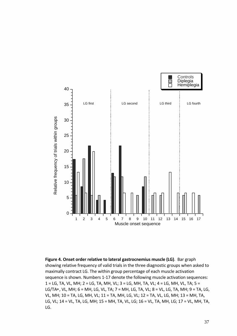
37
0
5
10
15
20
25
30
35
40
1 2 3 4 5 6 7 8 9 10 11 12 13 14 15 16 17
ControlsDiplegiaHemiplegia
Rel
ativ
e fre
quen
cy o
f tria
ls w
ithin
gro
ups
Muscle onset sequence
LG first LG second LG third LG fourth
Figure 4. Onset order relative to lateral gastrocnemius muscle (LG). Bar graph showing relative frequency of valid trials in the three diagnostic groups when asked to maximally contract LG. The within group percentage of each muscle activation sequence is shown. Numbers 1‐17 denote the following muscle activation sequences: 1 = LG, TA, VL, MH; 2 = LG, TA, MH, VL; 3 = LG, MH, TA, VL; 4 = LG, MH, VL, TA; 5 = LG/TA∗, VL, MH; 6 = MH, LG, VL, TA; 7 = MH, LG, TA, VL; 8 = VL, LG, TA, MH; 9 = TA, LG, VL, MH; 10 = TA, LG, MH, VL; 11 = TA, MH, LG, VL; 12 = TA, VL, LG, MH; 13 = MH, TA, LG, VL; 14 = VL, TA, LG, MH; 15 = MH, TA, VL, LG; 16 = VL, TA, MH, LG; 17 = VL, MH, TA, LG.

38
Human movement can be innate, learned, voluntary, automated, complex and/or reflex mediated. Different regions and tracts in the central nervous system are involved in the different types of movement and thus a lesion to the developing brain (as in CP) can affect motor function differently depending on the location and extent of the lesion and also on the maturational stage of the CNS at the time when an injury/lesion occurs. There are previous studies that have shown the altered muscle sequencing seen in children with CP during a range of complex movements controlled by automated motor programs such as; sitting, balance control, locomotion and reach and grasp (Eliasson et al., 1991;Leonard et al., 1991;Brogren et al., 1998;Woollacott and Shumway‐Cook, 2005). Automated motor programs for repetitive movements have been located to the spinal cord (Dietz et al., 1994). Through practice and learning. motor program are refined. When a motor program shifts from being new to automatised, the anatomical representation in the brain shifts as well (Jenkins et al., 1994;Toni et al., 1998). A voluntary simple movement of the lower limb, such as the task explored in this study, is conveyed through the primary motor cortex via the corticospinal tract with descending signals through the brain stem and spinal cord. A recent study in TD children and children with CP suggests that deficits in motor coordination (eye, head, hand movements) for accurate reaching are coupled (Saavedra et al., 2009). Saavedra et al, showed that compared to TD children, children with CP were significantly delayed in their ability to isolate their eye, head and hand movements, suggesting that more extensive synergies present compared to those seen, for example, during sitting or walking in children with TD. In summary, abnormal sequencing has been found in children with CP for complex automated movements (such as locomotion) as well as for isolated movements of different body parts that involve more complex synergies. In Study I we have shown that altered temporal sequencing of muscles can exist also for simple voluntary movements aimed at activating only one muscle. Therefore, we conclude that aberrant sequencing is present in different aspects of movement in CP, irrespective of the simplicity or complexity of the task. When group means for the absolute time (=time from the onset of the prime mover to onset of the other lower extremity muscles) were analyzed for each group of children in Study I, for both groups of children with CP, the other lower extremity muscles were activated earlier than in children with TD, except when the children were performing a MVIC of medial hamstring muscle (MH) In this cases, children with TD activated the vastus lateralis (VL) slightly earlier than children with diplegia. See Figure 5.
39
Figure 5. Time (ms) from EMG onset of prime mover to other recorded muscles. 5a = maximum contraction of Vastus Lateralis muscle (VL); 5b = maximum contraction of Medial Hamstrings muscle (MH); 5c = maximum contraction of Tibialis Anterior muscle (TA); and, 5d = maximum contraction of Lateral Gastrocnemius muscle (LG). Significant differences between the groups were seen as follows:
• Knee extension: When vastus lateralis quadriceps (VL) was the prime mover, all groups were significantly different with aspect to the TA onset time (P=0.05). Post hoc analysis revealed that children with hemiplegic CP activated TA earlier than children with TD.
• Knee flexion: When the medial hamstring (MH) was the prime mover there were significant group differences in TA onset time (P=0.02). Post‐hoc analysis confirmed that children with diplegic CP (P=0.01) and hemiplegic CP(P=0.02) activated TA significantly earlier than TD children..
• Ankle dorsiflexion: When tibialis anterior (TA) was the prime mover significant group differences with regard to MH onset time(P=0.008) were seen. Post hoc analysis showed that children with diplegic CP activated MH earlier than children with TD (P=0.002)
The shorter latencies in children with CP included also adjacent muscles and were not confined to antagonists, which suggests altered neural circuitry. As for muscle recruitment patterns, latencies to onset of other muscles were especially short for the children with CP when distal muscles were prime movers (Figure 5). Figure 6 is an example of the EMG activity seen during a MVIC of LG for the 3 different diagnostic groups.
40
A, TD B,Di C,He Figure 6. Example of EMG activity during maximum voluntary contractions of the lateral gastrocnemius muscle. The vertical line indicates muscle onset of the LG muscle. A= TD child first muscle to actvate is the intended LG muscle, TA is second. B= Child with diplegic CP, MH preactivates , LG and TA simultaneous onset. C= Child with hemiplegic CP , MH preactivates, LG second. For the aspects of motor control examined in Study I, i.e. muscle sequencing and latencies, differences between the children with CP and children with TD were more pronounced when the prime mover was a distal muscle. The children with CP included in the study had hemiplegic or diplegic CP. Eight of the 12 children with diplegic CP and 1 child with hemiplegic CP were born prematurely (gestational age 28‐36 weeks). Neuroradiology data were not available for the children with CP in the current study. However, it has been shown, (e.g. in the "European cerebral palsy study") that a high proportion of children with diplegic CP have periventricular white matter lesion of immaturity (Bax et al., 2006). White matter lesions of immaturity include periventricular leukomalacia ("PVL") and periventricular haemorrhage ("PVH"). In the study by Bax et al, 351 children with CP underwent magnetic resonance imaging (MRI) of the brain. White matter damage of immaturity was found in 71.3% of the children with diplegia and in 34.1% of those with hemiplegia. In the periventricular white matter, the corticospinal fibres pass in a strict somatotopic, pattern (Figure 7). The fibres to the most distal muscles, such as the toe muscles, pass more medially. As a rule of thumb, the more extensive the lesion the more likely it is that proximal muscles are affected. For the children with diplegia (three quarters of whom were born prematurely) included in Study I, one can assume, based on the findings of the Bax et al study (Bax et al., 2006), that a large proportion had white matter injury of immaturity . In these cases corticospinal fibers involved in control of distal muscles are likely to be affected in all children. In contrast, proximal muscles would only be affected in children with more severe lesions in the periventricular white matter. This can explain the increased

41
Figure 7.Schematic diagram of corticospinal tracts from their origin in the motor cortex, with descent past the periventricular region and into the internal capsule. The locus of the periventricular leukomalacia (marked square areas) would be expected to affect, particularly, descending fibers for lower extremity more than the laterally
placed fibers for upper extremity and face. Fibers to the distal lower extremity such as ankle and foot have the most medial placement. level of involvement of distal muscles, which was observed in our study. Therefore, we explored our data further with regard to relationships between gross motor function involvement, expressed as GMFCS levels, and recruitment patterns. There was, however, no obvious association between GMFCS levels and aberrant recruitment patterns. In this study, one of the limitations is the relatively small sample size, particularly when performing analyzes within the different GMFCS levels. Thus, our negative findings, based on visual inspection of the data, might be caused by the small sample size. Another possible explanation for the pronounced distal involvement might be that in children with lesions that involve the motor cortex, proximal and axial muscles have greater subcortical and also bilateral projections (Lawrence and Kuypers, 1968a;Lawrence and Kuypers, 1968b). These proximal muscles are thus less vulnerable than distal muscles that will receive their impulses through crossed, (contralateral) corticospinal fibres only. In Study II, the amount of co‐activity in the non‐prime mover muscles (the muscles that were not intended to contract) were studied. For each child data analysis involved a normalization process (described in the methods section) and the data of each child were expressed as % EMGmax. All children, TD and those with CP, had some co‐activity in lower extremity muscles during MVIC. Statistically significant differences in co‐activity were seen between the three different groups for all examined muscles except the ankle dorsiflexor (TA). Post‐hoc analysis showed differences mainly between the children with spastic diplegia and the TD children. When looking at the situations in which a muscle was not prime mover (i.e. three situations/muscle for four different muscles) children with CP more often reached above 40‐50% EMGmax than did TD children (i.e. children with CP had co‐activity levels that corresponded to more than 40% of the maximum activity in the muscle); 10 of 12 times in diplegia and 8 of 12 times in hemiplegia versus 3 of 12 times in comparison children. In two children with diplegia, mean EMG co‐activity exceeded 100%; specifically, 101.7% EMGmax for the TA during MH MVIC and 105.7% EMGmax for the MH during LG MVIC.

42
When the co‐activity of all muscles was averaged over all test conditions we found that children with TD showed mean co‐activity of 28% EMGmax compared to 65% for children with diplegia and 52% for children with hemiplegia. Thus, the amount of co‐activity recorded for the children with TD is lower than those found in children with CP. The amount recorded in the children with TD probably reflects the normal mechanisms needed to enhance stability. A recent review focusing on knee dynamics found antagonistic hamstring co‐activity reported in all six eligible studies that evaluated co‐activity or co‐contraction during functional movements in different age groups (Ford et al., 2008). In Study II children with diplegic CP had more than double and children with hemiplegic CP close to double the amount of co‐activity observed in TD children. An explanatory model for the increased level of co‐contraction seen in CP is presented by Brouwer and Ashby (Brouwer and Ashby, 1991). In a study in which they used transcranial magnetic stimulation (TMS) to elicit motor activity in TA of healthy controls and subjects with CP (adults or young adults), they showed that magnetic stimulation elicited equal facilitation in the soleus motor neuron and the TA motor neuron in the subjects with CP, whereas healthy subjects only showed TA motor neuron activity. The study by Brouwer and Ashby provides evidence in humans that injury to the immature brain and corticospinal tract results in altered corticospinal projections. The authors hypothesized that in CP, the altered projections appear to be directed equally to the motor neurons of both agonist and antagonist ankle muscles. This may explain why voluntary movements in these subjects are often disrupted by co‐contraction of flexor and extensor muscles. Myklebust et al (1986) described that a tendon tap in the newborn infant elicited muscle activation in both agonist and antagonist muscle (Myklebust et al., 1986). During ontogeny, this spreading reflex activity is reduced and in healthy adults a tendon tap will only elicit a stretch reflex of the agonist muscle. In CP, however, the immature reflex spread often remains. This indicates a failure to suppress spinal circuitry and defects in reciprocal inhibition via corticospinal control of Ia inhibitory interneurons. Increased levels of co‐activity observed in our study thus might also be caused by defect segmental reflex control. Surprisingly, in Study II, muscles adjacent (proximal or distal) to the prime mover showed higher means for co‐activity than did antagonist muscles. Often was this the case not only for children with CP but also for children with typical motor development. This finding is unexpected since co‐activity ‐‐ if one looks upon it as a measure of co‐contraction ‐‐ physiologically increases stability at a joint and thus should be largest at the antagonist. However then studies of this type in which several muscle groups are being simultaneously assessed in healthy controls are scarce. In one of the few available studies in which several muscles were examined simultaneously, only the agonist/antagonist pair had been evaluated for co‐activity (Gontijo et al., 2008). Thus, we currently do not fully understand to what extent the physiological co‐contraction also affects adjacent muscles during voluntary movements such as the one explored in this study. In a recent study in children and adolescents with CP and healthy controls, six agonist/antagonist pairs were examined in both lower extremities during a cycling test
43
(Johnston et al., 2007). The study found significantly higher levels (often four times as high) of co‐contraction in all the antagonists examined for the subjects with CP. Despite this study being different from ours in the sense that a complex movement was assessed, the study by Johnston et al too provides evidence that, from an EMG point of view, children and adolescents with CP use, or activate, their muscles differently from subjects with TD. They (Johnston et al) stated that "In general, subjects with CP displayed earlier onset and later offset of muscle activity… than subjects with TD…subjects with CP may have experienced differences due to decreased strength and motor control and therefore activated as many muscles as possible to both stabilize the joints and allow movement" (Johnston et al., 2007). It is likely that in the study by Johnston et al, abnormal synergies, changed latencies and co contraction patterns in muscles adjacent to the intended prime movers are shown, similar to our Study I and II. Previous to Study II, three other studies have examined co‐activity during MVIC in children with CP and children with TD (Ikeda et al., 1998;Elder et al., 2003;Stackhouse et al., 2005). However, the amount of observed co‐activity varied between the different studies. In addition, two of those three studies examined torque (Ikeda, Elder), and one study quantified muscle activation, muscle fatigue and contractile properties thus obtaining a measure for weakness (Stackhouse et al., 2005). All three studies analyzed agonist /antagonist pairs. In addition, the study by Stackhouse et al examined both proximal and distal muscle pairs but not simultaneously as in Study II. In the other two studies the quadriceps/hamstring muscles (Ikeda et al., 1998)and tibialis anterior/ gastrocnemius pairs (Elder et al., 2003)were assessed. In summary, these three studies found excessive co‐activity and reduced torque in children with CP when compared to the healthy controls (Ikeda et al., 1998;Elder et al., 2003;Stackhouse et al., 2005). In all three studies the authors conclude that the observed co‐contraction/co‐activity might partially be responsible for the observed weakness. These studies are discussed in detail in the reprint of Study II. In both Study I and Study II bilateral recordings could have added more to the understanding of the deviant motor control observed in children with CP. From studies that examine children with unilateral CP with TMS and fMRI, aberrant cortical circuits and corticospinal pathways from the motor cortex to the hand have been shown (Carr et al., 1993;Carr, 1996;Staudt et al., 2002;Vandermeeren et al., 2003b;Eyre et al., 2007). With regard to our study, altered latencies and motor onset orders also in the non‐active leg detected in a standardized position could have given insight into the organisation of altered corticospinal pathways that may affect the ipsilateral leg. Adding a measure of strength to study II would have been interesting and would also have added to the value of the study. The above mentioned three previous studies have shown a varied range of excess co‐activity and weakness in children with CP compared to TD children. Thus, additional data on strength could have contributed to the understanding not only with regard to levels of co‐activity but also with regard to relation to weakness. The finding that co‐activation was highest not in the

44
antagonist but a muscle adjacent to the prime mover is intriguing. In what way affects the co‐activity in an adjacent muscle the net torque in the intended joint? Will voluntary drive be dispersed to other joints and is this ,for example, important during strength training? In what way should strength training protocols for people with CP be constructed in order to maximize output and strengthen the targeted muscles? Study III In Study III, seven original reports and a large number of abstracts were identified for review. We included only these seven original articles in the review. A total of 112 children, aged 2 ‐ 20 years, were studied in the seven articles (see Table 4). One study addressed arms and legs of children with dyskinetic cerebral palsy (Denislic and Meh, 1995), while the remaining six studies dealt with intervention in spastic CP (Koman et al., 1993;Wall et al., 1993;Koman et al., 1994;Cosgrove et al., 1994;Calderon‐Gonzalez et al., 1994;Corry et al., 1997). Three studies focused on treatment of upper extremities (Wall et al., 1993;Denislic and Meh, 1995;Corry et al., 1997) and four on lower extremities (Koman et al., 1993;Koman et al., 1994;Cosgrove et al., 1994;Calderon‐Gonzalez et al., 1994) . Since BoNT‐A treatment was novel, particularly in children, the review also focused on therapeutic aspects and treatment procedures. Doses and injection intervals varied considerably among the different studies, making comparisons difficult. Sometimes the doses were defined in total units/kg BW (Koman et al., 1993;Koman et al., 1994;Cosgrove et al., 1994;Corry et al., 1997), while in other cases, as in adult practice, the number of units injected per individual or muscle was documented, but without any information about patient weight (Wall et al., 1993;Calderon‐Gonzalez et al., 1994). Another issue was the problem with the two non‐equivalent products. In the review we stated that the injection does not have an immediate effect and the delay may vary depending on the etiology of the treated condition. The reported duration of effect varied between 2‐6 months, which made us conclude that a variable interval between subsequent injections could be suggested. After an additional decade of BoNT‐A treatment and new indications, we now know that this is true. There are individual differences for when to repeat treatment in patients suffering from the same disorder, as well as differences that come into play when treating different disorders. For example, cervical dystonia requires treatment every 12‐14 weeks (Comella et al., 2000), while duration of effect for axillary hyperhidrosis is 7 months (Lowe et al., 2007). Quite surprisingly, critical assessment of the included studies revealed that only one study, by Corry et al, incorporated several aspects of a high‐level study design ‐ randomization and double‐blinding, with well‐described, validated assessment methods, and application of statistical methods (Corry et al., 1997). See Study III

45
(reprint: Table II) and Table 4. for the overall study design, methods of assessment, and statistical analysis for the included studies. Table 4. Studies included in review
Reference
Material N Age(y)
Diagnosis
Design Statistics
Koman et al 1993
n=3; 3‐11 n=8; 2‐13 n=16; 4‐16
severe quadriplegia; nonambulatory severe diplegia; nonambulatory di‐ & hemiplegia; ambulatory
open study no statistics (2 cohorts) t‐test (1 cohort)
Koman et al 1994
n=12; 4‐11 di‐ & hemiplegia;
ambulatory placebo, double blind no statistics
Cosgrove et al 1994
n=11; 2‐17 n=7; 2‐17 n=8; 2‐17
severe quadriplegia; nonambulatory diplegia hemiplegia
open study t‐test
Wall et al 1993
n=5 No age
given
hemiplegia, upper limb
prospective,
blind evaluation
Calderon‐ Gonzalez et al 1994
n=4; 2‐20 yr. n=10; 2‐20 yr. n=1; 2‐20 yr.
quadriplegia diplegia dystonic hemiplegia
open (A‐B‐A) t‐test
Corry et al 1997
n=12 4‐19 yr.
n=1 4‐19 yr.
n=1 4‐19 yr.
hemiplegia
tetraplegia
quadriplegia
randomized
placebo
double blind Denislic & Meh 1995
n=13; 7‐15 yr. dyskinesia
Unspecified

46
An additional study, by Koman et al, was also randomized and double‐blinded, but unfortunately the authors of this study did not reveal how their statistics were analyzed (Koman et al., 1994). A third study, of upper extremity treatment, was not blinded to either investigators or patients, but did apply an attractive model in which a blinded panel rated pre‐ and post‐intervention photographs and videos (Wall et al., 1993). A total of 3 of the 7 included studies did not use or describe statistical methods. Study III (reprint: Table II) provides an overview of the assessment methods used. Five studies investigated spasticity, three used the Ashworth or Modified Ashworth scale, and two relied on parental assessments and reports. In general, a reduction of muscle tone and spasticity was reported in these various studies. Range of motion (ROM) was a frequently measured parameter, but described clearly in only one study (Corry et al., 1997) . Three studies analyzed gait visually for which they used the physician rating scale (PRS) (Table 5). To the best of our knowledge, this scale was first described by Koman et al in the first study included in this review (Koman et al., 1993). At the time of the review no validity or reliability study whatsoever had been undertaken to evaluate the method. However, this scale was subsequently used in several studies to assess gait in children with CP (Koman et al., 1994;Corry et al., 1998;Koman et al., 2000;Ubhi et al., 2000). Table 5. Physician rating scale (PRS) Definition Right Left Crouch Severe (>20° hip, knee, ankle) 0 0 Moderate (5‐ 20° hip, knee, ankle) 1 1 Mild (<5° hip, knee, ankle) 2 2 None 3 3 Knee Recurvatum >5° 0 0 Recurvatum 0‐5° 1 1 Neutral (no recurvatum) 2 2 Foot contact Toe 0 0 Toe‐heel 1 1 Flat 2 2 Occasional heel‐toe 3 3 Heel‐toe 4 4 Change Worse ‐1 ‐1 None 0 0 Better 1 1

47
A reliability study evaluating the method showed that "the PRS had excellent intraobserver reliability, but poor interobserver reliability for children with CP" (Maathuis et al., 2005). A modified version of the PRS known as the Observational Gait Scale (OGS) was assessed for reliability and validity in video recordings of 20 children/adolescents, filmed at the time of a 3D‐gait analysis (Mackey et al., 2003). The validity of the OGS score was determined by comparing it with the 3‐D gait analysis. Some aspects of the OGS (knee and foot position in midstance, initial foot contact, and heel rise) had acceptable inter‐ and intra‐rater reliability and high validity in a comparison compared to the 3D‐gait analysis, while other aspects (such as base of support and hindfoot position) were associated with lower reliability and validity could not be confirmed by 3D‐gait data (Mackey et al., 2003) . In recent years, another visual gait scale, the Edinburgh Visual Gait Score, has been used (Read et al., 2003). This scale, however, also has a poor inter‐observer reliability and consequently it is recommended that longitudinal assessments of a patient should be performed by one observer only (Maathuis et al., 2005). Instrumented gait analysis, that includes computerized kinematics (description of motion) and kinetics (description of forces and torque) as well as EMG and video recording, are often used to assess gait in children with CP. Three dimensional gait analysis is also considered to be the gold standard for gait assessment. But the resources necessary for a fully equipped gait laboratory are not available everywhere and the procedure itself is costly, time consuming and unsuitable for younger children or children with severe cognitive problems. This justifies the need to develop the simplified observational methods described. Reliability and validity studies exist, as well as normal reference values for assessment of gait in adults and normal children using 3D‐gait analysis (Kadaba et al., 1989;Ounpuu et al., 1991;Steinwender et al., 2000). In children with CP the intra‐subject repeatability for different aspects of 3D gait analysis is lower compared to the analysis of TD children. Furthermore, the general repeatability for the assessment of kinetics is better than for kinematics (Steinwender et al., 2000). However, even considering the shortcomings of the visual scales, our recommendation still remains unchanged: for research purposes 3D‐gait analyses should be used. At the time of the review we were surprised to notice that the muscle tone reduction and increased ROM described in the study by Corry et al (1997) did not result in any fine motor functional improvement as assessed by the ability to pick up coins. Today, 11 years later, few published reports have been able to prove functional gains despite a reduction in muscle tone. Overall we found that study designs, assessment methods, and lack of statistical analysis in those early papers were major flaws that limited the ability to interpret the results of intervention with BoNT‐A. For future research we recommended as a minimum that the Ashworth scale be used, despite its limitations, to assess and document spasticity. For joint ROM we noted that the best results were obtained when the same examiner performed the examination pre‐ and posttreatment, and recommended including information about the examiner in published reports.

48
We discouraged use of the PRS and instead recommended use of 3D‐gait analysis to better evaluate gait changes. No attempts to objectively assess function were made in any of the 3 papers that included children with tetraplegia. We recommend use of the GMFM to assess gross motor function in nonambulatory children (Russell et al., 1989). We were concerned that premature registration of BoNT‐A products to treat children for the indication of CP could hamper necessary and important future research. The available scientific literature raised more questions than answers and our final recommendation was to further improve evaluation of BoNT‐A before this treatment could be accepted for general use in children with CP. Study IV Shortly after publication of our review article, Study III, Botox® was licensed for use for the indication spastic equinus in children over two years of age with CP in Sweden and some additional European countries. We realized this development would be likely to hamper efforts to conduct a large‐scale prospective RCT in Sweden to evaluate BoNT‐A intervention. Two studies were designed: Study IV, a pragmatic approach to prospectively follow all children receiving BoNT‐A at our hospital, and Study V, a randomized clinical trial to evaluate the effect of BoNT‐A in young children. In 1997 we started treating the first pilot patients with intramuscular injections of BoNT‐A. We set up a clinic where a pediatric neurologist (KT) and a pediatric orthopedic surgeon (YHÅ) examined all children with spasticity from greater Stockholm as well as some referred from the island of Gotland at the request of the child’s attending physician. A total of 133 children from this clinic started treatment with BoNT‐A over a five‐year period, 1997‐2001. One hundred and twenty‐four (93%) of the patients had spasticity associated with CP. Other causes of spasticity included traumatic brain injury or spina bifida. The majority (120) of patients received injections into the muscles of the lower extremities. Study IV is an analysis of the 94 patients with a diagnosis of CP whose lower extremities were treated. The average age of included patients at the time of the first injection was 5 years 4 months (range 11mo–17y 8mo). Of these patients, 50% had spastic diplegic CP, 22% hemiplegic CP, 25% tetraplegic CP, and 3% dyskinetic CP. The GMFCS level was I in 29%, II in 15%, III in 16%, IV in 17%, and V in 23% of the patients. Maximum follow‐up time in the study was 3 years 7 months, with a median of 1 year 6 months. (See Table I of reprint). The number of injections given in each muscle varied from 1 to 8, median 2 injections in any given muscle. (See Table II of reprint). After the first injection treatment continued if there were objective signs of benefit

49
(such as reduced muscle tone, increased ROM or gain in GMFM) and parents, PT and physician agreed on further injections. Eighty‐five percent (N=80) of patients continued treatment. The remaining 14 patients (15%) terminated treatment after a median of two follow‐up assessments (range 0–2); 10 exhibited no response and four experienced adverse effects including excessive weakness or painful injections. No serious adverse events in the complete cohort of 133 patients were reported at any time during the study. Our muscle‐by‐muscle analysis of long‐term effects in this cohort of patients included muscle tone and joint ROM, as stated in the methods section. In addition one or several of the following assessment tools were used in a majority of patients; muscle strength, selective motor control, Gross Motor Function Measure (GMFM), and three‐dimensional gait analysis. However these methods were not always assessed at regular intervals and with differences in how each patient was assessed, making the data difficult to analyze at the group level. Statistical analysis revealed that the initial muscle tone‐reducing effect previously documented by others could be maintained longer in the examined muscle groups than was previously reported for children. In addition, each repeated injections in the gastrocnemius muscle resulted in a significant reduction in muscle tone of nearly 1 AS compared with the muscle tone prior to that specific injection (Figure 8). Out of the muscles examined in our study we were able to document the longest significant muscle‐tone reducing effect in the gastrocnemius muscles bilaterally , see Study IV (reprint:Figure 1). Moreover, we found significant reduction of muscle tone in the hamstring and hip adductors bilaterally, compared to baseline values, which lasted for several 90‐day (or quarterly) intervals (Figure 9,10). Prior to this study, earlier studies reported an immediate tone‐reducing effect after BoNT‐A injections, but as we commented in Study III, only the work by Corry et al (Corry et al., 1997) with a 12‐week follow‐up was strongly evidence based. Subsequent studies have confirmed the muscle tone‐reducing effect of intramuscular injections of BoNT‐A in children with CP. Three RCTs have shown a muscle tone reduction lasting 6 months after lower extremity muscle injections with BoNT‐A (Flett et al., 1999;Love et al., 2001;Reddihough et al., 2002) and additional studies have confirmed a short‐term (3 months) effect (Hazneci et al., 2006;Bjornson et al., 2007).

50
Figure 8. : Effect of repeated injections to gastrocnemius muscle (only injections without concurrent casting included). (a, b) Mean change in muscle tone, measured as Ashworth score [with 95% confidence interval (CI) bars] before and after each injection for the left (a) and right (b) gastrocnemius muscles, respectively.(c, d) Corresponding change (with 95% CI bars) in ankle‐joint dorsal extension after each injection.
51
-2
-1.5
-1
-0.5
0
0.5
1
1.5
2Change relative to baseline in
right hamstring toneMuscle tone in A
shworth score
90- day intervals
0 0.5 1 2 3 4 5 6 7 8 9 10 11
Figure 9: Longitudinal change in muscle tone, relative to the baseline values (0) (taken before the first injection), after injections to right hamstring muscle, measured over time from the first treatment in 90‐day intervals for the right hamstring muscle. Ninety‐five per cent confidence interval bars (CIs) are given; a CI crossingthe baseline indicates no significant change.
-2
-1.5
-1
-0.5
0
0.5
1
1.5
2Change relative to baseline in left
hip adductor muscle tone
Change in m
uscletone AS
90-day intervals
0 0.5 1 2 3 4 5 6 7 8 9 10 11 12
Figure 10: Longitudinal change in muscle tone relative to the baseline values(taken before the first injection), after injections to left (hip) adductor muscle, measured over time from the first treatment in 90‐day intervals for the left adductor muscle. Ninety‐five per cent confidence interval bars (CIs) are given; a CI crossing the baseline indicates no significant change. In a recently published paper of a study initiated in the 1990s, a British group conducted an elegant double‐blind RCT with placebo saline injections or injections of BoNT‐A "as needed" in lower extremity muscles for a total of two years (Moore et al., 2008). The 64 children included in this study did not display any significant reduction in AS after 1 or 2 years in the BoNT‐A treatment group, compared with the placebo group. In order to compare our findings with those of others, we must turn to the adult literature. Our results concur with the outcome of a study in adults who received BoNT‐A treatment for an average of 15.8 years, a considerably longer follow‐up time, mainly for the indication of cervical or craniocervical dystonia, in which the treatment proved efficacious and reduced muscle tone after repeated injections (Mejia et al., 2005). In this regard a recent paper by Hägglund and Wagner is very interesting (Hagglund and Wagner, 2008). This is the first study ever to describe the natural development of spasticity over a 10‐year period in a large cohort of about 500 children with CP.

52
Spasticity was assessed using the Modified Ashworth scale. These authors used the Bohannon and Smith version of the scale (Bohannon and Smith, 1987). In this version of the scale, level 0= normal muscle tone. The Ashworth scale was dichotomized for statistical analysis. One category included levels 0‐1 and the other levels 2‐4. The study showed that muscle tone in children with CP, as measured by the modified Ashworth scale, increases up to 4 years of age and then decreases up to 12 years of age. Interestingly, a similar development was seen when children who had been operated with selective dorsal rhizotomy, intrathecal baclofen pump, or tendo‐achilles lengthening were excluded from the analysis. Could these new findings possibly explain the longitudinal decrease in muscle tone found in this study? We think not, since all gastrocnemius muscle injections, which were individually evaluated before and after each intervention, showed an obvious treatment effect of muscle tone reduction. In other words, we could show a significant reduction in muscle tone of nearly one AS even when children received a fourth injection of BoNT‐A in the gastrocnemius muscle, compared with muscle tone prior to injection (Figure 8). In our analysis we found that although BoNT‐A treatment initially resulted in increased joint ROM, this was not an enduring effect. A return to baseline ROM values or below was soon seen, reflecting continued shortening of the spastic muscles. Since this study did not include a control group we are forced to assess the results in relation to our limited knowledge of natural disease progression in CP, which has shown that children with CP experience a progressive decrease in joint ROM that ultimately threatens ambulation (Johnson et al., 1997;Bell et al., 2002;Gough et al., 2004). Therefore any reduction of progressive decrease in ROM cannot be detected by comparisons with baseline values; only significant changes can be registered. A thorough review of the current literature reveals very limited information about long‐term results for how ROM evolves after BoNT‐A intervention. An open multicenter study in which 207 children from 9 different centers were enrolled is often cited as an example of enduring effect (Koman et al., 2001). In our opinion there are some concerns about this study that limit the generalizability of the data since these researchers used shorter injection intervals (2.4 months), which may be more effective, but also associated with the risk of antibody formation (antibody formation being a common cause to secondary resistance where prior good responders loose their benefit of subsequent BoNT‐A treatment). In addition, they compared active dorsal extension before and after treatment, and follow‐up data were obtained from only 14 of the 207 enrolled patients 1 year after treatment (Koman et al., 2001). Lately additional evidence has been published in two studies by Bjornson et al and Graham et al that supports our findings (Bjornson et al., 2007;Graham et al., 2008). In a randomized controlled trial, Bjornson and coworkers found significant improvement in ankle ROM after 12 weeks, but not at 24 weeks after injecting BoNT‐A in the gastrocnemius muscle (Bjornson et al., 2007). In early 2008 Graham and co‐workers published a three‐year prospective RCT (Graham et al., 2008). They compared 6‐monthly BoNT‐A injections in the hip adductors and hamstring muscles combined with 6 hours of daily abduction orthotic use, to no intervention at all, for their effect on hips at risk of subluxation in 90 children with bilateral CP. The

53
Ashworth scale and the Tardieu scale was used to identify when a "particular muscle group should be reinjected". Thus we can assume that a muscle tone reduction was seen after injections. The Graham study revealed the same rate of progressive hip displacement in both the intervention and control groups. Therefore BoNT‐A treatment, even in combination with a rigorous daily splinting regimen, was unable to prevent hip displacement or reduction in hip abduction (Graham et al., 2008). The results presented in our Study IV showing a different impact on muscle tone and ROM challenges the accepted theory of contracture development. Clearly, contracture development is not solely attributable to increased muscle tone, but also depends on processes on which BoNT‐A injections do not have a favorable effect. Molecular changes, (Lieber et al., 2004;Ponten et al., 2007) including deposition of collagen (Booth et al., 2001), have been described in spastic muscles and during contracture development. The cause of these molecular changes is not known, but a recent 11‐year follow‐up indicates an association between immobility and hip contractures/dislocation rather than a relation between muscle tone and contracture development (Soo et al., 2006). However many clinicians argue that contracture development is secondary to spasticity and the study in spastic mice by Cosgrove and Graham (Cosgrove and Graham, 1994) (discussed in detail in the introduction p.9‐ 10) is often put forward as supportive. But as has previously been stated in this thesis ‐‐what remains unclear is exact mechanistic explanatory model to contracture formation. In a study by Williams et al (Williams et al., 1998) the tibialis anterior muscle of rabbit was progressively stretched at a rate mimicking the one used in human surgical limb lengthening. The stretching added new sarcomeres in serial giving length to the muscle fibres but concomitantly increased deposition of collagen type III, altered muscle stiffness, and resulted in greatly reduced ROM. Severe contractures are also a common complication of limb lengthening procedures. The researchers hypothesize that the connective tissue components are less able to adapt than are the contractile components. The same authors then performed a second study were stretching was performed at three different rates; low, medium and high (Williams et al., 1999). At the low rate, there was no change in the passive tension or collagen content compared to muscles from sham‐operated animals, and range of movement was significantly greater than at the other rates. While muscle distracted at a high rate, resulted in almost total loss of joint movement combined with changes in the connective tissue (Williams et al., 1999). A speculative explanation to our findings is thus that the first injection results in a rather sudden elongation of the injected muscle followed by a secondary increased deposition of collagen type III resulting in an initial but not lasting increase in ROM. The risk exists that repeated BoNT‐A injections supports the (as yet unknown) mechanisms underlying the development of contractures? Study V This study followed 15 children (mean age at study start 16 months) with spastic unilateral or bilateral CP for 3.5 years during the period in life when they advanced from "pull to standing" (baseline) to walking independently (see time axis in

54
Methods section, p. 25). It is the first RCT in which very young children received BoNT‐A injections in spastic muscles. We used goniometric assessments to establish ROM, a reliable method – especially when measurements are taken by an experienced PT, as in our study (Mutlu et al., 2007). In the control group, both ankle and knee ROM gradually decreased after 1 and 3.5 years, with a significant difference from baseline in both joints at 3.5 years. Reduction in ankle ROM at 3.5 years was ‐8.9° (95% CI ‐15.7° to ‐2.0°), (P=0.013), while reduction in knee ROM (i.e. increased popliteal angle) at 3.5 years was 11.1° (95% CI 4.2° to 18.0°); (P=0.003). This reduction in joint ROM reflects slow contracture development. In contrast, the BoNT‐A group showed increased ROM after 1 year of active treatment. Knee ROM significantly improved in relation to baseline prior to treatment start (‐12.4° (95% CI –21.5° to –3.6°), (P=0.009). Change in ROM also differed significantly from the changes seen in the control group. Three and a half years after study start, joint ROM in the BoNT‐A group returned to baseline, which is consistent with the findings in Study IV where there was an initial, temporary beneficial effect on the development of contractures, which subsequently continued despite ongoing treatment. A similar trend is also seen in this study on young children. Study IV, involving older children with CP, clearly showed that protracted treatment periods with BoNT‐A injections could not prevent contracture development. Results from this study (Figure 11) partially support this conclusion since an initial increase in ROM within the BoNT‐A group is followed by a decrease. However, since this study has the advantage of a control group, we can see in Figure 11 that while ROM continued to decrease in the control group during the study period, early treatment may have a beneficial effect on the progression of contractures by shifting progression along the "joint contracture curve". In the present study only gastrocnemius muscles were injected, but both the hamstring and the gastrocnemius muscle‐ being a biarticular muscle, act as knee flexors, thus a relaxed gastrocnemius muscles will contribute to a reduced popliteal angle, since the gastrocnemius muscle contributes with the initial flexion of the fully extended knee. Changes in knee ROM are seldom discussed in relation to gastrocnemius muscle injections thus we can not compare our findings to other published reports. An increased popliteal angle is considered to cause reduced velocity during gait and difficulties in seating due to a corresponding backward tilt of the pelvis. Popliteal angel reduction after hamstring lengthening has shown improved sitting position (Elmer et al., 1992) and a large review on sitting position in CP also found support for a neutral to slightly forward orientation in sitting on upper‐extremity function (Stavness, 2006). Thus one can assume that a reduction in popliteal angle has a positive functional effect even if this study failed to show this connection when evaluating GMFM, PEDI and gait. A reliable tool for spasticity assessment is crucial in research. Despite the lack of convincing pediatric reliability studies, we chose one of the most commonly used

55
tools in written papers and clinical pediatric practice, the Modified Ashworth scale, mainly due to the age of the subjects at study start (Clopton et al., 2005). Other methods include computer controlled dynamometers or stretch reflex testing but these methods is difficult to perform in very young children. In a previous study intrarater reliability was better than interrater (Fosang et al., 2003). In this study the same PT performed all assessments. The Ashworth scale is an ordinal scale. Thus we initially performed a ranking model for statistical analysis. However limited sample size reduced the possible output and we decided to perform descriptive statistics as this has most often been the methods employed in published papers. Muscle tone was measured in both the ankle and knee joints. All children in the study displayed increased muscle tone at baseline, but while the increased muscle tone in the control group was relatively constant in both joints at the 1 and 3.5 year assessments, it was clearly reduced in the BoNT‐A group. The change in the ankle joint from baseline was significantly reduced after 3.5 years in the BoNT‐A group, while in the knee joint the change in tone was significantly different between the two groups at 3.5 years (Figure 11). This study failed to detect any significant reduction in muscle tone after one year of active treatment (six months after the most recent injection of BoNT‐A in the BoNT‐A group). But sample size in the study was limited and insufficient to allow changes of the magnitude reported to be detected. In addition, as was previously discussed, the duration of the tone reducing‐effect after an injection differs among individuals and the majority of children will require reinjection on a regular 6‐month basis. Thus the reduction in remaining muscle tone after 6 months might be very small. The RCT study by Bjornson confirmed a significant reduction in muscle tone 12 weeks after injection, but no significant differences were found between the control group and the BoNT‐A group at 6 months post injection (Bjornson et al., 2007). However, especially considering the fact that these children were followed during a period in life when spasticity is increasing according to a recent study (Hagglund and Wagner, 2008), the present data suggest that BoNT‐A can also be effective in reducing muscle tone in young children with CP. As spasticity was‐‐and by many still is‐‐believed to correlate with a reduction in function in CP (Gage JR and Schwartz M, 2004), many studies have focused on functional gains after BoNT‐A intervention. The GMFM‐88 and GMFM‐66 have often been used as assessment tools in the past decade. We found statistically significant increases over time in the GMFM‐66 in both the intervention and the control groups. The BoNT‐A group displayed an increase of 13.6 points (P=<0.0001) during the first year, and 23.6 points (P=0.0002) from study start to the last follow‐up. The control group exhibited an increase of 10.1 points (P=0.0001) and 20.9 points (P= <0.0001), respectively, during the same time periods. Likewise, when all six scales of PEDI were evaluated both groups had a significant increase in all six scales at 1 and 3.5 years. However, no significant differences were found between the BoNT‐A group and the control group at any time.

56
A. Ankle dorsal extension ROM
B. Plantar flexor muscle tone
-20,00
-15,00
-10,00
-5,000
0,000
5,000
10,00
15,00
-0,5 0 0,5 1 2 2 3 3 4
Change in Joint range of m
otion
Years
*
-2,500
-2,000
-1,500
-1,000
-0,5000
0,000
0,5000
1,000
-0,5 0 0,5 1 2 2 3 3 4
Ashw
orth score
Years
*
-20,00
-10,00
0,000
10,00
20,00
-0,5 0 0,5 1 2 2 3 3 4
Change in Joint range of m
otion
Years
*
*
** **
-1,500
-1,000
-0,5000
0,000
0,5000
1,000
-0,5 0 0,5 1 2 2 3 3 4
Ashw
orth score
Years
**
C. Popliteal angle ROM D. Knee flexor muscle tone
* significant change from baseline group ** significant differences between groups with respect to changes from baseline
Figure 11. A+C: Longitudinal development of ankle and knee ROM, Y‐axis shows joint ROM change from baseline with SE bars were an increase in ROM of the ankle is positive while a reduction of the popliteal angle is desired. B+D longitudinal change in muscle tone of plantarflexor muscle and knee flexor muscle, Y‐axis shows muscle tone (Ashworth score) change from baseline with SE bars were an reduction is wanted.

57
Distinct motor development curves have been created based on GMFCS level (Rosenbaum et al., 2002). Through these we learn that the GMFM scores for children with poor gross motor function will plateau as early as age three, while the curve for children at GMFCS level II flattens out at age 6. Thus the GMFM could have been an appropriate method for detecting functional benefit in the group of children represented by this study. Since the start of this study, other RCTs have used the GMFM and found conflicting results after BoNT‐A treatment (Love et al., 2001;Boyd et al., 2001;Reddihough et al., 2002;Bjornson et al., 2007;Moore et al., 2008). In three studies statistical analysis of the GMFM was unable to demonstrate significant differences between the BoNT‐A and control groups (Boyd et al., 2001;Reddihough et al., 2002;Moore et al., 2008). In contrast, two studies found significant functional improvement in BoNT‐A groups compared with controls (Love et al., 2001;Bjornson et al., 2007). Interestingly, however, both these later studies also included a parent satisfaction assessment. One study in which parental satisfaction was analyzed using a VAS scale found that treatment group parents were more satisfied than control group parents, but satisfaction scores did not correlate with changes in function or technical outcomes, suggesting that this may be a placebo effect (Love et al., 2001). In the second double‐blind study by Bjornson et al, despite documented functional improvement at 6 months in the BoNT‐A group, family satisfaction with outcomes was no different between the two groups (Bjornson et al., 2007). The authors conclude that communication is essential to ensure realistic treatment expectations. The Pediatric Evaluation of Disability Inventory (PEDI) was recently used in a prospective, double‐blind, 2‐year follow‐up RCT that compared the effect of repeated injections of BoNT‐A in lower extremities with placebo injections. This study failed to detect any significant changes between the groups (Moore et al., 2008). Few studies have been able to detect functional improvement after BoNT‐A treatment. It has been hypothesized that this is due to the heterogeneity of patients and symptoms. In a recent study, individual goals for BoNT‐A treatment were set using the goal attainment scaling method in a randomized, blinded, multiple baseline (cross over) study (Steenbeek et al., 2005). This study showed a clinically relevant improvement in individual rehabilitation goals, after BoNT‐A intervention in children with cerebral palsy. Adding the goal attainment scaling method to our study might have been useful to detect a functional improvement . Children with CP typically walk on their toes (digitigrade, equinus gait), displaying premature calf muscle contraction and increased coactivation (Leonard et al., 1991). Could the disturbance in gait pattern transformation in children with CP be caused by the increased muscle stretch reflexes and enhanced tone in calf muscles that prevent a heel strike from developing? If so, could a reduction of calf muscle tone early in bipedal locomotion in children with CP encourage plantigrade transformation and improve the gait pattern? This is the hypothesis that we wanted to test. At almost 5 years of age, 5 children in the BoNT‐A group and 8 children in the control group were able to participate in a 3D‐gait analysis. One child in the BoNT‐A group was cognitively delayed and failed to comply with instructions and one child in

58
the control group had developed a more dyskinetic motor pattern that ruled out gait analysis. Studies that uses 3D‐gait analysis to evaluate treatment effects have typically focused on one or several single parameters, such as “time to toe‐off” or “peak dorsiflexion in swing”, at a time, but gait is a complex dynamic activity and alterations at one joint level will affect others as has been shown in dynamic simulations (Higginson et al., 2006;Hicks et al., 2008). For this purpose we choose to use the Gillette Gait Index (GGI) method developed for the analysis of 3D‐gait data in CP (see Methods) (Schutte et al., 2000;Hillman et al., 2007). The mean GGI in the BoNT‐A group was 385.0 (SD=278.4) and for the children with CP in the control group it was 700.0 (SD=619.0), while for TD children it was 15.5 (SD=4.5). No significant difference was found between the two groups of children with CP. Therefore we found no support for our hypothesis. Obviously, however, for this measurement each child lacked baseline data, since gait could only be reported at the final assessment. Original group differences, regardless of treatment, could influence the outcome, especially because the small groups became even smaller due to the drop out rate. Study V has several limitations. The greatest perhaps are the small sample size and uneven number of subjects in each group. In retrospect, the inclusion criteria were restrictive and difficult to achieve without long study duration. Subject inclusion criteria were especially restrictive relative to including only children that were quite young, whose motor developmental level lie within a narrow window (“nearly ready to pull to standing” at recruiting), and who needed treatment mainly of calf muscles that affected ankle control. The result was a protracted study duration complicated by further delay necessitated by the study design which required another 3 ½ years, or more, before subjects achieved the motor ability and maturity needed to participate in 3‐D gait analysis. Additional concern emerge in that, despite the randomization process, the two groups varies in baseline characteristics for muscle tone and ROM with the children in the BoNT‐A group showing less joint ROM and somewhat higher muscle tone than the control group. This complicates data interpretation despite basing comparisons on change scores relative to baseline.

59
5 CONCLUSIONS AND CLINICAL IMPLICATIONS The activation of muscles differs between children with CP and children with typical development when performing a voluntary movement. In this thesis voluntary movement is studied when a maximum voluntary isometric contraction of a lower extremity muscle is performed and proximal as well as distal agonist/antagonistic muscle pairs are examined simultaneously. Substantial variability was observed not only in the agonist/antagonist pairs. But also in the muscles that are proximal or distal to the contracting muscle, which also display aberrant EMG activity. Altogether differences in muscle activation can be observed in three different ways;
• Children with CP display a large amount of variability in the order in which the four muscles are activated during a specific muscle activity. This is especially apparent when the muscle that is intended to be active is distally located.
• Children with CP also display shorter times or latencies to the activation of other muscles than the prime mover compared to children with typical development, and this is also more apparent in the more distal muscles.
• Children with CP express twice as much co‐activity in antagonist and adjacent muscles during maximum voluntary isometric contraction of all four of the lower extremity muscles tested, compared to children with typical development.
Parallel phenomena are known to exist in complex automated movements. We therefore conclude that aberrant sequential muscle activation occurs in different aspects of movement in CP, irrespective of the simplicity or complexity of the task. When the long‐term effect (up to 3.5 years) of botulinum toxin A injections to the lower extremity muscles of children with CP, that were at average 5.5 years when they started intervention, were evaluated, the results suggest that BoNT‐A can be effective in the reduction of muscle tone over a longer period, but not in the prevention of development of contractures in spastic muscles. The dissociation between the effects on muscle tone and ROM indicates that development of contractures is not associated with increased muscle tone alone, but may also be caused by other mechanisms. Repeated injections of botulinum toxin A into the gastrocnemius muscle displayed a repeated and predictable tone reducing effect that was maintained over time. However the effect on ankle joint ROM was significantly improved only after the first injection. Early treatment of botulinum toxin A in the gastrocnemius muscle during the second year of life in children with spastic CP appears to have a short‐term effect in increasing ROM and to influence muscle tone and contracture progression also after 3.5 years. The long‐term effect on plantigrade gait pattern remains inconclusive.

60
CLINICAL IMPLICATIONS
All, the aspects of muscle activation reported above are likely to hamper children with CP, and cause their voluntary movement to be less predictable and possibly weaker. From an intervention perspective careful mapping possibly including the use of EMG, before making decisions on the type and extent of intervention should probably be recommended. As an example, reduction of focal spasticity (either surgically or by local injections) in a child that displays excessive co‐activity in adjacent muscle groups during voluntary movement, may not give the functional benefits expected from this intervention. When children with CP perform strength training programs, they are also likely to strengthen other muscles than those intended, i.e. antagonists or adjacent muscles. From this perspective when designing strength training exercise, programs that include many different muscle groups could be more effective in strengthening particularly weak muscles. Whenever reduction in muscle tone to a targeted muscle or group of muscles is the aim of the treatment, such as treatment of pain due to spasticity, spasticity interfering with the use of orthotics or spasticity causing functional limitations such as scissoring, repeated injections of botulinum toxin A could be expected to have a sustained and predictable spasticity reducing effect in most patients. From the results obtained in this thesis the use of botulinum toxin A cannot be recommended as an effective treatment to increase long‐term joint ROM in older children with CP, for any of the investigated lower extremity joints. For very young children with CP, early botulinum toxin A treatment seems to have an initial effect in increasing joint ROM. However, since the study is small, additional more extensive studies are needed before any recommendations on early treatment can be given.

61
6 APPENDIX
Subject characteristics Number of subjects Gillette Gait Index (GGI)
(SD)Typical motor develop. (TD) 16 15.5 (4.5) CP, GMFCS level I 6 169 (102) CP, GMFCS level II CP, GMFCS level III
3 4
577 (224) 1212 (394)
Unilateral CP, affected limb 5 192 (107,5) Table 77. Illustrates the GGI index for children with TD and all children included in Study V subdivided by GMFCS level.
62
7 ACKNOWLEDGEMENTS I would like to express my sincere gratitude to all those who have directly or indirectly contributed to the completion of this work. Special thanks go to: All the children and their parents that have participated in the studies, without you; Ebba, Amanda, Hampus, Dan, Ismail, Rebecca, Oliver, Alice, Ella, Wiktor and many more there would have been less knowledge and understanding today. Associate Professor Yvonne Haglund‐Åkerlind, my supervisor, friend and colleague for enjoyable cooperation, good clinical skills and constructive discussions. Professor Hans Forssberg, my supervisor for providing an excellent research climate at the Neuropediatric unit, Department of Woman and Child Health at Karolinska Institutet, and for being a brilliant researcher with the ability to pick up and elaborate on the uniqueness of results. Professors Loretta Knutson and Gary Soderbergh previously working at Southwest Missouri State University, Springfield, MO, USA, my co‐authors and “US‐supervisors” for support, advice, stimulating discussions, text editing and for being such good friends. Kicki Löwing, co‐author and friend for in an excellent, warm and enthusiastic manner, taking care of study patients and for being a statistical encyclopedia. My co‐authors Fredrik Granath and Lanie Gutierrez Farewik, for being such experts in your fields and making data analysis a thrilling experience. Researchers and collaborators Eva Broström and Brigitte Vollmer, for generously helping me with all kind of matters, for sharing knowledge, inspiring discussions, collaboration and not least for friendship. Collaborators and researchers Eva Brogren‐Carlberg, Ammi Bexelius, Ann‐Christine Eliasson, Lena Krumlinde‐Sundholm, Karin Thews, Åsa Bartonek, and Robert Petersson, all very important in the pursuit of the studies. My research colleagues at the Neuropediatric research unit at Karolinska Institutet, Linda Holmström, Eva Pontén, Stefan Gantelius, Pähr Engström and Åsa Hedberg, to mention a few, for making work so enjoyable. None‐Marie Kemp, Astrid Häggblad and Mikael Reimeringer at the Department of Woman and Child Health, KI, for always being knowledgeable, helpful and professional.

63
Carolina Luhr‐Nilsson, co‐worker and friend for skilfully handling the intrathecal baclofen pump patients while I was on research leave. Kerstin Dahlström and Christina Strömbeck clinical colleagues who are responsible for the staffing at the Neuropediatric unit at Astrid Lindgren Children’s Hospital, Karolinska University Hospital, for being supportive in making it possible for me to be off clinical duty to conclude this thesis. All important, co‐workers and colleagues at the Neuropediatric unit at Astrid Lindgren Children’s Hospital. It’s a privilege to be able to work with so inspiring and competent people. The staff at Mac Keith Press, Developmental Medicine and Child Neurology Editorial Office, for being helpful and responsive. The following people have contributed by being very important aspects of my personal life; My father Bo who, together with my late mother, affectionately raised me, my brother Anders Ekesbo a photoshop wizard. My supportive, energetic sister and friend, Anna Ekesbo‐Freisinger and her large and loud family. My family, Joakim and our children Antonia, Carl Johan, Hugo and Marika for being the joy and pride of my life and for always providing good laughs. The work has been financially supported through the regional agreement on medical training and clinical research (ALF) between Stockholm County Council and Karolinska Institutet, and educational grants from Stiftelsen Frimurare barnhuset i Stockholm, Norrbacka‐Eugeniastiftelsen, Stiftelsen Kempe‐Carlgrenska Fonden, Stiftelsen Sunnerdahls Handikappfond and RBU:s forskningsstiftelse.

64
8 REFERENCES
1. Constitution of the World Health Organization as adopted by the International Health Conference, New York, 19‐22 June, 1946. 1948.
2. World Health Organisation. International classification of functioning,disability
and health: ICF., 2001. 2001. Geneva, World Health Organisation.
3. (2004). Economic costs associated with mental retardation, cerebral palsy, hearing loss, and vision impairment‐‐United States, 2003. MMWR Morb.Mortal.Wkly.Rep. 53, 57‐59.
4. Abbink,J.H., van der,B.A., and van der Glas,H.W. (1998). Detection of onset and termination of muscle activity in surface electromyograms. J.Oral Rehabil. 25, 365‐369.
5. Ackman,J.D., Russman,B.S., Thomas,S.S., Buckon,C.E., Sussman,M.D., Masso,P., Sanders,J., D'Astous,J., and Aiona,M.D. (2005). Comparing botulinum toxin A with casting for treatment of dynamic equinus in children with cerebral palsy. Dev.Med.Child Neurol. 47, 620‐627.
6. Ahl,L.E., Johansson,E., Granat,T., and Carlberg,E.B. (2005). Functional therapy for children with cerebral palsy: an ecological approach. Dev.Med.Child Neurol. 47, 613‐619.
7. Albright,A.L., Barry,M.J., Shafton,D.H., and Ferson,S.S. (2001). Intrathecal baclofen for generalized dystonia. Dev.Med.Child Neurol. 43, 652‐657.
8. Baker,R., Jasinski,M., Maciag‐Tymecka,I., Michalowska‐Mrozek,J., Bonikowski,M., Carr,L., MacLean,J., Lin,J.P., Lynch,B., Theologis,T., Wendorff,J., Eunson,P., and Cosgrove,A. (2002). Botulinum toxin treatment of spasticity in diplegic cerebral palsy: a randomized, double‐blind, placebo‐controlled, dose‐ranging study. Dev.Med.Child Neurol. 44, 666‐675.
9. Barwood,S., Baillieu,C., Boyd,R., Brereton,K., Low,J., Nattrass,G., and Graham,H.K. (2000). Analgesic effects of botulinum toxin A: a randomized, placebo‐controlled clinical trial. Dev.Med.Child Neurol. 42, 116‐121.
10. Bax,M., Tydeman,C., and Flodmark,O. (2006). Clinical and MRI correlates of cerebral palsy: the European Cerebral Palsy Study. JAMA 296, 1602‐1608.
11. Bax,M.C. (1964). TERMINOLOGY AND CLASSIFICATION OF CEREBRAL PALSY. Dev.Med.Child Neurol. 11, 295‐297.
12. Bell,K.J., Ounpuu,S., DeLuca,P.A., and Romness,M.J. (2002). Natural progression of gait in children with cerebral palsy. J.Pediatr.Orthop. 22, 677‐682.
13. Berger,W., Altenmueller,E., and Dietz,V. (1984). Normal and impaired development of children's gait. Hum.Neurobiol. 3, 163‐170.

65
14. Bhasin,T.K., Brocksen,S., Avchen,R.N., and Van Naarden,B.K. (2006). Prevalence of four developmental disabilities among children aged 8 years‐‐Metropolitan Atlanta Developmental Disabilities Surveillance Program, 1996 and 2000. MMWR Surveill Summ. 55, 1‐9.
15. Bjornson,K., Hays,R., Graubert,C., Price,R., Won,F., McLaughlin,J.F., and Cohen,M. (2007). Botulinum toxin for spasticity in children with cerebral palsy: a comprehensive evaluation. Pediatrics 120, 49‐58.
16. Bohannon,R.W. and Smith,M.B. (1987). Interrater reliability of a modified Ashworth scale of muscle spasticity. Phys.Ther. 67, 206‐207.
17. Booth,C.M., Cortina‐Borja,M.J., and Theologis,T.N. (2001). Collagen accumulation in muscles of children with cerebral palsy and correlation with severity of spasticity. Dev.Med.Child Neurol. 43, 314‐320.
18. Boyd,R.N., Dobson,F., Parrott,J., Love,S., Oates,J., Larson,A., Burchall,G., Chondros,P., Carlin,J., Nattrass,G., and Graham,H.K. (2001). The effect of botulinum toxin type A and a variable hip abduction orthosis on gross motor function: a randomized controlled trial. Eur.J.Neurol. 8 Suppl 5, 109‐119.
19. Breau,L.M., Camfield,C.S., McGrath,P.J., and Finley,G.A. (2003). The incidence of pain in children with severe cognitive impairments. Arch.Pediatr.Adolesc.Med. 157, 1219‐1226.
20. Brogren,E., Hadders‐Algra,M., and Forssberg,H. (1998). Postural control in sitting children with cerebral palsy. Neurosci Biobehav Rev 22, 591‐596.
21. Brouwer,B. and Ashby,P. (1991). Altered corticospinal projections to lower limb motoneurons in subjects with cerebral palsy. Brain 114, 1395‐1407.
22. Burke,R.E., Fahn,S., and Gold,A.P. (1980). Delayed‐onset dystonia in patients with "static" encephalopathy. J Neurol.Neurosurg.Psychiatry 43, 789‐797.
23. Burtner,P.A., Qualls,C., and Woollacott,M.H. (1998). Muscle activation characteristics of stance balance control in children with spastic cerebral palsy. Gait.Posture. 8, 163‐174.
24. Butler,C. and Campbell,S. (2000). Evidence of the effects of intrathecal baclofen for spastic and dystonic cerebral palsy. AACPDM Treatment Outcomes Committee Review Panel. Dev.Med.Child Neurol. 42, 634‐645.
25. Calderon‐Gonzalez,R., Calderon‐Sepulveda,R., Rincon‐Reyes,M., Garcia‐Ramirez,J., and Mino‐Arango,E. (1994). Botulinum toxin A in management of cerebral palsy. Pediatr.Neurol. 10, 284‐288.
26. Carr,L.J. (1996). Development and reorganization of descending motor pathways in children with hemiplegic cerebral palsy. Acta Paediatr.Suppl 416, 53‐57.
27. Carr,L.J., Cosgrove,A.P., Gringras,P., and Neville,B.G. (1998). Position paper on the use of botulinum toxin in cerebral palsy. UK Botulinum Toxin and Cerebral Palsy Working Party. Arch.Dis.Child 79, 271‐273.
28. Carr,L.J., Harrison,L.M., Evans,A.L., and Stephens,J.A. (1993). Patterns of central motor reorganization in hemiplegic cerebral palsy. Brain 116, 1223‐1247.
29. Chen,C.L., Wu,C.Y., Wong,A.M., Cheng,P.T., Hong,W.H., and Chen,H.C. (2003). Correlation of polyelectromyographic patterns and clinical motor

66
manifestations in children with cerebral palsy. Am.J Phys Med.Rehabil. 82, 627‐635.
30. Clopton,N., Dutton,J., Featherston,T., Grigsby,A., Mobley,J., and Melvin,J. (2005). Interrater and intrarater reliability of the Modified Ashworth Scale in children with hypertonia. Pediatr.Phys Ther 17, 268‐274.
31. Comella,C.L., Jankovic,J., and Brin,M.F. (2000). Use of botulinum toxin type A in the treatment of cervical dystonia. Neurology 55, S15‐S21.
32. Corry,I.S., Cosgrove,A.P., Duffy,C.M., McNeill,S., Taylor,T.C., and Graham,H.K. (1998). Botulinum toxin A compared with stretching casts in the treatment of spastic equinus: a randomised prospective trial. J.Pediatr.Orthop. 18, 304‐311.
33. Corry,I.S., Cosgrove,A.P., Walsh,E.G., McClean,D., and Graham,H.K. (1997). Botulinum toxin A in the hemiplegic upper limb: a double‐blind trial. Dev.Med.Child Neurol. 39, 185‐193.
34. Cosgrove,A.P., Corry,I.S., and Graham,H.K. (1994). Botulinum toxin in the management of the lower limb in cerebral palsy. Dev.Med.Child Neurol. 36, 386‐396.
35. Cosgrove,A.P. and Graham,H.K. (1994). Botulinum toxin A prevents the development of contractures in the hereditary spastic mouse. Dev.Med.Child Neurol. 36, 379‐385.
36. Dahlin,M., Knutsson,E., and Nergardh,A. (1993). Treatment of spasticity in children with low dose benzodiazepine. J.Neurol.Sci. 117, 54‐60.
37. Damiano,D.L., Vaughan,C.L., and Abel,M.F. (1995). Muscle response to heavy resistance exercise in children with spastic cerebral palsy. Dev.Med.Child Neurol. 37, 731‐739.
38. Davis RB, Õunpuu s, Tyburski G, and Gage,J.R. (1991). A gait analysis data collection and reduction technique. Human Movement Science 10, 575‐587.
39. de Paiva,A., Meunier,F.A., Molgo,J., Aoki,K.R., and Dolly,J.O. (1999). Functional repair of motor endplates after botulinum neurotoxin type A poisoning: biphasic switch of synaptic activity between nerve sprouts and their parent terminals. Proc.Natl.Acad.Sci.U.S.A 96, 3200‐3205.
40. Delcomyn,F. (1980). Neural basis of rhythmic behavior in animals. Science 210, 492‐498.
41. Denislic,M. and Meh,D. (1995). Botulinum toxin in the treatment of cerebral palsy. Neuropediatrics 26, 249‐252.
42. Dhawlikar,S.H., Root,L., and Mann,R.L. (1992). Distal lengthening of the hamstrings in patients who have cerebral palsy. Long‐term retrospective analysis. J.Bone Joint Surg.Am. 74, 1385‐1391.
43. Di Fabio,R.P. (1987). Reliability of computerized surface electromyography for determining the onset of muscle activity. Phys.Ther. 67, 43‐48.
44. Dietz,V., Colombo,G., and Jensen,L. (1994). Locomotor activity in spinal man. Lancet 344, 1260‐1263.
67
45. Dolly JO and Lawrence G (2007). Mechanistic basis for the theraupetic effectiveness of botulinum toxin A on over‐active cholinergic nerves. In Clinical uses of Botilinum Toxins, Ward AB and Barnes MP, eds. (New York: Cambridge University Press), pp. 9‐26.
46. Dong,M., Yeh,F., Tepp,W.H., Dean,C., Johnson,E.A., Janz,R., and Chapman,E.R. (2006). SV2 is the protein receptor for botulinum neurotoxin A. Science 312, 592‐596.
47. Elder,G.C., Kirk,J., Stewart,G., Cook,K., Weir,D., Marshall,A., and Leahey,L. (2003). Contributing factors to muscle weakness in children with cerebral palsy. Dev.Med.Child Neurol. 45, 542‐550.
48. Eliasson,A.C., Gordon,A.M., and Forssberg,H. (1991). Basic co‐ordination of manipulative forces of children with cerebral palsy. Dev.Med.Child Neurol. 33, 661‐670.
49. Eliasson,A.C., Krumlinde‐Sundholm,L., Rosblad,B., Beckung,E., Arner,M., Ohrvall,A.M., and Rosenbaum,P. (2006). The Manual Ability Classification System (MACS) for children with cerebral palsy: scale development and evidence of validity and reliability. Dev.Med.Child Neurol. 48, 549‐554.
50. Elmer,E.B., Wenger,D.R., Mubarak,S.J., and Sutherland,D.H. (1992). Proximal hamstring lengthening in the sitting cerebral palsy patient. J.Pediatr.Orthop. 12, 329‐336.
51. Engsberg,J.R., Ross,S.A., and Park,T.S. (1999). Changes in ankle spasticity and strength following selective dorsal rhizotomy and physical therapy for spastic cerebral palsy. J Neurosurg. 91, 727‐732.
52. Eyre,J.A., Smith,M., Dabydeen,L., Clowry,G.J., Petacchi,E., Battini,R., Guzzetta,A., and Cioni,G. (2007). Is hemiplegic cerebral palsy equivalent to amblyopia of the corticospinal system? Ann.Neurol. 62, 493‐503.
53. Fenn WO and Garvey PH (1934). The measurement of the elasticity and viscosity of skeletal muscles in normal and pathological cases: a study of so‐called "muscle‐tonus". Journal of Clinical Investigation 13, 383‐397.
54. Flett,P.J., Stern,L.M., Waddy,H., Connell,T.M., Seeger,J.D., and Gibson,S.K. (1999). Botulinum toxin A versus fixed cast stretching for dynamic calf tightness in cerebral palsy. J.Paediatr.Child Health 35, 71‐77.
55. Ford,K.R., van den,B.J., Myer,G.D., Shapiro,R., and Hewett,T.E. (2008). The effects of age and skill level on knee musculature co‐contraction during functional activities: a systematic review. Br.J.Sports Med. 42, 561‐566.
56. Forssberg H (1992). A Neural Control Model for Human Locomotion Development: Implications for Therapy. In Movement Disorders in Children., Forssberg H and H. Hirschfeld, eds. (Basel: Karger), pp. 174‐181.
57. Forssberg H. (2003). Impaired motor programming in children with Cerebral Palsy. In Aktuelle Neuropädiatrie 2002, Korinthenberg R., ed. (Nurnberg: Novartis Pharma Verlag), pp. 163‐180.
58. Forssberg,H. (1999). Neural control of human motor development. Curr.Opin.Neurobiol. 9, 676‐682.
59. Forssberg,H., Eliasson,A.C., Redon‐Zouitenn,C., Mercuri,E., and Dubowitz,L. (1999). Impaired grip‐lift synergy in children with unilateral brain lesions. Brain 122, 1157‐1168.

68
60. Forssberg,H., Grillner,S., and Halbertsma,J. (1980). The locomotion of the low spinal cat. I. Coordination within a hindlimb. Acta Physiol Scand. 108, 269‐281.
61. Forssberg,H. and Tedroff,K.B. (1997). Botulinum toxin treatment in cerebral palsy: intervention with poor evaluation? Dev.Med.Child Neurol. 39, 635‐640.
62. Fosang,A.L., Galea,M.P., McCoy,A.T., Reddihough,D.S., and Story,I. (2003). Measures of muscle and joint performance in the lower limb of children with cerebral palsy. Dev.Med.Child Neurol. 45, 664‐670.
63. Friden,J. and Lieber,R.L. (2003). Spastic muscle cells are shorter and stiffer than normal cells. Muscle Nerve 27, 157‐164.
64. Fry,N.R., Gough,M., McNee,A.E., and Shortland,A.P. (2007). Changes in the volume and length of the medial gastrocnemius after surgical recession in children with spastic diplegic cerebral palsy. J.Pediatr.Orthop. 27, 769‐774.
65. Fry,N.R., Gough,M., and Shortland,A.P. (2004). Three‐dimensional realisation of muscle morphology and architecture using ultrasound. Gait.Posture. 20, 177‐182.
66. Gage JR (2004). A qualitatative description of normal gait. In The treatment of Gait problems in Cerebral Palsy, Gage JR, ed. (London: Mac Keith Press), pp. 42‐70.
67. Gage JR and Schwartz M (2004). Pathological gait and leverarm dysfunction. In The treatment of Gait problems in Cerebral Palsy, Gage JR, ed. (London: Mac Keith Press), pp. 180‐204.
68. Gage,J.R. and Novacheck,T.F. (2001). An update on the treatment of gait problems in cerebral palsy. J.Pediatr.Orthop.B 10, 265‐274.
69. Gerloff,C., Braun,C., Staudt,M., Hegner,Y.L., Dichgans,J., and Krageloh‐Mann,I. (2006). Coherent corticomuscular oscillations originate from primary motor cortex: evidence from patients with early brain lesions. Hum.Brain Mapp. 27, 789‐798.
70. Ghez C and Krakauer J (2000). The Organization of Movement. In Principles of Neural Science, Schwartz. J. Jessell. T. Kandel ER, ed. pp. 649‐673.
71. Gilmartin,R., Bruce,D., Storrs,B.B., Abbott,R., Krach,L., Ward,J., Bloom,K., Brooks,W.H., Johnson,D.L., Madsen,J.R., McLaughlin,J.F., and Nadell,J. (2000). Intrathecal baclofen for management of spastic cerebral palsy: multicenter trial. J.Child Neurol. 15, 71‐77.
72. Gontijo,A.P., Mancini,M.C., Silva,P.L., Chagas,P.S., Sampaio,R.F., Luz,R.E., and Fonseca,S.T. (2008). Changes in lower limb co‐contraction and stiffness by toddlers with Down syndrome and toddlers with typical development during the acquisition of independent gait. Hum.Mov Sci. 27, 610‐621.
73. Gormley ME, Krach LE, and Murr S (2004). Non‐operative treatment. In The treatment of Gait problems in Cerebral Palsy, Gage JR, ed. (London: Mac Keith Press), pp. 245‐272.
74. Gough,M., Eve,L.C., Robinson,R.O., and Shortland,A.P. (2004). Short‐term outcome of multilevel surgical intervention in spastic diplegic cerebral palsy compared with the natural history. Dev.Med.Child Neurol. 46, 91‐97.
75. Gough,M., Fairhurst,C., and Shortland,A.P. (2005). Botulinum toxin and cerebral palsy: time for reflection? Dev.Med.Child Neurol. 47, 709‐712.

69
76. Graham,H.K., Aoki,K.R., Autti‐Ramo,I., Boyd,R.N., Delgado,M.R., Gaebler‐Spira,D.J., Gormley,M.E., Guyer,B.M., Heinen,F., Holton,A.F., Matthews,D., Molenaers,G., Motta,F., Garcia Ruiz,P.J., and Wissel,J. (2000). Recommendations for the use of botulinum toxin type A in the management of cerebral palsy. Gait.Posture. 11, 67‐79.
77. Graham,H.K., Boyd,R., Carlin,J.B., Dobson,F., Lowe,K., Nattrass,G., Thomason,P., Wolfe,R., and Reddihough,D. (2008). Does botulinum toxin a combined with bracing prevent hip displacement in children with cerebral palsy and "hips at risk"? A randomized, controlled trial. J.Bone Joint Surg.Am. 90, 23‐33.
78. Gribble,P.L., Mullin,L.I., Cothros,N., and Mattar,A. (2003). Role of cocontraction in arm movement accuracy. J Neurophysiol. 89, 2396‐2405.
79. Grillner,S. (2006). Biological pattern generation: the cellular and computational logic of networks in motion. Neuron 52, 751‐766.
80. Hagglund,G. and Wagner,P. (2008). Development of spasticity with age in a total population of children with cerebral palsy. BMC.Musculoskelet.Disord. 9, 150.
81. Haley,s., Coster,W., Ludlow,L., Haltiwanger,J., and Andrellos,P. (1992). Pediatric Evaluation of Disability Inventory (PEDI).
82. Hambleton P, Pickett AM, and Shone CC (2007). Botulinum toxin: from menace to medicine. In Clinical uses of Botulinum Toxins, Ward AB and Barnes MP, eds. (New York: Cambridge University Press), pp. 27‐44.
83. Hazneci,B., Tan,A.K., Guncikan,M.N., Dincer,K., and Kalyon,T.A. (2006). Comparison of the efficacies of botulinum toxin A and Johnstone pressure splints against hip adductor spasticity among patients with cerebral palsy: a randomized trial. Mil.Med. 171, 653‐656.
84. Hebb DO (1949). The organization of Behavoiur. (New York: John Wiley).
85. Heinen,F., Molenaers,G., Fairhurst,C., Carr,L.J., Desloovere,K., Chaleat,V.E., Morel,E., Papavassiliou,A.S., Tedroff,K., Ignacio Pascual‐Pascual,S., Bernert,G., Berweck,S., Di Rosa,G., Kolanowski,E., and Krageloh‐Mann,I. (2006). European consensus table 2006 on botulinum toxin for children with cerebral palsy. Eur.J Paediatr.Neurol. 10, 215‐225.
86. Hemingway,C., McGrogan,J., and Freeman,J.M. (2001). Energy requirements of spasticity. Dev.Med.Child Neurol. 43, 277‐278.
87. Hicks,J.L., Schwartz,M.H., Arnold,A.S., and Delp,S.L. (2008). Crouched postures reduce the capacity of muscles to extend the hip and knee during the single‐limb stance phase of gait. J Biomech. 41, 960‐967.
88. Higginson,J.S., Zajac,F.E., Neptune,R.R., Kautz,S.A., Burgar,C.G., and Delp,S.L. (2006). Effect of equinus foot placement and intrinsic muscle response on knee extension during stance. Gait.Posture 23, 32‐36.
89. Hillman,S.J., Hazlewood,M.E., Schwartz,M.H., van der Linden,M.L., and Robb,J.E. (2007). Correlation of the Edinburgh Gait Score with the Gillette Gait Index, the Gillette Functional Assessment Questionnaire, and dimensionless speed. J Pediatr.Orthop. 27, 7‐11.

70
90. Himmelmann,K., Hagberg,G., Beckung,E., Hagberg,B., and Uvebrant,P. (2005). The changing panorama of cerebral palsy in Sweden. IX. Prevalence and origin in the birth‐year period 1995‐1998. Acta Paediatr. 94, 287‐294.
91. Himmelmann,K., Hagberg,G., Wiklund,L.M., Eek,M.N., and Uvebrant,P. (2007). Dyskinetic cerebral palsy: a population‐based study of children born between 1991 and 1998. Dev.Med.Child Neurol. 49, 246‐251.
92. Himpens,E., Van den,B.C., Oostra,A., Calders,P., and Vanhaesebrouck,P. (2008). Prevalence, type, distribution, and severity of cerebral palsy in relation to gestational age: a meta‐analytic review. Dev.Med.Child Neurol. 50, 334‐340.
93. Hodges,P.W. and Bui,B.H. (1996). A comparison of computer‐based methods for the determination of onset of muscle contraction using electromyography. Electroencephalogr.Clin.Neurophysiol. 101, 511‐519.
94. Hof,A.L. (2001). Changes in muscles and tendons due to neural motor disorders: implications for therapeutic intervention. Neural Plast. 8, 71‐81.
95. Holmström L, Vollmer B, Tedroff K, Islam M, Persson JKE, Kitts A, Forssberg H, and Eliasson AC. Hand function in relation to brain lesion and cortical organization in children with unilateral spastic cerebral palsy. Manuscript in progress 2009.
96. Houlihan,C.M., O'Donnell,M., Conaway,M., and Stevenson,R.D. (2004). Bodily pain and health‐related quality of life in children with cerebral palsy. Dev.Med.Child Neurol. 46, 305‐310.
97. Hoving,M.A., Evers,S.M., Ament,A.J., van Raak,E.P., and Vles,J.S. (2007a). Intractable spastic cerebral palsy in children: a Dutch cost of illness study. Dev.Med.Child Neurol. 49, 397‐398.
98. Hoving,M.A., Kranen‐Mastenbroek,V.H., van Raak,E.P., Spincemaille,G.H., Hardy,E.L., Vles,J.S., and On Behalf Of The Dutch Study Group On Child Spasticity (2006). Placebo controlled utility and feasibility study of the H‐reflex and flexor reflex in spastic children treated with intrathecal baclofen. Clin.Neurophysiol. 117, 1508‐1517.
99. Hoving,M.A., van Raak,E.P., Spincemaille,G.H., Palmans,L.J., Sleypen,F.A., and Vles,J.S. (2007b). Intrathecal baclofen in children with spastic cerebral palsy: a double‐blind, randomized, placebo‐controlled, dose‐finding study. Dev.Med.Child Neurol. 49, 654‐659.
100. Huet de la Tour, Tardieu,C., Tabary,J.C., and Tabary,C. (1979). Decrease of muscle extensibility and reduction of sarcomere number in soleus muscle following a local injection of tetanus toxin. J.Neurol.Sci. 40, 123‐131.
101. Hughes,R. and Whaler,B.C. (1962). Influence of nerve‐ending activity and of drugs on the rate of paralysis of rat diaphragm preparations by Cl. botulinum type A toxin. J.Physiol 160, 221‐233.
102. Ikeda,A.J., Abel,M.F., Granata,K.P., and Damiano,D.L. (1998). Quantification of cocontraction in spastic cerebral palsy. Electromyogr.Clin.Neurophysiol. 38, 497‐504.
103. Ireys,H.T., Anderson,G.F., Shaffer,T.J., and Neff,J.M. (1997). Expenditures for care of children with chronic illnesses enrolled in the Washington State Medicaid program, fiscal year 1993. Pediatrics 100, 197‐204.

71
104. Ito,J., Araki,A., Tanaka,H., Tasaki,T., Cho,K., and Yamazaki,R. (1996). Muscle histopathology in spastic cerebral palsy. Brain Dev. 18, 299‐303.
105. Jahnsen,R., Villien,L., Aamodt,G., Stanghelle,J.K., and Holm,I. (2004). Musculoskeletal pain in adults with cerebral palsy compared with the general population. J.Rehabil.Med. 36, 78‐84.
106. Jahnsen,R., Villien,L., Stanghelle,J.K., and Holm,I. (2003). Fatigue in adults with cerebral palsy in Norway compared with the general population. Dev.Med.Child Neurol. 45, 296‐303.
107. Jankovic,J. (2004). Botulinum toxin in clinical practice. J.Neurol.Neurosurg.Psychiatry 75, 951‐957.
108. Jenkins,I.H., Brooks,D.J., Nixon,P.D., Frackowiak,R.S., and Passingham,R.E. (1994). Motor sequence learning: a study with positron emission tomography. J.Neurosci. 14, 3775‐3790.
109. Johnson,D.C., Damiano,D.L., and Abel,M.F. (1997). The evolution of gait in childhood and adolescent cerebral palsy. J Pediatr.Orthop. 17, 392‐396.
110. Johnston,T.E., Barr,A.E., and Lee,S.C. (2007). Biomechanics of submaximal recumbent cycling in adolescents with and without cerebral palsy. Phys.Ther. 87, 572‐585.
111. Kadaba,M.P., Ramakrishnan,H.K., Wootten,M.E., Gainey,J., Gorton,G., and Cochran,G.V. (1989). Repeatability of kinematic, kinetic, and electromyographic data in normal adult gait. J.Orthop.Res. 7, 849‐860.
112. Kay,R.M., Rethlefsen,S.A., Fern‐Buneo,A., Wren,T.A., and Skaggs,D.L. (2004). Botulinum toxin as an adjunct to serial casting treatment in children with cerebral palsy. J.Bone Joint Surg.Am. 86‐A, 2377‐2384.
113. Koman,L.A., Brashear,A., Rosenfeld,S., Chambers,H., Russman,B., Rang,M., Root,L., Ferrari,E., Garcia de Yebenes,P.J., Smith,B.P., Turkel,C., Walcott,J.M., and Molloy,P.T. (2001). Botulinum toxin type a neuromuscular blockade in the treatment of equinus foot deformity in cerebral palsy: a multicenter, open‐label clinical trial. Pediatrics 108, 1062‐1071.
114. Koman,L.A., Mooney,J.F., III, Smith,B., Goodman,A., and Mulvaney,T. (1993). Management of cerebral palsy with botulinum‐A toxin: preliminary investigation. J.Pediatr.Orthop. 13, 489‐495.
115. Koman,L.A., Mooney,J.F., III, Smith,B.P., Goodman,A., and Mulvaney,T. (1994). Management of spasticity in cerebral palsy with botulinum‐A toxin: report of a preliminary, randomized, double‐blind trial. J.Pediatr.Orthop. 14, 299‐303.
116. Koman,L.A., Mooney,J.F., III, Smith,B.P., Walker,F., and Leon,J.M. (2000). Botulinum toxin type A neuromuscular blockade in the treatment of lower extremity spasticity in cerebral palsy: a randomized, double‐blind, placebo‐controlled trial. BOTOX Study Group. J Pediatr.Orthop. 20, 108‐115.
117. Korpi,E.R., Grunder,G., and Luddens,H. (2002). Drug interactions at GABA(A) receptors. Prog.Neurobiol. 67, 113‐159.
118. Kuhtz‐Buschbeck,J.P., Sundholm,L.K., Eliasson,A.C., and Forssberg,H. (2000). Quantitative assessment of mirror movements in children and adolescents with hemiplegic cerebral palsy. Dev.Med.Child Neurol. 42, 728‐736.

72
119. Lance JW (1980). Spaticity: Disordered Motor Control. In Symposium synopsis, Y. R. Feldmann RG and Koella WP, eds. (Chicago: Year Book Medical Publishers), pp. 484‐494.
120. Langerak,N.G., Lamberts,R.P., Fieggen,A.G., Peter,J.C., van der,M.L., Peacock,W.J., and Vaughan,C.L. (2008). A prospective gait analysis study in patients with diplegic cerebral palsy 20 years after selective dorsal rhizotomy. J.Neurosurg.Pediatrics 1, 180‐186.
121. Law,M., King,G., King,S., Kertoy,M., Hurley,P., Rosenbaum,P., Young,N., and Hanna,S. (2006). Patterns of participation in recreational and leisure activities among children with complex physical disabilities. Dev.Med.Child Neurol. 48, 337‐342.
122. Lawrence,D.G. and Kuypers,H.G. (1968a). The functional organization of the motor system in the monkey. I. The effects of bilateral pyramidal lesions. Brain 91, 1‐14.
123. Lawrence,D.G. and Kuypers,H.G. (1968b). The functional organization of the motor system in the monkey. II. The effects of lesions of the descending brain‐stem pathways. Brain 91, 15‐36.
124. Lebiedowska,M.K., Gaebler‐Spira,D., Burns,R.S., and Fisk,J.R. (2004). Biomechanic characteristics of patients with spastic and dystonic hypertonia in cerebral palsy. Arch.Phys.Med.Rehabil. 85, 875‐880.
125. Leonard,C.T. (1992). Neural and Neurobehavioral Changes Associated with Perinatal Brain Damage. In Movement Disorders in Children, Forssberg H and H. Hirschfeld, eds. (Basel: Karger), pp. 50‐56.
126. Leonard,C.T., Hirschfeld,H., and Forssberg,H. (1991). The development of independent walking in children with cerebral palsy. Dev.Med.Child Neurol. 33, 567‐577.
127. Leonard,C.T., Moritani,T., Hirschfeld,H., and Forssberg,H. (1990). Deficits in reciprocal inhibition of children with cerebral palsy as revealed by H reflex testing. Dev.Med.Child Neurol. 32, 974‐984.
128. Lieber,R.L., Steinman,S., Barash,I.A., and Chambers,H. (2004). Structural and functional changes in spastic skeletal muscle. Muscle Nerve 29, 615‐627.
129. Lin,J.P. (2003). The cerebral palsies: a physiological approach. J.Neurol.Neurosurg.Psychiatry 74 Suppl 1, i23‐i29.
130. Lin,J.P. and Brown,J.K. (1992). Peripheral and central mechanisms of hindfoot equinus in childhood hemiplegia. Dev.Med.Child Neurol. 34, 949‐965.
131. Lin,J.P., Brown,J.K., and Brotherstone,R. (1994a). Assessment of spasticity in hemiplegic cerebral palsy. I. Proximal lower‐limb reflex excitability. Dev.Med.Child Neurol. 36, 116‐129.
132. Lin,J.P., Brown,J.K., and Brotherstone,R. (1994b). Assessment of spasticity in hemiplegic cerebral palsy. II: Distal lower‐limb reflex excitability and function. Dev.Med.Child Neurol. 36, 290‐303.
133. Loeb GE and Ghez C (2000). The Motor unit and Muscle Action. In Principles of Neural Science, Schwartz. J. Jessell. T. Kandel ER, ed. (New York: McGraw‐Hill), pp. 674‐694.

73
134. Love,S.C., Valentine,J.P., Blair,E.M., Price,C.J., Cole,J.H., and Chauvel,P.J. (2001). The effect of botulinum toxin type A on the functional ability of the child with spastic hemiplegia a randomized controlled trial. Eur.J.Neurol. 8 Suppl 5, 50‐58.
135. Lowe,N.J., Glaser,D.A., Eadie,N., Daggett,S., Kowalski,J.W., and Lai,P.Y. (2007). Botulinum toxin type A in the treatment of primary axillary hyperhidrosis: a 52‐week multicenter double‐blind, randomized, placebo‐controlled study of efficacy and safety. J.Am.Acad.Dermatol. 56, 604‐611.
136. Löwing K, Bexelius A, and Brogren,E. Activity focused and goal directed therapy for children with cerebral palsy – do goals make a difference? Manuscript submitted 2008.
137. Maathuis,K.G., van der Schans,C.P., van Iperen,A., Rietman,H.S., and Geertzen,J.H. (2005). Gait in children with cerebral palsy: observer reliability of Physician Rating Scale and Edinburgh Visual Gait Analysis Interval Testing scale. J.Pediatr.Orthop. 25, 268‐272.
138. Mackey,A.H., Lobb,G.L., Walt,S.E., and Stott,N.S. (2003). Reliability and validity of the Observational Gait Scale in children with spastic diplegia. Dev.Med.Child Neurol. 45, 4‐11.
139. Majnemer,A., Shevell,M., Law,M., Birnbaum,R., Chilingaryan,G., Rosenbaum,P., and Poulin,C. (2008). Participation and enjoyment of leisure activities in school‐aged children with cerebral palsy. Dev.Med.Child Neurol. 50, 751‐758.
140. Malaiya,R., McNee,A.E., Fry,N.R., Eve,L.C., Gough,M., and Shortland,A.P. (2007). The morphology of the medial gastrocnemius in typically developing children and children with spastic hemiplegic cerebral palsy. J.Electromyogr.Kinesiol. 17, 657‐663.
141. Mall,V., Heinen,F., Siebel,A., Bertram,C., Hafkemeyer,U., Wissel,J., Berweck,S., Haverkamp,F., Nass,G., Doderlein,L., Breitbach‐Faller,N., Schulte‐Mattler,W., and Korinthenberg,R. (2006). Treatment of adductor spasticity with BTX‐A in children with CP: a randomized, double‐blind, placebo‐controlled study. Dev.Med.Child Neurol. 48, 10‐13.
142. Mayston,M.J., Harrison,L.M., and Stephens,J.A. (1999). A neurophysiological study of mirror movements in adults and children. Ann.Neurol. 45, 583‐594.
143. McLaughlin,J., Bjornson,K., Temkin,N., Steinbok,P., Wright,V., Reiner,A., Roberts,T., Drake,J., O'Donnell,M., Rosenbaum,P., Barber,J., and Ferrel,A. (2002). Selective dorsal rhizotomy: meta‐analysis of three randomized controlled trials. Dev.Med.Child Neurol. 44, 17‐25.
144. Mejia,N.I., Vuong,K.D., and Jankovic,J. (2005). Long‐term botulinum toxin efficacy, safety, and immunogenicity. Mov Disord. 20, 592‐597.
145. Michelsen,S.I., Uldall,P., Hansen,T., and Madsen,M. (2006). Social integration of adults with cerebral palsy. Dev.Med.Child Neurol. 48, 643‐649.
146. Michelsen,S.I., Uldall,P., Kejs,A.M., and Madsen,M. (2005). Education and employment prospects in cerebral palsy. Dev.Med.Child Neurol. 47, 511‐517.
147. Moore,A.P., Ade‐Hall,R.A., Smith,C.T., Rosenbloom,L., Walsh,H.P., Mohamed,K., and Williamson,P.R. (2008). Two‐year placebo‐controlled trial of botulinum toxin A for leg spasticity in cerebral palsy. Neurology 71, 122‐128.

74
148. Moseley CF (1992). Physilogic effects of soft tissue surgery. In Diplegic child: Evaluation and management, Sussman MD, ed. (Rosemont: pp. 259‐270.
149. Murphy,K.P., Molnar,G.E., and Lankasky,K. (2000). Employment and social issues in adults with cerebral palsy. Arch.Phys.Med.Rehabil. 81, 807‐811.
150. Mutch,L., Alberman,E., Hagberg,B., Kodama,K., and Perat,M.V. (1992). Cerebral palsy epidemiology: where are we now and where are we going? Dev.Med.Child Neurol. 34, 547‐551.
151. Mutlu,A., Livanelioglu,A., and Gunel,M.K. (2007). Reliability of goniometric measurements in children with spastic cerebral palsy. Med.Sci.Monit. 13, 323‐329.
152. Myklebust,B.M., Gottlieb,G.L., and Agarwal,G.C. (1986). Stretch reflexes of the normal infant. Dev.Med.Child Neurol. 28, 440‐449.
153. Nashner,L.M., Shumway‐Cook,A., and Marin,O. (1983). Stance posture control in select groups of children with cerebral palsy: deficits in sensory organization and muscular coordination. Exp.Brain Res. 49, 393‐409.
154. Naumann,M. and Jankovic,J. (2004). Safety of botulinum toxin type A: a systematic review and meta‐analysis. Curr.Med.Res.Opin. 20, 981‐990.
155. Naumann,M., So,Y., Argoff,C.E., Childers,M.K., Dykstra,D.D., Gronseth,G.S., Jabbari,B., Kaufmann,H.C., Schurch,B., Silberstein,S.D., and Simpson,D.M. (2008). Assessment: Botulinum neurotoxin in the treatment of autonomic disorders and pain (an evidence‐based review): report of the Therapeutics and Technology Assessment Subcommittee of the American Academy of Neurology. Neurology 70, 1707‐1714.
156. O'Sullivan,M.C., Miller,S., Ramesh,V., Conway,E., Gilfillan,K., McDonough,S., and Eyre,J.A. (1998). Abnormal development of biceps brachii phasic stretch reflex and persistence of short latency heteronymous reflexes from biceps to triceps brachii in spastic cerebral palsy. Brain 121, 2381‐2395.
157. Ostensjo,S., Carlberg,E.B., and Vollestad,N.K. (2004). Motor impairments in young children with cerebral palsy: relationship to gross motor function and everyday activities. Dev.Med.Child Neurol. 46, 580‐589.
158. Ounpuu,S., Gage,J.R., and Davis,R.B. (1991). Three‐dimensional lower extremity joint kinetics in normal pediatric gait. J.Pediatr.Orthop. 11, 341‐349.
159. Palisano,R., Rosenbaum,P., Walter,S., Russell,D., Wood,E., and Galuppi,B. (1997). Development and reliability of a system to classify gross motor function in children with cerebral palsy. Dev.Med.Child Neurol. 39, 214‐223.
160. Palisano,R.J., Cameron,D., Rosenbaum,P.L., Walter,S.D., and Russell,D. (2006). Stability of the gross motor function classification system. Dev.Med.Child Neurol. 48, 424‐428.
161. Peacock,W.J. and Arens,L.J. (1982). Selective posterior rhizotomy for the relief of spasticity in cerebral palsy. S.Afr.Med.J. 62, 119‐124.
162. Peacock,W.J., Arens,L.J., and Berman,B. (1987). Cerebral palsy spasticity. Selective posterior rhizotomy. Pediatr.Neurosci. 13, 61‐66.
163. Peacock,W.J. and Staudt,L.A. (1990). Spasticity in cerebral palsy and the selective posterior rhizotomy procedure. J.Child Neurol. 5, 179‐185.
75
164. Peacock,W.J. and Staudt,L.A. (1991). Functional outcomes following selective posterior rhizotomy in children with cerebral palsy. J.Neurosurg. 74, 380‐385.
165. Pearson K,G.J. (2000). Spinal Reflexes. In Principles of Neural Science, Schwartz. J. Jessell. T. Kandel ER, ed. (New York: McGraw‐Hill), pp. 713‐736.
166. Ponten,E., Gantelius,S., and Lieber,R.L. (2007). Intraoperative muscle measurements reveal a relationship between contracture formation and muscle remodeling. Muscle Nerve 36, 47‐54.
167. Poon,D.M. and Hui‐Chan,C.W. (2008). Hyperactive stretch reflexes, co‐contraction, and muscle weakness in children with cerebral palsy. Dev.Med.Child Neurol.
168. Read,H.S., Hazlewood,M.E., Hillman,S.J., Prescott,R.J., and Robb,J.E. (2003). Edinburgh visual gait score for use in cerebral palsy. J.Pediatr.Orthop. 23, 296‐301.
169. Reddihough,D.S., King,J.A., Coleman,G.J., Fosang,A., McCoy,A.T., Thomason,P., and Graham,H.K. (2002). Functional outcome of botulinum toxin A injections to the lower limbs in cerebral palsy. Dev.Med.Child Neurol. 44, 820‐827.
170. Romei,M., Galli,M., Motta,F., Schwartz,M., and Crivellini,M. (2004). Use of the normalcy index for the evaluation of gait pathology. Gait.Posture 19, 85‐90.
171. Rose,J. and McGill,K.C. (1998). The motor unit in cerebral palsy. Dev.Med.Child Neurol. 40, 270‐277.
172. Rose,J. and McGill,K.C. (2005). Neuromuscular activation and motor‐unit firing characteristics in cerebral palsy. Dev.Med.Child Neurol. 47, 329‐336.
173. Rosenbaum,P., Paneth,N., Leviton,A., Goldstein,M., Bax,M., Damiano,D., Dan,B., and Jacobsson,B. (2007). A report: the definition and classification of cerebral palsy April 2006. Dev.Med.Child Neurol.Suppl 109, 8‐14.
174. Rosenbaum,P.L., Walter,S.D., Hanna,S.E., Palisano,R.J., Russell,D.J., Raina,P., Wood,E., Bartlett,D.J., and Galuppi,B.E. (2002). Prognosis for gross motor function in cerebral palsy: creation of motor development curves. JAMA 288, 1357‐1363.
175. Ross,S.A. and Engsberg,J.R. (2007). Relationships between spasticity, strength, gait, and the GMFM‐66 in persons with spastic diplegia cerebral palsy. Arch.Phys.Med.Rehabil. 88, 1114‐1120.
176. Ross,S.A., Engsberg,J.R., Olree,K.S., and Park,T.S. (2001). Quadriceps and Hamstring Strength Changes as a Function of Selective Dorsal Rhizotomy Surgery and Rehabilitation. Pediatr.Phys.Ther. 13, 2‐9.
177. Russell,D.J., Avery,L.M., Rosenbaum,P.L., Raina,P.S., Walter,S.D., and Palisano,R.J. (2000). Improved scaling of the gross motor function measure for children with cerebral palsy: evidence of reliability and validity. Phys.Ther. 80, 873‐885.
178. Russell,D.J., Rosenbaum,P.L., Cadman,D.T., Gowland,C., Hardy,S., and Jarvis,S. (1989). The gross motor function measure: a means to evaluate the effects of physical therapy. Dev.Med.Child Neurol. 31, 341‐352.
179. Russell,D.J., Rosenbaum,PM., and Avery LM (2002). Gross Motor Function Measure (GMFM66 and GMFM88) User's Manual. (Mac Keith Press), pp. ‐244.

76
180. Russo,R.N., Miller,M.D., Haan,E., Cameron,I.D., and Crotty,M. (2008). Pain characteristics and their association with quality of life and self‐concept in children with hemiplegic cerebral palsy identified from a population register. Clin.J Pain 24, 335‐342.
181. Saavedra,S., Joshi,A., Woollacott,M., and van Donkelaar,P. (2009). Eye hand coordination in children with cerebral palsy. Exp.Brain Res. 192, 155‐165.
182. Samilson,R.L. (1981). Current concepts of surgical management of deformities of the lower extremities in cerebral palsy. Clin.Orthop.Relat Res. 99‐107.
183. Sanner G (1999). Cerebral pares. In Barnhabilitering vid rörelsehinder och andra neurologiskt betingade funktionshinder, Bille B and Olow I, eds. (Stockholm: Liber AB), pp. 73‐105.
184. Satila,H., Iisalo,T., Pietikainen,T., Seppanen,R.L., Salo,M., Koivikko,M., Autti‐Ramo,I., and Haataja,R. (2005). Botulinum toxin treatment of spastic equinus in cerebral palsy: a randomized trial comparing two injection sites. Am.J.Phys.Med.Rehabil. 84, 355‐365.
185. Schindler‐Ivens,S.M. and Shields,R.K. (2004). Soleus H‐reflex recruitment is not altered in persons with chronic spinal cord injury. Arch.Phys.Med.Rehabil. 85, 840‐847.
186. Scholtes,V.A., Dallmeijer,A.J., Knol,D.L., Speth,L.A., Maathuis,C.G., Jongerius,P.H., and Becher,J.G. (2007). Effect of multilevel botulinum toxin a and comprehensive rehabilitation on gait in cerebral palsy. Pediatr.Neurol. 36, 30‐39.
187. Schutte,L.M., Narayanan,U., Stout,J.L., Selber,P., Gage,J.R., and Schwartz,M.H. (2000). An index for quantifying deviations from normal gait. Gait.Posture. 11, 25‐31.
188. Schwartz,L., Engel,J.M., and Jensen,M.P. (1999). Pain in persons with cerebral palsy. Arch.Phys Med.Rehabil. 80, 1243‐1246.
189. Scott,A.B. (1980). Botulinum toxin injection into extraocular muscles as an alternative to strabismus surgery. J.Pediatr.Ophthalmol.Strabismus 17, 21‐25.
190. Sinkjaer,T., Toft,E., Larsen,K., Andreassen,S., and Hansen,H.J. (1993). Non‐reflex and reflex mediated ankle joint stiffness in multiple sclerosis patients with spasticity. Muscle Nerve 16, 69‐76.
191. Soo,B., Howard,J.J., Boyd,R.N., Reid,S.M., Lanigan,A., Wolfe,R., Reddihough,D., and Graham,H.K. (2006). Hip displacement in cerebral palsy. J Bone Joint Surg.Am. 88, 121‐129.
192. Stackhouse,S.K., Binder‐Macleod,S.A., and Lee,S.C. (2005). Voluntary muscle activation, contractile properties, and fatigability in children with and without cerebral palsy. Muscle Nerve 31, 594‐601.
193. Staudt,M., Grodd,W., Gerloff,C., Erb,M., Stitz,J., and Krageloh‐Mann,I. (2002). Two types of ipsilateral reorganization in congenital hemiparesis: a TMS and fMRI study. Brain 125, 2222‐2237.
194. Stavness,C. (2006). The effect of positioning for children with cerebral palsy on upper‐extremity function: a review of the evidence. Phys.Occup.Ther.Pediatr. 26, 39‐53.
77
195. Steenbeek,D., Meester‐Delver,A., Becher,J.G., and Lankhorst,G.J. (2005). The effect of botulinum toxin type A treatment of the lower extremity on the level of functional abilities in children with cerebral palsy: evaluation with goal attainment scaling. Clin.Rehabil. 19, 274‐282.
196. Steinwender,G., Saraph,V., Scheiber,S., Zwick,E.B., Uitz,C., and Hackl,K. (2000). Intrasubject repeatability of gait analysis data in normal and spastic children. Clin.Biomech.(Bristol., Avon.) 15, 134‐139.
197. Stout J,G.J. and Van Heest AE (2004). Hemiplegia, pathology and treatment. In The treatment of Gait problems in Cerebral Palsy, Gage JR, ed. (London: Mac Keith Press), pp. 314‐344.
198. Surveillance of Cerebral Palsy in Europe (SCPE) (2000). Surveillance of cerebral palsy in Europe: a collaboration of cerebral palsy surveys and registers. Dev.Med.Child Neurol. 42, 816‐824.
199. Sutherland,D.H., Kaufman,K.R., Wyatt,M.P., Chambers,H.G., and Mubarak,S.J. (1999). Double‐blind study of botulinum A toxin injections into the gastrocnemius muscle in patients with cerebral palsy. Gait.Posture 10, 1‐9.
200. Svob,S.D., Vlainic,J., Jazvinscak,J.M., and Pericic,D. (2008). Differential effects of diazepam treatment and withdrawal on recombinant GABA(A) receptor expression and functional coupling. Brain Res.
201. Tabary,J.C., Tabary,C., Tardieu,C., Tardieu,G., and Goldspink,G. (1972). Physiological and structural changes in the cat's soleus muscle due to immobilization at different lengths by plaster casts. J.Physiol 224, 231‐244.
202. Toni,I., Krams,M., Turner,R., and Passingham,R.E. (1998). The time course of changes during motor sequence learning: a whole‐brain fMRI study. Neuroimage. 8, 50‐61.
203. Tonson,A., Ratel,S., Le Fur,Y., Cozzone,P., and Bendahan,D. (2008). Effect of maturation on the relationship between muscle size and force production. Med.Sci.Sports Exerc. 40, 918‐925.
204. Ubhi,T., Bhakta,B.B., Ives,H.L., Allgar,V., and Roussounis,S.H. (2000). Randomised double blind placebo controlled trial of the effect of botulinum toxin on walking in cerebral palsy. Arch.Dis.Child 83, 481‐487.
205. van Galen GP and de Jong WP (1995). Fitts' law as the outcome of a dynamic noise filtering model of motor control. Human Movement Science 14.
206. van Galen,G.P. and van Huygevoort,M. (2000). Error, stress and the role of neuromotor noise in space oriented behaviour. Biol.Psychol. 51, 151‐171.
207. van Roon,D., Steenbergen,B., and Meulenbroek,R.G. (2005). Trunk use and co‐contraction in cerebral palsy as regulatory mechanisms for accuracy control. Neuropsychologia 43, 497‐508.
208. Vandermeeren,Y., Bastings,E., Fadiga,L., and Olivier,E. (2003a). Long‐latency motor evoked potentials in congenital hemiplegia. Clin.Neurophysiol. 114, 1808‐1818.
209. Vandermeeren,Y., Sebire,G., Grandin,C.B., Thonnard,J.L., Schlogel,X., and De Volder,A.G. (2003b). Functional reorganization of brain in children affected with congenital hemiplegia: fMRI study. Neuroimage. 20, 289‐301.

78
210. Wall,S.A., Chait,L.A., Temlett,J.A., Perkins,B., Hillen,G., and Becker,P. (1993). Botulinum A chemodenervation: a new modality in cerebral palsied hands. Br.J.Plast.Surg. 46, 703‐706.
211. Walsh EG (1992a). In Muscles, Masses and Motion. The Physiology of Normality, Hypertonicity, Spasticity and Rigidity, (Oxford: Mac Keith Press), pp. 1‐21.
212. Walsh EG (1992b). Muscles, Masses and Motion. The Physiology of Normality, Hypertonicity, Spasticity and Rigidity. (Oxford: Mac Keith Press), pp. 67‐77.
213. Westbom,L., Hagglund,G., and Nordmark,E. (2007). Cerebral palsy in a total population of 4‐11 year olds in southern Sweden. Prevalence and distribution according to different CP classification systems. BMC.Pediatr. 7, 41.
214. Wiley,M.E. and Damiano,D.L. (1998). Lower‐extremity strength profiles in spastic cerebral palsy. Dev.Med.Child Neurol. 40, 100‐107.
215. Williams,P., Kyberd,P., Simpson,H., Kenwright,J., and Goldspink,G. (1998). The morphological basis of increased stiffness of rabbit tibialis anterior muscles during surgical limb‐lengthening. J.Anat. 193, 131‐138.
216. Williams,P., Simpson,H., Kyberd,P., Kenwright,J., and Goldspink,G. (1999). Effect of rate of distraction on loss of range of joint movement, muscle stiffness, and intramuscular connective tissue content during surgical limb‐lengthening: a study in the rabbit. Anat.Rec. 255, 78‐83.
217. Williams,P.E. and Goldspink,G. (1976). The effect of denervation and dystrophy on the adaptation of sarcomere number to the functional length of the muscle in young and adult mice. J.Anat. 122, 455‐465.
218. Williams,P.E. and Goldspink,G. (1978). Changes in sarcomere length and physiological properties in immobilized muscle. J.Anat. 127, 459‐468.
219. Woollacott,M.H. and Burtner,P. (1996). Neural and musculoskeletal contributions to the development of stance balance control in typical children and in children with cerebral palsy. Acta Paediatr.Suppl 416, 58‐62.
220. Woollacott,M.H. and Shumway‐Cook,A. (2005). Postural dysfunction during standing and walking in children with cerebral palsy: what are the underlying problems and what new therapies might improve balance? Neural Plast. 12, 211‐219.
221. Wren,T.A., Do,K.P., Hara,R., Dorey,F.J., Kay,R.M., and Otsuka,N.Y. (2007). Gillette Gait Index as a gait analysis summary measure: comparison with qualitative visual assessments of overall gait. J Pediatr.Orthop. 27, 765‐768.
222. Ziv,I., Blackburn,N., Rang,M., and Koreska,J. (1984). Muscle growth in normal and spastic mice. Dev.Med.Child Neurol. 26, 94‐99.