chlamydomonas reinhardtii deï¬ne new modulators of eyespot
TRANSCRIPT
INVESTIGATION
Miniature- and Multiple-Eyespot Loci inChlamydomonas reinhardtii Define NewModulators of Eyespot Photoreceptionand AssemblyJoseph S. Boyd,* Mary Rose Lamb,† and Carol L. Dieckmann*,1*Department of Molecular and Cellular Biology, University of Arizona, Tucson, Arizona 85721, and †Department ofBiology, University of Puget Sound, Tacoma, Washington 98416
ABSTRACT The photosensory eyespot of the green alga Chlamydomonas reinhardtii is a model system forthe study of organelle biogenesis and placement. Eyespot assembly and positioning are governed byseveral genetic loci that have been identified in forward genetic screens for phototaxis-defective mutants.These include the previously described miniature-eyespot mutant min1, the multiple-eyespot mutant mlt1,the eyeless mutants eye2 and eye3, and two previously uncharacterized eyespot mutants, min2 andmlt2. Inthis study, effects of miniature- and multiple-eyespot mutations and their combinations on the localizationand expression levels of the rhodopsin photoreceptor channelrhodopsin-1 (ChR1) and the localization of theeyespot-assembly proteins EYE2 and EYE3 were examined. min2 mutants assemble a properly organized,albeit nonfunctional, eyespot that is slightly smaller than wild-type; however, combination of the min2 andmlt1 mutations resulted in drastic reduction of photoreceptor levels. Both stationary-phase mlt1 and mlt2cells have supernumerary, mislocalized eyespots that exhibit partial or total dissociation of the eyespotlayers. In these mutant strains, photoreceptor patches in the plasma membrane were never associated withpigment granule arrays in the chloroplast stroma unless EYE2 was present in the intervening envelope. Thedata suggest that MIN2 is required for the photoreceptive ability of the eyespot and that MLT2 playsa major role in regulating eyespot number, placement, and integrity.
KEYWORDS
eyespotphotoreceptionorganellebiogenesis
MIN2MLT2
Sensory organelles, such as the primary cilium, are specialized fordetection of external stimuli and often occupy defined positions withinthe cell to facilitate specific cellular responses (Pazour and Witman2003; Bornens 2008). The eyespot of the biflagellate, unicellular greenalga Chlamydomonas reinhardtii is an asymmetrically localized, pho-tosensory organelle that mediates directional light perception andallows the cell to respond to varying light levels by either swimming
toward a source of low-intensity light (positive phototaxis) or awayfrom high-intensity light (negative phototaxis) (Witman 1993;Kreimer 1994, Hegemann 1997). Light-induced plasma membranedepolarization is sensed by Ca2+-responsive proteins, eliciting changesin the flagellar beat pattern and swimming orientation (Nultsch 1983;Kamiya and Witman 1984; Hegemann et al. 1990). The eyespot of C.reinhardtii brings together multiple cellular compartments, compris-ing components in both the chloroplast and plasma membrane as wellas maintaining a characteristic association with the cytoplasmic cyto-skeletal system. The eyespot, which in wild-type C. reinhardtii cellshas an average diameter of one micrometer (Harris 1989), resides ina defined position in the cell in association with the daughter four-membered (D4) microtubule rootlet and is situated 45� from the planeof the flagella (Holmes and Dutcher 1989), an arrangement necessaryfor mediating proper photoresponses (Foster and Smyth 1980;Kamiya andWitman 1984). The C. reinhardtii eyespot is not inheritedfrom the mother cell but forms anew each cell cycle (Holmes andDutcher 1989). The eyespot comprises an average of 120 carotenoid-
Copyright © 2011 Boyd et al.doi: 10.1534/g3.111.000679Manuscript received July 5, 2011; accepted for publication September 23, 2011This is an open-access article distributed under the terms of the CreativeCommons Attribution Unported License (http://creativecommons.org/licenses/by/3.0/), which permits unrestricted use, distribution, and reproduction in anymedium, provided the original work is properly cited.Supporting information is available online at http://www.g3journal.org/lookup/suppl/doi:10.1534/g3.111.000679/-/DC11Corresponding author: Department of Molecular and Cellular Biology, Universityof Arizona, P.O. Box 210106, Tucson, AZ 85721-0106. E-mail: [email protected]
Volume 1 | November 2011 | 489
filled pigment granules (Melkonian and Robenek 1984) arranged intwo to four layers in the chloroplast. The pigment granule arrays arestacked between layers of thylakoid membrane and are tightly ap-posed to the chloroplast envelope (Melkonian and Robenek 1984;Dieckmann 2003; Kreimer 2009) and appear as a distinct spot whenviewed in the light microscope (see Figure 1A).
The membranes in the eyespot region are highly specialized. Theouter chloroplast envelope membrane is characterized by a highparticle density observed by electron microscopy (Melkonian andRobenek 1980). Directly overlaying the pigment granule compartmentis a particle-dense region of plasma membrane containing light-gatedrhodopsin photoreceptors, channelrhodopsins 1 and 2 (ChR1 andChR2) (Sineshchekov et al. 2002; Nagel et al. 2002; Nagel et al.2003). The D4 rootlet is postulated to guide the photoreceptors tothe site of eyespot assembly (Mittelmeier et al. 2011), where they formand maintain a stable association with eyespot proteins in the chlo-roplast envelope (Boyd et al. 2011a).
Unraveling the mechanisms that unify disparate cellular elementsinto a single functional system such as the eyespot is a formidablechallenge that has been assisted by forward genetic approaches.Mutations in C. reinhardtii have identified several loci required foreyespot biogenesis, structure, and positioning, including MIN1, EYE2,EYE3, and MLT1 (Lamb et al. 1999). Both the eye2 and eye3 mutantslack eyespots and are unable to phototax at low-light intensity. EYE2,a thioredoxin-family protein, localizes to the chloroplast envelope com-partment of the eyespot, likely directing the site for assembly of theeyespot pigment granule arrays (Boyd et al. 2011a). The EYE3 protein isa ser/thr kinase of the ABC1 family localized to the eyespot pigmentgranules, and it is required for the biogenesis or stability of the granules(Boyd et al. 2011a). The min1 mutant possesses a miniature eyespotcharacterized by disorganized and nonmembrane-apposed pigmentgranules in the chloroplast stroma when grown in medium lackingacetate as a carbon source (Lamb et al. 1999; Boyd et al. 2011a).
MIN1 is a C2/LysM-domain protein (Mittelmeier et al. 2008) presentin the eyespot proteome (Schmidt et al. 2006). The mlt1 mutant hasmultiple eyespots that can form in either longitudinal hemisphere of thecell. The pigment granule arrays in eyespots of the mlt1 mutant appearstructurally normal by electron microscopy (Lamb et al. 1999). Theputative MLT1 protein is not predicted to possess a chloroplast-targetingsequence and has no functional domains or homology to other pro-teins in the databases (Boyd et al. 2011b). Although previous studieshave revealed much information regarding the factors that governthe coordination of the eyespot-assembly process and regulate thestructural aspects of eyespot formation, many remain to be identi-fied. Here we report the characterization of two additional eyespotmutants, the miniature-eyespot mutant min2 and the multiple-eyespot mutant mlt2. Both theMIN2 andMLT2 loci map to existingclusters of eyespot-assembly genes and define novel factors thatindicate the existence and integration of multiple processes in eye-spot assembly and photoreceptive function.
MATERIALS AND METHODS
Chlamydomonas strains and mediaChlamydomonas strains used in this study are listed in Table 1. Chla-mydomonas reinhardtii wild-type strains 137c mt+ (CC-125) and mt2
(CC-124), were obtained from the Chlamydomonas Stock Center (Uni-versity of Minnesota, St. Paul, MN). Strains min1-1 (CC-4305) andmlt1-1 (CC-4304) were originally obtained following UV-mutagenesisof strain 137c mt+ (Lamb et al. 1999). Strain min2-1 (59-1; CC-4318)was a spontaneous mutation isolated following mutagenesis of strain137c mt+ with the CRY-1 insertion (Nelson et al. 1994). The mutationinmin2-1 is unlinked to the insertion. Strains 33 (CC-4317) andmlt2-1(2-8; CC-4320) were isolated following 5-fluorodeoxyuridine-inducedmutagenesis of strain 137c mt+. Strains were maintained on solid tris-acetate-phosphate (TAP) medium or TAP supplemented with 0.2 mg/mLarginine (for arginine auxotrophic strains). Liquid cultures weregrown in modified Sager and Granick medium I with added Hut-ner’s trace elements (R medium) or without acetate (M medium), orin M medium lacking nitrogen (M-N medium) (Harris 1989).
Genetic screens and phototaxis assaysMutagenized Chlamydomonas reinhardtii strains were screened usinga simple assay for phototactic ability. Strains were patched on solidTAP medium plus arginine and inoculated into 1.2 mL liquid M-Nmedium in test tubes. Cultures were grown overnight at 25� andassayed for phototaxis by placement in a covered box with a narrowslit at the bottom for illumination. Phototaxis-defective (ptx2) ornonswimming strains were observed by bright field microscopy.
Genetic analysisFresh cultures from plates were grown for two days on solid Rmedium containing one-tenth of the normal nitrogen source at 25�
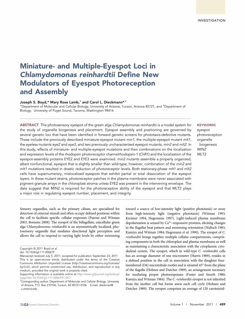
Figure 1 Phenotypic characterization of Chlamydomonas reinhardtiiminiature-eyespot mutants. (A–D) Bright field micrographs. Arrows in-dicate eyespots. Bars, 5 mm. (A) Wild-type cell. (B) The min1 mutanthas a miniature, equatorially localized eyespot. (C) The eyespot ofmin2 cells is equatorially localized and slightly smaller than wild-type.(D) min1 min2 double-mutant cell. Over half of the cells of this mutantstrain have no eyespot observable by bright field light microscopy.Eyespots of the remainder of cells in the population range in size fromultramini to approximately min1-sized.
n Table 1 Chlamydomonas strains used in this study
Strain Genotype/Comments Reference/Source
137c mt+ Wild-type Harris (1989)137c mt2 Wild-type Harris (1989)33 eye3-3 This study59-1 min2-1 This study12-10 mlt1-1 Lamb et al. (1999)2-8 mlt2-1 This study12-12 min1-1 mt+ Lamb et al. (1999)
490 | J. S. Boyd, M. R. Lamb, and C. L. Dieckmann

under continuous illumination. Cells were inoculated into 1 mL M-Nmedium, incubated four hours at 25�, and then 200 mL of each culturewere combined and allowed to mate for one hour under continuousillumination at 25�. Mating mixtures were plated on solid R mediumcontaining 4% agar and kept in the dark for at least four days. Dis-section and tetrad analysis were conducted according to standardmethods (Harris 1989). For complementation and dominance tests,eyespot mutant strains containing the arg7-2 allele were mated tomutant strains containing the arg7-8 allele. Mating mixtures wereplated on solid TAP medium, without arginine to select for diploids,which were then assayed for phototactic ability as described above.
Bright field microscopyCells from overnight liquid cultures were viewed according to theprotocol described in Mittelmeier et al. (2008).
Immunofluorescence microscopyPreparation of samples and immunofluorescence microscopy werecarried out according to the protocol described in Mittelmeier et al.(2008), except antibodies against EYE2, EYE3, and ChR1 were directlyconjugated to fluorophores (Alexa Fluor 488, Alexa 594, or allophy-cocyanin) using Zenon rabbit IgG–labeling kits (Invitrogen, Carlsbad,CA) following the manufacturer’s protocol. Monoclonal anti–acetylated a-tubulin (Clone 6-11B-1, Sigma, St. Louis, MO) wasdetected with goat anti-mouse secondary antibodies conjugated toAlexa Fluor 568 or 647 at a dilution of 1:1000 or Cy5 (MolecularProbes) at a dilution of 1:200.
ImmunoblottingImmunoblotting was carried out according to the protocol describedin Mittelmeier et al. (2008), except primary antibodies were used atthe following dilutions: 1:500 rabbit polyclonal anti-EYE2, 1:500 rabbitpolyclonal anti-ChR1, and 1:10,000 mouse anti-tubulin (clone B-5-1-2; Sigma). Blots were probed with goat anti-rabbit horseradish perox-idase at a dilution of 1:5,000 and/or goat anti-mouse horseradishperoxidase (Pierce, Rockford, IL) at a dilution of 1:10,000 in 1%NFDM-TBS-T for 2 hr at room temperature. Protein levels wereestimated from a digital image of the blot using the National Institutesof Health (NIH) ImageJ software Gel Anaylzer function. For eachsample, the anti-ChR1 and anti-EYE2 signal were normalized to theantitubulin signal.
Preparation of figuresFigures were prepared using Adobe Illustrator (Adobe Systems) andMicrosoft Word (Microsoft). Micrographs were minimally adjustedfor brightness and contrast using NIH ImageJ software, cropped inAdobe Photoshop, and reduced from the original size in AdobeIllustrator.
RESULTS
MIN2 is required for phototaxis and propereyespot sizeTo expand the collection of known eyespot mutants, a genetic screenfor strains defective in phototaxis was conducted following insertionalmutagenesis of wild-type strain 137c mt+ with the CRY-1 gene, whichencodes ribosomal protein S14 (Nelson Et Al., 1994). A motile, ptx2
strain was isolated that exhibited an equatorially localized miniatureeyespot by bright field microscopy (Figure 1C) and was named min2-1.The mutation in min2-1 was found to be unlinked to the CRY-1
insertion. The average area of eyespots measured in a min2 popu-lation was 0.85 6 0.16 mm2 (74% of wild-type area) compared withan average area of 0.38 6 0.09 mm2 for min1 cells (34% of wild-type area) and average area of wild-type eyespots of 1.2 6 0.24 mm2
(Table 2).As previously described, ChR1 photoreceptor localization is
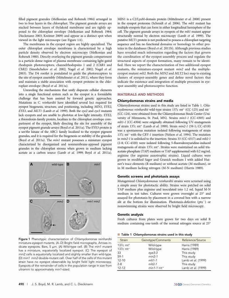
perturbed in the min1 mutant, appearing as stripes or multiple spotsalong the D4 rootlet (Mittelmeier et al. 2008; Figure 2B). If MIN2 hasa role in promotion of eyespot organization comparable to that ofMIN1, similar ChR1 localization patterns might be expected to beobserved in the min2 strain. However, ChR1 staining patterns inmin2 cells double-stained with antibodies directed against ChR1 andacetylated tubulin were unaffected, the photoreceptor patch retainingits nearly wild-type elliptical shape and rootlet association (Figure 2C).min1 min2 double-mutant cells were typified by multiple ChR1patches associated with the rootlet (Figure 2D), a phenotype similarto that of the min1 single mutant. When viewed by electron micros-copy, the pigment granule spot in min1 cells grown in medium lack-ing acetate as a carbon source is a disorganized aggregation in thechloroplast stroma that lacks apposition to the chloroplast envelopeand plasma membranes (Lamb et al. 1999). This finding was corrob-orated by immunofluorescence staining for ChR1 and the pigmentgranule marker EYE3; in photoautotropically grown min1 cells, thestromal pigment granules were separated from the photoreceptor(Figure 2E). By contrast, staining for both EYE3 and ChR1 in min2cells demonstrated that the photoreceptor patch directly overlayedpigment granule layers (Figure 2F). Thus, the min2 mutation doesnot affect the overall assembly and/or maintenance of the eyespotlayers.
min2 exacerbates the eyespot-assembly defect of min1
min1 mutant cells assemble a miniature, disorganized eyespot whengrown in medium lacking acetate (Figure 1B), butmin1 cells grown inacetate-containing medium assemble eyespots that are more orga-nized and closer in morphology to wild-type (Lamb et al. 1999; Mit-telmeier et al. 2008). The eyespot morphology of min2 did not appearto differ between cells from cultures grown photoautotrophically in Mmedium and cells from cultures grown mixotrophically in acetate-containing (R) medium (Figure 3, E and F). The combination ofthe min1 and min2 mutations resulted in cells with an intensifiedeyespot-assembly defect. Of min1 min2 cells scored following over-night growth in M medium, 58% had no observable eyespot by brightfield microscopy (Table 4). The remainder of cells in the populationhad a miniature eyespot that appeared to be approximatelymin1-sized(see Figure 1D). In addition, min1 min2 cells grown mixotrophicallywere unable to assemble a larger eyespot (Figure 3, G and H). Thus,eyespots can assemble in the absence of both MIN1 and MIN2 func-tion, but the lack of MIN2 function exacerbates the eyespot-assemblydefect of min1 mutants. The increased severity of the eyespot defectsin min1 min2 cells compared with those of min1 cells is suggestivethat MIN2 is needed for aspects of eyespot assembly distinct fromthose governed by MIN1.
n Table 2 Eyespot area of miniature-eyed mutants
StrainAverage Eyespot
Area (mm2) SD (mm2)% Wild-Type
Area n
Wild-type 1.2 0.24 100 100min1 0.38 0.09 32 100min2 0.85 0.16 71 100
Volume 1 November 2011 | Eyespot Mutants in C. reinhardtii | 491
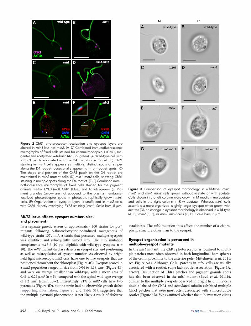
MLT2 locus affects eyespot number, size,and placementIn a separate genetic screen of approximately 200 strains for ptx2
mutants following 5-fluorodeoxyuridine-induced mutagenesis ofwild-type strain 137c mt+, a strain that possessed multiple eyespotswas identified and subsequently named mlt2. The mlt2 mutationcomplements mlt1-1 (10 ptx+ diploids with wild-type eyespots, n =10). The mlt2 mutant displays defects in eyespot size and positioningas well as misregulation of eyespot number. As observed by brightfield light microscopy, mlt2 cells have one to five eyespots that arepositioned throughout the chloroplast (Figure 4C). Eyespots scored ina mlt2 population ranged in size from 0.04 to 1.39 mm2 (Figure 4E)and were on average smaller than wild-type, with a mean area of0.496 0.29 mm2 (n = 54) compared with the typical wild-type averageof 1.2 mm2 (strain 137c). Interestingly, 21% of mlt2 cells have twopyrenoids (Figure 4D), but the strain had no observable growth defect(supporting information, Figure S1 and Table S1), suggestive thatthe multiple-pyrenoid phenomenon is not likely a result of defective
cytokinesis. The mlt2 mutation thus affects the number of a chloro-plastic structure other than to the eyespot.
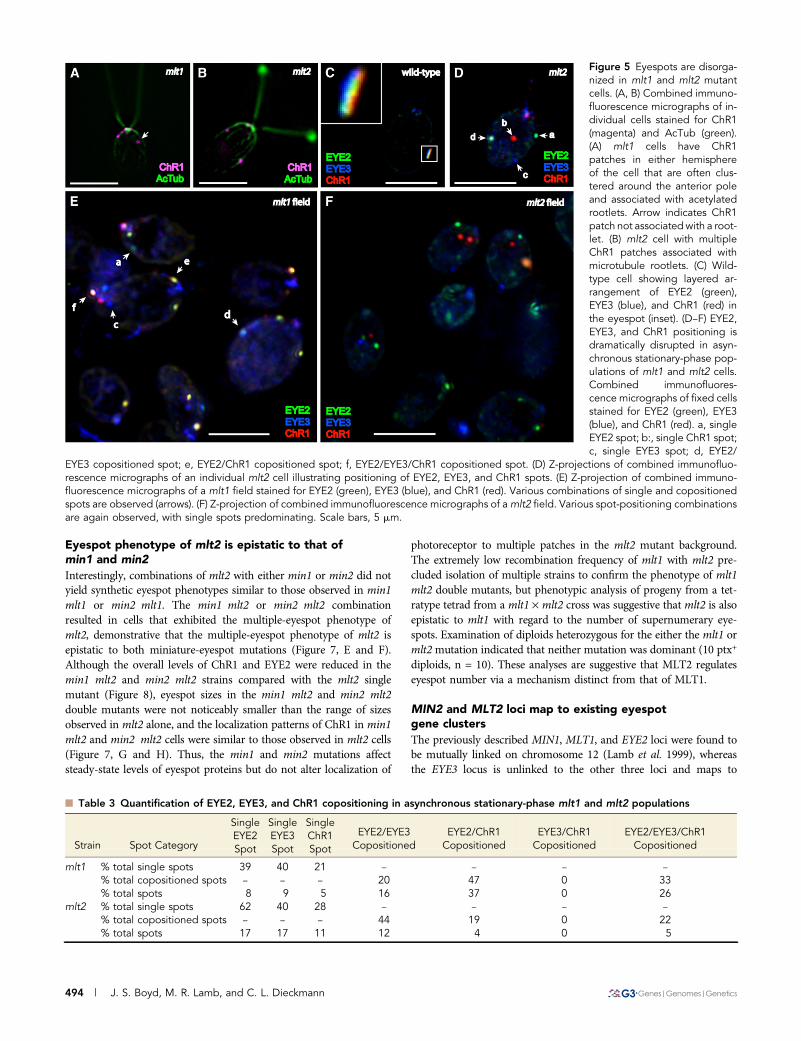
Eyespot organization is perturbed inmultiple-eyespot mutantsIn the mlt1 mutant, the ChR1 photoreceptor is localized to multi-ple patches most often observed in both longitudinal hemispheresof the cell in proximity to the anterior pole (Mittelmeier et al. 2011;see Figure 5A). Although ChR1 patches in mlt1 cells are usuallyassociated with a rootlet, some lack rootlet association (Figure 5A,arrow). Disjunction of ChR1 patches and pigment granule spotshas also been observed in the mlt1 mutant (Boyd et al. 2011b).Similar to the multiple eyespots observed in bright field, mlt2 cellsdouble-labeled for ChR1 and acetylated tubulin exhibited multipleChR1 patches that were most often associated with a microtubulerootlet (Figure 5B). We examined whether the mlt2mutation elicits
Figure 2 ChR1 photoreceptor localization and eyespot layers arealtered in min1 but not min2. (A–D) Combined immunofluorescencemicrographs of fixed cells stained for channelrhodopsin-1 (ChR1, ma-genta) and acetylated a-tubulin (AcTub, green). (A) Wild-type cell witha ChR1 patch associated with the D4 microtubule rootlet. (B) ChR1staining in min1 cells appears as multiple, distinct spots or stripesalong the D4 rootlet, occasionally appearing in off-rootlet spots. (C)The shape and position of the ChR1 patch on the D4 rootlet aremaintained in min2 mutant cells. (D) min1 min2 cells, showing ChR1staining in multiple spots along the D4 rootlet. (E–F) Combined immu-nofluorescence micrographs of fixed cells stained for the pigmentgranule marker EYE3 (red), ChR1 (blue), and AcTub (green). (E) Pig-ment granules (arrow) are not apposed to the plasma membrane-localized photoreceptor spots in photoautotrophically grown min1cells. (F) Organization of eyespot layers is unaffected in min2 cells,with ChR1 directly overlaying EYE3 staining (inset). Scale bars, 5 mm.
Figure 3 Comparison of eyespot morphology in wild-type, min1,min2, and min1 min2 cells grown without acetate or with acetate.Cells shown in the left column were grown in M medium (no acetate)and cells in the right column in R (+ acetate). Whereas min1 cellsassemble a more organized, slightly larger eyespot when grown withacetate (D), no change in eyespot morphology is observed in wild-type(A, B), min2 (E, F), or min1 min2 cells (G, H). Scale bars, 5 mm.
492 | J. S. Boyd, M. R. Lamb, and C. L. Dieckmann
similar effects on the organization of eyespot components. In wild-type cells, distinct layering of ChR1, EYE2, and EYE3 is observedin the eyespot (Boyd et al. 2011a; Figure 5C). In min1 and eye3mutants, in which the pigment granule layers are either disruptedor absent, EYE2 was most often observed copositioned with ChR1(Boyd et al. 2011a). If the multiple-eyespot mutations predomi-nantly affect the association of pigment granule layers with the restof the eyespot, EYE2 might retain association with ChR1. In actu-ality, mlt1 and mlt2 were found to have dramatic effects on orga-nization of eyespot components. In both mlt1 and mlt2 cells grownto stationary phase (�1.0 · 107 cells/mL), EYE2, EYE3, and ChR1spots were visible singly (i.e. without either of the other two pro-
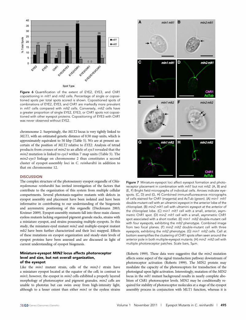
teins associated) and in various combinations of copositioned spots(Figure 5F), including copositioning of EYE2 and EYE3 withoutChR1. The distance between single spots was highly variable andcould be relatively large in some cells (Figure 5D). Strikingly,copositioning of EYE3 and ChR1 was never observed in mlt1 ormlt2 cells unless EYE2 was also present in the copositioned spot(Table 3 and Figure 6), demonstrating that EYE2 is required for thechloroplast envelope link between the plasma membrane and pig-ment granule layers in the eyespot.
The extent of EYE2, EYE3, and ChR1 copositioning wasdistributed variably in populations of both mlt1 and mlt2 cells (Table3); however, mlt1 cells had a greater proportion of copositioned spots,while single spots of all three proteins predominated in stationary-phase mlt2 cells (Figure 6). Eyespots in mlt2 cells did show a largedegree of copositioning in logarithmic growth phase (approx. 8.3 ·105 cells/mL), with 77% of spots consisting of EYE2, EYE3, and ChR1that were mutually copositioned. At this cell density, 32% of mlt2 cellshad one pigment granule spot, 55% had two pigment granule spots,and 13% had three or more pigment granule spots (n = 166). To-gether, these data are suggestive that to some extent MLT1, andespecially MLT2, play roles in limiting formation of supernumeraryeyespots and are required for maintenance of the supramolecularorganization of the eyespot and that in the absence of MLT1 orMLT2 gene function, disintegration of the eyespot structure can occurafter biogenesis of the organelle.
Miniature-eyespot mutations suppress themultiple-eyespot defect of mlt1
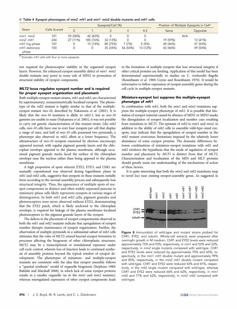
To further characterize the effects of eyespot mutations on assembly ofthe organelle, eyespots were examined in double-mutant combina-tions of mini- and multiple-eyespot mutants. Of eyespots scored bybright field microscopy in a population of min2 mlt1 double-mutantcells (n = 244), 76% had one eyespot, 13% had two eyespots, and 11%had no observable eyespot, in sharp contrast to the mostly multiple-eyespot phenotype of mlt1 cells after the same growth period (Table2). Eyespots of double-mutant cells ranged in size from ultraminiatureto approximatelymin2-sized, and ranged in position from the anteriortip of the chloroplast lobe (Figure 7B) to an approximately equatorialposition. Amongmin2 mlt1 cells with two eyespots, the proportion ofeyespots on the same vs. opposite sides of the cell did not differ sub-stantially from the proportions observed in mlt1 populations (Table4). These data imply that the min2 mutation suppresses the eyespotnumber defect of mlt1, but it does not alter the defect in eyespotplacement.
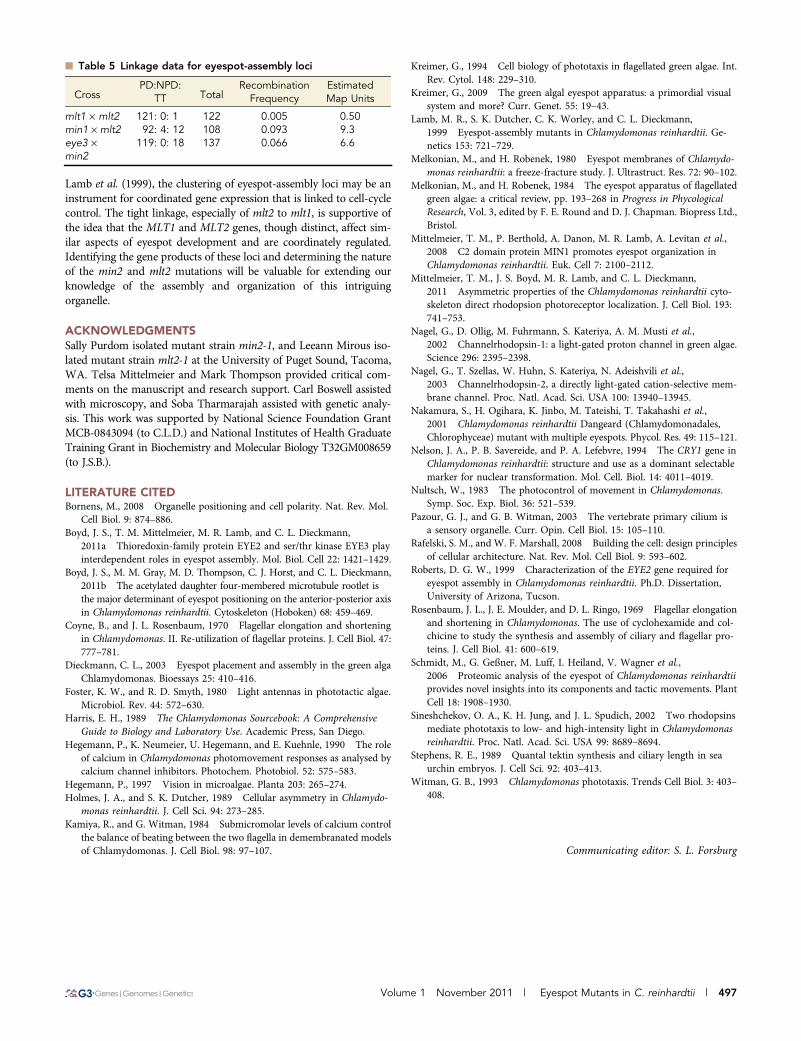
The combination of the min1 mutation with mlt1 produces a syn-thetic phenotype in which double-mutant cells are either eyeless orpossess a single ultraminiature spot of unorganized pigment granulesat or near the anterior tip of the chloroplast lobe (Lamb et al. 1999;Figure 7A). ChR1 localization in the min1 mlt1 double mutant mir-rored eyespot position as observed by bright field microcopy, appear-ing as a single, small spot near the anterior of the cell (Figure 7C). Asimilar phenotype was observed in somemin2 mlt1 cells, with a smallChR1 spot appearing at or near the anterior of the cell (Figure 7D).However, most min2 mlt1 cells had no observable ChR1 staining,paralleled in immunoblot analysis by drastically reduced overallChR1 levels compared with wild-type and either single mutant (Figure8). Mutation of either miniature-eyespot locus in the mlt1 backgroundthus results in magnified attenuation of ChR1 expression or stabilitywhen compared with the reduction observed in either the min1 ormin2 single mutants.
Figure 4 Characterization of multiple-eyespot mutant phenotypes.Arrows in A–C indicate eyespots. (A)mlt1 cell with two eyespots on thesame side of cell. (B) mlt1 cell with an eyespot on both sides. (C) mlt2cell with three eyespots in various positions. Combined image fromtwo focal planes. (D) Bright field micrograph of a mlt2 cell with twopyrenoids (arrows). Two pyrenoids are present in 21% of mlt2 cells,and 79% have one pyrenoid (n = 102). Bars, 5 mm. (E) Distribution ofeyespot sizes in wild-type (n = 100 eyespots) and a mlt2 population(n = 54 eyespots). Eyespots in the mlt2 mutant are distributed overa wide size range but are, on average, smaller than wild-type.
Volume 1 November 2011 | Eyespot Mutants in C. reinhardtii | 493
Eyespot phenotype of mlt2 is epistatic to that ofmin1 and min2
Interestingly, combinations of mlt2 with either min1 or min2 did notyield synthetic eyespot phenotypes similar to those observed in min1mlt1 or min2 mlt1. The min1 mlt2 or min2 mlt2 combinationresulted in cells that exhibited the multiple-eyespot phenotype ofmlt2, demonstrative that the multiple-eyespot phenotype of mlt2 isepistatic to both miniature-eyespot mutations (Figure 7, E and F).Although the overall levels of ChR1 and EYE2 were reduced in themin1 mlt2 and min2 mlt2 strains compared with the mlt2 singlemutant (Figure 8), eyespot sizes in the min1 mlt2 and min2 mlt2double mutants were not noticeably smaller than the range of sizesobserved in mlt2 alone, and the localization patterns of ChR1 in min1mlt2 and min2 mlt2 cells were similar to those observed in mlt2 cells(Figure 7, G and H). Thus, the min1 and min2 mutations affectsteady-state levels of eyespot proteins but do not alter localization of
photoreceptor to multiple patches in the mlt2 mutant background.The extremely low recombination frequency of mlt1 with mlt2 pre-cluded isolation of multiple strains to confirm the phenotype of mlt1mlt2 double mutants, but phenotypic analysis of progeny from a tet-ratype tetrad from a mlt1 · mlt2 cross was suggestive that mlt2 is alsoepistatic to mlt1 with regard to the number of supernumerary eye-spots. Examination of diploids heterozygous for the either the mlt1 ormlt2mutation indicated that neither mutation was dominant (10 ptx+
diploids, n = 10). These analyses are suggestive that MLT2 regulateseyespot number via a mechanism distinct from that of MLT1.
MIN2 and MLT2 loci map to existing eyespotgene clustersThe previously described MIN1, MLT1, and EYE2 loci were found tobe mutually linked on chromosome 12 (Lamb et al. 1999), whereasthe EYE3 locus is unlinked to the other three loci and maps to
Figure 5 Eyespots are disorga-nized in mlt1 and mlt2 mutantcells. (A, B) Combined immuno-fluorescence micrographs of in-dividual cells stained for ChR1(magenta) and AcTub (green).(A) mlt1 cells have ChR1patches in either hemisphereof the cell that are often clus-tered around the anterior poleand associated with acetylatedrootlets. Arrow indicates ChR1patch not associated with a root-let. (B) mlt2 cell with multipleChR1 patches associated withmicrotubule rootlets. (C) Wild-type cell showing layered ar-rangement of EYE2 (green),EYE3 (blue), and ChR1 (red) inthe eyespot (inset). (D–F) EYE2,EYE3, and ChR1 positioning isdramatically disrupted in asyn-chronous stationary-phase pop-ulations of mlt1 and mlt2 cells.Combined immunofluores-cence micrographs of fixed cellsstained for EYE2 (green), EYE3(blue), and ChR1 (red). a, singleEYE2 spot; b:, single ChR1 spot;c, single EYE3 spot; d, EYE2/
EYE3 copositioned spot; e, EYE2/ChR1 copositioned spot; f, EYE2/EYE3/ChR1 copositioned spot. (D) Z-projections of combined immunofluo-rescence micrographs of an individual mlt2 cell illustrating positioning of EYE2, EYE3, and ChR1 spots. (E) Z-projection of combined immuno-fluorescence micrographs of a mlt1 field stained for EYE2 (green), EYE3 (blue), and ChR1 (red). Various combinations of single and copositionedspots are observed (arrows). (F) Z-projection of combined immunofluorescence micrographs of amlt2 field. Various spot-positioning combinationsare again observed, with single spots predominating. Scale bars, 5 mm.
n Table 3 Quantification of EYE2, EYE3, and ChR1 copositioning in asynchronous stationary-phase mlt1 and mlt2 populations
Strain Spot Category
SingleEYE2Spot
SingleEYE3Spot
SingleChR1Spot
EYE2/EYE3Copositioned
EYE2/ChR1Copositioned
EYE3/ChR1Copositioned
EYE2/EYE3/ChR1Copositioned
mlt1 % total single spots 39 40 21 – – – –% total copositioned spots – – – 20 47 0 33% total spots 8 9 5 16 37 0 26
mlt2 % total single spots 62 40 28 – – – –% total copositioned spots – – – 44 19 0 22% total spots 17 17 11 12 4 0 5
494 | J. S. Boyd, M. R. Lamb, and C. L. Dieckmann
chromosome 2. Surprisingly, the MLT2 locus is very tightly linked toMLT1, with an estimated genetic distance of 0.50 map units, which isapproximately equivalent to 50 kbp (Table 5). We are at present un-certain of the position of MLT2 relative to EYE2. Analysis of tetradproducts from crosses of min2 to an allele of eye3 revealed that themin2 mutation is linked to eye3 within 7 map units (Table 5). Themin2-eye3 linkage on chromosome 2 thus constitutes a secondcluster of eyespot-assembly loci in C. reinhardtii in addition tothat on chromosome 12.
DISCUSSIONThe complex structure of the photosensory eyespot organelle of Chla-mydomonas reinhardtii has invited investigation of the factors thatcontribute to the organization of this system from multiple cellularcompartments. Several phototaxis-negative mutants with defects ineyespot assembly and placement have been isolated and have beeninformative in contributing to our understanding of the biogenesisand asymmetric positioning of this organelle (Dieckmann 2003;Kreimer 2009). Eyespot-assembly mutants fall into three main classes:eyeless mutants lacking organized pigment granule stacks, strains witha miniature eyespot, and strains possessing multiple eyespots. In thisstudy, the miniature-eyed mutant min2 and multiple-eyespot mutantmlt2 have been further characterized and their loci mapped. Effectsof these mutations on eyespot organization and steady-state levels ofeyespot proteins have been assessed and are discussed in light ofcurrent understanding of eyespot biogenesis.
Miniature-eyespot MIN2 locus affects photoreceptorlevel and size, but not overall organization,of the eyespotLike the min1 mutant strain, cells of the min2-1 strain havea miniature eyespot located at the equator of the cell; in contrast tomin1; however, the eyespot in min2 cells exhibited a properly layeredmorphology of photoreceptor and pigment granules. min2 cells areunable to phototax but can swim away from high-intensity light,although to a lesser extent than either min1 or the eyeless strains
(Roberts 1999). These data were suggestive that the min2 mutationaffects some aspect of the signal transduction pathway downstream ofphotoreceptor activation (Roberts 1999). The MIN2 protein maymodulate the capacity of the photoreceptors for transduction of thephotosignal upon light activation. Interestingly, mutation of theMIN2locus in the mlt1 mutant background results in nearly complete abo-lition of ChR1 photoreceptor levels. MIN2 may be conditionally re-quired for stability of photoreceptor molecules at a stage of the eyespotassembly process in conjunction with MLT1 function, whereas it is
Figure 6 Quantification of the extent of EYE2, EYE3, and ChR1copositioning in mlt1 and mlt2 cells. Percentage of single or coposi-tioned spots per total spots scored is shown. Copositioned spots ofcombinations of EYE2, EYE3, and ChR1 are markedly more prevalentin mlt1 cells compared with mlt2 cells. Conversely, mlt2 cells havea greater proportion of single EYE2, EYE3, or ChR1 spots not coposi-tioned with other eyespot proteins. Copositioning of EYE3 with ChR1was never observed without EYE2.
Figure 7 Miniature-eyespot loci affect eyespot formation and photo-receptor placement in combination with mlt1 but not mlt2. (A, B) and(E, F) Bright field micrographs of individual cells. Arrows indicate eye-spots. (C, D) and (G, H) Combined immunofluorescence micrographsof cells stained for ChR1 (magenta) and AcTub (green). (A) min1 mlt1double-mutant cell with an ultramini eyespot in the anterior lobe of thechloroplast. (B) min2 mlt1 cell with ultramini eyespot at the anterior ofthe chloroplast lobe. (C) min1 mlt1 cell with a small, anterior, asym-metric ChR1 spot. (D) min2 mlt1 cell with a small, asymmetric ChR1spot associated with a short rootlet. (E) min1 mlt2 double-mutant cellwith four eyespots, exhibiting the mlt2 phenotype. Combined imagefrom two focal planes. (F) min2 mlt2 double-mutant cell with threeeyespots, exhibiting the mlt2 phenotype. (G) min1 mlt2 cells. Cell atbottom exemplifies the clustering of ChR1 spots often seen around theanterior pole in both multiple-eyespot mutants. (H)min2 mlt2 cell withmultiple photoreceptor patches. Scale bars, 5mm.
Volume 1 November 2011 | Eyespot Mutants in C. reinhardtii | 495
not required for photoreceptor stability in the organized eyespotlayers. However, the enhanced eyespot-assembly defect of min1 min2double mutants may point to some role of MIN2 in promotion ofstructural stability of eyespot components.
MLT2 locus regulates eyespot number and is requiredfor proper eyespot organization and placementBoth multiple-eyespot mutant strains,mlt1 andmlt2, are characterizedby supernumerary, nonasymmetrically localized eyespots. The pheno-type of the mlt2 mutant is highly similar to that of the multiple-eyespot mutant mes-10, described by Nakamura et al. (2001). It islikely that the mes-10 mutation is allelic to mlt2-1, but as mes-10gametes are unable to mate (Nakamura et al. 2001), it was not possibleto carry out genetic characterization of this mutant strain. Like mlt2cells, mes-10 cells have one to over four eyespots per cell that displaya range of sizes, and half of mes-10 cells possessed two pyrenoids, aphenotype also observed in mlt2, albeit at a lower frequency. Theultrastructure of mes-10 eyespots examined by electron microscopyappeared normal, with regular pigment granule layers and the chlo-roplast envelope apposed to the plasma membrane, although occa-sional pigment granule stacks faced the surface of the chloroplastenvelope near the nucleus rather than being apposed to the plasmamembrane.
A high proportion of spots wherein EYE2, EYE3, and ChR1 aremutually copositioned was observed during logarithmic phase inmlt1 and mlt2 cells, suggestive that eyespots in these mutants initiallyform according to the normal assembly process and subsequently losestructural integrity. Thus, the appearance of multiple spots of eye-spot components in distinct and often widely separated punctae instationary-phase cells likely represents eyespots at various stages ofdisintegration. In both mlt1 and mlt2 cells, pigment granules andphotoreceptors were never observed without EYE2, demonstratingthat the EYE2 patch, which is likely anchored to the chloroplastenvelope, is required for linkage of the plasma membrane–localizedphotoreceptors to the pigment granule layers of the eyespot.
The defects in the placement of eyespot compartments observed inboth the mlt1 and mlt2 mutants indicate that upregulation of eyespotnumber disrupts maintenance of eyespot organization. Further, theobservation of multiple pyrenoids in a substantial subset of mlt2 cellsintimates that the roles of MLT2 extend beyond eyespot formation toprocesses affecting the biogenesis of other chloroplastic structures.MLT2 may be a transcriptional or translational repressor undercell-cycle control, wherein loss of function leads to continued synthe-sis of assembly proteins beyond the typical window of eyespot de-velopment. The phenotypes of miniature- and multiple-eyespotmutants are consistent with the idea that eyespot assembly followsa “quantal synthesis” model of organelle biogenesis (Stephens 1989;Rafelski and Marshall 2008), in which lack of some eyespot proteinsresults in a smaller organelle (as in the min1 and min2 mutants),whereas misregulated expression of other eyespot components leads
to the formation of multiple eyespots that lose structural integrity ifother critical proteins are limiting. Application of this model has beendemonstrated experimentally in studies on C. reinhardtii flagella(Rosenbaum et al. 1969; Coyne and Rosenbaum 1970). It would beinformative to follow expression of eyespot-assembly genes during thecell cycle in multiple-eyespot mutants.
Miniature-eyespot loci suppress the multiple-eyespotphenotype of mlt1
In combination with mlt1, both the min1 and min2 mutations sup-press the multiple-eyespot phenotype of mlt1. It is possible that lim-itation of eyespot material caused by absence of MIN1 or MIN2 masksthe disregulation of eyespot localization and number cues resultingfrom mutations in MLT1. The epistasis of mlt2 to min1 and min2, inaddition to the ability of mlt2 cells to assemble wild-type–sized eye-spots, may indicate that the upregulation of eyespot number in themlt2 mutant overcomes limitations imposed by the relatively lowerabundance of some eyespot proteins. The dramatic differences be-tween combinations of miniature-eyespot mutations with mlt1 andmlt2 reinforce the hypothesis that the mode of regulation of eyespotnumber and placement by MLT1 is distinct from that of MLT2.Characterization and localization of the MIN and MLT proteinsshould greatly assist our understanding of the mechanisms of actionof these factors.
It is quite interesting that both the min2 and mlt2 mutations mapto novel loci near existing eyespot-assembly genes. As suggested in
n Table 4 Eyespot phenotypes of min2 mlt1 and min1 min2 double mutants and mlt1 cells
Strain Cells ScoredEyespots/Cell (%) Position of Multiple Eyespots in Cella
0 1 2 3 4-5 Same Opposite
min1 min2 101 59 (58%) 42 (42%) 0 0 0 N/Amin2 mlt1 244 27 (11%) 185 (76%) 32 (13%) 0 0 19 (59%) 13 (41%)mlt1 log phase 107 0 15 (14%) 80 (75%) 7 (7%) 5 (5%) 40 (46%) 47 (54%)mlt1 stationary
phase104 0 0 35 (34%) 56 (54%) 13 (12%) 42 (46%) 49 (54%)
aExcludes mlt1 cells with four or more eyespots.
Figure 8 Immunoblot of wild-type and mutant strains probed forChR1, EYE2, and tubulin. Whole-cell extracts were prepared afterovernight growth in M medium. ChR1 and EYE2 levels were reducedapproximately 75% and 93%, respectively, in min1 and 53% and 32%,respectively, in min2 single mutants compared with wild-type. ChR1and EYE2 levels were reduced by approximately 75% and 65%, re-spectively, in the min1 mlt1 double mutant and approximately 99%and 85%, respectively, in the min2 mlt1 double mutant comparedwith wild-type. ChR1 and EYE2 were reduced 43% and 41%, respec-tively, in the mlt2 single mutant compared with wild-type, whereasChR1 and EYE2 were reduced 66% and 62%, respectively, in min1mlt2 and 77% and 62%, respectively, in min2 mlt2 compared withwild-type.
496 | J. S. Boyd, M. R. Lamb, and C. L. Dieckmann
Lamb et al. (1999), the clustering of eyespot-assembly loci may be aninstrument for coordinated gene expression that is linked to cell-cyclecontrol. The tight linkage, especially of mlt2 to mlt1, is supportive ofthe idea that the MLT1 and MLT2 genes, though distinct, affect sim-ilar aspects of eyespot development and are coordinately regulated.Identifying the gene products of these loci and determining the natureof the min2 and mlt2 mutations will be valuable for extending ourknowledge of the assembly and organization of this intriguingorganelle.
ACKNOWLEDGMENTSSally Purdom isolated mutant strain min2-1, and Leeann Mirous iso-lated mutant strain mlt2-1 at the University of Puget Sound, Tacoma,WA. Telsa Mittelmeier and Mark Thompson provided critical com-ments on the manuscript and research support. Carl Boswell assistedwith microscopy, and Soba Tharmarajah assisted with genetic analy-sis. This work was supported by National Science Foundation GrantMCB-0843094 (to C.L.D.) and National Institutes of Health GraduateTraining Grant in Biochemistry and Molecular Biology T32GM008659(to J.S.B.).
LITERATURE CITEDBornens, M., 2008 Organelle positioning and cell polarity. Nat. Rev. Mol.
Cell Biol. 9: 874–886.Boyd, J. S., T. M. Mittelmeier, M. R. Lamb, and C. L. Dieckmann,
2011a Thioredoxin-family protein EYE2 and ser/thr kinase EYE3 playinterdependent roles in eyespot assembly. Mol. Biol. Cell 22: 1421–1429.
Boyd, J. S., M. M. Gray, M. D. Thompson, C. J. Horst, and C. L. Dieckmann,2011b The acetylated daughter four-membered microtubule rootlet isthe major determinant of eyespot positioning on the anterior-posterior axisin Chlamydomonas reinhardtii. Cytoskeleton (Hoboken) 68: 459–469.
Coyne, B., and J. L. Rosenbaum, 1970 Flagellar elongation and shorteningin Chlamydomonas. II. Re-utilization of flagellar proteins. J. Cell Biol. 47:777–781.
Dieckmann, C. L., 2003 Eyespot placement and assembly in the green algaChlamydomonas. Bioessays 25: 410–416.
Foster, K. W., and R. D. Smyth, 1980 Light antennas in phototactic algae.Microbiol. Rev. 44: 572–630.
Harris, E. H., 1989 The Chlamydomonas Sourcebook: A ComprehensiveGuide to Biology and Laboratory Use. Academic Press, San Diego.
Hegemann, P., K. Neumeier, U. Hegemann, and E. Kuehnle, 1990 The roleof calcium in Chlamydomonas photomovement responses as analysed bycalcium channel inhibitors. Photochem. Photobiol. 52: 575–583.
Hegemann, P., 1997 Vision in microalgae. Planta 203: 265–274.Holmes, J. A., and S. K. Dutcher, 1989 Cellular asymmetry in Chlamydo-
monas reinhardtii. J. Cell Sci. 94: 273–285.Kamiya, R., and G. Witman, 1984 Submicromolar levels of calcium control
the balance of beating between the two flagella in demembranated modelsof Chlamydomonas. J. Cell Biol. 98: 97–107.
Kreimer, G., 1994 Cell biology of phototaxis in flagellated green algae. Int.Rev. Cytol. 148: 229–310.
Kreimer, G., 2009 The green algal eyespot apparatus: a primordial visualsystem and more? Curr. Genet. 55: 19–43.
Lamb, M. R., S. K. Dutcher, C. K. Worley, and C. L. Dieckmann,1999 Eyespot-assembly mutants in Chlamydomonas reinhardtii. Ge-netics 153: 721–729.
Melkonian, M., and H. Robenek, 1980 Eyespot membranes of Chlamydo-monas reinhardtii: a freeze-fracture study. J. Ultrastruct. Res. 72: 90–102.
Melkonian, M., and H. Robenek, 1984 The eyespot apparatus of flagellatedgreen algae: a critical review, pp. 193–268 in Progress in PhycologicalResearch, Vol. 3, edited by F. E. Round and D. J. Chapman. Biopress Ltd.,Bristol.
Mittelmeier, T. M., P. Berthold, A. Danon, M. R. Lamb, A. Levitan et al.,2008 C2 domain protein MIN1 promotes eyespot organization inChlamydomonas reinhardtii. Euk. Cell 7: 2100–2112.
Mittelmeier, T. M., J. S. Boyd, M. R. Lamb, and C. L. Dieckmann,2011 Asymmetric properties of the Chlamydomonas reinhardtii cyto-skeleton direct rhodopsion photoreceptor localization. J. Cell Biol. 193:741–753.
Nagel, G., D. Ollig, M. Fuhrmann, S. Kateriya, A. M. Musti et al.,2002 Channelrhodopsin-1: a light-gated proton channel in green algae.Science 296: 2395–2398.
Nagel, G., T. Szellas, W. Huhn, S. Kateriya, N. Adeishvili et al.,2003 Channelrhodopsin-2, a directly light-gated cation-selective mem-brane channel. Proc. Natl. Acad. Sci. USA 100: 13940–13945.
Nakamura, S., H. Ogihara, K. Jinbo, M. Tateishi, T. Takahashi et al.,2001 Chlamydomonas reinhardtii Dangeard (Chlamydomonadales,Chlorophyceae) mutant with multiple eyespots. Phycol. Res. 49: 115–121.
Nelson, J. A., P. B. Savereide, and P. A. Lefebvre, 1994 The CRY1 gene inChlamydomonas reinhardtii: structure and use as a dominant selectablemarker for nuclear transformation. Mol. Cell. Biol. 14: 4011–4019.
Nultsch, W., 1983 The photocontrol of movement in Chlamydomonas.Symp. Soc. Exp. Biol. 36: 521–539.
Pazour, G. J., and G. B. Witman, 2003 The vertebrate primary cilium isa sensory organelle. Curr. Opin. Cell Biol. 15: 105–110.
Rafelski, S. M., and W. F. Marshall, 2008 Building the cell: design principlesof cellular architecture. Nat. Rev. Mol. Cell Biol. 9: 593–602.
Roberts, D. G. W., 1999 Characterization of the EYE2 gene required foreyespot assembly in Chlamydomonas reinhardtii. Ph.D. Dissertation,University of Arizona, Tucson.
Rosenbaum, J. L., J. E. Moulder, and D. L. Ringo, 1969 Flagellar elongationand shortening in Chlamydomonas. The use of cyclohexamide and col-chicine to study the synthesis and assembly of ciliary and flagellar pro-teins. J. Cell Biol. 41: 600–619.
Schmidt, M., G. Geßner, M. Luff, I. Heiland, V. Wagner et al.,2006 Proteomic analysis of the eyespot of Chlamydomonas reinhardtiiprovides novel insights into its components and tactic movements. PlantCell 18: 1908–1930.
Sineshchekov, O. A., K. H. Jung, and J. L. Spudich, 2002 Two rhodopsinsmediate phototaxis to low- and high-intensity light in Chlamydomonasreinhardtii. Proc. Natl. Acad. Sci. USA 99: 8689–8694.
Stephens, R. E., 1989 Quantal tektin synthesis and ciliary length in seaurchin embryos. J. Cell Sci. 92: 403–413.
Witman, G. B., 1993 Chlamydomonas phototaxis. Trends Cell Biol. 3: 403–408.
Communicating editor: S. L. Forsburg
n Table 5 Linkage data for eyespot-assembly loci
CrossPD:NPD:
TT TotalRecombinationFrequency
EstimatedMap Units
mlt1 · mlt2 121: 0: 1 122 0.005 0.50min1 · mlt2 92: 4: 12 108 0.093 9.3eye3 ·min2
119: 0: 18 137 0.066 6.6
Volume 1 November 2011 | Eyespot Mutants in C. reinhardtii | 497