chondrocelect, common name - characterised … common name: characterised viable autologous...
TRANSCRIPT
European Medicines Agency Evaluation of Medicines for Human Use
7 Westferry Circus, Canary Wharf, London E14 4HB, UK Tel. (44-20) 74 18 84 00 Fax (44-20) 74 18 84 16
E-mail: [email protected] http://www.emea.europa.eu
© European Medicines Agency, 2009. Reproduction is authorised provided the source is acknowledged.
EMEA/724428/2009
ASSESSMENT REPORT
FOR
ChondroCelect
Common name: characterised viable autologous cartilage cells expanded ex vivo expressing specific marker proteins
Procedure No. EMEA/H/C/000878
Assessment Report as adopted by the CHMP with
all information of a commercially confidential nature deleted.
Medici
nal p
roduc
t no l
onge
r auth
orise
d

Page 2 of 38
TABLE OF CONTENTS
1. BACKGROUND INFORMATION ON THE PROCEDURE........................................... 3 1.1 Submission of the dossier ........................................................................................................ 3 1.2 Steps taken for the assessment of the product.......................................................................... 3
2. SCIENTIFIC DISCUSSION................................................................................................. 5 2.1 Introduction.............................................................................................................................. 5 2.2 Quality aspects ......................................................................................................................... 6 2.3 Non-clinical aspects ............................................................................................................... 10 2.4 Clinical aspects ...................................................................................................................... 13 2.5 Pharmacovigilance................................................................................................................. 31 2.6 Overall conclusions, risk/benefit assessment and recommendation ...................................... 35
Medici
nal p
roduc
t no l
onge
r auth
orise
d

Page 3 of 38
1. BACKGROUND INFORMATION ON THE PROCEDURE 1.1 Submission of the dossier The Applicant TiGenix NV submitted on 01 June 2007 an application for Marketing Authorisation to the European Medicines Agency (EMEA) for ChondroCelect, through the centralised procedure falling within the Article 3(1) and point 1 of Annex of Regulation (EC) No 726/2004. The eligibility to the centralised procedure was agreed upon by the EMEA/CHMP on 26 September 2006. The legal basis for this application refers to: A - Centralised / Article 8(3) / New active substance. Scientific Advice: The Applicant received Scientific Advice from the CHMP on 28 April 2008 (EMEA/151996/2206). The Scientific Advice pertained to quality, non-clinical and clinical aspects of the dossier. Licensing status: The product was not licensed in any country at the time of submission of the application. The Rapporteur and Co-Rapporteur appointed by the CHMP and the evaluation teams were: Rapporteur: Christian K. Schneider Co-Rapporteur: Jaana Kallio As ChondroCelect is an Advanced Therapy medicinal product, the advanced therapy regulation was applicable to this procedure. Therefore, during the CHMP meeting of 12 – 13 February 2009, a CAT Rapporteur, a CAT Co-Rapporteur and a CHMP Co-ordinator were appointed. Rapporteur: Egbert Flory Co-Rapporteur: Paula Salmikangas 1.2 Steps taken for the assessment of the product • The application was received by the EMEA on 01 June 2007. • The procedure started on 20 June 2007. • The Rapporteur's first Assessment Report was circulated to all CHMP members on 03
September 2007. The Co-Rapporteur's first Assessment Report was circulated to all CHMP members on 03 September 2007. In accordance with Article 6(3) of Regulation (RC) No 726/2004, the Rapporteur and Co-Rapporteur declared that they had completed their assessment report in less than 80 days.
• During the meeting on 18 October 2007, the CHMP agreed on the consolidated List of Questions to be sent to the Applicant. The final consolidated List of Questions was sent to the Applicant on 18 October 2007.
• The Applicant submitted the responses to the CHMP consolidated List of Questions on 28 April 2008.
• The final report of inspections carried out at the manufacturing site in Belgium on 13-14 December 2007 and 20-21 May 2008 was issued on 17 June 2008.
• The Rapporteurs circulated the Joint Assessment Report on the Applicant’s responses to the List of Questions to all CHMP members on 09 June 2008.
• During the CHMP meeting on 26 June 2008, the CHMP agreed on a list of outstanding issues to be addressed in writing and in an oral explanation by the Applicant.
• The Applicant submitted the responses to the CHMP consolidated List of Outstanding Issues on 03 September 2008.
• The Rapporteurs circulated the Joint Assessment Report on the Applicant’s responses to the List of Outstanding Issues to all CHMP members on 11 September 2008.
Medici
nal p
roduc
t no l
onge
r auth
orise
d

Page 4 of 38
• During a meeting of an Ad Hoc Expert group / Biologics Working Party on 13 October 2008, experts were convened to address questions raised by the CHMP.
• During the CHMP meeting on 23 October 2008, outstanding issues were addressed by the Applicant during an oral explanation before the CHMP. The CHMP agreed on a second list of outstanding issues to be addressed in writing and in an oral explanation by the Applicant.
• During the CHMP meeting of 12 – 13 February 2009 Dr. Egbert Flory was appointed as CAT Rapporteur and Dr Paula Salmikangas was appointed as CAT CoRapporteur.
• The Applicant submitted the responses to the CHMP consolidated second List of Outstanding Issues on 24 April 2009.
• The Rapporteurs circulated the Joint Assessment Report on the Applicant’s responses to the 2nd List of Outstanding Issues to all CHMP and CAT members on 11 May 2009.
• During the CAT meeting on 14 May 2009, outstanding issues were addressed by the Applicant during an oral explanation before the CAT.
• During the CAT meeting on 14 May 2009, a 3rd List of Outstanding Issues was adopted by CAT. The CHMP endorsed the 3rd LoOI on 29 May 2009.
• The Applicant submitted the responses to the third List of Outstanding Issues on 03 June 2009. • The Rapporteurs circulated the Joint Assessment Report on the Applicant’s responses to the 3rd
List of Outstanding Issues to all CHMP and CAT members on 12 June 2009. • The Applicant provided the letter of undertaking on follow-up measures to be fulfilled post-
authorisation on 23 June 2009. • On 24 June 2009, the CAT, in the light of the overall data submitted and the scientific
discussion within the Committee, issued a positive draft opinion for granting a Marketing Authorisation to ChondroCelect by written procedure including the recommendation under Article 14(2) of Regulation (EC) No 1394/2007 that the Marketing Authorisation Holder performs the studies and additional activities detailed in the Pharmacovigilance Plan and in the Efficacy Follow-up plan, as agreed in version 4 (dated 22/06/2009) of the Risk Management Plan (RMP) presented in Module 1.8.2. of the Marketing Authorisation Application and any subsequent updates of the RMP agreed by the CAT.
• During the meeting on 25 June 2009, the CHMP, in the light of the overall data submitted and the scientific discussion within the Committee, issued a positive opinion for granting a Marketing Authorisation to ChondroCelect including the recommendation under Article 14(2) of Regulation (EC) No 1394/2007 that the Marketing Authorisation Holder performs the studies and additional activities detailed in the Pharmacovigilance Plan and in the Efficacy Follow-up plan, as agreed in version 4 (dated 22/06/2009) of the Risk Management Plan (RMP) presented in Module 1.8.2. of the Marketing Authorisation Application and any subsequent updates of the RMP agreed by the CHMP.
Medici
nal p
roduc
t no l
onge
r auth
orise
d

Page 5 of 38
2. SCIENTIFIC DISCUSSION 2.1 Introduction Joint surface defects can originate after trauma, after osteochondritis dissecans or can be caused by an underlying genetic predisposition. The healing capacity of articular cartilage is poor and damaged articular cartilage is thought to be a precursor to the development of osteoarthritis. Damaged articular cartilage can result in pain, loss of joint function and disability. An early intervention on symptomatic cartilage lesions may prevent or delay irreversible changes in the joint surface. Currently, there is no uniform approach to managing significant knee cartilage defects. Interventions that aim to provide symptomatic relief include debridement, lavage and rehabilitation. Interventions intended to re-establish the cartilage surface include marrow stimulation techniques (i.e. microfracture (MF), abrasion arthroplasty or drilling), mosaicplasty and autologous chondrocyte implantation (ACI). Microfracture is frequently used as treatment for patients with smaller articular cartilage defects of the knee (for lesions < 4cm2). It induces cartilage repair by penetrating the subchondral bone and stimulating bleeding and thus the formation of a fibrin clot, which is considered to stimulate fibro cartilage formation, and has been shown to result in functional improvements within the first 2 years following treatment. For larger lesions particularly those exceeding 4cm2, however, this procedure is not recommended. Mosaicplasty takes advantage of the limited self-renewal capacity of the joint surface by fitting one or several osteochondral plugs, obtained from a low weight bearing area of the joint into a mosaic. It transforms large defects into several small defects that can be repaired spontaneously by the surrounding tissue and by the invading bone marrow derived skeletal precursors/mesenchymal stem cells. The ACI procedure was first developed in 1994 described by Brittberg et al. (1994) using a first generation autologous chondrocyte product. In the following years many groups could demonstrate the benefit and formation of ‘cartilage repair tissue’ with long-lasting stability and symptomatic relief between two and nine years after ACI treatment. For larger lesion sizes exceeding 4cm², ACI is also considered a suitable treatment option. ChondroCelect by Tigenix nv is a medicinal product for use in ACI treatment. ChondroCelect is a suspension of approximately 10,000 cartilage cells per microliter of medium for autologous use. The cells have been obtained by ex vivo expansion of chondrocytes isolated from a biopsy of the articular cartilage from the patient’s knee. Treatment with ChondroCelect comprises a two-step surgical procedure. In the first step a cartilage biopsy is obtained arthroscopically from healthy articular cartilage from a lesser weight bearing area of the patient’s knee, approximately 4 weeks prior to implantation. Chondrocytes are isolated from the biopsy by enzymatic digestion, expanded in vitro, characterised and delivered as a suspension of 1 x 10⁴ cells/μl for implantation in the same patient. During the second step of the procedure the expanded chondrocyte suspension is implanted in an open-knee surgery. In the pivotal study a periosteal flap was harvested from the medial tibia, sutured into the defect, with the cambium layer facing the subchondral bone, and sealed with fibrin glue. In future applications the defect will be covered with the help of a biodegradable membrane. The dosage of the cell suspension is defined as 0.8 to 1 million cells per cm² defect size. Hence, depending on the defect size measured at biopsy procurement, 4 or 8 or 12 million cells are formulated into 1 or 2 or 3 vial(s) of 4 million cells/ 0.4 ml excipient. The claimed indication for ChondroCelect is repair of single symptomatic cartilaginous defects of the femoral condyle of the knee (ICRS grade III or IV) in adults.
Medici
nal p
roduc
t no l
onge
r auth
orise
d
Page 6 of 38
2.2 Quality aspects Introduction
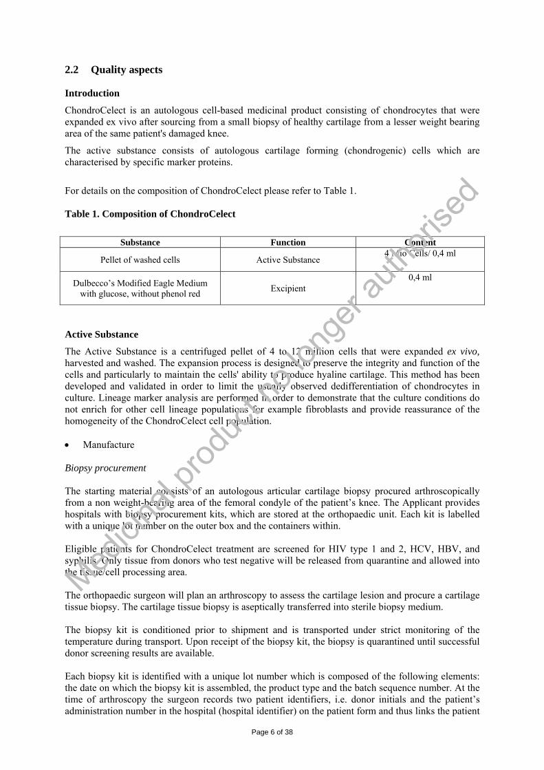
ChondroCelect is an autologous cell-based medicinal product consisting of chondrocytes that were expanded ex vivo after sourcing from a small biopsy of healthy cartilage from a lesser weight bearing area of the same patient's damaged knee.
The active substance consists of autologous cartilage forming (chondrogenic) cells which are characterised by specific marker proteins.
For details on the composition of ChondroCelect please refer to Table 1. Table 1. Composition of ChondroCelect
Substance Function Content
Pellet of washed cells Active Substance 4 Mio Cells/ 0,4 ml
Dulbecco’s Modified Eagle Medium with glucose, without phenol red Excipient
0,4 ml
Active Substance
The Active Substance is a centrifuged pellet of 4 to 12 million cells that were expanded ex vivo, harvested and washed. The expansion process is designed to preserve the integrity and function of the cells and particularly to maintain the cells' ability to produce hyaline cartilage. This method has been developed and validated in order to limit the usually observed dedifferentiation of chondrocytes in culture. Lineage marker analysis are performed in order to demonstrate that the culture conditions do not enrich for other cell lineage populations for example fibroblasts and provide reassurance of the homogeneity of the ChondroCelect cell population. • Manufacture Biopsy procurement The starting material consists of an autologous articular cartilage biopsy procured arthroscopically from a non weight-bearing area of the femoral condyle of the patient’s knee. The Applicant provides hospitals with biopsy procurement kits, which are stored at the orthopaedic unit. Each kit is labelled with a unique lot number on the outer box and the containers within. Eligible patients for ChondroCelect treatment are screened for HIV type 1 and 2, HCV, HBV, and syphilis. Only tissue from donors who test negative will be released from quarantine and allowed into the tissue/cell processing area. The orthopaedic surgeon will plan an arthroscopy to assess the cartilage lesion and procure a cartilage tissue biopsy. The cartilage tissue biopsy is aseptically transferred into sterile biopsy medium. The biopsy kit is conditioned prior to shipment and is transported under strict monitoring of the temperature during transport. Upon receipt of the biopsy kit, the biopsy is quarantined until successful donor screening results are available. Each biopsy kit is identified with a unique lot number which is composed of the following elements: the date on which the biopsy kit is assembled, the product type and the batch sequence number. At the time of arthroscopy the surgeon records two patient identifiers, i.e. donor initials and the patient’s administration number in the hospital (hospital identifier) on the patient form and thus links the patient
Medici
nal p
roduc
t no l
onge
r auth
orise
d

Page 7 of 38
identifiers to the lot number of the biopsy kit. These three codes are designed to form a unique combination, and the biopsy lot number is used throughout the entire process to identify the donor’s tissue and all lot related materials and documentation. Manufacturing process The manufacturing process of the Active Substance consists of the following steps:
• Biopsy digestion • Expansion culture • Cell culture harvest and wash
Biopsy digestion Before further processing, the appearance of the biopsy medium is verified with respect to clarity and colour. The tissue is minced under aseptic conditions and bone fragments are removed. The cartilage fragments are then transferred to allow for dissociation of the cartilage tissue fragments and to release the chondrocytes from the tissue matrix. Chondrocytes are isolated, washed, counted and seeded in tissue culture flasks in culture medium. Expansion culture The isolated cells are transferred to an incubator with humidified atmosphere and replenished with fresh culture medium in regular intervals. The flasks are regularly inspected. When the cultures reach confluence the cells are dissociated from the flask surface and subcultured in fresh tissue culture flasks until the appropriate number of expanded cells has been reached. The spent medium is pooled from every flask and sampled for microbiological testing The total number of passage numbers should remain lower or equal than 3 Cell culture harvest and wash At the end of culture the cells are trypsinized and collected, centrifuged and washed thoroughly. Cell viability is verified and a gram stain is performed on the collected wash solution. The cells obtained as a pellet at this stage are considered the Active Substance (i.e. living human autologous cartilage forming cells). In-process controls and specifications The in-process controls of the manufacturing process have been clearly specified. Critical parameters have been included as in process controls to routinely confirm the quality of the Medicinal product by testing the biopsy and cell culture for aspects like e.g. medium appearance, pH, microbiology, cell morphology, purity, cell viability and cell yield. Appropriate operating ranges have been defined. As the manufacturing process is a continuous process and the active substance is not stored in between, no formal Active Substance specifications have been set. Process validation and characterisation of Active Substance The Applicant used a series of functional tests capable to characterise the cells and suitable to validate the manufacturing process. These functional assays include a cell culture (3D cell culture assay), an in vitro assay in animal models and cellular expression patterns of genes relevant for cartilage and chondrocyte biology. The validation of the manufacturing process has been adequately performed. Some minor issues on the acceptance criteria for some parameters followed during the validation are still outstanding. The Applicant has committed to explore further the specification limits for the functional assay and to
Medici
nal p
roduc
t no l
onge
r auth
orise
d

Page 8 of 38
further define the acceptance criteria for process validation. On basis of the validation approach, the data collected and the commitment on further exploration of the specification limits, the comparability and consistency of lots produced by the proposed manufacturing process have been demonstrated. Medicinal product
The manufacturing process from the Active Substance to the Medicinal product is a continuous process without intermediate holding steps. The cell pellet is immediately resuspended in the excipient nutrient medium and packaged for shipment. The dosage is defined as 0.8 to 1 million cells per cm2 defect size. Hence, depending on the defect size measured at biopsy procurement, 4 or 8 or 12 million cells are formulated into 1 or 2 or 3 vial(s) of 4 million cells/ 0.4 ml excipient/ vial. Concentration of the Medicinal product is 10,000 cells formulated per microliter excipient. • Pharmaceutical development The Manufacture of ChondroCelect Medicinal product involves the formulation of the cell pellet in the excipient medium and subsequent filling into glass vials. The Applicant has conducted studies to demonstrate the suitability of the transport medium to serve as the excipient of the Medicinal product. The Medicinal product is composed of the Active Substance (a pellet of washed cells) and an aqueous nutrient medium (Dulbecco’s Modified Eagle Medium with glucose). • Manufacture of the product The Medicinal product is manufactured, routinely controlled and batch released by Tigenix (Leuven, Belgium). Operations are in compliance with Good Manufacturing Practice (GMP). The re-suspension of the Active Substance, the cell pellet in medium, without any intermediate holding steps yields the Medicinal product. The product is filled into a 1 ml clear, V-shaped, type 1 borosilicate glass vial, which is closed with a grey chlorobutyl /45 stopper. Vial and cap are manually crimp sealed with an aluminium tear-off seal. The glass vial complies with Ph.Eur. requirements. The chlorobutyl stopper is made of material that has “low extractables” characteristics and is certified by the manufacturer to be compliant with all applicable procedures and specifications. Based on the cell counts from the final harvest, the Medicinal product is formulated to contain 10,000 cells /µl and 0.4 ml of the cell suspension is filled per vial. Each vial thus contains a total of 4 million cells. Depending on the total amount of cells needed to treat a specific lesion, up to three vials are filled and provided within the falcon flask. Since the dosage is 0.8 to 1 million cells per cm2, the defective area, which may be treated with ChondroCelect, is limited to 15 cm2. • Adventitious agents The raw materials of biological origin used in the production of ChondroCelect include collagenase, fetal bovine serum and porcine trypsin. All raw materials, which are sourced directly or indirectly from animal material, are subjected to a risk analysis procedure and a compliance check with the appropriate legislative requirements. Certificate of suitability from the European Directorate for Quality of Medicinal Products (EDQM) have been provided. A valid EDQM Certificate of Suitability for foetal bovine serum (FBS) has been provided. Absence from bovine viruses according to Ph. Eur. 01/2008/2262, monograph Serum bovinum and EMEA Guideline (CPMP/BWP/1793/02) has been demonstrated. In addition, test methods have been described in details and virus inactivation results using gamma irradiation have been provided. Overall, sufficient data is provided to exclude a risk of TSE transmission through ChondroCelect. The risk of transmitting TSE by ChondroCelect is thus considered very remote.
Medici
nal p
roduc
t no l
onge
r auth
orise
d

Page 9 of 38
• Product specification The quality control program performed on the Medicinal product for ChondroCelect includes a test for sterility by Ph. Eur., testing for absence of Mycoplasma by Ph. Eur., testing for absence of endotoxin by Ph. Eur. and gram staining. Dosage and cell viability are confirmed prior to release. Visual tests for absence of particles and vial integrity are performed.
All analytical methods are performed according to Ph. Eur. where applicable and are validated according to ICH guidelines.
Compliance with the product specifications has been demonstrated, and the provided data is considered acceptable. The Company has committed to provide additional data in support of the product specification post-marketing. • Stability of the product Stability was addressed by analyzing various lots at time point 0 and time point 48 h. The data reveal no major changes. Hence, a shelf life of 48h is justified for ChondroCelect. • GMO ChondroCelect is composed of non-modified human autologous cells. The cells administered to the patient are likely to remain in the implantation site and are not released to the environment. Furthermore, incidental cell leakage is expected to result in metabolism, as is the case for natural release of cells within the body. Therefore the use of ChondroCelect is unlikely to result in any risk to the environment, due to its nature and also because the product is not released into the environment. Discussion on chemical, pharmaceutical and biological aspects Information on development, manufacture and control of ChondroCelect Active Substance and Finished product have been presented in a satisfactory manner. The results of the tests carried out indicate satisfactory consistency and uniformity of the manufacturing process and finished product, and these in turn lead to the conclusion that the product should have a satisfactory and uniform performance in the clinic. At the time of CAT opinion, there were a number of minor unresolved quality issues related to the specification limits, biodegradable membrane and storage conditions (see above). These shortcomings, however, have no impact on the Risk-benefit balance of the product. The Applicant provided a Letter of Undertaking and committed to resolve these as follow-up measures after the opinion, within an agreed timeframe.
Medici
nal p
roduc
t no l
onge
r auth
orise
d

Page 10 of 38
2.3 Non-clinical aspects Introduction Non-clinical studies were performed as combined pharmacodynamic / pharmacokinetic (distribution) / toxicological studies in the ectopic mouse (nu/nu model) and in orthotopic models in sheep and goats. These studies were non-GLP which is not in conformity with the pharmaceutical standards. However, these deficiencies were considered by CHMP to be tolerable in view of the specificity of the development programme for this particular product. In addition human data were supported by adequate clinical studies and did not raise any safety concerns. Pharmacology • Primary pharmacodynamics Nude mouse model Nude mice received an intramuscular injection of human articular chondrocytes expanded according to the ChondroCelect culture process. Implants retrieved from nude mice at 2 weeks post injection were subjected to histological staining. Based on these characteristics, the cartilage implants were considered of hyaline-like nature. Compared to normal adult human articular cartilage, the cartilage implants were hypercellular and lacked the typical columnar organisation. A further mouse study was performed using early or late passage expanded human articular chondrocytes. When injected intramuscularly into nude mice, late passage expanded cells did not form any cartilage tissue. In contrast, early passage expanded human articular chondrocytes formed a cartilage implant. Studies in a large animal species The importance of phenotypic stability for inducing in vivo hyaline-like cartilage formation was investigated by comparing goat or human articular chondrocytes of different passages. The histological score of implants indicated a loss of stable cartilage-forming potential at higher passages. In goats ChondroCelect-like chondrocytes showed a more stable cartilage forming potential than dedifferentiated chondrocytes. Implantation of ChondroCelect-like autologous chondrocytes resulted in an improved repair efficacy compared to dedifferentiated chondrocytes or dermal fibroblasts as observed in an improved repair in the defect centre, and improved repair tissue integration. Dedifferentiated autologous chondrocytes induced moderate defect filling with poor repair tissue and no or minimal basal and/or lateral integration. In all animals, partial or complete delamination of periosteal flap and fissures in the grafted area and surrounding cartilage was observed. The repair of the cartilage defect was evaluated by the Modified O’Driscoll (MOD) scores that represent a scoring system to assess late-stage cartilage regeneration. The number of data points was very limited and the MOD score obtained in goats have shown a non-valid correlation with the histology score obtained for the same cell preparations in nude mice. As observed for goat chondrocytes, the in vivo cartilage-forming capacity of human articular chondrocytes in nude mice was progressively lost during in vitro cell culture from passage 2-3 onwards. Chondrocytes expanded to higher passage numbers did not form an implant when injected into the thigh of nude mice. In another study in goats comparing passage 1 versus passage 5 expanded human articular chondrocytes according to ChondroCelect culture process all animals showed poor repair, possibly due to an immunological reaction to the human cells. In a goat study ChondroCelect-like autologous chondrocytes controls were performed with or without periosteal flap. Goats sacrificed up to 53 weeks post-implantation of ChondroCelect-like autologous chondrocytes showed normal mobility, almost complete filling of the cartilage lesion with hyaline-like cartilage or hyaline-like cartilage/fibrocartilage. However, goats sacrificed at various weeks post-implantation showed some degree of bone front ingrowths into the defect. The degree of bone front ingrowths into the defect at 52 weeks was most prominent in animals implanted with ChondroCelect-like cells.
Medici
nal p
roduc
t no l
onge
r auth
orise
d

Page 11 of 38
• Secondary pharmacodynamics The potential formation of other tissue types as a consequence of the loss of phenotypic stability during the in vitro expansion process was investigated. Further secondary pharmacodynamic studies were not performed. • Safety pharmacology programme ChondroCelect is administered locally. No direct effect of the cells or an effect of secreted pharmacologically active substances on CNS, cardiac or respiratory system is considered for this cell therapy medicinal product, thus the omission of safety pharmacological studies is in line with the guideline on human cell based medicinal products (EMEA/CHMP/410869/2006). • Pharmacodynamic drug interactions No formal pharmacodynamic drug interaction studies have been performed, since the Applicant justified that the intended clinical use and the applied surgical procedures are not associated with potential concerns regarding pharmacodynamic interactions with pre-, peri- or post-operatively administered medicinal products. Fibrin sealants are broadly employed in orthopaedic surgery as an adjunct to haemostasis during total knee prosthesis replacement or as mechanical seal of the outside margins of the membrane used to cover the defect in ACI. Fibrin sealant products differ significantly in their quantitative and qualitative composition, of the active substance and the excipients, thus it cannot be excluded that certain fibrin glues have, due to their composition, a negative effect on the viable cells and/or membrane. Compatibility data for the fibrin glue TissuCol (Tisseel) have demonstrated the safe and effective use of this sealant with ChondroCelect in non-clinical studies. No interaction studies with any other type of fibrin glues were performed. However the concomitant use of Quixil in the pivotal clinical trial did not reveal any safety signal so far. Pharmacokinetics Two studies were performed in goats to evaluate the persistence of cells in the inflicted cartilage defect as well as the potential migration of cells outside the implantation site. These studies with fluorescently-tagged ChondroCelect-like autologous chondrocytes demonstrated that implanted cells become a structural part of newly formed cartilage. No pharmacokinetic drug interaction studies were performed. This is in line with the draft guideline on human cell based medicinal products (EMEA/CHMP/410869/2006). Toxicology • Single dose toxicity Female NMRI nu/nu mice received intramuscular or subcutaneous injections of human articular chondrocytes (freshly isolated or expanded according to the ChondroCelect culture process), goat articular chondrocytes, pig articular or epiphysial chondrocytes, a combination of pig articular chondrocytes and human periosteal cells, goat dermal fibroblasts, immortalized cell lines with chondrocyte characteristics or human synovial membrane-derived mesenchymal stem cells. Two deaths unlikely to be caused by ChondroCelect were observed, all other animals treated were normal and healthy during the course of the experiment, regardless of the number and type of cells administered. In a sheep study, 70% if the animals receiving an implantation of either autologous articular chondrocytes expanded according to the ChondroCelect culture process, freshly isolated allogeneic articular chondrocytes, freshly isolated human articular chondrocytes or freshly isolated human stem cells showed penetration of cells in subchondral bone, partly with granulomatous reaction. Two of these animals also showed complete penetration of underlying bone marrow. In goats either autologous cells or human articular chondrocytes, expanded according to the ChondroCelect culture process were implancted via an ACI procedure. In one study an extensive set of safety parameters was monitored. Clinical and laboratory signs observed occurred with low incidence,
Medici
nal p
roduc
t no l
onge
r auth
orise
d

Page 12 of 38
were of short duration and are considered related to the surgical procedure including anaesthesia and/or post-surgery immobilization. Animals treated with autologous cells showed no major differences concerning macroscopic and microscopic findings of the femoral condyle as compared to control animals. As part of the goat study, the macroscopic, histological and biochemical composition of the synovium and synovial fluid was investigated 10 and 52 weeks post-implantation with particular attention to inflammation and ectopic cartilage or bone formation. At 10 weeks, ca. 70 % of all animals showed various degrees of synovitis. In a feasibility study in sheep the majority of the animals showed penetration of the transplanted cells in subchondral bone. In two cases complete penetration of underlying bone marrow was observed. Similar findings were observed in long-term studies in goats. In addition these animals showed complete penetration of underlying bone marrow. The observed synovitis and the reported penetration of the transplanted cells in the subchondral bone have been identified in the RMP as potential safety concerns related to the use of the product that warrant a specific statement under section 5.3 ‘Preclinical safety data’ of the SmPC. None of these potential concerns are found to have an impact on the safe clinical application of ChondroCelect. Further risk minimization actions or additional non-clinical data are not considered necessary. No effects on body systems or systemic toxicity was seen in the mice, sheep or goats as expected for this kind of autologous cell therapy medicinal product applied locally in this compartment. • Repeat dose toxicity (with toxicokinetics) As the observation period of the single dose studies described above were up to 12 weeks in mice, 14 weeks in sheep and 53 weeks in goats, these studies are considered to be sufficient to assess the long-term effects of ChondroCelect. Therefore the omission of repeat-dose toxicity studies is in line with the EMEA guideline EMEA/CHMP/410869/2006. • Genotoxicity The omission of genotoxicity studies in the development program for ChondroCelect is in line with EMEA/CHMP/410869/2006. • Carcinogenicity In order to address the carcinogenic potential of ChondroCelect, the Applicant performed an in vitro study to evaluate senescence of human articular chondrocytes after serial passaging, using ChondroCelect culture conditions. Cells were kept beyond the routine cell culturing as suggested in EMEA/CHMP/410869/2006. The results provide sufficient evidence that immortalisation of human chondrocytes during limited time in in vitro culture conditions would not occur, and that the risk of tumorigenic growth is negligible. In view of these results, the absence of standard carcinogenicity studies was considered to be acceptable. • Reproduction Toxicity Taking into account the nature of the product and its intended clinical use the risk for reproductive and developmental toxicity is considered to be negligible. Therefore, the omission of reproductive and developmental toxicity studies in the development program for ChondroCelect is acceptable and in line with the EMEA guideline EMEA/CHMP/410869/2006. • Local tolerance Local tolerance was an integral part of the toxicological studies. Therefore, no dedicated local tolerance studies with ChondroCelect. were deemed necessary. Pharmaco-toxicological studies in the
Medici
nal p
roduc
t no l
onge
r auth
orise
d

Page 13 of 38
orthotropic animal model showed that implantation of human or allogeneic chondrocytes causes an immune response to the CBMP, resulting in poor repair. • Other toxicity studies Ecotoxicity/environmental risk assessment ChondroCelect is composed of non-modified human autologous cells. The cells administered to the patient are likely to remain in the implantation site and are not released to the environment. Therefore the use of ChondroCelect is unlikely to result in any risk to the environment, due to its nature and also because the product is not released into the environment. Consequently, the absence of environmental studies is in line with the EMEA guideline EMEA/CHMP/SWP/4447/00. 2.4 Clinical aspects Introduction GCP The GCP inspection highlighted the amount of missing data on the structural endpoint and the change to the ICRSII read-out in the pivotal study as major concerns. Most of the concerns related to the ICRSII were resolved during the procedure. The generation of new slides from the original repair biopsy has increased the ICRSII data base from 73% to 93%. It was, however, acknowledged that the a priori determined primary efficacy end point, the MODs score (on the basis of which the for example a priori sample size calculations and power analysis were performed), was post hoc disregarded as invalid and a new primary end point, the ICRSII was developed within the course of the study, the conclusion being that this GCP non compliance cannot, as such, be post hoc rectified. Pharmacokinetics Studies on absorption, distribution, metabolism and excretion have not been performed Conventional ADME studies are usually not relevant for a cell based medicinal product. The body distribution/migration studies are part of the non-clinical development program. This is acceptable considering the nature and origin (autologous) of the product. Pharmacodynamics
Conventional pharmacodynamic studies for ChondroCelect have not been performed. The pharmacodynamic parameter “histological evaluation” was part of the efficacy assessment in the phase III trial. The ChondroCelect score is a functional test which suggests a correlation between the gene expression profile of chondrocytes and hyaline cartilage formation in vivo in animal models and was used also in the phase III study (see Overview on quality and non-clinical development as regards the discussion on validity of this score). In the pivotal study a periosteal flap was used to seal the defect and maintain the chondrocyte suspension in situ. Clinical efficacy • Dose response study(ies) No dose-response studies have been performed. The dose selection was based on a combination of animal studies conducted by TiGenix, published literature and experience in humans with ACI. On the basis of this information the dose of between 0.8 and 1 × 10⁶ cells/cm2 was used.
Medici
nal p
roduc
t no l
onge
r auth
orise
d
Page 14 of 38
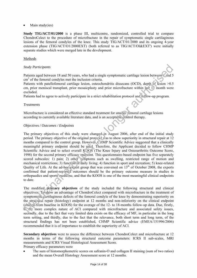
• Main study(ies) Study TIG/ACT/01/2000 is a phase III, multicentre, randomized, controlled trial to compare ChondroCelect to the procedure of microfracture in the repair of symptomatic single cartilaginous lesions of the femoral condyles of the knee. This study TIG/ACT/01/2000 and its ongoing 4-year extension phase (TIG/ACT/O1/2000EXT) (both referred to as TIG/ACT/OI&EXT') were initially separate studies which were merged late in the development. Methods Study Participants Patients aged between 18 and 50 years, who had a single symptomatic cartilage lesion between 1 and 5 cm2 of the femoral condyles met the inclusion criteria. Patients with patellofemoral cartilage lesion, osteochondritis dissecans (OCD), depth of lesion >0.5 cm, prior meniscal transplant, prior mosaicplasty and prior microfracture within last 12 month were excluded. Patients had to agree to actively participate in a strict rehabilitation protocol and follow-up program. Treatments Microfracture is considered an effective standard treatment for smaller femoral cartilage lesions according to currently available literature data, and is an acceptable control therapy. Objectives / Outcomes / Endpoints The primary objectives of this study were changed in August 2006, after end of the initial study period. The primary objective of the original protocol was to show superiority in structural repair at 12 months compared to the control group. However, CHMP Scientific Advice suggested that a clinically meaningful primary endpoint should be used. Therefore, the Applicant decided to follow CHMP Scientific Advice and to select overall KOOS (The Knee Injury and Osteoarthritis Outcome Score, 1998) for the second primary efficacy endpoint. This questionnaire-based endpoint has five separately scored subscales: 1) pain; 2) other symptoms such as swelling, restricted range of motion and mechanical restrictions; 3) function in daily living; 4) function in sport and recreation; 5) knee-related Quality of Life. At the ad-hoc expert group that was convened on 13th of October 2008, the experts confirmed that patient-reported outcomes should be the primary outcome measure in studies in orthopaedics and sports medicine, and that the KOOS is one of the most meaningful clinical endpoints to date. The modified primary objectives of the study included the following structural and clinical objectives: To show an advantage of ChondroCelect compared with microfracture in the treatment of symptomatic cartilaginous defects of the femoral condyle of the knee by demonstrating superiority on the structural repair (histology) endpoint at 12 months and non-inferiority on the clinical endpoint (change from baseline in KOOS) for the average of the 12- to 18-months follow-up data. Due, firstly, to the more complex nature of ACI compared with microfracture and associated safety issues, secondly, due to the fact that very limited data exists on the efficacy of MF, in particular in the long term setting, and thirdly, due to the fact that the relevance, both short term and long term, of the structural findings has not been established, CHMP Scientific advice (EMEA/151996/2006) recommended that it is of importance to establish the superiority of ACI. Secondary objectives were to assess the difference between ChondroCelect and microfracture at 12 months in terms of the following structural outcome parameters: ICRS II sub-scales, MRI measurements and ICRS Visual Histological Assessment Score. Primary efficacy parameters were • The sum of histomorphometric scores on safranin-O and collagen II staining (sum of two ratios)
and the mean Overall Histology Assessment score at 12 months.
Medici
nal p
roduc
t no l
onge
r auth
orise
d

Page 15 of 38
• The change from baseline in Overall KOOS score averaged over 12 and 18 months. Sample size and Randomisation Sample size calculation was based on the score component of the MODS (primary endpoint according protocol). Due to a lack on information on the expected variability, the sample size was determined using only the categorization (success/failure) element of the MODS. Anticipating a 30% success rate for the microfracture group and a 60% success rate for the ChondroCelect procedure, it was calculated that with 112 (56 per group) a one-sided test at the 2.5% level would have 90% power to detect such a difference. However, this primary endpoint was disregarded as invalid and a new histological end point was developed during the conduct of the study. Randomisation was performed via a central IVRS. Blinding (masking) All clinical assessments were performed by independent evaluators at each site. Two central histopathologists who were blinded to the treatment allocation completed the histopathological and histomorphometric assessments of the biopsies. The scoring of the MRI scans was also performed centrally by two independent musculoskeletal radiologists, who were blinded to the treatment allocation. The randomisation was performed using the “minimisation” method, in order to balance groups for the most important prognostic factors. Statistical methods All analyses were performed for the FAS population (i.e. all patients randomized who underwent the surgical procedure). For the primary efficacy parameter additional (secondary) analyses were to be performed for the ITT and PP population respectively. RESULTS Participant flow
Medici
nal p
roduc
t no l
onge
r auth
orise
d

Page 16 of 38
Recruitment The study was performed in 13 centres in 4 countries. Conduct of the study The initial 12-months trial protocol was dated 22 October 2001. Seven protocol amendments were subsequently implemented. The first two amendments were implemented prior to inclusion of any patient.
Medici
nal p
roduc
t no l
onge
r auth
orise
d

Page 17 of 38
Baseline data The randomisation to ChondroCelect and microfracturing groups was successful for age (mean age 33.9 years and 33.9 years, respectively, gender (61% and 67% males), and weight (mean 78.1 kg and 80.6 kg). There was a higher proportion of patients with a BMI >30 kg/m2 in the microfracture group than in the ChondroCelect group (9.8% versus 5.3%) and a slightly higher proportion of patients in the microfracture group whose onset of symptoms was acute compared to the ChondroCelect group. The median duration of time since onset of knee injury was slightly longer in the ChondroCelect group than in the microfracture group (2.0 years versus 1.6 years). The presence of concomitant cartilage lesions was comparable in both groups (30% versus 25%). More patients in the ChondroCelect treatment group, compared to patients in the microfracture group, had undergone previous knee surgery (88% versus 77%). The lesions of the femoral condyle that were treated with ChondroCelect or microfracture were ICRS grade III or IV, except for one patient with a grade II lesion in the ChondroCelect group. Thirty per cent (17/57) of patients in the ChondroCelect group and 25% (15/61) of patients in the microfracture group had additional concomitant cartilage lesions (data from Clinical Overview). The mean surface area of cartilage defect post-debridement was similar in both treatment groups (mean 2.64 and 2. 44, respectively). Numbers analysed The patient disposition is seen in the Table below.
The full analysis set (FAS) was used for the main efficacy analysis. Six patients in the ChondroCelect group were excluded from FAS because acceptable products could not be prepared from the biopsies. All patients were included in the safety analysis. Seven patients in both groups were lost for follow up between months 12 and 18. Outcomes and estimation Primary endpoints The results of the analysis of structural repair and of the clinical endpoint are presented in the Table below: Parameter Treatment N Adjusted
Mean (SE) Difference (95% CI) p-value
ChondroCelect 47 1.01 (0.08) Histomorphometric endpoint Microfracture 54 0.75 (0.08)
0.26 (0.09, 0.44) 0.003
ICRS II at 12 ChondroCelect 49 55.11 (3.98) 10.92 (2.63, 19.21) 0.0103
Medici
nal p
roduc
t no l
onge
r auth
orise
d
Page 18 of 38
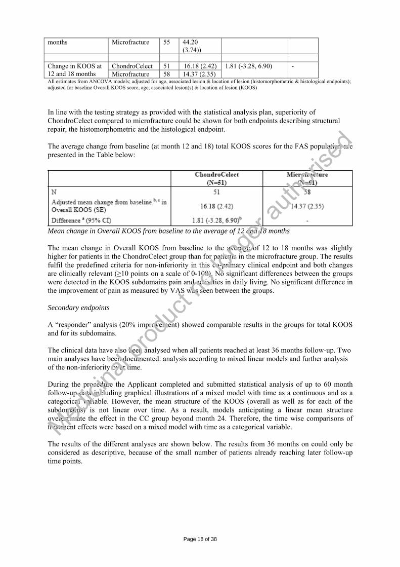
months Microfracture 55 44.20 (3.74))
ChondroCelect 51 16.18 (2.42) Change in KOOS at
12 and 18 months Microfracture 58 14.37 (2.35) 1.81 (-3.28, 6.90) -
All estimates from ANCOVA models; adjusted for age, associated lesion & location of lesion (histomorphometric & histological endpoints); adjusted for baseline Overall KOOS score, age, associated lesion(s) & location of lesion (KOOS) In line with the testing strategy as provided with the statistical analysis plan, superiority of ChondroCelect compared to microfracture could be shown for both endpoints describing structural repair, the histomorphometric and the histological endpoint. The average change from baseline (at month 12 and 18) total KOOS scores for the FAS population are presented in the Table below:
Mean change in Overall KOOS from baseline to the average of 12 and 18 months The mean change in Overall KOOS from baseline to the average of 12 to 18 months was slightly higher for patients in the ChondroCelect group than for patients in the microfracture group. The results fulfil the predefined criteria for non-inferiority in this co-primary clinical endpoint and both changes are clinically relevant (≥10 points on a scale of 0-100). No significant differences between the groups were detected in the KOOS subdomains pain and activities in daily living. No significant difference in the improvement of pain as measured by VAS was seen between the groups. Secondary endpoints A “responder” analysis (20% improvement) showed comparable results in the groups for total KOOS and for its subdomains. The clinical data have also been analysed when all patients reached at least 36 months follow-up. Two main analyses have been documented: analysis according to mixed linear models and further analysis of the non-inferiority over time. During the procedure the Applicant completed and submitted statistical analysis of up to 60 month follow-up data including graphical illustrations of a mixed model with time as a continuous and as a categorical variable. However, the mean structure of the KOOS (overall as well as for each of the subdomains) is not linear over time. As a result, models anticipating a linear mean structure overestimate the effect in the CC group beyond month 24. Therefore, the time wise comparisons of treatment effects were based on a mixed model with time as a categorical variable. The results of the different analyses are shown below. The results from 36 months on could only be considered as descriptive, because of the small number of patients already reaching later follow-up time points.
Medici
nal p
roduc
t no l
onge
r auth
orise
d

Page 19 of 38
Mixed model with time as a categorical variable – all data up to month 36
p-value for the time by treatment interaction: 0.070 Treatment effect at month 36 (CC – MI): 7.655, p-value: 0.0481 Mixed model with time as a categorical variable – all data up to month 60
p-value for the time by treatment interaction: 0.1192 Treatment effect at month 36 (CC – MI): 7.148 p-value: 0.0514 Treatment effect at month 48 (CC – MI): 6.893 p-value: 0.1493 Treatment effect at month 60 (CC – MI): 5.771 p-value: 0.4616 The additional analyses indicate that Change (increase) from baseline in KOOS in the ChondroCelect group (CC) is numerically more pronounced when compared to the Microfracture group (MI). This observation is true not only for the overall KOOS but also for the subdomains. The additional mixed model analysis with time as a categorical variable does not give statistically significant differences at months 36, 48 and 60 (when considering multiplicity). The second analysis which was performed was the further analysis of the non-inferiority over time. The Applicant has calculated the 95% CI for the treatment difference for each consecutive visit (i.e.
Medici
nal p
roduc
t no l
onge
r auth
orise
d

Page 20 of 38
months 2, 3, 6, 9, 12, 18, 24, 30, 36, 48, and 60) in order to assess a possible non-inferiority of CC (compared to MF). For data missing as a result of treatment failure, a last observation carried forward (LOCF) approach was applied. The results for the change from baseline over time in Overall KOOS are presented in the table below. Table 1 Overall KOOS – change from baseline till 36 months Time point Treatment
group N Mean Difference LS
mean SE Difference 95% CI
CC 51 6.66 4.71 2.43 2 months MF 59 6.42
0.24 6.81 2.35
-2.10 -7.19, 2.98
CC 51 12.51 11.65 2.30 3 months MF 59 10.44
2.07 11.66 2.23
-0.02 -4.84, 4.81
CC 51 14.27 13.29 2.40 6 months MF 59 13.18
1.09 14.18 2.32
-0.89 -5.91, 4.14
CC 51 17.63 15.74 2.51 9 months MF 56 13.83
3.80 13.78 2.48
1.96 -3.33, 7.25
CC 51 16.96 14.64 2.55 12 months MF 57 13.54
3.42 13.09 2.48
1.55 -3.83, 6.93
CC 44 18.45 19.19 2.79 18 months MF 51 15.50
2.95 18.47 2.87
0.72 -5.33, 6.78
CC 45 19.38 19.65 3.28 24 months MF 52 13.09
6.29 15.19 3.29
4.46 -2.63, 11.55
CC 43 20.71 18.49 3.47 30 months MF 51 15.16
5.55 15.22 3.39
3.27 -3.92, 10.46
CC 41 22.14 21.25 3.60 36 months MF 50 14.69
7.45 15.83 3.48
5.42 -2.09, 12.94
Source data: TIGACT01&EXT, efficacy tables – analysis of month 36 data, 11 May 2009 The data show that the (unadjusted) mean change from baseline is higher for patients in the ChondroCelect group than for patients in the microfracture group at all time points and with a gradual increase over time (which is also found in the adjusted means). Non-inferiority of ChondroCelect compared to microfracture is confirmed at all time points as the lower limit of the 95% CI for the difference between the adjusted means is above the pre-defined delta of -9% points for all time points. Neither BMI nor Gender have a significant influence on the overall KOOS.
Medici
nal p
roduc
t no l
onge
r auth
orise
d

Page 21 of 38
Treatment failures Treatment failure is defined as “the decision by the participating orthopaedic surgeon to proceed with a re-intervention (i.e. new procedure) on the same defect (index lesion) based on persistence or recurrence of symptoms as reported by the patient”. In the context of this definition, any operation on the involved knee that involves the index lesion to a clinically relevant extent (i.e. 20% or more of its surface), or is intended as a result of clinical treatment failure is considered a re-intervention. Generally, the surgeon relies on MRI and/or arthroscopic assessment to confirm the patient’s complaints are caused by failure of the therapeutic intervention on the index lesions and to exclude possible other causes (e.g. a new lesion). After 36 months post-surgery follow-up, the total number of treatment failures is 2 for the ChondroCelect group and 7 for the microfracture group (p=0.178). However, as the inefficacy of the therapeutic procedure is considered a serious AE, since requiring a surgical re-intervention with hospitalisation, it was felt important to include all known treatment failures up to the 36 month time point and not only those that effectively occurred within a 36-months post-surgery timeframe. As a result, the cumulative number of treatment failures becomes 5 for the ChondroCelect group (9.8%) and 9 for the microfracture group (15%) (p=0.569), as outlined in the table below. These failure rates are concurring with failure rates reported in published literature on ACI and microfracture (Peterson et al., 2000; Peterson et al., 2002; Peterson et al., 2003; Micheli et al., 2006; Wood et al., 2006; Minas et al., 2009; Mithoefer et al., 2009). Table 2 TIG/ACT/01&EXT – treatment failures
ChondroCelect
Microfracture AEs reported over a 36 month post-operative period
N % N %
P-value*
Total number of treated patients 51 100% 61 100% - Treatment failures after 36 months follow-up
2 3.9% 7 11% 0.178
Treatment failures - all cases known at time of 36 months database cut-off
5 9.8% 9 15% 0.569
As is shown in table 2, fewer re-interventions for inefficacy were reported in patients treated with ChondroCelect compared to microfracture (i.e. 5 out of 51 patients [9.8%] versus 9 out of 61 patients [15%], respectively). Treatment failures in the ChondroCelect treated patients were all associated with some degree of periost loosening or graft delamination whereas in the microfracture group the majority of re-interventions were reported to be associated with insufficient or inadequate repair tissue formation. As the use of a biological membrane is expected to result in less friction at the graft surface (i.e. less hypertrophy and less crepitations), the use of a biological membrane instead of periost may possibly reduce the frequency of treatment failures upon treatment with ChondroCelect. Ancillary analyses • Analysis performed across trials (pooled analyses and meta-analysis) N/A • Clinical studies in special populations N/A • Supportive study(ies)
Medici
nal p
roduc
t no l
onge
r auth
orise
d

Page 22 of 38
Prospective, long-term follow-up study of patients in the Belgian Armed Forces treated with ChondroCelect (TIG/ACT/02) Methodology and baseline data This study is a prospective, non-comparative, open-label study of 2 to 5 years’ duration in 20 patients with single and multiple symptomatic cartilage defects, in any location of the knee, who underwent CCI using ChondroCelect. Patients satisfying the inclusion and exclusion criteria were administered ChondroCelect during an arthrotomy which occurred approximately 4 weeks after the arthroscopic procurement of cartilage. Approximately 1 week following arthrotomy, they were discharged from the hospital and invited for regular follow-up visits for up to 5 years after CCI. Clinical outcome, knee pain and activity levels are assessed before (pre-operatively) and after CCI by the KOOS, visual analogue scale (VAS) for pain, Activity Rating Scale (ARS) and military tests for physical fitness (MTLG). Data on pre-defect activity levels were available for the ARS and were expected to be available for MTLG. Secondary objectives of this study included to assess the extent to which the ARS and MTLG scores return to their pre-defect levels within a 5-year post-operative follow-up period. An additional efficacy endpoint listed in the SAP is to assess the change from preoperative baseline in MRI measurements. A final objective is to assess the safety of CCI with ChondroCelect in this specific patient population with single or multiple symptomatic cartilage defects in the knee of any location. Patients with symptomatic cartilage defects in the knee of any location were eligible for inclusion if they, were between 18-50 years of age, had a total cumulative cartilage defect between 1 and 21 cm2 and agreed to adhere to the rehabilitation regimen and the restrictions with regard to concomitant medication. The study population enrolled was characterized by a male predominance (80%), a relatively high age (65% ≥40 years), relatively high body mass index (BMI) (60% >25 kg/m2), and a relatively recent onset of symptoms (mean: 0.9 years; median 0.5 years, range: 0 - 4 years). A femoral cartilage lesion was reported in 95% (19/20) of patients, a patellar lesion in 40% (8/20) and a tibial lesion in 15% (3/20). A total of 35 lesions were reported in 20 patients. The majority of patients had only one lesion (60%;12/20), whilst the remaining patients had two (10%; 2/20), three (25%; 5/20) or four (5%; 1/20) lesions. One of the patients with three lesions and the patient with four lesions each had three lesions treated with CCI. All other patients (18/20; 90%) had only single lesions treated with CCI. Of the 7 patients with multiple lesions who did not have all their lesions treated with CCI, four had their other lesions treated with either shaving (4 lesions in 3 patients) or microfracture (1 lesion in 1 patient) and three had untreated lesions. Of all reported lesions, 80% were reported to be of ICRS Grade III or IV. Of 24 femoral lesions reported in 19 patients, 21 were treated with CCI. All femoral lesions were ICRS grade III-IV. Two of the femoral lesions not treated with CCI were ICRS Grade IV and were located on the trochlea, one of these had been treated with microfracture at arthroscopy; the third untreated femoral lesion was also located on the trochlea and was shaved at arthroscopy (lesion grade unknown). Of 8 patellar lesions reported in 8 patients, three were treated with CCI (one ICRS grade II, one ICRS grade III and one ICRS grade IV). Of the five untreated patella lesions, three were ICRS Grade I and two were ICRS Grade III. One of the grade I patella lesions not treated with CCI and one of the grade III patella lesions not treated with CCI had been shaved at arthroscopy. Three tibial lesions were reported in 3 patients, none of which were treated with CCI. Two of the tibial lesions were ICRS grade II and one was ICRS grade II to III. One of the grade II tibial lesions had been shaved at arthroscopy. Of all enrolled patients, 60% (12/20) had undergone previous cartilage repair surgery at least once (45% [9/20] debridement, 10% [2/20]) microfracture, 5% [1/20]) abrasion arthroplasty, and 5% [1/20]) multiple osteochondral autologous grafts), 60% had a previous meniscus operation, and 20% had previous ligament surgery at baseline. Two patients (9%) had an ACL repair and five patients (23%) had meniscus surgery during the arthroscopy performed for the harvest biopsy. The lesion size treated with ChondroCelect was 2.33 cm² (SD 1.16; range, 0.8 - 9.2 cm2).
Medici
nal p
roduc
t no l
onge
r auth
orise
d

Page 23 of 38
Results
At 24 months following CCI, the patients’ clinical status was improved compared to baseline: mean change in Overall KOOS 28.3 [95% CI 11.28, 45.1; n=9]); VAS (mean change -37.3 [95% CI -63.2, -11.5; n=9]); ARS total score at 24 months was 1.0 (95% CI: -0.2 to 2.2; n=7) indicating a trend towards improved.
The percentage of patients with asymptomatic knees (patient categorization derived from KOOS scores) was increased from none at baseline (0/19 patients) to 38% at Month 18 (5/13 patients) and 56% at Month 24 (5/9 patients).
There was a trend towards an improvement in the patients’ activity level at 18 and 24 months compared to pre-operative baseline, although their activity level remained below their pre-defect levels. The data on MTLG were insufficient to draw any conclusion.
Overall discussion on Efficacy The efficacy evaluation of ChondroCelect is based on one pivotal study. At 12 months post-surgery, structural assessments (histology and MRI) were performed, and at 12 to 18 months post-surgery the clinical outcome was assessed. In line with the testing strategy as provided with the statistical analysis
Medici
nal p
roduc
t no l
onge
r auth
orise
d

Page 24 of 38
plan, superiority of ChondroCelect compared to microfracture could be shown for both endpoints describing structural repair The ICRSII had been developed within the trial, and the validity of this new tool had not been assessed prior to starting the trial. This finding could not be corrected post-hoc. However, the need to develop a suitable assessment method for structural repair was acknowledged, as well as the fact that the new ICRSII score was developed in a blinded manner. A further issue was that many of the tissue sections could not be assessed which lead to 20% of missing data. In the course of the procedure the missing data were provided, which strengthened the superiority claim of structural repair. With respect to the clinical component (change in overall KOOS) non-inferiority was proven. The clinical non-inferiority in the KOOS at 12-18 months was explained by the fact that cartilage requires a longer time to be repaired, given the bradytrophic nature of human joint cartilage and the long time required for differentiation and functional repair. However, statistically significant superiority over microfracture at later time points could not be shown, although the formal requirements to demonstrate non-inferiority at 36 months are fulfilled. In the supportive study TIG/ACT/02 the results of the informal interim analysis show a trend towards clinical benefit. However, only 9/20 patients reached the 24 month time point by the time of the analysis and were assessed for efficacy. The contribution of this study to the benefit/risk analysis of the product is small. Clinical safety • Patient exposure A total of 463 patients have been exposed to ChondroCelect. In the two clinical studies 71 patients w were treated with ChondroCelect, and 61 underwent microfracture treatment. Twenty-two (22) patients were included in the expanded access program and 370 patients were included in the compassionate use program. Safety data from 334 patients are available from the compassionate use program. In both the clinical studies and programs, the absolute dose of ChondroCelect received was determined by the size of the lesion(s) treated. • Adverse events First, the overall frequencies of adverse events (AEs) between the two groups are summarised. Then, those AEs that occurred more frequently in the ChondroCelect group as compared to the microfracture group are discussed. Comparative AE frequencies (TIG/ACT/01&EXT – ChondroCelect vs microfracture) Table 3 provides an overview of the frequencies of treatment-emergent adverse events (TEAEs) in both treatment groups in the pivotal clinical trial (ChondroCelect and microfracture). Overall, patients treated with either ChondroCelect or microfracture show a similar frequency pattern of TEAEs. A slightly larger proportion of the patients in the ChondroCelect group experienced at least one TEAE when compared to microfracture (98% versus 82%); a similar pattern is also observed when only the related TEAEs are considered (78% versus 62%). The number of patients that experienced a severe TEAE, however, is very similar in both treatment groups. In contrast, the number of serious adverse events (SAEs) or the number of patients with an adverse event (AE) leading to discontinuation, was higher in the microfracture group (respectively 9.8% versus 18%, and 0% versus 4.9%). The totality of these data suggests that, despite the observed excess of TEAEs in the ChondroCelect group, the patient’s functionality was not mayoral impacted. There is thus no indication that patients treated with ChondroCelect in the 2-step ACI procedure are significantly more impaired by AEs than patients treated by a 1-step microfracture technique.
Medici
nal p
roduc
t no l
onge
r auth
orise
d

Page 25 of 38
Table 3 TIG/ACT/01&EXT - Overall comparative AE frequencies
ChondroCelect
Microfracture AEs reported over a 36 month post-operative period°
N % N % Total number of treated patients 51 100% 61 100% Patients with at least one TEAE
50 98% 50 82%
Patients with at least one severe TEAE
14 27% 15 25%
Patients with at least one related TEAE
40 78% 38 62%
Patients with at least one treatment-emergent SAE
5 9.8% 11 18%
Patients with at least one AE leading to discontinuation
0 0.0% 3 4.9%
Reference: Database cut-off TIG/ACT/01&EXT (13-Feb-2008) ° AEs are presented as number of patients experiencing at least one AE A similar pattern in frequencies of TEAEs between both treatment groups is further confirmed when the AEs are summarised by body system (Table 4). Up to the 36 months time point, the highest incidence of TEAEs in both treatment groups were observed in the following four body systems: i) Musculoskeletal & Connective Tissue Disorders, ii) Infections and Infestations, iii) Injury, Poisoning & Procedural Complications, and iv) General Disorders & Administration Site Disorders. The incidence in the Musculoskeletal & Connective Disorders as well as in the Injury, Poisoning & Procedural Complications body system was higher in the ChondroCelect group as compared to the microfracture group (respectively 92% versus 77%, p=0.038; and 41% versus 25%, p=0.070). These observed differences relate to some specific AEs in the ChondroCelect group, and will be further discussed here below. For all other body systems, the frequency in AEs was quite similar between both treatment groups and no statistical differences could be found. Table 4 TIG/ACT/01&EXT – Summary of treatment-emergent AEs by body system
ChondroCelect
Microfracture AEs reported over a 36 month post-operative period°
N % N %
P-value*
Total number of treated patients 51 100% 61 100% - Musculoskeletal & Connective Tissue Disorders
47 92% 47 77% 0.038
Infections & Infestations
30 59% 33 54% 0.703
Injury, Poisoning & Procedural Complications
21 41% 15 25% 0.070
General Disorders & Administration Site Disorders
18 35% 15 25% 0.298
Gastrointestinal Disorders
13 25% 11 18% 0.364
Nervous System Disorders
9 18% 18 30% 0.185
Psychiatric Disorders
9 18% 9 15% 0.798
Skin & Subcutaneous Tissue Disorders
6 12% 4 6.6% 0.508
Medici
nal p
roduc
t no l
onge
r auth
orise
d

Page 26 of 38
Surgical & Medical Procedures
5 9.8% 3 4.9% 0.465
Investigations
4 7.8% 6 9.8% 0.753
Vascular Disorders
4 7.8% 5 8.2% 1.000
Cardiac Disorders
3 5.9% 1 1.6% 0.329
Immune System Disorders
2 3.9% 3 4.9% 1.000
Respiratory, Thoracic & Mediastinal Disorders
1 2.0% 5 8.2% 0.217
Reference: Database cut-off TIG/ACT/01&EXT (13-Feb-2008) ° AEs are presented as number of patients; an AE is counted only once per patient * Comparison of treatment groups by Fisher’s exact test When applying a conservative statistical significance of p<0.1, a selection of those AEs that are more frequently observed in the ChondroCelect group as compared to the microfracture group up to the 36 months time point is obtained. These are summarised in Table 5. As could be expected, four of these selected AEs categorised in the Musculoskeletal & Connective Tissue Disorders group (i.e. cartilage hypertrophy, joint swelling, joint crepitation, and joint effusion), whereas influenza-like illness is categorised in the General Disorders & Administration Site Disorders group (of note: this body system also includes the treatment failures, which will be discussed in a later section). Graft complication is categorised in the Injury, Poisoning & Procedural Complications group. The vast majority of the ChondroCelect specific AEs all occurred during the first 18 months post-surgery, with the exception of joint effusion (Table 5). Each of these events will be discussed in further detail in the paragraphs below. Table 5 TIG/ACT/01&EXT – ChondroCelect specific AEs
Period between 0-18 months
Period between 0-36 months AEs reported post-operatively°
CC (N=51)
MF (N=61)
CC (N=51)
MF (N=61)
P-value*
Cartilage hypertrophy
14 8 14 (27%)
8 (13%)
0.093
Joint swelling
11 3 11 (22%)
4 (6.6%)
0.026
Joint crepitation
7 3 9 (18%)
4 (6.6%)
0.082
Joint effusion
4 5 12 (24%)
6 (9.8%)
0.070
Influenza-like illness
4 0 4 (7.8%)
0 (0.0%)
0.040
Graft Complication
3 0 3 (5.9%)
0 (0.0%)
0.091
Reference: Database cut-off TIG/ACT/01&EXT (13-Feb-2008) ° Selected based on p<0.1, AEs are presented as number of patients; an AE is counted only once per patient * Comparison of treatment groups by Fisher’s exact test
Medici
nal p
roduc
t no l
onge
r auth
orise
d

Page 27 of 38
Symptomatic cartilage hypertrophy Symptomatic cartilage hypertrophy is an undesirable AE that may result in physical impairment requiring surgical arthroscopic intervention. Symptomatic cartilage hypertrophy is generally resolved after arthroscopic shaving during day-care arthroscopy. The events of cartilage hypertrophy included both those events that were symptomatic and those that were asymptomatic. The reporting of the latter type of (asymptomatic) hypertrophy occurred mostly at 1 year, as it was observed at the 12-month arthroscopic endpoint biopsy procedure related to the clinical protocol. Of the 14 ChondroCelect-treated patients who had AEs of cartilage hypertrophy recorded, 7 had symptomatic AEs (7/51 [14%]). The other 7 patients had AEs that were asymptomatic. In the microfracture group, 7 of the 8 patients had asymptomatic AEs of cartilage hypertrophy and one was symptomatic (1/61 [2%]). The difference between the two treatment groups of the clinically relevant symptomatic hypertrophy is statistically significant (p = 0.022). It is worth noting that the majority of these events occurred in the first 18 months post-surgery, indicating that this event is related to the regenerative phase of the repair tissue. All reported AEs of cartilage hypertrophy were mild or moderate in severity in both treatment groups. None was recorded as severe and none was reported as serious. In the pivotal clinical trial, a periosteal flap was used to cover the ChondroCelect implant as this was at that time the standard surgical procedure. However, the use of a periosteal flap to cover the cultured chondrocytes is also generally considered to involve a risk of cartilage hypertrophy. Indeed, literature data indicate that tissue hypertrophy can be related to the periosteal flap that is used to cover the defect before injection of the cells (Gooding et al., 2006). In recent publications, the potential risk of hypertrophy was reported to be reduced with the use of biological membranes without periosteal cells (Haddo et al, 2004; Gooding et al., 2006; Steinwachs and Kreuz, 2007). In current clinical practice, the use of a periosteal cover has decreased over the last years in favour of the use of biological membranes. The preference for collagen membranes was also confirmed by the experts in the ad-hoc scientific advisory group organised by EMEA on October 13, 2008. It is anticipated that the frequency of hypertrophy as observed in the clinical trial can be reduced when a biological membrane is used., This is supported by a comparison of the symptomatic hypertrophy frequency observed in the pivotal trial population (7/51 patients, i.e. 14%) and the patients treated under compassionate use (6/334 patients, i.e. 1.8%). Joint swelling The reported frequency of joint swelling is higher after ChondroCelect than after microfracture and is mainly explained by the arthrotomy performed for the ChondroCelect implantation. Knee swelling suggests the accumulation of fluid in and/or around the knee. It is a well-described symptom after arthrotomy as a result of the inflammatory synovial reaction due to incision (Muckle, 1984). This is further confirmed by analysing the temporal relationship of the reported joint swelling events with surgery, showing a high frequency and a significant difference with microfracture in the first weeks after the intervention. 7 of the 11 patients reported with joint swelling after ChondroCelect experienced the AE in the first 4 weeks after intervention, compared to none after microfracture (p=0.003). This earlier onset in the ChondroCelect group is linked to the arthrotomy procedure. After this initial 4-week post-operative period, no significant differences between the 2 groups were reported (i.e. 3 patients in the ChondroCelect group versus 4 patients in the microfracture group). Post-operative swelling is not associated with a significant risk and is temporary. No events of joint swelling were recorded as severe during the study, and none was reported as serious. Compassionate use program Comparison of TIG/ACT/01&EXT and compassionate use AEs In Table 6, a comparison is made between the safety results of the pivotal trial population and the patients treated under compassionate use, this latter population being considered to be more representative for the real-life situation. In this table, only those AEs that are considered related to
Medici
nal p
roduc
t no l
onge
r auth
orise
d

Page 28 of 38
ChondroCelect or to the surgical intervention are reported. Overall, the frequencies of AEs are consistently lower in the compassionate use population as compared to the TIG/ACT/01&EXT population. This is likely explained by a relative underreporting of AEs in the real-life situation, outside the controlled environment of a clinical trial. Assuming a 50% underreporting rate, the overall frequency for most of the reported AEs becomes similar to the frequencies observed in the clinical trial. Table 6 TIG/ACT/01&EXT and CUP – comparison of most frequent related† AEs
TIG/ACT/01&EXT
CUP AEs reported post-operatively°
N % N % Total number of patients 51 100% 334* 100% Patients with at least one related AE
40 78% 155 45%
Arthralgia (knee pain)
24 47% 67 20%
Cartilage hypertrophy - symptomatic (Total)
7
(14)
14%
(27%)
6 1.8%
Joint crepitation
9 18% 17 5.1%
Joint swelling
7 14% 23 6.9%
Joint effusion
5 9.8% 24 7.2%
Treatment failure
5‡ 9.8% 9 2.7%
Reference: TIG/ACT/01&EXT database cut-off 13 February 2008 (36 months), CUP database cut-off 7 January 2009 ° AEs are presented as number of patients; an AE is counted only once per patient * Number of patients contributing to the safety population at time of database cut-off (7 January 2009) † Related to ChondroCelect or surgical intervention ‡ All cases known at the time of the 36 months database cut-off As can be seen from the table, knee pain (Arthralgia) is the most frequently reported treatment-related AE in both the pivotal study (24/51 patients, i.e. 47%) and the CUP (67/334 patients, i.e. 20%). In the pivotal study, a similar frequency of Arthralgia was observed in the ChondroCelect (47%) and microfracture (43%) groups up to the 36 months cut-off. This AE was not discussed in the overall analysis of the TIG/ACT/01&EXT data as the difference between the two treatment groups was not significant (p=0.704). The incidence of symptomatic cartilage hypertrophy was reduced to 1.8% in the CUP patients compared to 14% in the pivotal study. It can be assumed that the 6 reported AEs in the CUP patients are all symptomatic AEs, as these patients did not undergo an endpoint biopsy. In the patients treated under compassionate use, ChondroCelect has been covered with a biological membrane (ChondroGide®) in the majority of the patients in contrast to the use of a periosteal flap in the pivotal trial. The lower incidence of cartilage hypertrophy observed in the CUP is in line with other reports published in the literature on low hypertrophy rates with a biological membrane (Haddo et al, 2004; Gooding et al., 2006; Steinwachs and Kreuz, 2007), and illustrates that a similar benefit of using a biological membrane can also be obtained for ChondroCelect. The relatively high incidence of joint effusion in the compassionate use patients (7.2% of patients in the CUP as compared to 9.8% in the pivotal trial) is considered to result from the fact that in this population more salvage cases with more complex concomitant knee pathology have been treated. A detailed assessment of these cases reveals that in all patients but 2, the effusion was considered to be
Medici
nal p
roduc
t no l
onge
r auth
orise
d

Page 29 of 38
of mild (14) or moderate (8) intensity. None of the 24 cases was reported to be serious. In 15 cases, the joint effusion was considered to be related to the surgical procedure (3 unknown, 6 not related to surgery), and in 7 patients the event was considered to be related to ChondroCelect. For all other AEs, similar or slightly lower frequencies are observed when considering a 50% underreporting in the CUP (i.e. joint crepitation [18% versus 5.1%], joint swelling [14% versus 6.9%], and treatment failures [9.8% versus 2.7%]). • Serious adverse event/deaths/other significant events Twenty four serious adverse events (SAEs) were reported in 16 patients (8 SAEs in 7 patients in the TIG/ACT/01&EXT study and 16 SAEs in 9 patients in the TIG/ACT/02 study). Only one of the 24 cases was considered to be related (possibly) to ChondroCelect. In this case, the ChondroCelect transplant was considered to have failed possibly because of loosening of the periosteal flap, and was removed; microfracture was subsequently performed. The majority of cases (14/24; 58%) were considered to be unrelated to the study procedure. In the pivotal TIG/ACT/01&EXT there were 10 SAEs in 8 patients recorded in the microfracture group. All cases recorded for microfracture-treated patients were considered either unrelated or unlikely to be related to the surgical procedure. From additional safety information gathered from the EAP and compassionate use program, there have been 26 SAEs reported for 18 patients (17 SAEs in 10 of the 22 patients in the EAP, 9 SAEs in 8 of the 163 patients in the compassionate use program for whom safety data are available). Of these, only one case was considered by the surgeon to be related (possibly) to ChondroCelect. In this case the patient experienced a deficit in knee mobilisation of moderate severity, approximately 2 months after the implantation of ChondroCelect (secured with ChondroGide). The patient underwent a procedure to mobilize the knee under anaesthesia. There were no patient deaths recorded during the study. No patients are recorded as being discontinued from the study due to SAEs. • Laboratory findings No laboratory findings related to the ChondroCelect treatment were reported. • Safety in special populations Twelve paediatric patients were treated in the compassionate use program with ChondroCelect. Five of the 11 (45%) paediatric patients had no reported AE. The remaining 6 paediatric patients reported a total of 11 AEs. None of the AEs reported was considered serious. None of the AEs was considered to be related to ChondroCelect; most were considered to be related to surgery (7/11 events; 64%). Most AEs (8/11; 73%) were of mild or moderate intensity and did not require any intervention or medical therapy. Three events were recorded as being of severe intensity, one event of muscle atrophy (n=2) and arthralgia (n=1). There was one pregnancy during ChondroCelect therapy. The mother developed pre.eclampsia, had a premature birth of a normal child. The use of ChondroCelect is not recommended during pregnancy, mainly because of the surgical procedures. • Safety related to drug-drug interactions and other interactions No investigations have been performed • Discontinuation due to adverse events See efficacy part for discontinuation due to treatment failure.
Medici
nal p
roduc
t no l
onge
r auth
orise
d

Page 30 of 38
• Post marketing experience N/A • Other relevant safety information A clinically meaningful event that is not reported in the classical AE capturing relates to the impact of the procedures on the subchondral bone. Detailed assessment of the MRI data of the pivotal clinical trial revealed that treatment with ChondroCelect resulted in less subchondral bone reaction as compared to microfracture. A difference of 0.45 on the 0-3 global scale was observed in favour of ChondroCelect (p=0.0559). In addition, the incidence of subchondral bone plate elevation was shown to be higher in the microfracture group when compared to the ChondroCelect group (51.5% compared to 25%, respectively). The difference in subchondral bone reactions relates to the difference in surgical intervention. In microfracture, chondral defects are treated by recruiting blood and cell populations including mesenchymal stem cells from the underlying bone marrow. By physically disturbing the osteochondral junction (upon puncturing the subchondral bone), an osteochondral defect is de facto created, and the biology of the cartilage defect significantly altered. In contrast, in ACI cells are re-implanted into the defect without damaging the subchondral bone. The consequences of the injury and its increased subchondral bone reaction and moving up of the bone front might lead to poorer repair tissue, decreased durability of the repair tissue, and consequently increased risk of treatment failure (Mithoefer et al., 2009). The moving of the bone front (ultimately leading to intra-lesional osteophytes) might also have consequences for future re-interventions. Indeed, Minas et al., 2009 published that prior treatment affecting the subchondral bone such as microfracture increases the failure rate of subsequent regenerative procedures. Finally, the biological stability of the osteochondral junction can be of importance in the development of osteoarthritis, instability of the subchondral bone and progressive bone damage being important factors in progression to the disease (Dieppe and Lohmander, 2005; McQueen, 2007). It is also anticipated that in genetically predisposed patients, disturbing the osteochondral junction presents an additional risk for a fast progression to osteoarthritis (Luyten et al., 2009). Discussion on Safety Microfracture is performed in one arthroscopic procedure with either local (spinal) or general anaesthesia, while ACI requires 2 interventions: an arthroscopy to inspect the defect and to obtain the biopsy specimen, and open knee surgery (arthrotomy) in general anaesthesia for chondrocyte implant four weeks later. In the case of patients included in the ChondroCelect arm of the pivotal trial a second incision was made over the medial tibia to harvest the periosteal flap which was needed to cover the chondrocyte suspension. The most likely AEs observed when treating with ChondroCelect are arthralgia, symptomatic cartilage hypertrophy, joint crepitations, joint swelling, and joint effusion. Arthralgia is an expected and common consequence of knee surgery and occurs in both treatment arms. The data provided on the CUP program as well as data from published literature suggest that it may be possible to reduce ChondroCelect-related cartilage hypertrophy by the use of a collagen membrane. This modification will reduce the potential for morbidity associated with the harvest of the periosteal flap. Joint crepitation is a mild complication and occurs also in the normal population. Joint swelling is another complication observed at a higher rate in the ChondroCelect group. It is a consequence of the arthrotomy surgical intervention and can as such not be avoided. However, it is a mild and transient complication. The incidence of joint effusion was higher after 18 months post-intervention than in the period after the surgery, and the reason for this is not completely clear. Analysis of the individual cases indicate that the underlying knee disease status as well as potentially higher physical activity rate might be related to this occurrence. The cases were not severe or serious, and do therefore not represent a major safety signal. Overall, the safety profile of ChondroCelect is considered acceptable. Considering that cartilage hypertrophy may be reduced by use of a physical seal the main difference is related to arthrotomy and the implantation procedure. No complications were seen in relation to the arthroscopic harvest biopsy procedure.
Medici
nal p
roduc
t no l
onge
r auth
orise
d

Page 31 of 38
Considering the higher treatment failures after microfracture which require more surgical re-interventions microfracture and ChondroCelect implantation have a balanced safety profile. The CHMP was of the opinion that the indications (section 4.1 of the SPC) should be reworded as follows: Repair of single symptomatic cartilage defects of the femoral condyle of the knee (International Cartilage Repair Society [ICRS] grade III or IV) from 2 cm² onwards in adults. Concomitant asymptomatic cartilage lesions (ICRS grade I or II) might be present Demonstration of efficacy is based on a randomised controlled trial evaluating the efficacy of ChondroCelect in patients with lesions between 1-5cm2. The scientific reasons for this change are described below: CHMP considered that putting a precise lower boundary would prevent physicians from treating patients on-label who have lesions in sizes formally below 2cm2, for whom, however, ChondroCelect treatment might nevertheless be medically indicated by the individual decision of the treating physician. Likewise, CHMP recognised the greater need for the treatment of larger lesions, but was concerned about the amount of data available for larger lesions. Therefore, in line with previous similar scenarios, the CHMP has taken the approach to put an indication that is permissive, but to point out the limitations of the data and to clearly inform the prescribing physician in section 4.1. A wording like this is usually perceived as stronger as compared to a wording in section 5.1, thus being more in line with the overall principles of the CAT draft opinion. This allows the physician to take an informed decision for a particular patient’s situation based on the knowledge of the availability of evidence from the pivotal study. 2.5 Pharmacovigilance Detailed description of the Pharmacovigilance system as agreed at CAT The CAT considered that the Pharmacovigilance system as described by the Applicant fulfils the legislative requirements. Risk Management Plan as agreed at CAT The MAA submitted a risk management plan, which included a risk minimisation plan and efficacy follow-up plan. Table Summary of the risk management plan Important potential risks
Proposed pharmacovigilance Activities
Proposed risk minimisation activities
Safety concern
(routine and additional) (routine and additional)
Partial or complete delamination of the periost flap, synovitis, subchondral bone injuries
Routine pharmacovigilance Proactive training of
orthopaedic surgeons and their staff on the use of the product and the associated procedures
Solicited adverse reaction
Information in section 5.3 of the SPC on the findings of synovitis and subchondral bone injuries.
Pro-active training in the framework of a controlled distribution system.
Medici
nal p
roduc
t no l
onge
r auth
orise
d
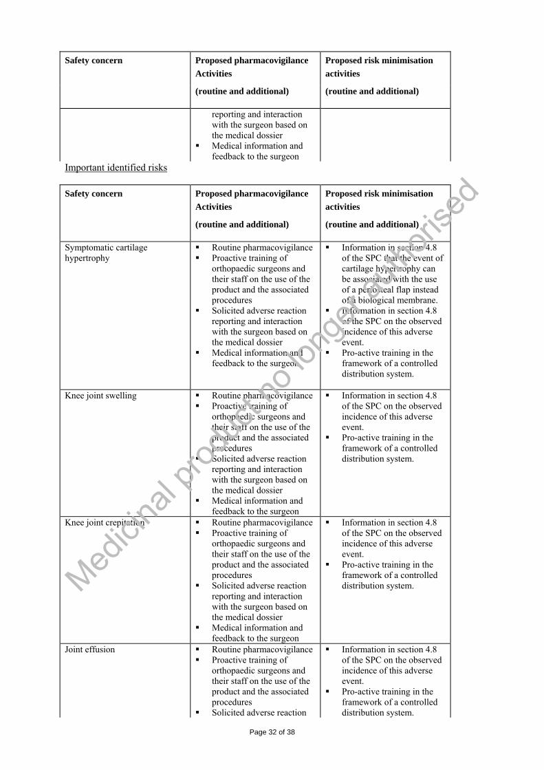
Page 32 of 38
Proposed pharmacovigilance Activities
Proposed risk minimisation activities
Safety concern
(routine and additional) (routine and additional)
reporting and interaction with the surgeon based on the medical dossier
Medical information and feedback to the surgeon
Important identified risks
Proposed pharmacovigilance Activities
Proposed risk minimisation activities
Safety concern
(routine and additional) (routine and additional)
Symptomatic cartilage hypertrophy
Routine pharmacovigilance Proactive training of
orthopaedic surgeons and their staff on the use of the product and the associated procedures
Solicited adverse reaction reporting and interaction with the surgeon based on the medical dossier
Medical information and feedback to the surgeon
Information in section 4.8 of the SPC that the event of cartilage hypertrophy can be associated with the use of a periosteal flap instead of a biological membrane.
Information in section 4.8 of the SPC on the observed incidence of this adverse event.
Pro-active training in the framework of a controlled distribution system.
Knee joint swelling Routine pharmacovigilance
Proactive training of orthopaedic surgeons and their staff on the use of the product and the associated procedures
Solicited adverse reaction reporting and interaction with the surgeon based on the medical dossier
Medical information and feedback to the surgeon
Information in section 4.8 of the SPC on the observed incidence of this adverse event.
Pro-active training in the framework of a controlled distribution system.
Knee joint crepitation Routine pharmacovigilance Proactive training of
orthopaedic surgeons and their staff on the use of the product and the associated procedures
Solicited adverse reaction reporting and interaction with the surgeon based on the medical dossier
Medical information and feedback to the surgeon
Information in section 4.8 of the SPC on the observed incidence of this adverse event.
Pro-active training in the framework of a controlled distribution system.
Joint effusion Routine pharmacovigilance Proactive training of
orthopaedic surgeons and their staff on the use of the product and the associated procedures
Solicited adverse reaction
Information in section 4.8 of the SPC on the observed incidence of this adverse event.
Pro-active training in the framework of a controlled distribution system.
Medici
nal p
roduc
t no l
onge
r auth
orise
d
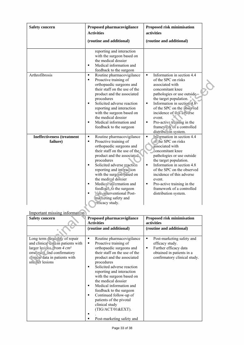
Page 33 of 38
Proposed pharmacovigilance Activities
Proposed risk minimisation activities
Safety concern
(routine and additional) (routine and additional)
reporting and interaction with the surgeon based on the medical dossier
Medical information and feedback to the surgeon
Arthrofibrosis Routine pharmacovigilance Proactive training of
orthopaedic surgeons and their staff on the use of the product and the associated procedures
Solicited adverse reaction reporting and interaction with the surgeon based on the medical dossier
Medical information and feedback to the surgeon
Information in section 4.4 of the SPC on risks associated with concomitant knee pathologies or use outside the target population.
Information in section 4.8 of the SPC on the observed incidence of this adverse event.
Pro-active training in the framework of a controlled distribution system.
Ineffectiveness (treatment failure)
Routine pharmacovigilance Proactive training of
orthopaedic surgeons and their staff on the use of the product and the associated procedures
Solicited adverse reaction reporting and interaction with the surgeon based on the medical dossier
Medical information and feedback to the surgeon
Non-interventional Post-marketing safety and efficacy study.
Information in section 4.4 of the SPC on risks associated with concomitant knee pathologies or use outside the target population.
Information in section 4.8 of the SPC on the observed incidence of this adverse event.
Pro-active training in the framework of a controlled distribution system.
Important missing information Safety concern Proposed pharmacovigilance
Activities Proposed risk minimisation activities
(routine and additional) (routine and additional)
Long term durability of repair and clinical data in patients with larger lesions (from 4 cm² onwards), and confirmatory clinical data in patients with smaller lesions
Routine pharmacovigilance Proactive training of
orthopaedic surgeons and their staff on the use of the product and the associated procedures
Solicited adverse reaction reporting and interaction with the surgeon based on the medical dossier
Medical information and feedback to the surgeon
Continued follow-up of patients of the pivotal clinical study (TIG/ACT/01&EXT).
. Post-marketing safety and
Post-marketing safety and efficacy study.
Further efficacy data obtained in patients in a confirmatory clinical study. Med
icina
l prod
uct n
o lon
ger a
uthori
sed

Page 34 of 38
efficacy study.
The CAT, having considered the data submitted, was of the opinion that the risk management system should be requested according to the Article 14 (2) of the Regulation (EC) 1394/2007. There are the following particular causes for concern:
• There were deficiencies in the conduct of the pre-authorisation studies and uncertainties related to the result of the submitted single pivotal trial.
• There is unknown long-term durability of the product efficacy. • Benefit/risk of the product is significantly influenced by the level of compliance with the
defined procedures throughout the treatment with ChondroCelect, from the biopsy harvest till the correct physiotherapy.
The CAT, having considered the data submitted in the application is of the opinion that the following risk minimisation activities are necessary for the safe and effective use of the medicinal product: The Marketing Authorisation Holder (MAH) shall ensure that the medicinal product will be distributed only to Healthcare Establishments that meet criteria described in the Risk Management Plan. The Marketing Authorisation Holder (MAH) shall ensure, prior to the distribution of the product to a particular Healthcare Establishment, that all surgeons and other healthcare professionals involved in the handling and administration of ChondroCelect or its components, as well as those involved in follow-up of patients treated with ChondroCelect in the Healthcare Establishment, receive training as per the educational programme described in the Risk Management Plan. The educational programme for healthcare professionals contains the following components:
• Training material for Surgeons • Training material for other Healthcare Professionals • Informed consent for the patients to be signed prior to the treatment with ChondroCelect
The training materials for Surgeons shall include the following key messages and components:
• Summary of Product Characteristics • The biopsy harvest procedure • The surgical checklist to be completed at the operating theatre immediately prior to the first
incision confirming the right patient, the right product, the right side of the implantation, and the type of biological membrane and fibrin sealant to be used in the procedure.
• The implantation procedure by knee-joint arthrotomy • The follow-up protocol
The training material for other Healthcare Professionals shall include the following key messages and components:
• Summary of Product Characteristics • The need for screening of donors using patient questionnaire and laboratory tests for hepatitis C,
hepatitis B, HIV, and Syphilis • The handling of the biopsy harvest • The handling of ChondroCelect and its preparation for the implantation • The schedule of follow-up of patients • The recommended physiotherapy
The CAT also considered that performing of post-authorisation studies will need to be a part of the Pharmacovigilance plan and Efficacy follow-up plan presented in the Risk Management Plan.
Medici
nal p
roduc
t no l
onge
r auth
orise
d

Page 35 of 38
In the Risk Management Plan, the MAH commits to confirm and extend the pivotal clinical study data with an appropriately designed trial. The design should be subject to EMEA Scientific Advice, and agreed with the CAT. In the Risk Management Plan, the MAH also commits to further study efficacy and safety of ChondroCelect in large lesions. The design of such a study should be subject to EMEA Scientific Advice, and agreed with the CAT. The timetable for the conduct of the studies was agreed with the Applicant. The MAH commits to submit, within two weeks after the CHMP opinion, an update to the Risk Management Plan (RMP) to include the following points: a. Studies, and their protocol outlines as requested by CAT and CHMP to be reflected in the Pharmacovigilance plan and Efficacy Follow-up plan. b. Timetables of the actions in the Pharmacovigilance plan and in the Efficacy Follow-up plan:
- Submission of application for Scientific Advice to EMEA regarding all interventional studies
planned in the RMP – Date of CHMP opinion + 2 month - Start of the studies in a way recommended by the Scientific Advice – within 1 year after adoption of
the requested EMEA Scientific Advice - Annual reporting providing interim analysis of safety and efficacy from the studies. The CHMP agreed with the above 2.6 Overall conclusions, risk/benefit assessment and recommendation Quality The Applicant has made considerable progress towards the improvement of critical quality aspects in the manufacture and control of ChondroCelect. The outstanding major concerns raised during the procedure could be resolved and are now considered adequately addressed with data and/or follow-up commitments. A number of control measures as well as adequate tools to monitor functionality of the cells and to perform a robust process validation have been successfully implemented. Some activities related to the specification limits are still under development and await their final implementation. The Applicant has committed to address these minor outstanding issues through follow-up measures. In conclusion, information on development, manufacture and control of ChondroCelect Active Substance and finished product have been presented in a satisfactory manner. The results of the tests carried out indicate satisfactory consistency and uniformity of the manufacturing process and finished product, and these in turn lead to the conclusion that the product should have a satisfactory and uniform performance in the clinic. Non-clinical pharmacology and toxicology Primary pharmacodynamic studies were performed in two animal models, an ectopic model in nu/nu mice and an orthotopic model in goats. These pharmacodynamic studies were conducted non-GLP. The implications of this deficiency to the validity and significance of the safety data collected in the pivotal goat study are considered tolerable. The mouse ECFA assay was originally central on the one hand in validating the potency assay, and on the other hand in correlating the potency data with the cartilage repair in clinically relevant setting, i.e. implantation to knee. Since a direct correlation between the potency and the cartilage repair in patients could not be demonstrated, the animal data would have been invaluable in providing evidence for this
Medici
nal p
roduc
t no l
onge
r auth
orise
d

Page 36 of 38
interrelationship. Since the Applicant has later developed a new functional assay to follow potency of the Medicinal product during characterisation and process validation studies, the problems related to the validity of the ECFA assay in bridging the potency and clinical efficacy data is no more an issue. The studies in goats are adequate to demonstrate proof of principle in a clinically relevant setting. However, the set of data is very limited and the Applicant did not demonstrate a valid correlation between the ECFA histology score and cartilage repair in the orthotopic goat model. In addition, although the orthotopic goat model demonstrates the proof of concept of ChondroCelect-like chondrocytes, this model is not fully representing the human situation (such as using membrane/ fibrin sealant) and is limited for cartilage repair in long-term. Since data in humans are available, it is agreed that further data to obtain a real correlation between the ECFA histology score and cartilage repair in the goat model is not appropriate. Regarding the fibrin sealants used together with ChondroCelect compatibility data for the fibrin glue TissuCol (Tisseel) have demonstrated the safe and effective use of this sealant with ChondroCelect in non-clinical studies. The concomitant use of Quixil in the pivotal clinical trial did not reveal any safety signal so far. The concomitant use of fibrin glue has been addressed in the SPC. Efficacy In one pivotal, multicentre, randomized, controlled phase III study ACI was compared to Microfracture with regards to structural repair and KOOS. Results of the histological analysis of structural repair at 12 months favour ChondroCelect and the difference is statistically significant for both qualitative and quantitative analysis. It was, however, acknowledged that this end point was not in compliance with GCP as it was developed during the conduct of the study as the original a priori determined primary efficacy point was considered as invalid. The mean change in overall KOOS from baseline to the average of 12 to 18 months was slightly higher for patients in the ChondroCelect group than for patients in the microfracture group. The results fulfil the predefined criteria for non-inferiority and changes are clinically relevant. The clinical data have also been analysed when all patients reached at least 36 months follow-up. During the procedure the Applicant provided data for the pivotal clinical trial including graphical illustrations of a mixed model with time as a categorical variable. The additional mixed model analysis indicates that change (increase) from baseline in KOOS in the ChondroCelect group (CC) is numerically more pronounced when compared to the Microfracture group (MI), but does not give a statistically significant difference at months 36, 48 and 60 (when considering multiplicity) in favor of ChondroCelect. However, it does fulfill the formal requirement for non-inferiority. Neither BMI nor Gender has a significant influence on the overall KOOS. The results of time points beyond 36 months were to be taken only as descriptive, because of the small number of patients already reaching later follow-up time points (i.e. 48 and 60 months). Safety The overall safety summary shows that the main difference in treatment related adverse events compared to microfracture is related to the open knee surgery (arthrotomy) which causes an increase in joint swelling and possible joint effusion. Cartilage hypertrophy can be reduced by using a biomembrane to cover the lesion, and will therefore not pose a major safety concern in future applications of ChondroCelect. However, a higher number of patients in the microfracture arm have a treatment failure and require a subsequent surgical intervention. Therefore the short and long term complication rate is not higher for ChondroCelect compared to microfracture. The Applicant has presented an acceptable RMP including a proposal for a confirmatory randomized controlled trial and an observational follow-up study. From the safety database all the adverse reactions reported in clinical trials have been included in the Summary of Product Characteristics
Medici
nal p
roduc
t no l
onge
r auth
orise
d

Page 37 of 38
• User consultation The user/readability testing is considered acceptable. The information on user testing provided by the Applicant was found to be satisfactory. Risk-benefit assessment The CAT, having considered the data submitted, was of the opinion that the risk management system should be requested according to the Article 14 (2) of the Regulation (EC) 1394/2007. There are the following particular causes for concern:
• There were deficiencies in the conduct of the pre-authorisation studies and uncertainties related to the result of the submitted single pivotal trial.
• There is unknown long-term durability of the product efficacy. • Benefit/risk of the product is significantly influenced by the level of compliance with the
defined procedures throughout the treatment with ChondroCelect, from the biopsy harvest till the correct physiotherapy.
Therefore, fully produced Risk Management Plan, including Pharmacovigilance plan, Risk Minimisation plan and Efficacy Follow-up plan was required. The details are described above in the Chapter 3.5 – Pharmacovigilance. Benefits The benefits of the ACI technique using ChondroCelect for smaller lesions (up to 5 cm2) are based on the demonstration of superiority for structural repair and non-inferiority in the clinical analysis (KOOS) compared to the standard treatment microfracture. ACI techniques would particularly be suitable for larger defects (≥4 cm2), for which other suitable treatment does not exist. While limited data are available with ChondroCelect the literature data provided confirm this observation. The repair of full size chondrocyte defects and the restoration of functional cartilage need special consideration with the aim to reduce the risk of developing knee osteoarthritis on the long term.
Risks The combination of current conservative and invasive therapies for cartilage injuries is associated with reasonably good control of symptoms and physical function in the short term. However, it is less clear whether these therapies provide a good long term outcome or whether the injury will have long term consequences, such as secondary arthrosis. Balance Given that microfracture is considered an effective standard treatment for femoral cartilage lesions below 3-4cm² size, and given the proven statistical non-inferiority of ACI with ChondroCelect to microfracture as well as the balanced overall safety profile the final overall B/R is considered positive. However, considering the limited data as efficacy is only based on one single pivotal trial, follow up of the clinical efficacy is required. Recommendation Based on the CAT/CHMP review of data on quality, safety and efficacy, the CAT considered by majority that the risk-benefit balance of ChondroCelect in the treatment of repair of single symptomatic cartilaginous defects of the femoral condyles of the knee (ICRS grade III or IV) in adults (Concomitant asymptomatic cartilage lesions (ICRS grade I or II) might be present) was favourable and therefore recommended the granting of the marketing authorisation.
Medici
nal p
roduc
t no l
onge
r auth
orise
d

Page 38 of 38
The CAT, having considered the data submitted, was of the opinion that the risk management system should be requested according to the Article 14 (2) of the Regulation (EC) 1394/2007. There are the following particular causes for concern:
• There were deficiencies in the conduct of the pre-authorisation studies and uncertainties related to the result of the submitted single pivotal trial.
• There is unknown long-term durability of the product efficacy. • Benefit/risk of the product is significantly influenced by the level of compliance with the
defined procedures throughout the treatment with ChondroCelect, from the biopsy harvest till the correct physiotherapy.
The CHMP agreed with the Benefit-Risk assessment and recommendation for approval of the Marketing Authorisation for ChondroCelect as expressed by the CAT. The CHMP had a comment to amend the wording of the indication (section 4.1 of the SPC) as follows: Repair of single symptomatic cartilage defects of the femoral condyle of the knee (International Cartilage Repair Society [ICRS] grade III or IV) from 2 cm² onwards in adults. Concomitant asymptomatic cartilage lesions (ICRS grade I or II) might be present Demonstration of efficacy is based on a randomised controlled trial evaluating the efficacy of ChondroCelect in patients with lesions between 1-5cm2. The scientific reasons for this change are described below: CHMP considered that putting a precise lower boundary would prevent physicians from treating patients on-label who have lesions in sizes formally below 2cm2, for whom, however, ChondroCelect treatment might nevertheless be medically indicated by the individual decision of the treating physician. Likewise, CHMP recognised the greater need for the treatment of larger lesions, but was concerned about the limited amount of data available for larger lesions. Therefore, in line with previous similar scenarios, the CHMP has taken the approach to put an indication that is permissive, but to point out the limitations of the data and to clearly inform the prescribing physician in section 4.1. A wording like this is usually perceived as stronger as compared to a wording in section 5.1, thus being more in line with the overall outcome of the CAT draft opinion. This allows the physician to take an informed decision for a particular patient’s situation based on the knowledge of the availability of evidence from the pivotal study.
Medici
nal p
roduc
t no l
onge
r auth
orise
d