chondrogenesis of infrapatellar fat pad derived adipose stem...
TRANSCRIPT
Chondrogenesis of Infrapatellar Fat Pad Derived AdiposeStem Cells in 3D Printed Chitosan ScaffoldKen Ye1,2, Raed Felimban1,2, Kathy Traianedes3, Simon E. Moulton4, Gordon G. Wallace4,
Johnson Chung4, Anita Quigley3, Peter F. M. Choong1,2, Damian E. Myers1,2*
1 Department of Surgery, St Vincent’s Hospital, University of Melbourne, Fitzroy, Victoria, Australia, 2 Department of Orthopaedics, St Vincent’s Hospital, Fitzroy, Victoria,
Australia, 3 Departments of Medicine and Clinical Neurosciences, St Vincent’s Hospital, University of Melbourne, Fitzroy, Victoria, Australia, 4 Intelligent Polymer Research
Institute, University of Wollongong, ARC Centre of Excellence for Electromaterials Science (ACES), Squires Way, North Wollongong, New South Wales, Australia
Abstract
Infrapatellar fat pad adipose stem cells (IPFP-ASCs) have been shown to harbor chondrogenic potential. When combinedwith 3D polymeric structures, the stem cells provide a source of stem cells to engineer 3D tissues for cartilage repair. In thisstudy, we have shown human IPFP-ASCs seeded onto 3D printed chitosan scaffolds can undergo chondrogenesis usingTGFb3 and BMP6. By week 4, a pearlescent, cartilage-like matrix had formed that penetrated the top layers of the chitosanscaffold forming a ‘cap’ on the scaffold. Chondrocytic morphology showed typical cells encased in extracellular matrix whichstained positively with toluidine blue. Immunohistochemistry demonstrated positive staining for collagen type II andcartilage proteoglycans, as well as collagen type I. Real time PCR analysis showed up-regulation of collagen type II, aggrecanand SOX9 genes when IPFP-ASCs were stimulated by TGFb3 and BMP6. Thus, IPFP-ASCs can successfully undergochondrogenesis using TGFb3 and BMP6 and the cartilage-like tissue that forms on the surface of 3D-printed chitosanscaffold may prove useful as an osteochondral graft.
Citation: Ye K, Felimban R, Traianedes K, Moulton SE, Wallace GG, et al. (2014) Chondrogenesis of Infrapatellar Fat Pad Derived Adipose Stem Cells in 3D PrintedChitosan Scaffold. PLoS ONE 9(6): e99410. doi:10.1371/journal.pone.0099410
Editor: Gwendolen Reilly, University of Sheffield, United Kingdom
Received February 20, 2014; Accepted May 14, 2014; Published June 11, 2014
Copyright: � 2014 Ye et al. This is an open-access article distributed under the terms of the Creative Commons Attribution License, which permits unrestricteduse, distribution, and reproduction in any medium, provided the original author and source are credited.
Funding: This work was funded through the Australian Orthopaedics Association Research Foundation (AOA Research Foundation), Australian Research Council(ARC) and National Health and Medical Research Council (NHMRC) Postgraduate Scholarship for author K Ye (APP1017633). The authors have no other relevantaffiliations or financial involvement with any organization or entity with a financial interest in or financial conflict with the subject matter or materials discussed inthe manuscript apart from those disclosed. No writing assistance was utilized in the production of this manuscript. The funders had no role in study design, datacollection and analysis, decision to publish, or preparation of the manuscript.
Competing Interests: The authors have declared that no competing interests exist.
* Email: [email protected]
Introduction
Articular cartilage defects have limited capacity for self-
regeneration and healing. Cartilage damage often results in pain
and loss of function for the patient and often accelerates the
development of osteoarthritis in the joint. Current methods of
osteochondral repair aimed at improving symptoms and function
include microfracture, osteochondral grafting, and autologous
chondrocyte transplantation (ACT) [1,2,3,4,5,6]. However, there
are inadequacies with these procedures which include the
formation of fibrocartilage, donor site morbidity, hypertrophy of
implant, and suboptimal long term outcomes [7,8,9,10,11].
Tissue engineering may offer treatment options that could
overcome the limitations of current management options. The
combination of cells, scaffold and biochemical factors may provide
the possibility of true cartilage regeneration. Although the use of
autologous chondrocytes has yielded some good short term results,
long term results are equivocal [12,13,14,15,16]. Furthermore the
use of autologous chondrocytes is limited by major factors,
including donor site morbidity, and chondrocytes are limited in
number comprising of only 5–10% of cartilage tissue, thus require
expansion which may lead to dedifferentiation [7,17,18,19,20].
Due to these limitations, mature chondrocytes are not ideal
candidate cells to use in tissue engineering constructs.
Adult mesenchymal stem cells can overcome some of the
aforementioned issues. These cells can be derived from bone
marrow, fat, skin, muscle, periosteum, or cord blood
[21,22,23,24,25,26]. More recently, adipose tissue has become
an attractive source of adipose stem cells (ASC) due to the ease of
accessibility and great abundance [27,28]. Compared to bone
marrow, adipose tissue is reported to give a higher yield of stem
cells [29]. These cells have enormous capacity for proliferation
and differentiation into chondrocytes as shown in many groups
[27,28,30] Most have used ASCs derived from the stromal
vascular fraction (SVF) of liposuction [31]. However, some have
used adipose stem cells derived from the infrapatellar fat pad
(IPFP) during total knee arthroplasty because the removal of IPFP
improves surgical access and visualization, and reduces the chance
of impingement of the fat pad by the prosthesis. It may be that this
autologous source of stem cells is a suitable candidate cell for
repairing cartilage defects in the knee before total knee arthro-
plasty is required and may form part of a one-step surgical
procedure for autologous stem cell transplantation in the knee
[32].
Chitosan has been used widely in the tissue engineering field for
cartilage engineering [33]. It shares some structural characteristics
with various glycosaminoglycans and hyaluronic acid found in
native cartilage. It is usually biocompatible and degradation
products are often elements involved in the synthesis of cartilage,
PLOS ONE | www.plosone.org 1 June 2014 | Volume 9 | Issue 6 | e99410

such as chondroitin sulfate, hyaluronic acid, keratin sulfate and
glycosylated collagne type II) [34]. 3D structures can be designed
to mimic the native cartilage environment and thus, in theory,
should provide greater chance for cartilage regeneration [35].
Some studies have shown that chondrocytes require a 3D
environment to avoid dedifferentiation [20]. 3D printed structures
can also be engineered using computer-assisted drawing technol-
ogies (AutoCAD) and thus made to any shape or size to fill defects;
this greatly enhances their potential clinical use.
In the current study we have used ASC derived from IPFP that
was removed during total knee arthroplasty for osteoarthritis. Our
aim was to investigate in vitro chondrogenesis of IPFP-ASCs using
a 3D chitosan engineered scaffold.
Materials and Methods
Ethics StatementInfrapatellar fat pads were obtained intraoperatively from total
knee arthroplasties after informed written consent and approval
from Human Research Ethics Committee at St Vincent’s Hospital
(HREC-A 117/10). All necessary ethics protocols were adhered to
in the process of tissue harvest and use. Only patients with primary
osteoarthritis were selected. Patients with inflammatory arthritis
and with a history of prior knee surgery were excluded from
selection. A total of three patients (2 female, 1 male) with mean age
of 69 (aged 67, 69, 71; N = 3) were included in this study.
MaterialsMaterials used for IPFP-ASC isolation, culture and differenti-
ation are listed as the following: Dulbecco’s phosphate-buffered
saline (D-PBS), fetal bovine serum (FBS), antibiotic/antimycotic
solution (Amphotericin B, Penicillin, Streptomycin 1006), gluta-
max, L-ascorbic acid 2-phosphate, transforming growth factor
beta-3 (TGFb3), and HEPES buffer were purchased from
GIBCO, Life Technologies Corporation (Carlsbad, CA, USA);
Red cell lysis buffer, Dulbecco’s modified eagle medium (DMEM),
insulin-transferring-selenium (ITS), dexamethasone, and 0.1%
EDTA/0.25% trypsin were from Sigma-Aldrich (St. Louis, MO,
USA). All culture plates, conical tubes, well inserts were from
Corning Inc., (NY, USA) and cell filters from Millipore
(Darmstadt, Germany). Collagenase type 1 was purchased from
Worthington Biochemical Corporation (Lakewood, NJ, USA).
Human epidermal growth factor (hEGF), human fibroblastic
growth factor-2 (hFGF-2), and bone morphogenetic protein-6
(BMP6) were purchased from R&D Systems, Inc. (Minneapolis,
MN, USA).
Histology and immunohistochemistry reagents were purchased
as listed: Neutral buffered formalin (NBF), Mayer’s haematoxylin
and eosin (H&E), toluidine blue were from Sigma-Aldrich (St.
Louis, MO, USA). Hydrogen peroxide was from Merck Millipore
(Darmstadt, Germany); Proteinase K, rabbit serum, secondary
antibodies (biotinylated rabbit polyclonal anti-goat and anti-mouse
antibodies), and liquid DAB+ were purchased from Dako
(Glostrup, Denmark); Horseradish peroxidase (HRP)-conjugated
streptavidin was from the Vectastain ABC kit from Vector
Laboratories (Burlingame, CA, USA); Primary antibodies included
mouse monoclonal anti-human type II collagen IgG antibody (MP
Biomedical, Solon, OH, USA), goat polyclonal anti-human type I
collagen IgG antibody (SouthernBiotech, Birmingham, AL, USA),
and mouse monoclonal anti-human cartilage proteoglycan IgG
antibody (Merck Millipore, Billerica, MA, USA). Goat IgG isotype
control was from SouthernBiotech (Birmingham, AL, USA) and
mouse IgG isotype control was from Invitrogen, Life Technologies
Corporation (Carlsbad, CA, USA).
Reagents for qPCR included Trizol (Ambion, Life Technolo-
gies, Carlsbad, CA, USA), and all other RNA extraction materials
from Qiagen (Hilden, Germany). cDNA synthesis materials were
Figure 1. 3D printed chitosan scaffold. (A) Macroscopic image of a 3D printed chitosan scaffold showing strands of chitosan extruded in a 3Dlattice pattern overlying each other forming a 3D structure. (B) Scanning electron microscope (SEM) image showing the lattice network of chitosanfibres. Scale bars as indicated.doi:10.1371/journal.pone.0099410.g001
Figure 2. In vitro culture of cell-scaffold constructs using cellinserts. Scaffolds were cut using a 6 mm biopsy punch and seededwith 7.56105 ASCs in a 24 tissue culture plate well inserts with aninternal diameter of 6.5 mm and permeable membrane of 3.0 mm poresize.doi:10.1371/journal.pone.0099410.g002
Chondrogenesis IPFP-ASCs in 3D Scaffold
PLOS ONE | www.plosone.org 2 June 2014 | Volume 9 | Issue 6 | e99410
from Promega (Madison, WI, USA). Taqman probes were used
for the evaluation of collagen type I, II, SOX9, Aggrecan and
GAPDH genes (Invitrogen, Life Technologies Corporation,
Carlsbad, CA, USA).
Cell Isolation and CultureCell isolation and culture was based on a previously published
protocol of isolating cells from lipoaspirate material and adapted
for the isolation of cells from the IPFP [36]. Briefly, the IPFP was
immediately placed in sterile normal saline and processed within
30 minutes of harvest. Initially the tissue was washed several times
with PBS, to remove contaminating blood. Fibrous material such
as capsule or meniscus were dissected and discarded. The
remaining fat content was diced using a scalpel and digested with
0.2% Collagenase Type 1 for three hours at 37C under constant
agitation. The released cells, IPFP-ASCs, and materials were
filtered through a 100 mm nylon mesh and centrifuged at 400 g at
room temperature for five minutes to separate the stromal vascular
fraction (SVF) from the floating adipocytes. The supernatant was
discarded and the cell pellet resuspended in Red Cell Lysis Buffer
and incubated at room temperature for 10 minutes. This was then
filtered through a 40 mm nylon mesh before centrifugation at
400 g at room temperature for five minutes. The cells were
resuspended in PBS, counted and plated in monolayer culture (75
cm2 tissue culture flask) at 5 000 cells/cm2 in stromal media (SM)
containing DMEM supplemented with 10% FBS, 16 antibiotic/
antimycotic solution, 16 Glutamax, and 15 mM HEPES.
Cultures were maintained 48 hours at 37C in 5% CO2. The cells
were washed and media were replaced with expansion media
containing stromal media with 5 ng/ml human epidermal growth
factor (hEGF) and 1 ng/ml human fibroblastic growth factor
(hFGF). The cells were cultured until they reached 80%
confluence and then harvested with 0.1% EDTA/0.25% trypsin
and made into a single cell suspension for seeding onto the
chitosan scaffold.
Scaffold PreparationA 3% w/v medium molecular weight chitosan solution was
prepared in 2% v/v acetic acid (Sigma-Aldrich, St. Louis, MO,
USA). The solution was filtered and centrifuged to remove air
bubbles before loading into a disposable syringe (Nordson EFD)
fitted with a 200 mm diameter nozzle. The chitosan solution was
extrusion printed onto a glass slide immersed in a precipitating
bath of isopropyl alcohol using a custom modified computer
numerical control (CNC) milling machine (Sherline Products, CA).
The system was equipped with a three-axis positioning platform
and designed using EMC2 software (LinuxCNC). An attachment
for syringe deposition was built and connected to a controllable gas
flow regulator (1–100 psi). The regulator was controlled using a
Pololu SciLabs USB-to-serial microcontroller and with an in-
house software interface. Solutions were extrusion printed at
approximately 13 Psi onto a glass slide at a feed rate of 150 mm/
min, strand spacing of 0.25 mm, to a final size of
10 mm610 mm65 mm with a porosity of 250 mm. Scanning
electron microscopy was used to image the scaffold to show micro-
architecture of the 3D lattice structure using the Agilent 8500 FE-
SEM system (Agilent Technologies Inc, Santa Clara, CA, USA)
(Figure 1). The extruded 3D scaffolds were then neutralised in a
dilution series of ethanol and PBS over a period of two days.
24 scaffolds were made for this experiment. Three to four 6 mm
plugs were cut using a skin biopsy punch and randomly allocated.
Six scaffolds were used for each time point and for each condition.
Two were used for histological analysis. Four were used to harvest
RNA for PCR gene analysis. A total of 72 6 mm plugs of scaffolds
were used for 3 biological replicates of IPFP ASCs.
Chondrogenic Differentiation and CultureConfluent IPFP-ASCs, cells at third passage, were harvested,
counted and resuspended in chondrogenic medium (CM) consist-
ing of DMEM-high glucose, 1% FBS, 1% ITS, 100 nM
Dexamethasone, 50 mg/ml ascorbic acid, 16 antibiotic/antimy-
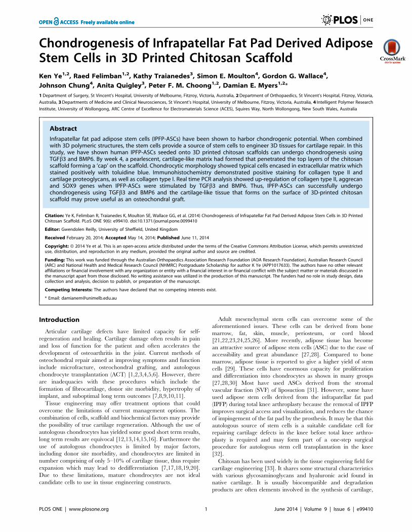
Figure 3. Macroscopic image of cell-scaffold construct at week4. (A) Macroscopic image showing a cartilage-like cap (black arrow)derived after 7.56105 IPFP-ASCs were seeded onto a 6 mm plug of 3Dprinted chitosan (white arrow) scaffold to form a cell-scaffold unit inchondrogenic media after 4 weeks. (B) Macroscopic image showing novisible signs of cartilage formation on the chitosan scaffold (whitearrow) after 4 weeks in control media without growth factors. Thescaffolds were cut using a 6 mm skin biopsy punch. The image wastaken using a Canon EOS 55D with macro zoom lens at 100 mm. Thescale bar represents 1 mm.doi:10.1371/journal.pone.0099410.g003
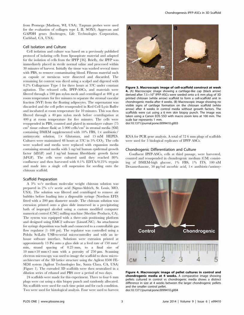
Figure 4. Macroscopic image of pellet cultures in control andchondrogenic media at 4 weeks. A comparative image showingpellets cultured in control vs chondrogenic media shows a distinctdifference in size at 4 weeks between the larger chondrogenic pelletsand the smaller control pellet.doi:10.1371/journal.pone.0099410.g004
Chondrogenesis IPFP-ASCs in 3D Scaffold
PLOS ONE | www.plosone.org 3 June 2014 | Volume 9 | Issue 6 | e99410

cotic, 10 ng/ml TGFb3 and 10 ng/ml BMP6. Scaffolds were cut
using a 6 mm biopsy punch (Kai Medical, Honolulu, HI, USA)
and seeded with 7.56105 ASCs in a 24 Transwell tissue culture
plate well inserts with an internal diameter of 6.5 mm and 3.0 mm
pore size (Figure 2). Cells were seeded on top of the scaffold and
1.2 ml of chondrogenic or control media was used in each well.
These were incubated at 37C in 5% CO2 for 14 and 28 days and
media changed three times per week. The same protocol was used
for media without growth factors, and served as the negative
control. Cells were also cultured by themselves using micromass
pellet culture. 250,000 cells were centrifuged at 400 g for 5
minutes in 15 ml centrifuge tubes to form cell pellets and cultured
in 0.5 ml of chondrogenic or control media for the same time
period as cells cultured on the chitosan scaffolds.
Histology and ImmunohistochemistryAfter 14 and 28 days of culture, cell pellets and cell-scaffold
constructs were harvested for histological and immunohistochem-
ical analysis using standard techniques of fixation, dehydration
and paraffin embedding. Pellets and cell-scaffold constructs were
fixed in 10% NBF overnight at 4C and processed and imbedded at
the histopathology laboratory, pathology department at St
Vincent’s Hospital, Melbourne, sectioned into 6 mm sections and
incubated overnight at 37C. The sections were deparaffinised,
rehydrated through graded ethanol, and stained with haematox-
ylin & eosin (H&E) and toluidine blue (TB) (for glycosaminogly-
cans (GAGs)).
Accumulation of collagen types I and II, and cartilage-specific
proteoglycan was assessed by immunohistochemistry. Sections
were treated with 0.3% hydrogen peroxide (H2O2) for five
minutes, antigen retrieval using Proteinase K for four minutes
and were blocked using 10% normal rabbit serum (NRS) for 30
minutes at room temperature. These sections were incubated with
the following primary antibodies; mouse monoclonal anti-human
type II collagen antibody (1:500), goat polyclonal anti-human type
I collagen (1:500), and mouse monoclonal anti-human cartilage
proteoglycan antibody (1:500) for 60 minutes at 37C. Isotype
negative controls were used at the same concentration as their
respective primary antibodies. Subsequently, sections were incu-
bated using biotinylated rabbit polyclonal anti- goat and rabbit
anti-mouse antibodies as secondary antibodies for 30 minutes
followed by horseradish peroxidase (HRP)-conjugated streptavidin
treatment according to the manufacturer’s instructions. The
reaction was developed as a brown precipitation using peroxidase
substrate 3,3-diaminobenzidine (DAB) for 5 minutes. Sections
were counterstained with haematoxylin, dehydrated, cleared, and
mounted with Pertex.
Quantitative Real Time PCR (qPCR)Week 2 and 4 cell-scaffold constructs were pulverized in liquid
nitrogen using a small mortar and pestle and then homogenized in
1 ml of Trizol solution, and RNA was extracted and purified using
a combination of the Trizol method and silica membrane-based
commercial extraction kit (QIAGEN, RNeasy mini kit) according
to the manufacturer’s protocol. RNA from pre-differentiated cells
(day 0) was also extracted as Time zero samples. The RNA
concentration and purity were measured using a NanoDrop
spectrophotometer (Peqlab, Erlangen, Germany) and the Agilent
2100 BioAnalyzer (Agilent Technologies, Santa Clara, CA, USA).
Complimentary DNA copies were reverse transcribed from
Figure 5. Histology of week 4 pellet culture in control andchondrogenic media. Control media shows cells that appear morefibroblastic in morphology as compared to chondrocytic morphologywith extracellular matrix in the chondrogenic pellet. Chondrogenicpellets are larger, well rounded and stains positively for toluidine blue.Magnification 206. Scale bars at 50 mm as indicated.doi:10.1371/journal.pone.0099410.g005
Figure 6. Histology of week 4 chitosan-ASC constructs in chondrogenic and control media. There is a paucity of cellular attachment andgrowth in control media and lack of toluidine blue staining. Chitosan fibres are clearly stained by eosin. The cells that have grown and attached to thechitosan structure under chondrogenic media shows extracellular matrix deposition with strong toluidine blue staining, suggesting the presence ofproteoglycans. Magnification 206. A–D represent whole tissue images with scale bars at 500 mm as indicated. E–F represents magnified images withscale bars at 20 mm as indicated.doi:10.1371/journal.pone.0099410.g006
Chondrogenesis IPFP-ASCs in 3D Scaffold
PLOS ONE | www.plosone.org 4 June 2014 | Volume 9 | Issue 6 | e99410

200 ng total RNA for all samples using oligo-dT primers and
omniscript reverse transcriptase kit according to the recommen-
dations of the manufacturer. qPCR was performed using standard
TaqMan Probe-Based Gene Expression Analysis protocols using
commercial available probes for collagen types I and II, SOX 9,
and Aggrecan. The Taqman primer ID for each gene was as
follows: COL1A2 (Hs00164099_m1), COL2A1
(Hs00264051_m1), SOX9 (Hs01165814_m1), and ACAN
(Hs00153936_m1). GAPDH was used as housekeeping gene for
relative quantification of gene expression (Hs02758991_g1).
Liquid handling was performed by the CAS1200 series robot by
Corbett Robotics (Corbett Life Sciences, Qiagen, Hilden,
Germany). Subsequent PCR reaction was performed using the
Lightcycler 480 (Roche, Basel, Switzerland).
Data AnalysisRelative quantification was derived and analyzed using the
Second Derivative Maximum method through the Lightcycler 480
software version 1.5. Subsequent numerical data analysis of
relative quantification of qPCR results was performed in Microsoft
Excel 2010 and GraphPad Prism 6.0 (GraphPad Software, La
Jolla, CA, USA) using the 2(2DCT) method. Means, standard
deviations (SD) and errors (SEM), and 95% confidence limits were
calculated for each set of results. The t-test was used to assess
significant between two sets of data. Friedman’s test was used to
assess significance over three or more sets of data. Statistical testing
was verified by a statistician.
Results
Cell CultureIPFP-ASCs cultured for up to four weeks on a 3D chitosan
scaffold in chondrogenic media (TGFb3 and BMP6), developed a
pearlescent, white and shiny cartilage-like tissue ‘cap’ (Figure 3A).
Conversely, cell-scaffold constructs cultured in control media
showed no visible signs of cartilage-like tissue formation
(Figure 3B). Similarly, chondrogenic pellets were larger and
rounder than control pellets and also had a shiny white
appearance macroscopically (Figure 4).
Histology and ImmunohistochemistryH&E staining of chondrogenic pellets showed a change in
cellular morphology towards a chondrocytic phenotype showing
larger cells encapsulated in lacunae when compared to control
pellets. Positive toluidine blue of chondrogenic pellets contrasts
with a lack of toluidine blue staining in control pellets.
Furthermore, in chondrogenic pellets, gradient differentiation of
toluidine blue staining intensity can be seen on magnification,
together with more tangential morphology of cells near the surface
(Figure 5).
H&E staining of the cell-scaffold construct show a paucity of
cells attached to the chitosan in the negative control, and the cells
that did attach appeared to be fibroblastic in appearance and
lacked features of chondrocytic morphology. In contrast, cell-
scaffold constructs cultured in chondrogenic media showed
cellular aggregation throughout the upper layers of the chitosan
scaffold forming what can be seen macroscopically as a ‘cap’ of
cartilaginous tissue. These cells exhibit features of chondrocytic
morphology as seen in the chondrogenic pellets as well as staining
for toluidine blue (Figure 6). The chitosan fibres were stained pink
by the eosin and therefore easily recognizable in the H&E stain.
The chitosan fibres did not stain for toluidine blue and therefore
appeared like empty spaces in the toluidine blue stain.
Immunohistochemistry staining of chondrogenic and control
pellets for the presence of collagen type II, cartilage proteoglycan,
and collagen type I at week 4 is shown in Figure 7. When
compared to control pellets and isotype controls, chondrogenic
pellets stained strongly for collagen type II and proteoglycan in the
extracellular matrix. Collagen type I was present in both the
control and the chondrogenic pellets. Similar patterns of staining
can be seen in the cell-scaffold constructs (Figure 8). Strong
staining for collagen type II and cartilage proteoglycans is seen
throughout cartilaginous ‘cap’. There was also staining of collagen
type I throughout, which appears to co-localise with collagen type
II in some parts. All immunohistochemistry staining show the cells
are encased in their lacunae consisting of extracellular matrix
consisting of predominantly collagen type II. Some non-specific
staining of chitosan fibres is also present in the proteoglycan stain.
Figure 7. Immunohistochemistry of week 4 pellet culture incontrol and chondrogenic media. Collagen type II and proteogly-can is expressed in chondrogenic pellet compared with no expressionin control media. Collagen type I is expressed in both control andchondrogenic pellet. Magnification 206. Scale bars at 50 mm asindicated.doi:10.1371/journal.pone.0099410.g007
Chondrogenesis IPFP-ASCs in 3D Scaffold
PLOS ONE | www.plosone.org 5 June 2014 | Volume 9 | Issue 6 | e99410

As shown in the histological stains, there is a paucity of cellular
attachment in the control cell-scaffold constructs.
Gene ExpressionThere was an increase in all mRNA expression levels of
chondrogenic markers in both chondrogenic pellets and cell-
scaffold constructs tested from week 0 to week 4. It is important to
note that there were undetectable levels of expression of collagen
type II in the cells prior to plating. In contrast, collagen type II
expression was present at week 2 and was increased significantly
by week 4 in both pellets and cell-scaffold constructs (p,0.05)
(Figure 9). However, collagen type I was present at time zero in the
cells and by week 4 there was comparable expression levels of
collagen type II gene with collagen type I. Collagen type I
expression remained consistent over the four-week culture period
and any changes across the pellets or cell-scaffold constructs were
not statistically significant.
At week 4 cell-scaffold constructs and pellets cultured in control
media (i.e. without TGFb3 and BMP6) had collagen type II gene
expression levels that were undetectable, and only low levels of
SOX9 and aggrecan genes were expressed. All chondrogenic
genes examined were expressed at a higher level when cultured in
chondrogenic media. The expression of collagen type II and
aggrecan at week 4 were significantly greater in the chondrogenic
group compared with the control group in both pellets and cell-
scaffold constructs (p,0.05). Figure 10 illustrates the chondrogenic
gene expression data between of chondrogenic media and control
media groups at week 4.
Discussion and Conclusion
This study demonstrates the infrapatellar fat pad is a reliable
and abundant source of adipose stem cells with chondrogenic
differentiation capacity that can readily be accessed during surgery
as an autologous material. The volume of the material produced
means that small harvest of autologous IPFP can yield adequate
stem cell numbers for the possible repair of quite substantial areas
of cartilage damage. Table 1 shows the studies that have used
IPFP for chondrogenesis in the past and the combination of
TGFb3 and BMP6 is unique. In a previous study, we have
characterized the chondrogenesis of IPFP-ASCs using this
combination of growth factors to demonstrate their chondrogenic
potential [37]. In this study we have demonstrated the ability for
these cells to undergo chondrogenesis not only by themselves in
pellet form but also attach, proliferate and differentiate on a 3D
printed chitosan scaffold which may serve as a delivery mechanism
for these cells into a site of cartilage repair.
In our experiments, all three samples of IPFP-ASCs did not
express detectable levels of collagen type II and SOX9 genes at
week 0. However, it is clear the expression of these markers
increased substantially over four weeks (p,0.05). The increase in
collagen type II expression was significant. Collagen type II
expression remained undetectable at week 4 in the control group.
This suggests the addition of TGFb3 and BMP6 has had a
profound effect on chondrogenic differentiation and the stimula-
tion of production of collagen type II in the IPFP-ASCs. The
macroscopic changes of the cell aggregates and the morphological
changes in the cells also clearly demonstrated the progressive
Figure 8. Immunohistochemistry of week 4 chitosan-ASC constructs in chondrogenic and control media. There is a paucity of cellularattachment and growth in control media and lack of collagen type II and proteoglycan expression. The cells that have grown and attached to thechitosan structure under chondrogenic media strongly expresses collagen type II, proteoglycan and also collagen type I, indicating the formation ofhyaline-like cartilage. Magnification 206. A-H represent whole tissue images with scale bars at 500 mm as indicated. I-P represent magnified imageswith scale bars at 20 mm as indicated.doi:10.1371/journal.pone.0099410.g008
Chondrogenesis IPFP-ASCs in 3D Scaffold
PLOS ONE | www.plosone.org 6 June 2014 | Volume 9 | Issue 6 | e99410

development of a chondrogenic phenotype. Collagen type I
expression remained unchanged from pre-plated cells to week 4
(under chondrogenic conditions). The expression of collagen type I
by IPFP-ASCs is consistent with previous studies using these cells
[32,38,39,40].
Histologically, there is evidence of some co-localization of
collagen type II and type I expression which may provide evidence
of early developmental progression at 4 weeks in vitro, of the cell-
scaffold constructs toward a more chondrogenic phenotype.
Collagen type I expression was important to investigate, not only
because it is found in fibrocartilage, but because it is expressed in
early chondrogenesis as part of the transformation that occurs
from mesenchymal cells to chondrocytes. Therefore, collagen type
1 expression is also a marker of early chondrogenesis. This is
consistent with the pre-natal development of the knee joint, which
starts with a condensation of the mesenchyme between the two
long bones prior to the distinct development of the articular
surfaces of the long bones [41]. In our study, cell-matrix constructs
were maintained for only four weeks and may indicate the need to
extend the time period for further clarification of the in vitro
development sequence. Changes to the composition and structure
of the scaffold over time may also impact the production of
collagen type I in the cells. This possibility was not investigated in
this study, however similar observations were made by other
studies of this nature [32].
SOX9 plays a significant role in chondrogenesis, and is also
present in other tissues such as the notochord, otic vesicle, neural
tube, brain and the developing gonads [42]. In terms of
chondrogenesis, it is an important transcription factor in the
activation of collagen type II gene during the process of
chondrogenesis [42]. Collagen type II is predominantly found in
adult articular (hyaline) cartilage and also occurs to a smaller
extent in fibrocartilage tissue such as intervertebral disc and
meniscus [43]. The presence of SOX 9 in our study is consistent
with the chondrogenic differentiation of the IPFP-ASCs under the
influence of TGFb3 and BMP6. The concomitant increase in
collagen type II gene expression, and in particular its continued
increase over 4 weeks in culture is indicative of the beginning of
hyaline cartilage formation. Furthermore the expression of
Figure 9. Relative gene expression of chondrogenic pellets andcell-scaffold constructs over time. Collagen type II gene expression,which is not expressed at week 0, is significantly increased over time inboth pellet and cell-scaffold cultures (*p,0.05; Friedman’s test). Otherchondrogenic genes such as aggrecan and SOX9 all increase over timehowever they were less significant. Collagen type I was present fromthe outset in both cultures and remained elevated throughout,however changes were not statistically significant. These resultsrepresent 3 separate experiments using 3 separate biological replicates(N = 3) as well as triplicate internal replicates for each qPCR reaction.Mean plotted with error bars representing the SEM.doi:10.1371/journal.pone.0099410.g009
Figure 10. Relative chondrogenic gene expression betweencontrol and chondrogenic pellets and cell-scaffold constructsat 4 weeks. Collagen type II gene was not expressed in all controlcultures for both pellets and cell-scaffolds at week 4. Both collagen typeII and aggrecan genes were significantly increased by week 4 overcontrol groups (*p,0.05; t-test). The increase in SOX9 was lesssignificant between the control groups and the chondrogenic groupsat 4 weeks. These results represent 3 separate experiments using 3separate biological replicates (N = 3) as well as triplicate internalreplicates for each qPCR reaction. Mean plotted with error barsrepresenting the SEM.doi:10.1371/journal.pone.0099410.g010
Chondrogenesis IPFP-ASCs in 3D Scaffold
PLOS ONE | www.plosone.org 7 June 2014 | Volume 9 | Issue 6 | e99410

collagen type II and aggrecan protein in the extracellular matrix,
as demonstrated by immunohistochemistry, is a hallmark of
hyaline cartilage production [43]. This indicates that the
phenotypic expression of the gene products is conclusive for
chondrogenesis and hyaline cartilage formation.
The limited the number of biological samples available for this
study may attribute to the variability in qPCR results. Ideally,
IPFPs from healthy individuals may provide different expression
profiles and it has been suggested that mesenchymal stem cells
obtained from patients with advanced osteoarthritis reduces its
chondrogenic potential [44]. Similar limitations were noted by a
previous study in the use of fat pad obtained from patients with
osteoarthritis and the potential loss of chondrogenic potential of
ASCs [32]. However, it was impossible due to ethical reasons for
us to compare the chondrogenic potential of ASCs from healthy
versus osteoarthritic IPFP as it involves removing healthy tissue
from donors. Therefore, the most available sources of IPFPs were
from patients with osteoarthritis undergoing total knee replace-
ments. However, to improve the quality of fat pad, patients with
previous surgery to the knee were excluded to reduce the
possibility of fibrosis and scar tissue formation within the fat
pad. Likewise, patients with autoimmune or inflammatory arthritis
undergoing total knee replacements were excluded to reduce the
impact of either their disease process or specific auto-immune
medications on the potential for cells within the IPFP to proliferate
and differentiate. Despite controlling for those factors, the disease
process of osteoarthritis may still affect the behavior of cells within
the IPFP. If the development of stem cells and biomaterials could
improve the management of patients with osteoarthritis, then the
use of IPFP from patients with osteoarthritis becomes clinically
relevant and applicable.
Although previous studies using IPFP-ASCs have successfully
induced chondrogenesis using various growth factors and culture
methods, our results differ in the formation of a ‘cap’ of
cartilaginous-like tissue above the chitosan scaffold. The 250 mm
porosity of the chitosan scaffold may have limited the penetration
of the IPFP-ASCs into the 3D chitosan lattice scaffold due to the
self-aggregation nature and extracellular matrix production ability
of these cells. These two factors, based on the observations we
have seen in this study, influences how the cells behave and attach
to the scaffold. In contrast, cells cultured in control media did not
exhibit self-aggregation and extracellular matrix production
abilities as those under chondrogenic influence, and therefore
did not seem to adhere to the chitosan scaffold well, resulting in a
paucity of cellular attachment as shown in the histological samples.
Therefore, it seems a porosity of 250 mm is more than sufficient for
non-chondrogenic cells to penetrate and pass through the scaffold
network. However, once cells undergo chondrogenic differentia-
tion, wider pore sizes may be required to accommodate cellular
aggregation and enhanced extracellular matrix production to
achieve full penetration of the chitosan scaffold, Indeed, in this
study, IPFP-ASCs may have not utilized the chitosan as a
substitute for the extracellular matrix which they inherently
produced, but rather seemed to use the chitosan scaffold for
cellular support only. It is possible to capitalize on this interaction
such that initially, the 3D nature of the scaffold provides a
platform for the cells to naturally aggregate, form a spheroid-like
structure and expand in the framework of the scaffold. We have
seen this phenomenon in micromass cultures of these cells without
scaffold [38,39]. We believe this more accurately mimics the
behavior of the formation of an osteochondral unit whereby
chondrocytes are fully surrounded in their own matrix supported
at their base by subchondral bone which provides strength and
support. Whether our cell and chitosan construct behaves like a
biphasic ‘implant’ in vivo remains to be seen. Further in vivo
studies are required to assess the behavior of this engineered
construct.
In conclusion, infrapatellar fat pad-derived adipose stem cells
appear to provide an excellent source of cells with chondrogenic
potential. Our results demonstrate the combination of TGFb3 and
BMP6 strongly promotes chondrogenesis with these cells in a 3D
chitosan scaffold. This cell-scaffold construct may provide the basis
of a viable chondral graft suitable for in vivo implantation.
Author Contributions
Conceived and designed the experiments: KY RF KT SEM GGW PFMC
DEM. Performed the experiments: KY RF KT JC. Analyzed the data: KY
KT AQ DEM. Contributed reagents/materials/analysis tools: KY KT
SEM GGW JC PFMC DEM. Wrote the paper: KY KT PFMC DEM.
References
1. Breinan HA, Martin SD, Hsu HP, Spector M (2000) Healing of canine articularcartilage defects treated with microfracture, a type-II collagen matrix, or
cultured autologous chondrocytes. J Orthop Res 18: 781–789.
2. Steadman JR, Rodkey WG, Rodrigo JJ (2001) Microfracture: surgical technique
and rehabilitation to treat chondral defects. Clin Orthop Relat Res: S362–369.
3. Steadman JR, Miller BS, Karas SG, Schlegel TF, Briggs KK, et al. (2003) Themicrofracture technique in the treatment of full-thickness chondral lesions of the
knee in National Football League players. J Knee Surg 16: 83–86.
4. Hangody L, Fules P (2003) Autologous osteochondral mosaicplasty for the
treatment of full-thickness defects of weight-bearing joints: ten years of
experimental and clinical experience. J Bone Joint Surg Am 85-A Suppl 2:25–32.
5. Brittberg M, Lindahl A, Nilsson A, Ohlsson C, Isaksson O, et al. (1994)
Treatment of deep cartilage defects in the knee with autologous chondrocyte
transplantation. N Engl J Med 331: 889–895.
6. Zheng MH, Willers C, Kirilak L, Yates P, Xu J, et al. (2007) Matrix-inducedautologous chondrocyte implantation (MACI): biological and histological
assessment. Tissue Eng 13: 737–746.
7. Bedi A, Feeley BT, Williams RJ III (2010) Management of articular cartilage
defects of the knee. J Bone Joint Surg Am 92: 994–1009.
Table 1. List of studies using IPFP as the stem cell source and the growth factors used.
Study Cell Source Culture Type Growth Factors Used
Dragoo et al (2003) [29] IPFP-ASC Micromass with fibrin IGF1/FGF2
Khan et al (2008) [39] IPFP-ASC 3D cell aggregate IGF1/TGFb3
Lee et al (2008) [38] IPFP-ASC Pellet culture TGFb1/BMP7
Jurgens et al (2009) [32] IPFP-ASC 3D PLA-CPL Scaffold TGFb1
Buckley et al (2010) [40] IPFP-ASC (Porcine) Agarose hydrogel TGFb3
Abbreviations: IGF (insulin growth factor), TGFb (transforming growth factor beta), BMP (bone morphogenetic protein), PLA-CPL (poly L-lactic-co-E-caprolactone).doi:10.1371/journal.pone.0099410.t001
Chondrogenesis IPFP-ASCs in 3D Scaffold
PLOS ONE | www.plosone.org 8 June 2014 | Volume 9 | Issue 6 | e99410

8. Bae DK, Yoon KH, Song SJ (2006) Cartilage healing after microfracture in
osteoarthritic knees. Arthroscopy 22: 367–374.9. Mithoefer K, Williams RJ III, Warren RF, Wickiewicz TL, Marx RG (2006)
High-impact athletics after knee articular cartilage repair: a prospective
evaluation of the microfracture technique. Am J Sports Med 34: 1413–1418.10. Mithoefer K, Williams RJ III, Warren RF, Potter HG, Spock CR, et al. (2005)
The microfracture technique for the treatment of articular cartilage lesions in theknee. A prospective cohort study. J Bone Joint Surg Am 87: 1911–1920.
11. Gooding CR, Bartlett W, Bentley G, Skinner JA, Carrington R, et al. (2006) A
prospective, randomised study comparing two techniques of autologouschondrocyte implantation for osteochondral defects in the knee: Periosteum
covered versus type I/III collagen covered. Knee 13: 203–210.12. Behrens P, Bitter T, Kurz B, Russlies M (2006) Matrix-associated autologous
chondrocyte transplantation/implantation (MACT/MACI)-5-year follow-up.Knee 13: 194–202.
13. Zaslav K, Cole B, Brewster R, DeBerardino T, Farr J, et al. (2009) A prospective
study of autologous chondrocyte implantation in patients with failed priortreatment for articular cartilage defect of the knee: results of the Study of the
Treatment of Articular Repair (STAR) clinical trial. Am J Sports Med 37: 42–55.
14. Bartlett W, Skinner JA, Gooding CR, Carrington RW, Flanagan AM, et al.
(2005) Autologous chondrocyte implantation versus matrix-induced autologouschondrocyte implantation for osteochondral defects of the knee: a prospective,
randomised study. J Bone Joint Surg Br 87: 640–645.15. Marcacci M, Berruto M, Brocchetta D, Delcogliano A, Ghinelli D, et al. (2005)
Articular cartilage engineering with Hyalograft C: 3-year clinical results. ClinOrthop Relat Res: 96–105.
16. Gobbi A, Kon E, Berruto M, Filardo G, Delcogliano M, et al. (2009)
Patellofemoral full-thickness chondral defects treated with second-generationautologous chondrocyte implantation: results at 5 years’ follow-up. Am J Sports
Med 37: 1083–1092.17. Chung C, Burdick JA (2008) Engineering cartilage tissue. Adv Drug Deliv Rev
60: 243–262.
18. Goessler UR, Bugert P, Bieback K, Sadick H, Baisch A, et al. (2006) In vitroanalysis of differential expression of collagens, integrins, and growth factors in
cultured human chondrocytes. Otolaryngol Head Neck Surg 134: 510–515.19. Goessler UR, Bugert P, Bieback K, Baisch A, Sadick H, et al. (2004) Expression
of collagen and fiber-associated proteins in human septal cartilage duringin vitro dedifferentiation. Int J Mol Med 14: 1015–1022.
20. Darling EM, Athanasiou KA (2005) Rapid phenotypic changes in passaged
articular chondrocyte subpopulations. J Orthop Res 23: 425–432.21. Lee KH, Song SU, Hwang TS, Yi Y, Oh IS, et al. (2001) Regeneration of
hyaline cartilage by cell-mediated gene therapy using transforming growth factorbeta 1-producing fibroblasts. Hum Gene Ther 12: 1805–1813.
22. Mandl EW, van der Veen SW, Verhaar JA, van Osch GJ (2002) Serum-free
medium supplemented with high-concentration FGF2 for cell expansion cultureof human ear chondrocytes promotes redifferentiation capacity. Tissue Eng 8:
573–580.23. Tuan RS, Boland G, Tuli R (2003) Adult mesenchymal stem cells and cell-based
tissue engineering. Arthritis Res Ther 5: 32–45.24. French MM, Rose S, Canseco J, Athanasiou KA (2004) Chondrogenic
differentiation of adult dermal fibroblasts. Ann Biomed Eng 32: 50–56.
25. Asakura A, Komaki M, Rudnicki M (2001) Muscle satellite cells aremultipotential stem cells that exhibit myogenic, osteogenic, and adipogenic
differentiation. Differentiation 68: 245–253.
26. Nakahara H, Goldberg VM, Caplan AI (1991) Culture-expanded human
periosteal-derived cells exhibit osteochondral potential in vivo. J Orthop Res 9:
465–476.
27. Zuk PA, Zhu M, Ashjian P, De Ugarte DA, Huang JI, et al. (2002) Human
adipose tissue is a source of multipotent stem cells. Mol Biol Cell 13: 4279–4295.
28. Ogawa R, Mizuno S (2010) Cartilage regeneration using adipose-derived stem
cells. Curr Stem Cell Res Ther 5: 129–132.
29. Dragoo JL, Samimi B, Zhu M, Hame SL, Thomas BJ, et al. (2003) Tissue-
engineered cartilage and bone using stem cells from human infrapatellar fat
pads. J Bone Joint Surg Br 85: 740–747.
30. Awad HA, Wickham MQ, Leddy HA, Gimble JM, Guilak F (2004)Chondrogenic differentiation of adipose-derived adult stem cells in agarose,
alginate, and gelatin scaffolds. Biomaterials 25: 3211–3222.
31. Zuk PA, Zhu M, Mizuno H, Huang J, Futrell JW, et al. (2001) Multilineage cells
from human adipose tissue: implications for cell-based therapies. Tissue Eng 7:
211–228.
32. Jurgens WJ, van Dijk A, Doulabi BZ, Niessen FB, Ritt MJ, et al. (2009) Freshly
isolated stromal cells from the infrapatellar fat pad are suitable for a one-step
surgical procedure to regenerate cartilage tissue. Cytotherapy 11: 1052–1064.
33. Suh JK, Matthew HW (2000) Application of chitosan-based polysaccharide
biomaterials in cartilage tissue engineering: a review. Biomaterials 21: 2589–2598.
34. Abarrategi A, Lopiz-Morales Y, Ramos V, Civantos A, Lopez-Duran L, et al.
(2010) Chitosan scaffolds for osteochondral tissue regeneration. J Biomed Mater
Res A 95: 1132–1141.
35. Woodfield TB, Malda J, de Wijn J, Peters F, Riesle J, et al. (2004) Design of
porous scaffolds for cartilage tissue engineering using a three-dimensional fiber-
deposition technique. Biomaterials 25: 4149–4161.
36. Estes BT, Diekman BO, Gimble JM, Guilak F (2010) Isolation of adipose-
derived stem cells and their induction to a chondrogenic phenotype. Nat Protoc
5: 1294–1311.
37. Felimban R, Ye K, Traianedes K, Di Bella C, Crook J, et al. (2014)
Differentiation of stem cells from human infrapatellar fat pad: Characterization
of cells undergoing chondrogenesis. Tissue Eng Part A.
38. Lee SY, Nakagawa T, Reddi AH (2008) Induction of chondrogenesis andexpression of superficial zone protein (SZP)/lubricin by mesenchymal
progenitors in the infrapatellar fat pad of the knee joint treated with TGF-
beta1 and BMP-7. Biochem Biophys Res Commun 376: 148–153.
39. Khan WS, Tew SR, Adesida AB, Hardingham TE (2008) Human infrapatellar
fat pad-derived stem cells express the pericyte marker 3G5 and show enhanced
chondrogenesis after expansion in fibroblast growth factor-2. Arthritis Res Ther
10: R74.
40. Buckley CT, Vinardell T, Thorpe SD, Haugh MG, Jones E, et al. (2010)
Functional properties of cartilaginous tissues engineered from infrapatellar fat
pad-derived mesenchymal stem cells. J Biomech 43: 920–926.
41. Merida-Velasco JA, Sanchez-Montesinos I, Espin-Ferra J, Rodriguez-VazquezJF, Merida-Velasco JR, et al. (1997) Development of the human knee joint. Anat
Rec 248: 269–278.
42. Lefebvre V, de Crombrugghe B (1998) Toward understanding SOX9 function
in chondrocyte differentiation. Matrix Biol 16: 529–540.
43. Eyre D (2002) Collagen of articular cartilage. Arthritis Res 4: 30–35.
44. Murphy JM, Dixon K, Beck S, Fabian D, Feldman A, et al. (2002) Reduced
chondrogenic and adipogenic activity of mesenchymal stem cells from patients
with advanced osteoarthritis. Arthritis Rheum 46: 704–713.
Chondrogenesis IPFP-ASCs in 3D Scaffold
PLOS ONE | www.plosone.org 9 June 2014 | Volume 9 | Issue 6 | e99410