choose your models wisely: how different murine bone marrow-derived dendritic cell protocols...
TRANSCRIPT

Journal of Controlled Release xxx (2014) xxx–xxx
COREL-07258; No of Pages 9
Contents lists available at ScienceDirect
Journal of Controlled Release
j ourna l homepage: www.e lsev ie r .com/ locate / jconre l
Choose yourmodels wisely: Howdifferentmurine bonemarrow-deriveddendritic cell protocols influence the success of nanoparticulate vaccinesin vitro
Heleen Dewitte a,1, Rein Verbeke a,1, Karine Breckpot b, Roosmarijn E. Vandenbroucke c,d, Claude Libert c,d,Stefaan C. De Smedt a,⁎, Ine Lentacker a
a Lab for General Biochemistry and Physical Pharmacy, Faculty of Pharmacy, Ghent University, Ottergemsesteenweg 460, 9000 Ghent, Belgiumb Laboratory of Molecular and Cellular Therapy, Department of Immunology-Physiology, Medical School of the Vrije Universiteit Brussel (VUB), Laarbeeklaan 103, 1050 Jette, Belgiumc Inflammation Research Center (IRC), VIB, Technologiepark 927, 9052 Ghent, Belgiumd Molecular Mouse Genetics Unit, Department of Biomedical Molecular Biology, Ghent University, Technologiepark 927, 9052 Ghent, Belgium
⁎ Corresponding author at: Laboratory of General BiochHarelbekestraat 72, 9000 Ghent, Belgium. Tel.:+32 9 264
E-mail address: [email protected] (S.C. De S1 Both authors contributed equally to this work.
http://dx.doi.org/10.1016/j.jconrel.2014.06.0240168-3659/© 2014 Elsevier B.V. All rights reserved.
Please cite this article as: H. Dewitte, et al.,influence the success of nanoparticulate vac
a b s t r a c t
a r t i c l e i n f oArticle history:Received 11 April 2014Accepted 15 June 2014Available online xxxx
Chemical compounds studied in this article:DOTAP (PubChem CID: 6437371)DOPE (PubChem CID: 9546757)CholEsteryl BODIPY® FL C12(PubChem CID: 70682631)SIINFEKL (PubChem CID: 71311993)
Keywords:Dendritic cellmRNACancer vaccinationNanoparticle
Dendritic cell (DC)-based cancer vaccination has shown great potential in cancer immunotherapy. As a result,novel nanoparticles aiming to load DCs with tumor antigens are being developed and evaluated in vitro. Forthis,murine bonemarrow-derivedDCs (BM-DCs) aremost commonly used asmodel DCs. However,many differ-ent protocols exist to generate these cells. Therefore, we investigated to what extent different BM-DC cultureprotocols impact on the immunobiology of the cells, as well as their response to particulate antigens. We evalu-ated 4 different BM-DC protocols with 2 main variables: bovine serum and cytokine combinations. Our resultsshow distinct differences in yield, phenotypical maturation status and the production of immune stimulatoryand immune suppressive cytokines by the different BM-DCs. Importantly, we demonstrate that the antigen-loading of these different BM-DCs via transfection with mRNA lipoplexes results in large differences in transfec-tion efficiency aswell as in the capacity ofmRNA-transfected BM-DCs to stimulate antigen-specific T cells. Thus, itis clear that the BM-DCmodel can have significant confounding effects on the evaluation of novel nanoparticulatevaccines. To take this into accountwhen testing novel particulate antigen-delivery systems in BM-DCmodels, wepropose to (1) perform a thorough immunological characterization of the BM-DCs and to (2) not only judge aparticle's potential for cancer vaccination based on transfection efficiency, but also to include an evaluation offunctional end-points such as T cell activation.
© 2014 Elsevier B.V. All rights reserved.
1. Introduction
Since thefirst discovery of dendritic cells (DCs) as a novel cell type inmice by noble prize-winner Ralph M. Steinman, DCs came to be knownas nature's adjuvant [1]. This title was awarded to them due to theirunique capacity to elicit antigen-specific immune responses. DCs con-tinuously sample their environment, engulfing foreign material andpresenting it on their surface to naïve T cells. Importantly, DCs can pro-vide the necessary co-stimulatory signals to activate T cells andpromotetheir differentiation into cytotoxic T lymphocytes (CTLs) and helper Tcells (Th) in case of CD8+ and CD4+ T cells respectively. The more theDC's role in initiating immunity was unraveled, the more its potentialuse inmedicine came to light. For instance, bymodifying DCs to presenttumor-associated antigens (TAAs) or HIV-antigens, specific antitumor
emistry and Physical Pharmacy,80 78; fax: +32 9 264 81 89.medt).
Choose your models wisely:cines in vitro, J. Control. Relea
or anti-HIV immune responses can be evoked [2–4]. Research in thisfield of therapeutic DC vaccines flourished and has resulted in theFDA-approval of the first DC-based vaccine in 2010 (Provenge®,marketed by Dendreon) [5,6].
In search of novel strategies for effective antigen-loading of DCs tocombat cancer or viral infections, experiments are generally performedon in vitro generated DCs. Themain reason for this is the low number ofin vivo DCs that can be isolated from different tissues [7–9]. As a result,different protocols were established for the in vitro production of DCs,generated frommurine bonemarrow precursors or from the peripheralbloodmonocytes found in humanblood [10–13]. By culturing thesemu-rine and humanmonocytic precursors for a number of days in the pres-ence of appropriate cytokines, their differentiation into murine bonemarrow-derived DCs (BM-DCs), and human monocyte-derived DCs(MoDCs) will be induced. This way, convenient DC models for in vivostudies of the DC functionality, phenotype, immunogenic potentialand antigen-presentation capacity were established. Importantly,thesemodels gained in popularity among researchers who are develop-ing novel particulate systems for the delivery of protein or nucleic acid
How different murine bone marrow-derived dendritic cell protocolsse (2014), http://dx.doi.org/10.1016/j.jconrel.2014.06.024

2 H. Dewitte et al. / Journal of Controlled Release xxx (2014) xxx–xxx
antigens toDCs.However, it shouldbenoted that although thesemethodsresult in the generation of large numbers of cells with DC-like properties,they do not seem to correspond to any of the DC subsets that under nor-mal circumstances populate themouse lymphnodes. Instead, they rather,but not completely, resemble “emergency” or “inflammatory” DCs thatonly occur in inflamed lymphnodes [14–16]. Therefore, in vitro generatedmonocyte-derived DCs need to be considered as simplified models for acomplex in vivo situation, and conclusions from experiments usingthese cells should be drawn with the necessary caution. An additionalcritical factor that complicates the use of DCs in vitro, is the plethora ofprotocols that are available for the generation of these cells. The differen-tiation of bonemarrow into BM-DCs can be achievedwith either GM-CSF(Granulocyte-macrophage colony-stimulating factor) alone or in combi-nation with IL-4 (interleukin-4), but also other factors, including Flt3L(FMS-related tyrosine kinase 3 ligand) or IL-3 have been employed at dif-ferent concentration ratios [14,17,18]. Changing the cocktail and concen-trations of cytokines can result in major differences in DC phenotype. Inaddition, other factors such as cell culture medium, the timeframe forDC generation from bone marrow precursors, additional purificationsteps, mouse age and especially growth factors that are present in thesera that are routinely added to the culture medium, may decide thetype of BM-DCs that are produced. Obviously, these differences make itdifficult to compare the results obtained in different in vitro models. Forone, the variability of BM-DCs generated via different protocols hasalready led to substantial debate on an immunological level. For instance,Lutz et al. observed major differences in the immune stimulating proper-ties of BM-DCs based on the serum and cytokine cocktails used duringtheir differentiation from bone marrow cells [19].
Importantly, this variability in BM-DC protocols can also be observedin research on novel antigen-delivery systems. In recent years, numerousreports on the design of novel biomaterial systems to deliver antigenicmaterial to DCs, packaged in nano- and microparticles have been pub-lished. These delivery vehicles are becoming increasingly complex, oftencontaining antigen-coding nucleic acid sequences (plasmid DNA andmRNA) instead of proteins or peptide antigens and aiming for controlledor triggered antigen-delivery to antigen-presenting cells [20–22]. In theinitial evaluation of these particles, BM-DCs are often used as modelDCs. And here as well, a vast diversity in the BM-DC generation protocolsis observed. We hypothesize that these changes in the culture protocolsfor BM-DCs in vitro have important repercussions for their response toparticulate antigens, and that a careful choice of the BM-DC protocol iswarranted in order to give newly designed delivery systems a full chanceof success.
In this study, we aim to investigate the effect of using four differentlygeneratedmodel DCs on their immunological properties, as well as theirresponse to particulate antigen. More specifically, we chose to uselipoplexes containing antigen-coding mRNA. mRNA is particularly inter-esting for the delivery of antigens to DCs, as it offers several advantages:(a) by introducing antigen mRNA, the antigenic protein will end up inthe DC cytoplasm and will therefore preferentially be presented inMHC-I which results in the induction of CD8+ CTLs; (b) by introducingthe nucleic acid sequence encoding an entire protein, immuneresponses against multiple epitopes can be induced; (c) mRNA istranslated in the cytoplasm and therefore does not need to cross thenuclearmembrane, in contrast to plasmidDNA and (d)mRNA is not con-sidered as a gene therapeutic, as it does not encompass the risk of geno-mic integration [23]. For these reasons, we used particles consisting ofmRNA complexed to cationic lipids, and evaluated their use in differentBM-DC models.
2. Materials and methods
2.1. Dendritic cell culture
Primary murine bone marrow-derived DC (BM-DC) cultures weregenerated from C57BL/6 mice. Female C57BL/6 mice were purchased
Please cite this article as: H. Dewitte, et al., Choose your models wisely:influence the success of nanoparticulate vaccines in vitro, J. Control. Relea
from Harlan Laboratories and housed in an SPF facility according tothe regulations of the Belgian law and the local Ethical Committee. Micewere euthanized and bone marrow was flushed from the hind limbs.The red blood cells in the resulting single cell suspension were lysed(Pharm Lyse Buffer, BD Biosciences, Erembodegem, Belgium) and the col-lected cells were seeded in 100 mm Not TC-Treated polystyrene CultureDishes (Corning®, Amsterdam, The Netherlands) at 2 × 10⁶ cells ml−1
in 15ml. The cell culturemediumusedwas RPMI 1640 (Gibco-Invitrogen,Merelbeke, Belgium) supplemented with penicillin/streptomycin/L-glutamine (1%, Gibco-Invitrogen) and β-mercaptoethanol (50 μm,Gibco-Invitrogen) and 5% serum. Two different types of serum wereused: Fetal Bovine Serum (FBS 5%, Batch n°RSE30013, HyClone™, Pierce,Rockford, IL, USA) and FetalClone™ I (FCI 5%, Batch n°AXD36551,HyClone™). To promote differentiation of the monocytes into BM-DCs,cytokines were added: GM-CSF alone (20 ng ml−1, Peprotech, Rock Hill,NJ) or a combination of GM-CSF (10 ng ml−1) with IL-4 (10 ng ml−1,Peprotech). On day 3 of the culture, an additional 15 ml completecytokine-supplemented culture medium containing GM-CSF(40 ng ml−1) or GM-CSF and IL-4 (both at 20 ng ml−1) was added.On day 5, all cells were collected by centrifugation (5 min at 300 g),resuspended in the appropriate culture medium at 106 cells ml−1 andseeded in 24 well plates for experiments (5 × 10⁵ cells per well). Forevery experiment, 1 single batch of bone marrow cells was used togenerate the 4 different BM-DC cultures in order to exclude bias dueto animal-related effects.
2.2. Cell yield, viability and purity
At day 5, when the cells were collected from the petri dishes, cellyield was determined by counting the collected cells with trypan blueexclusion of dead cells (Sigma-Aldrich, Bornem, Belgium). DC purityand cell viability were evaluated the next day, via anti-CD11c-allophycocyanin (APC) surface staining (eBiosciences, Vienna, Austria)and a SYTOX® green nucleic acid stain (Molecular Probes/Invitrogen,Merelbeke, Belgium), respectively. The cells were collected fromthe wells, washed in FACS buffer (phosphate buffered saline (PBS,Gibco-Invitrogen), supplemented with 5% bovine serum albumin, BSA(Sigma-Aldrich, Bornem, Belgium)) and incubated with a staining buffercontaining both the antibody and 45 nM SYTOX® green for 30 min at4 °C. After additional washing steps, the cells were analyzed by flow cy-tometry using a FACSCalibur and CellQuest Pro software (BDBiosciences).
2.3. Microscopy
Transmission microscopy images of the cells obtained via the differ-ent BM-DC generation protocols were recorded using a Nikon C1si con-focal laser scanning module attached to a motorized Nikon TE2000-Einverted microscope (Nikon Benelux, Brussels, Belgium), equippedwith a Plan APO 40× DIC water immersion objective lens (Nikon) andsuitable optical elements to obtain differential interference contrast(DIC) transmission images. For this, the cells were seeded in 35 mmMatTek glass bottom culture dishes (MatTek Corporation, MA, USA)on day 5 of the culture, and imaged on day 6.
2.4. BM-DC phenotype analysis
The effect of different sera or cytokine combinations on the DCphenotype was investigated by examining the expression of the matu-ration markers CD40, CD86 and MHC-II on the DC surface. For this, thecells (at day 6 of the culture) were either untreated, or supplementedwith Escherichia coli derived lipopolysaccharide (1 μg ml−1 LPS,Sigma-Aldrich) to induce maturation. 24 h after LPS addition, the cellswere collected, washed with flow buffer and surface stained for theDC marker CD11c-APC in combination with staining for either CD40-phycoerythrin (PE), CD86-PE or MHC-II-fluorescein isothiocyanate
How different murine bone marrow-derived dendritic cell protocolsse (2014), http://dx.doi.org/10.1016/j.jconrel.2014.06.024

3H. Dewitte et al. / Journal of Controlled Release xxx (2014) xxx–xxx
(FITC) (all BD Biosciences) for 30 min at 4 °C. After additional washingsteps, the cells were analyzed by flow cytometry.
2.5. mRNA
Luciferase, eGFP, and ovalbumin (OVA) mRNA were produced byin vitro transcription from pBlue-Luc-A50, pGEM4Z-GFP-64A andpGEM-Ii80tOVA plasmids [24]. The plasmids were purified using aQIAquick PCR purification kit (Qiagen, Venlo, The Netherlands) andlinearized using Dra I (for the pBlue plasmid) or Spe I (for the pGEMplasmids) restriction enzymes (Promega, Leiden, TheNetherlands). Lin-earized plasmids were used as templates for the in vitro transcriptionreaction using the T7mMessagemMachine kit (Ambion, Life Technolo-gies, Ghent, Belgium). The resulting capped and polyadenylatedmRNAswere purified by DNase I digestion, LiCl precipitation and washed with70% ethanol. The mRNA concentration was determined by measuringthe absorbance at 260 nm. mRNAs were stored in small aliquots at−80 °C at a concentration of 1 µg µl–1.
2.6. mRNA lipoplexes
Cationic liposomes, containing 50% DOTAP (1,2-dioleoyl-3-trimethylammonium-propane) and 50% DOPE (1,2-dioleoyl-sn-glycero-3-phosphoethanolamine) (both Avanti Polar Lipids, Alabaster,AL) were prepared by transferring the appropriate amounts of lipids,dissolved in chloroform into a round-bottom flask. To prepare fluores-centmRNA lipoplexes, 1 mol.% CholEsteryl-BODIPY® FL C12 (MolecularProbes/Invitrogen) was added. The chloroform was evaporated undernitrogen and the resulting lipid film was rehydrated in RNase-freewater (Ambion) to obtain a final lipid concentration of 1 mg ml–1. Theresulting DOTAP/DOPE liposomes were sonicated for 15 min in a bathsonicator (Branson Ultrasonics, Dansbury, USA), after which they weremixed with mRNA to obtain mRNA lipoplexes at a cationic lipid-to-mRNA charge (N/P) ratio of 10 in OptiMem® (Gibco-Invitrogen). Theproduced liposomes and lipoplexes were subjected to a size and zetapotential quality control prior to use, using a Malvern Zetasizer nano-ZS (Malvern Instruments Ltd, Worcestershire, UK).
2.7. mRNA lipoplex loading and transfection of DCs
Uptake and transfection efficiency of mRNA lipoplexes were evaluat-ed in the four differently generated BM-DCs. Lipoplex loading was per-formed on BM-DCs at day 6 in 24 well plates. The cell culture mediumwas removed and the mRNA lipoplexes, dispersed in OptiMem® wereadded for 2 h at 37 °C (1 μgmRNA perwell, N/P=10). For uptake exper-iments, fluorescent mRNA lipoplexes containing luciferase mRNA wereused. After the 2 h incubation period, the cells were collected, washedwith FACS buffer and extracellular fluorescence was quenched usingtrypan blue (1:1 diluted in FACS buffer for 5 min at RT). Then, the cellswere washed, surface stained for CD11c-APC and the uptake of fluores-centmRNA lipoplexeswas evaluated byflowcytometry. The transfectionefficiency of the lipoplexes was evaluated using eGFP mRNA, and afterthe 2 h incubation of the BM-DCs with the particles in OptiMem®, thecellswere re-cultured in the appropriate cell culturemedia. After 24 h in-cubation at 37 °C, the eGFP-transfected cells were collected, surfacestained for CD11c-APC and expression of eGFP was analyzed by flow cy-tometry. Untreated cells served as a negative control.
2.8. In vitro T cell activation assay
In order to assess the potential of the BM-DCs to prime antigen-specific CD8+ T cells, an in vitro OT-I activation assay was performed.In this assay, OVA lipoplex transfected DCs were co-cultured with OT-Icells, which have a transgenic T cell receptor that recognizes the MHC-Irestricted OVA-peptide SIINFEKL. Untreated and eGFP-transfected DCsserved as negative controls. As a positive control, DCs loaded with
Please cite this article as: H. Dewitte, et al., Choose your models wisely:influence the success of nanoparticulate vaccines in vitro, J. Control. Relea
SIINFEKL peptide (1 μg ml−1, Eurogentec, Seraing, Belgium) were used.5 h after transfection, the cells were matured for 2 h with LPS. Then,the DCs were collected, washed, and seeded per 104 DCs in a U-bottom96 well plate (Falcon, BD Biosciences), for co-incubation with 105 OT-Icells (derived from the spleens of OT-I transgenic mice, Charles River).After 5 days, the cells were collected, surface stained for CD8-APC(BD Pharmingen) and the T cell activation marker CD25-PE (MACSMiltenyi, Leiden, The Netherlands) and analyzed by flow cytometry.
2.9. ELISA
Supernatants of untreated and LPS-stimulated DCs were screenedfor the presence of IL-10 and IL12-p70. Supernatants of DC-T-cellco-cultures were assayed for IFNγ and IL-2. Cytokine concentrationswere measured via ELISA (all Ready-SET-Go!® ELISA kits, eBioscience)according to the manufacturer's instructions.
2.10. Statistical analysis
All data are presented asmean± standard deviation (SD). Presenteddata are representative for at least 3 independent experiments per-formed on cells derived from the bone marrow of different donor mice,except for the uptake experiments. Statistical analyses were performedusing a one-way ANOVA with Bonferroni correction.
3. Results
3.1. DC yield, purity and viability
We tested a limited number of variations that are commonly en-countered in murine BM-DC generation protocols. Based on the previ-ous results, we chose to use BM-DCs at day 6 of the culture as this, inour hands, results in the largest cell yield and the lowest percentagesof mature DCs in untreated samples. A first variable we tested is the cy-tokine combination that is used to induce differentiation into BM-DCs.Initially, co-supplementation with GM-CSF and IL-4 was used, whereasmore recent studies are performed on cells that were generated withGM-CSF alone. In order to investigate the influence of IL-4,we generatedBM-DCs with cytokine supplementation of GM-CSF in the presence orabsence of IL-4. A second important parameter is the serum that isadded to the BM-DC culture medium as a source of nutritional andgrowth factors. In general, using different batches of fetal bovineserum (FBS) is known to cause variation in cell viability and growthrates due to large batch-to-batch variation in the levels of both definedand undefined growth factors present. Besides batch differences, vary-ing types of serum have been used and tested for BM-DC generation.Therefore,we chose to use twodifferent types of serawithin the cell cul-ture medium: regular FBS and FetalClone™ I serum (FCI). According tothe manufacturers' information, the latter is supplemented with addi-tional growth factors, but contains lower levels of certain immunosup-pressive growth factors such as transforming growth factor beta-1(TGF-β1). By changing these two parameters, we obtained four differ-ent protocols for the generation of BM-DCs and first investigated theinfluence of these parameters on cell yield, cell viability and DC purity.
A first observation after 6 days of culturing murine bone marrowcells in the four different culture media, was the difference in cell num-ber. When the growth media are supplemented with FCI, expansion ofthe cell number is observed, with an 80% increase in cell count whenGM-CSF is used alone, and 40% increase when a combination of GM-CSF and IL-4 is used. In case of FBS-supplementation on the otherhand, the cell number decreases after 6 days of culture, resulting in a40% and even 60% reduction in cell count when cultured with GM-CSFalone or combined with IL-4, respectively.
In all cases, the resulting cell population is quite heterogeneous, as canbe seen in transmissionmicroscopy images in Fig. 1A. These show that inall 4 BM-DC cultures, the cells can grow both adherent to the petri dish
How different murine bone marrow-derived dendritic cell protocolsse (2014), http://dx.doi.org/10.1016/j.jconrel.2014.06.024

4 H. Dewitte et al. / Journal of Controlled Release xxx (2014) xxx–xxx
surface, as well as suspendedwithin the culture medium. In addition, thesize and shape of the cells can largely vary. In all populations, a fraction ofthe cells display aDC-like appearance,with dendrites protruding from thecell membranes. To evaluate the viability of the cells after 6 days of
Please cite this article as: H. Dewitte, et al., Choose your models wisely:influence the success of nanoparticulate vaccines in vitro, J. Control. Relea
culture, a SYTOX® green nucleic acid stain was performed. As shown inFig. 1B, although there is a trend towards lower cell viability when IL-4is added to the medium, the differences observed were not significant,and for all protocols tested, the percentage of viable cells exceeded 80%.
In order to determine exactly how many BM-DCs can be obtainedfrom each of the 4 culture circumstances, the cells were surface-stainedfor CD11c, a well-known DC marker, and analyzed by flow cytometry.The results in Fig. 1C and D indeed support the microscopic ascertain-ments, indicating that a portion of the cells in the population expressesCD11c and can therefore be identified as BM-DCs. A percentage ofCD11c positive cells over 80% can be reached when cells are cultured ina medium supplemented with FBS and GM-CSF alone, whereas the DCpurity in the other cultures is limited to 50%. When these purity resultsare combinedwith the differences in the total cell yield at day 6 of the cul-ture, FCI-supplemented medium will provide the largest number of DCs,and GM-CSF alone is superior over a combination of GM-CSF and IL-4.
Often, researchers selectively collect either the suspension or theadherent fraction to use in their experiments. In our hands, CD11c+
BM-DCs were found at high percentages in both suspension and adher-ent fractions, therefore, we chose to collect and use all cells for furtherexperiments.
3.2. DC phenotype
An important phenotypical parameter that can provide informationon the immunological properties of BM-DCs, is their maturation status.During maturation, DCs shift in function from antigen-uptake toantigen-presentation, a process that is accompanied by an increased ex-pression of different molecules that are required for effective antigen-presentation. As their presence is a crucial prerequisite for DCs to be-come effective T cell activators, we evaluated to what extent well-known maturation markers CD40, CD86 and MHC-II are present onthe surface of the BM-DCs generated via the different protocols. Forthis, both untreated (blank) and BM-DCs where maturation was in-duced by co-incubating them with bacteria-derived lipopolysaccharide(LPS)were surface-stained for both theDCmarker CD11c and thediffer-ent maturation markers.
The results of flow cytometric analysis can be found in Fig. 2. In thecase of the blank DCs, the same trends could be observed for all matura-tion markers tested. First of all, BM-DCs generated in the presence ofIL-4 exhibit a more mature phenotype, which demonstrate that IL-4enhances the maturation process. Secondly, changing the serum in thecell culture media also has a significant effect on the phenotype of thegenerated BM-DCs. In general, supplementation with FBS results in in-creased maturation marker expression compared to FCI. For effectiveactivation of effector cytotoxic T lymphocytes by DCs, a fully matureDC phenotype is crucial. Even more so, numerous researchers have re-ported that antigen presentation by immature or partially mature DCswill induce antigen-specific tolerance rather than immunity [25]. There-fore, we studied the phenotypical properties of the different BM-DC cul-tures after overnight incubation with LPS, a known toll-like receptor 4(TLR4) ligand, which is expected to induce complete maturation.Indeed, in all of the DC populations, there is a marked increase in thematuration marker expression level. However, these maximal levelsvary gravely. Especially in the case of BM-DCs cultured with FCIand GM-CSF alone, the percentage of fully mature CD86 andMHC-II ex-pressing cells is limited to 40%, whereas percentages over 90% can be
Fig. 1. Morphology, viability and purity of different BM-DC cultures. Images show (A)transmission microscopy images of BM-DCs generated via the 4 different protocols onday 6 of the culture. Black arrows indicate cellswith aDC-like appearance; (B) graphic rep-resentation of cell viability measured via SYTOX® green nucleic acid staining (n=3); (C)graph showing the purity of the BM-DC cultures based on staining for CD11c (n=6),withrepresentative scatterplots shown in (D). Results were analyzed with a one-way ANOVAwith Bonferroni correction. There were no significant differences between the BM-DCcultures with respect to cell viability (B). Regarding the DC purity (C), the observed differ-ences were all statistically significant (p b 0.001).
How different murine bone marrow-derived dendritic cell protocolsse (2014), http://dx.doi.org/10.1016/j.jconrel.2014.06.024
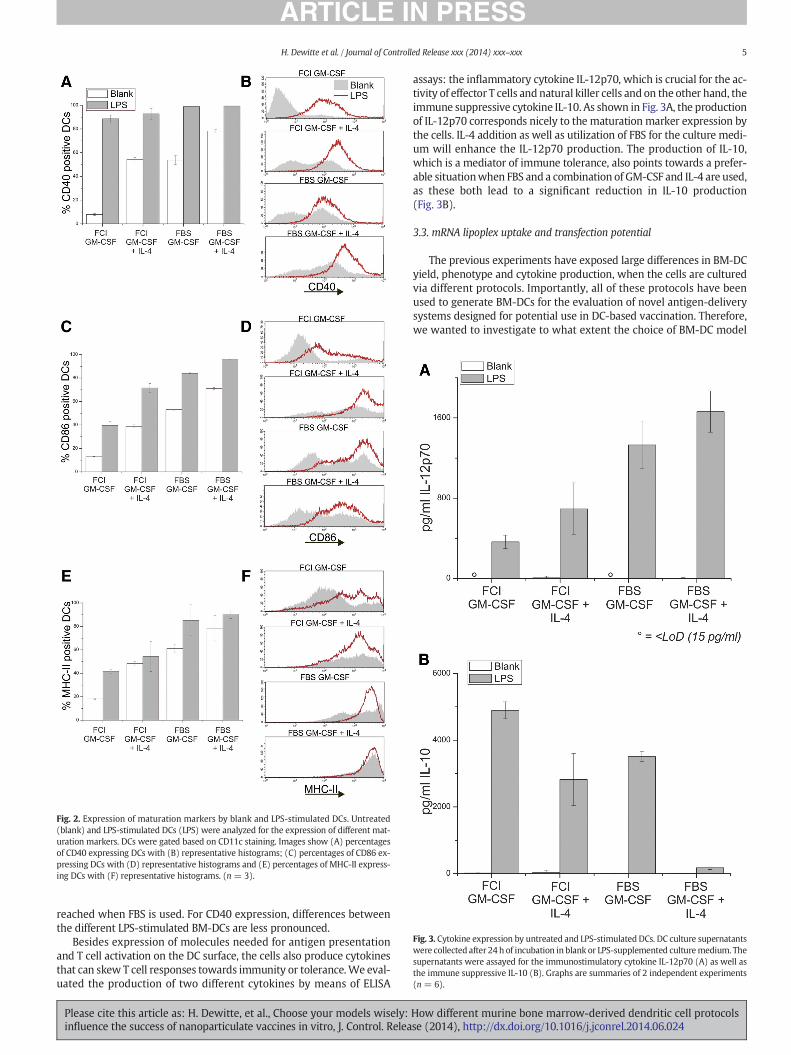
Fig. 2. Expression of maturation markers by blank and LPS-stimulated DCs. Untreated(blank) and LPS-stimulated DCs (LPS) were analyzed for the expression of different mat-uration markers. DCs were gated based on CD11c staining. Images show (A) percentagesof CD40 expressing DCs with (B) representative histograms; (C) percentages of CD86 ex-pressing DCs with (D) representative histograms and (E) percentages of MHC-II express-ing DCs with (F) representative histograms. (n = 3).
Fig. 3. Cytokine expression by untreated and LPS-stimulated DCs. DC culture supernatantswere collected after 24 h of incubation in blank or LPS-supplemented culturemedium. Thesupernatants were assayed for the immunostimulatory cytokine IL-12p70 (A) as well asthe immune suppressive IL-10 (B). Graphs are summaries of 2 independent experiments(n = 6).
5H. Dewitte et al. / Journal of Controlled Release xxx (2014) xxx–xxx
reached when FBS is used. For CD40 expression, differences betweenthe different LPS-stimulated BM-DCs are less pronounced.
Besides expression of molecules needed for antigen presentationand T cell activation on the DC surface, the cells also produce cytokinesthat can skew T cell responses towards immunity or tolerance.We eval-uated the production of two different cytokines by means of ELISA
Please cite this article as: H. Dewitte, et al., Choose your models wisely:influence the success of nanoparticulate vaccines in vitro, J. Control. Relea
assays: the inflammatory cytokine IL-12p70, which is crucial for the ac-tivity of effector T cells and natural killer cells and on the other hand, theimmune suppressive cytokine IL-10. As shown in Fig. 3A, the productionof IL-12p70 corresponds nicely to the maturation marker expression bythe cells. IL-4 addition as well as utilization of FBS for the culture medi-um will enhance the IL-12p70 production. The production of IL-10,which is a mediator of immune tolerance, also points towards a prefer-able situationwhen FBS and a combination of GM-CSF and IL-4 are used,as these both lead to a significant reduction in IL-10 production(Fig. 3B).
3.3. mRNA lipoplex uptake and transfection potential
The previous experiments have exposed large differences in BM-DCyield, phenotype and cytokine production, when the cells are culturedvia different protocols. Importantly, all of these protocols have beenused to generate BM-DCs for the evaluation of novel antigen-deliverysystems designed for potential use in DC-based vaccination. Therefore,we wanted to investigate to what extent the choice of BM-DC model
How different murine bone marrow-derived dendritic cell protocolsse (2014), http://dx.doi.org/10.1016/j.jconrel.2014.06.024
6 H. Dewitte et al. / Journal of Controlled Release xxx (2014) xxx–xxx
could influence the results obtained with particulate antigens. As amodel antigen-delivery system, we chose mRNA lipoplexes, sincemRNA is becoming increasingly investigated as a source of antigen,and this requires more of the DCs' own machinery for translation and
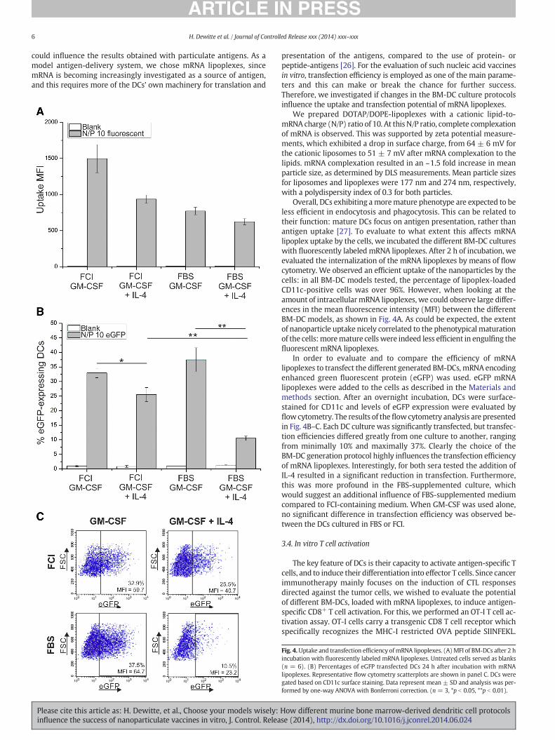
Fig. 4.Uptake and transfection efficiency ofmRNA lipoplexes. (A) MFI of BM-DCs after 2 hincubation with fluorescently labeled mRNA lipoplexes. Untreated cells served as blanks(n = 6). (B) Percentages of eGFP transfected DCs 24 h after incubation with mRNAlipoplexes. Representative flow cytometry scatterplots are shown in panel C. DCs weregated based on CD11c surface staining. Data represent mean ± SD and analysis was per-formed by one-way ANOVA with Bonferroni correction. (n = 3, *p b 0.05, **p b 0.01).
Please cite this article as: H. Dewitte, et al., Choose your models wisely:influence the success of nanoparticulate vaccines in vitro, J. Control. Relea
presentation of the antigens, compared to the use of protein- orpeptide-antigens [26]. For the evaluation of such nucleic acid vaccinesin vitro, transfection efficiency is employed as one of the main parame-ters and this can make or break the chance for further success.Therefore, we investigated if changes in the BM-DC culture protocolsinfluence the uptake and transfection potential of mRNA lipoplexes.
We prepared DOTAP/DOPE-lipoplexes with a cationic lipid-to-mRNA charge (N/P) ratio of 10. At this N/P ratio, complete complexationof mRNA is observed. This was supported by zeta potential measure-ments, which exhibited a drop in surface charge, from 64 ± 6 mV forthe cationic liposomes to 51 ± 7 mV after mRNA complexation to thelipids. mRNA complexation resulted in an ~1.5 fold increase in meanparticle size, as determined by DLS measurements. Mean particle sizesfor liposomes and lipoplexes were 177 nm and 274 nm, respectively,with a polydispersity index of 0.3 for both particles.
Overall, DCs exhibiting amoremature phenotype are expected to beless efficient in endocytosis and phagocytosis. This can be related totheir function: mature DCs focus on antigen presentation, rather thanantigen uptake [27]. To evaluate to what extent this affects mRNAlipoplex uptake by the cells, we incubated the different BM-DC cultureswith fluorescently labeled mRNA lipoplexes. After 2 h of incubation, weevaluated the internalization of the mRNA lipoplexes by means of flowcytometry. We observed an efficient uptake of the nanoparticles by thecells: in all BM-DC models tested, the percentage of lipoplex-loadedCD11c-positive cells was over 96%. However, when looking at theamount of intracellularmRNA lipoplexes, we could observe large differ-ences in the mean fluorescence intensity (MFI) between the differentBM-DC models, as shown in Fig. 4A. As could be expected, the extentof nanoparticle uptake nicely correlated to the phenotypical maturationof the cells:moremature cellswere indeed less efficient in engulfing thefluorescent mRNA lipoplexes.
In order to evaluate and to compare the efficiency of mRNAlipoplexes to transfect the different generated BM-DCs, mRNA encodingenhanced green fluorescent protein (eGFP) was used. eGFP mRNAlipoplexes were added to the cells as described in the Materials andmethods section. After an overnight incubation, DCs were surface-stained for CD11c and levels of eGFP expression were evaluated byflow cytometry. The results of theflowcytometry analysis are presentedin Fig. 4B–C. Each DC culture was significantly transfected, but transfec-tion efficiencies differed greatly from one culture to another, rangingfrom minimally 10% and maximally 37%. Clearly the choice of theBM-DC generation protocol highly influences the transfection efficiencyof mRNA lipoplexes. Interestingly, for both sera tested the addition ofIL-4 resulted in a significant reduction in transfection. Furthermore,this was more profound in the FBS-supplemented culture, whichwould suggest an additional influence of FBS-supplemented mediumcompared to FCI-containing medium. When GM-CSF was used alone,no significant difference in transfection efficiency was observed be-tween the DCs cultured in FBS or FCI.
3.4. In vitro T cell activation
The key feature of DCs is their capacity to activate antigen-specific Tcells, and to induce their differentiation into effector T cells. Since cancerimmunotherapy mainly focuses on the induction of CTL responsesdirected against the tumor cells, we wished to evaluate the potentialof different BM-DCs, loaded with mRNA lipoplexes, to induce antigen-specific CD8+ T cell activation. For this, we performed an OT-I T cell ac-tivation assay. OT-I cells carry a transgenic CD8 T cell receptor whichspecifically recognizes the MHC-I restricted OVA peptide SIINFEKL.
How different murine bone marrow-derived dendritic cell protocolsse (2014), http://dx.doi.org/10.1016/j.jconrel.2014.06.024
7H. Dewitte et al. / Journal of Controlled Release xxx (2014) xxx–xxx
Please cite this article as: H. Dewitte, et al., Choose your models wisely:influence the success of nanoparticulate vaccines in vitro, J. Control. Relea
Therefore, if these OT-I cells are stimulated by mature DCs that presentSIINFEKL in the context of MHC-I, these cells will be activated. Thisactivation is accompanied by an increased expression of CD25 on theT cell surface.
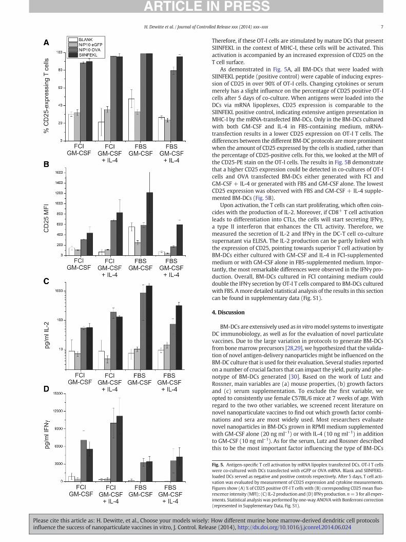
As demonstrated in Fig. 5A, all BM-DCs that were loaded withSIINFEKL peptide (positive control) were capable of inducing expres-sion of CD25 in over 90% of OT-I cells. Changing cytokines or serummerely has a slight influence on the percentage of CD25 positive OT-Icells after 5 days of co-culture. When antigens were loaded into theDCs via mRNA lipoplexes, CD25 expression is comparable to theSIINFEKL positive control, indicating extensive antigen presentation inMHC-I by the mRNA-transfected BM-DCs. Only in the BM-DCs culturedwith both GM-CSF and IL-4 in FBS-containing medium, mRNA-transfection results in a lower CD25 expression on OT-I T cells. Thedifferences between the different BM-DC protocols aremore prominentwhen the amount of CD25 expressed by the cells is studied, rather thanthe percentage of CD25-positive cells. For this, we looked at the MFI ofthe CD25-PE stain on the OT-I cells. The results in Fig. 5B demonstratethat a higher CD25 expression could be detected in co-cultures of OT-Icells and OVA transfected BM-DCs either generated with FCI andGM-CSF + IL-4 or generated with FBS and GM-CSF alone. The lowestCD25 expression was observed with FBS and GM-CSF + IL-4 supple-mented BM-DCs (Fig. 5B).
Upon activation, the T cells can start proliferating, which often coin-cides with the production of IL-2. Moreover, if CD8+ T cell activationleads to differentiation into CTLs, the cells will start secreting IFNγ,a type II interferon that enhances the CTL activity. Therefore, wemeasured the secretion of IL-2 and IFNγ in the DC-T cell co-culturesupernatant via ELISA. The IL-2 production can be partly linked withthe expression of CD25, pointing towards superior T cell activation byBM-DCs either cultured with GM-CSF and IL-4 in FCI-supplementedmedium or with GM-CSF alone in FBS-supplemented medium. Impor-tantly, the most remarkable differences were observed in the IFNγ pro-duction. Overall, BM-DCs cultured in FCI containing medium coulddouble the IFNγ secretion by OT-I T cells compared to BM-DCs culturedwith FBS. Amore detailed statistical analysis of the results in this sectioncan be found in supplementary data (Fig. S1).
4. Discussion
BM-DCs are extensively used as in vitromodel systems to investigateDC immunobiology, as well as for the evaluation of novel particulatevaccines. Due to the large variation in protocols to generate BM-DCsfrom bonemarrow precursors [28,29], we hypothesized that the valida-tion of novel antigen-delivery nanoparticles might be influenced on theBM-DC culture that is used for their evaluation. Several studies reportedon a number of crucial factors that can impact the yield, purity and phe-notype of BM-DCs generated [30]. Based on the work of Lutz andRossner, main variables are (a) mouse properties, (b) growth factorsand (c) serum supplementation. To exclude the first variable, weopted to consistently use female C57BL/6 mice at 7 weeks of age. Withregard to the two other variables, we screened recent literature onnovel nanoparticulate vaccines to find out which growth factor combi-nations and sera are most widely used. Most researchers evaluatenovel nanoparticles in BM-DCs grown in RPMI medium supplementedwith GM-CSF alone (20 ng ml–1) or with IL-4 (10 ng ml–1) in additionto GM-CSF (10 ng ml–1). As for the serum, Lutz and Rossner describedthis to be the most important factor influencing the type of BM-DCs
Fig. 5. Antigen-specific T cell activation by mRNA lipoplex transfected DCs. OT-I T cellswere co-cultured with DCs transfected with eGFP or OVA mRNA. Blank and SIINFEKL-loaded DCs served as negative and positive controls respectively. After 5 days, T cell acti-vation was evaluated by measurement of CD25 expression and cytokine measurements.Figures show (A) % of CD25 positive OT-I T cells with (B) corresponding CD25mean fluo-rescence intensity (MFI); (C) IL-2 production and (D) IFNγ production. n=3 for all exper-iments. Statistical analysis was performed by one-way ANOVAwith Bonferroni correction(represented in Supplementary Data, Fig. S1).
How different murine bone marrow-derived dendritic cell protocolsse (2014), http://dx.doi.org/10.1016/j.jconrel.2014.06.024

8 H. Dewitte et al. / Journal of Controlled Release xxx (2014) xxx–xxx
generated. Therefore we chose to work with two different types of bo-vine serum: FBS and FCI.
After 6 days of culturing red blood cell-depleted murine bone mar-row cells in the different cell culture media, the percentage of CD11c+
DCs ranges from 39 to 80%. The highest BM-DC purity can be reachedwhen FBS serum and GM-CSF alone are used. In accordance with theprevious reports, addition of IL-4 to the medium reduces the yield andpurity of the BM-DCs [30]. It should be noted that other factors, suchas the duration of the BM-DC culture, or the implementation of addi-tional and often quite complex purification steps, can result in cultureswith larger percentages of CD11c+ cells [10,28,31]. Often, cells growingadherent to the culture dishes are excluded in order to increase BM-DCpurity. In our hands, however, for all culture media tested, the adherentcell fraction contained a substantial portion of CD11c-expressingBM-DCs. This confirms the previous results obtained by Li and Lu, whoshowed that there is no reason to discard adherent cells in BM-DC cul-tures as junk cells [32]. Overall, practically all BM-DC protocols will fi-nally result in a cell population containing a (large) fraction of CD11c+
BM-DCs as well as a fraction of non-DCs. These two subpopulationsare expected to behave differentlywith regard to e.g. endocytotic capac-ity, maturation marker expression and cytokine production. Therefore,in order to assess the impact of novel nano- and micromaterials onDCs in vitro, it is important to selectively investigate the response ofCD11c+ cells, rather than the total cell population. Assays where no fur-ther selection or gating of CD11c-expressing cells is possible (e.g. cellcounts, many cytotoxicity assays, DC-T cell co-cultures, other assaysperformed on the total population), can be impacted by these large var-iations in BM-DC purity. This should be taken into account in the designand set-up of new experiments.
Substantial differences in surface marker expression can also existwithin the CD11c-positive population. This was made obvious byperforming antibody staining against maturation markers CD40, CD86and MHC-II, where we demonstrated the presence of both immatureas well as mature BM-DCs in all cultures. Importantly, addition of IL-4increases the proportion ofmature DCs. This can be explained by the in-duction of “spontaneous” maturation of generated BM-DCs, which isknown to be enhanced by the presence of IL-4 [28,33,34]. In addition,replacing FBS by FCI induces a significant reduction ofmaturationmark-er expression by the BM-DCs. In the study of Lutz et al., high serumlevels of the liver enzymes glutamic oxaloacetic transaminase (GOT)and lactate dehydrogenase (LDH) have been correlated with high per-centages of mature cells. For the batches of FBS and FCI we used, thiscorrelation was not observed: the FCI used contained high levels ofboth liver enzymes, whereas merely a small fraction of the DCs exhibit-ed amature phenotype. Even though the effect of the serum compoundis prominent, due to the complexity of these bovine products, we couldnot pinpoint the exact cause of the maturation-differences.
In nanoparticle research, a first step towards success is the efficientuptake of the nanomaterials by the target cells. In case of DCs, their in-ternalization capacity is closely related to their maturation status: al-though mature DCs are still capable of performing receptor-mediatedendocytosis, a specific endocytosis as well as phagocytosis andmacropinocytosis are markedly reduced in mature DCs compared totheir immature counterparts [27]. This was also evidenced by themRNA lipoplex uptake experiment, showing less extensive mRNAlipoplex uptake by BM-DCs that exhibit a more mature phenotype.Therefore, ideally, antigen-containing nanoparticleswould be evaluatedin immature BM-DCs. However, a striking observation was the reducedmaturation capacity of themore immature BM-DC subsets. For example,BM-DCs culturedwith FCI and GM-CSF alone, exhibit the lowest portionof mature DCs in untreated samples, but upon TLR4 ligation maximally40% of the BM-DCs will upregulate CD86 and MHC-II. In contrast, wellover 80% of BM-DCs generated in the presence of FBS medium, willupregulate these markers upon LPS stimulation. This reduced respon-siveness tomaturation stimuli is nicely confirmed by cytokinemeasure-ments, showing that these specific BM-DCs also produce low levels of
Please cite this article as: H. Dewitte, et al., Choose your models wisely:influence the success of nanoparticulate vaccines in vitro, J. Control. Relea
immune stimulatory IL-12p70 and high levels of the immune suppres-sive IL-10. Such phenotypical properties are expected to impact the im-mune responses that can be induced by these specific DCs. In addition, itis important to consider that for DC vaccines to be effective, efficientantigen-presentation by fully mature DCs is required. Since differentreports havemade clear that co-formulation of antigens andmaturationstimuli will result in superior immune responses, the purpose ofnanoparticulate vaccines has shifted from mere antigen delivery, todual-modality particles that should simultaneously load DCs with anti-gens, and induce maturation of DCs [35,36]. Therefore, immune stimu-lants such as the TLR ligands CpG and poly(I:C) have beenincorporated into nanoparticulate vaccines [37]. As these dual-modality particles aim to induce complete maturation, it would un-doubtedly be disadvantageous to evaluate them in a maturation-resistant BM-DCmodel. Therefore, a careful choice and characterizationof the BM-DC model for the intended purpose is required.
In addition to immunological properties, we wished to assess towhat extent novel nanoparticulate vaccines can lead to different resultswhen tested in one BM-DC culture versus another. For this, we chose toworkwithmRNA lipoplexes, sincemRNA is an increasingly investigatedsource of antigen in novel DC vaccines [26]. When DCs are loaded withnucleic acid sequences encoding tumor antigens, the transfection effi-ciency is used as a critical measure for the amount of antigen delivered,and hence the main criterion to evaluate new nanoparticulate vaccines.An evaluation of the transfection efficiency of mRNA lipoplexes in our 4different models consistently, revealed large differences, ranging from10% to 37% mRNA expressing BM-DCs, even though the exact samenanoparticles were used. If transfection efficiency is the main parame-ter, it is now clear that the model BM-DC used for transfection experi-ments is a confounding factor. Do we then use the right means ofscoring novel transfection-based particulate vaccines? First of all, itshould, at all times, be kept in mind that the final end-point of the vac-cine is to elicit antigen-specific T cell responses. And this is probably nota question of numbers. As reviewed byGilboa andVieweg, high efficien-cy transfection of DCs with antigen does not necessarily correlate withimproved immune stimulatory effects [38]. In addition, it was previous-ly suggested that no analytical method can compare to the sensitivity ofT cells to detect presented antigens [39,40]. Therefore, as a concludingexperiment, we evaluated to which extent the observed transfectionefficiencies could lead to activation of antigen-specific CD8+ T cellsin vitro. After co-culturing naïve OVA-specific CD8+ T cells with mRNAlipoplex-loaded BM-DCs, significant antigen-specific T cell activationcould be observed in all models. However, the differences observed inthe extent of T cell activation point towards influences of both pheno-typical parameters aswell as variations in transfection efficiency. For in-stance, BM-DCs grown in FCI and GM-CSF-supplemented mediumshowed low expression of molecules involved in antigen presentation(MHC-II) and T cell activation (CD86 and IL-12p70). As a result, evenafter efficient loadingwith antigen-mRNA,merely low T cell stimulationcould be detected. A similar reasoning is valid for phenotypically supe-rior BM-DCs (e.g. BM-DCs resulting from culture with FBS in the pres-ence of IL-4), where a markedly low transfection efficiency is thelikely cause for the deficient activation of T cells.
To conclude, in vitro data obtained with novel nanoparticles are notonly the result of the nanoparticle's capacity to load DCs with antigenicmaterial. Although BM-DCs are routinely used as amodel for in vivoDCs,they should always be considered as simplified models for a complexin vivo situation. By screening a limited number of parameters, wewere able to demonstrate extensive differences in BM-DC yield and im-munological properties.Moreover,we showed that the BM-DCmodel inwhich novel nanoparticles are tested, acts as an important confoundingfactor in both transfection efficiency and T cell activation assays. Thisnot only makes it hard to evaluate the potential of novel nanoparticlesfor DC vaccination purposes, but also renders it difficult to comparethe results obtained in different research groups using differentBM-DC models. We wish to stress that our aim was not to identify an
How different murine bone marrow-derived dendritic cell protocolsse (2014), http://dx.doi.org/10.1016/j.jconrel.2014.06.024

9H. Dewitte et al. / Journal of Controlled Release xxx (2014) xxx–xxx
optimal BM-DC protocol, but to expose the bias of the model on thefunctional outcome of the nanoparticle. Therefore, we propose to:(1) include a thorough immunological characterization of the BM-DCsmodel used; (2) in addition to transfection efficiency, always evaluatefunctional end-points, such as T cell activation. Only if these two criteriaare met, can the impact of the model's confounding effect be assessed,and can the true value of a novel particulate vaccine be revealed.
Supplementary data to this article can be found online at http://dx.doi.org/10.1016/j.jconrel.2014.06.024.
Acknowledgments
The authors would like to thank Sandra Van Lint and LauraWayteckfor their assistance with the OT-I proliferation assay. They further wishto thank Carlo Heirman, Elsy Vaeremans and Petra Roman for theirhelp with the mRNA production. Heleen Dewitte is a doctoral fellowof the Institute for the Promotion of Innovation through Science andTechnology in Flanders, Belgium (IWT-Vlaanderen). Ine Lentacker andKarine Breckpot are postdoctoral fellows of the Research Foundation-Flanders, Belgium (FWO-Vlaanderen). This project was funded throughthe FWO grant G016513N.
References
[1] R.M. Steinman, Z.A. Cohn, Identification of a novel cell type in peripheral lymphoidorgans of mice, J. Exp. Med. 137 (1973) 1142–1162.
[2] J. Banchereau, R.M. Steinman, Dendritic cells and the control of immunity, Nature392 (1998) 245–252.
[3] J.I. Mayordomo, T. Zorina, W.J. Storkus, L. Zitvogel, C. Celluzzi, L.D. Falo, C.J. Melief, S.T. Ildstad, W.M. Kast, A.B. Deleo, et al., Bone marrow-derived dendritic cells pulsedwith synthetic tumour peptides elicit protective and therapeutic antitumour immu-nity, Nat. Med. 1 (1995) 1297–1302.
[4] W. Lu, L.C. Arraes, W.T. Ferreira, J.M. Andrieu, Therapeutic dendritic-cell vaccine forchronic HIV-1 infection, Nat. Med. 10 (2004) 1359–1365.
[5] C.S. Higano, P.F. Schellhammer, E.J. Small, P.A. Burch, J. Nemunaitis, L. Yuh, N.Provost, M.W. Frohlich, Integrated data from 2 randomized, double-blind,placebo-controlled, phase 3 trials of active cellular immunotherapy withsipuleucel-T in advanced prostate cancer, Cancer 115 (2009) 3670–3679.
[6] E.J. Small, P.F. Schellhammer, C.S. Higano, C.H. Redfern, J.J. Nemunaitis, F.H. Valone, S.S. Verjee, L.A. Jones, R.M. Hershberg, Placebo-controlled phase III trial of immuno-logic therapy with sipuleucel-T (APC8015) in patients with metastatic, asymptom-atic hormone refractory prostate cancer, J. Clin. Oncol. 24 (2006) 3089–3094.
[7] C. Brocks, H. Graefe, H. Frenzel, R. Pries, B. Wollenberg, Isolation of human myeloiddendritic cells from tumor tissue and peripheral blood, In Vivo 20 (2006) 239–242.
[8] V. Pena-Cruz, S. Ito, M. Oukka, K. Yoneda, C.C. Dascher, F. Von Lichtenberg, M. Sugita,Extraction of human Langerhans cells: a method for isolation of epidermis-residentdendritic cells, J. Immunol. Methods 255 (2001) 83–91.
[9] C.E. Jacome-Galarza, S.K. Lee, J.A. Lorenzo, H.L. Aguila, Identification, characteriza-tion, and isolation of a common progenitor for osteoclasts, macrophages, anddendritic cells from murine bone marrow and periphery, J. Bone Miner. Res. 28(2013) 1203–1213.
[10] K. Inaba, M. Inaba, N. Romani, H. Aya, M. Deguchi, S. Ikehara, S. Muramatsu, R.M.Steinman, Generation of large numbers of dendritic cells from mouse bone marrowcultures supplemented with granulocyte/macrophage colony-stimulating factor, J.Exp. Med. 176 (1992) 1693–1702.
[11] N. Romani, S. Gruner, D. Brang, E. Kampgen, A. Lenz, B. Trockenbacher, G.Konwalinka, P.O. Fritsch, R.M. Steinman, G. Schuler, Proliferating dendritic cell pro-genitors in human blood, J. Exp. Med. 180 (1994) 83–93.
[12] S. Tuyaerts, J.L. Aerts, J. Corthals, B. Neyns, C. Heirman, K. Breckpot, K. Thielemans, A.Bonehill, Current approaches in dendritic cell generation and future implications forcancer immunotherapy, Cancer Immunol. Immunother. 56 (2007) 1513–1537.
[13] M.B. Lutz, S. Rossner, Factors influencing the generation of murine dendritic cellsfrom bone marrow: the special role of fetal calf serum, Immunobiology 212(2007) 855–862.
[14] K. Shortman, S.H. Naik, Steady-state and inflammatory dendritic-cell development,Nat. Rev. Immunol. 7 (2007) 19–30.
Please cite this article as: H. Dewitte, et al., Choose your models wisely:influence the success of nanoparticulate vaccines in vitro, J. Control. Relea
[15] J.A. Villadangos, P. Schnorrer, Intrinsic and cooperative antigen-presenting functionsof dendritic-cell subsets in vivo, Nat. Rev. Immunol. 7 (2007) 543–555.
[16] A.T. Satpathy, X.D. Wu, J.C. Albring, K.M. Murphy, Re(de)fining the dendritic celllineage, Nat. Immunol. 13 (2012) 1145–1154.
[17] K. Brasel, T. De Smedt, J.L. Smith, C.R.Maliszewski, Generation ofmurine dendritic cellsfrom flt3-ligand-supplemented bone marrow cultures, Blood 96 (2000) 3029–3039.
[18] Y. Xu, Y. Zhan, A.M. Lew, S.H. Naik, M.H. Kershaw, Differential development ofmurine dendritic cells by GM-CSF versus Flt3 ligand has implications for inflamma-tion and trafficking, J. Immunol. 179 (2007) 7577–7584.
[19] M. Menges, T. Baumeister, S. Rossner, P. Stoitzner, N. Romani, A. Gessner, M.B. Lutz,IL-4 supports the generation of a dendritic cell subset from murine bone marrowwith altered endocytosis capacity, J. Leukoc. Biol. 77 (2005) 535–543.
[20] M.L. De Temmerman, H. Dewitte, R.E. Vandenbroucke, B. Lucas, C. Libert, J.Demeester, S.C. De Smedt, I. Lentacker, J. Rejman, mRNA-lipoplex loadedmicrobubble contrast agents for ultrasound-assisted transfection of dendritic cells,Biomaterials 32 (2011) 9128–9135.
[21] Z.P. Zhang, Y.J. Guo, S.S. Feng, Nanoimmunotherapy: application of nanotechnologyfor sustained and targeted delivery of antigens to dendritic cells, Nanomedicine-Uk7 (2012) 1–4.
[22] W. De Haes, J. Rejman, C. Pollard, C. Merlin, M. Vekemans, E. Florence, S.C. De Smedt,J. Grooten, G. Vanham, S. De Koker, E. Van Gulck, Lipoplexes carrying mRNAencoding Gag protein modulate dendritic cells to stimulate HIV-specific immuneresponses, Nanomedicine-Uk 8 (2013) 77–87.
[23] S. Van Lint, C. Heirman, K. Thielemans, K. Breckpot, mRNA: from a chemicalblueprint for protein production to an off-the-shelf therapeutic, Hum. Vaccin.Immunother. 9 (2013).
[24] S. Van Meirvenne, L. Straetman, C. Heirman, M. Dullaers, C. De Greef, V. VanTendeloo, K. Thielemans, Efficient genetic modification of murine dendritic cellsby electroporation with mRNA, Cancer Gene Ther. 9 (2002) 787–797.
[25] M.B. Lutz, G. Schuler, Immature, semi-mature and fully mature dendritic cells:which signals induce tolerance or immunity? Trends Immunol. 23 (2002) 445–449.
[26] S. Van Lint, K. Thielemans, K. Breckpot, mRNA: delivering an antitumor message?Immunotherapy 3 (2011) 605–607.
[27] C.D. Platt, J.K. Ma, C. Chalouni, M. Ebersold, H. Bou-Reslan, R.A.D. Carano, I. Mellman,L. Delamarre, Mature dendritic cells use endocytic receptors to capture and presentantigens, Proc. Natl. Acad. Sci. U. S. A. 107 (2010) 4287–4292.
[28] M.B. Lutz, IL-3 in dendritic cell development and function: a comparison with GM-CSF and IL-4, Immunobiology 209 (2004) 79–87.
[29] H.C. O'Neill, H.L. Wilson, Limitations with in vitro production of dendritic cells usingcytokines, J. Leukoc. Biol. 75 (2004) 600–603.
[30] J.W. Wells, D. Darling, F. Farzaneh, J. Galea-Lauri, Influence of interleukin-4 on thephenotype and function of bone marrow-derived murine dendritic cells generatedunder serum-free conditions, Scand. J. Immunol. 61 (2005) 251–259.
[31] T. Kalantari, E. Kamali-Sarvestani, G.X. Zhang, F. Safavi, E. Lauretti, M.E. Khedmati, A.Rostami, Generation of large numbers of highly purified dendritic cells from bonemarrow progenitor cells after co-culture with syngeneic murine splenocytes, Exp.Mol. Pathol. 94 (2013) 336–342.
[32] G.B. Li, G.X. Lu, Adherent cells in granulocyte-macrophage colony-stimulatingfactor-induced bone marrow-derived dendritic cell culture system are qualifieddendritic cells, Cell. Immunol. 264 (2010) 4–6.
[33] M.B. Lutz, M. Schnare, M. Menges, S. Rossner, M. Rollinghoff, G. Schuler, A. Gessner,Differential functions of IL-4 receptor types I and II for dendritic cell maturation andIL-12 production and their dependency on GM-CSF, J. Immunol. 169 (2002)3574–3580.
[34] Y. Yamaguchi, H. Tsumura, M. Miwa, K. Inaba, Contrasting effects of TGF-beta 1 andTNF-alpha on the development of dendritic cells from progenitors in mouse bonemarrow, Stem Cells 15 (1997) 144–153.
[35] E. Schlosser, M. Mueller, S. Fischer, S. Basta, D.H. Busch, B. Gander, M. Groettrup, TLRligands and antigen need to be coencapsulated into the same biodegradable micro-sphere for the generation of potent cytotoxic T lymphocyte responses, Vaccine 26(2008) 1626–1637.
[36] K. Zaks, M. Jordan, A. Guth, K. Sellins, R. Kedl, A. Izzo, C. Bosio, S. Dow, Efficientimmunization and cross-priming by vaccine adjuvants containing TLR3 or TLR9agonists complexed to cationic liposomes, J. Immunol. 176 (2006) 7335–7345.
[37] Y.R. Lee, Y.H. Lee, S.A. Im, I.H. Yang, G.W. Ahn, K. Kim, C.K. Lee, Biodegradablenanoparticles containing TLR3 or TLR9 agonists together with Antigen enhanceMHC-restrictedpresentation of the antigen, Arch. Pharm. Res. 33 (2010) 1859–1866.
[38] E. Gilboa, J. Vieweg, Cancer immunotherapy with mRNA-transfected dendritic cells,Immunol. Rev. 199 (2004) 251–263.
[39] F. Grunebach, M.R. Muller, A. Nencioni, P. Brossart, Delivery of tumor-derived RNAfor the induction of cytotoxic T-lymphocytes, Gene Ther. 10 (2003) 367–374.
[40] S.K. Nair, D. Boczkowski,M.Morse, R.I. Cumming, H.K. Lyerly, E. Gilboa, Induction of pri-mary carcinoembryonic antigen (CEA)-specific cytotoxic T lymphocytes in vitro usinghuman dendritic cells transfected with RNA, Nat. Biotechnol. 16 (1998) 364–369.
How different murine bone marrow-derived dendritic cell protocolsse (2014), http://dx.doi.org/10.1016/j.jconrel.2014.06.024