chpt 20 kwast lecture sp12 ked - university of illinois
TRANSCRIPT

© 2012 Pearson Education, Inc.
PowerPoint® Lecture Presentations prepared by Jason LaPres Lone Star College—North Harris
20 The Heart
NOTE: Presentations extensively modi6ied for use in MCB 244 & 246 at the University of Illinois by Drs. Kwast & Brown (2011-‐2012)

© 2012 Pearson Education, Inc.
Chapter 20 Learning Objectives
1. Describe the anatomy of the heart and vasculature including major blood vessels, chambers, and valves.
2. Describe excitation-contraction coupling in cardiac tissue including electrical and ionic events.
3. Describe the physical events during the cardiac cycle including atrial and ventricular systole and diastole.
4. Define cardiac output, describe factors that affect stroke volume, heart rate and contractility and how these change during physical activity.
© 2012 Pearson Education, Inc.
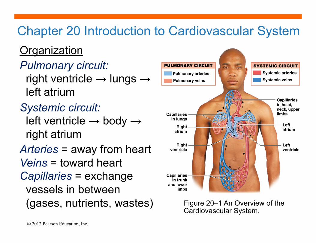
Chapter 20 Introduction to Cardiovascular System Organization
Pulmonary circuit: right ventricle → lungs → left atrium
Systemic circuit: left ventricle → body → right atrium
Arteries = away from heart Veins = toward heart Capillaries = exchange vessels in between (gases, nutrients, wastes) Figure 20–1 An Overview of the
Cardiovascular System.

© 2012 Pearson Education, Inc.
20-1 Heart Anatomy: Thoracic Location
• Heart is fist sized, < 1 lb., beats, ~100,000 times/day moving 8000 liters of blood/day
Figure 20–2a The Location of the Heart in the Thoracic Cavity
• Lies left of midline, between 2nd rib and 5th intercostal space, posterior to sternum, in the pericardial cavity of the mediastinum (the region between the two pleural cavities, which also contains the great vessels, thymus, esophagus and trachea).

© 2012 Pearson Education, Inc.
20-1 Heart Anatomy: Thoracic Location
Figure 20–2b The Location of the Heart in the Thoracic Cavity: The heart is surrounded by the pericardial sac, which consists of dense
network of collagen fibers that stabilizes the position of the heart (and major vessels) within the mediastinum.
© 2012 Pearson Education, Inc.
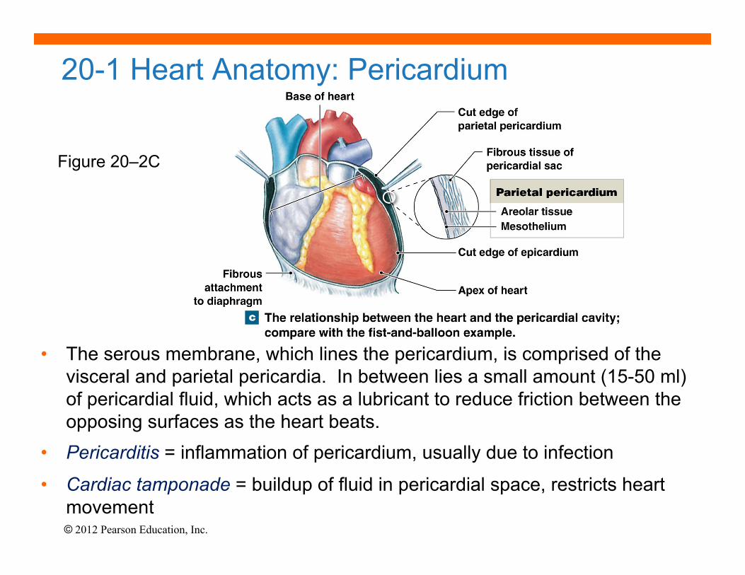
20-1 Heart Anatomy: Pericardium
• The serous membrane, which lines the pericardium, is comprised of the visceral and parietal pericardia. In between lies a small amount (15-50 ml) of pericardial fluid, which acts as a lubricant to reduce friction between the opposing surfaces as the heart beats.
• Pericarditis = inflammation of pericardium, usually due to infection
• Cardiac tamponade = buildup of fluid in pericardial space, restricts heart movement
Figure 20–2C
© 2012 Pearson Education, Inc.
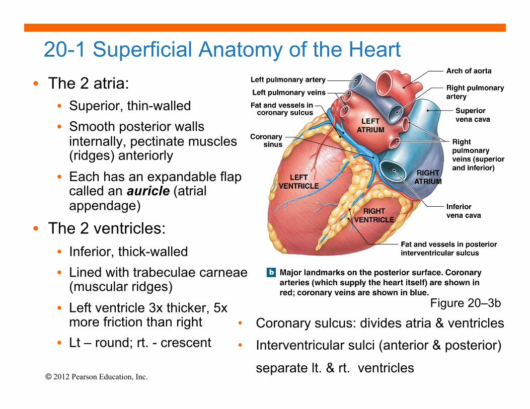
• The 2 atria: • Superior, thin-walled • Smooth posterior walls
internally, pectinate muscles (ridges) anteriorly
• Each has an expandable flap called an auricle (atrial appendage)
• The 2 ventricles: • Inferior, thick-walled • Lined with trabeculae carneae
(muscular ridges) • Left ventricle 3x thicker, 5x
more friction than right • Lt – round; rt. - crescent
20-1 Superficial Anatomy of the Heart
• Coronary sulcus: divides atria & ventricles • Interventricular sulci (anterior & posterior)
separate lt. & rt. ventricles
Figure 20–3b
© 2012 Pearson Education, Inc.
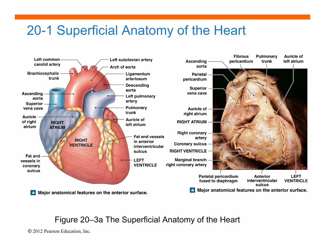
20-1 Superficial Anatomy of the Heart
Figure 20–3a The Superficial Anatomy of the Heart
© 2012 Pearson Education, Inc.
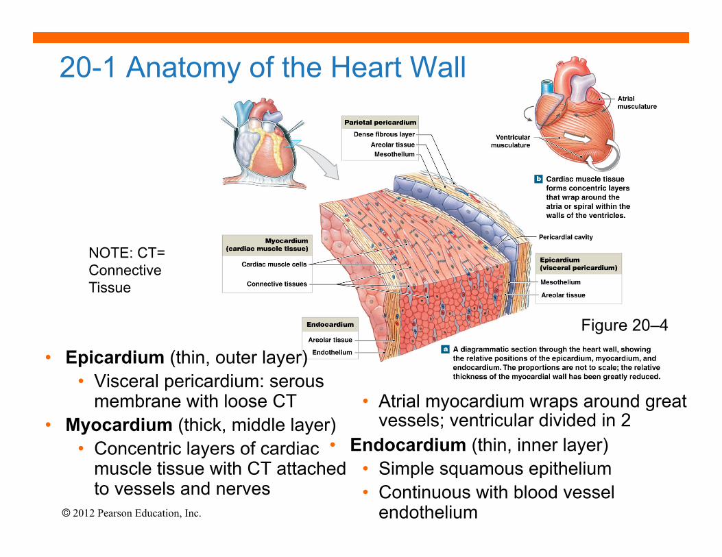
NOTE: CT= Connective Tissue
Figure 20–4
20-1 Anatomy of the Heart Wall
• Epicardium (thin, outer layer) • Visceral pericardium: serous
membrane with loose CT • Myocardium (thick, middle layer)
• Concentric layers of cardiac muscle tissue with CT attached to vessels and nerves
• Atrial myocardium wraps around great vessels; ventricular divided in 2
• Endocardium (thin, inner layer) • Simple squamous epithelium • Continuous with blood vessel
endothelium

© 2012 Pearson Education, Inc.
• Muscle cells = cardiocytes • actin & myosin sliding filaments, but
small w/ single nucleus • rich in mitochondria • Cells connected by intercalated discs
= desmosomes + gap junctions • Propagate action potential and convey
timing & force of contraction • Contractions are all or none; longer
contraction phase than skeletal muscle
20-1 Anatomy of Cardiac Muscle
Figure 20–5

© 2012 Pearson Education, Inc.
20-1 Comparison of Cardiac and Skeletal Muscle Characteristics

© 2012 Pearson Education, Inc.
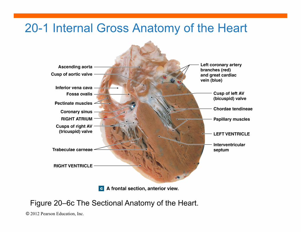
20-1 Internal Anatomy of the Heart: Septa & Atrioventricular Valves
• Interatrial septum: separates atria • Interventricular septum: separates
ventricles
• Atrioventricular (AV) valves: • Connect rt. atrium to rt. ventricle
(tricuspid) and lt. atrium to lt. ventricle (bicuspid or mitral)
• Permit one-way blood flow: atria → ventricles
• Cusps attached to chordae tendineae from papillary muscles on ventricle wall
• Papillary muscles prevent cusps from swinging into atria; during ventricular contraction pressure closes valves
Figure 20–6a&b
© 2012 Pearson Education, Inc.
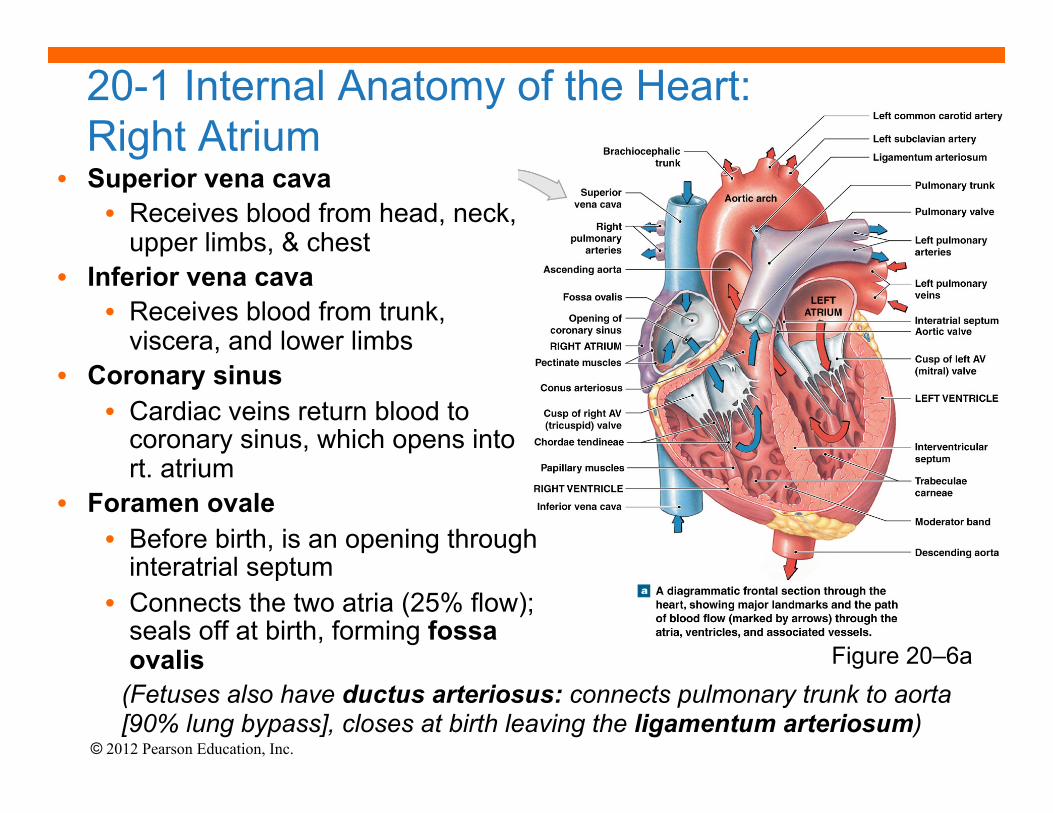
• Superior vena cava • Receives blood from head, neck,
upper limbs, & chest • Inferior vena cava
• Receives blood from trunk, viscera, and lower limbs
• Coronary sinus • Cardiac veins return blood to
coronary sinus, which opens into rt. atrium
• Foramen ovale • Before birth, is an opening through
interatrial septum • Connects the two atria (25% flow);
seals off at birth, forming fossa ovalis
20-1 Internal Anatomy of the Heart: Right Atrium
(Fetuses also have ductus arteriosus: connects pulmonary trunk to aorta [90% lung bypass], closes at birth leaving the ligamentum arteriosum)
Figure 20–6a
© 2012 Pearson Education, Inc.
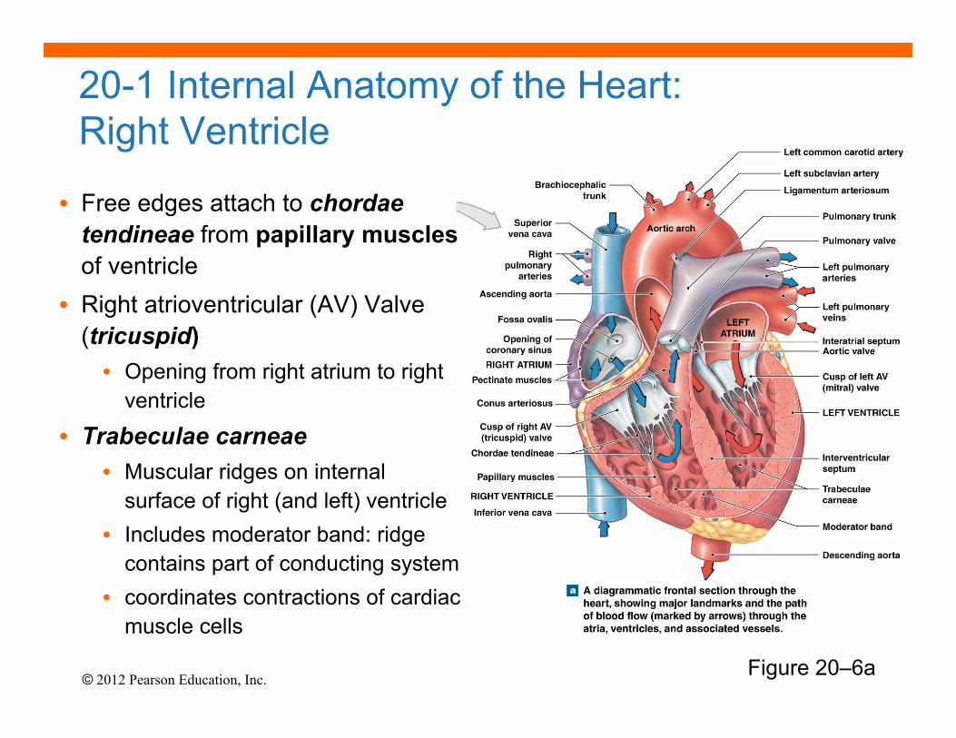
20-1 Internal Anatomy of the Heart: Right Ventricle
Figure 20–6a
• Free edges attach to chordae tendineae from papillary muscles of ventricle
• Right atrioventricular (AV) Valve (tricuspid) • Opening from right atrium to right
ventricle
• Trabeculae carneae • Muscular ridges on internal
surface of right (and left) ventricle • Includes moderator band: ridge
contains part of conducting system • coordinates contractions of cardiac
muscle cells
© 2012 Pearson Education, Inc.
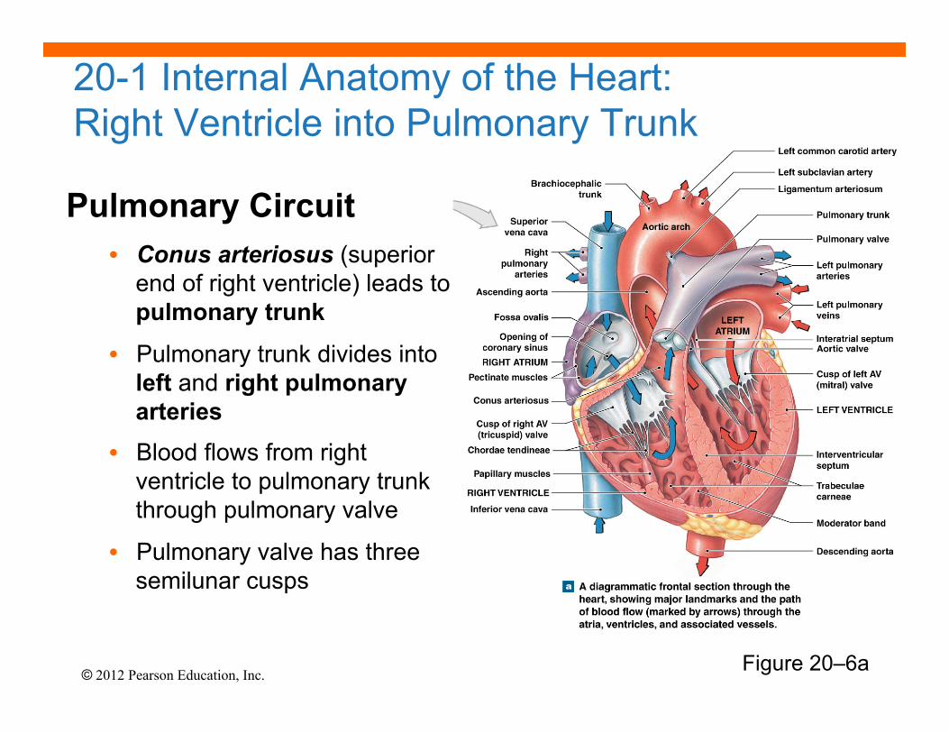
Pulmonary Circuit • Conus arteriosus (superior
end of right ventricle) leads to pulmonary trunk
• Pulmonary trunk divides into left and right pulmonary arteries
• Blood flows from right ventricle to pulmonary trunk through pulmonary valve
• Pulmonary valve has three semilunar cusps
Figure 20–6a
20-1 Internal Anatomy of the Heart: Right Ventricle into Pulmonary Trunk

© 2012 Pearson Education, Inc.
The Left Atrium § Lt. & rt. pulmonary veins
deliver oxygenated blood to left atrium
§ Passes to left ventricle through left AV (bicuspid or mitral) valve
The Left Ventricle § Holds same volume as right
but thicker and more powerful muscle
§ No moderator band
20-1 Anatomy of the Heart: Left Atrium and Ventricle
Figure 20–6a
Systemic Circulation Blood leaves left ventricle through aortic valve into ascending aorta (aortic arch) and becomes descending aorta
© 2012 Pearson Education, Inc.
20-1 Internal Gross Anatomy of the Heart
Figure 20–6c The Sectional Anatomy of the Heart.

© 2012 Pearson Education, Inc.
• Right ventricle wall is thinner and develops less pressure than left ventricle (why?--lungs are close to heart and pulmonary vessels are short and wide)
• Left ventricle must produce 4-6x more pressure than right
• When left ventricle contracts, distance between base & apex decreases as well as diameter
• Contraction and bulging of the round left ventricle into the crescent-shaped right ventricle helps eject blood from right ventricle as well (see 20-7b)
20-1 Internal Anatomy of the Heart: Comparison of Right & Left Ventricles
Figure 20–7 Structural Differences between the Left and Right Ventricles
© 2012 Pearson Education, Inc.
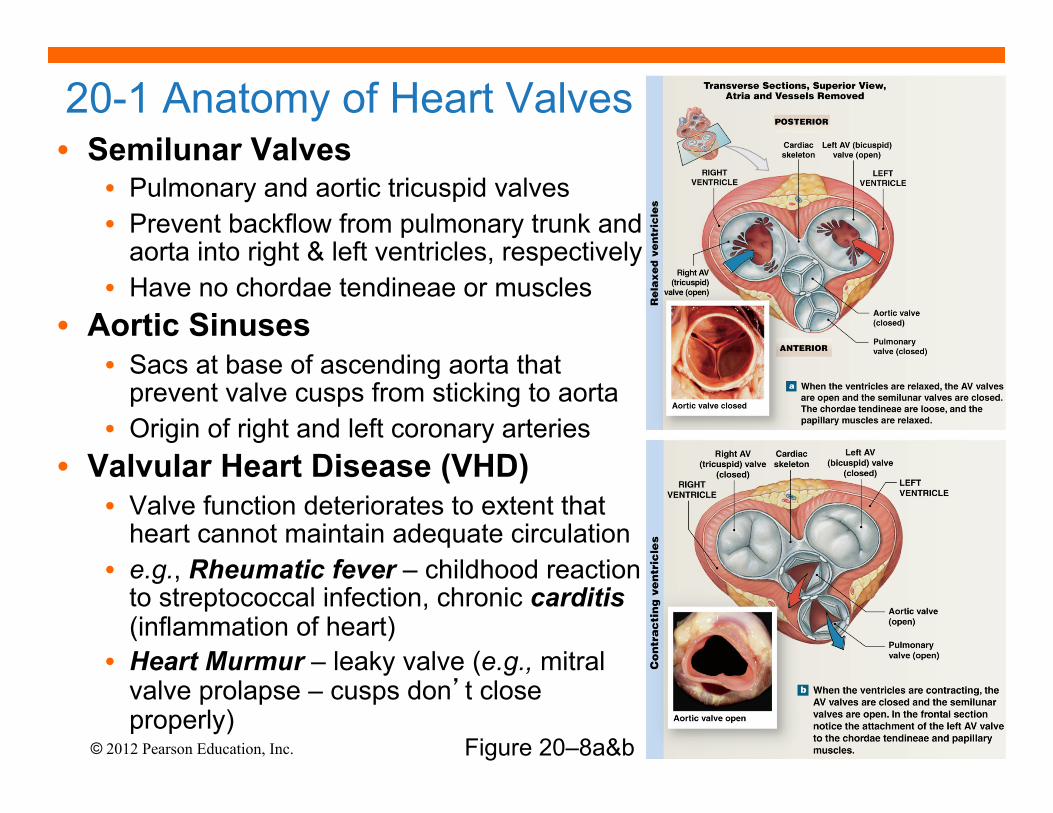
• Semilunar Valves • Pulmonary and aortic tricuspid valves • Prevent backflow from pulmonary trunk and
aorta into right & left ventricles, respectively • Have no chordae tendineae or muscles
• Aortic Sinuses • Sacs at base of ascending aorta that
prevent valve cusps from sticking to aorta • Origin of right and left coronary arteries
• Valvular Heart Disease (VHD) • Valve function deteriorates to extent that
heart cannot maintain adequate circulation • e.g., Rheumatic fever – childhood reaction
to streptococcal infection, chronic carditis (inflammation of heart)
• Heart Murmur – leaky valve (e.g., mitral valve prolapse – cusps don’t close properly)
20-1 Anatomy of Heart Valves
Figure 20–8a&b
© 2012 Pearson Education, Inc.
• Connective Tissues • Physically support cardiac muscle fibers
• Distribute forces of contraction
• Add strength and prevent overexpansion of heart
• Elastic fibers return heart to original shape after contraction
• The Cardiac (Fibrous) Skeleton • Four bands around heart valves and bases of pulmonary
trunk and aorta
• Stabilize valves
• Electrically insulate ventricular cells from atrial cells
20-1 Connective Tissue and the Cardiac Fibrous Skeleton

© 2012 Pearson Education, Inc.
• Heart is <1% body mass but requires 5% blood
• Blood flow to heart may ↑ 9x during vigorous activity
• Coronary Arteries (lt. & rt.) originate at aortic sinuses and then branch out
• Cardiac veins return deoxygenated coronary blood into right atrium
• High blood pressure & elastic rebound forces blood through coronary arteries between ventricular contractions
• Arterial anastomoses = interconnections between arteries (helps stabilize coronary blood flow despite pressure differences between lt. and rt. coronary arteries)
20-1 Coronary Circulation
Figure 20–9a&b
© 2012 Pearson Education, Inc.
• Right Coronary Artery -- supplies blood to rt. atrium, portions of both ventricles and cells of sinoatrial (SA) & atrioventricular (AV) nodes
– gives rise to marginal arteries (surface of rt. Ventricle)
– Supplies posterior interventricular artery
20-1 Coronary Circulation
• Left Coronary Artery -- supplies blood to lt. atrium, lt. ventricle and interventricular septum − gives rise to circumflex artery and
anterior interventricular artery
• Cardiac Veins: small veins drain into great cardiac vein which drains into the coronary sinus and eventually into the rt. atrium (at base of the inferior vena cava)
Figure 20–9a&c

© 2012 Pearson Education, Inc.
• Coronary Artery Disease (CAD) - partial or complete block of coronary circulation, results in coronary ischemia
• Can lead to myocardial infarction (heart attack): heart tissue denied oxygen.
• Common symptom of CAD: angina pectoralis pain in the chest as a result of the ischemia
• CAD treatments include drugs that block sympathetic stimulation (e.g., propranolol) vasodilators (e.g., nitroglycerin) and calcium channel blockers
20-1 Coronary Circulation and Disease
• Plaques can be removed surgically via catheter (laser or “roto-rooter”) or via balloon angioplasty; stents (wire mesh) used to keep artery open
• Coronary bypass surgery - use healthy veins (from legs) to create anatomoses around blockages; most people have 4 major coronary arteries, hence “quadruple bypass surgery” Figure 20–10
© 2012 Pearson Education, Inc.
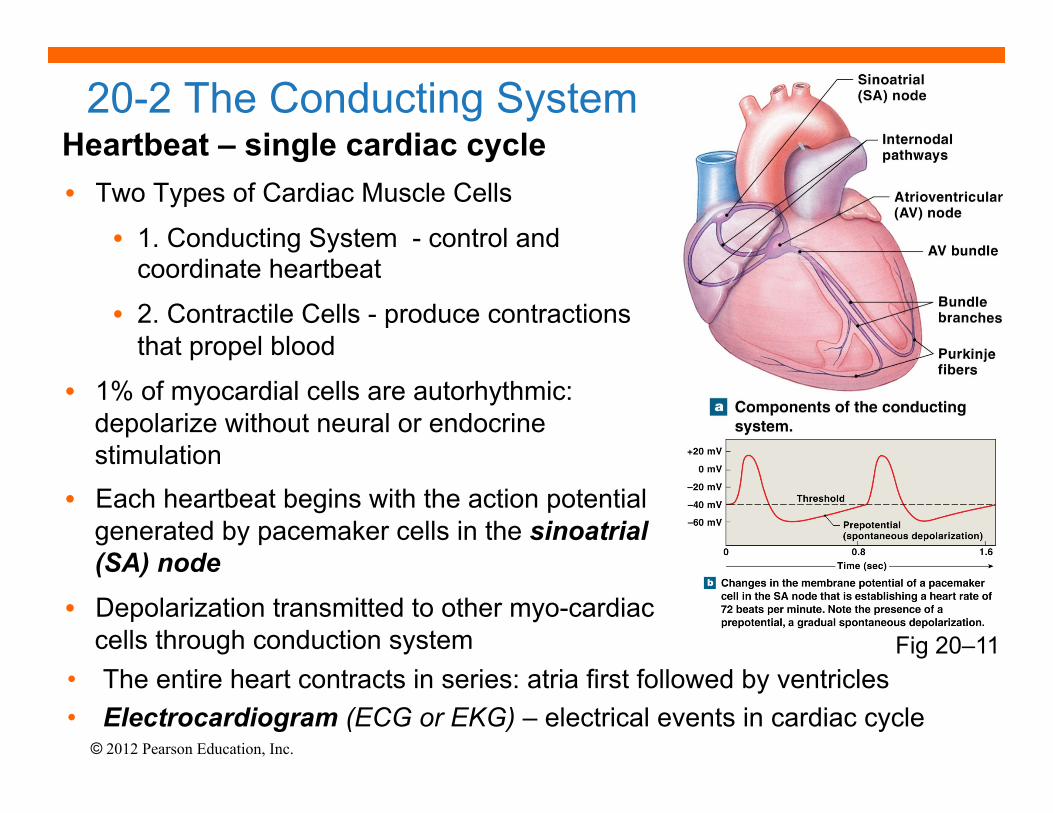
Heartbeat – single cardiac cycle • Two Types of Cardiac Muscle Cells
• 1. Conducting System - control and coordinate heartbeat
• 2. Contractile Cells - produce contractions that propel blood
• 1% of myocardial cells are autorhythmic: depolarize without neural or endocrine stimulation
• Each heartbeat begins with the action potential generated by pacemaker cells in the sinoatrial (SA) node
• Depolarization transmitted to other myo-cardiac cells through conduction system
20-2 The Conducting System
• The entire heart contracts in series: atria first followed by ventricles • Electrocardiogram (ECG or EKG) – electrical events in cardiac cycle
Fig 20–11
© 2012 Pearson Education, Inc. Fig 20–11
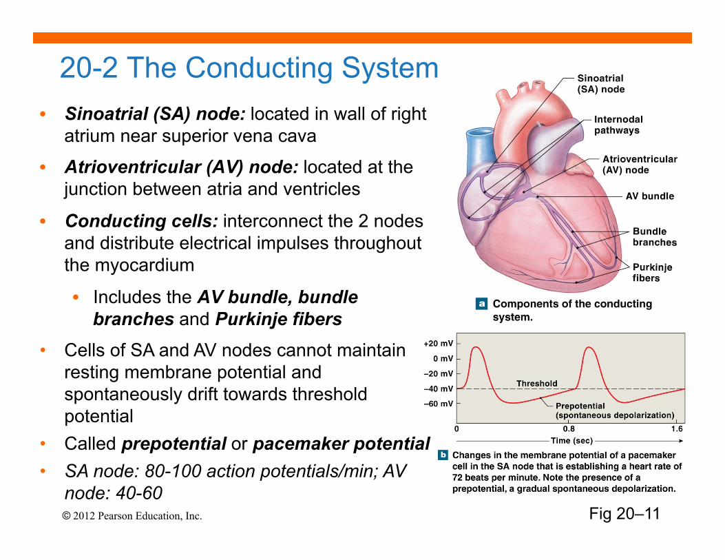
20-2 The Conducting System • Sinoatrial (SA) node: located in wall of right
atrium near superior vena cava
• Atrioventricular (AV) node: located at the junction between atria and ventricles
• Conducting cells: interconnect the 2 nodes and distribute electrical impulses throughout the myocardium
• Includes the AV bundle, bundle branches and Purkinje fibers
• Cells of SA and AV nodes cannot maintain resting membrane potential and spontaneously drift towards threshold potential
• Called prepotential or pacemaker potential • SA node: 80-100 action potentials/min; AV
node: 40-60

© 2012 Pearson Education, Inc.
20-2 Impulse Conduction Through Heart
Fig 20–12 Impulse Conduction
© 2012 Pearson Education, Inc.
• Overall heart rate set by SA node • Resting heart rate (sinus rhythm) ~75bpm set by SA
node (80-100) + parasympathetic stimulation (Vagus) • Max rate 230 bpm set by AV node max; inefficient
pumping above 180 bpm • Abnormal Pacemaker Function
• Bradycardia: abnormally slow heart rate (<60 bpm)
• Tachycardia: abnormally fast heart rate (>100 bpm)
• Ectopic pacemaker • Abnormal cells partially or completely bypass conducting
system; disrupts timing of ventricular contractions
20-2 The Conducting System
• Allometry “fact”?: 800 million beats lifespan
© 2012 Pearson Education, Inc.
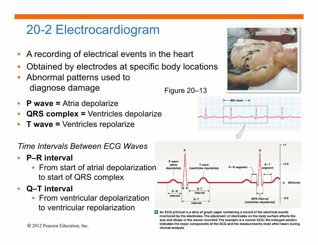
20-2 Electrocardiogram
Figure 20–13
• A recording of electrical events in the heart • Obtained by electrodes at specific body locations • Abnormal patterns used to diagnose damage
• P wave = Atria depolarize • QRS complex = Ventricles depolarize • T wave = Ventricles repolarize
Time Intervals Between ECG Waves • P–R interval
• From start of atrial depolarization to start of QRS complex
• Q–T interval • From ventricular depolarization to ventricular repolarization
© 2012 Pearson Education, Inc.
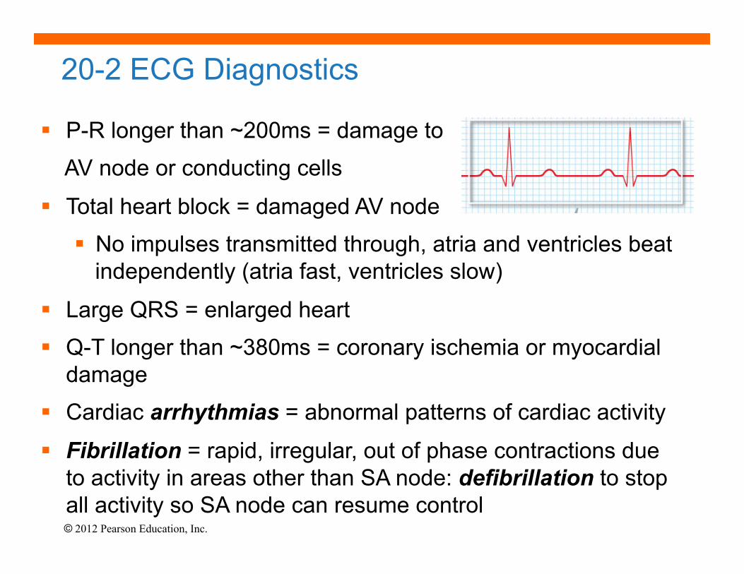
20-2 ECG Diagnostics
§ P-R longer than ~200ms = damage to
AV node or conducting cells
§ Total heart block = damaged AV node
§ No impulses transmitted through, atria and ventricles beat independently (atria fast, ventricles slow)
§ Large QRS = enlarged heart
§ Q-T longer than ~380ms = coronary ischemia or myocardial damage
§ Cardiac arrhythmias = abnormal patterns of cardiac activity
§ Fibrillation = rapid, irregular, out of phase contractions due to activity in areas other than SA node: defibrillation to stop all activity so SA node can resume control

© 2012 Pearson Education, Inc.
20-2 Cardiac Muscle Cell Action Potential Once threshold (~-75mV) reached, AP proceeds rapidly in 3 steps:
Figure 20–15 The Action Potential in Skeletal and Cardiac Muscle

© 2012 Pearson Education, Inc.
20-2 The Cardiac Action Potential
• Refractory Period • Period when AP cannot be elicited with a second stimulus
OR requires > than normal stimulus
• Absolute – no AP no matter what the stimulus because Na+ channels are either open and/or inactivated
• Relative – can elicit a 2nd AP with stronger-than-normal stimulus
• Length of cardiac action potential in ventricular cell • 250–300 ms:
• 30 times longer than skeletal muscle fiber
• long refractory period prevents summation and tetany

© 2012 Pearson Education, Inc.
20-2 Role of Calcium Ions and Energetics for Cardiac Contraction • 20% of Ca2+ for contraction enters through slow, voltage-
sensitive calcium channels in plasma membrane (extracellular sources)
• Influx triggers release of calcium ion reserves from sarcoplasmic reticulum (SR)
• As slow calcium channels close, intracellular Ca2+ is pumped into SR or out of cell
• Energy for Cardiac Contractions
• From mitochondrial breakdown of fatty acids and glucose
• Oxygen from circulating hemoglobin and internal myoglobin
© 2012 Pearson Education, Inc.
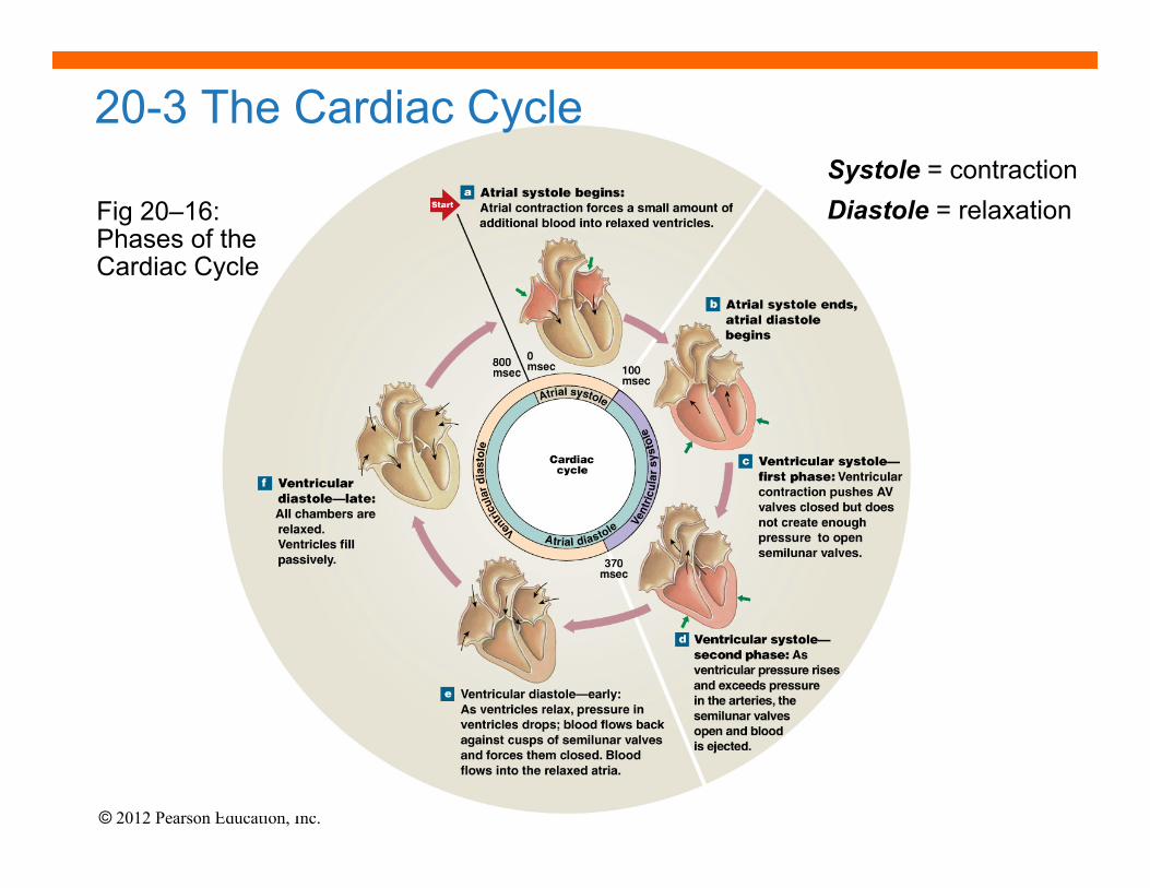
20-3 The Cardiac Cycle
Fig 20–16: Phases of the Cardiac Cycle
Systole = contraction Diastole = relaxation

© 2012 Pearson Education, Inc.
20-3 The Cardiac Cycle
Fig 20–17
8 Steps in the Cardiac Cycle 1. Atrial systole begins
▪ Atrial contraction begins ▪ Rt. & lt. AV valves remain open
2. Atria “top off” ventricles ▪ Filling ventricles
3. Atrial systole ends ▪ AV valves close
▪ Ventricles contain max vol. known as end-diastolic volume (EDV)
4. Ventricular systole begins ▪ Isovolumetric ventricular contraction ▪ Pressure in ventricles rises with AV valves shut

© 2012 Pearson Education, Inc.
20-3 The Cardiac Cycle
Fig 20–17
5. Ventricular ejection
▪ Semilunar valves open ▪ Blood flows into pulmonary and aortic trunks ▪ Stroke volume (SV) = 60% of end-diastolic volume
6. Ventricular pressure falls
▪ Semilunar valves close ▪ Ventricles contain end-systolic volume (ESV), about 40% of end-diastolic volume
7. Ventricular diastole ▪ Ventricular pressure is higher than atrial pressure
▪ All heart valves are closed ▪ Ventricles relax (isovolumetric relaxation)

© 2012 Pearson Education, Inc. Fig 20–17
8. AV valves open & passive atrial filling begins.
▪ When ventricular pressure falls below atrial pressure, the AV valves open ▪ Blood then flows from the atria into the ventricles while both are in diastole
20-3 The Cardiac Cycle
© 2012 Pearson Education, Inc.
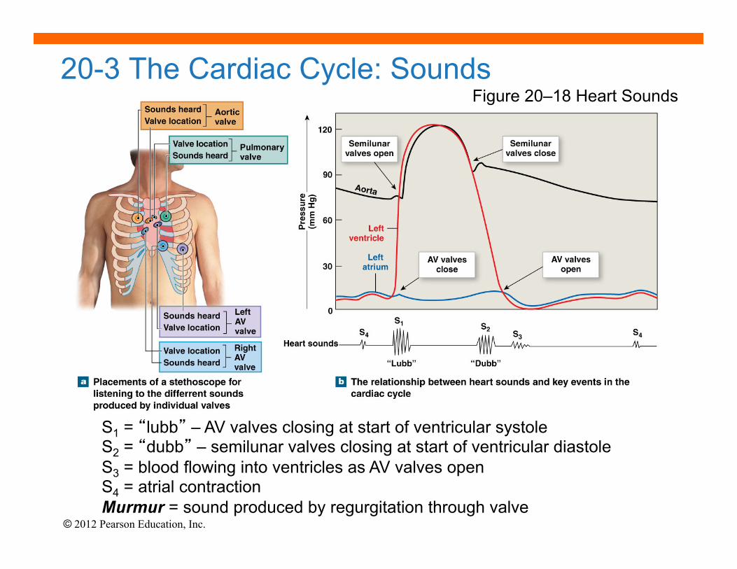
20-3 The Cardiac Cycle: Sounds
S1 = “lubb” – AV valves closing at start of ventricular systole S2 = “dubb” – semilunar valves closing at start of ventricular diastole S3 = blood flowing into ventricles as AV valves open S4 = atrial contraction Murmur = sound produced by regurgitation through valve
Figure 20–18 Heart Sounds

© 2012 Pearson Education, Inc.
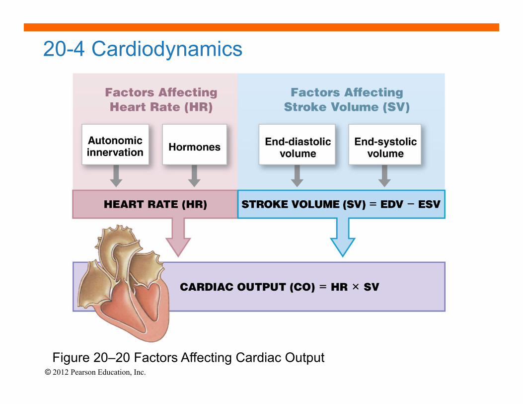
Cardiodynamics = movement & force generated by cardiac contractions • End-diastolic volume (EDV) – am’t of blood in ventricles at end of diastole • End-systolic volume (ESV) – am’t blood in ventricles at end of systole • Stroke volume (SV) – am’t of blood ejected in single beat (ml/beat)
• SV = EDV – ESV • Ejection fraction
• The percentage of EDV represented by SV • Cardiac output (CO) (ml/min)
• The volume pumped by ventricle in 1 minute • CO = HR x SV (heart rate [beats/min] times stroke volume)
20-4 Cardiodynamics
Fig 20-19
© 2012 Pearson Education, Inc.
20-4 Cardiodynamics
Figure 20–20 Factors Affecting Cardiac Output

© 2012 Pearson Education, Inc.
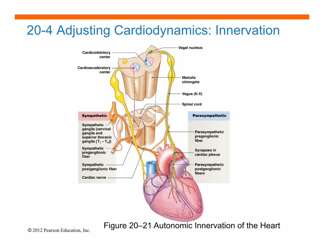
20-4 Adjusting Cardiodynamics: Innervation
• Autonomic innervation • Both sympathetic (NE) and parasympathetic (ACh) innervation of SA
node, AV node and atrial myocardium
• Sympathetic dominates in ventricles • Cardiac centers of medulla oblongata monitor blood pressure
(baroreceptors) arterial O2 and CO2 levels (chemoreceptors)
• cardioacceleratory center controls sympathetic neurons (increases heart rate – positive chronotropic effect)
• cardioinhibitory center controls parasympathetic neurons (slows heart rate – negative chronotropic effect)
• Autonomic tone • Dual innervation maintains resting tone by releasing ACh (and NE) • At rest, parasympathetic tone reduces SA node inherent rate
(80-100 bpm) to ~70 bpm
© 2012 Pearson Education, Inc.
20-4 Adjusting Cardiodynamics: Innervation
Figure 20–21 Autonomic Innervation of the Heart
© 2012 Pearson Education, Inc.
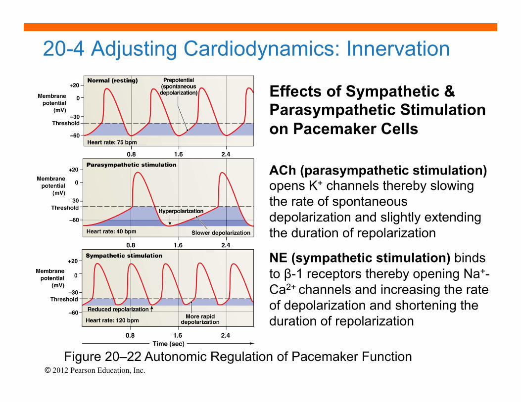
20-4 Adjusting Cardiodynamics: Innervation
Effects of Sympathetic & Parasympathetic Stimulation on Pacemaker Cells ACh (parasympathetic stimulation) opens K+ channels thereby slowing the rate of spontaneous depolarization and slightly extending the duration of repolarization
NE (sympathetic stimulation) binds to β-1 receptors thereby opening Na+-Ca2+ channels and increasing the rate of depolarization and shortening the duration of repolarization
Figure 20–22 Autonomic Regulation of Pacemaker Function
© 2012 Pearson Education, Inc.
20-4 Adjusting Cardiodynamics: Reflexes, Hormones & Drugs • Atrial or Bainbridge Reflex
• Increased venous return activates stretch receptors in right atrium → ↑ sympathetic activity → ↑ HR
• Hormonal Effects on Heart Rate • Epinephrine (E), Norepinephrine (NE) and Thyroid hormone
(thyroxine) ↑ HR and contractile strength (positive inotropic effect) by acting on SA node
• β-1 blockers (hypertensive drugs) block E & NE effects • Other Heart Rate Effectors
• Caffeine: rapid depolarization of SA node, ↑ HR • Nicotine: stimulates sympathetic neurons, ↑ HR • Changes in K+, Ca2+, temperature, etc.
© 2012 Pearson Education, Inc.
20-4 Cardiodynamics: Stroke Volume Adjustments • End diastolic volume (EDV) is affected by
1. Venous return 2. Filling time
↑ EDV → ↑ preload (amount of ventricular stretch) → ↑ SV (Frank-Starling Principle) • End systolic volume (ESV) is affected by
1. Preload 2. Contractility = force produced during contraction (inotropic) 3. Afterload = tension the ventricle must produce to open the semilunar valve and eject blood (↑ by any factor that restricts arterial blood flow)
• ↑ ESV → ↓ SV

© 2012 Pearson Education, Inc.
20-4 Cardiodynamics: Stroke Volume Adjustments
Figure 20–23

© 2012 Pearson Education, Inc.
20-4 Cardiodynamics: Contractility • Sympathetic stimulation – positive inotropic effect
• NE released by postganglionic fibers of cardiac nerves AND both E & NE released by suprarenal (adrenal) medullae
• Increases ejection fraction and decreases ESV
• Parasympathetic activity – negative inotropic effect
• Acetylcholine released by Vagus nerve
• Hormones – can have neg. or pos. inotropic effects • Many pharmaceutical drugs mimic E, NE and thyroxine actions
• β-1 Receptors Mimetics (e.g., isoproterenol, dopamine, dobutamine)
• β-1 Receptor Blockers (e.g., propranolol, timolol, metoprolol) • Others affect Ca2+ (e.g., digitalis [inhibit SR uptake], nifedipine and
verapamil [channel blockers])

© 2012 Pearson Education, Inc.
20-4 Summary of Factors Affecting Cardiac Output
Figure 20–24 A Summary of the Factors Affecting Cardiac Output