chromoendoscopy in magnetically guided capsule endoscopy
TRANSCRIPT
Mewes et al. BioMedical Engineering OnLine 2013, 12:52http://www.biomedical-engineering-online.com/content/12/1/52
RESEARCH Open Access
Chromoendoscopy in magnetically guidedcapsule endoscopyPhilip W Mewes1,2*†, Stefan Foertsch1,3†, Aleksandar Lj Juloski2, Elli Angelopoulou1, Stefan K Goelder4,Dirk Guldi3, Joachim Hornegger2 and Helmut Messmann4
*Correspondence:[email protected]†Equal contributors1Pattern Recognition Lab,University of Erlangen-Nuremberg,Martensstrasse 3, Erlangen,Germany2Siemens AG, Healthcare Sector,Allee Am Roethelheimpark 2,Erlangen 91052, GermanyFull list of author information isavailable at the end of the article
Abstract
Background: Diagnosis of intestinal metaplasia and dysplasia via conventionalendoscopy is characterized by low interobserver agreement and poor correlation withhistopathologic findings. Chromoendoscopy significantly enhances the visibility ofmucosa irregularities, like metaplasia and dysplasia mucosa. Magnetically guidedcapsule endoscopy (MGCE) offers an alternative technology for upper GI examination.We expect the difficulties of diagnosis of neoplasm in conventional endoscopy totransfer to MGCE. Thus, we aim to chart a path for the application of chromoendoscopyon MGCE via an ex-vivo animal study.
Methods: We propose a modified preparation protocol which adds a staining step tothe existing MGCE preparation protocol. An optimal staining concentration isquantitatively determined for different stain types and pathologies. To that end 190 pigstomach tissue samples with and without lesion imitations were stained with differentdye concentrations. Quantitative visual criteria are introduced to measure the quality ofthe staining with respect to mucosa and lesion visibility. Thusly determined optimalconcentrations are tested in an ex-vivo pig stomach experiment under magneticguidance of an endoscopic capsule with the modified protocol.
Results: We found that the proposed protocol modification does not impact thevisibility in the stomach or steerability of the endoscopy capsule. An average optimalstaining concentration for the proposed protocol was found at 0.4% for Methyleneblue and Indigo carmine. The lesion visibility is improved using the previously obtainedoptimal dye concentration.
Conclusions: We conclude that chromoendoscopy may be applied in MGCE andimproves mucosa and lesion visibility. Systematic evaluation provides importantinformation on appropriate staining concentration. However, further animal andhuman in-vivo studies are necessary.
Keywords: Methylene blue, Indigo carmine, Staining
BackgroundAlthough incidence and mortality are decreasing, gastric cancer with 738.000 casesworldwide in 2008 is the 2nd most lethal digestive neoplasm in the world [1]. Intestinalmetaplasia and dysplasia are precursors of cancer [2]. The identification of these lesionsand follow-up of afflicted patients could lead to early diagnosis and treatment, and thusenhance the survival of the patient [3,4]. Esophagogastroduodenoscopy (EGD) is themost
© 2013 Mewes et al.; licensee BioMed Central Ltd. This is an Open Access article distributed under the terms of the CreativeCommons Attribution License (http://creativecommons.org/licenses/by/2.0), which permits unrestricted use, distribution, andreproduction in any medium, provided the original work is properly cited.
Mewes et al. BioMedical Engineering OnLine 2013, 12:52 Page 2 of 16http://www.biomedical-engineering-online.com/content/12/1/52
common procedure for diagnosis and treatment. However, for the detection of metapla-sia and dysplasia conventional EGD is characterized by low interobserver agreement andpoor correlation with histopathologic findings [5,6].Various techniques are available for enhancing and highlighting mucosa irregulari-
ties and for increasing the visibility of structures which lie under the surface of themucosa. The most important methods include narrow band imaging, confocal laserendomicroscopy, magnification endoscopy, optical coherence tomography and chro-moendoscopy [7-10]. These techniques have often been compared against each other, orin combination in terms of their impact in diagnostic accuracy (e.g. in [11,12]). How-ever a substantial difference between chromoendoscopy and all competing techniqueslies in the lack for additional hardware. Chromoendoscopy requires no modification ofthe hardware of the imaging system itself.Furthermore, chromoendoscopy in EGD and colonoscopy has been shown to signifi-
cantly enhance the visibility of mucosa irregularities, like metaplasia and dysplasia [13].Chromoendoscopy consists of the topical application of different stains to improve tissuevisibility, localization and characterization for the purpose of better diagnosis. Chro-moendoscopy usually consists of four steps for absorptive stains and three steps forcontrast stains: (1) Application of an acid solution to dissolve gastric mucus, (2) localapplication of a stain, (3) (only for absorptive stains) washing of the respective region withwater and (4) visual inspection of the stained regions for diagnostic purposes. In (1)-(3)the application of dye is performed locally using the working channel of the endoscopeand different spray catheters (direct method) under the visual guidance of the endoscope.For colonoscopy the passive application of stain with a dye-powder filled capsule has alsobeen reported [14,15]. In this procedure a capsule with dye powder is given to the patientafter administration of a bowel cleansing solution such as PEG. Between the dye adminis-tration and the colonoscopy examination a waiting time is needed. Application of the dyein the morning and examination in the afternoon was reported as a sufficiently large timespan [14]. Though the procedure was found to be feasible, difficulties have been reporteddue to the inhomogeneous application of the stain [16]. The oral application of dye forexamination of the stomach without using a spray catheter (indirect method) has beendescribed in [15,17].Recently, different approaches for magnetically guided capsule endoscopes (MGCE)
for gastric and small bowel examinations were presented [18-23]. In a clinical study onhumans, MGCE showed the feasibility of gastric exploration with a guided capsule endo-scope [18,19]. In this specific study the stomach was filled with water and the capsule wasnavigated from the outside using an external magnetic field. An operator could controlthe motion of the capsule during the examination using feedback from real-time gas-tric imaging provided by two capsule camera sensors. Hence, he could obtain a sufficientnumber of stomach-surface images with diagnostic value.We expect that known difficulties in the diagnosis of neoplasia, regarding interobserver
agreement in conventional endoscopy, transfer to MGCE. MGCE could, thus, benefitfrom chromoendoscopy in the same way classic endoscopy does. However, compared tothe direct application of stain in EGD and most colonoscopy procedures, in MGCE onlyindirect application is possible. No acid preparation and washing of the gastric mucosa ispossible. Furthermore, the water in which the capsule is maneuvered, must not be stainedat levels which reduce the overall visibility. Competing methods such as narrow-band
Mewes et al. BioMedical Engineering OnLine 2013, 12:52 Page 3 of 16http://www.biomedical-engineering-online.com/content/12/1/52
imaging in the diagnosis of colorectal neoplasia are difficult to integrate in a capsuleendoscope [11]. Furthermore, as inmany endoscopic techniques the exact impact of chro-moendoscopy and the technical details have not yet been established [24,25]. For example,in literature ([5,26,27]) one can find three different concentrations of Methylene blue dyeand application times for the examination of gastric neoplasm. The search for an optimalconcentration for a staining procedure in animal and human trials has been reported afew times, but without the support of a thorough analysis. In [28] an optimal stainingconcentration for simultaneous confocal laser endomicroscopy and chromoendoscopywith Cresyl violet is assessed in mices, but without an objective criterion. In [29] an opti-mal staining concentration for endocytoscopy was accessed in an ex-vivo animal studyin which freshly resected porcine esophagus, stomach, and colon were examined. Imagecontrast and staining status were evaluated by experts for each organ to determine thebest concentration. Results were transferred to resected human organs. The problem of amissing systematic study for classic chromoendoscopy in the stomach transfers to MGCEand becomes more severe through the challenges of the indirect application of stain.In this paper we evaluate the possible application of chromoendoscopy to MGCE
in an ex-vivo animal study. First, we propose a modification to the MGCE prepa-ration protocol in order to incorporate a staining procedure for chromoendoscopy.Second, we present a method to systematically assess an optimal concentration of dyefor the proposed protocol modification. The optimization is conducted in experimentsusing pig stomach tissue samples and with respect to the best visibility of tissue ofdifferent histological or pathological nature. Third, we transfer these results to an ex-vivo pig stomach experiment under magnetic guidance of a capsule endoscope. Theseexperiments should determine: a) the overall under-water visibility after the proposedpassive staining protocol; and b) the mucosa and lesion visibility with the optimized dyeconcentration.The guidance magnet is technically similar to the one used for the human study [18,19].
All navigation functions of that study are also available in our setup. The system is ajoint development of SIEMENS Healthcare and Olympus Medical Systems Corp. Itsmain components are: (1) A guidance magnet that consists of a set of electromagneticcoils defining a working volume and enabling the operator to control a capsule endo-scope with 5 degrees of freedom (DOF). The magnetic flux density has a maximumof 100 millitesla. (2) A capsule endoscope of 31mm length manufactured by Olym-pus Medical Systems Corp. with a build-in permanent magnet and two CCD cameraseach transmitting 2 frames per second in real time to an external receiver attachedto the patient’s body. (3) A display showing the capsule images to the operator. (4)A set of joysticks allowing the operator to maneuver the capsule inside the stomach.The orientation of an electromagnetic (EM) field orients the capsule in the stomach.The EM field together with an EM gradient field generate forces on the capsule endo-scope of less than 1 millinewton. These are sufficient for translational movements. Moredetails about the hardware and software design of the guidance magnet can be found in[30]. The guidance of the capsule is performed based on real-time imaging provided bythe capsule endoscope inside the pig stomach.In [31] a pig stomach study was presented to improve mucosa visibility in MGCE
using Methylene blue. This study was limited to only a few cases and a single dye.The magnetic steerability was only simulated using a plastic support and there was
Mewes et al. BioMedical Engineering OnLine 2013, 12:52 Page 4 of 16http://www.biomedical-engineering-online.com/content/12/1/52
no systematic evaluation of an optimized dye concentration prior to the experiments.Whereas in this paper the magnetic steerability is achieved with a capsule guidance mag-net. The study is conducted with a large number of pig stomachs and leads to a systematicassessment of two optimal dye types.
MethodsModification of preparation protocol for MGCE
The established preparation protocol used for the existing human MGCE study with 43patients consists of three administrations of tap water prior to the examination:[18,19]
ExistingMGCE preparation protocol
E.1 500 mL of clear water at room temperature one hour and 15 minutes prior to theexamination and after overnight fasting.
E.2 400 mL of clear water at room temperature 15 minutes prior to the examinationfollowed by light exercises.
E.3 400 mL of clear water at near body temperature, immediately before theexamination.
All applications are given orally. Steps E.1 and E.2 are primarily intended for cleaningthe stomach. Step E.3 aims at expanding the stomach in order to obtain enough space forthe capsule to be maneuvered and for complete visibility of the stomach mucosa withoutgastric folds overlapping each other and eventually hiding relevant mucosa parts. StepE.3 cannot be modified since it is crucial for the guidance of the capsule inside the stom-ach. We expect the water to be stained to such an extent that the general visibility isreduced when the stain is directly applied prior to E.3. Hence, we propose to fit a stainingstep between steps E.1 and E.2 of the existing preparation protocol. In order to con-duct further experiments with animal phantoms the following protocol is adopted for pigstomachs:
ModifiedMGCE preparation protocol for pig stomachs
M.1 2000 mL of clear water at room temperature that is emptied out of the stomachimmediately after administration. The purpose of this step is still the cleaning ofthe stomach from mucus and/or remaining food.
M.2 100 mL of dye, followed by 5 minutes of massage and kneading the stomach tosimulate peristaltic movement, followed by emptying the stomach from the dye.This step applies the dye to the stomach walls. Massage and kneading the stomachsimulates peristaltic motion and is performed under the assumption that the dyewould be naturally scattered over all anatomical areas of the stomach in the naturalcase. Simulating digestive peristalsis via massage a stomacher bag or a pulley systemto create peristaltic motion in a mechanical stomach model was reported in [32,33].
M.3 500 mL of clear water at room temperature, which remains in the stomach for 5minutes and is afterwards emptied out. This step is similar to E.2 of the standardMGCE procedure but this time it also evacuates the remaining dye.
M.4 2000 mL of clear water at near body temperature, immediately before theexamination (same purpose as E.3).
Mewes et al. BioMedical Engineering OnLine 2013, 12:52 Page 5 of 16http://www.biomedical-engineering-online.com/content/12/1/52
All applications are performed through the esophagus and intend to simulate the oralapplication of water and dye. Using a combination of water, to expand the stomach, anddye at the same time was assumed to be incompatible with MGCE since the visibility ofthe capsule would deteriorate. The emptying is performed by gently squeezing the stom-ach and imitates the natural evacuation of stomach content into the small intestine. Theamount of water for washing and expanding the stomach (step M.1 and M.4) was set tolarger values due to the larger size of a pig’s stomach.
Target lesion and lesion imitations
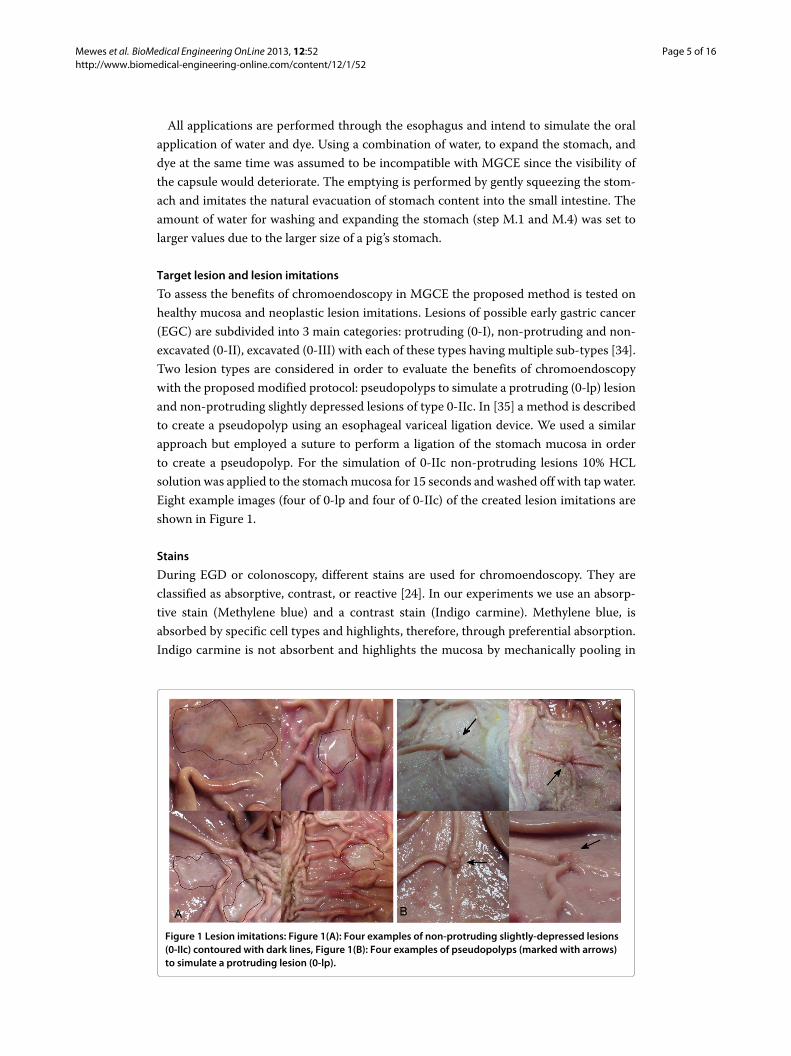
To assess the benefits of chromoendoscopy in MGCE the proposed method is tested onhealthy mucosa and neoplastic lesion imitations. Lesions of possible early gastric cancer(EGC) are subdivided into 3 main categories: protruding (0-I), non-protruding and non-excavated (0-II), excavated (0-III) with each of these types having multiple sub-types [34].Two lesion types are considered in order to evaluate the benefits of chromoendoscopywith the proposed modified protocol: pseudopolyps to simulate a protruding (0-lp) lesionand non-protruding slightly depressed lesions of type 0-IIc. In [35] a method is describedto create a pseudopolyp using an esophageal variceal ligation device. We used a similarapproach but employed a suture to perform a ligation of the stomach mucosa in orderto create a pseudopolyp. For the simulation of 0-IIc non-protruding lesions 10% HCLsolution was applied to the stomachmucosa for 15 seconds and washed off with tap water.Eight example images (four of 0-lp and four of 0-IIc) of the created lesion imitations areshown in Figure 1.
Stains
During EGD or colonoscopy, different stains are used for chromoendoscopy. They areclassified as absorptive, contrast, or reactive [24]. In our experiments we use an absorp-tive stain (Methylene blue) and a contrast stain (Indigo carmine). Methylene blue, isabsorbed by specific cell types and highlights, therefore, through preferential absorption.Indigo carmine is not absorbent and highlights the mucosa by mechanically pooling in
Figure 1 Lesion imitations: Figure 1(A): Four examples of non-protruding slightly-depressed lesions(0-IIc) contoured with dark lines, Figure 1(B): Four examples of pseudopolyps (marked with arrows)to simulate a protruding lesion (0-lp).
Mewes et al. BioMedical Engineering OnLine 2013, 12:52 Page 6 of 16http://www.biomedical-engineering-online.com/content/12/1/52
cervices between epithelial cells, fat or depressed lesions and other irregularities. Thelesion imitations of type 0-IIc (early gastric cancer) are stained with indigo carmineas described in [36]. For the staining of fine details of the mucosa Methylene blue isused.
Visual criteria of an optimal stain concentration
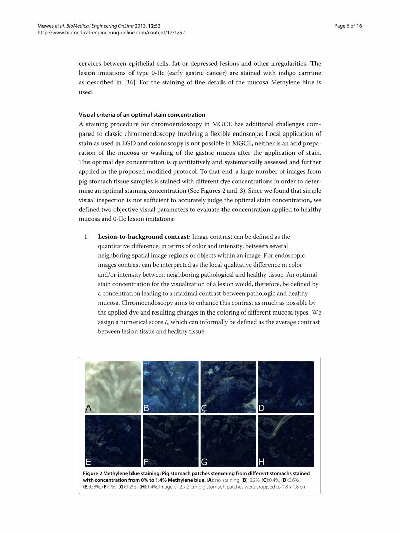
A staining procedure for chromoendoscopy in MGCE has additional challenges com-pared to classic chromoendoscopy involving a flexible endoscope: Local application ofstain as used in EGD and colonoscopy is not possible in MGCE, neither is an acid prepa-ration of the mucosa or washing of the gastric mucus after the application of stain.The optimal dye concentration is quantitatively and systematically assessed and furtherapplied in the proposed modified protocol. To that end, a large number of images frompig stomach tissue samples is stained with different dye concentrations in order to deter-mine an optimal staining concentration (See Figures 2 and 3). Since we found that simplevisual inspection is not sufficient to accurately judge the optimal stain concentration, wedefined two objective visual parameters to evaluate the concentration applied to healthymucosa and 0-IIc lesion imitations:
1. Lesion-to-background contrast: Image contrast can be defined as thequantitative difference, in terms of color and intensity, between severalneighboring spatial image regions or objects within an image. For endoscopicimages contrast can be interpreted as the local qualitative difference in colorand/or intensity between neighboring pathological and healthy tissue. An optimalstain concentration for the visualization of a lesion would, therefore, be defined bya concentration leading to a maximal contrast between pathologic and healthymucosa. Chromoendoscopy aims to enhance this contrast as much as possible bythe applied dye and resulting changes in the coloring of different mucosa types. Weassign a numerical score Ic which can informally be defined as the average contrastbetween lesion tissue and healthy tissue.
Figure 2 Methylene blue staining: Pig stomach patches stemming from different stomachs stainedwith concentration from 0% to 1.4%Methylene blue. (A): no staining, (B): 0.2%, (C):0.4%, (D):0.6%,(E):0.8%, (F):1% , (G):1.2% , (H):1.4%. Image of 2 x 2 cm pig stomach patches were cropped to 1.8 x 1.8 cm.

Mewes et al. BioMedical Engineering OnLine 2013, 12:52 Page 7 of 16http://www.biomedical-engineering-online.com/content/12/1/52
Figure 3 Pig stomach patches with lesion imitation stained with different concentrations of Indigocarmine. (A) no staining, (B) 0.2%, (C) 0.4%, (D) 0.6%, (E) 0.8%, (F) 1%. Non-protruding slightly-depressedlesions (0-IIc) are contoured with red outlines. Image of 2 x 2 cm pig stomach patches were cropped to 1.8 x1.8 cm.
For the computation of the contrast score Ic two regions Rl and Rh are defined forthe image region with lesions and the healthy tissue respectively. Since the contrastin an image can be described as the quantitative difference between different imageregions. A measurement of the contrast Ic between both regions can therefore bedenotes as
Ic = ∣∣g(Rl) − g(Rh)∣∣ (1)
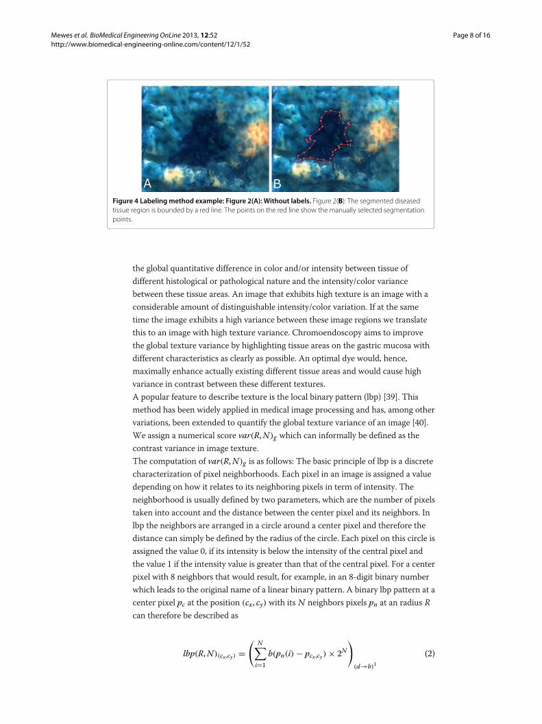
where g(·) refers to the gray-scale conversion of the original color image. Tocompute such a contrast score, for each image the two regions Rl and Rh aremanually chosen inside a region with lesions and the healthy tissue. This labelingprocess does not segment the exact border between healthy and diseased tissue.Manual segmentation is always subject to the expert. More notably an exactsegmentation is relatively unimportant, compared to the overall contrast betweentwo image regions. Hence an exact region segmentation is not necessary. Figure 4shows an example of a manually segmented diseased tissue region. The value on agray scale image lies between 0 and 255. Hence the score for thelesion-to-background contrast would also lie between these two values, where asmall value would imply poor contrast and a high value would indicate an imagewith high contrast between lesion and background. In [29] such an approach wasdefined to determine the optimal contrast between the cytoplasm and the nucleifor endocytoscopy.
2. Global texture variance: Various texture measurements are known fromcomputer vision in general and in particular from medical image processing for thepurpose of automatic segmentation, classification or content based image retrieval[37,38].Image texture can be described as a measurement of spatial arrangement anddistribution of intensity and/or color in a image. Within these arrangements anddistributions the variance can be measured. For images in endoscopy this can be
Mewes et al. BioMedical Engineering OnLine 2013, 12:52 Page 8 of 16http://www.biomedical-engineering-online.com/content/12/1/52
Figure 4 Labeling method example: Figure 2(A): Without labels. Figure 2(B): The segmented diseasedtissue region is bounded by a red line. The points on the red line show the manually selected segmentationpoints.
the global quantitative difference in color and/or intensity between tissue ofdifferent histological or pathological nature and the intensity/color variancebetween these tissue areas. An image that exhibits high texture is an image with aconsiderable amount of distinguishable intensity/color variation. If at the sametime the image exhibits a high variance between these image regions we translatethis to an image with high texture variance. Chromoendoscopy aims to improvethe global texture variance by highlighting tissue areas on the gastric mucosa withdifferent characteristics as clearly as possible. An optimal dye would, hence,maximally enhance actually existing different tissue areas and would cause highvariance in contrast between these different textures.A popular feature to describe texture is the local binary pattern (lbp) [39]. Thismethod has been widely applied in medical image processing and has, among othervariations, been extended to quantify the global texture variance of an image [40].We assign a numerical score var(R,N)g which can informally be defined as thecontrast variance in image texture.The computation of var(R,N)g is as follows: The basic principle of lbp is a discretecharacterization of pixel neighborhoods. Each pixel in an image is assigned a valuedepending on how it relates to its neighboring pixels in term of intensity. Theneighborhood is usually defined by two parameters, which are the number of pixelstaken into account and the distance between the center pixel and its neighbors. Inlbp the neighbors are arranged in a circle around a center pixel and therefore thedistance can simply be defined by the radius of the circle. Each pixel on this circle isassigned the value 0, if its intensity is below the intensity of the central pixel andthe value 1 if the intensity value is greater than that of the central pixel. For a centerpixel with 8 neighbors that would result, for example, in an 8-digit binary numberwhich leads to the original name of a linear binary pattern. A binary lbp pattern at acenter pixel pc at the position (cx, cy) with its N neighbors pixels pn at an radius Rcan therefore be described as
lbp(R,N)(cx,cy) =( N∑
i=1b(pn(i) − pcx,cy) × 2N
)(d→b)1
(2)
Mewes et al. BioMedical Engineering OnLine 2013, 12:52 Page 9 of 16http://www.biomedical-engineering-online.com/content/12/1/52
where
b(x) ={1 if x ≥ 00 if x < 0
(3)
where d → b denotes the conversion of the previous term from a decimal to abinary number and the coordinates of each neighbors pn(i) can be calculated for acenter pixel at the position (cx, cy) as pn(i)x = cx + R × cos(2 × π × i
N ) andpn(i)y = cy + R × sin(2 × π × i
N ). The decimal number lbp(R,N)(cx,cy) needs thento be converted to a binary number in order to obtain the actual pattern. If theposition of an neighbor pixel is not exactly in the center of the pixel grid, its value isbilinearly interpolated out of a 2 × 2 neighborhood. According to [41] Eq. 2 can bemodified to measure the local texture variance. For a pixel pc at the position (cx, cy)with its N neighbors pixels pn at an radius R the texture variance is computed as
var(R,N)(cx,cy) = 1N
N∑i=1
(pn(i) − μ)2. (4)
with μ = 1N
∑Ni=1 pn(i). To globally measure the texture variance for a entire
image Eq. 4 needs to be applied to the entire image. Thus Eq. 4 is extended to
var(R,N)g = 1h × v
h∑k=1
v∑l=1
1N
N∑i=1
(pn(i) − μ)2. (5)
where the coordinates of each neighbors pn(i) for k and l are computed aspn(i)x = k + R × cos(2 × π × i
N ) and pn(i)y = l + R × sin(2 × π × iN ). h and v
denote the horizontal and vertical image dimensions. All computation are done onimages that have been previously converted to gray scale images.For the experiments conducted for this paper the score var(R,N)g is computed for5 different radii (R = 10 : 10 : 50) and the final result is averaged. This ensures thatdifferent texture frequencies are considered for the computation of the final score.This approach takes into consideration that the distribution of different tissuetypes is irregular as well.
Experimental evaluation of optimal staining concentration
Experiments for lesion-to-background contrast
To evaluate the optimal staining concentration 2 × 2 cm patches of pig stomach mucosawere cut. To compute the optimal lesion-to-background score, Ic, each time 10 patcheswere stained with a particular Indigo carmine concentration. Concentrations range from0.2% to 1.6% in steps of 0.2%. Ten patches were left unstained. All patches were pre-pared with lesion imitations of type 0-IIc as described previously. Five out of the 10patches stem from the fundus region and five from the pyloric region of the stom-ach. None of the patches stained with the same concentration were cut from the samestomach. In total, we examined 90 patches for these experiments: 5 patches per concen-tration and per stomach region and 9 different concentrations, including 10 unstainedpatches. The patches were placed in a glass container containing the respective dye con-centration for 15 seconds and then washed with flowing lukewarm water for another15 seconds. The patches were then photographed with a commercially-available indus-trial CCD camera (Point Grey Flea 2 FL2G-50S5M/C/ICX655 2/3 SuperHAD CCD,
Mewes et al. BioMedical Engineering OnLine 2013, 12:52 Page 10 of 16http://www.biomedical-engineering-online.com/content/12/1/52
Point Grey Research Inc 12051 Riverside Way Richmond, BC, Canada). Such a scientific-quality and high-resolution apparatus decreases the amount of negative effects onthe computation due to visual artifacts such as image distortion and sensor noise.It also allows for an accurate quantitative evaluation of the impact of different dyeconcentrations.
Experiments global texture variance
To evaluate the global texture variance score, var(R,N)g , experiments were conductedin the same way as for the lesion-to-background score. However, the patches this timewere not prepared with lesion imitations. Furthermore, in this set of experiments thesamples were stained with aMethylene blue concentration of 0.2% to 2.0% in steps of 0.2%and 0.2% to 1.6% in steps of 0.2% for fundus patches and pylorus patches respectively.100 patches in total were examined in this evaluation.
Computation of scores and results
Pictures of 1400 x 1200 pixels were taken from each stained patch. These images wereconverted to gray-scale and the contrast score, Ic, as well as the texture variance score,var(R,N)g , were computed for the respective patches.
Ex-vivo Pig stomach experiments under magnetic guidance
To verify our results, the proposed modification of the preparation protocol andthe newly determined optimal dye concentration were combined and tested oncomplete pig stomachs. In order to assess the effects of the proposed staining param-eters, pig stomachs with lesion imitations were examined with magnetically guidedcapsule endoscopes, including lesion imitations, with and without applied staining.The pig stomachs were harvested fresh from a slaughterhouse with the esophagus andduodenum attached.
1. Each pig stomach was prepared with either protruding (0-lp) lesions ornon-protruding slightly depressed lesions of type 0-IIc. To prepare the stomach,first a 6 cm long cut in the fundus region was performed, the stomach was turnedinside out using this cut and the artificial lesion imitations were applied. Eightlesion imitations of one type were distributed on all anatomical regions of thestomach. After applying the lesion imitations the stomach was turned back and thecut was stitched.
2. The modified preparation protocol previously described was applied to thestomach without the staining step M.2.
3. A capsule endoscope was introduced through the esophagus. The esophagus andduodenum were closed with clips to prevent water from running out of thestomach. The stomach was placed on a foam support with a stomach-shaped hole.This support maintained the natural shape of the stomach. The stomach wascovered with a plastic sheet and the receiving antennas for the capsule endoscopewere placed on this sheet.
4. The stomach was placed in the guidance magnet. In approximately 10 minutes allrelevant anatomical regions of the stomach were visualized with the capsuleendoscope.
Mewes et al. BioMedical Engineering OnLine 2013, 12:52 Page 11 of 16http://www.biomedical-engineering-online.com/content/12/1/52
5. The stomach was removed from the magnet and the water was flushed out.6. The modified preparation protocol was again applied to the same stomach this
time including the staining step M.2. For stomachs prepared with for 0-IIc lesionimitations, a 0.2% or 0.6% Indigo carmine solution was applied, depending onwhich anatomical section of the stomach the lesion was brought on. For theevaluation of general mucosa visibility and visibility of 0-lp lesions imitation 0.4%Methylene blue was applied.
7. Step 4 was repeated with the stained stomach. For better comparison of stainingand unstained lesion imitation special emphasis was put on capturing lesionimitations from approximately the same viewpoint and viewing angle as with theunstained stomach. Lesion imitations of the same type applied to the same stomachwere compared with and without staining from nearly the same viewpoint.
ResultsEvaluation of an optimal dye concentration
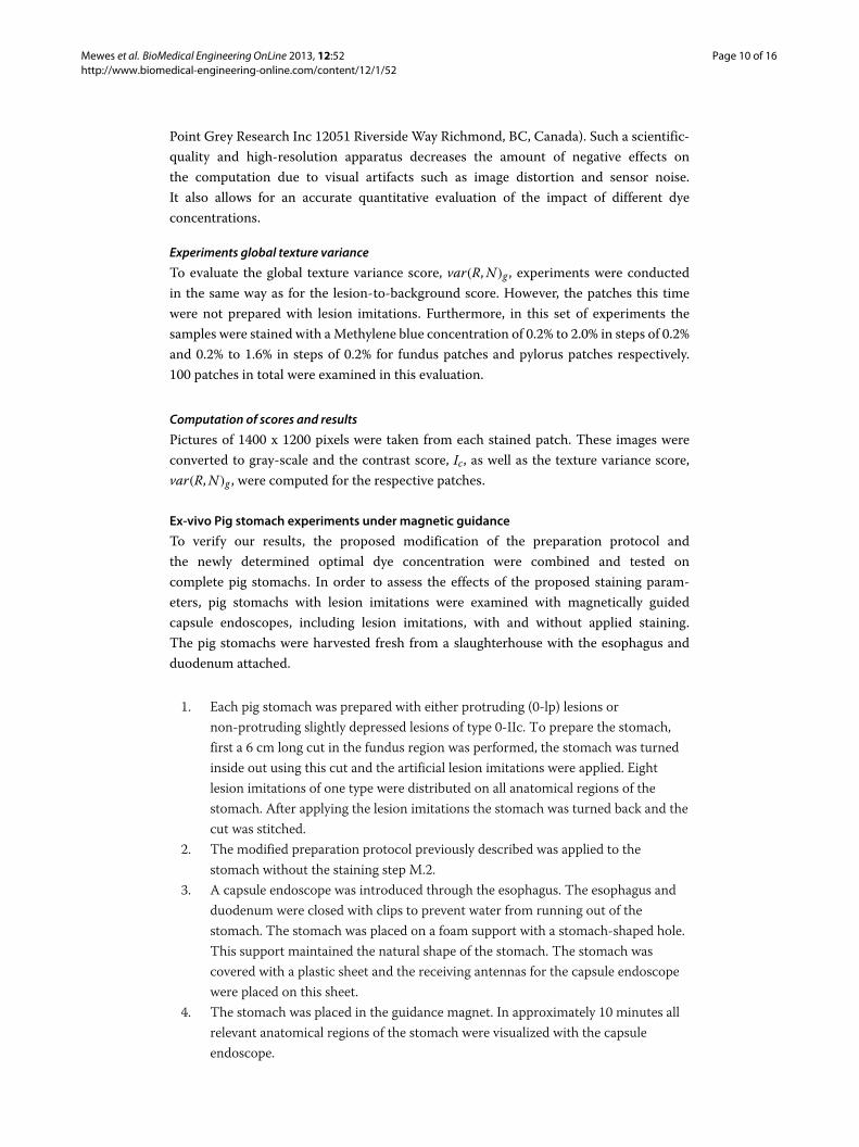
In total 190 tissue samples have been examined to determine an optimal concentration ofdye for the best lesion and mucosa visibility. Figure 5 summarizes these results. Each datapoint represents the score computed for one particular dye concentration averaged overfive patches. The yellow line with circle markers (right figure) shows the texture variancescore, var(R,N)g , for the Methylene blue concentration series from 0.0% to 2.0% withpig stomach patches stemming from the fundus region. A peak for the score appears atthe 0.4% Methylene blue solution. The blue line with square markers shows the texturevariance score, var(R,N)g , for the Methylene blue concentration series from 0.0% to 1.6%with pig stomach patches stemming from the pylorus region. A peak for the score occursat the 0.4% Methylene blue solution.The yellow line with circle markers (left figure) shows the lesions-to-background con-
trast score, Ic, for the indigo carmine concentration series from 0.0% to 1.6% with pigstomach patches stemming from the fundus region. A peak for the score occurs at the
Figure 5 Results of staining optimization: Left Figure: Yellow line with circle markers: Ic score for thefundus stomach patches stained with Indigo carmine solutions from 0.2% - 1.6%. Blue line with squaremarkers: Ic score for the pyloric stomach patches stained with indigo carmine solutions from 0.2% - 2.0%.Right Figure: Yellow line with circle markers: var(R,N)g score for patches from the fundus stomach region,staining was performed with Methylene blue solutions from 0.2% - 2.0%. Blue line with square markers:var(R,N)g score for patches from the pylorus stomach region, staining was performed with Methylene bluesolutions from 0.2% -1.6%.
Mewes et al. BioMedical Engineering OnLine 2013, 12:52 Page 12 of 16http://www.biomedical-engineering-online.com/content/12/1/52
0.2% indigo carmine solution. The blue line with square markers shows the lesions-to-background contrast score, Ic, for the indigo carmine concentration series from 0.0% to2.0% with pig stomach patches stemming from the pylorus region. A peak for the score isformed at the 0.6% Indigo carmine solution.Note that there is a 120% increase in the computed texture variance score,
var(R,N)g , between the non stained tissue and the maximal score within the con-centration series for the pylorus tissue sample and 105% increase for the fundus.For the lesions-to-background score, Ic, we noted an increase of 205% for the pylorus and469% for the fundus samples.
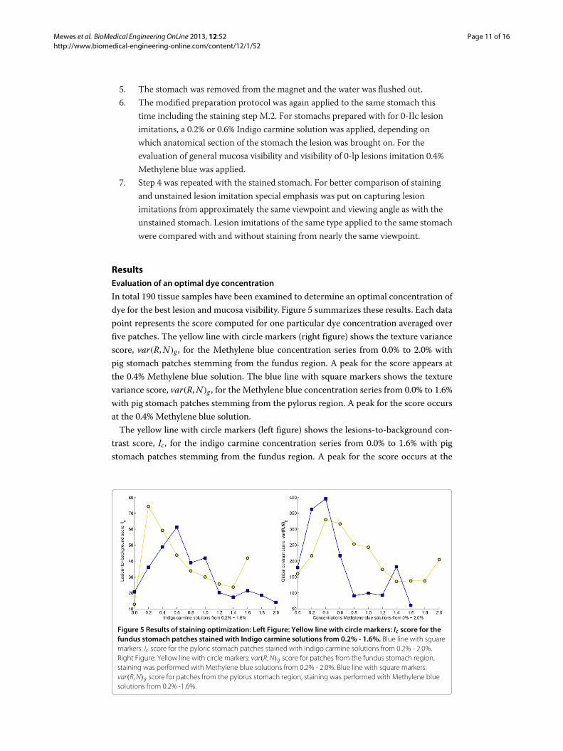
Application on a optimal dye in an ex-vivo experiment
Results from the previously conducted search for an optimal concentration were trans-ferred to and applied to ex-vivo experiments with six pig stomachs prepared with lesionimitation as previously described. Visual inspection shows that the general mucosa visi-bility and the lesion visibility are enhanced through the staining procedure (See Figure 6).Quantitative analysis supports this observation. The lesions-to-background score Ic andthe texture variance score var(R,N)g were computed for the images stemming from theex-vivo experiments. The texture variance score, var(R,N)g , in the pylorus region ofthe stomach was increased by 123% between the unstained and stained tissue patchescompared to the 120% increase in the experimental pig stomach patches. The lesion-to-background score, Ic, was increased by 95% between the unstained and stained lesions inthe pylorus region and is, therefore, only half as significant compared to the experimentalresults computed for the pig stomach patches.
Modification of preparation protocol
A modified preparation protocol for MGCE with chromoendoscopy was proposed. Theprotocol was tested in an ex-vivo pig stomach study under magnetic guidance of a capsuleendoscope. No negative impact on the underwater visibility or steerability of the capsulecould be observed.
DiscussionStaining procedure
The proposed modification to the preparation protocol did not have any negative effectson the examination in general. The visibility in the water-filled stomach was not dis-turbed by the additional stain applied during the modified protocol. This also holds truefor the steerability of the capsule. Since the stain did not significantly change the viscos-ity of the water, the mechanical forces applied to the capsule by the magnetic fields werenot affected either. Anatomical structures and landmarks (such as pylorus or cardia) areenhanced by the stain and can be easily spotted, making visual navigation easier for theoperator. The darker appearance of the mucosa did not result in a underexposed and darkimage in MGCE because the electronic shutter time adjustment of the capsule was ableto correct for the dimmer setup. Application of water and dye for the proposed modi-fied staining protocol has all been performed through the esophagus in order to simulateactual oral application. The oral application of dye for the examination of the stomachinvolving chromoendoscopy has been previously reported in humans.This suggests that
Mewes et al. BioMedical Engineering OnLine 2013, 12:52 Page 13 of 16http://www.biomedical-engineering-online.com/content/12/1/52
Figure 6 Experiments in a pig stomach under magnetic guidance: Comparison of stained andunstained pig stomachs examined with a guided capsule endoscope. Top: Lesion imitation of type 0-IIcwith 0.3% Indigo carmine staining. (A) left: without staining, (A) right: with staining. Bottom: Lesion imitationof type 0-Ip with 0.4% Methylene blue staining. (B) left: without staining, (B) right: with staining.
the modified staining protocol is applicable to capsule examinations in the stomach incombination with MGCE.
Evaluation of optimal stain concentration
190 pig stomach mucosa patches were stained with different staining concentrations andlesion imitations in order to systematically find an optimal staining concentration in thesubsequent ex-vivo experiments. Compared to previous studies ([28,29]) objective crite-ria were established in order to determine the optimal concentrations. The experimentsprovide a clear result on the optimal staining concentration for different lesion types anddifferent dye types. The staining concentration for an optimal lesion-to-background con-trast differs when the dye (Indigo carmine) is applied to lesion imitations in differentanatomical regions of the stomach. Those differences in optimal staining concentrationsfor different anatomical regions were previously observed in [28] due to differences inthe cell epithelium. In our work, we also observe these differences for different stom-ach regions. Lesion imitations are applied to the pylorus and cardia region. Endocrineglands in the pylorus region produce additional mucus that dilutes the 10% HCL acidused for lesion imitations. The damage to the epithelium is therefore modest comparedto the damage generated in the cardia region. A dye with higher concentration has, thus,a stronger effect on the staining of the damaged mucosa. The staining concentrationsfor the enhancement of global texture variance with Methylene blue is the same for both

Mewes et al. BioMedical Engineering OnLine 2013, 12:52 Page 14 of 16http://www.biomedical-engineering-online.com/content/12/1/52
anatomical regions. As a vital stain, Methylene blue is absorbed by specific cell types.Since neck cells (isthmus of the gland) and enteroendocrine cells (base of the gland) aresimilar in both regions the optimal dye concentration is also the same.
Ex-vivo pig stomach experiments
Optimal staining concentrations were transferred to ex-vivo experiments using a com-plete pig stomach under magnetic guidance. The lesion and mucosa visibility shows asimilar behavior compared to the pig stomach tissue samples. The proposedmethodologycould, therefore, be further generalized for the assessment of dye concentration in capsuleendoscopy and/or endoscopy in general. Furthermore, we showed that results of optimalstaining concentration obtained with a high quality camera could be transfered to the ex-vivo stomach experiments where a capsule endoscope with a low resulution camera wasused.
Technical limitations of ex-vivo experiments
Although freshly resected pig stomachs were used for all experiments the staining charac-teristics might be different from in-vivo experiments. This also holds for the authenticityof lesion imitations.
Future outcomes
Magnetically guided capsule endoscopes are an active field of research. The popular andestablished technique of chromoendoscopymay find its way to humanMGCE proceduresso that capsule examination and diagnosis can benefit from improved tissue localizationand characterization.
ConclusionFrom the first ex-vivo experiments combining chromoendoscopy and magneticallyguided capsule endoscopy we conclude that chromoendoscopy may be applied in MGCEand improves the mucosa and lesion visibility. Systematic evaluation provides importantinformation on appropriate staining concentrations for MGCE examination with respectto different dyes and anatomical differentiation. However, further animal and humanex-vivo and in-vivo studies are necessary before transferring this technique into clinicaltrials.
AbbreviationsMGCE: Magnetically guided capsule endoscopy; DOF: Degrees of freedom; EGD: Esophagogastroduodenoscopy; EGC:Early gastric cancer; lbp: Local binary pattern; EM: Electromagnetic.
Competing interests• P. W. Mewes: PhD thesis financially supported by Siemens AG (Healthcare Sector), Employee Siemens AG
(Healthcare Sector)• S. Foertsch: PhD thesis financially supported by Siemens AG (Healthcare Sector)• A. Lj. Juloski: Employee Siemens AG (Healthcare Sector)• E. Angelopoulou: Salary: Spouse: Management Position Siemens AG (Healthcare Sector) Spouse: Ownership
interest: Stock Shareholder Siemens AG (Healthcare Sector)• A. Hornegger: Head of Pattern Recognition Lab: Joint projects with Siemens AG (Healthcare Sector)• D. Guldi: The author disclosed no financial relationships relevant to this publication• H. Messmann: The author disclosed no financial relationships relevant to this publication• S.K. Goelder: The author disclosed no financial relationships relevant to this publication.
Authors’ contributionsPWM conception and design of the study, analysis and interpretation of data, drafting of manuscript. SF studysupervision , conception and design of the study. ALjJ critical revision of the manuscript for important intellectual

Mewes et al. BioMedical Engineering OnLine 2013, 12:52 Page 15 of 16http://www.biomedical-engineering-online.com/content/12/1/52
content, drafting and revision of the manuscript, approval of the final version of the manuscript. EA critical revision of themanuscript for important intellectual content, drafting and revision of the manuscript, approval of the final version of themanuscript. SKG study supervision, drafting and revision of the manuscript, approval of the final version of themanuscript. DG approval of the final version of the manuscript. JH approval of the final version of the manuscript. HMstudy supervision, drafting and revision of the manuscript, approval of the final version of the manuscript. All authorsread and approved the final manuscript.
AcknowledgementsWe acknowledge support by Deutsche Forschungsgemeinschaft and Friedrich-Alexander-Universität Erlangen-Nürnbergwithin the funding programme Open Access Publishing.
Author details1Pattern Recognition Lab, University of Erlangen-Nuremberg, Martensstrasse 3, Erlangen, Germany. 2Siemens AG,Healthcare Sector, Allee Am Roethelheimpark 2, Erlangen 91052, Germany. 3Department of Chemistry and Pharmacy,Friedrich-Alexander University, Egerlandstr. 3, Erlangen 91058,Germany. 4Medizinische Klinik III, Klinikum Augsburg,Stenglinstr.2, Augsburg 86156, Germany.
Received: 8 March 2013 Accepted: 5 June 2013Published: 11 June 2013
References1. Ferlay J, Shin H, Bray F, Forman D, Mathers C, Parkin D: Estimates of worldwide burden of cancer in
2008:GLOBOCAN 2008. Int J Cancer 2010, 127(12):2893–2917.2. Morson B, Sobin L, Grundmann E, Johansen A, Nagayo T, Serck-Hanssen A: Precancerous conditions and
epithelial dysplasia in the stomach. J Clin Pathol 1980, 33(8):711.3. Ang T, Khor C, Gotoda T: Diagnosis and endoscopic resection of early gastric cancer. Singapore Med J 2010,
51(2):93–100.4. Parkin D, Bray F, Ferlay J, Pisani P: Global cancer statistics, 2002. CA: A Cancer J Clin 2005, 55(2):74–108.5. Dinis-Ribeiro M, da Costa-Pereira A, Lopes C, Lara-Santos L, Guilherme M, Moreira-Dias L, Lomba-Viana H, Ribeiro A,
Santos C, Soares J, et al.:Magnification chromoendoscopy for the diagnosis of gastric intestinal metaplasiaand dysplasia. Gastrointest Endosc 2003, 57(4):498–504.
6. Lin B, Shun C, Wang T, Lin J: Endoscopic diagnosis of intestinal metaplasia of stomach: Accuracy judged byhistology. Hepato-gastroenterol 1999, 46(25):162–166.
7. Sano Y, Emura F, Ikematsu H: Narrow-band imaging. In Colonoscopy: Principles and Practice, Second Edition. Editedby Waye JD, Rex DK, Williams CB: Wiley Online Library; 2009:514–526.
8. Kiesslich R, Goetz M, Vieth M, Galle PR, Neurath MF, et al: Confocal laser endomicroscopy. Gastrointest Endosc ClinNorth Am 2005, 15(4):715.
9. Guelrud M, Herrera I, Essenfeld H, Castro J: Enhancedmagnification endoscopy: a new technique to identifyspecialized intestinal metaplasia in Barrett’s esophagus. Gastrointest Endosc 2001, 53(6):559–565.
10. Sivak Jr MV, Kobayashi K, Izatt JA, Rollins AM, Ung-Runyawee R, Chak A, Wong RC, Isenberg GA, Willis J:High-resolution endoscopic imaging of the GI tract using optical coherence tomography. Gastrointest Endosc2000, 51(4):474–479.
11. Chiu H, Chang C, Chen C, Lee Y, Wu M, Lin J, Shun C, Wang H: A prospective comparative study of narrow-bandimaging, chromoendoscopy, and conventional colonoscopy in the diagnosis of colorectal neoplasia. Gut2007, 56(3):373–379.
12. Neumann H, Neurath MF, Mudter J: New endoscopic approaches in IBD.World J Gastroenterol: WJG 2011, 17:63.13. Ratiu N, Rath H, Büttner R, Gelbmann C, Klebl F, Kullmann F, Rogler G, Strauch U, Schölmerich J, Messmann H: The
effect of chromoendoscopy on the diagnostic improvement of gastric ulcers by endoscopists with differentlevels of experience. Rom J Gastroenterol 2005, 14(3):239–244.
14. Mitooka H, Fujimori T, Maeda S, Nagasako K:Minute flat depressed neoplastic lesions of the colon detected bycontrast chromoscopy using an indigo carmine capsule. Gastrointest Endosc 1995, 41(5):453–459.
15. Kida M, Kobayashi K, Saigenji K: Routine chromoendoscopy for gastrointestinal diseases: indications revised.Endosc 2003, 35(7):590.
16. Araujo S, Costa A, Caravatto P, Dumarco R, Genzini T, Miranda M: Efficacy of contrast chromoendoscopy of thecolon after oral administration of indigo carmine dye. Arquivos de Gastroenterologia 2002, 39:153–157.
17. Bhutani M, Tandon R: Advances in Gastrointestinal Endoscopy by Bhutani. Jaypee Brothers, Medical Publishers; 2001.18. Rey J, Ogata H, Hosoe N, Ohtsuka K, Ogata N, Ikeda K, Aihara H, Pangtay I, Hibi T, Kudo S, et al.: Feasibility of
stomach exploration with a guided capsule endoscope. Endosc 2010, 42(7):541–545.19. Rey J-F, Ogata H, Hosoe N, Ohtsuka K, Ogata N, Ikeda K, Aihara H, Pangtay I, Hibi T, Kudo S, Ikeda K, Kudo S, et al.:
Blinded nonrandomized comparative study of gastric examination with a magnetically guided capsuleendoscope and standard videoendoscope. Gastrointest Endosc 2012, 75(2):373–381.
20. Swain P, Toor A, Volke F, Keller J, Gerber J, Rabinovitz E, Rothstein R: Remote magnetic manipulation of a wirelesscapsule endoscope in the esophagus and stomach of humans (with). Gastrointest Endosc 2010,71(7):1290–1293.
21. Ciuti G, Donlin R, Valdastri P, Arezzo A, Menciassi A, Morino M, Dario P, et al.: Robotic versus manual control inmagnetic steering of an endoscopic capsule. Endosc 2010, 42(2):148–152.
22. Kósa G, Jakab P, Jólesz F, Hata N: Swimming capsule endoscope using static and RF magnetic field of MRI forpropulsion. In Robotics and Automation,2008. ICRA 2008.IEEE International Conference on. Piscataway Township: IEEE;2008:2922–2927.
23. Sendoh M, Ishiyama K, Arai K: Fabrication of magnetic actuator for use in a capsule endoscope.Magnetics, IEEETrans 2003, 39(5):3232–3234.
Mewes et al. BioMedical Engineering OnLine 2013, 12:52 Page 16 of 16http://www.biomedical-engineering-online.com/content/12/1/52
24. Canto M, et al.: Staining in gastrointestinal endoscopy: the basics. Endosc 1999, 31:479–486.25. Peitz U, Malfertheiner P: Chromoendoscopy: from a research tool to clinical progress. Dig Dis 2003,
20(2):111–119.26. Halperin D, Belmonte R, Sanchez L, Mohar A, Guarner J, Herrera R:Methylene blue staining for the endoscopic
diagnosis of intestinal metaplasia in the stomach. Gastroenterol 1998, 114:A605–A605.27. Canto M, Setrakian S, Petras R, Blades E, Chak A, Sivak M, et al.:Methylene blue selectively stains intestinal
metaplasia in Barrett’s esophagus. Gastrointest Endosc 1996, 44:1–7.28. Goetz M, Toermer T, Vieth M, Dunbar K, Hoffman A, Galle P, Neurath M, Delaney P, Kiesslich R: Simultaneous
confocal laser endomicroscopy and chromoendoscopy with topical cresyl violet. Gastrointest Endosc 2009,70(5):959–968.
29. Kodashima S, Fujishiro M, Takubo K, Kammori M, Nomura S, Kakushima N, Muraki Y, Tateishi A, Kaminishi M, OmataM: Ex-vivo study of high-magnification chromoendoscopy in the gastrointestinal tract to determine theoptimal staining conditions for endocytoscopy. Endosc 2006, 38(11):1115–1121.
30. Keller H, Juloski A, Kawano H, Bechtold M, Kimura A, Takizawa H, Kuth R:Method for Navigation and Control of aMagnetically Guided Capsule Endoscope in the Human Stomach. In Biomedical Robotics and Biomechatronics(BioRob),2010 3rd IEEE RAS and EMBS International Conference on. Piscataway Township: IEEE; 2012:859–865.
31. Mewes P, Foertsch S, Angelopoulou E, Guldi D, Messmann H: ChromoendoscopyWith automatic lesionenhancement in magnetically guided capsule endoscopy: a feasibility study. Gastroenterol 2011,140(5):S–S750.
32. Koseki S, Mizuno Y, Sotome I:Modeling of pathogen survival during simulated gastric digestion. Appl EnvironMicrobiol 2011, 77(3):1021–1032.
33. Kong F, Singh RP: A human gastric simulator (HGS) to study food digestion in human stomach. J Food Sci 2010,75(9):E627–E635.
34. Lambert R: Endoscopic classification review group. Update on the Paris classification of superficialneoplastic lesions in the digestive tract. Endosc 2005, 37(6):570–578.
35. Wang TE, Wang HY, Lin CC, Chen TY, Chang CW, Chen CJ, Chen M: Simulating a target lesion for endoscopicsubmucosal dissection training in an ex vivo pig model. Gastrointest Endosc 2011, 74(2):398–402.
36. Lee B, Kim G, Park D, Kim D, Jeon T, Park S, You H, Ryu D, Kim D, Song G: Acetic acid-indigo carminechromoendoscopy for delineating early gastric cancers: its usefulness according to histological type. BMCGastroenterol 2010, 10:97.
37. Kovalev V, Kruggel F, Gertz H, Von Cramon D: Three-dimensional texture analysis of MRI brain datasets.MedImaging, IEEE Trans 2001, 20(5):424–433.
38. Glatard T, Montagnat J, Magnin I: Texture based medical image indexing and retrieval: application to cardiacimaging. In Proceedings of the 6th ACM SIGMM International Workshop onMultimedia Information Retrieval. New York:ACM; 2004:135–142.
39. Ojala T, Pietikainen M, Harwood D: Performance evaluation of texture measures with classification based onkullback discrimination of distributions. In Pattern Recognition,1994. Vol. 1-Conference A:Computer Vision & ImageProcessing., Proceedings of the 12th IAPR International Conference on, Volume 1: Piscataway Township: IEEE;1994:582–585.
40. Guo Z, Zhang L, Zhang D: Rotation invariant texture classification using LBP variance (LBPV) with globalmatching. Pattern Recognit 2009, 43(3):706–719.
41. Ojala T, Pietikainen M, Mäenpää T:Multiresolution gray-scale and rotation invariant texture classification withlocal binary patterns. IEEE Trans Pattern Anal Mach Intel 2002, 24(7):971–987.
doi:10.1186/1475-925X-12-52Cite this article as:Mewes et al.:Chromoendoscopy inmagnetically guided capsule endoscopy. BioMedical EngineeringOnLine 2013 12:52.
Submit your next manuscript to BioMed Centraland take full advantage of:
• Convenient online submission
• Thorough peer review
• No space constraints or color figure charges
• Immediate publication on acceptance
• Inclusion in PubMed, CAS, Scopus and Google Scholar
• Research which is freely available for redistribution
Submit your manuscript at www.biomedcentral.com/submit