chromosomal analysis
TRANSCRIPT

CHROMOSOMAL ANALYSIS
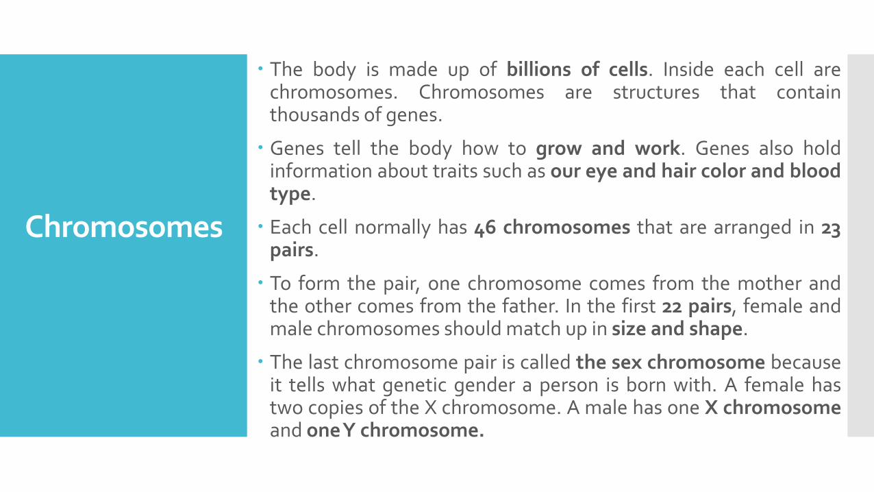
Chromosomes
The body is made up of billions of cells. Inside each cell arechromosomes. Chromosomes are structures that containthousands of genes.
Genes tell the body how to grow and work. Genes also holdinformation about traits such as our eye and hair color and bloodtype.
Each cell normally has 46 chromosomes that are arranged in 23pairs.
To form the pair, one chromosome comes from the mother andthe other comes from the father. In the first 22 pairs, female andmale chromosomes should match up in size and shape.
The last chromosome pair is called the sex chromosome becauseit tells what genetic gender a person is born with. A female hastwo copies of the X chromosome. A male has one X chromosomeand oneY chromosome.

Chromosome abnormalities have long beenrecognised as an important cause of learningdisability and multiple malformationsyndromes; 0.8% of live born infants havenumerical or structural chromosomalanomalies resulting in an abnormal phenotype.
The identification of such anomalies isimportant, both clinically and for accurate geneticcounselling

Karyotypes can be used to screen for
and confirm chromosomal
abnormalities such as Down's
syndrome and Cat Eye Syndrome, and
there are several different types of
abnormalities which may be detected.
Chromosomal abnormalities:
Trisomies in which there are three copies of one ofthe chromosomes rather than two
Monosomies in which only one copy (instead of two)is present
Chromosome deletions in which part of achromosome is missing
Chromosome translocations in which a part of onechromosome is attached to another chromosome(and vice versa in balanced translocations.)

Examples of trisomies
include
Down syndrome (trisomy 21)
Edward syndrome (trisomy 18)
Patau syndrome (trisomy 13)
Klinefelter's syndrome (XXY and other variations) - Klinefelter'ssyndrome occurs in 1 in 500 newborn males
Triple X syndrome (XXX)

An example of monosomy
includes
Turner syndrome (X0) or monosomy X –
Roughly 10% of first trimester miscarriages are due to Turner syndrome,
but this monosomy is present in only around 1 in 2,500 live female births

Examples of chromosomal
deletions include
Cri-du-Chat syndrome
(missing chromosome 5)
Williams syndrome
(missing chromosome 7)

Chromosome translocations
Translocations - There are many examples oftranslocations including translocation Downsyndrome. Robertsonian translocations are fairlycommon, occurring in roughly 1 in 1000 people.
Mosaicism is a condition in which some cells in thebody have a chromosomal abnormality while othersdo not. For example, mosaic Down syndrome ormosaic trisomy 9. Full trisomy 9 is not compatiblewith life, but mosaic trisomy 9 may result in a livebirth.
What Is a Karyotype
Test?
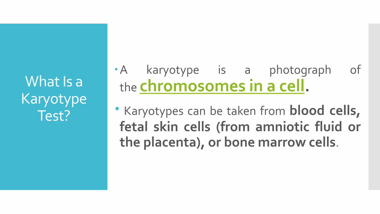
A karyotype is a photograph of
the chromosomes in a cell.
Karyotypes can be taken from blood cells,fetal skin cells (from amniotic fluid orthe placenta), or bone marrow cells.

WHEN IT'S DONE
There are many situations in which a karyotype may be
recommended. These might include
Infants or children who have medical conditions whichsuggest a chromosomal abnormality that has not yet beendiagnosed.
Adults who have symptoms suggestive of a chromosomalabnormality (for example, men with Klinefelter's diseasemay go undiagnosed until puberty or adulthood.) Some ofthe mosaic trisomy disorders may also go undiagnosed.
Infertility: A genetic karyotype may be done for infertility.As noted above, some chromosomal abnormalities may goundiagnosed until adulthood. A woman with Turnersyndrome or a man with one of the variants of Klinefelter'smay not be aware of the condition until they are coping withinfertility.

WHEN IT'S DONE
There are many situations in which a karyotype may be
recommended. These might include
Prenatal testing: In some cases, such as translocation Downsyndrome, the condition may be hereditary and parents may betested if a child has been born with a Down syndrome. (It'simportant to note that most of the time Down syndrome is not ahereditary disorder but rather a chance mutation.)
Stillbirth: A karyotype is often done as part of the testingfollowing a stillbirth.
Recurrent miscarriages: A parental karyotype of recurrentmiscarriages may give clues as to the reasons for thesedevastating recurring losses. It's thought that chromosomalabnormalities, such as trisomy 16, are the cause of at least 50% ofmiscarriages.
Leukemia: Karyotype testing may also be done to help diagnoseleukemias, for example, by looking for the Philadelphiachromosome found in some people with chronic myelogenousleukemia or acute lymphocytic leukemia.

P
r
o
c
e
d
u
r
e
o
f
K
a
r
y
o
t
y
p
i
n
g
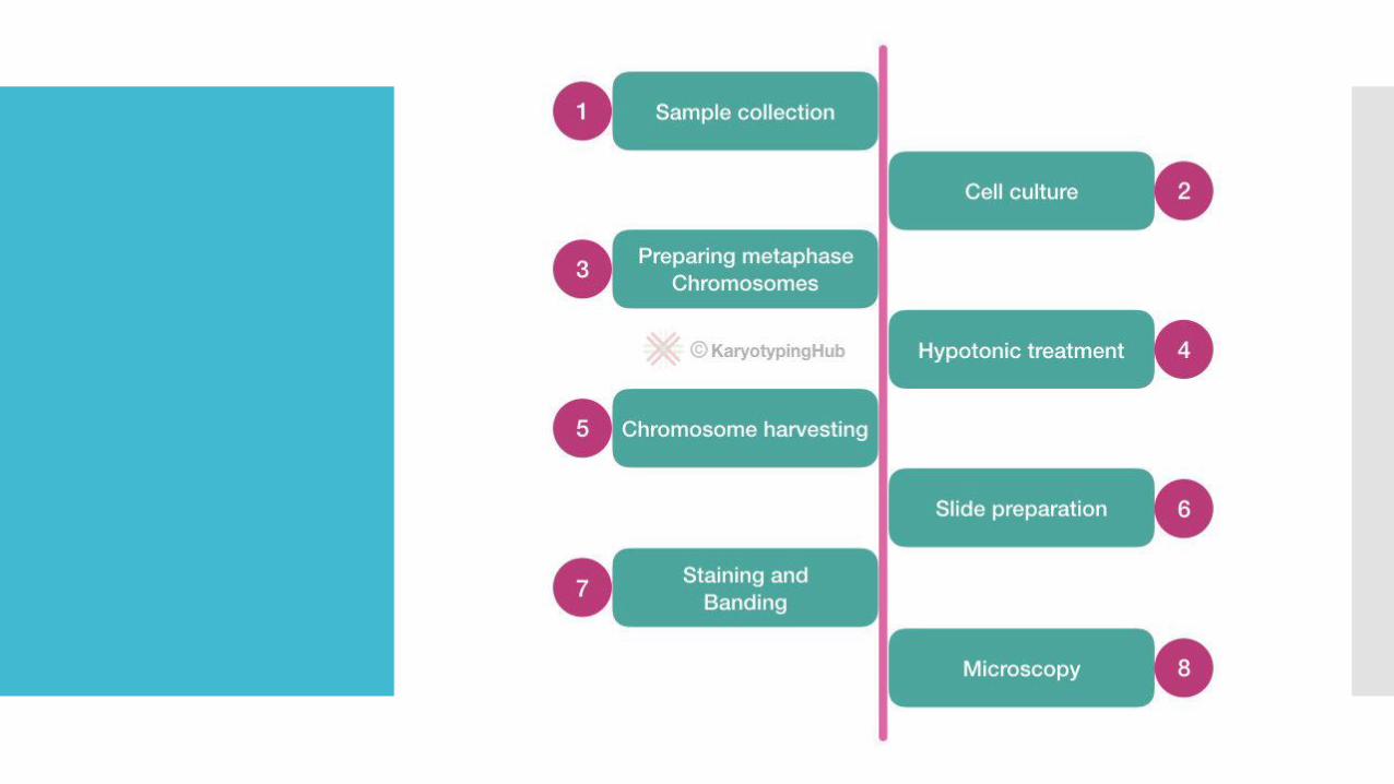

Sample collection:
Any bodily fluid having dividing cells can be cultured,however, blood, amniotic fluid, solid tissue, tumor,chorionic villi are common samples used in karyotyping.
The sample is collected in a sterile tube, heparin tube, or AFcollecting tube. The sample is immediately sent to thekaryotyping center. Transport the sample under 4°Ctemperature. It is essential to maintain an asepticconditions.
Notably, sample collection also as crucial as any othertechnique in karyotyping. The blood sample is collected inthe heparin tube. EDTA prevent cell division and thereforenot used.
Cell culture
The sample is culture using the culture media, usuallyready to use media like the complete RPMI-1640 areused, but we can also prepare our own culturing mediaby mixing various chemicals.
The final culture media must contain antibiotics, L-glutamine, or phytohemagglutinin M. Both areessential amino acids and must be needed in cellculture.

Preparing the metaphase chromosome
In metaphase, chromosomes are arranged well. Thus we needthat one for karyotyping. To fulfill the present aim we need toarrest all our cells at the metaphase stage of cell division.
Adding colchicine into the culture prevents the spindleformation and therefore arrests cells at metaphase.
The elongated forms of colchicine treated chromosomes.
Hypotonic treatment
In order to isolate chromosomes, we need to swell cellsto observe chromosomes.
Usually, the KCl is used as a hypotonic solution.
A special type of solution known as a hypotonicsolution is used to isolate chromosomes from the cells.The reason for doing this is to separate chromosomeswell on a slide.
Chromosomes harvesting
Now we need to harvest cells to remove the debris.Using the treatment of methanol and glacial acetic acidwith the process of centrifugation, cells are harvested.
Slide preparation
Unlike other slide preparation methods, the slidepreparation technique in karyotyping is a bit different.
Here to separate every chromosome properly we needto drop the cell suspension from some height. For thatpre-chilled, methanol treated slide is used so that oncethe cell suspension hits the slide, it is immediately fixedon the surface.

Staining & banding:
Now our slide is ready but to know whether the cells are cultured properlyor not, we have to first stain it with some color.
G-staining or Giemsa staining is used to ensure cell culture. Chromosomesare stained blue in Giemsa and looking like this, see the figure:
In order to encounter various anomalies and to study chromosomes, weneed to do banding. Various banding techniques are used for variousindications.
GTG banding, Q-banding, NOR-banding, C-banding, T-banding are somecommonly used banding techniques in cytogenetic labs.

Microscopy
First, the slide is observed under the 10X microscope lens tochoose the best and well-separated filed. Then it isobserved in the 45X and 100X oil-immersion lenses.
50 to 100 well-separated metaphase chromosome fields areobserved to validate the results. After that, a karyogram orkaryotype is prepared.
Chromosomes are arranged chronologically to detectfurther indications.

Advantages
The cytogenetic techniques, especially, the karyotyping method is utilized to observe chromosomes. In a PCR, we can’t amplify the entire genome or whole chromosome DNA.
To study chromosomal aberration we have to perform karyotyping, however, nowadays FISH, spectral karyotyping, and microarray like techniques are available.
The present method is powerful enough to identify numerical and structural chromosomal abnormalities.
It is a bit accurate and thus used in prenatal diagnosis and screening of chromosomal abnormalities.

Disadvantages
The disadvantages of karyotyping are more than advantages. It is a time-consuming technique.
High-end sophisticated instrumentation is a need.
Strict aseptic conditions are required throughout the protocol.
The chance of contamination is very high.
The chance of reaction failure is also very high.
A high level of expertise is needed to proceed and analyze the results.
Also, an expert’s touch is required to interpret the results.

Applications
Briefly, some of the applications are,
To study chromosome number and structure.
To identify chromosomal anomalies
To study centromere location
To study the telomeric regions
To identify the relative size of chromosomes
To differentiate the chromosomes of different species.