chronic irradiation with 222-nm uvc light induces neither
TRANSCRIPT

RESEARCH ARTICLE
Chronic irradiation with 222-nm UVC light
induces neither DNA damage nor epidermal
lesions in mouse skin, even at high doses
Kouji Narita1,2, Krisana Asano1,3, Yukihiro Morimoto4, Tatsushi Igarashi4, Akio Nakane1,3*
1 Department of Microbiology and Immunology, Hirosaki University Graduate School of Medicine, Hirosaki,
Aomori, Japan, 2 Institute for Animal Experimentation, Hirosaki University Graduate School of Medicine,
Hirosaki, Aomori, Japan, 3 Department of Biopolymer and Health Science, Hirosaki University Graduate
School of Medicine, Hirosaki, Aomori, Japan, 4 Ushio Inc., Chiyoda-ku, Tokyo, Japan
Abstract
Surgical site infections (SSIs) represent an important clinical problem associated with
increased levels of surgical morbidity and mortality. UVC irradiation during surgery has
been considered to represent a possible strategy to prevent the development of SSI. 254-
nm UVC induces marked levels of DNA damage by generating cyclobutyl pyrimidine dimers
(CPD) in microorganisms. However, this effect is elicited not only in microorganisms, but
also in human cells, and chronic exposure to 254-nm UVC has been established to repre-
sent a human health hazard. In contrast, despite short wavelength-UVC light, especially
222-nm UVC, having been demonstrated to elicit a bactericidal effect, single irradiation with
a high dose of 222-nm UVC energy has been reported to not induce mutagenic or cytotoxic
DNA lesions in mammalian cells. However, the effect of chronic irradiation with a high dose
of 222-nm UVC to mammalian cells has not been determined. In this study, it was demon-
strated that large numbers of CPD-expressing cells were induced in the epidermis of mice
following treatment with a small amount of single exposure 254-nm UVC, and then less than
half of these cells reduced within 24 h. Chronic 254-nm UVC irradiation was revealed to
induce sunburn and desquamation in mouse skin. Histological analysis demonstrated that
small numbers of CPD-expressing cells were detected only in hyperkeratotic stratum cor-
neum after chronic irradiation with a high dose of 254-nm UVC, and that significant hyperpla-
sia and intercellular edema were also induced in the epidermis of mice. In contrast, chronic
irradiation with 222-nm UVC light was revealed not to induce mutagenic or cytotoxic effects
in the epidermis of mice. These results indicated that 222-nm UVC light emitted from the
lamp apparatus (or device), which was designed to attenuate harmful light present in wave-
lengths of more than 230 nm, represents a promising tool for the reduction of SSI incidence
in patients and hospital staff.
PLOS ONE | https://doi.org/10.1371/journal.pone.0201259 July 25, 2018 1 / 9
a1111111111
a1111111111
a1111111111
a1111111111
a1111111111
OPENACCESS
Citation: Narita K, Asano K, Morimoto Y, Igarashi
T, Nakane A (2018) Chronic irradiation with 222-
nm UVC light induces neither DNA damage nor
epidermal lesions in mouse skin, even at high
doses. PLoS ONE 13(7): e0201259. https://doi.org/
10.1371/journal.pone.0201259
Editor: Albert J. Fornace, Jr, Georgetown
University, UNITED STATES
Received: April 16, 2018
Accepted: July 11, 2018
Published: July 25, 2018
Copyright: © 2018 Narita et al. This is an open
access article distributed under the terms of the
Creative Commons Attribution License, which
permits unrestricted use, distribution, and
reproduction in any medium, provided the original
author and source are credited.
Data Availability Statement: All relevant data are
within the paper.
Funding: This work was supported by USHIO Inc.,
Tokyo, Japan. The funder provided support in the
form of salaries for authors [Y. M. and T. I.] but did
not have any additional role in the study design,
data collection and analysis, decision to publish, or
preparation of the manuscript.
Competing interests: I have read the journal’s
policy and the authors of this manuscript Yukihiro
Morimoto and Tatsushi Igarashi have the following
Introduction
Surgical site infections (SSIs) are defined as infections that affect either the incision or deep tis-
sue at operation sites, typically occur within 30 days of an operative procedure and are associ-
ated with increased levels of surgical morbidity and mortality. Despite improvements in
preventive treatment, including antisepsis of hands and forearms, sterilization of surgical
instruments and preparation of the patient’s skin, SSIs remain a significant clinical problem
[1, 2].
Microbiota present on the patient’s skin and mucous membranes have been well established
to represent an important pathogen source of SSIs [3]. On the other hand, it has also been
reported that approximately 80–90% of bacterial contaminants found in wounds post-surgery
are due to air in the operating room. Hence, the reduction of airborne bacterial contaminants
may represent an effective strategy for decreasing the incidence of SSIs [4].
It has been well established that UVC light within the range of 240–280 nm elicits a signifi-
cant germicidal effect [5]. Germicidal lamps with an emission peak at 254-nm have been
widely used to kill and inactivate bacteria and viruses [6]. It has previously been reported that
UVC irradiation produced by a germicidal lamp reduced the number of environmental bacte-
ria in an operating room and lowered the risk of wound infection following joint replacement
surgery performed in the operating room [7]. Furthermore, it has been established that
254-nm UVC is directly absorbed by DNA in microorganisms and induces a variety of muta-
genic and cytotoxic DNA lesions [8]. Cyclobutyl pyrimidine dimers (CPD) caused by UV radi-
ation interrupt transcription, translation and replication of DNA leads to bacterial cell death
and viral inactivation [8]. It has been previously reported that chronic UV irradiation induces
skin damage. Chronic UVB irradiation induces a large number of CPD in mouse epidermal
cells. However, the number of cells retaining CPD decreased within several days and following
irradiation with UVB light, epidermal hyperplasia subsequently occurred in skin [9]. The
induction of tumorigenesis following chronic irradiation with a high dose of UVB light to
mouse skin was reported to be dependent on the accumulation of CPD as well as the degree of
epidermal hyperplasia [10]. Similarly, treatment with 254-nm UVC also induces CPD accumu-
lation in mammalian cells and epidermal hyperplasia, and repeated exposure to 254-nm UVC
for long durations of time is known to represent a human health hazard, cause dermatitis and
increase the risk of skin cancer [8, 11]. These studies demonstrated that when UVC irradiation
is applied for the prophylaxis of SSIs, use of personal protective equipment is necessary for
patients and hospital staff in order to prevent 254-nm UVC exposure during surgical proce-
dures [12].
UVC light with decreased wavelengths (~200–230 nm) has been reported to be harmless to
mammalian cells since it does not reach the nuclei, as the light is absorbed by proteins, particu-
larly by peptide bonds and other biomolecules [13, 14, 15]. It has been reported that irradiation
with 222-nm UVC light efficiently reduces bacterial counts of MRSA in vitro without inducing
mouse skin damage, which is typically induced by conventional germicidal UV exposure invivo [14]. We previously demonstrated that single irradiation with 222-nm UVC light reduces
the number of bacterial MRSA in mouse skin wounds immediately after the irradiation, and
does not induce CPD in mouse epidermal cells [16]. These studies demonstrated that single
irradiation with 222-nm UVC may be harmless to the epidermis of mice. Treatment with
222-nm UVC irradiation in operating rooms during surgery may represent an effective strat-
egy for the prophylaxis of SSIs without eliciting harmful effects to patients and hospital staff.
However, the effect of chronic 222-nm UVC irradiation to mammalian skin has not yet been
determined.
Chronic irradiation with 222-nm UVC light induces neither DNA damage nor epidermal lesions
PLOS ONE | https://doi.org/10.1371/journal.pone.0201259 July 25, 2018 2 / 9
competing interests: Yukihiro Morimoto and
Tatsushi Igarashi are provided support in the form
of salaries from Ushio Inc., Tokyo, Japan. This
does not alter our adherence to PLOS ONE policies
on sharing data and materials.
In this study, it was demonstrated that single irradiation with 254-nm UVC induced CPD-
retaining cells in the epidermis, which then decreased to almost half of the initial value a total
of 24 h after irradiation. Following the treatment of mice with chronic irradiation using a high
dose of 254-nm UVC light for 10 days, a small number of CPD-retaining cells were detected in
the hyperkeratotic stratum corneum alone. Conversely, significant hyperplasia and intercellu-
lar edema were induced in the epidermis of mice following chronic irradiation. In contrast,
chronic irradiation with a high dose of 222-nm UVC did not induce the CPD-retaining cells
or epidermal abnormalities in mouse dorsal skin.
Materials and methods
Mice and ethics statement
Seven-week-old female hairless mice (Hos: HR-1; Japan SLC, Inc., Hamamatsu, Japan) were
used in this study. Mice were maintained under specific pathogen-free conditions at the Insti-
tute for Animal Experimentation, Hirosaki University Graduate School of Medicine. All
experiments were carried out in strict accordance with the Guidelines for Animal Experimen-
tation of Hirosaki University. The experimental protocol was approved by the ethics commit-
tee of the Institute for Animal Experimentation, Hirosaki University Graduate School of
Medicine (Permit number: M16010).
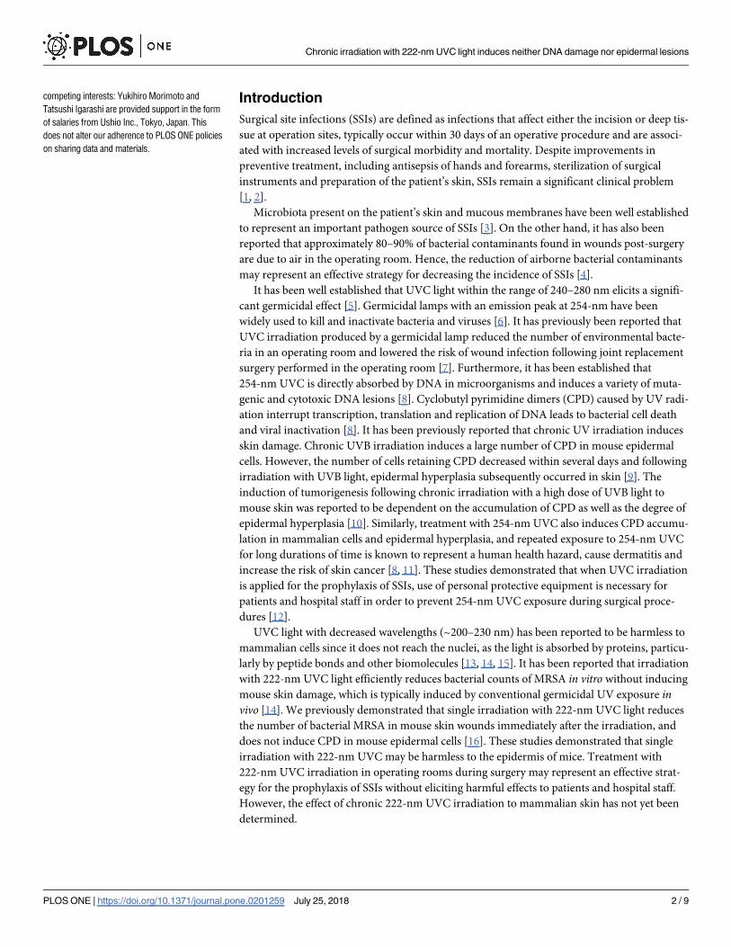
UVC light source
Two types of lamp devices were used for UVC light irradiation: one was mounted using a
krypton-chloride (Kr-Cl) excimer lamp and an optical filter that restricted spectra emitting
light ranging between 200-230-nm, of which the maximum output wavelength was 222-nm;
and the other was a conventional low-pressure mercury lamp named SUV-4 (AS ONE corp.
Osaka, Japan), which emitted a line spectrum at 254-nm. The 222-nm-emitting SafeZoneUVC
device (Ushio Inc. Tokyo, Japan) is composed of a lamp, air-cooling fan, mirrors and a custom
band-pass filter. The filter was used to block almost all wavelengths except for the dominant
222-nm emission wavelength (Fig 1). Irradiance emitted by 222-nm light was measured using
an S-172/UIT250 accumulated UV meter (Ushio Inc.), and was found to be 5 mW/cm2 at a
Fig 1. Measured spectra emitted from the Kr-Cl excimer lamp equipped with a band-pass filter.
https://doi.org/10.1371/journal.pone.0201259.g001
Chronic irradiation with 222-nm UVC light induces neither DNA damage nor epidermal lesions
PLOS ONE | https://doi.org/10.1371/journal.pone.0201259 July 25, 2018 3 / 9
distance of 10 mm from the emission window. Irradiance emitted by 254-nm light was deter-
mined by an S-254/UIT250 (Ushio Inc.), and was revealed to be 3 mW/cm2 at a distance of 20
mm from the window.
UVC irradiation
Two experiments were performed in this study: One to observe the recovery rate of CPD gen-
erated by irradiation subjected to the dorsal skin of hairless mice using a small amount of 254
nm-light, and the other to evaluate the toxicity associated with chronic irradiation. Groups of
five mice were anesthetized with isoflurane (Pfizer Japan Inc., Tokyo, Japan), and their dorsal
skins were then irradiated using 254-nm UVC at 75 mJ/cm2. We previously reported that mice
subjected to single irradiation treatment of 222-nm at 450 mJ/cm2 exhibited significantly
reduced MRSA bacterial counts on dorsal skin [16]. To evaluate the effect of chronic 254 or
222-nm UVC irradiation on dorsal skin, mice were anesthetized with isoflurane (Pfizer Japan
Inc.) and subsequently subjected to daily irradiation with 254-nm or 222-nm UVC light at a
dose of 450 mJ/cm2/day on days 1, 2, 3, 4, 5, 8, 9 and 10. Gross appearances of irradiated skin
were observed immediately after irradiation.
Histological and immunohistochemical analysis
The dorsal skins of mice were immediately harvested at 1, 3, 6 and 24 h time intervals post-
irradiation with 254-nm UVC at 75 mJ/cm2. In addition, the dorsal skins of mice subjected to
irradiation with UVC at 450 mJ/cm2 were collected 1 day after the last treatment. Skin tissues
were then fixed with 10% phosphate-buffered formalin overnight. Paraffin-embedded tissue
and 2 μm-thick tissue sections were prepared. After deparaffinization and rehydration of the
tissue sections, hematoxylin and eosin staining was performed. To detect CPD formation, skin
sections were prepared as described above, and antigen retrieval was performed by incubating
the sections in proteinase K solution (1:1000; Qiagen, Hilden, Germany). The sections were
then incubated with HRP-conjugated anti-CPD monoclonal antibodies (Kaniya Biomedical
Co., Seattle, WA) overnight at 4˚C. After rinsing three times in PBS for 5 min each, the color
reaction was developed by addition of diaminobenzidine. Following this, counterstaining was
performed with hematoxylin. CPD-positive cells were quantified by counting the cells in 10
random visual fields of each section (200× magnification).
Results
Time course of DNA damage repair following single 254-nm UVC
irradiation
To investigate DNA repair following CPD expression induced by single irradiation with
254-nm UVC, dorsal skin of hairless mice was irradiated with 254-nm UVC light at 75 mJ/
cm2. The results revealed that CPD-expressing cells were distributed in the stratum spinosum
immediately after irradiation. Furthermore, the distribution of CPD-expressing cells at 1 h
after irradiation was similar to that observed immediately after irradiation. CPD-expressing
cells were then detected in the upper stratum spinosum at 3 and 6 h after irradiation. A total of
24 h after irradiation, CPD-expressing cells appeared flattened in shape and were only detected
on surface of the epidermis (Fig 2A). In addition, 37% of keratinocytes expressed CPD imme-
diately after irradiation, and the number of CPD-expressing cells then gradually reduced to
13% a total of 24 h post-irradiation (Fig 2B).
Chronic irradiation with 222-nm UVC light induces neither DNA damage nor epidermal lesions
PLOS ONE | https://doi.org/10.1371/journal.pone.0201259 July 25, 2018 4 / 9

Effect of chronic irradiation with 222-nm UVC on epidermis of mouse skin
To investigate epidermal damage induced by chronic irradiation using 254-nm and 222-nm
UVC light, mice were irradiated a total of 8 times according to the aforementioned protocol.
As shown in Fig 3, sunburn and desquamation were observed in the dorsal skins of mice irra-
diated with 254-nm UVC, not with 222-nm UVC, on days 4 and 5. Although sunburn and des-
quamation symptoms were found to be attenuated on day 8, the abnormal findings were
exacerbated in the 254-nm UVC-irradiated mice on days 9 and 10. In the dorsal skin of
222-nm irradiated mice as well as non-irradiated mice, these abnormal findings were not
observed during evaluation (Fig 3). To further investigate the effect of chronic irradiation with
222-nm UVC and 254-nm UVC on the epidermis of mouse skin, histological analyses were
performed. The dorsal skins of mice that had been irradiated 8 times with 254-nm UVC exhib-
ited parakeratosis, epidermal hyperplasia, intracellular edema and mitotic figures in the stra-
tum spinosum. These histological findings were not observed in the epidermis of mice
irradiated with 222-nm UVC or in non-irradiated mice (Fig 4A). In contrast, CPD-expressing
cells in the skin of mice subjected to chronic irradiation with 254-nm UVC were detected only
in the hyperkeratotic stratum corneum, but not in the stratum spinosum. CPD-expressing
Fig 2. Time-course analysis of DNA damage repair following 254-nm UVC irradiation. Dorsal skins of mice
irradiated with 254-nm UVC at 75 mJ/cm2 were immediately collected post-treatment, as well as 1, 3, 6 and 24 h post-
irradiation. (A) CPD-expressing cells were detected by immunohistochemistry (200× magnification). (B) Percentages
of CPD-expressing cells per visual field (200× magnification) were enumerated (n = 5).
https://doi.org/10.1371/journal.pone.0201259.g002
Chronic irradiation with 222-nm UVC light induces neither DNA damage nor epidermal lesions
PLOS ONE | https://doi.org/10.1371/journal.pone.0201259 July 25, 2018 5 / 9
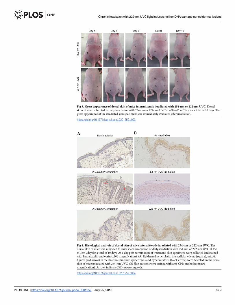
Fig 3. Gross appearance of dorsal skin of mice intermittently irradiated with 254-nm or 222-nm UVC. Dorsal
skins of mice subjected to daily irradiation with 254-nm or 222-nm UVC at 450 mJ/cm2/day for a total of 10 days. The
gross appearance of the irradiated skin specimens was immediately evaluated after irradiation.
https://doi.org/10.1371/journal.pone.0201259.g003
Fig 4. Histological analysis of dorsal skin of mice intermittently irradiated with 254-nm or 222-nm UVC. The
dorsal skin of mice was subjected to daily sham-irradiation or daily irradiation with 254-nm or 222-nm UVC at 450
mJ/cm2/day for a total of 10 days. At 1 day post-termination of treatment, skin specimens were collected and stained
with hematoxylin and eosin (x200 magnification). (A) Epidermal hyperplasia, intracellular edema (square), mitotic
figures (red arrow) in the stratum spinosum epidermidis and hyperkeratosis (black arrow) were detected on the dorsal
skin of mice irradiated with 254-nm UVC. (B) Skin sections were stained with anti-CPD antibodies (x400
magnification). Arrows indicate CPD-expressing cells.
https://doi.org/10.1371/journal.pone.0201259.g004
Chronic irradiation with 222-nm UVC light induces neither DNA damage nor epidermal lesions
PLOS ONE | https://doi.org/10.1371/journal.pone.0201259 July 25, 2018 6 / 9

cells were not detected in the epidermis of mice irradiated with 222-nm UVC or in non-irradi-
ated mice (Fig 4B).
Discussion
UVC irradiation emitted by germicidal lamps in operation rooms during surgery has been con-
sidered to represent a potential strategy for the reduction of SSIs, as airborne bacteria have been
revealed to be closely associated with the incidence of SSIs [7, 12, 17]. It has been established that
the mechanism of using 254-nm UVC light for bactericidal effects impairs the genetic materials in
which CPD is predominantly damaged in bacterial DNAs [6, 18]. However, exposure of human
cells to 254-nm UVC causes the formation of mutagenic and cytotoxic DNA lesions, which, if
exposed to for a sufficient duration of time, may lead to the initiation and progression of skin can-
cer [11]. Alternatively, mammalian cells have numerous repair systems for UV-induced DNA
lesions. Nucleotide excision repair (NER) is one of the most versatile and flexible repair systems
[19]. It has previously been reported that approximately 30% of CPD-retaining human epidermal
keratinocytes are repaired within 24 h post-UVB irradiation in vitro [20]. In addition, elimination
of cells damaged with UVC exposure by apoptosis is considered to be a protective function against
skin cancer [21]. Effect of chronic 222-nm UVC irradiation to apoptosis including via p53-depen-
dent intrinsic and CD95-dependent extrinsic pathways is not filly elucidated. In the present study,
the number of CPD-retaining cells in epidermis, which received single irradiation with 254-nm
UVC, gradually reduced to less than half of the preliminary total within 24 h (Fig 2B). It has been
reported that epidermal basal cells retain the ability to continuously proliferate, daughter cells
migrate toward the surface of the skin and that the estimated epidermal turnover period of mice
is 8–10 days [22]. Buonanno et al. reported that 254-nm UVC irradiation induced epidermal
hyperplasia in mouse skin and increased the percentage of epidermal cells expressing the prolifer-
ative marker Ki-67 by double compared with mice that had not been subjected to irradiation [11].
Fig 2A showed that single irradiation with 254-nm UVC induced polygonal CPD-expressing cells
in the epidermis, excluding the basal layer, immediately after irradiation. At 3 and 6 h time inter-
vals post-irradiation, CPD-expressing cells were revealed to be flattened and were detected in the
upper layer of epidermis; after a total of 24 h post-irradiation, CPD-expressing cells were detected
only in the stratum granulosum. These results suggested that decreased levels of CPD-expressing
cells in the epidermis of mice within 24 h post-irradiation was due to not only repair DNA lesions
by the NER system, but also the promotion of epidermal turnover induced by UVC irradiation.
It has been previously shown that 222 nm-UCV light displays comparable bactericidal proper-
ties with 254-nm UVC. S. aureus is known as the most common microbial cause of SSIs, account-
ing for 15–20% of nosocomial SSIs [23, 24]. At present, methicillin-resistant S. aureus (MRSA)
represents a major problem worldwide. We reported that irradiation with 222 nm-UCV reduced
the MRSA burden on the skin surface, as well as in the skin wounds, of mice immediately after
irradiation [16]. Ponnaiya et al. demonstrated that 222 nm-UVC light efficiently prevented
MRSA infection in a hairless mouse model of superficial skin incisions [12].
222-nm UVC light may effectively penetrate bacterial cells (diameter of<1 μm); however,
222-nm UVC light struggles to reach mammalian nuclei, as this UVC light is strongly
absorbed by proteins and other biomolecules in cells (diameter ranging approximately
between 10 and 25 μm) and markedly attenuated before reaching the nucleus [14, 25]. It has
also been established that 222-nm UVC light fails to penetrate stratum corneum, and thus
does not reach epidermal cells underlining stratum corneum [14]. Furthermore, single irradia-
tion with 222-nm UVC has been reported to elicit a bactericidal effect in mammalian cells
without inducing DNA lesions [14, 16, 12]. However, the effect of chronic irradiation with
222-nm UVC to mammalian cells remains unknown.
Chronic irradiation with 222-nm UVC light induces neither DNA damage nor epidermal lesions
PLOS ONE | https://doi.org/10.1371/journal.pone.0201259 July 25, 2018 7 / 9
In the present study, irradiation with 254-nm UVC light for 5 consecutive days induced sun-
burn and desquamation in the epidermis of mice. Despite these effects being attenuated after a
further 2 days of treatment, affected skin lesions were exacerbated following re-irradiation with
254-nm UVC for a further 3 consecutive days. In contrast, lesions were not observed in the dorsal
skin of mice consecutively irradiated with 222-nm UVC, or mice that were not subjected to irradi-
ation, during observation. Previous studies have reported that single irradiation with 254-nm
UVC induces the expression of Ki-67 in a large number of cells, which is strongly associated with
cell proliferation, significant epidermal hyperplasia and pre-mutagenic DNA lesions [11, 14, 26].
In this study, CPD-expressing cells were found to be distributed only in the hyperkeratotic stra-
tum corneum; whereas significant epidermal hyperplasia, intercellular edema in the stratum spi-
nosum and parakeratosis were observed in the epidermis of mice subjected to chronic 254-nm
UVC irradiation. Berton et al. suggested that the etiological role of chronic UVB irradiation in
tumorigenesis is strongly correlated with epidermal hyperplasia, rather than the amount of DNA
photodamage [10]. Sterenborg et al. reported that chronic 254-nm irradiation induces hyperkera-
tosis, and scaly tumors are produced in hyperkeratotic areas [27]. It may be suggested that the
induction of epidermal hyperplasia represents the harmful effect of chronic irradiation with
254-UVC light. Importantly, these histological lesions were not detected in the epidermis of mice
subjected to chronic irradiation with 222-nm UVC mice.
In the present study, we have demonstrated that chronic irradiation with 222-nm UVC
light did not induce a mutagenic or cytotoxic effect on the epidermis of mice. However, in
order to perform direct application of chronic 222-nm UVC light to human skin, further
investigation is required. The 222-nm UVC-emitting lamp represents a promising tool for the
reduction of SSI incidence in patients and hospital staff.
Author Contributions
Conceptualization: Kouji Narita, Yukihiro Morimoto, Tatsushi Igarashi, Akio Nakane.
Formal analysis: Kouji Narita.
Investigation: Kouji Narita, Yukihiro Morimoto.
Project administration: Akio Nakane.
Supervision: Akio Nakane.
Validation: Kouji Narita.
Writing – original draft: Kouji Narita, Krisana Asano, Yukihiro Morimoto, Tatsushi Igarashi,
Akio Nakane.
References1. Owens CD, Stoessel K. Surgical site infections: epidemiology, microbiology and prevention. J Hosp
Infect. 2008; 70(Suppl2):3–10. https://doi.org/10.1016/S0195-6701(08)60017-1
2. Reichman DE, Greenberg JA. Reducing surgical site infections: a review. Rev Obstet Gynecol. 2009; 2
(4):212–221. PMID: 20111657
3. Altemeier WA, Culbertson WR, Hummel RP. Surgical considerations of endogenous infections—
sources, types, and methods of control. Surg Clin North Am. 1968; 48(1):227–240. PMID: 5640439
4. Howorth FH. Prevention of airborne infection during surgery. Lancet. 1985; 1(8425):386–388. PMID:
2857432
5. Gupta A, Avci P, Dai T, Huang YY, Hamblin MR. Ultraviolet radiation in wound care: Sterilization and
stimulation. Adv Wound Care (New Rochelle). 2013; 2(8):422–437.
6. Chang JC, Ossoff SF, Lobe DC, Dorfman MH, Dumais CM, Qualls RG et al. UV inactivation of patho-
genic and indicator microorganisms. Appl Environ Microbiol 1985; 49(6): 1361–1365. PMID: 2990336
Chronic irradiation with 222-nm UVC light induces neither DNA damage nor epidermal lesions
PLOS ONE | https://doi.org/10.1371/journal.pone.0201259 July 25, 2018 8 / 9

7. Ritter MA, Olberding EM, Malinzak RA. Ultraviolet lighting during orthopaedic surgery and the rate of
infection. J Bone Joint Surg Am. 2007; 89(9):1935–1940. https://doi.org/10.2106/JBJS.F.01037 PMID:
17768189
8. Pfeifer GP, You YH, Besaratinia A. Mutations induced by ultraviolet light. Mutat Res. 2005; 571(1–
2):19–31. https://doi.org/10.1016/j.mrfmmm.2004.06.057 PMID: 15748635
9. Vink AA, Berg RJ, de Gruijl FR, Roza L, Baan RA. Induction, repair and accumulation of thymine dimers
in the skin of UV-B-irradiated hairless mice. Carcinogenesis. 1991; 12(5):861–864. PMID: 2029750
10. Berton TR, Mitchell DL, Fischer SM, Locniskar MF. Epidermal proliferation but not quantity of DNA
photodamage is correlated with UV-induced mouse skin carcinogenesis. J Invest Dermatol. 1997; 109
(3):340–347. PMID: 9284102
11. Pfeifer GP, Besaratinia A. UV wavelength-dependent DNA damage and human non-melanoma and
melanoma skin cancer. Photochem Photobiol Sci. 2012; 11(1):90–97. https://doi.org/10.1039/
c1pp05144j PMID: 21804977
12. Ponnaiya B, Buonanno M, Welch D, Shuryak I, Randers-Pehrson G, Brenner DJ. Far-UVC light pre-
vents MRSA infection of superficial wounds in vivo. PLoS ONE 2018; 13(2):e0192053. https://doi.org/
10.1371/journal.pone.0192053 PMID: 29466457
13. Buonanno M, Stanislauskas M, Ponnaiya B, Bigelow AW, Randers-Pehrson G, Xu Y et al. 207-nm UV
light-A promising tool for safe low-cost reduction of surgical site infections. II: In-Vivo Safety Studies.
PLoS ONE. 2016; 11(6):e0138418. https://doi.org/10.1371/journal.pone.0138418 PMID: 27275949
14. Buonanno M, Ponnaiya B, Welch D, Stanislauskas M, Randers-Pehrson G, Smilenov L et al. Germi-
cidal efficacy and mammalian skin safety of 222-nm UV Light. Radiat Res 2017; 187(4):483–491.
https://doi.org/10.1667/RR0010CC.1 PMID: 28225654
15. Welch D, Buonanno M, Grilj V, Shuryak I, Crickmore C, Bigelow AW et al. Far-UVC light: A new tool to
control the spread of airborne-mediated microbial diseases. Sci Rep 2018; 8(1):2752. https://doi.org/10.
1038/s41598-018-21058-w PMID: 29426899
16. Narita K, Asano K, Morimoto Y, Igarashi T, Hamblin MR, Dai T et al. Disinfection and healing effects of
222-nm UVC light on methicillin-resistant Staphylococcus aureus infection in mouse wounds. J Photo-
chem Photobiol B 2018; 178:10–18. https://doi.org/10.1016/j.jphotobiol.2017.10.030 PMID: 29101868
17. Gosden PE, MacGowan AP, Bannister GC. Importance of air quality and related factors in the preven-
tion of infection in orthopaedic implant surgery. J Hosp Infect. 1998; 39(3):173–180. PMID: 9699136
18. Dai T, Garcia B, Murray CK, Vrahas MS, Hamblin MR. UVC light prophylaxis for cutaneous wound
infections in mice. Antimicrob Agents Chemother. 2012; 56(16):3841–3848. https://doi.org/10.1128/
AAC.00161-12 PMID: 22564833
19. Rastogi RP, Richa, Kumar A, Tyagi MB, Sinha RP. Molecular mechanisms of ultraviolet radiation-
induced DNA damage and repair. J Nucleic Acids. 2010:592980. https://doi.org/10.4061/2010/592980
PMID: 21209706
20. Mallet JD, Dorr MM, Drigeard Desgarnier MC, Bastien N, Gendron SP, Rochette PJ. Faster DNA repair
of ultraviolet-induced cyclobutane pyrimidine dimers and lower sensitivity to apoptosis in human corneal
epithelial cells than in epidermal keratinocytes. PLoS ONE. 2016; 11(9):e0162212. https://doi.org/10.
1371/journal.pone.0162212 PMID: 27611318
21. Lee CH, Wu SB, Hong CH, Yu HS, Wei YH. Molecular Mechanisms of UV-induced apoptosis and its
effects on skin residential cells: The implication in UV-based phototherapy. Int J Mol Sci. 2013; 14
(3):6414–6435. https://doi.org/10.3390/ijms14036414 PMID: 23519108
22. Koster MI. Making an epidermis. Ann N Y Acad Sci. 2009; 1170:7–10. https://doi.org/10.1111/j.1749-
6632.2009.04363.x PMID: 19686098
23. Anderson DJ, Kaye KS. Staphylococcal surgical site infections, Infect. Dis. Clin. North Am. 2009; 23
(1):53–72. https://doi.org/10.1016/j.idc.2008.10.004 PMID: 19135916
24. Kaye KS, Anderson DJ, Choi Y, Link K, Thacker P, Sexton DJ. The deadly toll of invasive methicillin-
resistant Staphylococcus aureus infection in community hospitals. Clin Infect Dis. 2008; 46(10):1568–
1577. https://doi.org/10.1086/587673 PMID: 18419491
25. Goldfarb AR, Saidel LJ. Ultraviolet absorption spectra of proteins. Science 1951; 114(2954):156–157.
PMID: 14866175
26. Buonanno M, Randers-Pehrson G, Bigelow AW, Trivedi S, Lowy FD, Spotnitz HM et al. 207-nm UV
light—a promising tool for safe low-cost reduction of surgical site infections. I: in vitro studies. PLoS
ONE. 2013; 8(10):e76968. https://doi.org/10.1371/journal.pone.0076968 PMID: 24146947
27. Sterenborg HJ, van der Putte SC, van der Leun JC. The dose-response relationship of tumorigenesis
by ultraviolet radiation of 254 nm. Photochem Photobiol. 1988; 47 (2):245–253. PMID: 3344292
Chronic irradiation with 222-nm UVC light induces neither DNA damage nor epidermal lesions
PLOS ONE | https://doi.org/10.1371/journal.pone.0201259 July 25, 2018 9 / 9