chronic myeloproliferative disorders lectures/medicine deptt lec...chronic myeloproliferative...
TRANSCRIPT

Chronic Myeloproliferative Disorders
• Overproduction of one or more of the formed elements of the blood without significant dysplasia.
• Predilection to extramedullary hematopoiesis, myelofibrosis,• Transformation at varying rates to acute leukemia.

Chronic Myeloproliferative DisordersWHO Classification
• CML [Ph chromosome t(9;22)(q34;11), BCR/ABL‐positive]• Chronic neutrophilic leukemia(CNL) • Chronic eosinophilic leukemia(CEL) • Polycythemia vera(PV)• Primary myelofibrosis(PMF)• Essential thrombocytosis(ET)• Mastocytosis• Myeloproliferative neoplasms, unclassifiable

Chronic Myeloproliferative Disorders
CML/CNL/CEL• Myeloid• translocation between
chromosomes • Clinical course in years
• high rate of transformation into acute leukemia.
PV/PMF/ET• Erythroid or megakaryocytic• JAK2 mutation
• usually measured in decades,
• transformation to acute leukemia is uncommon

Polycythemia Vera
• Most common
• Clonal disorder involving a multipotent hematopoietic progenitor cell in which phenotypically normal red cells, granulocytes, and platelets accumulate in the absence of a recognizable physiologic stimulus.

JAK2 mutation
Janus Kinase 2 (JAK2) has tyrosine kinase activity and is involved in signal transduction from EPOR (erythropoietin receptor) to nucleus for gene expression

JAK2 mutation
• Single nucleotide JAK2 mutation (JAK2 V617F)– Valine to phenylalanine substition at codon 617
• Mutation occurs in pseudokinase (normally negative regulator of kinase activity) domain of JAK2 gene resulting in constitutively activated tyrosine kinase
• Exclusive to disorders of myeloid lineage and not observed in lymphoid neoplasms or solid tumors
• Mutation prevalence: PV (60‐90%), ET and PMF(30‐50%)

Incidence
• Median age of diagnosis is 60 but seen in wide age range between 20 and 85
• Slightly higher incidence in men than women (2.8 vs. 1.3 cases/100,000 per year, respectively)
• Treated patient survival is >10years

Causes of ErythrocytosisRelative erythrocytosis: Hemoconcentration, secondary to dehydration, androgens, or tobacco abuse
Absolute erythrocytosis
Hypoxia Carbon monoxide intoxication, High affinity hemoglobin, High altitude
Pulmonary disease Right‐to‐left shunts, Sleep‐apnea syndrome
Renal disease Renal artery stenosis , glomerulonephritis , Renal transplantation
Tumors Hypernephroma, Hepatoma ,Cerebellar hemangioblastoma,Uterine fibromyoma, Adrenal tumors,Meningioma ,Pheochromocytoma
,Drugs Androgens, Recombinant erythropoietinFamilialPolycythemia vera
Clinical Presentation
• Pruritus– Especially following vigorous rubbing of skin after warm bath or shower
– cell degranulation ,release of histamine, adenosine diphosphate from red cells or catecholamines from adrenergic vasoconstrictor nerves.

Clinical Presentation
• Erythromelalgia– Burning pain in feet or hands accompanied by erythema, pallor, or cyanosis in presence of palpable pulses
– Microvascular thrombotic complication

Clinical Presentation
• Thrombosis– Secondary to increases in blood viscosity and platelet number
– 15% of PV pts with a prior major thrombotic complication (ie CVA, MI, thrombophlebitis, DVT, PE)
– De novo presentation of thrombosis in pts with Budd‐Chiari syndrome and portal, splenic, or mesenteric vein thrombosis

Clinical Presentation
• GI sxs– High incidence of epigastric distress, h/o PUD, and gastroduodenal erosions on upper endoscopy
– Felt secondary to alterations in gastric mucosal blood flow due to altered blood viscosity and/or increased histamine release from tissue basophils
Physical Exam
• Splenomegaly• Facial plethora (ruddy cyanosis)• Hepatomegaly• Injection of conjunctival small vessels• Excoriation of skin suggesting severe pruritus• Stigmata of prior arterial or venous thrombotic event• Gouty arthritis• Erythromelalgia


Diagnostic Criteria
• Polycythemia Vera Study Group (1960s)• Major Criteria
– Increased red cell mass: Males ≥ 36ml/kg, Females ≥ 32ml/kg– Arterial oxygen saturation ≥ 92%– Splenomegaly
• Minor Criteria– Platelet count >400,000/microL– WBC >12,000/microL– Leukocyte alkaline phosphatase score >100– Vitamin B12 >900 pg/ml
• Requires all 3 major criteria or 2 major and 2 minor criteria
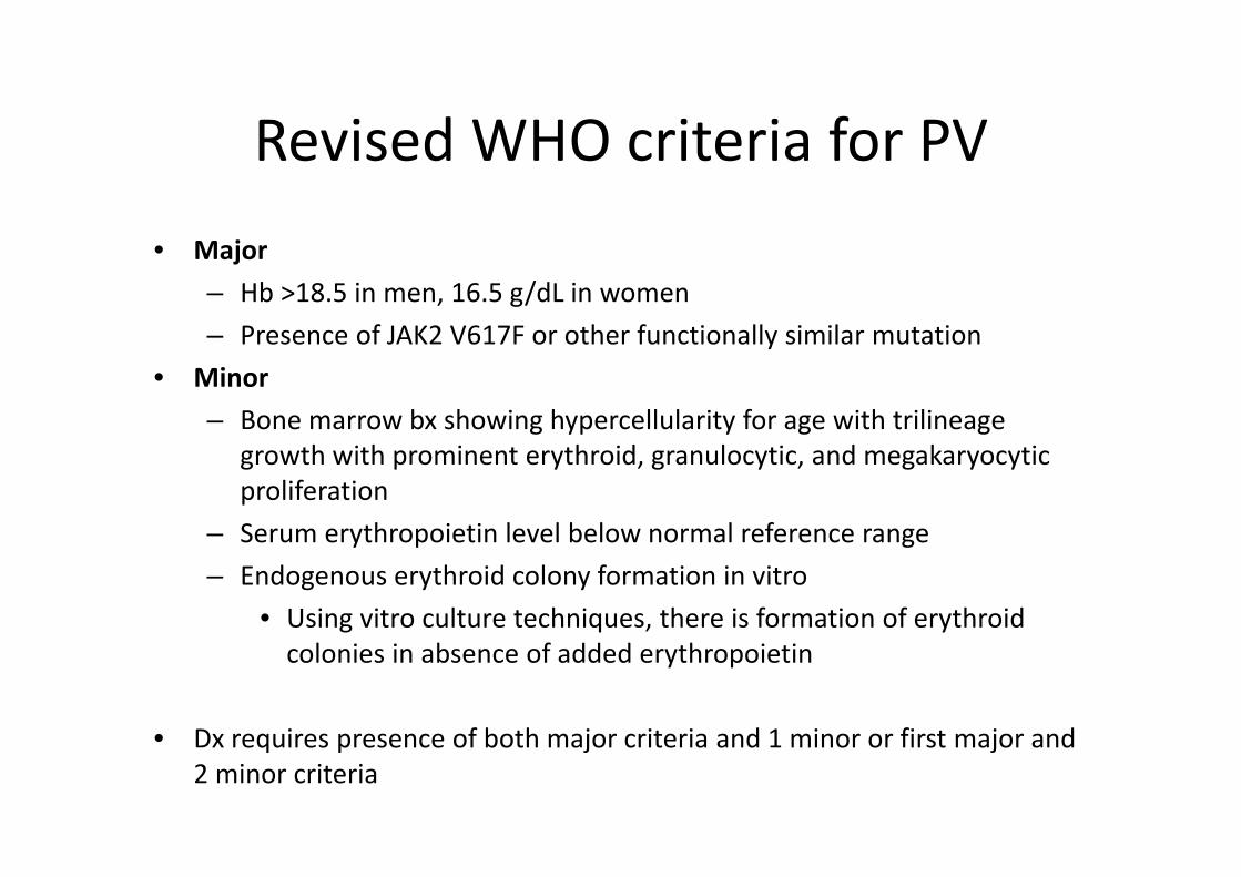
Revised WHO criteria for PV• Major
– Hb >18.5 in men, 16.5 g/dL in women– Presence of JAK2 V617F or other functionally similar mutation
• Minor– Bone marrow bx showing hypercellularity for age with trilineage
growth with prominent erythroid, granulocytic, and megakaryocytic proliferation
– Serum erythropoietin level below normal reference range– Endogenous erythroid colony formation in vitro
• Using vitro culture techniques, there is formation of erythroidcolonies in absence of added erythropoietin
• Dx requires presence of both major criteria and 1 minor or first major and 2 minor criteria

Treatment
• Phlebotomy– Goal is to reduce viscosity, reduce HCT to <45.
• Hydroxyurea– Reduced incidence of thrombosis compared to phlebotomy
– Effective in reducing blood counts although transient cytopenia may occur

TreatmentInterferon alpha
– IFN‐ reduces JAK2 V617F expression – Shown to provide relief from intractable pruritus and reduce spleen size
Anagrelide, a phosphodiesterase inhibitor, can reduce the platelet count and, if tolerated, is preferable to hydroxyurea because it lacks marrow toxicity
Allogeneic BMTmay be curative in young patients.

Primary Myelofibrosis
Least common, primarily afflicts individuals in their sixth decade or later
• Marrow fibrosis • Extramedullary hematopoiesis• Splenomegaly

Disorders Causing MyelofibrosisMalignant• Acute leukemia• CML• Hairy cell leukemia• Hodgkin disease• Idiopathic myelofibrosis• Lymphoma• Multiple myeloma• Myelodysplasia• Polycythemia vera• Systemic mastocytosis
Non malignant• HIV infection• Hyperparathyroidism• Renal osteodystrophy• SLE• Tuberculosis• Vitamin D deficiency• Gray platelet syndrome

Clinical features
• No signs or symptoms are specific for chronic IMF • Many patients are asymptomatic at presentation• Progressive anemia• Extramedullary hematopoiesis• HSM (SM is the hallmark)• Bone marrow fibrosis• Hypercatabolic syndromes (Fatigue, fevers, weight loss, night
sweats)• Evolution to acute leukemia

Diagnosis• suggested by peripheral blood smear
– normocytic anemia– increased or decreased number of granulocytes and platelets.
– myelophthisis• teardrop‐shaped RBCs (dacryocytes)• leukoerythroblastosis (nucleated RBCs and granulocyte precursors)
• confirmed by bone marrow biopsy– Usually dry tap

Bone Marrow Features
• Ineffective erythropoiesis• Dysplastic‐megakaryocyte hyperplasia • Increase in ratio of immature to total granulocytes• Reactive bone marrow fibrosis (polyclonal fibroblasts)• thickening and distortion of the bony trabeculae
(osteosclerosis)• Bcr‐abl negative

Prognosis• Median survival 3‐5 yrs• Adverse prognostic factors:
– Anemia– Age >64– Hypercatabolic sx (wt loss, fatigue, NS, fever)– WBC>30 or <4– Blasts>1%– Cytogenetics +8, 12p‐

Treatment Options
• No specific therapy exists • EPO; PRBCs• Danazol 200‐800mg/day improves anemia• Hydroxyurea for increased WBC or Plts• Busulfan & Melphalan• Allogeneic bone marrow transplantation is the only curative
treatment and should be considered in younger patients

Splenectomy
• Indications for surgery:– Mechanical discomfort (39%)– Portal HTN (11%)– Severe hypercatabolic symptoms (5%)– PRBC needed frequently (45%)

Essential Thrombocytosis
• Clonal disorder of unknown etiology involving a multipotenthematopoietic progenitor cell manifested clinically by overproduction of platelets without a definable cause.
• ET is an uncommon disorder, with an incidence of 1–2/100,000
• Distinct female predominance

Causes of Thrombocytosis• Malignancy• Infection• Myeloproliferative disorders: polycythemia vera, idiopathic
myelofibrosis, essential thrombocytosis, CML• Myelodysplastic disorders: 5q‐syndrome, idiopathic refractory
sideroblastic anemia• Postsplenectomy or hyposplenism• Hemorrhage• Iron deficiency anemia• Surgery• Rebound: Correction of vitamin B12 or folate deficiency, post‐
ethanol abuse• Hemolysis
Clinical Presentation
• Up to 50% ‐asymptomatic at presentation.• The remaining 50% of patients may have “vasomotor”
symptoms, thrombotic events, or hemorrhagic complications.

Clinical Presentation• Vasomotor symptoms include:
– Headache– Lightheadedness– Syncope – Atypical chest pain– Acral paresthesia– Livedo reticularis– Erythromelalgia
• Burning pain of the hands or feet associated with erythema and warmth– Transient visual disturbances
• Amaurosis fugax• Scintillating scotomata• Ocular Migraine

Thrombosis
• A common complication of ET.• Incidence rates in ET vary between 9 and 22%.• Thrombotic events include:
– Stroke– Transient ischemic attacks– Retinal artery or venous occlusions– Coronary artery ischemia– Pulmonary embolism– Hepatic or portal vein thrombosis– Deep vein thrombosis– Digital ischemia

Diagnostic Criteria
The Polycythemia Vera Study Group (PVSG) criteria include:
– A consistently elevated platelet count >600,000/ microL. – Megakaryocytic hyperplasia on bone marrow aspiration and biopsy.– Absence of the Philadelphia chromosome on routine cytogenetic
study.
(Molecular studies of the BCR/ABL gene rearrangement are now recommended to exclude cytogenetically masked cases of CML)

Diagnostic Criteria
The World Health Organization (WHO) criteria are similar and include:
– A sustained platelet count >600,000/microL.– Bone marrow biopsy showing proliferation of enlarged, mature
megakaryocytes.– No evidence of PV, CML, chronic idiopathic myelofibrosis,
myelodysplastic syndrome, or reactive thrombocytosis.

Therapeutic Agents• The most frequent symptoms are vasomotor and are easily managed with
low dose Aspirin (40 to 325 mg/day).• Hydroxyurea:
– Initial dose of 15 mg/kg per day po in divided doses.– Dose is adjusted to keep platelets between 100,000 and
400,000/microL.– Rapid onset of action, usually within 3‐5 days.– Teratogenic and should not be used in pregnancy, breast‐feeding, or
women with childbearing potential.
hydroxyurea plus aspirin is recommended as first line therapy in ET

Therapeutic Agents
• Anagrelide:
– An oral imidazoquinazoline derivative that inhibits platelet aggregation via platelet anti‐cyclic AMP phosphodiesterase activity.
– Lowers platelets via interference with megakaryocyte proliferation and maturation, resulting in platelet underproduction.
– Initial dose is 0.5 mg po tid or qid.– Dose is adjusted according to platelet count response and
symptomatology.
Therapeutic Agents
• Alpha Interferon– High cost and toxicity issues.– Reserved for use in high‐risk women of childbearing age, pregnant
women, and patients failing treatment with hydroxyurea.
• Platelet apheresis– Reduces the platelet count only in the short‐term.– Restricted to acute cerebrovascular complications, digital ischemia,
and rare life‐threatening situations.