cirugÍa y cirujanos - connecting repositories · cirugía y cirujanos. 2016;84(1):58---64 cirugÍa...
TRANSCRIPT

C
C
Mc
HNN
a
Db
c
S
RA
N
2B
irugía y Cirujanos. 2016;84(1):58---64
www.amc.org.mx www.elsevier.es/circir
CIRUGÍA y CIRUJANOSÓrgano de difusión científica de la Academia Mexicana de Cirugía
Fundada en 1933
LINICAL CASE
ultiple aneurysms splenic; surgical exclusion withonservation of the spleen�
éctor Bizueto-Rosasa,∗, José Ángel Barajas-Colóna, Ivan Delgadillo-de la Ob,ahieli Patricia Malo-Martíneza, Hugo Alonso Pérez-Gonzáleza,oemí Antonia Hernández-Pérezc
Servicio de Angiología, Hospital de Especialidades, Centro Médico Nacional La Raza, Instituto Mexicano del Seguro Social,.F., MexicoServicio de Angiología, Hospital de Tecamac, Instituto Mexicano del Seguro Social, D.F., MexicoServicio de Medicina Familiar y Laboral, Unidad de Medicina Familiar y Hospital General de Zona No. 29, Instituto Mexicano deleguro Social, D.F., Mexico
eceived 21 May 2014; accepted 10 November 2014vailable online 10 February 2016
KEYWORDSAneurysm;Splenic artery;Surgical resection;Conservation;Spleen
AbstractBackground: Aneurysm of the splenic artery is diagnosed when the diameter of the splenicartery is greater than 1 cm. It occupies third place among abdominal aneurysms. It is more fre-quent in women (4:1). It is associated with trauma, haemodynamics and local hormonal effectsduring pregnancy, portal hypertension (including the Caroli syndrome), arterial degeneration,atherosclerosis, and liver transplantation. It is difficult to diagnose, and it generally presentsas ruptured, thus once the diagnosis is made, the surgical approach is indicated due to its highmortality.Clinical case: Female of 66 years of age with a diagnosis of splenic artery aneurysm, withpulsing sensation at epigastric level of 8 months onset. On physical examination there is apalpable throbbing mass of 9 cm of diameter approximately, for which she was admitted. Thecomputed tomography angiography with reconstruction showed three splenic aneurysms. Twowere tied and the larger one was repaired by endo-aneurysmorrhaphy.Discussion: Visceral aneurysms are extremely rare. They are currently increasing and are the
third leading cause of cardiovascular death, as morbidity/mortality is high. The surgical treat- ment must be done selectively according to their size. Selection of the surgical techniquesdepends on the anatomic location and the need for revascularisation, the aetiology and theexperience of the surgeon.� Please cite this article as: Bizueto-Rosas H, Barajas-Colón JA, Delgadillo-de la O I, Malo-Martínez NP, Pérez-González HA, Hernández-PérezA. Aneurismas esplénicos múltiples; exclusión quirúrgica con conservación del bazo. Cirugía y Cirujanos. 2016;84:58---64.∗ Corresponding author: Puerto Zihuatanejo 18, Ampliación Fernando Casas Alemán, C.P. 07580, D.F., Mexico. Tel.: +52 (55) 4633 1881.
E-mail address: dr bizueto [email protected] (H. Bizueto-Rosas).
444-0507/© 2015 Academia Mexicana de Cirugía A.C. Published by Masson Doyma México S.A. This is an open access article under the CCY-NC-ND license (http://creativecommons.org/licenses/by-nc-nd/4.0/).

Multiple aneurysms splenic; surgical exclusion with conservation of the spleen 59
Conclusion: A review has been presented on the advances in diagnostic, and management,concluding that the best is to preserve the spleen, and whatever the technique it must beperformed by trained surgeons.© 2015 Academia Mexicana de Cirugía A.C. Published by Masson Doyma México S.A. Thisis an open access article under the CC BY-NC-ND license (http://creativecommons.org/licenses/by-nc-nd/4.0/).
PALABRAS CLAVEAneurisma;Arteria esplénica;Resección quirúrgica;Conservación;Bazo
Aneurismas esplénicos múltiples; exclusión quirúrgica con conservación del bazo
ResumenAntecedentes: El aneurisma de la arteria esplénica se diagnostica cuando el diámetro de la arte-ria esplénica es mayor de 1 cm. Ocupa el tercer lugar de los aneurismas abdominales y es másfrecuente en mujeres (4:1). Etiología: traumatismo, efectos locales hormonales y hemodinámi-cos del embarazo, hipertensión portal (incluyendo el síndrome de Caroli), degeneración arterial,aterosclerosis y postrasplante hepático. Es de difícil diagnóstico, generalmente comienzan comorotos, por lo que una vez hecho el diagnóstico el abordaje quirúrgico está indicado por su altamortalidad.Caso clínico: Mujer de 66 anos de edad con diagnóstico de aneurisma esplénico; sensación depulsación a nivel de epigastrio de 8 meses de evolución. A la exploración física se palpa masapulsátil de 9 cm de diámetro aproximadamente, por lo que se decide su hospitalización. Laangiotomografía con reconstrucción evidencia 3 aneurismas esplénicos. Dos se ligaron y en elmayor se realizó la endoaneurismorrafia, por no poder excluirlo.Discusión: Los aneurismas viscerales son sumamente raros; sin embargo, están actualmenteaumentando, siendo la tercera causa de muerte cardiovascular. La morbimortalidad es altay las posibilidades de supervivencia son escasas al detectarse tardíamente. Deben operarseselectivamente según su tamano; la selección de las técnicas quirúrgicas depende: de la loca-lización anatómica, de la necesidad de revascularización, de su etiología y, de la experienciadel cirujano.Conclusión: Se hizo una revisión de los avances diagnósticos y de manejo, concluyendo quelo mejor es preservar el bazo y cualquiera que sea la técnica, debe realizarse por cirujanoscapacitados.© 2015 Academia Mexicana de Cirugía A.C. Publicado por Masson Doyma México S.A. Estees un artículo Open Access bajo la licencia CC BY-NC-ND (http://creativecommons.org/licenses/by-nc-nd/4.0/).
aw2di
A
Tuatdtashort gastric vessels, and the left gastroepiploic artery.10
Background
Aneurysms of the splenic artery are rare but potentiallyfatal, with a prevalence of up to 10% in postmortemstudies.1 Aneurysm of the splenic artery represents 60%of all splanchnic arteries, and is the third most commonabdominal aneurysm, after aortic and iliac aneurysms.2 Itis defined as an abnormal dilatation of the splenic arteryof more than 1 cm in diameter. According to Al-Habbalet al.3 it was described for the first time by Beaussierin 1770, who observed it in autopsies. It was not until1920 that Hoegler made a preoperative diagnosis. The inci-dence of splenic artery aneurysms varies from 0.1% to10.4% in the general population.4 It is 4 times more com-mon in women than in men.3,5 Although the pathogenesishas not been fully clarified, risk factors include: trauma,local hormonal and haemodynamic effects during preg-nancy, portal hypertension (including Caroli syndrome) inup to 13%,6,7 fibrodysplasia of the media and atheroscle-
rosis. The development of a new splenic artery aneurysmafter liver transplantation can present up to 16 years aftertransplantation.8Ste
The diagnosis and appropriate treatment of splenic arteryneurysm is important because of the risk of rupture,hich increases significantly with a diameter greater than
cm, with 25---70% mortality depending on the underlyingisease.9 Advances in imaging techniques and minimallynvasive procedures have revolutionised treatment.
natomy
he splanchnic circulation includes the coeliac arteries, andpper and lower mesenteric arteries, which originate in thebdominal aorta. The splenic artery, branch of the coeliacrunk, bifurcates in the hilus of the spleen, it has a 5 mmiameter in men, and originates at 1.5 cm from the celiacrunk; it primarily irrigates the spleen and the pancreas,nd the great curvature of the stomach, together with the
eventy-five percent of splenic aneurysms affect the distalhird of the artery and 20% the middle third. They are gen-rally solitary and saccular in nature.11 The average size of

6
sr
uatplsiasnp
iapBihccei
sd
unt
tlia
sidtaa
tlseiilfsssapso
a
smlp
siaitolla
ercccpa
ocaataoftotfa
iiotapltti
seirdp
pas
0
plenic aneurysms on diagnosis is approximately 2 cm; theyarely exceed 3 cm.12
The natural history of splenic aneurysms is a grad-al increase in size and rupture. Eighty percent aresymptomatic and are diagnosed as a finding or becausehey rupture, the other 20% which are symptomatic canresent with abdominal pain in the epigastrium or in theeft hypochondrium radiating to the left shoulder (Kehr’sign) and haemodynamic instability.13 Other symptoms cannclude anorexia, nausea or vomiting, which are frequentlyttributed to a co-existing hiatus hernia or other diseasesuch as gallstones and peptic ulcer disease. However diag-osis is almost always from a chance discovery,14 because aulsatile mass is rarely palpated on clinical examination.
Spontaneous rupture of the aneurysm presents initiallyn 2---10% of patients;15 it is currently reduced to 3% withdvances in diagnosis. Occasionally a double rupture canresent within 48 h; this phenomenon was described byockerman in 1930.4,15 Secondary erosion of the aneurysm
nto adjacent viscera can cause gastrointestinal haemorr-age in 13% of patients due to rupture to the stomach,olon or pancreatic duct. Erosion in the splenic vein canause an arteriovenous fistula with portal hypertension, orven mesenteric steal syndrome and ischaemia of the smallntestine.4
Spontaneous rupture is the most serious complication ofplenic aneurysm, with 25% mortality; it is more frequenturing pregnancy, when mortality rises to 75---90%.16
As the population’s life expectancy rises, with the use ofltrasound and cross-sectional imaging,4,15 incidental diag-osis is also on the increase. Ultrasound has the advantagehat it can be used during pregnancy.
Once a suspected diagnosis has been made, digital sub-raction angiography is indicated, as it can define the preciseocation of the aneurysm, the collateral branches can benvestigated, and the source of bleeding, and other visceralneurysms can be documented or excluded.2,15
Ultrasound can be used for follow-up, however, multi-lice computed tomography is preferable. Indications forntervention are associated with the natural history of theisease, placing the emphasis on the factors which increasehe risk of spontaneous rupture. Calcification, advancedge and normotension do not preclude rupture of theneurysm.17
Treatment depends on the presentation, location andhe size of the aneurysm, and can involve conventional oraparoscopic surgery, endovascular embolisation, and exclu-ion of the aneurysm by stent. It is important to makevery effort to preserve the spleen in order to preservemmunological function, unless the aneurysm is locatedn the splenic hilus.17 However, there is evidence thatigature or embolisation of the splenic artery alters theunction of the spleen, even if it is preserved.18 Openurgery approaches can include splenectomy with exci-ion of the aneurysm, proximal and distal ligature of theplenic artery with or without resection of the aneurysm,nd endo-aneurysmorrhaphy.5 Partial splenectomy can beerformed for distal aneurysms, preserving the unaffected
plenic parenchyma. The mortality rate associated withpen surgery is 1.3%, with a morbidity rate of 9%.19Aneurysms of the splenic artery can be approached vian anterior or a lateral route. Using the latter approach the
sdta
H. Bizueto-Rosas et al.
hort gastric and left gastroepiploic vessels can be compro-ised, which increases the risk of splenic infarction, and the
ateral retroperitoneal approach preserves collateral splenicerfusion.20
Arca et al.5 conclude that a laparoscopic approach for aplenic aneurysm is a safe and feasible alternative, providedt is undertaken by an experienced surgeon using intraoper-tive ultrasound, which has been demonstrated to be lessnvasive than open surgery. A laparascopic approach is con-raindicated in haemodynamically unstable patients or withther signs of rupture.5 As with the open approach, theaparascopic approach can be either anterior or lateral. Aateral approach might be appropriate for central and distalneurysms.16
Transcatheter embolisation was first described by Probstt al. in 1978.21 The advances in digital subtraction angiog-aphy, and the development of a wide variety of arterialatheters and associated equipment have increased its suc-essful application in 85---100% of cases. It is currentlyonsidered the first line of treatment for the majority ofatients with splenic aneurysms, particularly for incidentalsymptomatic aneurysms.22,23
After computed tomography, embolisation shouldcclude the source of the aneurysm.19,24 It is contraindi-ated in splenic aneurysms located in the splenic hilus,24
nd is indicated when the aneurysm is difficult to managend/or in high risk patients. Complications include migra-ion of the coil and distal infarction, abscess formationnd, rarely, rupture of the aneurysm.24 Recanalisation canccur in up to 12.5% of patients. Embolisation can also failor technical reasons if the splenic artery is particularlyortuous. Some authors have reported a conglomerationf symptoms in embolised patients, which has beenermed postembolisation syndrome,19 characterised byever, abdominal pain, ileus, and occasionally pancreatitis,ffecting up to 39% of patients.
Recent innovations in the treatment of splenic aneurysmsnclude the use of endovascular stents. This is a minimallynvasive technique, which excludes aneurysmatic dilatationf the artery, preserving normal blood flow. The size andortuosity of the splenic artery, and the position of theneurysm limit the use of a stent. They are more appro-riate for proximal aneurysms.11 Stents have a significantlyower risk of splenic infarction compared to embolisation;hey also have the advantage over embolisation in situa-ions where preservation of the arterial flow to the spleens necessary for other reasons.25
Combined therapy in stages (embolisation followed byurgical resection) is recommended in specific situations,specially in the management of giant aneurysms andn patients with significant comorbidities.26 The use ofobot technology can enable a more precise surgical proce-ure, including anastomosis of small structures in selectedatients.27
False aneurysms of the splenic artery constitute a com-letely different clinical scenario. Most authors recommendctive management without delay, without taking size,ymptoms or rupture into account. Patients with a false
plenic aneurysm are usually affected by an underlyingisease, which is generally pancreatitis or pancreatic fis-ulae. Mortality, after open surgical intervention on falseneurysms close to the pancreatic head is from 16% to 50%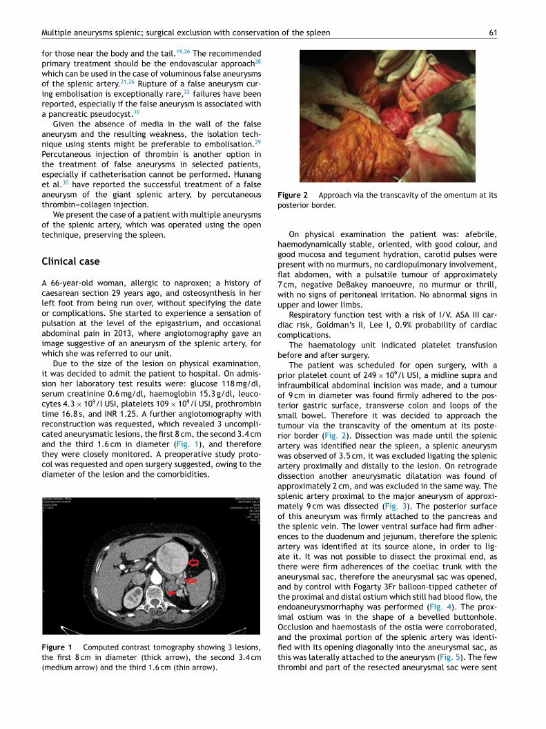
tion of the spleen 61
Fp
hgpfl7wu
dc
b
piotstr
Multiple aneurysms splenic; surgical exclusion with conserva
for those near the body and the tail.19,26 The recommendedprimary treatment should be the endovascular approach28
which can be used in the case of voluminous false aneurysmsof the splenic artery.21,26 Rupture of a false aneurysm cur-ing embolisation is exceptionally rare,22 failures have beenreported, especially if the false aneurysm is associated witha pancreatic pseudocyst.10
Given the absence of media in the wall of the falseaneurysm and the resulting weakness, the isolation tech-nique using stents might be preferable to embolisation.29
Percutaneous injection of thrombin is another option inthe treatment of false aneurysms in selected patients,especially if catheterisation cannot be performed. Hunanget al.30 have reported the successful treatment of a falseaneurysm of the giant splenic artery, by percutaneousthrombin---collagen injection.
We present the case of a patient with multiple aneurysmsof the splenic artery, which was operated using the opentechnique, preserving the spleen.
Clinical case
A 66-year-old woman, allergic to naproxen; a history ofcaesarean section 29 years ago, and osteosynthesis in herleft foot from being run over, without specifying the dateor complications. She started to experience a sensation ofpulsation at the level of the epigastrium, and occasionalabdominal pain in 2013, where angiotomography gave animage suggestive of an aneurysm of the splenic artery, forwhich she was referred to our unit.
Due to the size of the lesion on physical examination,it was decided to admit the patient to hospital. On admis-sion her laboratory test results were: glucose 118 mg/dl,serum creatinine 0.6 mg/dl, haemoglobin 15.3 g/dl, leuco-cytes 4.3 × 10⁹/l USI, platelets 109 × 109/l USI, prothrombintime 16.8 s, and INR 1.25. A further angiotomography withreconstruction was requested, which revealed 3 uncompli-cated aneurysmatic lesions, the first 8 cm, the second 3.4 cm
and the third 1.6 cm in diameter (Fig. 1), and thereforethey were closely monitored. A preoperative study proto-col was requested and open surgery suggested, owing to thediameter of the lesion and the comorbidities.Figure 1 Computed contrast tomography showing 3 lesions,the first 8 cm in diameter (thick arrow), the second 3.4 cm(medium arrow) and the third 1.6 cm (thin arrow).
awadasmoteaataateiOafitt
igure 2 Approach via the transcavity of the omentum at itsosterior border.
On physical examination the patient was: afebrile,aemodynamically stable, oriented, with good colour, andood mucosa and tegument hydration, carotid pulses wereresent with no murmurs, no cardiopulmonary involvement,at abdomen, with a pulsatile tumour of approximately
cm, negative DeBakey manoeuvre, no murmur or thrill,ith no signs of peritoneal irritation. No abnormal signs inpper and lower limbs.
Respiratory function test with a risk of I/V. ASA III car-iac risk, Goldman’s II, Lee I, 0.9% probability of cardiacomplications.
The haematology unit indicated platelet transfusionefore and after surgery.
The patient was scheduled for open surgery, with arior platelet count of 249 × 109/l USI, a midline supra andnfraumbilical abdominal incision was made, and a tumourf 9 cm in diameter was found firmly adhered to the pos-erior gastric surface, transverse colon and loops of themall bowel. Therefore it was decided to approach theumour via the transcavity of the omentum at its poste-ior border (Fig. 2). Dissection was made until the splenicrtery was identified near the spleen, a splenic aneurysmas observed of 3.5 cm, it was excluded ligating the splenicrtery proximally and distally to the lesion. On retrogradeissection another aneurysmatic dilatation was found ofpproximately 2 cm, and was excluded in the same way. Theplenic artery proximal to the major aneurysm of approxi-ately 9 cm was dissected (Fig. 3). The posterior surface
f this aneurysm was firmly attached to the pancreas andhe splenic vein. The lower ventral surface had firm adher-nces to the duodenum and jejunum, therefore the splenicrtery was identified at its source alone, in order to lig-te it. It was not possible to dissect the proximal end, ashere were firm adherences of the coeliac trunk with theneurysmal sac, therefore the aneurysmal sac was opened,nd by control with Fogarty 3Fr balloon-tipped catheter ofhe proximal and distal ostium which still had blood flow, thendoaneurysmorrhaphy was performed (Fig. 4). The prox-mal ostium was in the shape of a bevelled buttonhole.cclusion and haemostasis of the ostia were corroborated,nd the proximal portion of the splenic artery was identi-
ed with its opening diagonally into the aneurysmal sac, ashis was laterally attached to the aneurysm (Fig. 5). The fewhrombi and part of the resected aneurysmal sac were sent
62 H. Bizueto-Rosas et al.
Figure 3 Dissection of the splenic artery proximal to theaneurysm larger than 9 cm.
Figure 4 Opening the aneurysmal sac and by means of controlwith Fogarty 3Fr balloon catheter of the proximal and distalostium; an endo-aneurysmorrhaphy was then performed.
Figure 5 Amplified construction where the main lesion can beseen adhering to the coeliac trunk and the route of the splenicartery tangent to the aneurysm.
Figure 6 Computed tomography 48 h postoperatively revea-lp
fsn
nhlbtwsi
o
ga
D
Vdsl
ntcod
hadmbiatwt
5t
ing hypodense areas involving almost 50% of the splenicarenchyma.
or histopathological study and culture; the sac was thenutured. The spleen was observed to be hypertrophic witho evidence of areas of ischaemia during surgery.
The patient progressed well, afebrile, with abdomi-al pain of low intensity in the mesogastrium and leftypochondrium. Her platelet count lowered to 67.0 witheucocytosis of 11,000 and neutrophilia of 87.6, with noandaemia, serum amylase of 500 u/l, and therefore a con-rasted computed tomography was requested after 48 hhich revealed hypodense areas involving almost 50% of the
plenic parenchyma (Fig. 6). In subsequent biometries anncrease in platelet count to 99.0 was observed.
The patient was discharged home well and tolerating anral diet.
She was seen as an outpatient 15 days later and had pro-ressed well, and was discharged from her local hospitalfter one month with no changes in clinical parameters.
iscussion
isceral aneurysms are extremely rare. The first cases wereescribed 200 years ago with a micotic aneurysm of theuperior mesenteric artery. Case series reports are few andimited to no more than 10 patients.
Because they are so rare other possible differential diag-oses need to be considered, such as cystic pancreaticumours, which are common in women, pancreatic pseudo-ysts, although these can be ruled out if there is no historyf acute or chronic pancreatitis. Another extremely rareisease is neuroendocrine tumour.31
As we mentioned early, in the last 2 decades thereas been a 7-fold increase in the frequency of abdominalneurysms, and they are the third cause of cardiovasculareath. Fifty percent of women with splenic aneurysms areultiparous. In our country, with an ageing population, airth rate of 18.87 births per 1000 inhabitants, and increas-ngly frequent liver transplantations, this disease will haven increasing impact. Therefore this disease needs to beaken into consideration because its mortality is very highhen detected late. We should be aware of its presence as
31
he chances of survival are low.The splenic artery in men has an average diameter ofmm, and according to the definition of aneurysm, it isermed an aneurysm when the diameter exceeds 50% of the

tion
tirotot
cw
C
Atfta
adt
t
da
ndt
se
rTcm
C
T
R
Multiple aneurysms splenic; surgical exclusion with conserva
normal diameter of the vessel involved. Even so, surgery isindicated when its diameter is 2 cm. However, it is morecommon in women, who have smaller arteries with nar-rower diameters, and they are generally diagnosed when theaneurysm ruptures with very high mortality, and thereforemost should be operated when they are diagnosed.
Ideally, blood flow should be re-established after theaneurysm has been resected or excluded, or the splenicartery dissected and ligated at its portion proximal anddistal to the lesion, but in our case, the inflammatory pro-cess around the aneurysmal sac which was firmly adheredto the coeliac trunk, to the pancreatic tissue, and to thesplenic vein prevented dissection of the splenic artery nearto its source (Fig. 5), therefore, despite its originating inthe middle third, the lesion had to be opened and endo-aneurysmorrhaphy performed of the 2 ostia (primum nonnocere), and injury avoided to other structures such as thepancreas, the intestinal loops or compromising the integrityof the coeliac trunk; because as stated in the medical refer-ences ‘‘treatment depends on the manner of presentation,the location, and the size of the aneurysm’’.32
A ruptured aneurysm can manifest acutely, into the pan-creatic duct, and result in a clinical picture known ashemossucus pancreaticus.33
It has been mentioned that splenic artery aneurysms rup-ture more frequently, especially in pregnant women, andthat it has been found in postmortem studies that they canbe more common than aneurysms of the abdominal aorta.
Aneurysms should be operated selectively according totheir size, and this procedure should be performed as anemergency when they rupture and unstable symptoms, con-tained ruptures, aneurysms that are embolising, and thosethat are infected cause shock.31
Various surgical treatment options are offered, and theselection of one of the diverse techniques will dependon the anatomical location of the aneurysm, on the needfor revascularisation, its aetiology, and the experience ofthe surgeon.33 Preservation of the spleen is increasinglypreferred, we should not fail to mention that multipleaneurysms present in 22%,29 and the laparoscopic approachis being used in order to avoid large-scale surgery, ligatingthe splenic artery with or without splenectomy.5
With splenic infarction, as the efficient volume of thespleen is reduced, blood elements increase which accumu-late in the spleen, and result in an increased red blood cell,white blood cell and platelet count.
Complications which can present are abdominal pain,fever, rupture, splenic abscesses, pneumonia and septi-caemia, and many patients with splenic infarction canremain asymptomatic.
Diagnosis can be made by means of ultrasound or com-puted tomography. The initial treatment is medical, withanalgesics and anticoagulants. Surgery is only indicated forcases which present complications, such as splenic abscessor rupture, or when diagnosis is imprecise.34
Open surgery on visceral aneurysms is still the proce-dure of choice to repair aneurysms that have been diagnosedlate; however, minimally invasive endovascular techniques
can also offer advantages with conventional treatment, pro-viding, as Suso et al. mention,35 they are performed bya surgeon of proven experience. It is important to high-light that endovascular techniques are not free from risk,of the spleen 63
hey can cause the secondary aneurysm to rupture with thencrease in pressure when inserting the embolic material,esult in incomplete thrombosis of the aneurysm, and fail tobliterate the collateral branches which might be keepinghe aneurysm ‘‘pressurised’’,36 and cause the inadvertentcclusion of another vessel, migration of the coils, and infec-ion.
Elective resection of splenic aneurysms carries a risk ofomplications, such as: ischaemia of the spleen (up to 29%)36
hich requires splenectomy.
onclusions
neurysms of the splenic artery that are at low risk of rup-ure can be managed without intervention, with radiologicalollow-up every 6 months with ultrasound or computedomography, in order to evaluate the progression of theneurysm.
Surgical intervention should be considered if theneurysm is symptomatic, if it has enlarged over 2 cm iniameter or if it is found during pregnancy or at a reproduc-ive age.
All false aneurysms of the splenic artery should bereated as rapidly as possible.
The choice between embolisation and placing a stentepends on: the size, shape and site of the splenic arteryneurysm, and the experience of the surgeon.
Due to the anatomy of her splenic artery, our patient wasot a candidate for embolisation, because if an occludingevice or covered stent had been placed it would occludehe coeliac trunk.
A laparoscopic approach might be considered if expo-ure to radiation is contraindicated (pregnancy), or whenndovascular techniques fail or are not available.
Ruptured or complicated splenic aneurysms should beesolved by means of conventional open surgical technique.he inclusion of interventionist radiology might enable aombined approach (surgical and radiological), in the treat-ent of complex or difficult ruptured aneurysms.
onflict of interests
he authors have no conflict of interest to declare.
eferences
1. Bedford PD, Lodge B. Aneurysm of the splenic artery. Gut.1960;1:312---20.
2. Dave SP, Reis ED, Hossain A, Taub PJ, Kerstein MD, HollierLH. Splenic artery aneurysm in the 1990s. Ann Vasc Surg.2000;14:223---9.
3. Al-Habbal Y, Christophi C, Muralidharan V. Aneurysms of thesplenic artery. A review. Surgeon. 2010;8:223---31.
4. Messina LM, Shanley CJ. Visceral artery aneurysms. Surg ClinNorth Am. 1997;77:425---42.
5. Arca MJ, Gagner M, Heniford BT, Sullivan TM, Beven EG. Splenicartery aneurysms: methods of laparoscopic repair. J Vasc Surg.1999;30:184---8.
6. Puttini M, Aseni P, Brambilla G, Belli L. Splenic artery aneurysmsin portal hypertension. J Cardiovasc Surg. 1982;23:490---3.
7. Garbagna G, Cornalba G, Rota L. Splenic artery aneurysms inpatients with portal hypertension. Radiol Med. 1980;66:239---42.

6
1
1
1
1
1
1
1
1
1
1
2
2
2
2
2
2
2
2
2
2
3
3
3
3
3
3
4
8. Kóbori L, van der Kolk MJ, de Jong KP, Peeters PMJG,Klompmaker IJ, Kok T, et al. Splenic artery aneurysms inliver transplant patients. Liver Transplant Group. J Hepatol.1997;27:890---3.
9. Bizueto Rosas H, Guerra Ledezma J, Oropeza Martínez G.Tratamiento quirúrgico de los aneursimas de las arterias vis-cerales. Cir Cir. 1995;63:36---9.
0. Pasha SF, Gloviczki P, Stanson AW, Kamath PS. Splanchnic arteryaneurysms. Mayo Clin Proc. 2007;82:472---9.
1. Karaman K, Onat L, Sirvanci M, Olga R. Endovascular stent grafttreatment in a patient with splenic artery aneurysm. DiagnInterv Radiol. 2005;11:119---21.
2. Spittel JA, Fairbairn JF 2nd, Kincaid OW, ReMine WH. Aneurysmof the splenic artery. JAMA. 1961;175:452---6.
3. Sadat U, Dar O, Walsh S, Varty K. Splenic artery aneurysms inpregnancy --- a systematic review. Int J Surg. 2008;6:261---5.
4. Trastek VF, Pairolero PC, Joyce JW, Hollier LH, Bernatz PE.Splenic artery aneurysms. Surg. 1982;91:694---9.
5. Mattar SG, Lumsden AB. The management of splenicartery aneurysms: experience with 23 cases. Am J Surg.1995;169:580---4.
6. De Csepel J, Quinn T, Gagner M. Laparoscopic exclusion ofa splenic artery aneurysm using a lateral approach permitspreservation of the spleen. Surg Laparosc Endosc Percutan Tech.2001;11:221---4.
7. Hashizume M, Ohta M, Ueno K, Okadome K, Sugimachi K.Laparoscopic ligation of splenic artery aneurysm. Surgery.1993;113:352---4.
8. Nincheri Kunz M, Pantalone D, Borri A, Paolucci R, Pernice LM,Taruffi F, et al. Management of true splenic artery aneurysms.Two case reports and review of the literature. Minerva Chir.2003;58:247---56.
9. Guillon R, Garcier JM, Abergel A, Mofid R, Garcia V, ChahidT, et al. Management of splenic artery aneurysms and falseaneurysms with endovascular treatment in 12 patients. Cardio-vasc Intervent Radiol. 2003;26:256---60.
0. Moore SW, Guida PM, Schumacher HW. Splenic artery aneurysm.Bull Soc Int Chir. 1970;29:210---8.
1. Probst P, Castaneda-Zuniga WR, Gomes AS, Yonehiro EG,Delaney JP, Amplatz K. Nonsurgical treatment of splenic-artery
aneurysms. Radiology. 1978;128:619---23.2. McDermott VG, Shlansky-Golberg R, Cope C. Endovascularmanagement of splenic artery aneurysms and pseudoa-neurysms. Cardiovasc Intervent Radiol. 1994;17:179---84.
3
H. Bizueto-Rosas et al.
3. Yamamoto S, Hirota S, Maeda H, Achiwa S, Arai K, KobayashiK, et al. Transcatheter coil embolization of splenic arteryaneurysm. Cardiovasc Intervent Radiol. 2008;31:527---34.
4. Reidy JF, Rowe PH, Ellis FG. Splenic artery aneurysm emboliza-tion --- the preferred technique to surgery. Clin Radiol.1990;41:281---2.
5. Arepally A, Dagli M, Hofmann LV, Kim HS, Cooper M, Klein A.Treatment of splenic artery aneurysm with use of a stent-graft.J Vasc Interv Radiol. 2002;13:631---3.
6. Bakhos CT, McIntosh BC, Nukta FA, Fiedler PN, Denatale RW,Sweeney TF, et al. Staged arterial embolization and surgicalresection of a giant splenic artery aneurysm. Ann Vasc Surg.2007;21:208---10.
7. Antico A, Vesce G, Iob G, Parini U. Splenic artery aneurys-mectomy with combined laparoscopic-robotic technique: ourpreliminary experience and literature review. Surg LaparoscEndosc Percutan Tech. 2006;16:292.
8. Tihansky DP, Lluncor E. Transcatheter embolization of multi-ple mycotic splenic artery aneurysms: a case report. Angiology.1986;37:530---4.
9. Berceli SA. Hepatic and splenic artery aneurysms. Semin VascSurg. 2005;18:196---201.
0. Huang IH, Zuckerman DA, Matthews JB. Occlusion of agiant splenic artery pseudoaneurysm with percutaneousthrombin---collagen injection. J Vasc Surg. 2004;40:574---7.
1. Larrain DC, Fava M, Espinosa RG. Aneurisma de la arteriaesplénica. Diagnóstico diferencial y alternativas terapéuticas.Rev Méd Chile. 2005;133:943---6.
2. Escudero de Fez MD, Sabater Ortí L, Calvete Chornet J, CampsVilata B, Gómez Portilla A, Martínez León J. Hemoperito-neo por rotura de aneurisma de la arteria esplénica. Cir Esp.2001;70:160---3.
3. Wagner WH, Cossman DV, Treiman RL, Foran RF, Phillip M, Levin,et al. Hemosuccus pancreaticus from intraductal rupture of aprimary splenic artery aneurysm. J Vasc Surg. 1994;19:158---64.
4. Agolini SF, Shah KT, Goodreau JJ, McLoughlin TM Jr, Sinclair MC.Splenic infarction caused by a large thoracic aortic thrombus. JVasc Surg. 1997;26:1069---72.
5. Suso KI, Shimura T, Asao T, Nomoto K, Kanoh K, Tuboi K,et al. Laparoscopic resection of splenic artery aneurysm: a case
report. Hepatogastroenterology. 2002;49:1520---2.6. Abad C, Montesdeoca-Cabrera D, Sáez-Guzmán T. Aneurisma dela arteria esplénica. Revisión de dos casos intervenidos quirúr-gicamente. An Med Interna. 2006;23:130---2.