cisterns 360°
TRANSCRIPT
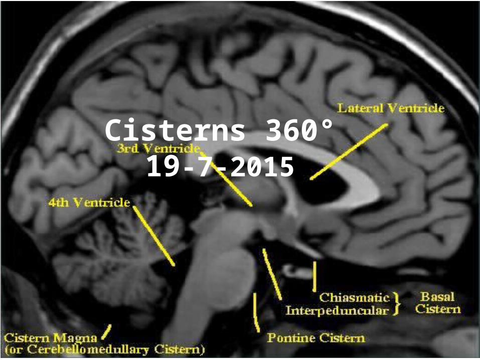
Cisterns 360°19-7-2015

Great teachers – All this is their work . I am just the reader of their books .
Prof. Paolo castelnuovo
Prof. Aldo Stamm Prof. Mario Sanna
Prof. Magnan

For Other powerpoint presentatioins of “ Skull base 360° ”
I will update continuosly with date tag at the end as I am getting more & more information
click
www.skullbase360.in - you have to login to slideshare.net with Facebook account for downloading.

Oculomotor cistern
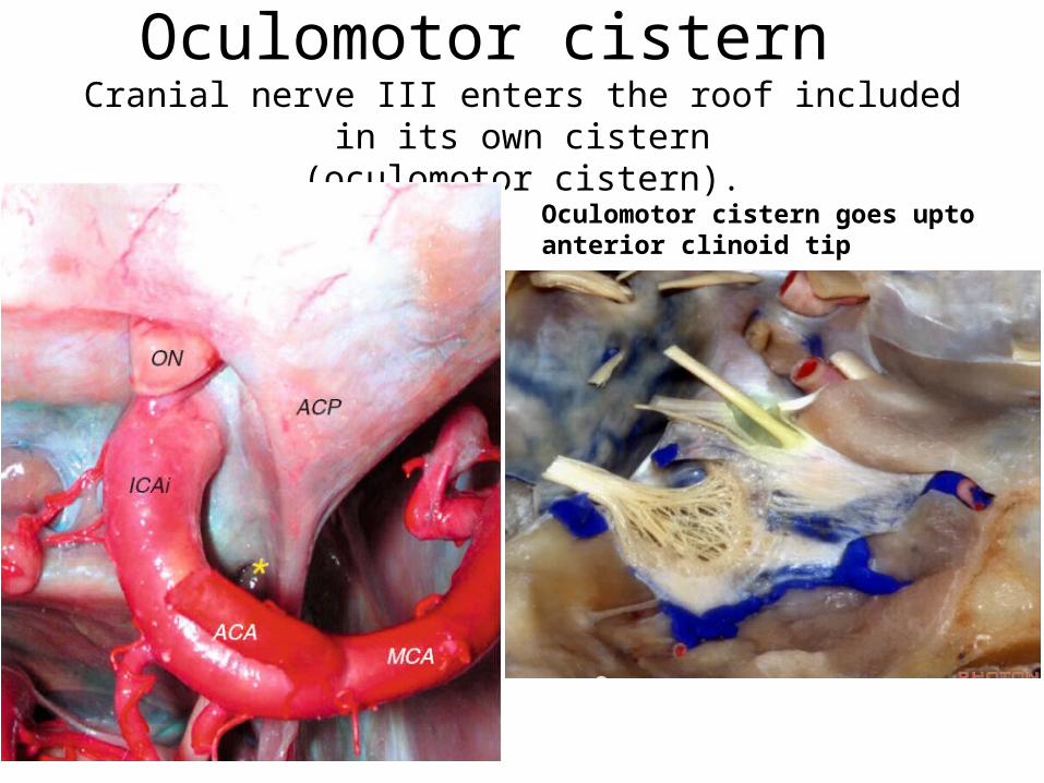
Oculomotor cistern Cranial nerve III enters the roof included in its own cistern
(oculomotor cistern).
Oculomotor cistern goes upto anterior clinoid tip
LT lamina terminalis cistern= Suprachiasmatic cistern
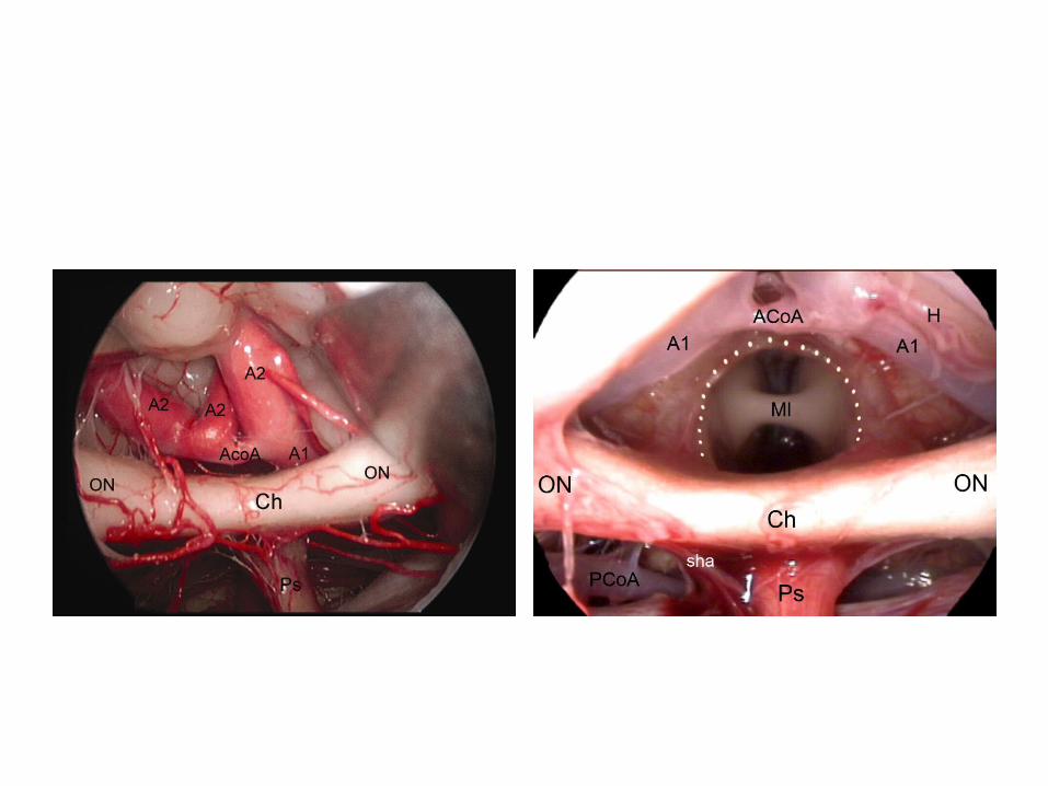
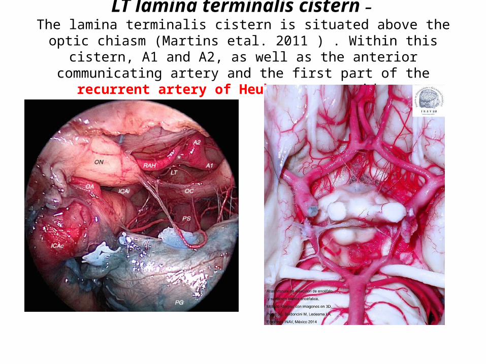
LT lamina terminalis cistern – The lamina terminalis cistern is situated above the optic chiasm (Martins etal. 2011 ) . Within this cistern, A1 and A2, as well as the anterior communicating
artery and the first part of the recurrent artery of Heubner, are evident.
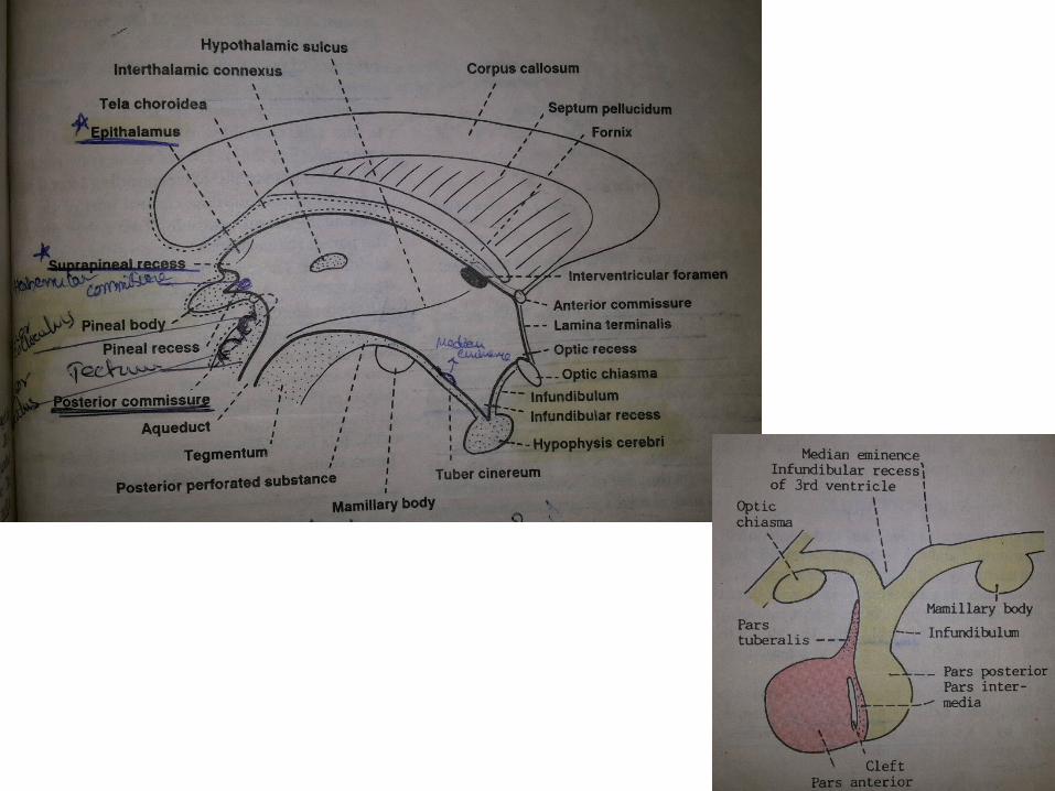
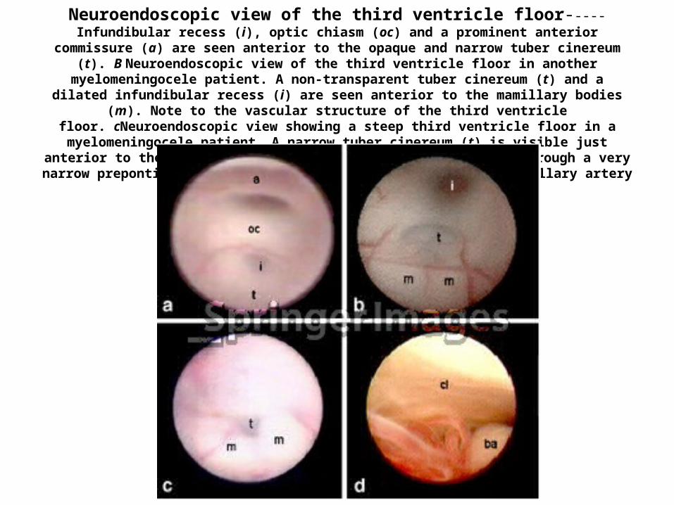
The space between a & oc is Lamina terminalis Neuroendoscopic view of the third ventricle floor-----Infundibular recess (i), optic chiasm (oc) and a prominent anterior commissure (a) are seen anterior to the opaque and narrow tuber cinereum (t). B Neuroendoscopic view of the third ventricle floor in another myelomeningocele patient. A non-transparent tuber cinereum (t) and a dilated infundibular recess (i) are seen anterior to the mamillary bodies (m). Note to the vascular structure of the third ventricle floor. cNeuroendoscopic view showing a steep third ventricle
floor in a myelomeningocele patient. A narrow tuber cinereum (t) is visible just anterior to the mamillary bodies (m). dNeuroendoscopic view through a very narrow prepontine cistern. Note the close proximity of
the basillary artery (ba) and clivus (cl)
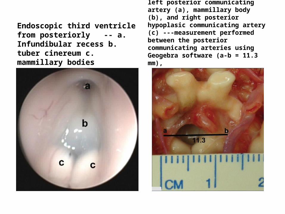
Endoscopic third ventricle from posteriorly -- a. Infundibular recess b. tuber cinereum c. mammillary bodies
left posterior communicating artery (a), mammillary body (b), and right posterior hypoplasic communicating artery (c) ---measurement performed between the posterior communicating arteries using Geogebra software (a-b = 11.3 mm),
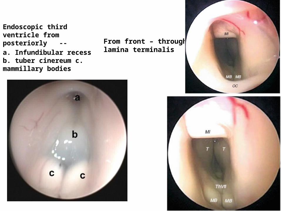
Endoscopic third ventricle from posteriorly -- a. Infundibular recess b. tuber cinereum c. mammillary bodies
From front – through lamina terminalis
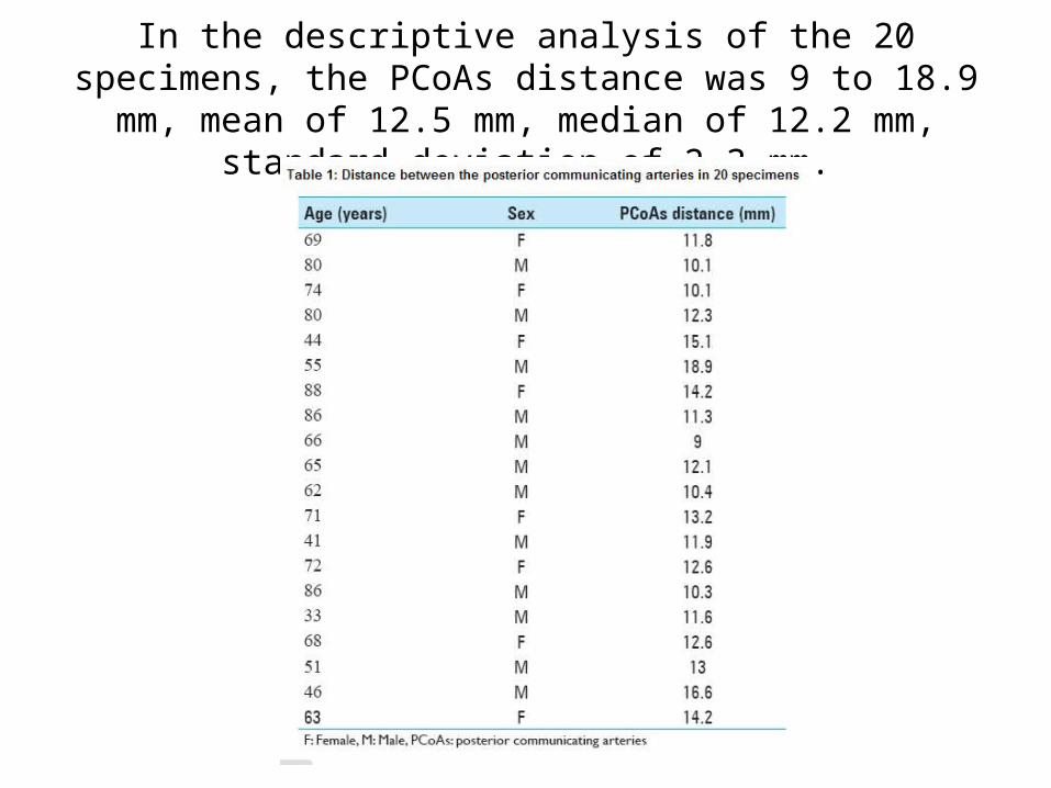
In the descriptive analysis of the 20 specimens, the PCoAs distance was 9 to 18.9 mm, mean of 12.5 mm, median of 12.2
mm, standard deviation of 2.3 mm.
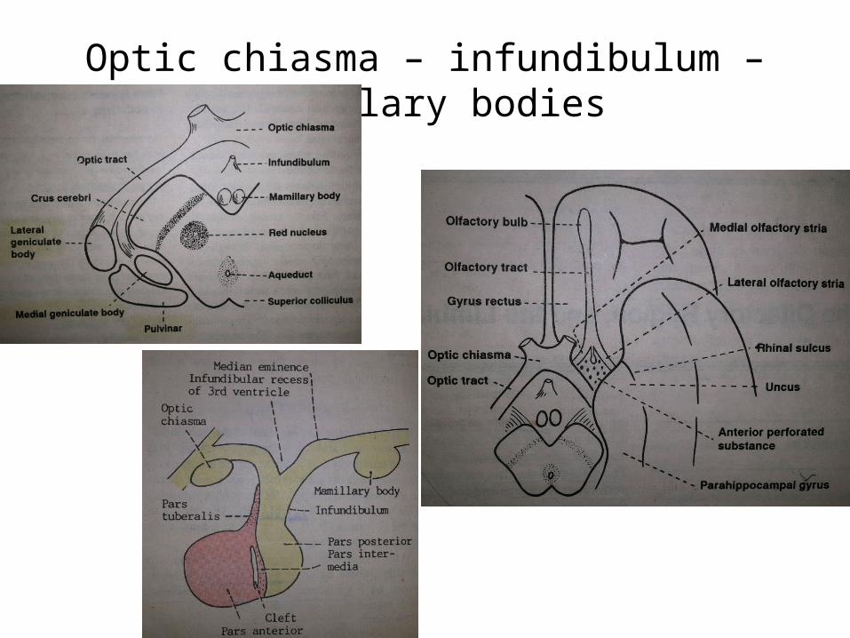
Optic chiasma – infundibulum – Mamillary bodies
Chiasmatic cistern
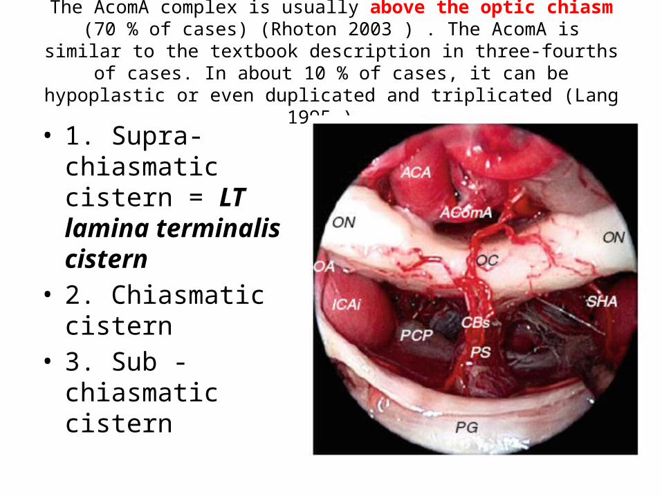
The AcomA complex is usually above the optic chiasm (70 % of cases) (Rhoton 2003 ) . The AcomA is similar to the textbook description in three-
fourths of cases. In about 10 % of cases, it can be hypoplastic or even duplicated and triplicated (Lang 1995 ) .
• 1. Supra-chiasmatic cistern = LT lamina terminalis cistern
• 2. Chiasmatic cistern• 3. Sub -chiasmatic
cistern
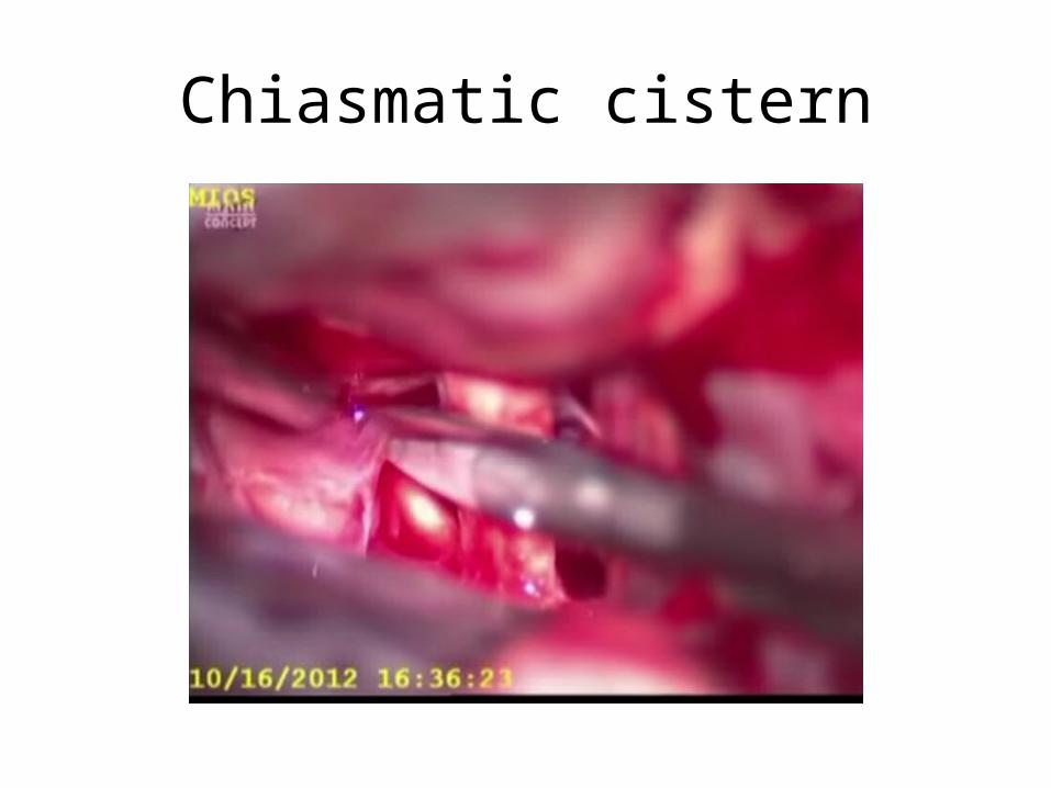
Chiasmatic cistern
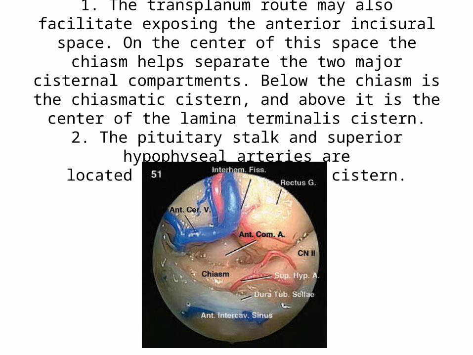
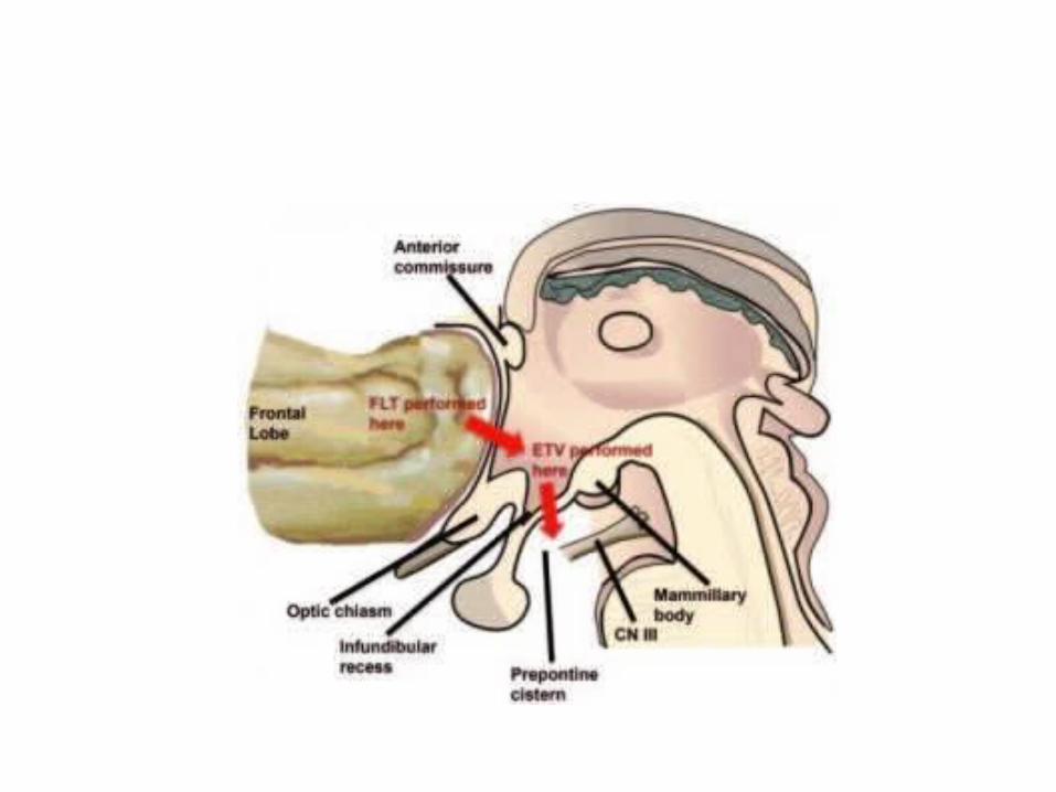
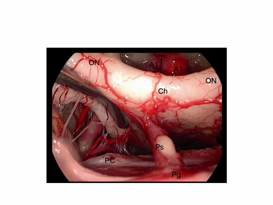
1. The transplanum route may also facilitate exposing the anterior incisural space. On the center of this space the chiasm
helps separate the two major cisternal compartments. Below the chiasm is the chiasmatic cistern, and above it is the center of the
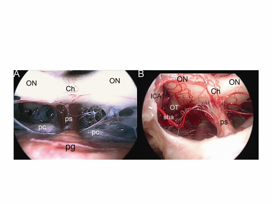
lamina terminalis cistern.2. The pituitary stalk and superior hypophyseal arteries are
located into the chiasmatic cistern.
Below the OT & A1 you will see PComA
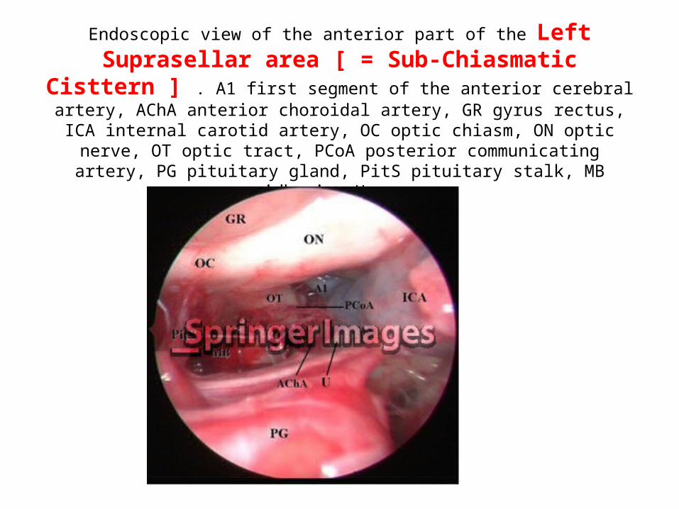
Endoscopic view of the anterior part of the Left Suprasellar area [ = Sub-Chiasmatic Cisttern ] . A1 first segment of the anterior cerebral artery, AChA
anterior choroidal artery, GR gyrus rectus, ICA internal carotid artery, OC optic chiasm, ON optic nerve, OT optic tract, PCoA posterior communicating artery, PG pituitary
gland, PitS pituitary stalk, MB midbrain, U uncus
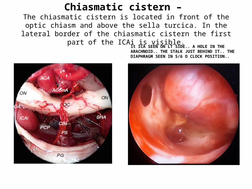
Chiasmatic cistern – The chiasmatic cistern is located in front of the
optic chiasm and above the sella turcica. In the lateral border of the chiasmatic cistern the first part of the ICAi is visible.
lt ICA SEEN ON LT SIDE.. A HOLE IN THE ARACHNOID.. THE STALK JUST BEHIND IT.. THE DIAPHRAGM SEEN IN 5/6 O CLOCK POSITION..
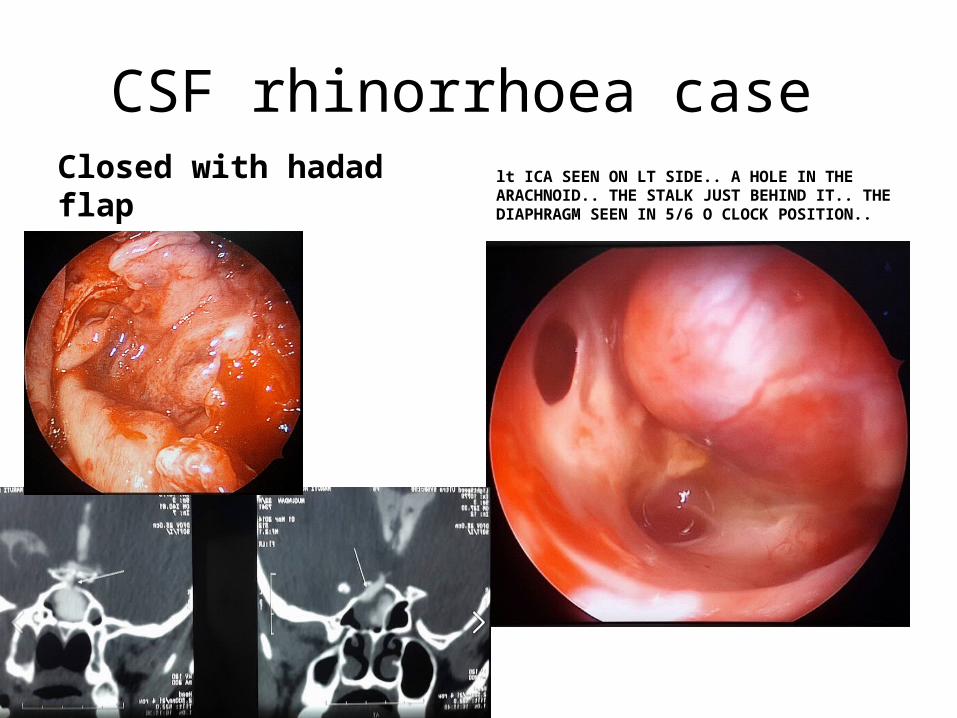
CSF rhinorrhoea case
Closed with hadad flap lt ICA SEEN ON LT SIDE.. A HOLE IN THE ARACHNOID.. THE STALK JUST BEHIND IT.. THE DIAPHRAGM SEEN IN 5/6 O CLOCK POSITION..
Sub-chiasmatic cistern = Supra sellar area = Supr-sellar cistern

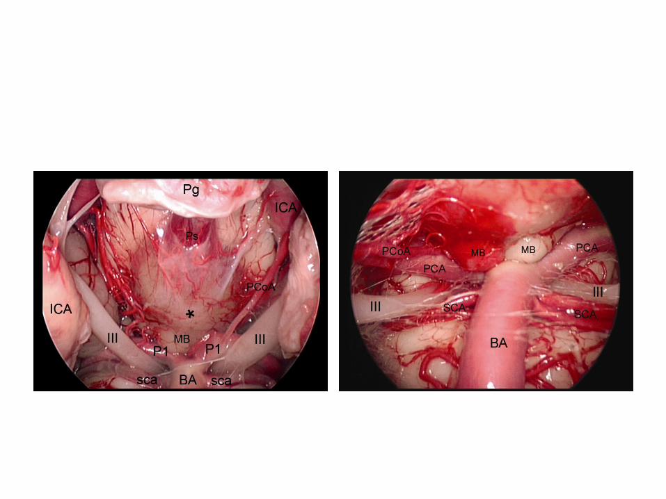
Observe here the Pcom (here labelled as ACoP in some language ) is parallel to 3rd nerve in infrachiasmatic cistern . Excellent photo .
Other points to note 1. 3rd nerve sandwitched between posterior cerebral artery & superior cerebellar artery . 2. On the left side 2 superior cerebellar
arteries present from the origin itself. 3. Diameter of Pcom varies on two sides. 4. Infra-chiasmastic cistern is nothing but suprasellar area
Lillequit’s membrane

Retrosellar
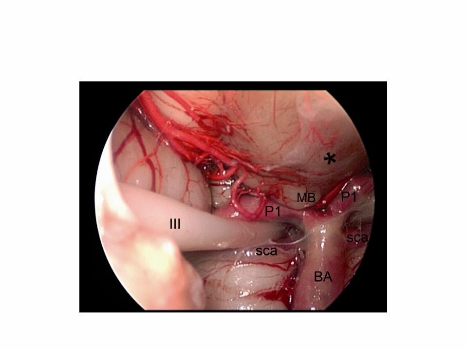
Interpeduncular cistern [= basal cistern ]
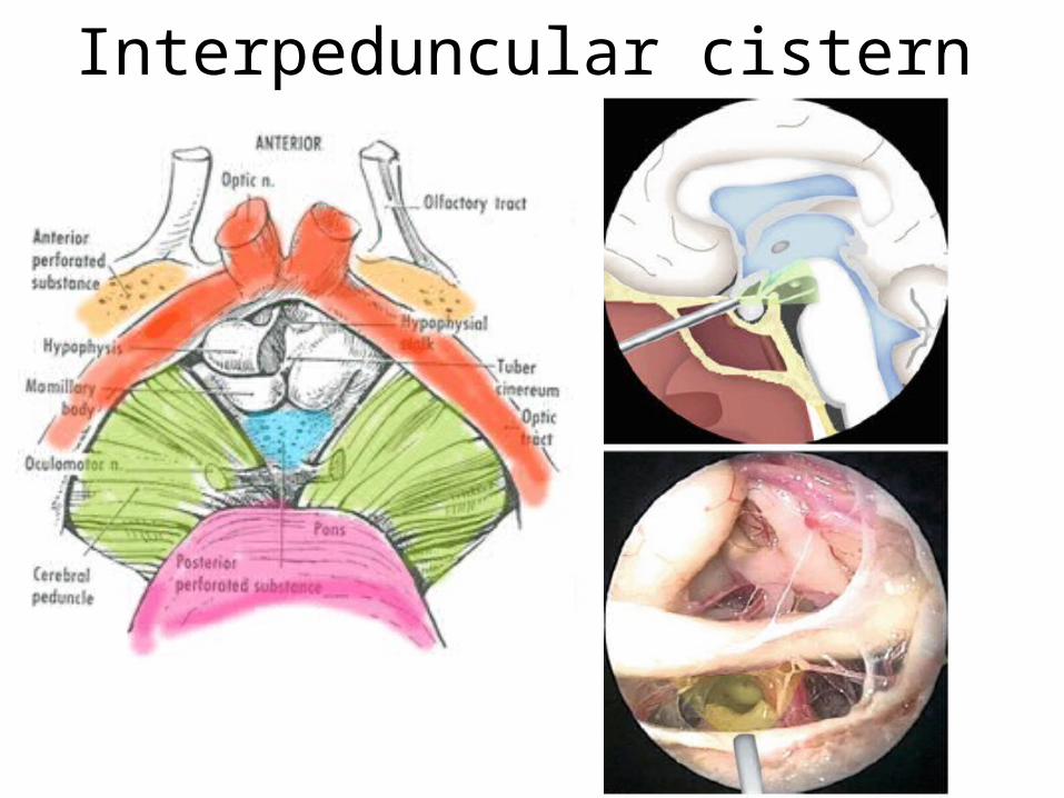
Interpeduncular cistern
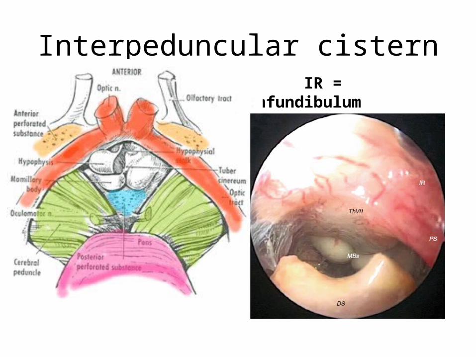
Interpeduncular cistern
IR = infundibulum
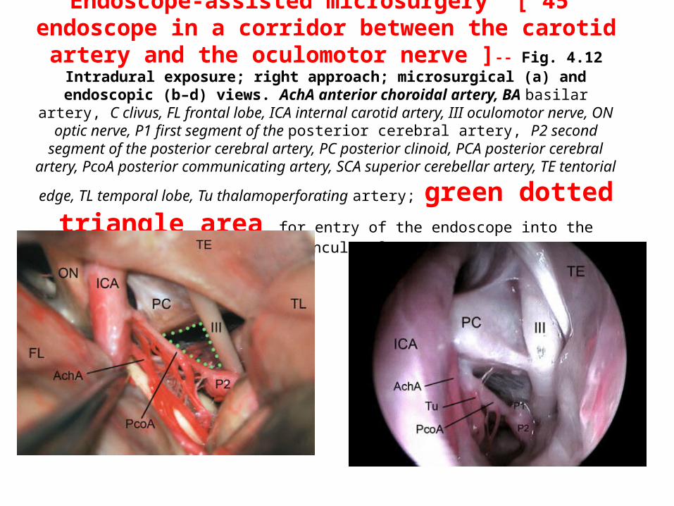
Endoscope-assisted microsurgery [ 45° endoscope in a corridor between the carotid artery and the oculomotor nerve ]-- Fig. 4.12
Intradural exposure; right approach; microsurgical (a) and endoscopic (b–d) views. AchA anterior choroidal artery, BA basilar artery, C clivus, FL frontal lobe, ICA internal carotid artery, III
oculomotor nerve, ON optic nerve, P1 first segment of the posterior cerebral artery, P2 second segment of the posterior cerebral artery, PC posterior clinoid, PCA posterior cerebral artery, PcoA
posterior communicating artery, SCA superior cerebellar artery, TE tentorial edge, TL temporal
lobe, Tu thalamoperforating artery; green dotted triangle area for entry of the endoscope into the interpeduncular fossa

Cadaveric dissection image demonstrating the close anatomical relationship of the posterior clinoid (PC) with both the intracranial carotid artery (ICCA)
and the posterior genu of the intracavernous carotid artery (P. CCA). AL, anterior lobe of the pituitary gland; PL, posterior lobe of the pituitary gland;
BA, basilar artery.green dotted triangle area for entry of the endoscope into the interpeduncular fossa
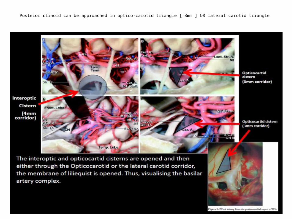
Posteior clinoid can be approached in optico-carotid triangle [ 3mm ] OR lateral carotid triangle
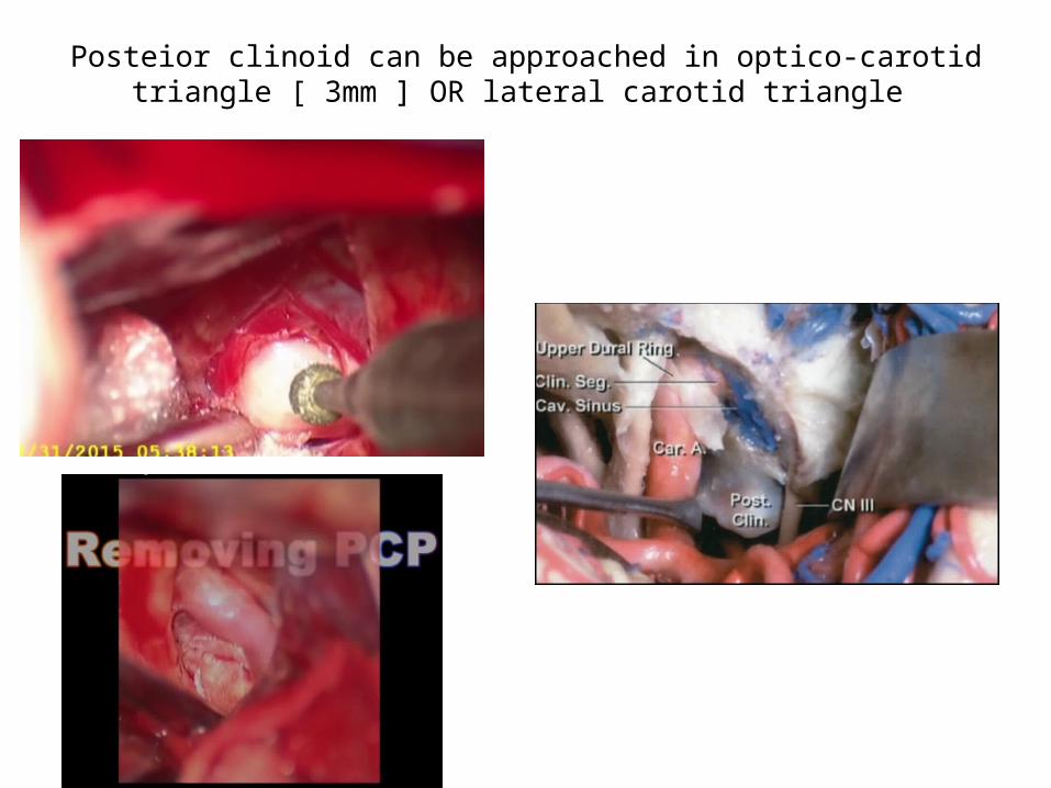
Posteior clinoid can be approached in optico-carotid triangle [ 3mm ] OR lateral carotid triangle
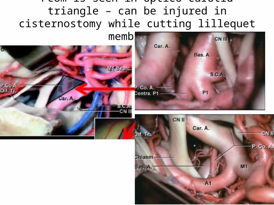
Pcom is seen in optico-carotid triangle – can be injured in cisternostomy while cutting lillequet membrane
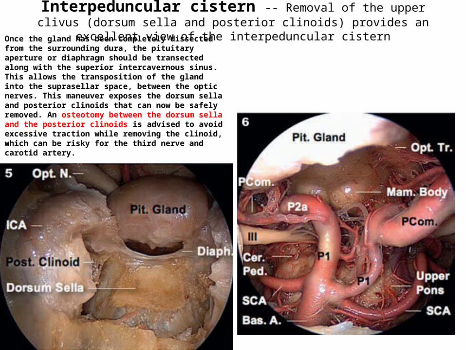
Interpeduncular cistern -- Removal of the upper clivus (dorsum sella and posterior clinoids) provides an excellent view of the interpeduncular cisternOnce the gland has been completely dissected from
the surrounding dura, the pituitary aperture or diaphragm should be transected along with the superior intercavernous sinus. This allows the transposition of the gland into the suprasellar space, between the optic nerves. This maneuver exposes the dorsum sella and posterior clinoids that can now be safely removed. An osteotomy between the dorsum sella and the posterior clinoids is advised to avoid excessive traction while removing the clinoid, which can be risky for the third nerve and carotid artery.
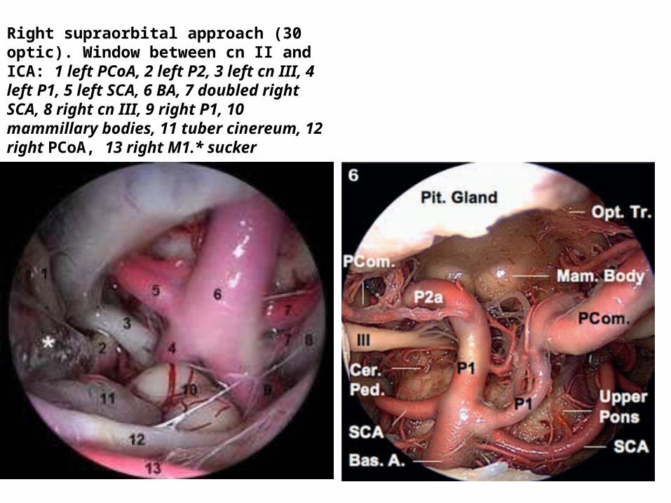
Right supraorbital approach (30 optic). Window between cn II and ICA: 1 left PCoA, 2 left P2, 3 left cn III, 4 left P1, 5 left SCA, 6 BA, 7 doubled right SCA, 8 right cn III, 9 right P1, 10 mammillary bodies, 11 tuber cinereum, 12 right PCoA, 13 right M1.* sucker
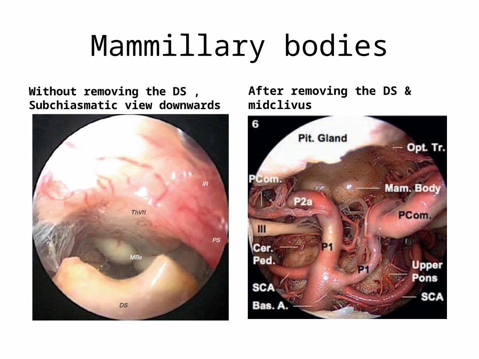
Mammillary bodiesWithout removing the DS , Subchiasmatic view downwards After removing the DS & midclivus
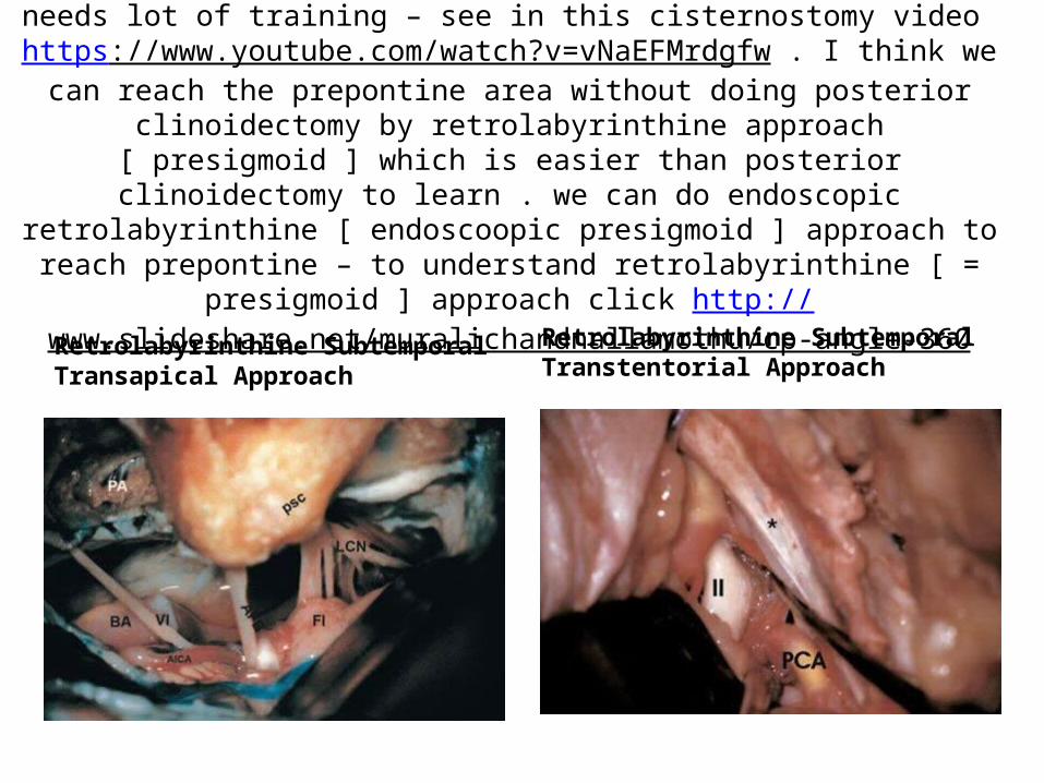
posterior clinoidectomy by middle cranial fossa approach needs lot of training – see in this cisternostomy video https://www.youtube.com/watch?v=vNaEFMrdgfw . I think we can reach the prepontine area without doing posterior clinoidectomy by
retrolabyrinthine approach [ presigmoid ] which is easier than posterior clinoidectomy to learn . we can do endoscopic retrolabyrinthine [ endoscoopic
presigmoid ] approach to reach prepontine – to understand retrolabyrinthine [ = presigmoid ] approach click http://
www.slideshare.net/muralichandnallamothu/cp-angle-360
Retrolabyrinthine Subtemporal Transapical Approach
Retrolabyrinthine Subtemporal Transtentorial Approach
cisternostomy in head injury video – by Dr. Iype Cherian - click
https://www.youtube.com/watch?v=vNaEFMrdgfw
and
https://youtu.be/NLTm8-rHzAA

For Other powerpoint presentatioins of “ Skull base 360° ”
I will update continuosly with date tag at the end as I am getting more & more information
click
www.skullbase360.in - you have to login to slideshare.net with Facebook account for downloading.