c.j. harvey author manuscript nih public access a, r.k ...faculty.fortlewis.edu/blake_d/publications...
TRANSCRIPT
Nrf2-regulated glutathione recycling independent of biosynthesisis critical for cell survival during oxidative stress
C.J. Harveya, R.K. Thimmulappaa, A. Singha, D.J. Blakea, G. Linga, N. Wakabayashia, J.Fujiib, A. Myersc, and S. Biswala,*
a Department of Environmental Health Science, Bloomberg School of Public Health, School of Medicine,Johns Hopkins University, Baltimore, MD 21205, USA
b Department of Biomolecular Function, Yamagata University, Yamagata, Japan
c Division of Allergy and Clinical Immunology, School of Medicine, Johns Hopkins University, Baltimore,MD 21205, USA
AbstractNuclear factor-erythroid 2 p45-related factor 2 (Nrf2) is the primary transcription factor protectingcells from oxidative stress by regulating cytoprotective genes, including the antioxidant glutathione(GSH) pathway. GSH maintains cellular redox status and affects redox signaling, cell proliferation,and death. GSH homeostasis is regulated by de novo synthesis as well as GSH redox state; previousstudies have demonstrated that Nrf2 regulates GSH homeostasis by affecting de novo synthesis. Wereport that Nrf2 modulates the GSH redox state by regulating glutathione reductase (GSR). Inresponse to oxidants, lungs and embryonic fibroblasts (MEFs) from Nrf2-deficient (Nrf2−/−) miceshowed lower levels of GSR mRNA, protein, and enzyme activity relative to wild type (Nrf2+/+).Nrf2−/− MEFs exhibited greater accumulation of glutathione disulfide and cytotoxicity compared toNrf2+/+ MEFs in response to t-butylhydroquinone, which was rescued by restoring GSR.Microinjection of glutathione disulfide induced greater apoptosis in Nrf2−/− MEFs compared toNrf2+/+ MEFs. In silico promoter analysis of the GSR gene revealed three putative antioxidant-response elements (ARE1, −44; ARE2, −813; ARE3, −1041). Reporter analysis, site-directedmutagenesis, and chromatin immunoprecipitation assays demonstrated binding of Nrf2 to two AREsdistal to the transcription start site. Overall, Nrf2 is critical for maintaining the GSH redox state viatranscriptional regulation of GSR and protecting cells against oxidative stress.
KeywordsNrf2; Oxidative stress; Glutathione; Glutathione reductase; Cigarette smoke; COPD; Emphysema;Free radicals
Because of their function (gas exchange) and structure (large surface area), the lungs areconstantly challenged with oxidative insults caused by exogenous air pollutants. Oxidativestress is involved in the pathogenesis of several chronic lung inflammatory diseases, includingchronic obstructive pulmonary disease (COPD), asthma, acute respiratory distress syndrome(ARDS), and pulmonary fibrosis. Glutathione (GSH) is a vital antioxidant that regulates thecellular redox status and protects airway epithelial cells from oxidant-induced lung injury andinflammation [1]. Oxidative stress causes perturbations in cellular GSH levels by eitheraffecting the biosynthesis or altering the ratio of intracellular reduced and oxidized forms of
*Corresponding author: E-mail address: [email protected] (S. Biswal).
NIH Public AccessAuthor ManuscriptFree Radic Biol Med. Author manuscript; available in PMC 2009 February 15.
Published in final edited form as:Free Radic Biol Med. 2009 February 15; 46(4): 443–453. doi:10.1016/j.freeradbiomed.2008.10.040.
NIH
-PA Author Manuscript
NIH
-PA Author Manuscript
NIH
-PA Author Manuscript
glutathione that affect multiple physiological responses. The key functions of GSH include (i)working as a direct antioxidant to aid in the removal of deleterious reactive oxygen speciesand reactive nitrogen species; (ii) acting as a cofactor for antioxidant enzymes, includingglutathione peroxidases, glutathione S-transferases, and glutaredoxins; (iii) recycling of otherantioxidants (such as ascorbic acid) [2]; (iv) protecting against inflammatory responses bymodulating redox-regulated signal transduction and redox-sensitive transcription factors (NF-κB and AP1) [3]; (v) regulating cell proliferation [4,5]; and (vi) protecting against apoptosis[6,7]. Elevated levels of glutathione disulfide (GSSG) can affect the function of multipleproteins via glutathionylation and cause cell death through lowering reduced GSH levels andincreasing accumulation of GSSG [8–10]. The contribution of protection against oxidativestress by regeneration of GSH from GSSG is unclear.
Glutathione reductase (GSR), a homodimeric flavoprotein (50-kDa subunits), regulatescellular GSH homeostasis by catalyzing the reduction of GSSG to GSH using NADPH as areducing cofactor [11]. Evolutionarily, GSR is well conserved in plants, bacteria, fungi, yeast,and mammals, suggesting that it is important for survival in aerobic environments [11]. GSRhas been shown to be critical for cell survival, and mice with suboptimal levels of GSR aremore prone to oxidative stress and its associated diseases [12]. However, GSR deficiencies areuncommon among humans, caused primarily by dietary riboflavin deficiencies [13,14] andtreatment with the anticancer agent 1,3-bis(2-chloroethyl)-1-nitrosourea (BCNU) [15,16].
The basic leucine-zipper transcription factor nuclear factor-erythroid 2 p45-related factor 2(Nrf2) has been shown to play a vital role in protecting cells from oxidative stress [17]. Inresponse to oxidative stress, Nrf2 dissociates from its cytosolic inhibitor Keap1, translocatesto the nucleus, and binds to antioxidant-response elements (AREs) in the promoters of targetgenes. This leads to transcriptional induction of several cellular defense genes, includingglutathione biosynthetic enzymes (glutathione cysteine ligase modifier subunit (GCLM) andglutathione cysteine ligase catalytic subunit (GCLC)), GSH-dependent antioxidant enzymes(glutathione peroxidase 2 (GPX2), glutathione S-transferases (GST), and heme oxygenase-1(HO-1)) [17]. However, disruption of Nrf2 in mice diminishes or abrogates the induction ofthese antioxidant genes, indicating their Nrf2-dependent regulation. We and others have shownthat Nrf2-defcient (Nrf2−/−) mice show enhanced sensitivity to a variety of lung inflammatorydiseases, including cigarette smoke (CS)-induced emphysema [18], allergic asthma [19],endotoxin [20], hyperoxia-induced acute lung injury [5], and bleomycin-induced lung fibrosis[21], in which increased oxidative stress results from impaired adaptive induction of GSH andGSH-dependent enzymes. More recently, we and others have shown that a decline in Nrf2 inthe lungs of patients with COPD contributes to pulmonary oxidant/antioxidant imbalance andpathogenesis of lung diseases [22–24]. In contrast, because of a functional mutation in theKeap1 gene, lung cancer tissues show constitutively higher Nrf2-dependent antioxidants,including GSH, which in turn cause therapeutic resistance to chemotherapeutic drugs throughattenuation of oxidative stress [25].
Several lines of evidence indicate that Nrf2 plays a key role in the regulation of cellular GSHhomeostasis: (i) there is low GSH or a loss of induction of GSH in Nrf2−/− cells and tissues[18,20]; (ii) Nrf2 regulates GSH biosynthesizing enzymes (GCLM, GCLC) [26]; (iii) Nrf2regulates the cysteine/glutamate exchange transporter that maintains intracellular GSH levelsby regulating cysteine influx [27]; and (iv) Nrf2 regulates GPX2 and GST, which use GSH asa cofactor [28]. In addition, we and others have shown that in response to oxidative stress,Nrf2−/− cells and tissues accumulate greater levels of GSSG than wild-type Nrf-2 cells [22].Because GSR is the key enzyme that mediates the reduction of GSSG and regeneration of GSH,we hypothesized that Nrf2 regulates GSR during oxidative stress. This study elucidates thecritical role of GSR in cell survival and molecular regulation of GSR by Nrf2 in response tooxidative stress.
Harvey et al. Page 2
Free Radic Biol Med. Author manuscript; available in PMC 2009 February 15.
NIH
-PA Author Manuscript
NIH
-PA Author Manuscript
NIH
-PA Author Manuscript

Materials and methodsAnimals and care
Nrf2−/− CD-1 (ICR) mice were generated as described [12]. All experimental proceduresconducted on the mice were performed in accordance with the standards established by theU.S. Animal Welfare Acts, set forth in the National Institutes of Health guidelines and thePolicy and Procedures Manual of the Johns Hopkins University Animal Care and UseCommittee.
Mouse exposure to cigarette smokeEight-week-old mice were divided into four groups (n=3 per group): I, air control Nrf2+/+ mice;II, experimental Nrf2+/+ mice; III, air control Nrf2−/− mice; and IV, experimental Nrf2−/− mice.Groups I and III were kept in a filtered-air environment, and groups II and IV were subjectedto CS according to a previously published protocol [18]. CS exposure was performed byburning 3R4F reference cigarettes (0.73 mg nicotine per cigarette, purchased from the TobaccoResearch Institute, University of Kentucky, Lexington, KY, USA) using a smoking machine(Model TE-10; Teague Enterprises, Woodland, CA, USA) as previously described [29]. Sixcigarettes were smoked simultaneously, resulting in an exposure level of100 mg/m3. Mice wereexposed to this concentration 5 hday for 1 day.
Cell cultureMouse embryonic fibroblasts (MEFs) were isolated from Nrf2+/+, Nrf2−/−, and Keap1−/− miceas previously described [20] and were cultured in Iscove’s modified Dulbecco’s mediumsupplemented with 10% FBS and 1% penicillin–streptomycin (Invitrogen, Carlsbad, CA,USA).
Knockdown of GSR by short interfering RNA (siRNA)Four GSR siRNA duplexes and siControl nontargeting siRNA 1 (SS siRNA) were obtainedfrom Dharmacon Research (Lafayette, CO, USA). First, we identified an siRNA duplex thatexhibits selective and maximal silencing of the GSR gene compared to SS siRNA. Briefly,Nrf2+/+ MEFs at 80% confluency were transfected with 20 pmol of siRNA duplexes usingLipofectamine 2000 and OPTI-MEM I reduced-serum medium (Invitrogen) according to theLipofectamine protocol. Concentrations of siRNAs were chosen on the basis of dose–responsestudies. Knockdown of the GSR gene was quantified by real-time reverse transcriptase-polymerase chain reaction (RT-PCR) 48 h after transfection. The sequences of the sense strandsof the siRNA duplexes were as follows: duplex 1, AGACGAAGCUGUUCAUAAGUU;duplex 2, GACCAUGAUUCCAGAUGUUUU; duplex 3,GACGGGACCCAAAUUCUAAUU; and duplex 4, CGUGAAUGUUGGAUGUGUAUU.Duplex 1 was shown to have the greatest knockdown efficiency (data not shown).
Overexpression of GSRThe vector for GSR overexpression was obtained from Open Biosystems (Huntsville, AL,USA) (Clone ID 6813295). The cDNA insert was digested out of the parent plasmid and ligatedinto the pUB6-V5-His mammalian expression vector (Invitrogen) with a ubiquitin promoter.After amplification in Escherichia coli and plasmid purification (Qiagen Sciences,Gaithersburg, MD, USA), the plasmid was transfected into Nrf2−/− MEFs using Lipofectamine2000 (Invitrogen) as described above.
Cytoprotective role of GSRMicroinjection of GSSG and measurement of apoptosis—Nrf2+/+ and Nrf2−/− MEFswere plated at a density of 1×10⁶ cells/ml in Mattek dishes. Approximately 100 cells per dish
Harvey et al. Page 3
Free Radic Biol Med. Author manuscript; available in PMC 2009 February 15.
NIH
-PA Author Manuscript
NIH
-PA Author Manuscript
NIH
-PA Author Manuscript
were injected with vehicle, phosphate-buffered saline (PBS), GSSG, or GSH (Sigma–Aldrich,St. Louis, MO, USA) at a concentration of 50 mM using a FemtoJet microinjector (EppendorfNorth America, Westbury, NY, USA). As a control for injection efficiency, cells were also co-injected with Alexa fluorhydrazine 596 (Invitrogen). After the 4-h incubation at 37°C in 5%CO2, the number of apoptotic cells was determined by annexin V Alexa fluor 488 (Invitrogen)staining following the manufacturer’s protocol. Data are expressed as the percentage ofapoptotic cells compared to total number of cells microinjected.
tert-Butylhydroquinone-induced cell death—tert-Butylhydroquinone (tBHQ)-inducedcytotoxicity was evaluated in (i) Nrf2+/+ and Nrf2−/− MEFs; (ii) Nrf2+/+ MEFs transfected withGSR siRNA or SS siRNA; and (iii) Nrf2−/− MEFs overexpressing GSR or plasmid vector.Briefly, MEFs were seeded in 96-well plates at a density of 1×10⁵ cells/ml and grownovernight. Cells were treated with tBHQ (150 μM) for 4 h and cell death was evaluated usinga 3-(4,5-dimethylthiazol-2-yl)-2,5-diphenyltetrazolium bromide (MTT; Sigma) reductionconversion assay [30]. Cell survival was expressed as absorbance relative to that of vehicle-treated controls. For GSR knockdown or overexpression experiments, 48 h after transfection,MEFs were challenged with tBHQ.
Measurement of oxidized and reduced glutathioneNrf2+/+ and Nrf2−/− MEFs were seeded in 96-well plates at a density of 1×10⁵ cells/ml andwere treated with or without BCNU (25 μM), an irreversible inhibitor of GSR, for 16 h.Intracellular levels of total glutathione, GSH, and GSSG were determined using an enzymaticrecycling assay as described previously [31].
GSR mRNA expression by quantitative real-time RT-PCRTotal RNA was extracted from lung tissues or cells with TRIzol reagent (Invitrogen) andreverse transcribed using the Superscript First Strand Synthesis system (Invitrogen) as per themanufacturer’s instructions. Quantitative real-time RT-PCR analyses of murine GSR wasperformed by using Assay on Demand (Cat. No. Mm00439151_m1) primers and probe setsfrom Applied Biosystems (Foster City, CA, USA). The assay was performed using the ABI7000 TaqMan system (Applied Biosystems). β-Actin was used for normalization.
Immunoblot analysis of GSRFor immunoblot analysis, 40 μg of total protein was separated by 4–12% sodium dodecylsulfate–polyacrylamide gel electrophoresis and transferred to PVDF membrane by semidryblotting. The PVDF membrane was blocked and incubated with polyclonal rabbit anti-GSRantibody (Santa Cruz Biotechnology, Santa Cruz, CA, USA) (1:1000) or rabbit anti-GAPDH(1:1000), followed by incubation with horseradish peroxidase-conjugated horse anti-rabbitsecondary antibody (Jackson ImmunoResearch Laboratories, West Grove, PA, USA), anddeveloped using an ECL chemiluminescence detection system (Amersham Biosciences,Piscataway, NJ, USA).
Immunohistochemical analysis of GSR in mouse lungsNrf2+/+ and Nrf2−/− mice were exposed to CS for 5 h. After 24 h, the mice were sacrificed, andthe lungs were processed for immunohistochemical analysis as described previously [28].Briefly, 6-μm-thick lung sections were heated at 95°C for 10 min in 10 mM citrate buffer (pH6) and allowed to cool for 10 min. The sections were blocked in 10% goat serum for 30 min;endogenous biotin was blocked using an avidin–biotin blocking kit (Zymed, South SanFrancisco, CA, USA). Sections were then incubated with 1.0 μg/ml rabbit anti-GSR antibody[32] for 18 h at 4°C. Nonimmune rabbit IgG (Jackson ImmunoResearch Laboratories) wasused as a negative control. Antibody binding was detected by incubation with biotinylated goat
Harvey et al. Page 4
Free Radic Biol Med. Author manuscript; available in PMC 2009 February 15.
NIH
-PA Author Manuscript
NIH
-PA Author Manuscript
NIH
-PA Author Manuscript
anti-rabbit antibody diluted 1:1000 (Jackson ImmunoResearch Laboratories). Antibodybinding was visualized by incubation with avidin–peroxidase conjugate (Vector Laboratories,Burlingame, CA, USA) for 30 min and with DAB (Zymed) for 5 min.
GSR enzyme activityTo measure the enzyme activity of GSR, mice were exposed to CS for 5 h and sacrificed 24 hlater. The lungs were excised (n=5 per group) and processed as described previously [18]. GSRactivity was measured via NADPH consumption using a spectrophotometer at 340 nm [11].
Identification of AREs in the promoter of GSRTo identify the presence and location of AREs in the GSR (Accession No. 14782) promoter,the 2-kb upstream region from the translation start site was downloaded from the NCBIdatabase (www.ncbi.nlm.nih.gov). The 2-kb sequence was used to search for AREs with thehelp of Genamics Expression 1.1 software (Hamilton, New Zealand) using the primary AREcore sequence (RTGAYNNNGCR) as the probe. The location of the transcription start site forthe mouse GSR gene was determined using Mouse Genome build 33, version 1, of the NCBIdatabase. The promoter sequences of mouse and human homologs of GSR were downloadedfrom the NCBI database and scanned for the presence of AREs.
Plasmids and mutagenesisThe 5′ flanking region of the mouse GSR promoter region was PCR amplified from genomicDNA isolated from murine blood with high-fidelity Taq polymerase (Applied Biosystems).The isolated PCR product was ligated into pCR2.1 (Invitrogen), and a KpnI–XhoI fragmentfrom this construct was cloned into the pGL3 Basic vector (Promega, Madison, WI, USA).Three deletion constructs (− 1253 to +624, −978 to +624, and −755 to +624) were generated.The primers used for amplification were as follows: AAGTCAAAGTAACGCTGGTGTTGG(ARE1–3 forward), CCCTTTTGTGATCAAGTAGGAGTT (ARE1–2 forward),ACAGGGTACTCTACCAGAAGGAAA (ARE1 forward), andCTGTTACCTAGCACTTTGCCCTTG (reverse primer for all constructs).
Individual AREs identified in the mouse GSR promoter region were PCR amplified from theARE1–3 constructs in the pGL3 Basic vector using high-fidelity Taq polymerase (AppliedBiosystems). The isolated PCR product was ligated into pCR2.1 (Invitrogen), and a KpnI–XhoI fragment from this construct was cloned into the pTAL luciferase reporter vector (BDBiosciences, San Jose, CA, USA). The forward primers used for amplification were as follows:ARE3 forward (−1073), ACTGGTTATTGCCTCACAACGGCA; ARE2 forward (− 857),TTAACTCGGTGATCTTAGCAATCA; and ARE1 forward (− 143),TGCAGGAATCCGAGAAGC. The reverse primers used for amplification were as follows:ARE3 reverse (−997), CCGGCAGAAAAGTTGGTTTCCCTT; ARE2 reverse (−725),CCTTCTTCCTTGATGATCTTGAAC; and ARE1 reverse (+14),ACTTCCGCGCATGGCGCT. Mutated (mu) ARE sequences were generated by using a site-directed mutagenesis kit from Stratagene (La Jolla, CA, USA). Primers containing the mu-ARE sequences (mu-ARE3, TTCTTATGACTTATTAGTATTTAA; mu-ARE2,AAGAAAGCCTTTTGGTCACTGTGA; mu-ARE1,GGCGCGGTTTTTAGTCACGGCGAC) were used for PCR amplification of the mutatedGSR promoter, and PCR products were digested with DpnI for 1 h to cleave the wild-typepromoter. The mutation in each promoter was verified by sequencing. The NQO1 ARE reporterconstruct contains a 41-bp rQR-ARE/EpRE inserted into a minimal promoter vector containinga TATA box and an initiator element [33].
Harvey et al. Page 5
Free Radic Biol Med. Author manuscript; available in PMC 2009 February 15.
NIH
-PA Author Manuscript
NIH
-PA Author Manuscript
NIH
-PA Author Manuscript

DNA transfection and luciferase activityCells were transfected at 80% confluence using Lipofectamine 2000 (Invitrogen). Briefly, cellswere seeded in 24-well plates at a density of 2×10⁵ cells/ml and grown overnight. Thesubsequent day, cells were transfected with 200 ng of plasmid DNA and 1 ng of pRL-TKplasmid (Promega). After 48 h, cells were lysed, and Renilla and firefly luciferase activitieswere measured using the dual luciferase assay kit (Promega) with a luminometer (EG&GWallac, Gaithersburg, MD, USA). For transfection efficiency, luciferase activity wasnormalized to Renilla luciferase activity.
Chromatin immunoprecipitation assayMEFs(Keap−/−, Nrf2+/+, and Nrf2−/−)were harvested and the chromatin immunoprecipitation(CHIP)assay was performed using a commercially available kit (Upstate Biotechnology, LakePlacid, NY, USA). Immunoprecipitates and total chromatin input were reverse cross-linked,DNA was isolated, and 1μl of DNA was used for PCR(35cycles)with primers specific for theindividual GSR AREs. The GSR primer sequences were as follows: (1) ARE2 forward,CCATCAAACTTTAACTCGGTGA; reverse GACTTGGGAGATAGAAGGAACG; (2)ARE3 forward, TGAGATTGACTGACACAATGGA; reverse,GATCACAAAAGGGAAACCAACT.
Statistical analysisResults are presented as the means±SE. Statistical comparisons were performed by pairedStudent t tests. A value of p < 0.05 was considered statistically significant.
ResultsNrf2-dependent expression of glutathione reductase in mouse lung after acute exposure toCS
The basal levels of mRNA expression of GSR in the lungs were similar in Nrf2+/+ andNrf2−/− mice as determined by real-time RT-PCR analysis. In response to CS exposure, themRNA expression of GSR showed an approximately fourfold induction in the lungs ofNrf2+/+ mice compared to air, whereas the lungs of Nrf2−/− mice showed no induction of GSR(Fig. 1A). In corroboration, immunoblot analysis showed no differences in the basal levels ofGSR protein in Nrf2+/+ and Nrf2−/− lungs; however, CS exposure elevated GSR protein onlyin the Nrf2+/+ lungs (Figs. 1B and C). Along the same line, GSR-specific activity also waselevated only in Nrf2+/+ lungs, whereas there was no induction in Nrf2−/− lungs after CSexposure (Fig. 1D). Immunohistochemical analysis of mouse lungs with anti-GSR antibodyshowed increased staining in airway epithelial cells of CS-exposed Nrf2+/+ mice, whereas veryweak or no staining was observed in lung sections of Nrf2+/+ air controls and air- or CS-exposedNrf2−/− mice (Fig. 1E). Taken together, these results indicate that induction of GSR in mouselungs during CS exposure is dependent on Nrf2.
tBHQ treatment induces GSR expression only in Nrf2+/+ MEFsIn support of in vivo data on CS exposure, we measured GSR expression in Nrf2+/+ andNrf2−/− MEFs after tBHQ (50 μM) treatment. Nrf2+/+ MEFs showed an ~2-fold increase inGSR mRNA and protein expression 16 h after tBHQ treatment compared to vehicle control(DMSO) (Figs. 2A and B). However, tBHQ treatment failed to induce GSR expression inNrf2−/− MEFs. Consistent with the in vivo data, basal levels of GSR expression were similarin Nrf2+/+ and Nrf2−/− MEFs (Fig. 2C). Disruption of Keap1 in MEF cells led to increasedbasal Nrf2 activity, with Keap−/− MEF cells displaying an ~1.9-fold increase in basal GSRmRNA expression relative to Nrf2+/+ cells (Fig. 2D).
Harvey et al. Page 6
Free Radic Biol Med. Author manuscript; available in PMC 2009 February 15.
NIH
-PA Author Manuscript
NIH
-PA Author Manuscript
NIH
-PA Author Manuscript
Oxidized glutathione induces greater apoptosis in Nrf2-deficient cellsTo determine the role of Nrf2 in conferring cytoprotection against intracellular GSSG,Nrf2+/+ and Nrf2−/− MEFs were microinjected with GSSG (50 μM), reduced glutathione (50μM), or PBS. Cells with an approximate intracellular volume of 1 pl (picoliter) were injectedwith 1 fl (femtoliter) of each treatment. Approximately 100 cells were injected with eachtreatment, and apoptosis was determined by annexin V binding 1.5 h after injection. MEFsinjected with the three treatments on a single plate are shown in Fig. 3A. PBS or GSHmicroinjection caused no cell death in both genotypes. However, GSSG induced greaterapoptosis in Nrf2−/− MEFs compared to wild-type MEFs (Fig. 3B). These results indicate agreater ability of Nrf2+/+ MEFs to detoxify GSSG compared to Nrf2−/− MEFs.
Restoration of GSR rescues Nrf2−/− MEFs from tBHQ-induced cell deathTo determine whether restoration of GSR protects Nrf2−/− MEFs from tBHQ-inducedcytotoxicity, Nrf2−/− MEFs were transfected with the GSR overexpression vector. After 48 hof transfection, an ~10-fold increase in the GSR mRNA expression in Nrf2−/− MEFs comparedto vector alone was noted (Fig. 3C). To determine the impact on tBHQ-induced cytotoxicity,GSR-overexpressing Nrf2−/− MEFs were treated with tBHQ (150 μM), and cell death wasassessed after 6 h. Overexpression of GSR in Nrf2−/− MEFs significantly attenuated tBHQ-induced cytotoxicity compared to Nrf2−/− MEFs transfected with vector alone (Fig. 3D).
Glutathione reductase maintains GSH redox state and is critical for cell survivalNext, we investigated whether knockdown of GSR in Nrf2+/+ MEFs sensitizes cells to tBHQ-induced cytotoxicity. MEFs transfected with GSR siRNA showed >90 and 65% reductionmRNA levels and enzyme activity of GSR, respectively, compared to mock and scrambledsiRNA (ssRNA) (Figs. 4A and B). MEFs transfected with GSR siRNA and ssRNA werechallenged with tBHQ (150 μM), and cell death was measured by MTT assay after 6 h. ThetBHQ treatment did not induce any significant cell death in ssRNA-transfected cells; however,~40% increased cell death was observed in cells transfected with GSR siRNA compared tocontrols (Fig. 4C). No cell death was observed in vehicle-treated cells transfected with SSsiRNA or GSR siRNA.
GSR regulates GSH homeostasisTo determine the role of GSR in the maintenance of GSH homeostasis, wild-type MEFs weretreated with BCNU, a selective inhibitor of GSR [34,35], and 16 h later, GSH and GSSG wereassessed. Consistent with previous reports, BCNU treatment significantly elevated totalglutathione compared to vehicle treatment (Fig. 4D); however, 75% of total glutathione wasGSSG in BCNU-treated cells, whereas only 5% of total glutathione was GSSG in vehicle-treated cells (Fig. 4E). An attempt was made to assess cell viability via an MTT assay afterBCNU and tBHQ treatment; however, treatment conditions resulted in total cell death. Theseresults emphasize the role of GSR in maintaining GSH/GSSG homeostasis and protectionagainst oxidative stress-induced cell death.
Nrf2−/− cells show enhanced accumulation of GSSG during oxidative stressConstitutively, Nrf2+/+ MEFs showed significantly higher levels of total glutathione comparedto Nrf2−/− MEFs (Fig. 4F). Interestingly, the percentage of the total glutathione that was GSSGwas greater in Nrf2−/− MEFs compared to Nrf2+/+ MEFs. In response to tBHQ treatment, therewas a significant decrease in total glutathione in MEFs of both genotypes. However, Nrf2−/−
MEFs showed greater accumulation of GSSG compared to Nrf2+/+ MEFs after tBHQ treatment(Fig. 4E). Taken together, these results demonstrate that Nrf2 deficiency results in greateraccumulation of cellular oxidized glutathione due to lower expression of GSR.
Harvey et al. Page 7
Free Radic Biol Med. Author manuscript; available in PMC 2009 February 15.
NIH
-PA Author Manuscript
NIH
-PA Author Manuscript
NIH
-PA Author Manuscript
Nrf2 regulates glutathione gene expression through the AREDetailed in silico analysis of the 2-kb promoter region 5′ upstream of the translational start siteof the mouse GSR genomic locus was performed using the core ARE sequenceRTGAYNNNGCR with the help of Genamics Expression 1.1 software as previously described[28]. Three putative AREs in the murine GSR promoter were indicated: ARE3 at −1041 to−1030, ARE2 at −813 to −802, and ARE1 at −44 to −33. Sequences of ARE2 and ARE1 werein reverse orientation, whereas ARE3 showed a forward orientation. Sequence homologybetween mouse and human GSR promoters was minimal (only 60%) (Fig. 5A); however, 90%sequence homology was observed between mouse and human GSR cDNA (data not shown).All three putative AREs in the GSR promoter showed high sequence homology to the NQO1ARE (Fig. 5B).
To investigate the functionality of these putative AREs in GSR promoter and Nrf2-dependentregulation, we cloned the 2-kb portion 5′ upstream of the translation start site of the GSR geneinto the pGL3 Basic luciferase reporter vector. Reporter constructs containing all three putativeAREs, two AREs, and one ARE were cloned into the pGL3 Basic vector. Two deletionconstructs (−1253 to +624 (ARE1–3), −978 to +624 (ARE1–2)) exhibited ~160-fold higherluciferase activity compared with the pGL3 Basic vector. The smallest clone containing theARE1 sequence (−755 to +624 (ARE1)) overlapping with the promoter demonstrated ~40-foldhigher reporter activity (Fig. 5C). The presence of all three AREs or two AREs (ARE2 andARE1) resulted in a 4-fold higher reporter activity compared to ARE1 alone. All three reporterconstructs containing all three putative AREs exhibited over 3- to 20-fold induction inKeap−/− cells (high Nrf2 activity) compared with Nrf2−/− cells (no Nrf2 activity). These datastrongly suggest that ARE sequences in the GSR promoter are active and function as enhancers.
To dissect the functionalities of individual ARE enhancer sequences, we cloned specificregions of the promoter-containing individual AREs into the pTAL luciferase vector withminimal promoter (ARE3, −1703 to −997; ARE2 −857 to −725; and ARE1, −143 to +14) andtransiently transfected them into Nrf2−/− and Keap−/− MEFs. The two distal ARE reporterconstructs (ARE3 and ARE2) showed significant induction of luciferase activity in Keap−/−
MEFs, whereas no induction was observed in Nrf2−/− MEFs. ARE3 showed the highestluciferase activity, followed by ARE2, whereas ARE1 showed no significant activity (Fig. 5D).On the other hand, transfection of individual mu-ARE reporter plasmids in which the consensusARE was mutated failed to induce luciferase activity in Keap−/− MEFs, and it was comparableto that in Nrf2−/− cells and or vector alone, indicating the specificity of the “ARE motifs” inthe induction of reporter activity (Fig. 5D). Taken together, these experiments demonstrate thefunctionality of the two distal AREs (ARE2 and ARE3) identified in the GSR promoter andthe regulatory role of Nrf2 in modulating GSR gene expression.
Nrf2 binds to ARE3 and ARE2 in the promoter of the GSR geneNext, we analyzed the constitutive Nrf2 binding to ARE3 and ARE2 by CHIP assay inNrf2+/+, Nrf2−/−, and Keap−/− MEFs. A CHIP assay was performed using a commerciallyavailable kit (Upstate Biotechnology). Immunoprecipitates and total chromatin input werereverse cross-linked, DNA was isolated, and 1 μl of DNA was used for PCR (35 cycles) withprimers specific for the individual GSR AREs present in the promoter. An anti-Nrf2 antibodywas used to confirm the binding of Nrf2 to the GSR promoter. Activation of Nrf2 by geneticdeletion of Keap1 enhanced the recruitment of Nrf2 to AREs in the GSR promoter asdemonstrated by enhanced amplification, with ARE3 showing the highest level of binding (Fig.6A). Nrf2 was present in undetectable levels in Nrf2−/− MEFs, and no amplification wasdetected using primers for the GSR AREs. Nonspecific antibody was used as a negative controlin these assays. Quantification of CHIP amplification was performed relative to input (Fig.6B).
Harvey et al. Page 8
Free Radic Biol Med. Author manuscript; available in PMC 2009 February 15.
NIH
-PA Author Manuscript
NIH
-PA Author Manuscript
NIH
-PA Author Manuscript
DiscussionThrough the use of Nrf2-disrupted models (cells and mice), our laboratory and others havepreviously reported that Nrf2 protects from cell death induced by multiple oxidants (H2O2,tBHQ, cigarette smoke, hyperoxia, and anticancer drugs) mainly by alleviating cellular ROSlevels [17]. Nrf2 positively regulates antioxidant and electrophile detoxification enzymes(NQO1, GSTs, and GPX2) as well as enzymes that directly regulate levels of glutathione(GCLM, GCLC). Because electrophilic detoxification and glutathione homeostasis arecoordinately regulated by Nrf2, it has been difficult to determine the contribution of eitherpathway in redox homeostasis. Here we report for the first time that Nrf2-dependent regulationof GSH redox status by GSR-mediated recycling of oxidized glutathione is critical for cellsurvival during oxidative stress, independent of the induction of GSH biosynthetic enzymes.The major findings of this study are: (i) a greater accumulation of GSSG in Nrf2−/− cells inresponse to tBHQ-induced oxidative stress; (ii) an enhanced cytotoxicity in Nrf2−/− cellsfollowing microinjection of GSSG; and (iii) protection from tBHQ-induced cytotoxicity afterrestoration of GSR in Nrf2−/− cells. Thus, minimizing the GSSG level in the cells through GSR-mediated recycling is a critical factor that determines survival during oxidative stress.
Multiple inflammatory disorders including COPD, ARDS, Alzheimer disease, Parkinsondisease, liver disease, heart attack, stroke, and diabetes, as well as HIV infection and AIDS,are associated with elevated GSSG and reduced GSH levels. GSR is the key enzyme thatmaintains the GSH redox state by converting GSSG to GSH and thus may play a vital role inprotecting against oxidative pathologies in these diseases. Chemical inhibition of GSR hasbeen demonstrated to significantly impair the survival of mice in response to hyperoxia [36].In contrast, over-expression of GSR has been reported to extend the survival of Drosophila byattenuating oxidative damage in response to hyperoxia [37]. Over-expression of GSR inmacrophages has been shown to protect from inflammatory diseases such as atherosclerosis[38].
Recent studies have indicated that oxidant-induced alterations in the cellular GSH-to-GSSGratio in favor of GSSG trigger apoptosis independent of ROS and GSH levels, suggesting thatGSSG alone is capable of inducing apoptosis [39,40]. Our study also demonstrates thatelevation of intracellular GSSG alone by microinjection is adequate to induce apoptosis (Fig.3). GSSG accumulation can modulate the mitochondrial membrane potential, which can triggerthe release of cytochrome c and mediate an apoptotic signaling cascade [8]. Cellular GSSGlevels can be elevated during oxidative stress mainly by two mechanisms: first, directinteraction of oxidants with GSH, causing depletion of cellular GSH, and second, differentialexpression of the GSH-metabolizing enzymes glutathione peroxidases and GSR. Glutathioneperoxidases (GPX) are major cellular antioxidative enzymes that protect from oxidativedamage and cytotoxicity caused by hydroperoxides (H2O2 and lipid hydroperoxides).However, GPX-mediated reduction of hydroperoxides results in enhanced GSSG formationthrough oxidation of GSH. Studies have shown that differential expression of GPX2 and GSRgreatly determines cell-specific GSH redox status as well as sensitivity to oxidants [12].Cadmium, at similar concentrations, induced a greater decrease in GSH/GSSG in C6 gliomacells compared to HepG2 cells. In HepG2 cells the GSR activity was almost threefold higherthan the GPX2 activity, whereas in C6 glioma cells the GPX2 activity was greater than theGSR activity [12]. These findings highlight the significance of the coexpression of GSR andGPX in determining sensitivity to oxidant-induced cytotoxicity.
Recently we reported that Nrf2 regulates GPX2, one of the major isoforms of glutathioneperoxidase, in lung cells as a response to oxidant or phytochemical treatment [28]. However,it was unclear whether other Nrf2-dependent antioxidative enzymes would protect from thehigh cellular levels of GSSG generated by GPX2 activity during oxidative stress. The major
Harvey et al. Page 9
Free Radic Biol Med. Author manuscript; available in PMC 2009 February 15.
NIH
-PA Author Manuscript
NIH
-PA Author Manuscript
NIH
-PA Author Manuscript

findings of this study in conjunction with previously published GPX2 data demonstrate thatGSR is a key antioxidant gene by which Nrf2 mediates cytoprotection during oxidative stress.
Nrf2 regulates its target gene transcripts by directly binding to a cis-enhancer element referredto as an antioxidant response element that functions in both forward and reverse orientations.In silico analysis of the GSR promoter revealed three potential AREs within 2 kb upstream ofthe translation start point. The sequences of all three AREs in the GSR promoter were similarto the prototypical AREs identified in Nrf2-regulated antioxidants such as NQO1 and HO-1.A luciferase reporter construct with two distal AREs (ARE2 and ARE3) showed significantinduction of luciferase activity only in Keap−/− and not in Nrf2−/− MEFs. Similarly, individualAREs cloned in luciferase reporter constructs with a minimal flanking promoter also showedsignificant induction of luciferase activity in Keap−/− MEFs, whereas activity in Nrf2−/− MEFswas equivalent to vector control. Mutation in the core sequence of the individual AREscompletely abolished the luciferase activity in Keap−/− and Nrf2+/+ MEFs. Similarly, CHIPanalysis revealed greater Nrf2 binding activity at ARE2 and ARE3 in Keap−/−MEFs. However,nuclear extracts from Nrf2−/− and Nrf2+/+ showed poor Nrf2 DNA binding activity. Theseresults corroborate the findings that Nrf2 upregulates expression of GSR (mRNA and protein)under inducible but not basal conditions, with induction of GSR being observed in Nrf2+/+
lungs and MEFs only in response to oxidative stress. Taken together, these results suggest thatthe two distal AREs in the GSR promoter are functional to a variable degree, and Nrf2 regulatesinducible but not basal expression of GSR via these AREs.
In conclusion, we report Nrf2-ARE-mediated regulation of GSR induction during oxidativestress. In addition to GSH biosynthesis, Nrf2-dependent regulation of the cellular GSH redoxstate through GSR is vital for protecting against oxidative stress-induced cellular redox signalperturbations and cell death in lungs and other organs.
AcknowledgementsThe authors thank Dr. Thomas W. Kensler for critical review of the manuscript, Dr. Masayuki Yamamoto for sharingthe Nrf2−/− mice, and Drs. Michael A. Trush and James P. Kehrer for discussion of chemical inhibitors of GSR. Thiswork was supported by NIH Grant HL081205, NHLBI SCCOR Grant P50HL084945, FAMRI, a Maryland CigaretteRestitution Fund research grant, GM079239, and NIEHS Center Grant ES03819. C.J.H. was supported by NIEHSTraining Grant ES07141.
References1. Rahman I, Biswas SK, Jimenez LA, Torres M, Forman HJ. Glutathione, stress responses, and redox
signaling in lung inflammation. Antioxid Redox Signaling 2005;7:42–59.2. May JM, Qu ZC, Whitesell RR, Cobb CE. Ascorbate recycling in human erythrocytes: role of GSH
in reducing dehydroascorbate. Free Radic Biol Med 1996;20:543–551. [PubMed: 8904295]3. Rahman I. Regulation of nuclear factor-kappa B, activator protein-1, and glutathione levels by tumor
necrosis factor-alpha and dexamethasone in alveolar epithelial cells. Biochem Pharmacol2000;60:1041–1049. [PubMed: 11007940]
4. Markovic J, Borras C, Ortega A, Sastre J, Vina J, Pallardo FV. Glutathione is recruited into the nucleusin early phases of cell proliferation. J Biol Chem 2007;282:20416–20424. [PubMed: 17452333]
5. Reddy NM, Kleeberger SR, Cho HY, Yamamoto M, Kensler TW, Biswal S, Reddy SP. Deficiency inNrf2–GSH signaling impairs type II cell growth and enhances sensitivity to oxidants. Am J RespirCell Mol Biol 2007;37:3–8. [PubMed: 17413030]
6. Armstrong JS, Steinauer KK, Hornung B, Irish JM, Lecane P, Birrell GW, Peehl DM, Knox SJ. Roleof glutathione depletion and reactive oxygen species generation in apoptotic signaling in a human Blymphoma cell line. Cell Death Differ 2002;9:252–263. [PubMed: 11859408]
7. Ghibelli L, Fanelli C, Rotilio G, Lafavia E, Coppola S, Colussi C, Civitareale P, Ciriolo MR. Rescueof cells from apoptosis by inhibition of active GSH extrusion. FASEB J 1998;12:479–486. [PubMed:9535220]
Harvey et al. Page 10
Free Radic Biol Med. Author manuscript; available in PMC 2009 February 15.
NIH
-PA Author Manuscript
NIH
-PA Author Manuscript
NIH
-PA Author Manuscript
8. Filomeni G, Rotilio G, Ciriolo MR. Glutathione disulfide induces apoptosis in U937 cells by a redox-mediated p38 MAP kinase pathway. FASEB J 2003;17:64–66. [PubMed: 12424221]
9. Yoon HS, Lee IA, Lee H, Lee BH, Jo J. Overexpression of a eukaryotic glutathione reductase genefrom Brassica campestris improved resistance to oxidative stress in Escherichia coli. Biochem BiophysRes Commun 2005;326:618–623. [PubMed: 15596144]
10. Walther UI, Czermak A, Muckter H, Walther SC, Fichtl B. Decreased GSSG reductase activityenhances cellular zinc toxicity in three human lung cell lines. Arch Toxicol 2003;77:131–137.[PubMed: 12632252]
11. Carlberg I, Mannervik B. Glutathione reductase. Methods Enzymol 1985;113:484–490. [PubMed:3003504]
12. Yang MS, Chan HW, Yu LC. Glutathione peroxidase and glutathione reductase activities are partiallyresponsible for determining the susceptibility of cells to oxidative stress. Toxicology 2006;226:126–130. [PubMed: 16887253]
13. el-Hazmi MA, Warsy AS. Glutathione reductase in the south-western province of Saudi Arabia—genetic variation vs. acquired deficiency. Haematologia (Budapest) 1989;22:37–42.
14. el-Hazmi MA, Warsy AS. Riboflavin status in Saudi Arabia—a comparative study in differentregions. Trop Geogr Med 1989;41:22–25. [PubMed: 2763342]
15. Frischer H, Ahmad T. Severe generalized glutathione reductase deficiency after antitumorchemotherapy with BCNU [1,3-bis(chloroethyl)-1-nitrosourea]. J Lab Clin Med 1977;89:1080–1091. [PubMed: 870569]
16. Frischer H. Erythrocytic glutathione reductase deficiency in a hospital population in the United States.Am J Hematol 1977;2:327–334. [PubMed: 602923]
17. Kensler TW, Wakabayashi N, Biswal S. Cell survival responses to environmental stresses via theKeap1–Nrf2–ARE pathway. Annu Rev Pharmacol Toxicol 2007;47:89–116. [PubMed: 16968214]
18. Rangasamy T, Cho CY, Thimmulappa RK, Zhen L, Srisuma SS, Kensler TW, Yamamoto M, PetracheI, Tuder RM, Biswal S. Genetic ablation of Nrf2 enhances susceptibility to cigarette smoke-inducedemphysema in mice. J Clin Invest 2004;114:1248–1259. [PubMed: 15520857]
19. Rangasamy T, Guo J, Mitzner WA, Roman J, Singh A, Fryer AD, Yamamoto M, Kensler TW, TuderRM, Georas SN, Biswal S. Disruption of Nrf2 enhances susceptibility to severe airway inflammationand asthma in mice. J Exp Med 2005;202:47–59. [PubMed: 15998787]
20. Thimmulappa RK, Lee H, Rangasamy T, Reddy SP, Yamamoto M, Kensler TW, Biswal S. Nrf2 isa critical regulator of the innate immune response and survival during experimental sepsis. J ClinInvest 2006;116:984–995. [PubMed: 16585964]
21. Cho HY, Reddy SP, Yamamoto M, Kleeberger SR. The transcription factor NRF2 protects againstpulmonary fibrosis. FASEB J 2004;18:1258–1260. [PubMed: 15208274]
22. Goven D, Boutten A, Lecon-Malas V, Marchal-Somme J, Amara N, Crestani B, Fournier M, LesecheG, Soler P, Boczkowski J, Bonay M. Altered Nrf2/Keap1–Bach1 equilibrium in pulmonaryemphysema. Thorax 2008;63:916–924. [PubMed: 18559366]
23. Malhotra D, Thimmulappa R, Navas-Acien A, Sandford A, Elliott M, Singh A, Chen L, Zhuang X,Hogg J, Pare P, Tuder RM, Biswal S. Decline in NRF2 regulated antioxidants in COPD lungs dueto loss of its positive regulator DJ-1. Am J Respir Crit Care Med 2008;178:592–604. [PubMed:18556627]
24. Suzuki M, Betsuyaku T, Ito Y, Nagai K, Nasuhara Y, Kaga K, Kondo S, Nishimura M. DownregulatedNF-E2-related factor 2 in pulmonary macrophages of aged smokers and COPD patients. Am J RespirCell Mol Biol 2008;39:673–682. [PubMed: 18566336]
25. Singh A, Misra V, Thimmulappa RK, Lee H, Ames S, Hoque MO, Herman JG, Baylin SB, SidranskyD, Gabrielson E, Brock MV, Biswal S. Dysfunctional KEAP1–NRF2 interaction in non-small-celllung cancer. PLoS Med 2006;3:e420. [PubMed: 17020408]
26. Erickson AM, Nevarea Z, Gipp JJ, Mulcahy RT. Identification of a variant antioxidant responseelement in the promoter of the human glutamate–cysteine ligase modifier subunit gene: revision ofthe ARE consensus sequence. J Biol Chem 2002;277:30730–30737. [PubMed: 12070177]
27. Sasaki H, Sato H, Kuriyama-Matsumura K, Sato K, Maebara K, Wang H, Tamba M, Itoh K,Yamamoto M, Bannai S. Electrophile response element-mediated induction of the cystine/glutamateexchange transporter gene expression. J Biol Chem 2002;277:44765–44771. [PubMed: 12235164]
Harvey et al. Page 11
Free Radic Biol Med. Author manuscript; available in PMC 2009 February 15.
NIH
-PA Author Manuscript
NIH
-PA Author Manuscript
NIH
-PA Author Manuscript
28. Singh A, Rangasamy T, Thimmulappa RK, Lee H, Osburn WO, Brigelius-Flohe R, Kensler TW,Yamamoto M, Biswal S. Glutathione peroxidase 2, the major cigarette smoke-inducible isoform ofGPX in lungs, is regulated by Nrf2. Am J Respir Cell Mol Biol 2006;35:639–650. [PubMed:16794261]
29. Witschi H, Espiritu I, Maronpot RR, Pinkerton KE, Jones AD. The carcinogenic potential of the gasphase of environmental tobacco smoke. Carcinogenesis 1997;18:2035–2042. [PubMed: 9395199]
30. Scudiero DA, Shoemaker RH, Paull KD, Monks A, Tierney S, Nofziger TH, Currens MJ, Seniff D,Boyd MR. Evaluation of a soluble tetrazolium/formazan assay for cell growth and drug sensitivityin culture using human and other tumor cell lines. Cancer Res 1988;48:4827–4833. [PubMed:3409223]
31. Rahman I, Kode A, Biswas SK. Assay for quantitative determination of glutathione and glutathionedisulfide levels using enzymatic recycling method. Nat Protoc 2006;1:3159–3165. [PubMed:17406579]
32. Kaneko T, Iuchi Y, Kawachiya S, Fujii T, Saito H, Kurachi H, Fujii J. Alteration of glutathionereductase expression in the female reproductive organs during the estrous cycle. Biol Reprod2001;65:1410–1416. [PubMed: 11673257]
33. Tirumalai R, Rajesh Kumar T, Mai KH, Biswal S. Acrolein causes transcriptional induction of phaseII genes by activation of Nrf2 in human lung type II epithelial (A549) cells. Toxicol Lett 2002;132:27–36. [PubMed: 12084617]
34. Kehrer JP. The effect of BCNU (carmustine) on tissue glutathione reductase activity. Toxicol Lett1983;17:63–68. [PubMed: 6623510]
35. Shinohara K, Tanaka KR. Mechanism of inhibition of red blood cell glutathione reductase activityby BCNU (1,3-bis(2-chloroethyl)-1-nitrosourea). Clin Chim Acta 1979;92:147–152. [PubMed:39688]
36. Kehrer JP, Paraidathathu T. Enhanced oxygen toxicity following treatment with 1,3-bis(2-chloroethyl)-1-nitrosourea. Fundam Appl Toxicol 1984;4:760–767. [PubMed: 6510607]
37. Mockett RJ, Sohal RS, Orr WC. Overexpression of glutathione reductase extends survival intransgenic Drosophila melanogaster under hyperoxia but not normoxia. FASEB J 1999;13:1733–1742. [PubMed: 10506576]
38. Qiao M, Kisgati M, Cholewa JM, Zhu W, Smart EJ, Sulistio MS, Asmis R. Increased expression ofglutathione reductase in macrophages decreases atherosclerotic lesion formation in low-densitylipoprotein receptor-deficient mice. Arterioscler Thromb Vasc Biol 2007;27:1375–1382. [PubMed:17363688]
39. Pias EK, Aw TY. Early redox imbalance mediates hydroperoxide-induced apoptosis in mitoticcompetent undifferentiated PC-12 cells. Cell Death Differ 2002;9:1007–1016. [PubMed: 12181751]
40. Pias EK, Aw TY. Apoptosis in mitotic competent undifferentiated cells is induced by cellular redoximbalance independent of reactive oxygen species production. FASEB J 2002;16:781–790.[PubMed: 12039859]
Harvey et al. Page 12
Free Radic Biol Med. Author manuscript; available in PMC 2009 February 15.
NIH
-PA Author Manuscript
NIH
-PA Author Manuscript
NIH
-PA Author Manuscript

Fig. 1.Nrf2-dependent induction of glutathione reductase in the lungs of mice in response to cigarettesmoke-induced oxidative stress. (A) Quantification of mRNA expression of GSR in Nrf2+/+
and Nrf2−/− lungs after CS exposure. Lungs were isolated from mice immediately after 5 h ofCS exposure and mRNA expression of GSR was analyzed by real-time RT-PCR. Data arepresented as the means±SD; n=3. (B) Protein expression of GSR in Nrf2+/+ and Nrf2−/− lungsafter CS exposure. Lungs were isolated from mice immediately after 5 h of CS, and GSRexpression was analyzed by Western blot. Immunoblot is a representative of two mouse lungsfrom three independent analyses. (C) Quantification of GSR protein expression in the lungs ofair- and CS-exposed Nrf2+/+and Nrf2−/− mice; n=6 per group. *Significant compared to aircontrols; p < 0.05. (D) GSR enzyme activity in the lungs of Nrf2+/+ and Nrf2− mice 24 h afterCS exposure. Enzyme activity was measured in lung lysates and expressed as nmol of NADPH
Harvey et al. Page 13
Free Radic Biol Med. Author manuscript; available in PMC 2009 February 15.
NIH
-PA Author Manuscript
NIH
-PA Author Manuscript
NIH
-PA Author Manuscript

oxidized/min/mg protein; n=4 per group. (E) Immunohistochemical analysis of GSR proteinin the lungs of Nrf2+/+ and Nrf2−/− mice after CS exposure. At 24 h post-CS exposure, lungswere fixed and processed for immunohistochemical analysis using anti-GSR antibody. Intensestaining was observed in the epithelial cells lining the small and large airways of CS-exposedNrf2+/+ mice (E-II), whereas weak or no staining was observed in the lung sections of the CS-exposed Nrf2−/− mice (E-IV) and the air-exposed Nrf2+/+ (E-I) and Nrf2−/− mice (E-III).Original magnification: 20×. Abbreviations are as follows: BV, blood vessel; AW, airway; andAE, airway epithelium. For (A) and (C), relative fold change is abbreviated RFC.
Harvey et al. Page 14
Free Radic Biol Med. Author manuscript; available in PMC 2009 February 15.
NIH
-PA Author Manuscript
NIH
-PA Author Manuscript
NIH
-PA Author Manuscript

Fig. 2.Activation of Nrf2 by tBHQ treatment and disruption of Keap1 induces GSR expression. (A)Expression of GSR mRNA in Nrf2+/+ and Nrf2−/− MEFs treated with tBHQ. MEFs werechallenged with either vehicle (DMSO) or tBHQ (50 μM) for 16 h. After challenge, mRNAexpression was analyzed by RT-PCR. The mRNA levels were normalized to GAPDHendogenous control; n=3. (B) Protein expression of GSR in Nrf2+/+ and Nrf2−/− MEFs treatedwith tBHQ. MEFs were challenged with tBHQ (50 μM), and 16 h later, GSR protein expressionwas analyzed by immunoblot. Immunoblot is representative of two biological duplicates ofthree independent analyses. (C) Quantification of GSR protein expression in the MEFs aftertBHQ treatment; n=6. (D) Basal expression of GSR mRNA in Nrf2+/+, Nrf2−/−, and Keap−/−
MEFs as analyzed by real-time RT-PCR. Data are represented as fold change relative (RFC)to Nrf2+/+ cells; n=3. *Significant compared to Nrf2+/+ or vehicle; p < 0.05.
Harvey et al. Page 15
Free Radic Biol Med. Author manuscript; available in PMC 2009 February 15.
NIH
-PA Author Manuscript
NIH
-PA Author Manuscript
NIH
-PA Author Manuscript

Fig. 3.Oxidized glutathione induces apoptosis in Nrf2+/+ and Nrf2−/− MEFs. Approximately 100MEFs per genotype were microinjected with GSSG (50 μM), GSH (50 μM), or PBS solution.Apoptosis was analyzed by annexin V binding 90 min after microinjection. (A)Photomicrographs illustrating annexin V staining in cells microinjected with GSSG comparedto no staining seen in cells microinjected with GSH and PBS. (B) Percentage of apoptotic cellsin Nrf2+/+ and Nrf2−/− MEFs 90 min after GSSG microinjection. Values presented arepercentage annexin V-positive cells of the total number of injected cells. (C) Transfection ofGSR overexpression vector increased GSR mRNA expression in Nrf2−/− MEFs 48 hposttransfection. *Significant relative to Nrf2+/+ vehicle, and †significant relative to Nrf2−/−
Harvey et al. Page 16
Free Radic Biol Med. Author manuscript; available in PMC 2009 February 15.
NIH
-PA Author Manuscript
NIH
-PA Author Manuscript
NIH
-PA Author Manuscript

MEF cells transfected with empty vector (p < 0.05). (D) Overexpression of GSR rescuesNrf2−/− MEFs from tBHQ-induced cytotoxicity. Results are presented as percentage cell deathrelative to untreated Nrf2+/+ MEFs; n=3. *Significant relative to control (p < 0.05).
Harvey et al. Page 17
Free Radic Biol Med. Author manuscript; available in PMC 2009 February 15.
NIH
-PA Author Manuscript
NIH
-PA Author Manuscript
NIH
-PA Author Manuscript

Fig. 4.Glutathione reductase maintains GSH redox state and is critical for cell survival. (A) GSRmRNA expression in Nrf2+/+ MEFs 48 h after GSR siRNA, SS siRNA, and mock transfection.Data are expressed as the relative fold change (RFC)±SD; n=3. (B) Basal GSR enzyme activityin MEFs 48 h after transfection with SS siRNA and GSR siRNA. Enzyme activity wasmeasured in cell lysates and is expressed as nmol of NADPH oxidized/min/mg protein; n=3.(C) tBHQ-induced cell death in MEFs transfected with GSR siRNA. After 48 h of GSR siRNAtransfection, MEFs were treated with tBHQ (150 μM) for 4 h, and cell death was analyzed byMTT assay; n=3. (D) Levels of total GSH in MEFs 16 h after BCNU, a potent inhibitor of GSRtreatment; n=3. (E) Levels of reduced GSH and GSSG are expressed as a percentage of totalGSH. Significant compared to vehicle control at p<0.05. (F) Levels of total, oxidized, andreduced glutathione in Nrf2+/+ and Nrf2−/− MEFs 24 h after treatment with tBHQ (100 μM).For each treatment and genotype, reduced GSH and GSSG levels are presented as relativepercentages of total glutathione in control Nrf2+/+ vehicle-treated cells (RTC).
Harvey et al. Page 18
Free Radic Biol Med. Author manuscript; available in PMC 2009 February 15.
NIH
-PA Author Manuscript
NIH
-PA Author Manuscript
NIH
-PA Author Manuscript

Harvey et al. Page 19
Free Radic Biol Med. Author manuscript; available in PMC 2009 February 15.
NIH
-PA Author Manuscript
NIH
-PA Author Manuscript
NIH
-PA Author Manuscript
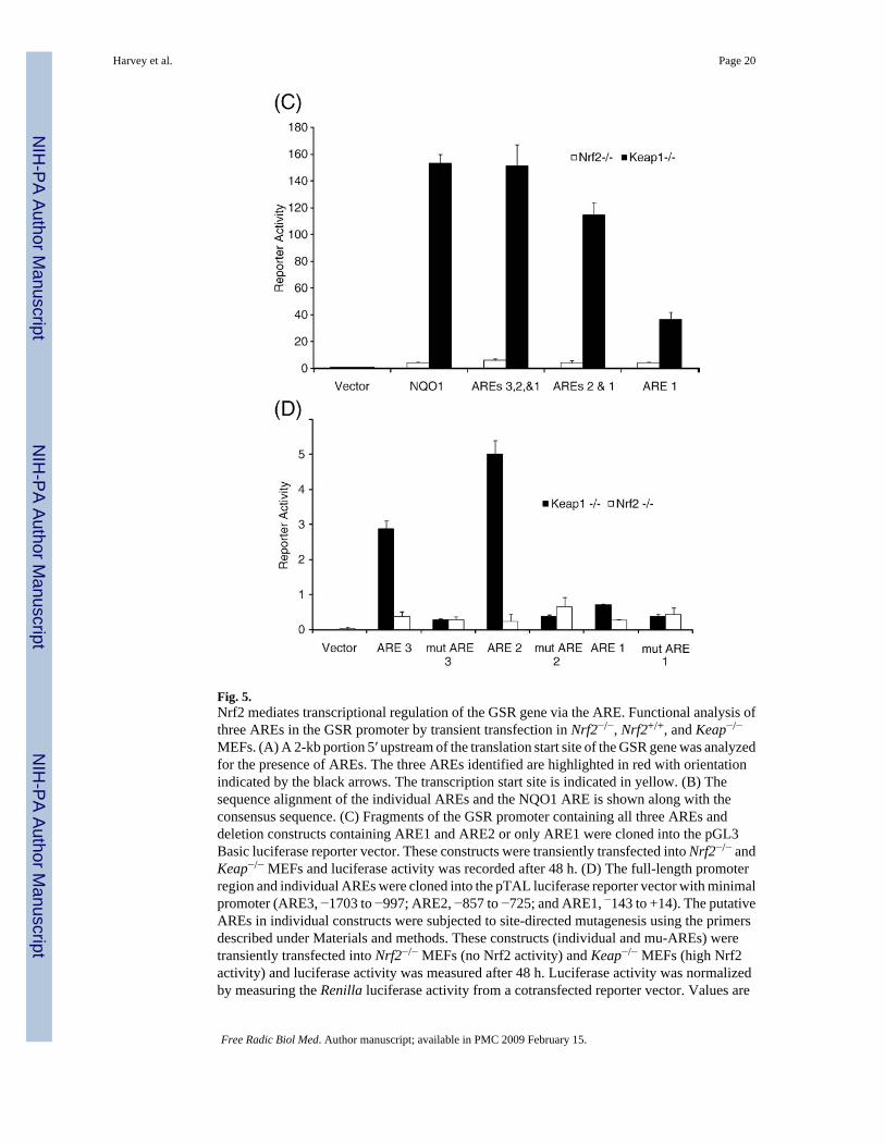
Fig. 5.Nrf2 mediates transcriptional regulation of the GSR gene via the ARE. Functional analysis ofthree AREs in the GSR promoter by transient transfection in Nrf2−/−, Nrf2+/+, and Keap−/−
MEFs. (A) A 2-kb portion 5′ upstream of the translation start site of the GSR gene was analyzedfor the presence of AREs. The three AREs identified are highlighted in red with orientationindicated by the black arrows. The transcription start site is indicated in yellow. (B) Thesequence alignment of the individual AREs and the NQO1 ARE is shown along with theconsensus sequence. (C) Fragments of the GSR promoter containing all three AREs anddeletion constructs containing ARE1 and ARE2 or only ARE1 were cloned into the pGL3Basic luciferase reporter vector. These constructs were transiently transfected into Nrf2−/− andKeap−/− MEFs and luciferase activity was recorded after 48 h. (D) The full-length promoterregion and individual AREs were cloned into the pTAL luciferase reporter vector with minimalpromoter (ARE3, −1703 to −997; ARE2, −857 to −725; and ARE1, −143 to +14). The putativeAREs in individual constructs were subjected to site-directed mutagenesis using the primersdescribed under Materials and methods. These constructs (individual and mu-AREs) weretransiently transfected into Nrf2−/− MEFs (no Nrf2 activity) and Keap−/− MEFs (high Nrf2activity) and luciferase activity was measured after 48 h. Luciferase activity was normalizedby measuring the Renilla luciferase activity from a cotransfected reporter vector. Values are
Harvey et al. Page 20
Free Radic Biol Med. Author manuscript; available in PMC 2009 February 15.
NIH
-PA Author Manuscript
NIH
-PA Author Manuscript
NIH
-PA Author Manuscript

means±SE from three different experiments (p<0.05). *Significant compared to Nrf2−/−. (Forinterpretation of the references to colour in this figure legend, the reader is referred to the webversion of this article.)
Harvey et al. Page 21
Free Radic Biol Med. Author manuscript; available in PMC 2009 February 15.
NIH
-PA Author Manuscript
NIH
-PA Author Manuscript
NIH
-PA Author Manuscript

Fig. 6.CHIP analysis demonstrating Nrf2 binding to GSR AREs. (A) Nuclear extracts from Nrf2−/−,Nrf2+/+, and Keap−/− MEFs were used to assess the Nrf2 binding activity to ARE3 and ARE2.(A) CHIP assay was performed with Nrf2−/−, Nrf2+/+, and Keap−/− MEFs using anti-Nrf2antibody and rabbit IgG1. (B) Quantification of CHIP assay results ((GSR promoter binding-antibody)/input ratio). Quantification was performed on representative CHIP data from n=3independent experiments.
Harvey et al. Page 22
Free Radic Biol Med. Author manuscript; available in PMC 2009 February 15.
NIH
-PA Author Manuscript
NIH
-PA Author Manuscript
NIH
-PA Author Manuscript