class iii correction using an inter-arch spring-loaded module · class iii correction using an...
TRANSCRIPT
Vanlaecken et al. Progress in Orthodontics 2014, 15:32http://www.progressinorthodontics.com/content/15/1/32
RESEARCH ARTICLE Open Access
Class III correction using an inter-archspring-loaded moduleRobert Vanlaecken2, Michael O Williams3, Thomas Razmus4, Erdogan Gunel5, Chris Martin1 and Peter Ngan1*
Abstract
Background: A retrospective study was conducted to determine the cephalometric changes in a group of Class IIIpatients treated with the inter-arch spring-loaded module (CS2000®, Dynaflex, St. Ann, MO, USA).
Methods: Thirty Caucasian patients (15 males, 15 females) with an average pre-treatment age of 9.6 years weretreated consecutively with this appliance and compared with a control group of subjects from the Bolton-Brush Studywho were matched in age, gender, and craniofacial morphology to the treatment group. Lateral cephalograms weretaken before treatment and after removal of the CS2000® appliance. The treatment effects of the CS2000® appliancewere calculated by subtracting the changes due to growth (control group) from the treatment changes.
Results: All patients were improved to a Class I dental arch relationship with a positive overjet. Significant sagittal,vertical, and angular changes were found between the pre- and post-treatment radiographs. With an average treatmenttime of 1.3 years, the maxillary base moved forward by 0.8 mm, while the mandibular base moved backward by 2.8 mmtogether with improvements in the ANB and Wits measurements. The maxillary incisor moved forward by 1.3 mm andthe mandibular incisor moved forward by 1.0 mm. The maxillary molar moved forward by 1.0 mm while the mandibularmolar moved backward by 0.6 mm. The average overjet correction was 3.9 mm and 92% of the correction was due toskeletal contribution and 8% was due to dental contribution. The average molar correction was 5.2 mm and 69% of thecorrection was due to skeletal contribution and 31% was due to dental contribution.
Conclusions: Mild to moderate Class III malocclusion can be corrected using the inter-arch spring-loaded appliance withminimal patient compliance. The overjet correction was contributed by forward movement of the maxilla, backward anddownward movement of the mandible, and proclination of the maxillary incisors. The molar relationship was correctedby mesialization of the maxillary molars, distalization of the mandibular molars together with a rotation of the occlusalplane.
BackgroundTreatment of Class III malocclusions may includegrowth modification, camouflage with orthodontic toothmovement and orthognathic surgery [1]. In young patientswith deficient maxilla, facemask is the appliance of choicewhereas in patients with a normal maxilla and prognathicmandible, the chin cup appliance is usually preferred. InClass III patients with no growth remaining, fixed appliancewith Class III elastics are usually used to camouflagethe malocclusion [2]. However, most of these appliancesrequire patient cooperation. If patients do not wear the
* Correspondence: [email protected] of Orthodontics, West Virginia University School of Dentistry,1073 Health Science Center North, P.O. Box 9480, MorgantownWV 26506, USAFull list of author information is available at the end of the article
© 2014 Vanlaecken et al.; licensee Springer. ThiCommons Attribution License (http://creativecoreproduction in any medium, provided the orig
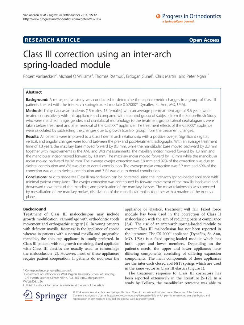
appliance or elastics, treatment will fail. Fixed forcemodule has been used in the correction of Class IImalocclusion with the aim of reducing patient compliance[3,4]. The use of an inter-arch spring-loaded module tocorrect Class III malocclusion has not been reported inthe literature. The CS 2000® appliance (Dynaflex, St. Ann,MO, USA) is a fixed spring-loaded module which hasboth upper and lower members. Depending on thepatient's needs, the upper and lower appliances havediffering components consisting of differing expansioncomponents. The main components of these appliancesare the inter-arch closed coil NiTi springs which are usedin the same vector as Class III elastics (Figure 1).The treatment response to Class III correctors has
been reported extensively in the literature [5-12]. In astudy by Tollaro, the mandibular retractor was able to
s is an Open Access article distributed under the terms of the Creativemmons.org/licenses/by/2.0), which permits unrestricted use, distribution, andinal work is properly cited.
A
B CFigure 1 The CS2000® appliance (A), TB SAG appliance (B), andMSX 2000 appliance (C).
Vanlaecken et al. Progress in Orthodontics 2014, 15:32 Page 2 of 11http://www.progressinorthodontics.com/content/15/1/32
rotate the mandible downward and backward to com-pensate for the excessive mandibular growth [5]. Baikfound that the Frankel regulator III can correct ClassIII malocclusion in growing patients by a backwardand downward rotation of the mandible and lingualtipping of the lower incisors [6]. Garattini used aBionator III appliance to correct Class III malocclusion andconcluded that the majority of changes can be attributed todentoalveolar changes [9]. Similar results were noted byKidner with the use of a Class III twin block appliance [8].However, Proffit noted that these changes are not skeletalin origin, but mainly dentoalveolar [2]. These appliancesallowed the maxillary molars to migrate mesially and holdthe lower molars in place. They also proclined the upperincisors and retroclined the lower incisors, rotate theocclusal plane and/or the chin posterior, but have nomajor effect on the skeletal growth of the mandibleor maxilla.The facemask appliance has been used in the correction
of Class III patients with maxillary deficiency [10,11,13-18].The goal of this appliance is to provide skeletal correctionby protracting the maxilla and limiting the growth of themandible. While this is thought to be the main effect, daSilva Filho also noted that this appliance also rotated themandible down and back together with distalization of themandibular teeth and mesialization of the maxillary teeth
[13-16]. Ngan et al. reported on the treatment response ofClass III patients to expansion and facemask therapy[10]. The overjet correction was attributed to a forwardmovement of the maxilla, backward rotation of themandible, proclination of maxillary incisors, and retroclina-tion of the mandibular incisors. Baccetti looked at how ageaffects treatment outcomes with a bonded RPE andfacemask [11]. He found that in the early treatmentgroup (6.8 ± 0.6 years), a significant forward movement of‘A’ point occurred, while in the late treatment group(10.3 years ± 1.0 year), no significant A point movementwas achieved. During post-treatment, Baccetti found thatClass III growth patterns returned in the absence of anyskeletal retention appliances [17]. Westwood also found areturn to Class III growth patterns once treatment wascomplete and recommends an overcorrection duringfacemask treatment [18]. All of these appliances requiresignificant patient compliance in order to achieve areasonable treatment result. The objective of this studywas to determine the cephalometric changes in a group ofClass III patients treated with a fixed spring-loadedmodule that required minimal patient compliance(CS2000®, St. Ann, MO, USA) and compare the resultswith those reported by other Class III correctors.
MethodsThis study was approved by the Institutional ReviewBoard of West Virginia University. Approval was alsogranted from one of the authors (MW) for the use oforthodontic records from his office. Seventy-five patientswere treated consecutively by one of the authors (MW)with the CS2000® appliance. The inclusion criteria werethat all subjects had no previous orthodontic treatment.All subjects were in the mixed to early permanent dentitionages. All subjects had a Class III molar occlusion or amesial step and the pre-treatment Wits < 0. All subjectsrequired comprehensive orthodontic treatment togetherwith the CS2000® appliance. Patients with poor-qualityradiographs or missing radiographs were excludedfrom the study. The final sample consisted of 30 patients(15 males and 15 females) with a mean age of 9.6 ± 2.1 yearsand a range of ages 6 to 15 years. The mean age forthe male sample was 8.7 ± 1.7 years and the femalesample, 10.4 ± 2.2 years. The mean treatment timewas 1.3 ± 0.3 years. Lateral cephalograms were takenat pre-treatment (T1) and at completion of treatmentwith the CS2000® appliance (T2).The control group consisted of serial cephalometric
radiographs of 30 Class III subjects (15 boys, 15 girls)with no history of orthodontic treatment from theBolton-Brush Study. The control subjects were closelymatched in age, sex, and craniofacial morphology with theexperimental subjects (Table 1). Significant differenceswere found in 6 of the 26 cephalometric variables
Table 1 Comparison of pre-treatment craniofacialmorphology of control and treatment samples
Variable Control Treated Diff p value Sig
Mean SD Mean SD
Sagittal
OLp-A point 67.78 3.61 68.31 2.71 0.52 0.53 NS
OLp-B point 71.87 3.81 74.28 3.38 2.41 0.01 S
OLp-Pg 74.15 4.34 76.70 4.13 2.54 0.02 S
OLp-Co −9.74 2.09 −10.06 3.69 −1.32 0.09 NS
Wits −3.62 3.77 −4.22 2.14 −0.60 0.45 NS
Is-OLp 73.89 4.73 74.13 3.89 0.23 0.83 NS
Ii-OLp 73.20 3.92 74.12 3.85 0.92 0.36 NS
Overjet 0.69 2.43 0.01 2.29 0.68 0.26 NS
Ms-OLp 45.94 3.17 47.05 3.36 1.11 0.19 NS
Mi-OLp 49.24 3.08 49.63 3.19 0.38 0.63 NS
Molar relationship −3.30 1.82 −2.57 1.92 0.72 0.13 NS
Vertical
OLs-A point 27.04 3.92 29.40 5.22 2.36 0.06 NS
ANS-Me 59.34 4.22 56.40 4.96 −2.93 0.01 S
Is-NL 24.36 2.32 23.41 2.23 −0.85 0.15 NS
Ii-ML 34.10 3.07 34.05 2.48 −0.05 0.94 NS
Overbite 1.57 1.29 2.31 1.54 0.74 0.06 NS
Msc-NL 17.81 1.68 18.77 2.41 0.96 0.07 NS
Mic-ML 25.70 2.37 24.77 2.34 −0.93 0.11 NS
Angular
SNA 79.65 3.91 80.07 3.80 0.42 0.67 NS
SNB 77.39 2.77 80.39 3.59 3.09 0.01 S
ANB 2.36 3.32 −0.29 1.61 −2.66 0.01 S
SNL-NL 9.64 2.67 9.01 3.30 −0.63 0.41 NS
SNL-ML 38.83 4.97 32.81 5.01 −6.02 0.01 S
SNL-OLs 21.92 3.59 20.27 3.75 −1.65 0.06 NS
Is/SNL 100.17 6.73 102.24 9.02 2.07 0.31 NS
Ii/ML 86.65 6.73 85.81 7.16 −0.84 0.64 NS
Interincisal angle 134.20 7.68 139.12 13.30 4.92 0.08 NS
Overjet (Is/OLp − Ii/OLp), molar relationship (Ms/OLp −Mi/OLp), maxillary base(A/OLp), mandibular base (pg/OLp), maxillary incisor (Is/OLp), mandibularincisor (Ii/OLp), maxillary molar (Ms/OLp), and mandibular molar (Mi/OLp).
Vanlaecken et al. Progress in Orthodontics 2014, 15:32 Page 3 of 11http://www.progressinorthodontics.com/content/15/1/32
indicating that the starting form of the treatmentgroup has a more forward position of the mandibleand the control group has greater increase in thelower facial height and mandibular plane angle.The CS 2000® appliance is a fixed Class III corrector
consisting of an upper member, the Tooth Born Sagittal(TB SAG) appliance, and a lower member, the MSX2000 (Dynaflex, St. Ann, MO, USA), and an inter-archNiTi springs from upper-first molars to lower-firstbicuspids (Figure 1). The TB SAG is the upper memberof the CS2000® consisting of NiTi expansion springs fortransverse correction as well as NiTi springs in an
anterior-posterior direction that places a protractionforce on the pre-maxilla. It also provides an attachmentat the first molars for NiTi springs. The MSX 2000 isthe lower member of the CS2000® appliance that providesmandibular arch transverse correction with NiTi springs.It also provides an attachment at the first bicuspids forNiTi springs. Depending on the patient's needs, theupper and lower appliances have differing componentsconsisting of differing expansion components. The maincomponents of these appliances are the inter-archclosed-coil NiTi springs in the same vector as Class IIIelastics. When the inter-arch spring module is attached tothe pivot teeth, it will cause these teeth to resist the forceexpressed by the 150-g NiTi coil spring on each side.When the pivot teeth are coupled with the 300-g coilsprings, they will reinforce and serve as the anchorageteeth. This principle allows the inter-arch spring moduleto be a totally intraoral anchorage appliance and does notrely on the forehead and chin as anchorage as in the caseof protraction facemask therapy. Therefore, this appliancerequires minimal patient compliance.
Cephalometric analysisPre- and post-treatment lateral cephalograms fromboth groups were digitized using the Dolphin Imaging(Dolphin Imaging, Chatsworth, CA, USA) software toallow for landmark identification and adjusting formagnification. The cephalometric systems describedby Bjork [19] and Pancherz [20] were used to analyze thetreatment changes. The landmarks used are shown inFigures 2, 3, and 4. The magnification factor of thelateral cephalograms was found to be 6% both for thecontrol and treated subjects. The angular measurementswere located using the Dolphin Imaging system (DolphinImaging, Chatsworth, CA, USA) and reported to thenearest 0.1°. Films were printed 1:1 using a KodakESP 7250 printer (Kodak, Atlanta, GA, USA), andthen traced by one investigator using a #2 lead pencilon 0.003 in. matt cephalometric acetate tracing film(3 M Unitek, Monrovia, CA, USA). All linear measure-ments were performed using a digital caliper (accurate to0.01 mm) and reported to the nearest 0.1 mm. Analysis ofthe sagittal skeletal and dental changes were recordedalong the occlusal plane (OLs) and to the occlusal planeperpendicular (OLp) from the first cephalogram; thisformed the reference grid. The grid was then transferredto subsequent cephalograms by superimposing the tracingon the mid-sagittal cranial structures. The changes inoverjet and molar relationship were calculated using theformula depicted in Table 3.
Data analysisThe dentofacial morphology of the subjects in theexperimental group was compared using paired t tests.

Figure 2 Cephalometric landmarks and lines used forsagittal measurements.
Figure 4 Cephalometric landmarks and lines forangular measurements.
Vanlaecken et al. Progress in Orthodontics 2014, 15:32 Page 4 of 11http://www.progressinorthodontics.com/content/15/1/32
The starting forms of the control and experimentalsamples were compared with a two-tailed t test. Theskeletal and dental changes between the treated andcontrol sample at the two time periods were compared witha two-tailed t test. The confidence level was set at 95%.
Figure 3 Cephalometric landmarks and lines forvertical measurements.
Method errorThe error in locating, superimposing, and measuringthe changes of the landmarks by one examiner weremeasured on the cephalograms of 10 randomly selectedsubjects. All cephalograms were recorded twice inde-pendently on two separate occasions with a 2-weekinterval. For all the cephalometric variables, differencesbetween the independent repeated measurements of eachindividual before/after treatment were recorded. Theintraclass correlation coefficient of reliability (R) wasused to determine the reliability of cephalometricmeasurements. The R value ranged from 0 to 1.00,with R value greater than 0.90 indicating high reliability.The mean differences for all linear measurements wereless than 0.8 mm. The greatest mean error for angularmeasurement was 0.9° for the measurement of maxillarycentral incisal angle (Is/NSL) and mandibular centralincisal angle (Ii/ML).
ResultsCephalometric changesChanges in cephalometric measurements in patientstreated with the CS2000® appliance (T2-T1) are shown inTables 2, 3, and 4. The appliance effects were calculatedby subtracting the changes due to growth from thetreatment changes.
Sagittal differencesSignificant differences were found in all the sagittal variablesmeasured (Table 2). Figures 5 and 6 summarize theskeletal and dental contributions to the overjet and
Table 2 Individual sagittal changes (mm) between pre-treatment and post-treatment in 30 subjects
Overjet Maxillaryincisor
Mandibularincisor
Molarrelationship
Maxillarymolar
Mandibularmolar
Maxillarybase
Mandibularbase
Patient
1 4.7 3.7 −1.0 5.4 4.5 −0.9 1.3 0.1
2 0.8 4.7 3.9 −1.0 −2.6 −1.6 0.3 −1.4
3 −7.7 −5.4 2.3 11.8 4.1 −7.7 0.5 −5.3
4 0.4 1.0 0.6 7.5 2.7 −4.8 0.9 −1.0
5 6.0 −0.3 −6.3 6.2 −0.5 −6.7 −0.4 −3.0
6 11.5 3.8 −7.7 7.8 0.9 −6.9 0.9 −6.2
7 8.9 4.9 −4.0 6.4 2.4 −4.0 0.6 −1.8
8 0.6 −0.7 −1.3 7.7 3.6 −4.1 1.3 −2.5
9 9.8 5.0 −4.8 6.5 −1.6 −8.1 −0.1 −4.4
10 7.0 6.0 −1.0 9.4 2.8 −6.6 1.1 −1.9
11 2.1 2.2 0.1 1.8 1.3 −0.5 0.2 −2.6
12 5.1 2.0 −3.1 7.7 3.5 −4.2 0.5 −4.8
13 7.8 3.9 −3.9 −4.8 −3.5 1.3 −0.8 −4.9
14 4.2 2.1 −2.1 3.0 1.2 −1.8 0.0 −2.5
15 −0.5 4.0 4.5 5.9 2.7 −3.2 3.0 −0.2
16 0.2 −1.1 −1.3 0.4 −1.1 −1.5 0.4 −2.6
17 1.0 2.1 1.1 3.1 0.9 −2.2 1.6 −2.0
18 6.1 3.9 −2.2 10,6 6.6 −4.0 1.2 −2.2
19 5.3 1.6 −3.7 2.2 3.1 0.9 −0.4 −4.1
20 4.2 −0.1 −4.3 4.8 1.2 −3.6 1.3 −4.6
21 10.0 2.3 −7.7 9.1 1.8 −7.3 1.2 −7.5
22 11.3 6.1 −5.2 1.2 −0.2 −1.4 0.9 −3.0
23 7.4 8.5 1.1 7.1 6.3 −0.8 4.5 0.7
24 5.0 4.1 −0.9 6.1 4.7 −1.4 1.5 0.4
25 2.8 −4.5 −7.3 5.9 2.3 −3.6 −1.4 −5.5
26 3.4 4.0 0.6 7.0 4.1 −2.9 0.8 −0.6
27 0.4 3.2 2.8 −3.5 −0.7 2.8 0.7 2.1
28 5.8 −2.5 −8.3 7.4 0.0 −7.4 −0.4 −7.4
29 4.4 2.9 −1.5 8.4 2.9 −5.5 0.7 −2.6
30 −8.3 −3.0 5.3 4.2 0.5 −3.7 1.4 −3.4
Pooled
Mean 4.0* 2.1* −1.8* 5.2* 1.8* −3.4* 0.8* −2.8*
SD 4.7 3.2 3.7 3.9 2.4 2.8 1.1 3.7
Min −8.3 −5.4 −8.3 −4.8 −3.5 −8.1 −1.4 −8.3
Max 11.5 8.5 5.3 11.8 6.6 2.8 4.5 5.3
Overjet (Is/OLp − Ii/OLp), molar relationship (Ms/OLp −Mi/OLp), maxillary base (A/OLp), mandibular base (Pg/OLp), maxillary incisor (Is/OLp), mandibular incisor(Ii/OLp), maxillary molar (Ms/OLp), and mandibular molar (Mi/OLp).* = p<.05.
Vanlaecken et al. Progress in Orthodontics 2014, 15:32 Page 5 of 11http://www.progressinorthodontics.com/content/15/1/32
molar corrections from treatment. With an averagetreatment time of 1.3 years, all subjects were corrected to aClass I or overcorrected to a Class I or II dental archrelationship. Overjet and sagittal molar relationshipsimproved by an average of 3.9 and 5.2 mm, respectively.This was a result of a 0.8-mm forward maxillary movement,
a 2.8-mm backward movement of the mandible, a 1.3-mmlabial movement of the maxillary incisors, a 1.0-mm labialmovement of the mandibular incisors, a 1.0-mm mesialmovement of the maxillary molar, and a 0.6-mm distalmovement of the mandibular molars. The relationship ofthe maxillary base to the mandibular base relative to the
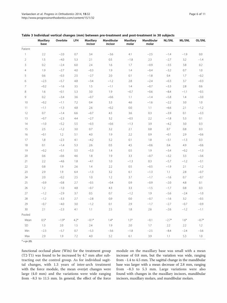
Table 3 Individual vertical changes (mm) between pre-treatment and post-treatment in 30 subjects
Maxillarybase
Overbite LFH Maxillaryincisor
Mandibularincisor
Maxillarymolar
Mandibularmolar
NL/SNL ML/SNL OL/SNL
Patient
1 2.2 −2.0 0.7 3.4 −3.6 4.1 −2.5 −1.4 −1.9 0.0
2 1.5 −4.0 5.3 2.1 0.5 −1.8 2.3 −2.7 3.2 −1.4
3 0.2 −2.4 6.0 2.4 1.6 1.7 −0.9 −3.5 3.8 0.2
4 1.9 −2.7 4.0 −0.3 1.9 1.4 −0.4 −3.2 0.7 1.0
5 0.6 −0.3 2.5 −2.7 2.0 0.1 −1.8 0.4 1.7 −0.2
6 −2.3 −5.7 4.8 −3.4 −1.2 2.8 −2.4 −0.3 3.7 −0.3
7 −0.2 −1.6 3.5 1.5 −1.1 1.4 −0.7 −3.3 2.8 0.6
8 1.6 −0.1 5.3 3.0 1.9 −0.7 −0.6 −8.4 −1.1 −0.5
9 0.2 −3.4 3.6 −0.7 −0.6 1.1 −1.4 −5.8 1.4 −3.0
10 −0.2 −1.1 7.2 0.4 3.3 4.6 −1.6 −2.2 3.0 1.0
11 −1.1 −1.3 4.8 2.6 −0.2 0.0 1.1 −6.6 2.1 −1.2
12 0.7 −1.4 6.6 −0.7 4.5 3.6 0.3 −3.9 0.1 −3.3
13 −0.7 −2.3 4.4 −2.7 3.2 −0.5 2.2 −1.8 5.3 0.1
14 −1.0 −5.2 5.5 −0.3 −0.6 −1.3 3.9 −4.2 3.0 0.3
15 2.5 −1.2 3.0 0.7 3.2 2.1 0.8 0.7 0.8 0.3
16 −0.1 1.2 5.1 4.0 1.9 2.2 0.9 −0.1 2.9 −0.6
17 1,8 −2.3 4.1 −4.2 5.2 0.1 1.8 −3.1 −1.3 0.3
18 0.1 −1.4 5.3 2.6 0.5 4.5 −0.6 0.4 4.9 −0.6
19 −0.2 −5.1 5.5 −5.3 1.4 0.5 1.9 −3.4 −0.2 −1.3
20 0.6 −0.6 4.6 1.8 1.9 3.3 −0.7 −3.2 3.3 −3.6
21 2.2 −4.6 1.8 −4.1 1.0 −1.3 0.3 −5.7 −1.2 −3.1
22 0.8 1.9 2.6 1.4 2.2 0.5 −0.5 −1.9 2.1 −1.2
23 2.9 1.9 6.4 −1.3 3.2 6.1 −1.3 1.1 2.8 −0.7
24 2.0 −0.2 2.5 1.0 1.2 3.7 −1.7 −1.6 0.7 −0.7
25 −0.9 −0.8 2.7 −0.5 −0.4 0.9 −0.9 −2.0 4.8 0.1
26 1.2 −1.0 4.8 −0.7 4.3 3.3 −1.5 −1.7 0.8 0.3
27 −1.2 −2.9 3.7 0.5 0.7 −1.2 1.9 −5.6 −2.4 −1.0
28 −1.2 −3.3 2.7 −2.8 0,9 0.0 −0.7 −1.6 3.2 −0.5
29 −0.7 −4.0 3.0 −1.2 0.1 2.9 −1.7 −2.7 −0.7 −0.9
30 2.1 −2.3 4.1 1.1 2.5 1.8 2.6 −2.6 −1.2 −1.1
Pooled
Mean 0.5* −1.9* 4.2* −0.1* 1.4* 1.5* −0.1 −2.7* 1.6* −0.7*
SD 1.3 2.0 1.5 2.4 1.9 2.0 1.7 2.2 2.2 1.2
Min −2.3 −5.7 0.7 −5.3 −3.6 −1.8 −2.5 −8.4 −2.4 −3.6
Max 2.9 1.9 7.2 4.0 5.2 6.1 3.9 1.1 5.3 1.0
* = p<.05.
Vanlaecken et al. Progress in Orthodontics 2014, 15:32 Page 6 of 11http://www.progressinorthodontics.com/content/15/1/32
functional occlusal plane (Wits) for the treatment group(T2-T1) was found to be increased by 4.7 mm after sub-tracting out the control group. As for individual sagit-tal changes, with 1.3 years of inter-arch treatmentwith the force module, the mean overjet changes werelarge (4.0 mm) and the variations were wide rangingfrom −8.3 to 11.5 mm. In general, the effect of the force
module on the maxillary base was small with a meanincrease of 0.8 mm, but the variation was wide, rangingfrom −1.4 to 4.5 mm. The sagittal change in the mandibularbase was larger with a mean decrease of 2.8 mm, rangingfrom −8.3 to 5.3 mm. Large variations were alsofound with changes in the maxillary incisors, mandibularincisors, maxillary molars, and mandibular molars.
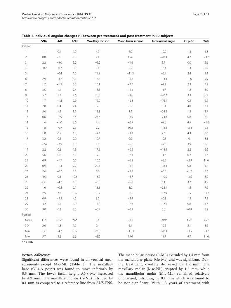
Table 4 Individual angular changes (°) between pre-treatment and post-treatment in 30 subjects
SNA SNB ANB Maxillary incisor Mandibular incisor Interincisal angle OLp-Co Wits
Patient
1 1.1 0.1 1.0 4.9 6.0 −9.0 1.4 1.8
2 0.0 −1.1 1.0 9.4 15.6 −28.3 4.7 −3.7
3 2.2 −3.0 5.2 −9.2 −4.6 8.7 0.0 5.6
4 −0.2 −0.7 0.5 0.1 5.5 −6.4 1.3 2.9
5 1.1 −0.4 1.6 14.8 −11.3 −5.4 2.4 5.4
6 2.9 −3.2 6.1 17.7 −6.8 −14.4 −1.0 9.9
7 1.2 −1.9 2.8 10.1 −3.7 −9.2 2.3 3.2
8 3.5 1.1 2.4 −8.3 −2.4 11.7 1.8 3.0
9 5.7 1.2 4.6 20.3 −1.6 −20.2 3.3 6.2
10 1.7 −1.2 2.9 16.0 −2.8 −16.1 0.3 6.9
11 2.8 0.4 2.4 −2.5 6.5 −6.1 4.0 0.1
12 5.0 1.2 3.7 15.1 8.9 −24.2 1.3 8.7
13 0.6 −2.9 3.4 23.6 −3.9 −24.8 0.8 8.0
14 1.6 −1.0 2.6 7.4 −0.9 −9.5 4.5 −1.0
15 1.8 −0.7 2.3 2.2 10.3 −13.4 −2.4 2.4
16 1.8 0.5 1.3 −4.1 −1.3 2.6 4.3 0.0
17 3.2 0.2 2.9 10.7 0.0 −9.5 −0.1 8.5
18 −2.4 −3.9 1.5 9.6 −6.7 −7.8 3.9 3.8
19 2.2 0.2 1.9 17.6 −0.5 −18.5 2.2 6.6
20 5.6 0.6 5.1 −7.5 −7.1 11.7 0.2 6.7
21 4.9 −1.7 6.6 10.6 −6.8 −2.5 −2.9 11.6
22 0.9 −1.4 2.2 20.4 −4.2 −18.4 0.8 4.2
23 2.6 −0.7 3.3 6.6 −3.8 −5.6 −1.2 8.7
24 −0.3 0.3 −0.6 16.2 −6.7 −10.0 −3.5 3.9
25 −3.1 −4.7 1.5 −5.0 −6.0 6.1 3.7 4.9
26 1.6 −0.3 2.1 18.3 3.0 −22.1 1.4 7.6
27 2.5 3.2 −0.7 10.2 5.0 −12.9 1.5 −1.2
28 0.9 −3.3 4.2 3.0 −5.4 −0.3 1.3 7.3
29 3.2 1.1 1.9 15.2 −2.3 −12.1 0.6 4.6
30 3.0 0.2 2.8 −0.4 −0.1 0.3 −0.3 3.2
Pooled
Mean 1.9* −0.7* 2.6* 8.1 −0.9 −8.9* 1.2* 4.7*
SD 2.0 1.8 1.7 9.4 6.1 10.6 2.1 3.6
Min −3.1 −4.7 −0.7 23.6 −11.3 −28.3 −3.5 −3.7
Max 5.7 3.2 6.6 −9.2 15.6 11.7 4.7 11.6
* = p<.05.
Vanlaecken et al. Progress in Orthodontics 2014, 15:32 Page 7 of 11http://www.progressinorthodontics.com/content/15/1/32
Vertical differencesSignificant differences were found in all vertical mea-surements except Mic-ML (Table 3). The maxillarybase (OLs-A point) was found to move inferiorly by0.5 mm. The lower facial height ANS-Me increasedby 4.2 mm. The maxillary incisor (Is-NL) intruded by0.1 mm as compared to a reference line from ANS-PNS.
The mandibular incisor (Ii-ML) extruded by 1.4 mm fromthe mandibular plane (Go-Me) and was significant. Dur-ing treatment, overbite decreased by 1.9 mm. Themaxillary molar (Msc-NL) erupted by 1.5 mm, whilethe mandibular molar (Mic-ML) remained relativelyunchanged, intruding by 0.1 mm which was found tobe non-significant. With 1.3 years of treatment with
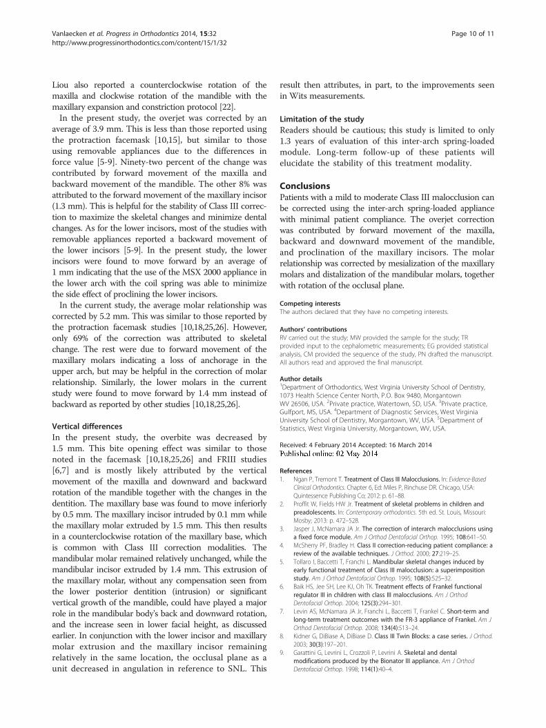
0.8mm
3.6mm
0.3mm
1.3mm
1.0mm
2.8mm
3.9mm
Overjet Correction (3.9)= Maxilla (0.8 mm) + Mx incisor (1.3) - Mandible (-2.8) - Md incisor (1.0)
Maxilla = OLp-A pt. (0.8 )
Mx incisor = Is-OLp (2.1) minus OLp-A pt. (0.8) = 1.3
Mandible = OLp-Pg (-2.8)
Mandibular incisor = Ii-OLp (-1.8) minus OLp-Pg (-2.8) = 1.0
Figure 5 Skeletal and dental contributions to overjet correction (T2-T1).
Vanlaecken et al. Progress in Orthodontics 2014, 15:32 Page 8 of 11http://www.progressinorthodontics.com/content/15/1/32
the force module, vertical maxillary base change wassmall with an average of 0.5 mm with a range of −2.3 to2.0 mm. The overbite reduction in individual subjectsranged from −5.7 to 1.9 mm. The lower face heightincreased in all subjects. No consistent pattern was foundin vertical changes of incisors and molars.
Angular differencesSignificant differences were found between all angu-lar measurements except Is/NL and Ii/ML (Table 4).SNA increased by 1.9° during treatment and was signifi-cant, while SNB remained decreased at 0.7°. ANB thusincreased by 2.6° during treatment and was found sig-nificant. The Is-NL was found to procline at 8.1° dur-ing treatment and the mandibular incisor retroclinedat 0.9° during treatment in relation to the mandibularplane (Go-Me), but were neither significant. The inter-incisal angle was found to increase by 8.9°. NL to SNwas found to decrease by 2.7° during treatment indi-cating a counterclockwise rotation of the palatal plane.ML to SN was found to increase by 1.6°, indicating aclockwise rotation of the mandibular plane. The OL to SNwas found to decrease by 0.7° during treatment, indicatinga counterclockwise rotation of the occlusal plane.
DiscussionThe use of an inter-arch spring-loaded module (CS2000®)to correct Class III malocclusion has not been reported inthe literature. However, different treatment modalities havebeen used to treat Class III malocclusions ranging fromprotraction facemasks [10,15,21], to removable appliancessuch as the Frankel III [8,9], Bionator III [10], modifiedtandem traction bow [12], and Class III Twin Block [11],to inter-arch protraction springs described by Liou [21,22].With the increase in popularity of skeletal anchoragedevices such as miniscrews and miniplates [23,24], moretreatment possibilities will be available.
Sagittal differencesIn the present study, significant changes were found inall the sagittal variables as compared to the controlgroup. The maxilla (A point) was found to move forwardby 0.8 mm over a period of 1.3 years. In a study withprotraction facemask [10], A point was found to moveforward by an average of 1.8 mm over a 6-month period.Baccetti found 2.3-mm- and 3.1-mm-forward movementsof A point in young patients treated using protractionfacemasks [11,17]. Most of the studies reported forwardmovements ranging from 1.5 to 3.4 mm [13-15]. Loiureported a 5.8-mm-forward movement of the maxilla in
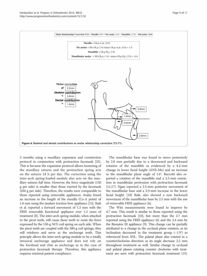
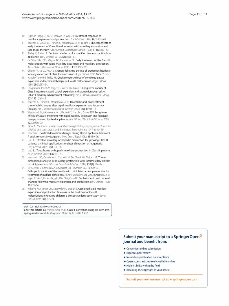
3.6mm
1.6mm
2.8mm
1.0mm
0.6mm
0.8mm
5.2mm
Molar Relationship Correction (5.2) = Maxilla (0.8) + Mx molar (1.0) – Mandible (-2.8) – Md molar (-0.6)
Maxilla = OLp-A pt. (0.8)
Mx molar = Ms-OLp (1.8) minus OLp-A pt. (0.8) = 1.0
Mandible = OLp-Pg (-2.8)
Mandibular molar = Mi-OLp (-3.4) minus OLp-Pg (-2.8) = -0.6
Figure 6 Skeletal and dental contributions to molar relationship correction (T2-T1).
Vanlaecken et al. Progress in Orthodontics 2014, 15:32 Page 9 of 11http://www.progressinorthodontics.com/content/15/1/32
3 months using a maxillary expansion and constrictionprotocol in conjunction with protraction facemask [25].This is because the expansion protocol allows loosening ofthe maxillary sutures and the protraction spring actson the sutures 24 h per day. The correction using theinter-arch spring-loaded module also acts on the max-illary sutures full time. However, the force magnitude (150g per side) is smaller than those exerted by the facemask(450 g per side). Therefore, the results were comparable tothose reported using removable appliances. Atalay foundan increase in the length of the maxilla (Co-A point) of1.8 mm using the tandem traction bow appliance [12]. Baiket al. reported a forward movement of 1.3 mm with theFRIII removable functional appliance over 1.3 years oftreatment [8]. The inter-arch spring module, when attachedto the pivot teeth, will cause these teeth to resist the forceexpressed by the 150-g NiTi coil spring on each side. Whenthe pivot teeth are coupled with the 300-g coil springs, theywill reinforce and serve as the anchorage teeth. Thisprinciple allows the inter-arch spring module to be a totallyintraoral anchorage appliance and does not rely onthe forehead and chin as anchorage as in the case ofprotraction facemask therapy. Therefore, this appliancerequires minimal patient compliance.
The mandibular base was found to move posteriorlyby 2.8 mm partially due to a downward and backwardrotation of the mandible as evidenced by a 4.2-mmchange in lower facial height (ANS-Me) and an increasein the mandibular plane angle of 1.6°. Baccetti also re-ported a rotation of the mandible and a 2.5-mm restric-tion in mandibular protrusion with protraction facemask[11,17]. Ngan reported a 2.5-mm posterior movement ofthe mandibular base and a 2.9-mm increase in the lowerfacial height. [10] Baik, also showed a new backwardmovement of the mandibular base by 2.5 mm with the useof removable FRIII appliance [6].The Wits measurements were found to improve by
4.7 mm. This result is similar to those reported using theprotraction facemask [10], but more than the 2.7 mmreported using the FRIII appliance [6] and the 2.4 mm bythe Bionator III appliance [9]. This change can be partiallyattributed to a change in the occlusal plane rotation, as itsinclination decreased in the treatment group (−1.9°) asreferenced from SNL. The palatal plane also rotated in acounterclockwise direction as its angle decrease 2.2 mmthroughout treatment as well. Similar change in occlusaland palatal planes rotating counter clockwise with treat-ment are seen with protraction facemask treatment [15].
Vanlaecken et al. Progress in Orthodontics 2014, 15:32 Page 10 of 11http://www.progressinorthodontics.com/content/15/1/32
Liou also reported a counterclockwise rotation of themaxilla and clockwise rotation of the mandible with themaxillary expansion and constriction protocol [22].In the present study, the overjet was corrected by an
average of 3.9 mm. This is less than those reported usingthe protraction facemask [10,15], but similar to thoseusing removable appliances due to the differences inforce value [5-9]. Ninety-two percent of the change wascontributed by forward movement of the maxilla andbackward movement of the mandible. The other 8% wasattributed to the forward movement of the maxillary incisor(1.3 mm). This is helpful for the stability of Class III correc-tion to maximize the skeletal changes and minimize dentalchanges. As for the lower incisors, most of the studies withremovable appliances reported a backward movement ofthe lower incisors [5-9]. In the present study, the lowerincisors were found to move forward by an average of1 mm indicating that the use of the MSX 2000 appliance inthe lower arch with the coil spring was able to minimizethe side effect of proclining the lower incisors.In the current study, the average molar relationship was
corrected by 5.2 mm. This was similar to those reported bythe protraction facemask studies [10,18,25,26]. However,only 69% of the correction was attributed to skeletalchange. The rest were due to forward movement of themaxillary molars indicating a loss of anchorage in theupper arch, but may be helpful in the correction of molarrelationship. Similarly, the lower molars in the currentstudy were found to move forward by 1.4 mm instead ofbackward as reported by other studies [10,18,25,26].
Vertical differencesIn the present study, the overbite was decreased by1.5 mm. This bite opening effect was similar to thosenoted in the facemask [10,18,25,26] and FRIII studies[6,7] and is mostly likely attributed by the verticalmovement of the maxilla and downward and backwardrotation of the mandible together with the changes in thedentition. The maxillary base was found to move inferiorlyby 0.5 mm. The maxillary incisor intruded by 0.1 mm whilethe maxillary molar extruded by 1.5 mm. This then resultsin a counterclockwise rotation of the maxillary base, whichis common with Class III correction modalities. Themandibular molar remained relatively unchanged, while themandibular incisor extruded by 1.4 mm. This extrusion ofthe maxillary molar, without any compensation seen fromthe lower posterior dentition (intrusion) or significantvertical growth of the mandible, could have played a majorrole in the mandibular body's back and downward rotation,and the increase seen in lower facial height, as discussedearlier. In conjunction with the lower incisor and maxillarymolar extrusion and the maxillary incisor remainingrelatively in the same location, the occlusal plane as aunit decreased in angulation in reference to SNL. This
result then attributes, in part, to the improvements seenin Wits measurements.
Limitation of the studyReaders should be cautious; this study is limited to only1.3 years of evaluation of this inter-arch spring-loadedmodule. Long-term follow-up of these patients willelucidate the stability of this treatment modality.
ConclusionsPatients with a mild to moderate Class III malocclusion canbe corrected using the inter-arch spring-loaded appliancewith minimal patient compliance. The overjet correctionwas contributed by forward movement of the maxilla,backward and downward movement of the mandible,and proclination of the maxillary incisors. The molarrelationship was corrected by mesialization of the maxillarymolars and distalization of the mandibular molars, togetherwith rotation of the occlusal plane.
Competing interestsThe authors declared that they have no competing interests.
Authors’ contributionsRV carried out the study; MW provided the sample for the study; TRprovided input to the cephalometric measurements; EG provided statisticalanalysis, CM provided the sequence of the study, PN drafted the manuscript.All authors read and approved the final manuscript.
Author details1Department of Orthodontics, West Virginia University School of Dentistry,1073 Health Science Center North, P.O. Box 9480, MorgantownWV 26506, USA. 2Private practice, Watertown, SD, USA. 3Private practice,Gulfport, MS, USA. 4Department of Diagnostic Services, West VirginiaUniversity School of Dentistry, Morgantown, WV, USA. 5Department ofStatistics, West Virginia University, Morgantown, WV, USA.
Received: 4 February 2014 Accepted: 16 March 2014
References1. Ngan P, Tremont T. Treatment of Class III Malocclusions. In: Evidence-Based
Clinical Orthodontics. Chapter 6, Ed: Miles P, Rinchuse DR. Chicago, USA:Quintessence Publishing Co; 2012: p. 61–88.
2. Proffit W, Fields HW Jr. Treatment of skeletal problems in children andpreadolescents. In: Contemporary orthodontics. 5th ed. St. Louis, Missouri:Mosby; 2013: p. 472–528.
3. Jasper J, McNamara JA Jr. The correction of interarch malocclusions usinga fixed force module. Am J Orthod Dentofacial Orthop. 1995; 108:641–50.
4. McSherry PF, Bradley H. Class II correction-reducing patient compliance: areview of the available techniques. J Orthod. 2000; 27:219–25.
5. Tollaro I, Baccetti T, Franchi L. Mandibular skeletal changes induced byearly functional treatment of Class III malocclusion: a superimpositionstudy. Am J Orthod Dentofacial Orthop. 1995; 108(5):525–32.
6. Baik HS, Jee SH, Lee KJ, Oh TK. Treatment effects of Frankel functionalregulator III in children with class III malocclusions. Am J OrthodDentofacial Orthop. 2004; 125(3):294–301.
7. Levin AS, McNamara JA Jr, Franchi L, Baccetti T, Frankel C. Short-term andlong-term treatment outcomes with the FR-3 appliance of Frankel. Am JOrthod Dentofacial Orthop. 2008; 134(4):513–24.
8. Kidner G, DiBiase A, DiBiase D. Class III Twin Blocks: a case series. J Orthod.2003; 30(3):197–201.
9. Garattini G, Levrini L, Crozzoli P, Levrini A. Skeletal and dentalmodifications produced by the Bionator III appliance. Am J OrthodDentofacial Orthop. 1998; 114(1):40–4.
Vanlaecken et al. Progress in Orthodontics 2014, 15:32 Page 11 of 11http://www.progressinorthodontics.com/content/15/1/32
10. Ngan P, Hagg U, Yiu C, Merwin D, Wei SH. Treatment response tomaxillary expansion and protraction. Eur J Orthod. 1996; 18(2):151–68.
11. Baccetti T, McGill JS, Franchi L, McNamara JA Jr, Tollaro I. Skeletal effects ofearly treatment of Class III malocclusion with maxillary expansion andface-mask therapy. Am J Orthod Dentofacial Orthop. 1998; 113(3):333–43.
12. Atalay Z, Tortop T. Dentofacial effects of a modified tandem traction bowappliance. Eur J Orthod. 2010; 32(6):655–61.
13. da Silva Filho OG, Magro AC, Capelozza FL. Early treatment of the Class IIImalocclusion with rapid maxillary expansion and maxillary protraction.Am J Orthod Dentofacial Orthop. 1998; 113(2):196–203.
14. Chong YH, Ive JC, Artun J. Changes following the use of protraction headgearfor early correction of Class III malocclusion. Angle Orthod. 1996; 66(5):351–62.
15. Nartallo-Turley PE, Turley PK. Cephalometric effects of combined palatalexpansion and facemask therapy on Class III malocclusion. Angle Orthod.1998; 68(3):217–24.
16. Pangrazio-Kulbersh V, Berger JL, Janisse FN, Bayirli B. Long-term stability ofClass III treatment: rapid palatal expansion and protraction facemask vsLeFort I maxillary advancement osteotomy. Am J Orthod Dentofacial Orthop.2007; 131(1):7–19.
17. Baccetti T, Franchi L, McNamara JA Jr. Treatment and posttreatmentcraniofacial changes after rapid maxillary expansion and facemasktherapy. Am J Orthod Dentofacial Orthop. 2000; 118(4):404–13.
18. Westwood PV, McNamara JA Jr, Baccetti T, Franchi L, Sarver DM. Long-termeffects of Class III treatment with rapid maxillary expansion and facemasktherapy followed by fixed appliances. Am J Orthod Dentofacial Orthop. 2003;123(3):306–20.
19. Bjork A. The face in profile: an anthoropological X-ray investigation of Swedishchildren and conscripts. Lund: Berlingska Boktrycheriet; 1947: p. 40–58.
20. Pancherz H. Vertical dentofacial changes during Herbst appliance treatment.A cephalometric investigation. Swed Dent J Suppl. 1982; 15:189–96.
21. Liou EJ. Effective maxillary orthopedic protraction for growing Class IIIpatients: a clinical application simulates distraction osteogenesis.Prog Orthod. 2005; 6(2):154–71.
22. Liou EJ. Toothborne orthopedic maxillary protraction in Class III patients.J Clin Orthod. 2005; 39(2):68–75.
23. Heymann GC, Cevidanes L, Cornelis M, De Clerck HJ, Tulloch JF. Three-dimensional analysis of maxillary protraction with intermaxillary elasticsto miniplates. Am J Orthod Dentofacial Orthop. 2010; 137(2):274–84.
24. De Clerck HJ, Cornelis MA, Cevidanes LH, Heymann GC, Tulloch CJ.Orthopedic traction of the maxilla with miniplates: a new perspective fortreatment of midface deficiency. J Oral Maxillofac Surg. 2009; 67(10):2123–9.
25. Ngan P, Yiu C, Hu A, Hagg U, Wei SHY, Gunel E. Cephalometric and occlusalchanges following maxillary expansion and protraction. Eur J Orthod. 1998;20:239–54.
26. Williams MD, Sarver DM, Sadowsky PL, Bradley E. Combined rapid maxillaryexpansion and protraction facemask in the treatment of Class IIImalocclusions in growing children: a prospective long-term study. SeminOrthod. 1997; 3(4):265–74.
doi:10.1186/s40510-014-0032-2Cite this article as: Vanlaecken et al.: Class III correction using an inter-archspring-loaded module. Progress in Orthodontics 2014 15:32.
Submit your manuscript to a journal and benefi t from:
7 Convenient online submission
7 Rigorous peer review
7 Immediate publication on acceptance
7 Open access: articles freely available online
7 High visibility within the fi eld
7 Retaining the copyright to your article
Submit your next manuscript at 7 springeropen.com