class iii malocclusion and bilateral cross-bite in an …class iii malocclusion, transverse...
TRANSCRIPT

Case Report
Class III malocclusion and bilateral cross-bite in an adult patient treated
with miniscrew-assisted rapid palatal expander and aligners
Luca Lombardoa; Antonella Carluccib; Bortolo Giuliano Mainoc; Anna Colonnab; EmanuelePaolettod; Giuseppe Sicilianie
ABSTRACTThis case report describes the use of a miniscrew-assisted rapid palatal expander and aligners tocorrect bilateral cross-bite and crowding in an adult patient with a Class III skeletal pattern. Adigitally designed surgical guide was three-dimensionally printed and used to accurately insert fourminiscrews into the palate; these were employed to anchor a novel miniscrew-assisted rapid palatalexpander appliance without any dental anchorage. Cone-beam computed tomograms before andafter miniscrew-assisted rapid palatal expander treatment demonstrated the orthopedic expansionof the maxilla without dental tipping. The patient was then fitted with aligners to correct crowdingand malocclusion. This case report demonstrates the successful treatment of an adult patient with anarrow maxilla and bilateral cross-bite using a nonsurgical, conservative treatment. (Angle Orthod.0000;00:000–000.)
KEY WORDS: Rapid palatal expander; Aligners; Miniscrew
INTRODUCTION
Nearly 30% of adult orthodontic patients present witha transverse maxillary deficiency and posterior cross-
bite. For many years, surgically assisted rapid palatalexpansion has been the treatment of choice to resolve
maxillary constriction in young adults, although several
authors have reported successful nonsurgical expan-sion in young and adult patients.1–5 However, Chang et
al.6 described possible side effects in nonsurgical palatal
expansion that, in adult patients, may produce dentoal-veolar tipping with unfavorable periodontal effects.
In 2010, Lee et al.7 introduced an appliance secured
to the palate by means of miniscrews, the miniscrew-
assisted rapid palatal expander (MARPE), which wasused to treat a 20-year-old patient with transversediscrepancy for mandibular prognathism, obviating theneed for orthognathic surgery. Expansion was suc-cessfully achieved with minimal damage to the teethand periodontium, and the authors concluded thatMARPE was an effective means of correcting trans-verse deficits. Moreover, as the miniscrews areanchored to the basal bone, the orthopedic forceexerted by the appliance results in pure skeletalmovement while minimizing unwanted dental effects.8
Based on the study by Lee et al., many authors haverecently developed novel skeletal expanders with theaid of miniscrews, and new MARPE devices have beenused to correct maxillary constriction in patients ofvarious ages.9–11 In addition, other authors havedeveloped a hybrid palatal expander, introducingsurgical guides (Miniscrew Assisted Palatal Appliance,MAPA system) for miniscrew insertion into the palate toprevent damage to the anatomical structures.12,13
Furthermore, to prevent undesirable tooth anchorageeffects at high risk of causing periodontal or rootdamage, a pure skeletal anchorage expander calledthe bone-anchored maxillary expander has beendescribed.14
CASE REPORT
This case report describes an adult female patientwith Class III malocclusion and bilateral cross-bite
a Research Assistant, Postgraduate School of Orthodontics,University of Ferrara, Ferrara, Italy.
b Research Fellow, Postgraduate School of Orthodontics,University of Ferrara, Ferrara, Italy.
c Visiting Professor, Postgraduate School of Orthodontics,University of Ferrara, Ferrara, Italy.
d Orthodontic Technician, Lab Orthomodul, Thiene, Italy.e Professor and Chairman, Postgraduate School of Orthodon-
tics, University of Ferrara, Ferrara, Italy.Corresponding author: Luca Lombardo, Research Assistant,
Postgraduate School of Orthodontics, University of Ferrara,Ferrara, Italy(e-mail: [email protected])
Accepted: February 2018. Submitted: November 2017.Published Online: May 1, 2018
� 0000 by The EH Angle Education and Research Foundation,Inc.
DOI: 10.2319/111617-790.1 Angle Orthodontist, Vol 00, No 00, 00001
treated successfully with a pure skeletal anchorage
maxillary expander and aligners.
Diagnosis and Etiology
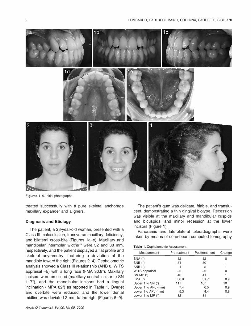
The patient, a 23-year-old woman, presented with a
Class III malocclusion, transverse maxillary deficiency,
and bilateral cross-bite (Figures 1a–e). Maxillary and
mandibular intermolar widths15 were 32 and 38 mm,
respectively, and the patient displayed a flat profile and
skeletal asymmetry, featuring a deviation of the
mandible toward the right (Figures 2–4). Cephalometric
analysis showed a Class III relationship (ANB 0, WITS
appraisal �5) with a long face (FMA 30.88). Maxillary
incisors were proclined (maxillary central incisor to SN
1178), and the mandibular incisors had a lingual
inclination (IMPA 828) as reported in Table 1. Overjet
and overbite were reduced, and the lower dental
midline was deviated 3 mm to the right (Figures 5–9).
The patient’s gum was delicate, friable, and translu-
cent, demonstrating a thin gingival biotype. Recession
was visible at the maxillary and mandibular cuspids
and bicuspids, and minor recession at the lower
incisors (Figure 1).
Panoramic and laterolateral teleradiographs were
taken by means of cone-beam computed tomography
Figures 1–4. Initial photographs.
Table 1. Cephalometric Assessment
Measurement Pretreatment Posttreatment Change
SNA (8) 82 82 0
SNB (8) 81 80 �1
ANB (8) 1 2 1
WITS appraisal �5 �5 0
SN MP (8) 40 41 1
FMA (8) 30.8 31.7 0.9
Upper 1 to SN (8) 117 107 10
Upper 1 to APo (mm) 7.4 6.5 0.9
Lower 1 o APo (mm) 5.3 4.4 0.8
Lower 1 to MP (8) 82 81 1
Angle Orthodontist, Vol 00, No 00, 0000
2 LOMBARDO, CARLUCCI, MAINO, COLONNA, PAOLETTO, SICILIANI

(CBCT; Figures 10–12), and an intraoral scan of thedental arches was performed. Axial CBCT slices at theupper cuspids and bicuspids and at the furcation of firstmolars clearly showed a maxillary traverse deficiencywith bilateral cross-bite (Figures 13–15). A three-dimensional skull model also revealed a diffuse paucityof buccal alveolar bone, in accordance with the clinicalfinding of gingival recession (Figures 16–17). Coronaland sagittal cross-sections were used to measurepalatal bone thickness (Figure 18). The patientreported a pronounced family history (both parents)of Class III and maxillary constriction, indicating thatthe malocclusion was genetic in origin.
Treatment Objectives
The primary objective was orthopedic correction ofthe posterior cross-bite by skeletal maxillary expansionwithout any dental compensation or worsening of theperiodontal situation. Additional objectives were toachieve molar and canine Class I, correct thecrowding, obtain ideal overjet (about 2.5 mm) andoverbite (about 2 mm), improve facial esthetics andincisor projection, and reduce black buccal corridorsduring smile.
Treatment Progress
To avoid any adverse effects on the upper teeth, abone-borne rapid palatal expander was selected.Because the maxilla was narrow and thin in the vertical
dimension, the MAPA system protocol was used to
insert four miniscrews into the palate.12,13 This protocol
enabled bicortical anchorage guaranteeing greater
resistance than that provided by orthopedic-loading
devices.16
First, the Standard Triangulation Language (STL)
files obtained from intraoral scans of the patient were
superimposed onto the CBCT Digital Imaging and
Communications in Medicine (DICOM) files. The
thicknesses of the palate were measured, and the
ideal positions for four virtual miniscrews were
identified (Figures 19–21). A three-dimensional tem-
plate was then designed and three-dimensionally
printed (MAPA system).12,13 It featured precisely
positioned cylindrical guide sleeves to enable the
correct placement of four miniscrews and rigorous
control of the direction of insertion (two 11-mm and
two 9-mm miniscrews, Ø 2 mm, Spider Screw,
Regular plus, HdC, Thiene, Italy; Figures 22–25). A
Polyvinyl Siloxane (PVS) impression of the upper arch
was then used to create the expansion device
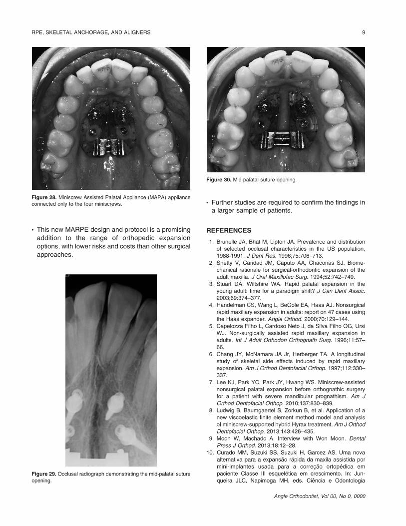
(Figures 26–28). The treatment protocol included
two activations per day17 until the mid-palatal suture
had opened and the constriction was corrected
(Figure 29). With 9 mm of appliance expansion, 7
mm of expansion was obtained at the maxillary first
molars, and 4 mm at the maxillary canines (Figure
30). Due to early contact between the upper and lower
second molars, the open bite was increased and the
Figures 5–9. Initial study models.
Angle Orthodontist, Vol 00, No 0, 0000
RPE, SKELETAL ANCHORAGE, AND ALIGNERS 3

device was left in situ for 2 months to stabilize the
expansion.
Postexpansion intraoral scans were taken and used
to plan aligner treatment. In this phase, interproximal
reduction to teeth 13 and 22, 35 and 43 was performed
to gain space and facilitate the derotation move-
ments.18–20 Then, 20 upper and lower individualized
F22 aligners (Sweden & Martina, Due Carrare, Italy)
were delivered to the patient after composite grip
points had been attached to the buccal surfaces of
teeth 13, 22, 23, 35, 44, and 45 and the lingual
surfaces of teeth 12, 11, 21, from 31 to 42 (Figures 31–
34), as prescribed by the digital set-up.
Each aligner was worn for 7 days and, after this
series, five upper and lower refinement aligners were
prescribed so that an acceptable result could be
Figures 10–12. Initial radiographs and cephalometric tracing.
Angle Orthodontist, Vol 00, No 00, 0000
4 LOMBARDO, CARLUCCI, MAINO, COLONNA, PAOLETTO, SICILIANI

achieved. Aligner treatment, therefore, lasted slighly
longer than 6 months (Figures 35–41). At the end ofthis phase, the four miniscrews were removed from the
palate. After 2 weeks, the peri-implant tissues had
completely healed (Figure 42).
Treatment Results
After 10 months, the treatment was complete. The
transverse constriction of the upper jaw had been
corrected and the bilateral cross-bite resolved. Com-
parison of the pre- and postoperative radiographs and
CBCT images reveal the maxillary expansion (Figures
43–47), which was also visible on dental casts (Figures
48–52). At the end of treatment, the patient displayed
Class I molar and canine relationships (due to an
increased positive tip, a slight edge-to-edge tendency
at the level of the canine was detectable). Cephalo-
metric data revealed an increase in the SNA (828) and
a reduction of the WITS index (Table 1). The data
reported in Table 1 also show that the vertical position
of the maxilla was relatively unchanged, but that the
FMA was slightly increased (31.78), as demonstrated
by the overall superimpositions (Figures 53–55). The
upper incisors had been extruded and uprighted while
the lower incisors remained unchanged (Table 1).
Measures of intermolar widths on the upper arches
before and after treatment showed an overall increase
in width of 6 mm (at the level of the palatal cusps of the
upper first molars; Table 2). Furthermore, all dental and
skeletal objectives had been achieved and a satisfac-
tory occlusal outcome was evident with no further
Figures 13–15. Initial cone-beam computed tomography axial slices.
Figures 16–17. Three-dimensional skull model showing diffuse
paucity of buccal alveolar bone.
Angle Orthodontist, Vol 00, No 0, 0000
RPE, SKELETAL ANCHORAGE, AND ALIGNERS 5

increase in recession. Although there had been somethinning of the buccal plates, there was still adequatecoverage of the maxillary cuspids, bicuspids, andmolar roots even after expansion, as shown in theCBCT slices (Figures 56–58).
The face appeared more symmetric, the patient’sprofile had been maintained, and the overall estheticshad been improved. The patient displayed a nice,broad smile, with improved incisor exposure and nobuccal corridors. The patient was instructed to wear thelast pair of aligners for retention due to the elasticpropriety of the thermoplastic material,21 and slightrestoration of tooth 22 was performed to achieveoptimal anterior tooth proportions.22 Upon completionof orthodontic treatment, the patient was offeredseveral periodontal surgery interventions to improve
the esthetics of the periodontal tissues. This multidis-ciplinary approach would have further enhanced thefinal outcome, providing results that could not beachieved by means of orthodontic treatment alone.Unfortunately, however, the patient refused surgery.
DISCUSSION
There is a strong consensus in the literature as to theefficacy of rapid maxillary expansion in growingpatients. However, in about 50% of cases, the reportedexpansion occurred at the mid-palatal suture, whereas
in the remaining 50% of cases it was brought about bydisplacement of the dentoalveolar complex.4 Age isconsidered a primary factor in the success of palatalexpansion, and this is based on the idea that it rapidly
Table 2. Skeletal Effects of Bone-Borne Rapid Maxillary Expander
Interdental Widthsa
Pretreatment Posttreatment Difference
U6 diameter (palatal
crown)
32 mm 38 mm 6 mm
U6 diameter (apex) 32.4 mm 38.1 mm 5.7 mm
U5 diameter (palatal
crown)
28.2 mm 33.6 mm 5.4 mm
U5 diameter (apex) 31.4 mm 37.4 mm 6 mm
U3 diameter (palatal
crown)
31 mm 34.4 mm 3.4 mm
a U5: Upper second premolar; U6: Upper first molar; U3: Uppercanine.
Figure 18. Cone-beam computed tomography cross-sections showing palatal bone thickness.
Figure 19. Cross-section of the maxilla and virtual position of the
miniscrews.
Angle Orthodontist, Vol 00, No 00, 0000
6 LOMBARDO, CARLUCCI, MAINO, COLONNA, PAOLETTO, SICILIANI

becomes inefficient after the early teens.23–25 In adults,surgery had long been considered the only option fororthopedic transverse correction. Nevertheless, manyauthors have reported cases of rapid maxillaryexpansion in adult patients based on the assumptionthat the correction of maxillary constriction results in adisplacement of the alveolar process associated withbuccal displacement of the teeth.26 However, rapidmaxillary expansion in adults can produce unwantedeffects, including lateral tipping of the posteriorteeth,27,28 extrusion,29,30 buccal root resorption,31,32
alveolar bone bending,33 fenestration of the buccalcortex,34,35 pain, and instability of the expansion.28,30,35
Carlson et al.17 and Mosleh et al.36 have reportedsuccessful outcomes in patients treated with MARPE,but these authors relied on an appliance anchoredpartially to the teeth. Winsauer et al.,14 on the otherhand, reported one case of a 30-year-old patientsuccessfully treated with bone-borne anchorage with-out unwanted dental effects.
To achieve true skeletal expansion, in this case apure skeletal anchorage expander was designed using
Figures 20–21. Digital Imaging and Communications in Medicine
(DICOM) and Standard Triangulation Language (STL) file superim-
position of intraoral patient maxilla.
Figures 22–24. Miniscrew Assisted Palatal Appliance (MAPA)
creation: three-dimensional–printed template for correct miniscrew
placement.
Angle Orthodontist, Vol 00, No 0, 0000
RPE, SKELETAL ANCHORAGE, AND ALIGNERS 7

a MAPA system to prevent any possible damage to theanatomical structures. Contrary to the belief thatnonsurgical palatal expansion is impossible in adultpatients, the posttreatment records of this adult patientclearly show skeletal expansion, verified by measure-ments of CBCT images and models (Table 2). Theposttreatment records of the patient show that thebuccal tipping of the teeth was well controlled37,38
(Figures 56–58; Table 3). The careful MARPE designand expansion protocol also resulted in a notableimprovement in the patient’s esthetics.39 Once ortho-pedic expansion of the upper jaw had been achieved,fully resolving the bilateral cross-bite, the patient wasthen fitted with aligners40; this confined the dentalmovements to the required teeth.
Such appliances as aligners can be extremely usefulin adult patients, especially in those with Class III orvertical discrepancy issues, as they maintain dentalcompensation without the need for other sources ofanchorage.41 Aligners also enable optimal oral hy-giene, especially in adults, in whom there is a greaterrisk of periodontal problems and a greater likelihood ofhaving a thin gingival biotype.42–44 A further advantageof aligner treatment is the favorable esthetics, whichmakes them better tolerated in patients, especiallyadults.
CONCLUSIONS
� The successful resolution of this case shows theefficacy of a combined protocol involving miniscrew-assisted rapid palatal expander and aligner treat-ment to resolve Class III malocclusion with bilateralcross-bite in an adult patient, despite the wide-spread belief that nonsurgical correction of suchcases is impossible. This orthopedic approachresulted in a better outcome than that previouslyreported in the literature, even those pertaining toyounger patients.
Figure 25. Miniscrews inserted into the palate after surgical guide
removal.
Figure 26. Polyvinyl Siloxane (PVS) impression showing the position
of the miniscrews.
Figure 27. Model of the patient’s maxilla used for appliance creation.
Table 3. Skeletal Effects of Bone-Borne Rapid Maxillary Expander
Buccolingual Angulation
Pretreatment Posttreatment Difference
16 angulation 99.18 99.78 0.68
26 angulation 99.18 99.28 0.18
15 angulation 92.28 92.28 08
25 angulation 90.68 928 1.48
13 angulation 102.28 1008 2.28
23 angulation 104.28 1018 3.28
Angle Orthodontist, Vol 00, No 00, 0000
8 LOMBARDO, CARLUCCI, MAINO, COLONNA, PAOLETTO, SICILIANI
� This new MARPE design and protocol is a promising
addition to the range of orthopedic expansion
options, with lower risks and costs than other surgical
approaches.
� Further studies are required to confirm the findings ina larger sample of patients.
REFERENCES
1. Brunelle JA, Bhat M, Lipton JA. Prevalence and distribution
of selected occlusal characteristics in the US population,
1988-1991. J Dent Res. 1996;75:706–713.
2. Shetty V, Caridad JM, Caputo AA, Chaconas SJ. Biome-
chanical rationale for surgical-orthodontic expansion of the
adult maxilla. J Oral Maxillofac Surg. 1994;52:742–749.
3. Stuart DA, Wiltshire WA. Rapid palatal expansion in the
young adult: time for a paradigm shift? J Can Dent Assoc.
2003;69:374–377.
4. Handelman CS, Wang L, BeGole EA, Haas AJ. Nonsurgical
rapid maxillary expansion in adults: report on 47 cases using
the Haas expander. Angle Orthod. 2000;70:129–144.
5. Capelozza Filho L, Cardoso Neto J, da Silva Filho OG, Ursi
WJ. Non-surgically assisted rapid maxillary expansion in
adults. Int J Adult Orthodon Orthognath Surg. 1996;11:57–
66.
6. Chang JY, McNamara JA Jr, Herberger TA. A longitudinal
study of skeletal side effects induced by rapid maxillary
expansion. Am J Orthod Dentofacial Orthop. 1997;112:330–
337.
7. Lee KJ, Park YC, Park JY, Hwang WS. Miniscrew-assisted
nonsurgical palatal expansion before orthognathic surgery
for a patient with severe mandibular prognathism. Am J
Orthod Dentofacial Orthop. 2010;137:830–839.
8. Ludwig B, Baumgaertel S, Zorkun B, et al. Application of a
new viscoelastic finite element method model and analysis
of miniscrew-supported hybrid Hyrax treatment. Am J Orthod
Dentofacial Orthop. 2013;143:426–435.
9. Moon W, Machado A. Interview with Won Moon. Dental
Press J Orthod. 2013;18:12–28.
10. Curado MM, Suzuki SS, Suzuki H, Garcez AS. Uma nova
alternativa para a expansao rapida da maxila assistida por
mini-implantes usada para a correcao ortopedica em
paciente Classe III esqueletica em crescimento. In: Jun-
queira JLC, Napimoga MH, eds. Ciencia e OdontologiaFigure 29. Occlusal radiograph demonstrating the mid-palatal suture
opening.
Figure 30. Mid-palatal suture opening.
Figure 28. Miniscrew Assisted Palatal Appliance (MAPA) appliance
connected only to the four miniscrews.
Angle Orthodontist, Vol 00, No 0, 0000
RPE, SKELETAL ANCHORAGE, AND ALIGNERS 9

casos clınicos baseado em evidencias cientıfica. Campinas:
Mundi Brasil; 2015:232–237.
11. Suzuki H, Moon W, Previdente LH, Suzuki SS, Garcez AS,
Consolaro A. Expansao rapida da maxila assistida com mini-
implantes ou MARPE: em busca de um movimento ortopedico
puro. Rev Clın Ortod Dental Press. 2016;15:110–125.
12. Maino G, Paoletto E, Lombardo L, Siciliani G. MAPA: a new
high-precisiom 3d method of palatal miniscrew placement.
EJCO. 2015,3:41–47.
13. Maino BG, Paoletto E, Lombardo L, Siciliani G. A three-
dimensional digital insertion guide for palatal miniscrew
placement. J Clin Orthod. 2016;50:12–22.
14. Winsauer H, Vlachojannis J, Winsauer C, Ludwig B, Walter
A. A bone-borne appliance for rapid maxillary expansion. J
Clin Orthod. 2013;47:375–381.
15. Lombardo L, Setti S, Molinari C, Siciliani G. Intra-arch
widths: a meta-analysis. Int Orthod. 2013 Jun;11(2):177–
192.
16. Lombardo L, Gracco A, Zampini F, Stefanoni F, Mollica F.
Optimal palatal configuration for miniscrew applications.
Angle Orthod. 2010;80:145–152.
17. Carlson C, Sung J, McComb RW, Machado AW, Moon W.
Microimplant-assisted rapid palatal expansion appliance to
orthopedically correct transverse maxillary deficiency in an
adult. Am J Orthod Dentofacial Orthop. 2016;149:716–728.
18. Sheridan JJ. Air-rotor stripping. J Clin Orthod. 1985;19:43–59.
19. Sheridan JJ. Air-rotor stripping update. J Clin Orthod.
1987;21:781–788.
20. Rossini G, Parrini S, Castroflorio T, Deregibus A, Debernardi
CL. Efficacy of clear aligners in controlling orthodontic tooth
movement: a systematic review. Angle Orthod. 2015;85:881–
889.
21. Lombardo L, Martines E, Mazzanti V, Arreghini A, Mollica F,
Siciliani G. Stress relaxation properties of four orthodontic
aligner materials: a 24-hour in vitro study. Angle Orthod.
2017;87:11–18.
22. Bolton WA. Disharmony in tooth size and its relation to the
analysis and treatment of malocclusion. Angle Orthod.
1958;28:113–130.
23. McNamara JA Jr, Brudon WL. Orthodontics and Dentofacial
Orthopedics. Needham, MA: Needham Press; 2001; 35–36.
24. Kerbs A. Midpalatal suture expansion studies by the implant
method over a seven-year period. Rep Congr Eur Orthod
Soc. 1964;40:131–142.
25. Handelman CS. Nonsurgical rapid maxillary alveolar
expansion in adults: a clinical evaluation. Angle Orthod.
1997;67:291–308.
26. Handelman C. Palatal expansion in adults: the nonsurgical
approach. Am J Orthod Dentofacial Orthop. 2011;140:462,
464, 466, 468.
27. Timms DJ. A study of basal movement with rapid maxillary
expansion. Am J Orthod. 1980;77:500–507.
28. Wertz RA. Skeletal and dental changes accompanying rapid
midpalatal suture opening. Am J Orthod. 1970;58:41–66.
29. Mommaerts MY. Transpalatal distraction as a method of
maxillary expansion. Br J Oral Maxillofac Surg. 1999;37:268–
272.
30. Zimring JF, Isaacson RJ. Forces produced by rapid maxillary
expansion. 3. Forces present during retention. Angle Orthod.
1965;35:178–186.
31. Barber AF, Sims MR. Rapid maxillary expansion and
external root resorption in man: a scanning electron
microscope study. Am J Orthod. 1981;79:630–652.
32. Langford SR, Sims MR. Root surface resorption, repair, and
periodontal attachment following rapid maxillary expansion
in man. Am J Orthod. 1982;81:108–115.
33. Shetty V, Caridad JM, Caputo AA, Chaconas SJ. Biome-
chanical rationale for surgical-orthodontic expansion of the
adult maxilla. J Oral Maxillofac Surg. 1994;52:742–749.
Figure 31. F22 virtual set-up.
Angle Orthodontist, Vol 00, No 00, 0000
10 LOMBARDO, CARLUCCI, MAINO, COLONNA, PAOLETTO, SICILIANI

34. Alpern MC, Yurosko JJ. Rapid palatal expansion in adults
with and without surgery. Angle Orthod. 1987;57:245–263.
35. Greenbaum KR, Zachrisson BU.The effect of palatal
expansion therapy on the periodontal supporting tissues.
Am J Orthod. 1982;81:12–21.
36. Mosleh MI, Kaddah MA, ElSayed FAA, ElSayed HS.
Comparison of transverse changes during maxillary
expansion with 4-point bone-borne and tooth-borne max-
illary expanders. Am J Orthod Dentofacial Orthop.
2015;148:599–607.
Figures 32–34. Grip-points and Interproximal reduction (IPR).
Figures 35–39. Intraoral photograph after the 20-aligner series.
Figures 40–41. Aligners in place.
Angle Orthodontist, Vol 00, No 0, 0000
RPE, SKELETAL ANCHORAGE, AND ALIGNERS 11

37. Christie KF, Boucher N, Chung CH. Effects of bonded rapid
palatal expansion on the transverse dimensions of themaxilla: a cone-beam computed tomography study. Am J
Orthod Dentofacial Orthop. 2010;137(suppl):S79–S85.
38. Lagravere MO, Carey J, Heo G, Toogood RW, Major PW.
Transverse, vertical, and anteroposterior changes from
bone-anchored maxillary expansion vs traditional rapid
maxillary expansion: a randomized clinical trial. Am J Orthod
Dentofacial Orthop. 2010;137:304.e1–e12.
39. Mirabella D1, Bacconi S, Gracco A, Lombardo L, Siciliani G.
Upper lip changes correlated with maxillary incisor move-
ment in 65 orthodontically treated adult patients. World J
Orthod. 2008;9:337–348.
40. Gu J, Tang JS, Skulski B, et al. Evaluation of Invisalign
treatment effectiveness and efficiency compared with con-
ventional fixed appliances using the Peer Assessment Rating
index. Am J Orthod Dentofacial Orthop. 2017;151:259–266.
41. Guarneri MP, Oliverio T, Silvestre I, Lombardo L, Siciliani G.
Open bite treatment using clear aligners. Angle Orthod.
2013;83:913–919.
42. Azaripour A, Weusmann J, Mahmoodi B, et al. Braces
versus Invisalignt: gingival parameters and patients’ satis-
faction during treatment: a cross-sectional study. BMC Oral
Health. 2015;15:69.
43. Abbate GM, Caria MP, Montanari P, et al. Periodontal health
in teenagers treated with removable aligners and fixed
orthodontic appliances. J Orofac Orthop. 2015;76:240–250.
44. Lombardo L, Ortan YO, Gorgun O, Panza C, Scuzzo G,
Siciliani G. Changes in the oral environment after placement
of lingual and labial orthodontic appliances. Prog Orthod.
2013;14:28.
Figure 42. Occlusal photograph after Miniscrew Assisted Palatal
Appliance (MAPA) and miniscrew removal.
Angle Orthodontist, Vol 00, No 00, 0000
12 LOMBARDO, CARLUCCI, MAINO, COLONNA, PAOLETTO, SICILIANI

Figures 43–44. Final photographs.
Angle Orthodontist, Vol 00, No 0, 0000
RPE, SKELETAL ANCHORAGE, AND ALIGNERS 13

Figures 45–47. Final radiographs and cephalometric tracing.
Angle Orthodontist, Vol 00, No 00, 0000
14 LOMBARDO, CARLUCCI, MAINO, COLONNA, PAOLETTO, SICILIANI
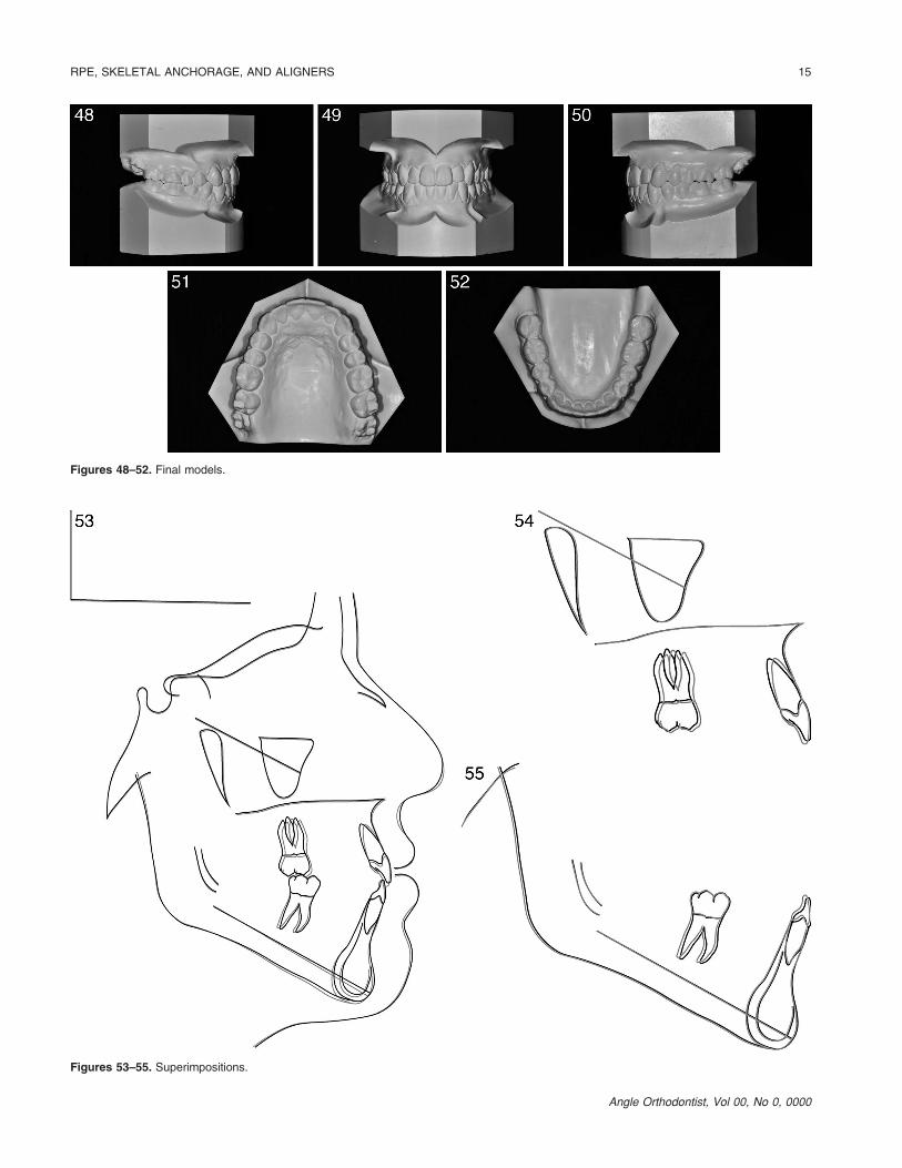
Figures 48–52. Final models.
Figures 53–55. Superimpositions.
Angle Orthodontist, Vol 00, No 0, 0000
RPE, SKELETAL ANCHORAGE, AND ALIGNERS 15

Figures 56–58. Final cone-beam computed tomography axial slices
showing the final angulation of the dentition.
Angle Orthodontist, Vol 00, No 00, 0000
16 LOMBARDO, CARLUCCI, MAINO, COLONNA, PAOLETTO, SICILIANI