clc chloride channels and transporters: a biophysical and...
TRANSCRIPT

Rev Physiol Biochem Pharmacol (2007)DOI 10.1007/112_2006_0605
G. Zifarelli · M. Pusch
CLC chloride channels and transporters:a biophysical and physiological perspective
Published online: 5 December 2006© Springer-Verlag 2007
Abstract Chloride-transporting proteins play fundamental roles in many tissues in theplasma membrane as well as in intracellular membranes. They have received increasingattention in the last years because crucial, and often unexpected and novel, physiologicalfunctions have been disclosed with gene-targeting approaches, X-ray crystallography, andbiophysical analysis. CLC proteins form a gene family that comprises nine members inmammals, at least four of which are involved in human genetic diseases. The X-ray struc-ture of the bacterial CLC homolog, ClC-ec1, revealed a complex fold and confirmed the an-ticipated homodimeric double-barreled architecture of CLC-proteins with two separate Cl–
ion transport pathways, one in each subunit. Four of the mammalian CLC proteins, ClC-1,ClC-2, ClC-Ka, and ClC-Kb, are chloride ion channels that fulfill their functional roles—stabilization of the membrane potential, transepithelial salt transport, and ion homeostasis—in the plasma membrane. The other five CLC proteins are predominantly expressed in in-tracellular organelles like endosomes and lysosomes, where they are probably important fora proper luminal acidification, in concert with the V-type H+-ATPase. Surprisingly, ClC-4,ClC-5, and probably also ClC-3, are not Cl– ion channels but exhibit significant Cl–/H+ an-tiporter activity, as does the bacterial homolog ClC-ec1 and the plant homolog AtCLCa. Thephysiological significance of the Cl–/H+ antiport activity remains to be established.
Overview and scope
The lipid bilayer that surrounds all living cells and the organelles inside eukaryotic cellspresents, by virtue of its fatty nature, an insurmountable electrostatic barrier for the diffu-sive passage of small inorganic ions like Na+, K+, Ca2+, Cl– and also small organic ionslike amino acids or HCO3
–. To overcome this barrier and to allow the exchange of thesesubstrates across the lipid bilayer in a controlled manner, nature has invented an incredible
G. Zifarelli · M. Pusch (�)CNR, Istituto di Biofisica,Via De Marini 6, 16149 Genova, Italye-mail: [email protected] · Tel.: +39-0106475-561/522 · Fax: +39-0106475-500

24 Rev Physiol Biochem Pharmacol (2007)
variety of different ion-transporting proteins, most of which allow the specific passage ofonly a very limited subset of ions. Transport proteins can be grossly subdivided into passivetransporters and active transporters. Conceptually, passive transporters can be regarded asenzymes that lower the activation energy for passive diffusion across the lipid bilayer. Themost important example of passive transporters are ion channels, which provide a selectivepore that allows a high-throughput transport, close to the diffusion limit in some cases, whilemaintaining exquisite selectivity. Active transporters couple the energy of the translocationof one substrate, or other energy sources such as ATP hydrolysis, to the transport of anothersubstrate, often in a strictly stoichiometric manner. One prominent example of this class ofproteins are the familiar P-type ion pumps and ion cotransporters. Active transport is gen-erally associated with the picture of an alternating access model of transport in which thetransporter exposes its ion binding sites alternatively to one or the other side of the mem-brane (see Tanford 1983). According to this mechanism, one or a few substrate moleculesare translocated for each transport cycle, leading to the slow transport rates seen for activetransporters, compared to those of ion channels (Hille 2001). As a consequence, in general,the architecture of active transporter proteins (see, e.g., Abramson et al. 2003; Toyoshima etal. 2000) is quite different from that of ion channels (see, e.g., Doyle et al. 1998; Miyazawaet al. 2003).
The present review focuses on anion-selective channels and, in particular, on Cl– chan-nels from the CLC family (Jentsch et al. 2002). However, as described below, the same basicarchitecture in the CLC family of proteins (Jentsch et al. 2005c) can be used to produce ei-ther active transporters (Accardi and Miller 2004; De Angeli et al. 2006; Picollo and Pusch2005; Scheel et al. 2005) or passive chloride channels (Bauer et al. 1991). Since a full ap-preciation of the physiological role of CLC proteins requires a molecular comprehension oftheir mechanism of transport, we will have to consider passive channel-mediated diffusionas well as the active antiport of protons and Cl– ions.
It is important to note that the CLC family represents only one of several classes ofproteins carrying out Cl– transport. A detailed treatment of such a vast and variegated ar-ray is beyond the scope of this review, but we nevertheless provide a brief overview of thephysiological roles of Cl– channels not belonging to the CLC branch.
The transport of Cl– (or any other ion) across the plasma membrane has two distinctconsequences: transport of the substrate and transport of electrical charge. The transport ofcharge is fundamental for the regulation of excitability in nerve and muscle, whereas thetransport of substrate is of paramount importance for epithelial physiology. In neurons andmuscle cells the membrane potential, Vm, is one of the most critical physiological variables.The activation of closed Cl– channels, or the inactivation of active Cl– channels, changesVm according to the equilibrium potential for Cl–, ECl. In most cases, the intracellular Cl–
concentration ([Cl–]int) is low, such that ECl is very negative and close to or even more nega-tive than EK. Low [Cl–]int is achieved by secondary active KCl cotransport proteins (Hübneret al. 2001). Thus Cl– channel activity in nerve and muscle generally dampens excitability,stabilizing a negative membrane potential. For the dampening and stabilization of the mem-brane potential not only is the value of ECl important, but also the chloride conductance, gCl,relative to other conductances, that is, a large gCl associated with a slightly depolarized EClwill nevertheless impede strong depolarization caused by a (relatively) small depolarizingconductance. A typical example, the skeletal muscle Cl– conductance that is provided by theClC-1 Cl– channel, is described in more detail below. In neurons, postsynaptic GABA andglycine receptors are the most important anion channels in the plasma membrane (Jentschet al. 2002). The traditional view is that their activation suppresses excitation (i.e., actionpotential firing) of the postsynaptic cell. It is clearly beyond the scope of this review to

Rev Physiol Biochem Pharmacol (2007) 25
describe these neuronal channels in detail. However, we would like to mention that acti-vation of GABA and glycine receptors is not always inhibitory: In the developing nervoussystem and in some specialized neuronal structures, [Cl–]int is relatively high, leading toa paradoxical excitatory effect of receptor activation (Marty and Llano 2005; Misgeld etal. 1986). GABA and glycine receptors are poorly selective for Cl–, showing a significantpermeability even to cations (Wotring et al. 2003). Physiologically, the permeability to bi-carbonate (HCO3
–) seems to be of particular relevance as it significantly contributes to a riseof [Cl–]int after GABA stimulation (see Marty and Llano 2005).
Apart from CLC proteins and GABA/glycine receptors, the only molecularly identifiedCl– channel is the “cystic fibrosis transmembrane conductance regulator,” CFTR (Riordanet al. 1989). CFTR is a widely expressed, but mostly epithelial, Cl– channel. Mutations inthe gene coding for CFTR cause cystic fibrosis (Tsui 1991), one of the most common lethalgenetic diseases. Structurally, CFTR belongs to the very large class of ABC transporters, butit seems to be the only channel member of this family of active transport proteins. Despiteextensive research in the 15 years since its cloning, the molecular mechanisms of channelgating by protein kinase A and intracellular ATP and also its physiological role are still rela-tively unclear. Excellent reviews about many aspects of CFTR have been published recently(Guggino 2004; Hanrahan and Wioland 2004; Riordan 2005).
Several important anion conductances have been described in various mammalian celltypes whose molecular identity is still unknown or in dispute. The most typical examplesare the swelling-activated Cl– channel, also known as VRAC (volume-regulated anion chan-nel) (Eggermont et al. 2001), and various types of calcium-activated Cl– channels. VRACis probably present in all animal cells and is activated by cell swelling, but the molecularmechanism leading to its activation is unknown (Eggermont et al. 2001). This channel isalso permeable to small organic solutes and has been proposed to be important for a processcalled regulatory volume decrease (RVD). Cellular volume regulation is essential for all celltypes to respond to osmotic challenges caused by changes of the extracellular medium aswell as to metabolically induced changes in intracellular osmolarity. The functional prop-erties of VRAC have been extensively studied, and several proteins have been proposed asmolecular correlates of VRAC, but none of these is generally accepted (see Jentsch et al.2002).
Ca2+ activated Cl– channels, CaCCs, are also found in many different cell systems in-cluding smooth muscle, epithelia, and olfactory receptors. Their activation, via an increaseof intracellular [Ca2+], generally leads to cell depolarization and thus, for example, smoothmuscle contraction or amplification of olfactory sensation (Hartzell et al. 2005). In epithelia,CaCC activation is responsible for transient Cl– (and water) secretion, for example, in sali-vary glands. Similar to VRAC, several proteins have been proposed as molecular correlatesof CaCCs, none of them being as yet fully accepted. Currently, the family of bestrophin pro-teins is under intense study as CaCC candidates (Hartzell et al. 2005), even though a definiteproof of their identity is still missing (see, e.g., Rosenthal et al. 2006).
Another example of a Cl– conductance for which the molecular association with a mem-brane protein is still lacking is the hyperpolarization- and cAMP-activated Cl– current mea-sured in choroid plexus cells (Kibble et al. 1996). This current superficially resembles ClC-2currents, but is found unaltered in ClC-2 knockout mice (Speake et al. 2002). Other exam-ples include an ATP-activated Cl– current described in mouse parotid acinar cells (Arreolaand Melvin 2003), and a proton-activated Cl– channel (Nobles et al. 2004), both sharingsome characteristics with VRAC.
Epithelial ion transporters are designed to allow massive but specific translocation ofsalts across the epithelial cell sheet. To allow for vectorial ion movement, transporters must
26 Rev Physiol Biochem Pharmacol (2007)
be expressed in a polarized manner. For example, the Na+-K+-ATPase is usually expressedon the basolateral membrane in epithelial cells. Thus it is important to understand the mech-anisms underlying the correct targeting of chloride channels and transporters to the apicalversus basolateral membrane. Very little is known about the targeting of the molecularlyidentified Cl– channels (CLC channels, GABA/glycine receptors, CFTR), even though sev-eral putative partner proteins of CLC channels, possibly important for targeting, have beenidentified in recent years (Dhani and Bear 2006) and are described in some detail below.
Cl– channels are not restricted to the plasma membrane but are also found in intracellularorganelles. Relatively little is known about the intracellular Cl– channels from in situ stud-ies. This is largely explained by the inaccessibility of the small intracellular organelles tostandard patch clamp techniques. As discussed in detail in later sections of this review, fiveof the nine mammalian CLC homologs reside in intracellular membranes, and their studythus opens new and promising perspectives for the understanding of the role of intracellularCl– channels and transporters.
The present review first describes the general mechanism underlying the function ofCLC proteins and then focuses on the biophysical properties and physiological and patho-physiological roles of mammalian, and in particular human, CLC members. For the mech-anistic aspects, two “model” CLCs have been most extensively studied. One is the Torpedochannel ClC-0, which, compared to many, physiologically more relevant, channels, has fa-vorable biophysical properties, for example, a relatively large single-channel conductance,and whose mechanisms of gating are best understood. The other model CLC is, of course,the bacterial ClC-ec1, for which we have detailed structural information and which can alsobe studied functionally. For reasons of space we do not attempt to cover the research on CLCproteins in other organisms like plants (Barbier-Brygoo et al. 2000; De Angeli et al. 2006),Caenorhabditis elegans (Strange 2003), or other model organisms or pathogens (see, e.g.,Salas-Casas et al. 2006).
Introduction: The CLC family of chloride-transporting proteins
The research in the CLC chloride channel field has always been accompanied, right from itsvery beginning, by a great number of unexpected findings and surprises. Already the firststep in the field, the identification of the Torpedo channel by Miller and coworkers (Whiteand Miller 1979), was a sort of accident (or artifact) in the quest of the authors to investigateacetylcholine-gated cation channels.
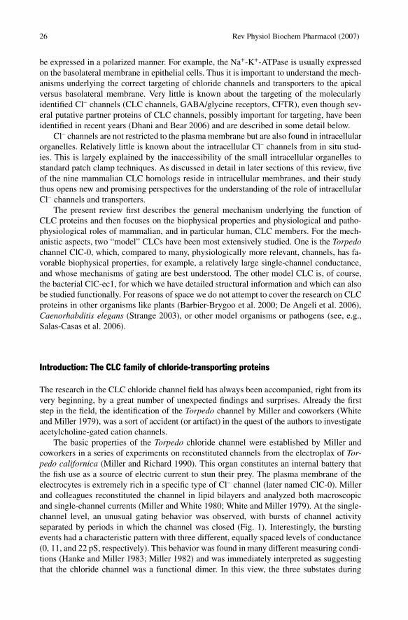
The basic properties of the Torpedo chloride channel were established by Miller andcoworkers in a series of experiments on reconstituted channels from the electroplax of Tor-pedo californica (Miller and Richard 1990). This organ constitutes an internal battery thatthe fish use as a source of electric current to stun their prey. The plasma membrane of theelectrocytes is extremely rich in a specific type of Cl– channel (later named ClC-0). Millerand colleagues reconstituted the channel in lipid bilayers and analyzed both macroscopicand single-channel currents (Miller and White 1980; White and Miller 1979). At the single-channel level, an unusual gating behavior was observed, with bursts of channel activityseparated by periods in which the channel was closed (Fig. 1). Interestingly, the burstingevents had a characteristic pattern with three different, equally spaced levels of conductance(0, 11, and 22 pS, respectively). This behavior was found in many different measuring condi-tions (Hanke and Miller 1983; Miller 1982) and was immediately interpreted as suggestingthat the chloride channel was a functional dimer. In this view, the three substates during

Rev Physiol Biochem Pharmacol (2007) 27
Fig. 1 Schematic (simulated) single-channel trace of the Torpedo channel ClC-0. Channel activity occurs inbursts that are separated by long closed periods. Within each bursts two open conductance levels (O1 and O2)are seen, where O2 has exactly twice the conductance of O1
the bursts would represent the independent opening and closing of two identical Cl– dif-fusion pathways, called protochannels; the dimeric channel complex may exist with bothprotochannels simultaneously open, with one open and one closed, or with both closed,generating the three conductance substates.
At all voltages tested, the frequency of substates during a burst followed a binomialdistribution as predicted for two independently opening and closing protochannels. More-over, the probability of a single protopore to be in its conducting state depended on voltageaccording to a Boltzmann distribution, as expected for a two-state mechanism. This is inagreement with the presence of two independently opening and closing Cl– pathways and incontrast with the presence of a single channel with different subconductance levels (Miller1982).
This model was strengthened by a study of DIDS (4,4′-diisothiocyanatostilbene-2,2′-disulfonate) inhibition of single-channel currents (Miller and White 1984). Addition of 10µM DIDS to the cis side of the chamber eliminated first the 22-pS conductance level and,subsequently, the 11-pS conductance level, that is, the bursting activity disappeared. The au-thors interpreted the finding as being due to the binding (and inhibition) of DIDS first to oneand then the other protopore. This strongly supported a model with two separated diffusionpathways (pores) each with a single open state rather than a single Cl– diffusion pathwaywith multiple conductance states.
Incidentally, the fact that DIDS inhibited the oriented channels only if added to the cisside of the preparation implied that the two protopores had the same orientation in the chan-nel complex.
The fact that the channel activity presented periods of activity (bursts) and periods ofno activity (Fig. 1) indicated that the two protochannels were not completely independentfrom each other. Therefore, it was suggested that there is an inactivating process that closesboth protochannels simultaneously and on a slower time scale (which was later defined asa common gate or slow gate) compared to the closing events within a burst (which wereattributed to what was later named fast gate) (Miller and Richard 1990).
Another peculiar feature of ClC-0 emerged from the inspection of the beginnings andthe endings of the bursts. Burst activity tended to begin with both protopores open and endedmore often with only one protopore open (Richard and Miller 1990). This time asymmetryimplies that the transitions between the possible states of the protopores are not in thermo-

28 Rev Physiol Biochem Pharmacol (2007)
dynamic equilibrium. The external source of free energy required to drive the irreversiblegating transitions was found to be the electrochemical gradient of Cl– (Richard and Miller1990). This finding anticipated one of the most bizarre characteristics of the CLC channelfamily, a gating mechanism mediated by the permeant anion.
The existence of a common gate has another fundamental implication: The two pro-tochannels must be intimately associated in a proteic complex—the double-barreled shotgunmodel was born (Miller 1982). On the basis of stability reasons it was suggested that the twoprotopores would be arranged symmetrically around an axis constituted from the interfacebetween the two subunits (Miller and White 1984).
These features, although solidly grounded on experiments that were elegant in their sim-plicity, were very original, not to say unfamiliar, for the “channel community,” and there-fore they stirred up considerable controversy. However, the progress made in the analysis ofchannel function and structure achieved throughout the last twenty years has spectacularlyconfirmed virtually all of them and provided deeper insights and new unexpected findingsthat we will try to summarize.
Cloning of the CLC family members
A critical turning point for the research on chloride channels was the cloning of the chan-nel from Torpedo marmorata, called ClC-0, with an elegant but extremely labor-intensiveexpression cloning strategy (Jentsch et al. 1990). This exposed ClC-0 to the use of the pow-erful tools of molecular biology and allowed, by homology, the identification of severalother CLC channels in organisms as diverse as animals, plants, yeast, archaebacteria, andeubacteria (Jentsch et al. 1999; Maduke et al. 2000).
Mammals possess nine different CLC genes, which, on the basis of sequence homology,can be grouped into three branches (Jentsch et al. 2002; Mindell and Maduke 2001). Thefirst branch comprises plasma membrane channels, ClC-1, ClC-2, ClC-Ka, and ClC-Kb,whereas members of the two other branches (ClC-3, ClC-4, and ClC-5 in one branch andClC-6 and ClC-7 in the other) function primarily in intracellular membranes.
The sequence, and structure, of CLC proteins bears no resemblance to any other classof membrane proteins. A very distinguishing element of all CLC channels and transporters,with respect to other Cl–-transporting membrane proteins, is their anion selectivity. First,members of the CLC family are practically completely impermeable to cations (except pro-tons). Second, among the halides Cl–, Br–, and I–, the selectivity and conductivity sequencefor CLC proteins is generally Cl–>Br–>I–. According to Wright and Diamond (Wrightand Diamond 1977) this indicates a high-field-strength anion binding site in the transportpathway. In contrast, most other Cl– channels (except CFTR) show an I–>Cl– preference,suggestive of a larger pore in which ions are not completely dehydrated.
Crystal structure of the bacterial ClC-ec1
So far it has not been possible to obtain crystal structures from eukaryotic CLC members,and, therefore, all the structural information (for the transmembrane region) available todate has come from investigation of prokaryotic CLC counterparts, an approach that hasbeen successful for a number of cation channels (Doyle et al. 1998; Zhou et al. 2001).

Rev Physiol Biochem Pharmacol (2007) 29
A projection structure of an Escherichia coli member of the CLC family, ClC-ec1, at6.5-Å resolution, supported the dimeric nature of the channel but could not provide anymolecular detail (Mindell et al. 2001). A much more thorough insight into the structure-function of CLC proteins was provided by two high-resolution structures of ClC-ec1 andStClC (from Salmonella typhimurium) obtained by Dutzler and coworkers (Dutzler et al.2002, 2003).
The biology of prokaryotic CLC proteins is still largely unexplored. In particular, it wasfound that ClC-ec1 is actually a Cl–/H+ antiporter (Accardi and Miller 2004), a character-istic that conflicts with its proposed role as a shunt conductance relevant for acid resistance(Iyer et al. 2002). More importantly, this finding raises a number of issues regarding thepossible extrapolation of features from the prokaryotic to the eukaryotic members of CLCfamily, some of which are discussed in later paragraphs. However, considering the sequenceconservation between prokaryotic CLCs and eukaryotic members of the family, especially inthe ion selectivity region (Maduke et al. 1999), there is confidence that the general structuralelements apply to the entire family.
ClC-ec1 is a dimer composed of two identical subunits of triangular shape (Fig. 2a). Thecontact surface area between subunits is extensive (~2,300 Å2), as expected because CLCchannels are thought to exist and function only as dimers (Dutzler et al. 2002), even if it isnot known at which stage of the biosynthesis dimerization occurs.
Each subunit contains Cl– ions at its center, indicating a putative ion conduction path-way, with a mutual distance between the two pores of ~39 Å. The largest part of ClC-ec1is embedded in the lipid bilayer, and only the N- and C-termini protrude into the cytoplasm(Fig. 2).
Each subunit consists of 18 α-helices (labeled A–R) organized in two topologically re-lated domains that span the membrane in opposite directions in an arrangement called “an-tiparallel architecture” that has been found also in the structure of the aquaporins (Lee et al.2005; Murata et al. 2000) and of a Na+/H+ antiporter from E. coli (Hunte et al. 2005).
The two domains are only weakly correlated in their sequence but show a significantsimilarity regarding the disposition of glycine residues (Dutzler et al. 2002). Some of thehelices are long and tilted by about 45° with respect to the membrane; others are short andpenetrate the membrane only halfway. The transmembrane structure is similar across thewhole CLC family. One fundamental difference lies in the presence of large C-terminal in-tracellular domains in all eukaryotic and some prokaryotic CLC proteins that are absent inClC-ec1 and StClC (Estévez and Jentsch 2002; Meyer and Dutzler 2006). Part of the isolatedC-terminus of ClC-0 has been recently crystallized (Meyer and Dutzler 2006). Its structureis described below.
In agreement with the fact that ClC-ec1 is not an ion channel allowing the passive diffu-sive flow of ions but a stoichiometrically coupled ion transporter, ClC-ec1 lacks a real pore.In the structures of ClC-ec1, the central Cl– ion is completely surrounded by protein and isnot “visible” from either side of the membrane. The putative transport pathway is 15 Å longand contains three ion-binding sites named Sint, Scen, and Sext, starting from the one closerto the intracellular space. The Sext site was found to be occupied by the negatively chargedside chain of a critical glutamate residue (Glu-148) in the wild-type structure, but binds a Cl–
ion if Glu-148 is mutated to alanine or glutamine; no water molecules have been detected inthe ion-binding region in the structures (Dutzler 2004; Dutzler et al. 2003) (Fig. 3).
Overall, the transport pathway across ClC-ec1 appears like a very narrow passage con-necting intracellular and extracellular vestibules (Dutzler et al. 2002, 2003). The vestibulesleading to the selectivity filter on both sides of the membrane contain basic (positivelycharged) amino acids, such as Arg-147 and Arg-451. The distribution of charges on the

30 Rev Physiol Biochem Pharmacol (2007)
Fig. 2a, b Overall structure of ClC-ec1 and CBS domains. In a, ClC-ec1 (PDB accession no. 1KPK) is shownin a ribbon representation viewed from the extracellular side. The two subunits of the dimeric complex areshown in green and orange, respectively. The two Cl– ions in the transport pathway of each subunit are shownin red (central chloride ion) and magenta (inner chloride ion). b Side view of ClC-ec1 assembled with thecytoplasmic C-terminal domains of ClC-0 from Torpedo marmorata (PDB accession no. 2D4Z). The relativeorientation has been arbitrarily fixed, because the exact spatial arrangement of the C-terminal domain withrespect to the membrane part is unknown
entire channel surface creates an electrostatic potential that probably funnels Cl– ions intothe pore entryways. The two pores of the dimer are separated by a large distance and by anelectronegative region on the extracellular surface (Dutzler et al. 2002). These findings areconsistent with the functional independence of the two pores in ClC-0 (Ludewig et al. 1996,1997b; Middleton et al. 1996).

Rev Physiol Biochem Pharmacol (2007) 31
Amino acids from four separate protein regions are brought together near the membranecenter to form the three ion-binding sites (Dutzler et al. 2002, 2003). These regions arehighly conserved in CLC proteins; they include GSGIP in helix D (106–110), G(K/R)EGPin helix F (146–150), GXFXP in helix N (355–359), and Tyr-455 in helix R (Fig. 3a). Thesesequences occur at the N-termini of α-helices, where polypeptide loops precede α-helicesD, F, and N. In agreement with this complex structural arrangement, several regions of CLCproteins influence pore properties like ion selectivity, single-channel conductance, and gat-ing (Estévez and Jentsch 2002; Ludewig et al. 1997a, 1996; Pusch et al. 1995a,. 1995b;Wollnik et al. 1997).
Helices D, F, N, and R are oriented with their N-terminus pointing toward the centralbinding site. Because of the helix dipole, this arrangement of helices is expected to createa favorable environment for anion binding. This is, for example, the mechanism hypothe-sized to be at work in KcsA to favor ion binding to the pore (Roux and MacKinnon 1999).However, some authors have raised doubts against the generalization of such a mechanismto ClC-ec1. On the basis of electrostatic calculations, Faraldo-Gomez and Roux (Faraldo-Gomez and Roux 2004) proposed that in ClC-ec1 the energetic cost for desolvation of theanions on transfer into the protein is contributed only marginally by long-range interaction
Fig. 3a–c The Cl– transport pathway and Cl- binding sites. a The position of the two Cl– binding sites ofClC-ec1 (coloring of subunits and chloride ions as in Fig. 2) with the protein regions involved in coordina-tion of the central Cl– ion shown in blue. b Detail of the amino acids coordinating the central Cl– ion in thewild-type ClC-ec1. c The central Cl- binding site in the structure of the mutant Glu-148-Gln (PDB accessionno. 1OTU). The side chain of Gln-148 is displaced from the permeation pathway, and a third Cl– ion (shownin blue) is present at the position occupied by the side chain of Glu-148 in the wild-type structure

32 Rev Physiol Biochem Pharmacol (2007)
with the helix macrodipole and comes mainly from favorable electrostatic interactions withthe backbone and side chains of residues that are not directly located in the permeationpathway.
This view is shared also by Cohen and Schulten (Cohen and Schulten 2004), who sug-gest, on the basis of molecular dynamics calculations, that the broken helix architecturedoes not constitute a prominent characteristic of the energy profile controlling Cl– conduc-tion and may possibly represent Nature’s design evolved to expose backbone amide groupsto the permeant anions.
In this respect, it is interesting to note that the bound Cl– ions do not make direct con-tact with a full positive charge from lysine or arginine residues. It has been speculated thata full positive charge would create a deep energy well and cause Cl– to bind too tightly,compromising the efficiency of transport (Dutzler 2004).
The Cl– ion at the Scen site is fully dehydrated and is coordinated by main chain amidenitrogen atoms from amino acids Ile-356 and Phe-357 and by side chain oxygen atomsfrom Ser-107 and Tyr-445 (Fig. 3b). On the basis of electrostatic calculations, however,it was hypothesized that the single most important favorable ion-side chain interaction inClCec-1 originates not from Ser-107 or Tyr-445 but from Lys-131. The side chain of thisresidue is located in the transmembrane helix E, completely buried within the protein, withits positively charged amino group pointing toward the chloride-binding sites, at a distanceof 7–9 Å (Faraldo-Gomez and Roux 2004) (Fig. 3b). Thus the stabilization seems to occurby a purely electrostatic, relatively long-range, interaction. These predictions are consistentwith a recent mutagenesis study of this residue in ClC-0 (Zhang et al. 2006).
Apart from the central binding pocket in which Cl– is coordinated by polar residuesand the extracellular exit in which charged residues form a putative gate, the channel poreis lined in its entirety by nonpolar, noncharged residues. The pore’s two conserved polarresidues, Ser-107 and Tyr-445, define Scen and provide an abrupt and significant narrowingof the pore. Their role is, however, not yet clear. For the ClC-0 channel, it was shown that thetyrosine is not responsible for the selectivity and the single-channel conductance (Accardiand Pusch 2003), whereas mutations of the serine residue slightly altered ion selectivity andreduced the single-channel conductance (Chen et al. 2003; Ludewig et al. 1996). Also, sim-ulation studies suggested that the interaction energy of Ser-107 and Tyr-445 with Cl– is notsignificant compared to the energy due to the strong electrical polarization of the protein(Cohen and Schulten 2004). It was therefore suggested that the most important role of theseresidues is to keep an anion permanently in the pore to prevent the formation of a proton-carrying continuous water file stretching across the channel or the passage of hydrophobicanions (Cohen and Schulten 2004).
The second ion binding-site, Sint, is at a distance of 6.5 Å from Scen, toward the intra-cellular side. It is located at the interface where the aqueous vestibule from the intracellularsolution meets the selectivity filter. The ion at this position is coordinated on one side bymain chain amide nitrogen atoms from the end of helix D and on the side where it is ex-posed to the vestibule is probably still hydrated.
In the first structure of ClC-ec1 (Dutzler et al. 2002), Sext was occupied by the sidechain of the glutamate at position 148, occluding the ion pathway (Fig. 3b). At that time, itwas believed that ClC-ec1 was a chloride ion channel, even if no direct electrophysiologicaldata were available yet. It was therefore hypothesized that the crystal structure captured thechannel in a state in which Cl– was occluded, that is, did not have direct access to intracel-lular or extracellular space, and that Cl– ions would activate conduction (gate the channelopen) entering the pore from the extracellular side and inducing a conformational changethat would displace the glutamate side chain.

Rev Physiol Biochem Pharmacol (2007) 33
This prediction was largely confirmed by a second structure of ClC-ec1 determined at2.5-Å resolution in combination with parallel electrophysiological measurements performedon ClC-0 (Dutzler et al. 2003). When the corresponding Glu-148 of ClC-ec1 was mutated inClC-0 into Ala (Glu-166-Ala), Gln (Glu-166-Gln), or Val (Glu-166-Val), it was found thatfast gating transitions were practically abolished (Dutzler et al. 2003). Interestingly, low-ering extracellular pH produced a similar open phenotype for wild-type ClC-0 (Chen andChen 2001; Dutzler et al. 2003), suggesting that the protonation of the glutamate side chainfrom the extracellular side opens the wild-type channel (Fig. 4). The crystal structures ofClC-ec1 in which Glu-148 was mutated to Ala and Gln presented an anion at Sext insteadof the Glu side chain (Dutzler et al. 2003) (Fig. 3c). It was therefore suggested that whenGlu-148 is mutated, the pore is open because it contains an uninterrupted queue of anionsconnecting the intracellular and the extracellular solutions.
In the structure of the Glu-148-Gln mutant of ClC-ec1, the side chain of Gln-148 isdirected toward the extracellular solution rather than into the pore (Fig. 3c), and it was spec-
Fig. 4a, b Effect of Cl– and H+ on the operation of the protopore gate of CLC channels and transporters.Cl– ions are indicated as red spheres. a Schematic representation of the transitions between the open and theclosed state of CLC channels and of the physicochemical factors influencing forward and backward rates.Protonation of the E166 (numbering of ClC-0) side chain allows Cl– flux. Possible additional rearrangementsin the pore region involved in channel opening are also indicated. The pathway that intracellular protons haveto follow to protonate E166 is not known, as indicated by question mark. b Schematic representation of theClC-ec1 transporter. Protonation of E148 (E166 in ClC-0) and E203 are required for the coupled Cl–/H+
antiport activity, but the pathway that intracellular protons have to follow to reach E148 after protonation ofE203 is not known. One possibility is that protons follow the Cl– permeation pathway. Another possibility isthat they reach E148 through a different route yet to be determined

34 Rev Physiol Biochem Pharmacol (2007)
ulated that this could be also the conformation assumed by the wild-type glutamate in theopen—presumably protonated—state (Dutzler et al. 2003). However, this point is still underdebate. For example, based on simulation studies, it was suggested that the side chain ofthe glutamate could swing out of the permeation pathway by a different type of movement(Bisset et al. 2005).
Sext is located between the N-termini of helices F and N, where amide nitrogen atomsform a cage surrounding the ion, and is only 4 Å apart from Scen. All three sites can simul-taneously be occupied by Cl– ions when the channel is open (Lobet and Dutzler 2006).
A very general point to be addressed is the extent to which the picture of the prokaryoticClC-ec1 provides an accurate description of the eukaryotic counterparts. Sequence align-ment exhibits a significant degree of conservation between bacterial and eukaryotic CLCchannels; the similarity is especially strong in the selectivity filter region. Mutational stud-ies on eukaryotic channels correlate well with the locations of key residues in the bacterialstructures. Chen and Chen, using the cysteine accessibility method, were able to show thatin ClC-0 the residues on the intracellular part of the putative helix R are arranged in an α-helical structure and line the wall of the ion permeation pathway as indicated by the crystalstructure of the ClC-ec1 (Chen et al. 2003). The results of Engh and Maduke, based on thesame approach, also suggest conservation of the overall architecture of the inner vestibulebetween ClC-0 and ClC-ec1 (Engh and Maduke 2005). Further support in this directioncame from a recent biochemical evaluation of the membrane domain boundaries of ClC-2(Ramjeesingh et al. 2006).
Estévez et al. showed that residues influencing the affinity of ClC-0 and ClC-1 for the in-tracellular inhibitors 9-anthracene carboxylic acid (9-AC) and p-chloro-phenoxy-acetic acid(CPA), partially overlapped with the Cl– binding pocket identified in the StClC structure (Es-tévez et al. 2003). It seems, therefore, that the structure of ClC-ec1 indeed provides a goodmodel for the description of other members of the CLC family. However, a potentially rele-vant difference between ClC-ec1 and CLC channels is the presence in the channels of moreArg and Lys residues near the pore (Corry et al. 2004). Moreover, the finding of Accardiand Miller that ClC-ec1 is not a chloride channel but a Cl–/H+ antiporter, with potentiallya completely different mechanism of action, suggests caution in the extrapolation of struc-tural features from ClC-ec1 to CLC channels (Accardi and Miller 2004). Subsequently, theeukaryotic ClC-4 and ClC-5 and the plant AtCLCa were also shown to be anion/proton an-tiporters and not chloride channels (De Angeli et al. 2006; Picollo and Pusch 2005; Scheelet al. 2005). It is surprising that members of the same protein family, sharing a fair degree ofhomology and high conservation in critical regions, behave in some cases as channels and inothers as transporters. At the moment there is no evidence regarding the molecular determi-nants of such a difference, and therefore we also do not know whether ClC-ec1 representsa better model for ClC-4 and ClC-5 compared to the CLC channels.
The identification of the major molecular determinant of the fast gate, Glu-148 (166 inClC-0), would explain two characteristics of the fast gate: (a) The fast gates of the two poresare independent because each pore contains its own glutamate residue and the conforma-tional change associated with the swing of the glutamate side chain is local and probablydoes not influence the other pore; and (b) the fast gate is coupled to Cl– permeation becauseCl– ions compete with the side chain of glutamate 166 for the occupancy of Sext and oncea Cl– ion occupies this site there is no obstacle to the permeation process. This would alsoexplain the relatively minor voltage dependence of gating of the kidney CLC channels, ClC-Ka and ClC-Kb, which carry a valine instead of a glutamate at the position equivalent to 166of ClC-0 (Kieferle et al. 1994; Waldegger and Jentsch 2000).

Rev Physiol Biochem Pharmacol (2007) 35
As detailed below, the fast gate can be opened by a mechanism that is favored at low in-tracellular pH. Presumably, protonation of Glu-166 results in increased open probability dueto neutralization of its side chain. For this second mechanism to occur, protons must accessthe Glu-166 side chain from the intracellular side (Fig. 4). Yin et al., on the basis of molec-ular simulations, suggested three proton pathways (Yin et al. 2004). One of these pathwaysinvolves glutamate residues at positions Glu-113, Glu-117, and Glu-203 that, interestingly,in ClC-0 are substituted by Lys, Leu, and Val, respectively. The residue Glu-203 in ClC-ec1was in fact suggested by Accardi et al. (Accardi et al. 2005) to be an internally accessibleacceptor for protons, as substitution of this residue with Gln completely abolished protonflux, underlining its importance for the mechanism of transport. Interestingly, all membersof the CLC family known to be ion channels (ClC-0, ClC-1, ClC-2, ClC-Ka, ClC-Kb, andrespective species homologs) present a Val in place of the Glu at position 203, suggest-ing a significant difference in the mechanism of transport between channel and antiportermembers of the CLC family.
However, despite all the pieces of information gathered so far, our picture of the mech-anism of gating is still incomplete; for example, some studies point to structural rearrange-ments of the pore associated with fast gate transitions, suggesting a larger conformationalchange than the one that would be produced by a simple swing of the Glu-148 side chain(Accardi and Pusch 2003; Traverso et al. 2003) (see “Use of CPA as a tool to explore the fastgate of ClC-0”). Moreover, a gating mechanism based solely on the movement of the Gluis unable to explain why the modulation of gating by Cl–ext is different from Cl–int (Chen2003).
Use of CPA as a tool to explore the fast gate of ClC-0
Small ligand molecules have been very useful tools to explore gating mechanisms of voltage-dependent cation channels (Hille 2001). A classic example is the identification of the ac-tivation gate of K+ channels by intracellularly applied tetraethylammonium (Armstrong1966). In a similar manner, the small organic acid CPA and related compounds have beenused as tools that interfere with the fast gate of ClC-0 (Accardi and Pusch 2003; Pusch etal. 2001; Traverso et al. 2003). CPA is the simplest derivative of 2-(p-chlorophenoxy)-3-phenylpropionic acid (CPP), a substance that is known to inhibit the macroscopic skele-tal muscle conductance (Conte-Camerino et al. 1988). Later studies on heterologously ex-pressed muscle ClC-1 revealed that CPP and analogs block ClC-1 exclusively from the in-tracellular side in a strongly voltage-dependent manner, leading to an apparent “shift” ofthe voltage dependence of opening (Aromataris et al. 1999; Liantonio et al. 2003; Puschet al. 2000). The binding site of CPA and the unrelated 9-AC was mapped on ClC-1 withconsiderable detail (Estévez et al. 2003). CPA and 9-AC bind to the channel in a partiallyhydrophobic pocket adjacent to the central Cl– binding site (when mapped onto the ClC-ec1structure), even though the precise orientation of the drug molecule is unknown (Estévez etal. 2003). However, the small single-channel conductance (Pusch et al. 1994) and the rel-atively complex gating of ClC-1 (Accardi and Pusch 2000) made it difficult to understandthe mechanism of CPP block in this channel. The prototype ClC-0 channel is more usefulin this respect. Employing the point mutant Cys-212-Ser simplifies the system even morebecause this single amino acid substitution almost completely abolishes the common gat-ing mechanism (Lin et al. 1999). CPA block of ClC-0 was extensively studied (Accardi andPusch 2003; Pusch et al. 2001). It was found that CPA binds to closed channels with an

36 Rev Physiol Biochem Pharmacol (2007)
about 20-fold higher affinity than to open channels. In this way, CPA stabilizes the closedstate and leads to an apparent “shift” of the voltage dependence of opening. Open channelblock is of low affinity and associated with rapid binding/unbinding kinetics (apparent KDin the 20 mM range), whereas closed channel inhibition has much slower kinetics (Accardiand Pusch 2003). As discussed above, fast gating of ClC-0 has been proposed to reflect onlythe reorientation of the carboxylate side chain of the Glu-166 residue (Dutzler et al. 2003),without any further conformational change of the protein. In this model, the relatively largedifference of the affinity and kinetics of open- and closed-channel binding of CPA is ratherunexpected, but might be explained by different electrostatic repulsion between CPA andother anions in the pore. However, a recent crystallographic study by Lobet and Dutzler(Lobet and Dutzler 2006) suggested that, in both open and closed states of the fast gate, allthree Cl– ion binding sites are equally maximally occupied by Cl– ions or by the carboxylateside chain of Glu-166. Thus the model advanced by Dutzler and colleagues appears unableto explain the characteristics of CPA block. Additional evidence in favor of a conformationalchange that accompanies opening of the fast gate was obtained by Accardi and Pusch fromdifferential effects of pore mutants on closed- and open-channel block by CPA. For exam-ple, the mutant Thr-481-Ser exclusively altered the closed-channel affinity, whereas othermutations mostly altered the open-channel block (Accardi and Pusch 2003). Also, the dataof Traverso et al. (Traverso et al. 2003), again using CPA as a tool, suggested that a con-formational change, in addition to the glutamate swing-out, accompanies opening of ClC-0protopores.
Thus several pieces of evidence argue against the simple gating model for the fast gateof ClC-0 in which the side chain of Glu-166 is the only moving part. Additional confor-mational changes, in particular on the intracellular side, would be more compatible withsome of the data. However, a more precise definition of the mechanism of the fast gate willprobably need direct structural information for a eukaryotic CLC homolog.
CBS domains
All eukaryotic CLC proteins have a long carboxy-terminal cytoplasmic region whose lengthranges from 155 (ClC-Ks) to 398 amino acids (ClC-1) (Estévez et al. 2004). The C-terminaldomain is essential for the functioning of the eukaryotic CLC proteins, as deletions andseveral point mutations in this region drastically affect transport activity and/or protein mat-uration and trafficking (see below). Indeed, several disease-causing mutations are foundwithin the C-terminus (Estévez and Jentsch 2002; Jentsch et al. 2002; Pusch 2002), but,despite some recent progress, its precise functional and physiological role is unknown. TheC-terminal region contains two so-called CBS domains (from cystathionine-β-synthase, thefirst protein in which these domains were identified). These structural domains normally oc-cur in pairs and are found in several unrelated proteins from all organisms (Bateman 1997;Ponting 1997).
Recently, the crystal structure of the isolated cytoplasmic domain of ClC-0 from Torpedomarmorata was solved by Dutzler and coworkers (Meyer and Dutzler 2006) (see Fig. 2b).As previously described for a different protein (Sintchak et al. 1996; Zhang et al. 1999),the two CBS domains have a triangular shape and are made of three β-strands and two α-helices. Similar to other CBS-containing proteins, the two CBS domains (i.e., CBS1 andCBS2) were found to interact at the level of the β-strands, forming a typical CBS1-CBS2complex. A portion of 95 residues of the linker between CBS1 and CBS2 was found to be

Rev Physiol Biochem Pharmacol (2007) 37
disordered in the crystal structure, but it is not clear yet whether this reflects a crystallo-graphic artifact or the intrinsic flexibility of the region. However, the residual C-terminalpart of the linker, encompassing 25 residues, is well ordered. Interestingly, channel functionwas not affected by the removal of residues that were part of the disordered linker region,whereas no functional channels were obtained if the truncation was made in the structurallywell-defined part of the linker region preceding CBS2 (Estévez et al. 2004).
Unfortunately, the protein did not associate in dimers in the crystallization conditionsused by Meyer and Dutzler (Meyer and Dutzler 2006), and therefore critical informationabout the subunits’ interaction had to be extrapolated from a modeling on the crystal struc-ture of TM0935, a protein from Thermotoga maritima (Miller et al. 2004). However, evenafter this procedure, the surface of the domain in contact with the transmembrane regionremained ambiguous, although CBS2 was suggested to be positioned closer to the pore thanCBS1 (Meyer and Dutzler 2006). Moreover, the C-terminal part of the cytoplasmic domain,which is predicted to be relevant in the interaction between CBS1 and CBS2, was not in-cluded in the construct used for the crystallization.
Several functions have been proposed for CBS domains. Alanine scanning mutagene-sis of the yeast Cl– transporter ScClC (gef1p) suggested that CBS domains influenced thesubcellular localization of the channel (Schwappach et al. 1998).
On truncation of ClC-0, ClC-1, and ClC-5 after the first CBS domain, the proteins didnot give rise to current. However, their function could be restored by coexpression of themissing C-terminal CBS domain, suggesting that CBS2 may function as an independentstructure (Maduke et al. 1998; Mo et al. 2004; Schmidt-Rose and Jentsch 1997). Estévez etal. showed that ClC-1 truncated after the CBS1 domain was not able to reach the plasmamembrane by itself but that the expression could be restored to a normal level in the pres-ence of the CBS2 domain in addition to a region of six amino acids at the N-terminal part ofCBS2 (Estévez et al. 2004). It was also shown that CBS domains from different CLC mem-bers could be exchanged without abolishing channel function, demonstrating that the overallarchitectural conservation of the domain may suffice, despite the low sequence conservation,to preserve their role.
A first hint that the C-terminal region of the channel could be functionally linked to theslow gate came from Jentsch and coworkers (Fong et al. 1998), who made use of mutationsin that region of the ClC-0 and of chimeric constructs and found that the C-terminal partis essential for functional expression of the channel and is involved in the operation of theslow gate. In particular, several point mutations in the CBS2 domain of ClC-0 and ClC-1were found to influence the slow gate (Estévez et al. 2004).
Scott and Hawley found that a purified fragment comprising the last 260 C-terminalresidues of ClC-2 was able to bind ATP and that mutations located in this region that areassociated with genetic diseases lead to defects in ATP binding (Scott et al. 2004). It is in-teresting to correlate these findings with a study of Niemeyer et al. (Niemeyer et al. 2004).Analyzing the functional consequence of the mutation G715E in ClC-2 that was proposed toinduce idiopathic generalized epilepsy (Haug et al. 2003), Niemeyer et al. could not find anygating alteration for the mutated channel but found that, in contrast to wild-type, it did notrespond to the substitution of ATP with AMP with accelerated opening and closing kinetics,even though the effects were relatively minor. Recently, it was suggested that the isolatedcarboxy terminus of ClC-5 folds in a predominantly α-helical structure and it is able to bindATP (Wellhauser et al. 2006). Interestingly, ATP modulates the activity of the common gateof ClC-1 channels such that increasing ATP concentration shifts the midpoint of the openprobability distribution toward depolarized potentials and reduces the fraction of channelsthat remain open at strong hyperpolarized potentials (Bennetts et al. 2005). Bennetts et al.

38 Rev Physiol Biochem Pharmacol (2007)
suggested that the interaction with ATP is mediated by the CBS domains (Bennetts et al.2005). Based on a homology model with the structure of a CBS dimer of IMPDH (inosinemonophosphate dehydrogenase) and in silico docking, they identified a putative ATP bind-ing pocket in a cleft between the two CBS domains of ClC-1 and confirmed their results,observing that mutations of residues that were predicted to interact with ATP reduced orablated the ability of ATP to modulate channel function (Bennetts et al. 2005). However,no ATP binding could be detected in the CBS1-CBS2 complex of ClC-0, even at very highATP concentrations (Meyer and Dutzler 2006). Physiologically, an increased ClC-1 activitydue to ATP depletion during metabolic stress would stabilize the membrane potential andreduce muscle excitability, thereby preserving the viability of muscle fibers. Such a mech-anism, however, has not been described in vivo. In fact, it is questionable that an increasedchloride conductance, via a shift of the voltage dependence of the open probability, is ableto suppress muscle excitation after nerve stimulation.
The fact that mutations in the CBS domains, per se or by affecting the ability to bindATP, interfere with the operation of the common gate requires an interaction of the trans-membrane part of the channel with the cytoplasmic terminus. An interesting possibility wassuggested by Estévez and coworkers (Estévez and Jentsch 2002; Estévez et al. 2004) toexplain this interaction: The last transmembrane helix R, whose N-terminal tyrosine coordi-nates a Cl– ion in the middle of the pore and whose C-terminus extends into the cytosol, isdirectly connected to the CBS1-CBS2 complex. This helix may therefore be the structurallink between the inner pore and CBS domains.
Additionally, CBS domains may be relevant in the interaction with other proteins. It hasbeen found that deleting CBS1 and/or CBS2 impairs the interaction of ClC-5 with cofilin,an actin-associated protein that is crucial in the regulation of albumin uptake by the proxi-mal tubule (Hryciw et al. 2003). Moreover, a PY motif is found between CBS1 and CBS2of ClC-5 that probably interacts with HECT-ubiquitin ligases to modulate the retention ofthe channel in the plasma membrane (Schwake et al. 2001), and a splice variant of ClC-3 displays a PDZ-binding motif at its extreme carboxy terminus that can interact with thescaffolding proteins EBP50 (ERM-binding phosphoprotein 50), PDZK1, and GOPC (Golgi-associated PDZ and coiled-coil motif-containing protein) (Gentzsch et al. 2003; Ogura et al.2002).
Gating of muscle-type ClC channels
According to the classic view, in voltage-dependent cation channels permeability and gat-ing are considered, to a first approximation, as independent processes implying the presenceof a permeable pore and of a separate structure that senses the transmembrane voltage andopens and closes the pore. This picture is completely inadequate for CLC channels. A firsthint of the strong coupling of gating and permeation in ClC-0 came from the time asymme-try of the single-channel bursts implying that the gating transitions were not in thermody-namic equilibrium (Richard and Miller 1990) (see “Introduction”). Such a situation impliesthe existence of an external energy input into the system that was identified as the chlorideelectrochemical potential, anticipating one of the most eccentric features of CLC channels,a gating process that is mediated by the permeant ion.
A thorough investigation of the properties of ClC-0 expressed in oocytes and CHO cellsallowed Pusch and coworkers to conclude that in ClC-0 permeation and gating are tightlylinked (Pusch et al. 1995a). They found that only permeant anions affect gating, that the ion

Rev Physiol Biochem Pharmacol (2007) 39
selectivity of conduction is reflected in the ion selectivity of gating, and that an anomalousmole fraction behavior in the conduction corresponds to a parallel behavior in the gating.Incidentally, the presence of such an anomalous mole fraction behavior showed for the firsttime that the channel pore contains more than one ion binding site, as was later confirmedby structural data (Dutzler et al. 2003).
As mentioned above, the conducting state of the ClC-0 channel is controlled by twodifferent mechanisms defined as the slow gate and the fast gate.
The slow gate controls the opening (and closing) of both pores simultaneously (Millerand White 1980; White and Miller 1979).
There are different factors affecting the operation of the slow gate, such as potential,chloride concentration, pH, and temperature. Hyperpolarized potentials favor the openingof the slow gate (Miller and Richard 1990). The steady-state activation of the slow gate canbe described by a Boltzmann function with a V1/2 of approximately –80 mV and an appar-ent gating valence of ~2 (Pusch et al. 1997). Moreover, the slow gate apparently does notdeactivate completely at depolarized voltages, leading to an offset of the open probabilityof the slow gate at positive voltages. Interestingly, this offset seems to correlate with theexpression level of ClC-0 in oocytes (Pusch et al. 1997).
Chen and Miller (Chen and Miller 1996), found in single-channel recordings, that in-creasing [Cl–]ext shortened the mean closed time and increased the mean open time of theslow gate. Also, [Cl–]int influences the operation of the slow gate. Decreasing intracellularCl– shifted the popen of the slow gate to more negative potentials and reduced the maximalactivation at the most negative voltages (Pusch et al. 1999). Temperature is another variablethat markedly influences the operation of the slow gate (Pusch et al. 1997). In particular,the kinetics of closing of the slow gate showed a Q10 of ~40 at 20°C, suggesting that thetransition between the open and the closed state requires a complex rearrangement of theprotein. The effect of an increase of temperature is on one hand to inactivate the channels ina more complete fashion at positive voltages and on the other hand to decrease the fractionof channels that can be activated by the slow gate at negative voltages (Pusch et al. 1997).However, the voltage of half-maximal activation is relatively independent of temperature.This complex behavior cannot be correctly described by a simple two-state model (open-closed states) but requires at least two open and two closed states for its description. Theeffect of temperature was assessed also on the single-channel level, with the finding thatincreasing the temperature increases the frequency of closure of the slow gate. As expected,single-channel currents increase with temperature, but the dependence is shallow, consistentwith a diffusion-regulated process (Pusch et al. 1997).
In ClC-1, which normally lacks the typical slow gate activation at negative voltages(Steinmeyer et al. 1991b), a hyperpolarization-activated component of the current becomesapparent at low pHext (5.5), which is reminiscent of the activation of the slow gate in ClC-0(Rychkov et al. 1996).
The mechanism responsible for the slow gating has not yet been identified. The fact thatthe slow gate acts on both pores simultaneously suggests, on the structural level, that it re-lies on subunit interactions (Estévez and Jentsch 2002), in agreement with the finding thatconcatemers comprising subunits of different CLC members led invariably to loss of slowgating transitions (Lorenz et al. 1996; Weinreich and Jentsch 2001).
The interaction of the subunits in the dimeric architecture of CLC proteins can involvethe interface between the transmembrane segments or the cytosolic portions that are of sub-stantial length in eukaryotic channels, or both.
Most ClC-1 mutations leading to dominant myotonia change the voltage dependence ofthe channel and most likely involve the slow gate (Pusch et al. 1995b; Saviane et al. 1999).

40 Rev Physiol Biochem Pharmacol (2007)
These mutations are scattered along the channel amino acid sequence (Pusch 2002) andtherefore prove that different regions of the channel probably interact to determine slow gatetransitions. However, several mutations cluster in helices at the dimer interface that probablyare important for subunit contacts: Mutations Val-286-Ala and Ile-290-Met change residuesin helix H, whereas mutations Phe-307-Ser, Ala-313-Thr, and Arg-317-Gln change residuesin helix I (Duffield et al. 2003; Pusch 2002). Moreover, several point mutations in ClC-0 thatare distant from the dimer interface have also been shown to eliminate slow gate transitions(Lin et al. 1999; Ludewig et al. 1996; Traverso et al. 2006). As explained in the section onthe CBS domains, the C-terminus also appears to be a major determinant of the slow gate(Estévez et al. 2004; Fong et al. 1998).
The fast protopore gate of ClC-0
The fast gate acts individually on the single pores of the dimer (Miller 1982). In single-channel recordings of ClC-0 incorporated into planar lipid bilayers, it was found that thefast gate operates in the milliseconds time range and the open probability of the single pro-topore increases with voltage with an apparent gating charge of ~1 (Miller 1982) and fol-lows a Boltzmann distribution as predicted for a two-state channel model (Hanke and Miller1983).
[Cl–]ext influences the open probability of the fast gate (Pusch et al. 1995a), with highextracellular Cl– favoring the opening of the channel, shifting the voltage dependence of theopen probability toward negative potentials (Fig. 4). Using single-channel recordings, Chenand Miller (Chen and Miller 1996) showed that the open probability approaches a nonzeroasymptote at very negative potentials, an effect that can be described as incomplete closureof the channel. The basis of this phenomenon is that the opening rate does not depend ina monotonic manner on voltage. At depolarized potentials the opening rate increases expo-nentially with voltage; at hyperpolarized voltages, however, the opening rate decreases atintermediate potentials but increases again at highly hyperpolarized potentials. The result isthat the opening rate has a minimum at negative voltages. On increase in [Cl–]ext, the volt-age activation curve shifts to the left along the voltage axis without significant change in theapparent gating charge.
The closing rate of the fast gate depends on voltage, decreasing exponentially with de-polarization. Importantly, the closing rate is only slightly affected by [Cl–]ext. Therefore,whereas the voltage dependence of the open probability is determined by both the openingand the closing rate, the external Cl– dependence derives almost completely from an effecton opening.
The operation of the fast gate depends also on [Cl–]int. In particular, the effect on theopening rate is very small, whereas lowering [Cl–]int substantially increases the closing rate(Chen and Miller 1996) (Fig. 4). As a result, increasing [Cl–]int shifts the steady-state activa-tion curve to the left, as with high [Cl–]ext. However, [Cl–]int exerts a more prominent effecton the degree of incomplete closure at hyperpolarized potentials, which was not observedchanging [Cl–]ext. In particular, as [Cl–]int increases, the asymptote of the open probabilityat negative voltages also increases (Chen and Miller 1996; Ludewig et al. 1997a).
These observations were rationalized by a model in which the fast gate of ClC-0 mayopen through two different routes with opposite voltage dependence (Chen and Chen 2001;Chen and Miller 1996). In one mode, opening is favored by membrane depolarization andis sensitive to [Cl–]ext. A plausible mechanism for this gate would be that Cl– first binds to

Rev Physiol Biochem Pharmacol (2007) 41
the channel and then travels through the pore to reach an inner binding site, spanning somedistance in the membrane electric field, as already suggested by Pusch (Pusch 1996; Puschet al. 1995a). The other mode does not depend on [Cl–]ext and is favored by hyperpolarizedpotentials (Chen and Chen 2001). A more quantitative analysis of the [Cl–]int dependence ofthe fast gate was performed by Chen et al. (Chen et al. 2003). Their results confirmed that[Cl–]int almost exclusively affects the closing rate (increasing [Cl–]int decreased the closingrate). The effect of [Cl–]int on the closing rate was saturable, suggesting that it is mediatedby a Cl–-binding site. This was confirmed by experiments in which Cl– was substitutedwith Br– and SO4
2–, showing how the impermeant ion SO42– did not have any such effect,
whereas Br–, which binds to the pore more tightly than Cl–, had a stronger effect (Chen etal. 2003).
The fast gate is also affected by alterations of the intrinsic electrostatic potential of thepore (Chen and Chen 2003; Zhang et al. 2006). In particular, mutating several residuesknown to line the pore or located close to it affected the closing rate, with very little ef-fect on the opening rate. Introducing positively charged residues (or removing negativelycharged residues) in the pore consistently increased the closing rate; vice versa, introducingnegatively charged residues decreased the closing rate. It seems therefore that increasing[Cl–]int and introducing more negative charges in the pore lead to a similar effect (Chen andChen 2003; Chen et al. 2003). Chen and coworkers proposed two mechanisms to explainthese results, both based on the assumption that Glu-166 is the fast gate in ClC-0. The neg-ative charge of the glutamate side chain could directly interact with charged residues in thepore region. In this scenario, negative charges in the inner pore would repel the negativecharge on the glutamate so that the gate would be more difficult to close, that is, it would bemore difficult for the carboxylate side chain of Glu-166 to occupy the Sext position. How-ever, as judged from the structure of ClC-ec1, some mutations tested in the study would bemore than 20 Å away from Glu-166. More importantly, the behavior of the double mutantE127K/K519E is not in agreement with this model (Chen and Chen 2003). The alternativepossibility is that the effect of the electrostatic potential of the pore on gating is mediated bythe permeant anion. For example, a more positive charge at the amino acid positions 127,515, and 519 that are located near Sint would decrease the ability of Cl– present at this siteto displace Cl– at Scen and at Sext. This, in turn, would decrease the ability of Cl– to competewith Glu-166 for Sext, leading to faster closing of the protopore gate.
This hypothesis is especially appealing because it would explain the behavior of themutant Glu-127-Gln, for which the effect on the fast gate mirrors the effect on channel con-ductance. However, not all the mutants affect both fast gate and conductance. Chen andcolleagues therefore suggested that the charge of residues in the pore and the charge carriedby the permeant ion both can contribute to the overall gating process and that the locationof the charge in the pore determines their relative contribution (Chen and Chen 2003; Chenet al. 2003).
Very recently it has been found that the residue K149 in ClC-0 (corresponding to K131in ClC-ec1), although not directly lining the pore, plays a very important role in the electro-statics of the channel, as mutations of this residue reduce the opening rate of the fast gate(Zhang et al. 2006). Interestingly, the mutation K131M in ClC-ec1 results in a perturbationof Cl–/H+ antiporter function (Accardi et al. 2005).
The electrostatics of the pore is also a major determinant of the single-channel conduc-tance of ClC-0 (Chen and Chen 2003). For example, it was found that mutations changingthe charge in the inner pore (e.g., Lys-519-Glu) reduce the conductance at “physiological”Cl– concentrations, but not at saturating [Cl–]int (Chen and Chen 2003). In contrast, for themutation Ser-123-Thr, which changed the highly conserved serine in the selectivity filter, the

42 Rev Physiol Biochem Pharmacol (2007)
decrease in conductance could not be rescued by manipulation of the internal Cl–. Notably,the mutant Tyr-512-Phe, located in the selectivity filter, produced an increase in conductanceof 30% compared to wild-type. This suggests that the regulation of channel conductance bymutations in the selectivity filter and in the channel inner mouth is different. The hydroxylgroups of Ser and Tyr are clearly shown in the ClC-ec1 structure to coordinate a Cl– ionat Scen and are conserved in the CLC family. The fact that mutations in the correspondingresidues in ClC-0 have such a different influence on conductance is still difficult to explainand may suggest a complex effect of these mutations on channel conductance and somedifference between the transporter and the channel members of CLC proteins.
The modulation of the fast gate by external protons was first studied by Chen and Chen(Chen and Chen 2001), who showed that reducing pHext increases the open probability,mostly at hyperpolarized potentials, almost exclusively increasing the opening rate (Fig. 4).The macroscopic effect of a decrease in pHext is therefore mostly an increase in the minimalopen probability (Pmin) at hyperpolarized potentials and not a shift of the popen(V) curve,which is instead seen on changing [Cl–]ext. Chen and Chen (Chen and Chen 2001) proposedthat the effect of pHext on the fast gate is not mediated by a change in the affinity of theCl– binding site that regulates channel opening (Chen and Miller 1996; Pusch 1996) andthat therefore the mechanism of pHext regulation must be intrinsically different from the[Cl–]ext-dependent channel opening. The regulation by external protons, Cl– independentand mostly effective at hyperpolarized potentials, is similar to one of the mechanisms ofopening described by Chen (Chen et al. 2003), potentially indicating that the two processesare linked (Chen and Chen 2001).
Moreover, the fact that the modulation by pHext is stronger at negative voltages is rem-iniscent of the action of [Cl–]int on the fast gate. Chen and Chen (Chen and Chen 2001)indeed suggested that the action of external protons is more pronounced at higher [Cl–]int.
The ClC-0 mutant Glu-166-Asp has a drastically reduced open probability compared towild-type (Traverso et al. 2006) and is thus expected to display an even stronger response tothe external pH. Traverso et al. (Traverso et al. 2006) found instead that decreasing pHext didnot increase outward currents. In particular, low pHext increased a persistent inward currentthat was characterized by a smaller single-channel conductance. These results suggestedthat Asp-166 can be protonated from the intracellular side in a voltage-dependent manneror from the extracellular side in a voltage-independent manner, resulting in open states ofdifferent conductance (Traverso et al. 2006). In ClC-1 it was found that decreasing pHextaffected the macroscopic current, mostly by increasing the steady-state component at theexpense of the deactivating portion. At variance with the behavior of external Cl– at lowpHint, it was found that at low pHext, external Cl–was not able to influence channel gating(Rychkov et al. 1996).
The influence of the internal pH on the fast gate transitions was investigated in the re-constituted Torpedo channel (Hanke and Miller 1983) (Fig. 4). Low pHint drives the pro-tochannel open without changing its conductance. The effect was interpreted in terms ofa shift of the voltage dependence of the open probability toward negative potentials. Hankeand Miller suggested that on opening of the channel a titratable group exposed to the intra-cellular solution changes its pK from 6 to 9 and that this change in pK underlies the ability ofprotons to drive the channel into its open state (Hanke and Miller 1983). In ClC-1, internalpH had a very similar effect (Rychkov et al. 1996). Hanke and Miller also investigated thepH dependence of the opening and closing rate constants. They found that at all pH valuestested, those rates vary exponentially with voltage and at all voltages both opening and clos-ing rate constants vary with proton concentration. However, with an increase in the protonconcentration, the closing rate constant decreases whereas the opening rate increases. There-

Rev Physiol Biochem Pharmacol (2007) 43
fore, the effect of pHint changes mainly translates into a shift of the popen along the voltageaxis. The pH dependence implies that a simple two-state model is insufficient to describe thechannel behavior and that a protonation reaction must be added to the scheme. Hanke andMiller (Hanke and Miller 1983) suggested, however, that the protonation step does not con-tribute to the voltage dependence of gating, which in their model is brought about only bythe transition between open and closed states. Such an interpretation was recently challengedby Pusch and coworkers, who investigated the pH dependence of the Glu-166-Asp ClC-0mutant (Traverso et al. 2006). This mutant strongly affects the operation of the fast gate, dra-matically reducing the open probability of the channel. This drastic effect of the conservativeGlu→Asp mutation (Traverso et al. 2006) probably reflects the sensitivity of ClC-0 gatingon the protonation state and flexibility of this key acidic residue. Lowering pHint increasedcurrent of the Glu→Asp mutant, in agreement with the behavior of the wild-type channel(Hanke and Miller 1983; Traverso et al. 2006). However, the pHint dependence of this mu-tant is not consistent with a model in which the protonation step is voltage independent,but could be better described by a model in which the protonation/deprotonation reactionscarry most of the voltage dependence. This suggestion also opens up new questions. It isreasonably well established that Glu-166 is the proton acceptor responsible for the regula-tion of the fast gate by pHext. On the other hand, we still do not know which residue(s) isinvolved in the control of the fast gate by intracellular protons. An interesting hypothesisis that opening of the fast gate requires the protonation of Glu-166. Protonation may occur,in a relatively voltage-independent manner, from the extracellular solution or, in a voltage-dependent manner, from the intracellular side. A protonation of Glu-166 (or Asp-166) fromthe intracellular side was also proposed recently by Miller as the possible major source ofvoltage dependence of the fast gate of ClC-0 (Miller 2006) (see Fig. 4).
Zinc and cadmium—inhibitors of ClC-0, ClC-1, and ClC-2
ClC-0, ClC-1, and ClC-2 are inhibited by Zn2+ and Cd2+ ions (Chen 1998; Clark et al. 1998;Kürz et al. 1997; Rychkov et al. 1997), confirming results obtained for the Cl– conductanceof frog skeletal muscle (Hutter and Warner 1967). A first mechanistic insight into the inter-action between Zn2+ ions and CLC channels came, however, from an analysis of the Zn2+
block of ClC-0 (Chen 1998). For ClC-0 the inhibition is reversible with an IC50 of 1–3 µM.The effect of Zn2+ did not seem to be mediated by an interaction with the fast gate, whosevoltage dependence of the open probability and of the kinetics remained unaltered in thepresence of Zn2+. The apparent on- and off-rates of Zn2+ inhibition were slow and showedpronounced temperature dependence, from which it was suggested that the inhibition wasunlikely to stem from a simple open channel block and probably involved a more compli-cated process (Chen 1998). In particular, the temperature dependence of the effect directlysuggested a possible link of the inhibition with the operation of the slow gate (Chen 1998;Pusch et al. 1997). It was found that indeed increasing Zn2+ concentration facilitated theslow gating process (Chen 1998). Specifically, the effect of Zn2+ on slow gating equilibriumappears to come mostly from an increase in the forward rate of inactivation. Interestingly,the mutation Cys-212-Ser in ClC-0, which was shown to eliminate the slow gating process,also drastically reduces the channel’s sensitivity to Zn2+ (Lin et al. 1999), further supportingthe association between the slow gate and the mechanism of Zn2+ inhibition.
As described below, the common gate of ClC-1 has quite different features from that inClC-0, such as, for example, an opposite voltage dependence, and vastly different kinetics

44 Rev Physiol Biochem Pharmacol (2007)
and temperature sensitivity. The IC50 for Zn2+ inhibition of ClC-1 has been found to be0.35 mM (Rychkov et al. 1997). In contrast to ClC-0 and ClC-2 (Chen 1998; Clark et al.1998), Zn2+ and Cd2+ block appear to be irreversible for ClC-1 (Kürz et al. 1997; Rychkovet al. 1997). Interestingly, also in ClC-1 the mutation Cys-277-Ser, corresponding to themutation Cys-212-Ser of ClC-0, drastically reduces the closure of the slow gate (Accardi etal. 2001) and virtually eliminates Zn2+ block, suggesting a similarity in the mechanism ofZn2+ block on the two channels (Duffield et al. 2005). At variance with ClC-0, however, inClC-1 the block by Zn2+ is too slow to be a simple function of the open probability of eitherthe fast or the putative slow gate. Moreover, the temperature dependence of Zn2+ inhibition(Q10 ~13°) is much higher than the Q10 of the putative slow gate, which is ~4° (Bennetts etal. 2001). Both elements indicate that in ClC-1 the mechanism of Zn2+ inhibition, althoughfounded on the interaction with the slow gate as in ClC-0, may present significant differ-ences, and Duffield et al. (Duffield et al. 2005) proposed that in ClC-1 Zn2+ acts by bindingto a closed substate of the common gate that has very low probability in the wild-type chan-nel and was therefore not previously identified.
Extracellular Cd2+ produces a concentration-dependent block of ClC-1 expressed in theSf-9 cell line, with an IC50 of 1 mM (Rychkov et al. 1997). It was suggested that ClC-1has at least two binding sites for Cd2+ in which His residues may play a prominent role(Rychkov et al. 1997).
Zúñiga et al. found that Cd2+ block of ClC-2 is mediated by an acceleration of the rateof deactivation (Zúñiga et al. 2004). Mutation of Cys-256 in ClC-2, corresponding to a cys-teine residue known to affect the operation of the slow gate in ClC-0 (Cys-212-Ser) (Linet al. 1999) and ClC-1 (Cys-277-Ser) (Accardi et al. 2001) and to drastically reduce Zn2+
block, also reduced the effect of Cd2+ compared to wild-type, indicating that Cd2+ wouldexert its action through an interaction with the gating machinery of the channel (Zúñiga etal. 2004). However, at variance with the action of Zn2+ on ClC-0 and ClC-1, Cd2+ affectedboth the fast and the slow gating process of ClC-2 (Yusef et al. 2006), indicating a strongcoupling between fast and slow gating, similar to what was proposed for ClC-1 (Accardi etal. 2001). Moreover, the mutation His-811-Ala in ClC-2, corresponding to a mutation thatcompletely and selectively abolishes slow gating in ClC-0 (Estévez et al. 2004) and that islocated in the highly conserved CBS2 domain, affected both fast and slow gating of ClC-2.Interestingly, combining this mutation with Glu-217-Val ablates all gating transitions (Yusefet al. 2006).
ClC-1—the skeletal muscle chloride channel
ClC-1 was cloned from rat skeletal muscle by homology screening with a probe derivedform the Torpedo ClC-0, with which it shares 54% sequence identity (Steinmeyer et al.1991b). It is predominantly expressed in skeletal muscle, where it accounts for the large Cl–
conductance responsible for the resting membrane potential (Bretag 1987; Steinmeyer et al.1991b). Low transcript levels could also be detected in kidney, heart, and smooth muscle(Steinmeyer et al. 1991b).
Analysis of dominant-negative mutations suggested that ClC-1 has a multimeric archi-tecture (Pusch et al. 1995b; Steinmeyer et al. 1994). This view was supported by Lorenzet al. (Lorenz et al. 1996), who showed that ClC-1 and ClC-2, on coexpression in Xenopusoocytes, form heterooligomers.

Rev Physiol Biochem Pharmacol (2007) 45
Even though a quantitative single-channel analysis of ClC-1 is difficult because of itssmall single-channel conductance (Pusch et al. 1994), an extension of the double-barreledarchitecture from ClC-0 to ClC-1 was strongly supported by inspection of the single-channelbehavior displaying two equidistant conductance levels of 1.2 and 2.4 pS (Saviane et al.1999). Incidentally, the small single-channel conductance explains why many previous at-tempts to detect its activity in intact muscle preparations failed.
Despite the similarities with ClC-0, there are a number of functional characteristics thatdifferentiate ClC-1 from ClC-0. In contrast to ClC-0, gating and permeation apparently donot seem to be so closely linked in ClC-1 (Rychkov et al. 1998) as anions like cyclamate andmethanesulfonate can have a substantial effect on gating without being permeant. However,these results can probably be explained by an external anion binding site with relatively highaffinity for organic anions in ClC-1 but not in ClC-0 (Rychkov et al. 2001). Occupation ofthis site by organic anions indirectly influences the occupation by chloride of deeper anionbinding sites. Gating of ClC-1 is similar to the fast gating of ClC-0 in that it also activateswith depolarization and can be described by a Boltzmann function with an apparent gatingcharge of ~0.9 (Pusch et al. 1994; Rychkov et al. 1996; Steinmeyer et al. 1991b).
Even if under normal conditions ClC-1 lacks a slow hyperpolarization-activated gate,such a gate becomes visible al low pHext and positive holding potentials (Rychkov et al.1996).
Under physiological pH conditions, ClC-1-mediated currents display a deactivationcomprising two exponential components (Rychkov et al. 1996). Accardi and Pusch (Accardiand Pusch 2000) showed that these components have time constants that are quite similarat negative voltages but grow apart as the voltage is increased. At a voltage of 200 mV theydiffer almost by a factor of 100, mimicking the difference between fast and slow gates inClC-0. In particular, investigation of the dependence of the two components on [Cl–]ext andpHint suggested that the faster gating components found for ClC-1 behaved very similarlyto the fast gate of ClC-0 and the slower component of ClC-1 was similar to the slowgate of ClC-0. It was therefore proposed that also for ClC-1 the two gating componentscorrelated with the operation of fast and slow gates. However, it was shown that the voltagedependence of the slow gate in ClC-1 is reversed compared to ClC-0 (Accardi and Pusch2000; Saviane et al. 1999).
The physiological role of ClC-1 is discussed below in the context of its involvement incongenital myotonia.
Myotonia
Myotonia—or muscle stiffness—is a symptom that is associated with various genetic dis-eases. In “chloride-channel” recessive (Becker) (Becker 1957) and dominant (Thomsen)(Thomsen 1876) myotonia congenita, myotonia is practically the only symptom. Musclediseases caused by mutations in the SCN4A sodium channel have overlapping but not iden-tical symptoms (Lehmann-Horn and Jurkat-Rott 1999).
Myotonia is caused by hyperexcitability of the muscle plasma membrane, such that nor-mal nerve stimulation produces an exaggerated and possibly repetitive firing of muscle ac-tion potentials (myotonic runs) (Adrian and Bryant 1974). About 80% of the resting con-ductance of skeletal muscle consists of a chloride conductance, gCl, the majority of which iscarried by ClC-1 (Steinmeyer et al. 1991a, 1991b). A marked reduction of gCl thus decreasesthe depolarizing and stabilizing conductance, causing hyperexcitability. In most neurons,

46 Rev Physiol Biochem Pharmacol (2007)
equivalently stabilizing and repolarizing conductances are mostly carried by K+ channels. Itis thought that in skeletal muscle a K+ conductance is not adequate for such a role becauseof an expected buildup of K+ ions in the restricted space inside the t-tubules (Cannon 2000;Pusch 2001). In fact, detubulation of rat skeletal muscle reduces gCl but not gK (Palade andBarchi 1977), However, the two studies that specifically investigated the subcellular local-ization of ClC-1 with immunofluorescence found the protein in the sarcolemma and not inthe t-tubules (Gurnett et al. 1995; Papponen et al. 2005).
In myotonic dystrophy (DM), for which myotonia is only one of many symptoms, it hasrecently been shown that the RNA coding for the ClC-1 protein is strongly reduced by analteration of its correct splicing (Berg et al. 2004; Charlet et al. 2002; Mankodi et al. 2002).
Dominant and recessive myotonia congenita are instead caused by mutations in CLCN1,the gene coding for ClC-1. A mouse model for recessive myotonia, the adr mouse (Mehrkeet al. 1988), helped to identify ClC-1 as the major skeletal muscle Cl– channel (Steinmeyeret al. 1991a). In the adr mouse, no ClC-1 protein is made because both alleles are practicallydestroyed by a homozygous transposon insertion. Similarly, most mutations that lead to re-cessive myotonia in humans either completely abolish channel function (like, e.g., early stopcodons) or drastically reduce channel function (see Pusch 2002 for an overview of possibleeffects of recessive mutations). There may be several reasons for the fact that heterozy-gous carriers of such recessive mutations (50% gene dosage) are generally asymptomatic.A 50% gene dosage could be functionally compensated at the RNA level (transcription,splicing, processing, turnover) or at the protein level (translation, processing, sorting, tar-geting, turnover). In fact, heterozygous adr mice show an almost unaltered muscle chlorideconductance (Chen et al. 1997). It remains, however, as an interesting problem if and howmuch the Cl– conductance is reduced in human heterozygous carriers of recessive mutations.Pharmacological experiments indicate that more than 50% of the Cl– conductance must beinhibited in order to cause myotonia (Furman and Barchi 1978). This observation and thefact that heterozygous carriers of recessive mutations are generally asymptomatic demon-strate that in order for a CLCN1 mutation to be inherited in a dominant manner it must pro-duce a dominant-negative effect. That is, it must reduce gCl more than a heterozygous lossof function, beyond the threshold that is necessary to precipitate myotonia. These considera-tions are in agreement with the fact that far more recessive than dominant CLCN1 mutationshave been described (Pusch 2002): Channel function is easily destroyed, for example, byearly stop codons, but a dominant-negative effect requires a specific association with a wild-type subunit. The first dominant mutation, P480L, was identified in descendants of Thom-sen, who himself suffered from the disease (Steinmeyer et al. 1994). When coexpressed withwild-type subunits in Xenopus oocytes, the mutation exerted a strong dominant-negative ef-fect, and this was the first indication that CLC channels are homomultimers (Steinmeyer etal. 1994). The mechanism of action remained unclear, however, and the initial estimate ofthe number of subunits (4) turned out to be wrong. Later, it was found that several dominantmutations, including P480L, exert a dominant-negative effect by “shifting” the voltage de-pendence of channel activation to more positive voltages, such that channels are less activeat the skeletal muscle resting membrane potential (Pusch et al. 1995b). In the context ofthe double-barreled structure of CLC channels with two separate gates (fast, protopore gateand slow, common gate) it was later found that most dominant mutations act primarily onthe common gate of ClC-1 (Aromataris et al. 2001; Saviane et al. 1999). It also must besaid, however, that the distinction between dominant and recessive forms of the disease isnot very clear-cut. The same mutation may appear as dominant in some pedigrees and asrecessive in others (Plassart-Schiess et al. 1998). Thus other factors, independent of ClC-1,seem to contribute to the severity of myotonia.

Rev Physiol Biochem Pharmacol (2007) 47
The shift of the voltage dependence is not the only dominant-negative mechanism. Forexample, the C-terminal truncation R894X has a quite strong dominant-negative effect,without an apparent change of the voltage dependence (Meyer-Kleine et al. 1995). Thismechanism remains to be identified. As we hope to have illustrated above, understanding thepathophysiology of myotonia provides a valuable insight into the general function of ClC-1.
ClC-2—a complex chloride channel of epithelial and nonepithelial cells
ClC-2 has been cloned from rat heart and brain. The 907-amino acid protein shares 49%identity with ClC-0 and 55% with ClC-1. It is broadly expressed in several tissues and incell lines of different origin such as epithelial, fibroblast, and neuronal (Thiemann et al.1992).
On expression in Xenopus oocytes, ClC-2 gave rise to currents that were slowly acti-vated and inwardly rectifying, unlike ClC-0 and ClC-1 (Gründer et al. 1992; Thiemann et al.1992). Moreover, these currents were activated only at unphysiological, hyperpolarized po-tentials. The instantaneous I-V curve observed after activation of the hyperpolarized currentrevealed a linear current-voltage relationship. Similar to ClC-0, iodide is less permeant thanchloride. Extracellular 9-anthracene carboxylic acid (1 mM) and diphenylaminecarboxylate(1 mM) inhibited the conductance by 50%, whereas 1 mM DIDS was almost ineffective(Thiemann et al. 1992).
Single-channel analysis applied on concatemeric constructs of ClC-0 and ClC-2 demon-strated a functional dimeric architecture of ClC-2 (Weinreich and Jentsch 2001) in analogywith ClC-0 (Bauer et al. 1991; Ludewig et al. 1996; Middleton et al. 1996; Miller and White1984) and ClC-1 (Saviane et al. 1999). Unfortunately, the single-channel conductance ofClC-2 is only 2.6 pS, a factor that has so far hampered attempts at a thorough characteriza-tion of the channel properties at the single-channel level.
Superfusion of oocytes expressing ClC-2 with hypotonic solution produced currentswith faster kinetics that were activated at less hyperpolarized potentials and therefore inthe physiological voltage range, suggesting that ClC-2 is involved in volume regulation(Furukawa et al. 1998; Gründer et al. 1992; Jentsch et al. 2002). In particular, the overallcurrent amplitude significantly increased on superfusion with hypotonic solution (Gründeret al. 1992). The activation was fully reversible and needed around 10 min to set in, sug-gesting that the effect was probably due to slow intracellular changes rather than to a directeffect on the channel. Hypertonicity did not have any effect on wild-type ClC-2. A chimericapproach allowed the identification of the N-terminal domain as determinant for the volumesensitivity of the channel (Gründer et al. 1992).
In particular, deletions in the first 31 amino acids led to constitutively open channels thatwere also unresponsive to hyper- or hypotonicity (when analyzed with the two-electrodevoltage-clamp technique), whereas upstream from this essential domain, deletions producedchannels with an intermediate phenotype. The effect of these domains was independent fromtheir position, as the N-terminal region could be transplanted to the C-terminus, retaining itseffect (Gründer et al. 1992).
Extracellular pH significantly affects the operation of ClC-2, with moderate acidifica-tion leading to channel activation already at ~ –30 mV and to increased steady-state currents(Jordt and Jentsch 1997). It was suggested that the mechanism of action is a shift of the volt-age dependence of the common gating mechanism, and Jordt and Jentsch proposed that, inanalogy with the effect of hyperpolarization and cell swelling, the response to extracellular

48 Rev Physiol Biochem Pharmacol (2007)
pH depends on the N-terminal domain (Jordt and Jentsch 1997). However, the role of theN-terminal domain of the channel is still not very clear. In fact, in contrast with the voltage-independent phenotype of the N-terminal deletion described previously with two-electrodevoltage-clamp recordings (Gründer et al. 1992), in inside-out patches the same constructgave rise to channels that conserved the characteristic activation at hyperpolarized poten-tials of the wild-type, albeit with a faster kinetics (Pusch et al. 1999). Similarly, deletions ofamino acids 16–61 of rClC-2 expressed in HEK cells, although producing faster opening andclosing kinetics compared to wild-type, did not produce significant changes in voltage andpH dependence (Varela et al. 2002). However, it was observed that with nystatin-perforatedpatches, which allow the selective exchange of cations between the cytoplasm and the pipettesolution, currents of the amino terminal-deleted mutant lost their voltage dependence (Varelaet al. 2002), suggesting that the differential effect of the deletion in different expression sys-tems and measuring conditions may depend on factors such as osmotic state of the cells,cytoskeleton structure integrity, or diffusible cytoplasmic components, as already discussedby Pusch et al. (Pusch et al. 1999).
In a study of currents in mouse parotid acinar cells that were probably mediated byClC-2, Arreola et al. found a bimodal pHext effect with a conductance maximum aroundpH 6.5 (Arreola et al. 2002). Interestingly, acidification to pH 5.5 applied during openingby hyperpolarization led first to a transient activation followed by inhibition, suggesting theexistence of two different proton-binding sites. Occupation of one of these can exert a stim-ulatory effect, but the site becomes accessible to extracellular protons only in the open stateof the channel (Arreola et al. 2002). Very similar results were found for guinea pig ClC-2expressed in HEK cells (Niemeyer et al. 2003). In particular, because the transient activationby external protons was ablated in the mutant Glu-217-Val, it was suggested that the residueGlu-217 is the acceptor site for protons responsible for the stimulatory effect of low pH onClC-2 (Niemeyer et al. 2003).
Mutating Lys-566, located at the end of the transmembrane-spanning domain, to gluta-mate was found to shift the voltage dependence of gating and to change the inward recti-fication of the open channel I-V relationship of wild-type ClC-2 to outward rectifying, inanalogy with the effect of the corresponding mutant (Lys-519-Glu) on the fast gate of ClC-0(Pusch et al.1999, 1995a). However, mutation of this lysine did not modify activation byhyperpolarization, cell swelling, and acidification. In contrast, mutations in helix I and thepreceding loop abolished all three modes of activation by constitutively opening the channelwithout changing its pore properties (Jordt and Jentsch 1997).
It has been clearly established that gating of ClC-2 depends on intracellular [Cl–], whoseincrease shifts the open probability of the channel toward positive potentials in a N-terminal-deleted construct of rat ClC-2 expressed in oocytes (Pusch et al. 1999), the full-length ratClC-2 expressed in HEK cells (Niemeyer et al. 2003), and the human ClC-2 (Haug et al.2003).
The role of extracellular chloride is still controversial, as Pusch et al. (Pusch et al.1999) showed that, surprisingly, decreasing [Cl–]ext increases the open probability of ClC-2, whereas Niemeyer et al. (Niemeyer et al. 2003) were not able to observe any effect ofextracellular Cl– on gating.
Activation of macroscopic ClC-2 currents follows a double exponential time course,with time constants differing roughly by one order of magnitude (de Santiago et al. 2005;Pusch et al. 1999; Zúñiga et al. 2004). Both time constants are voltage dependent, becomingfaster on hyperpolarization (Zúñiga et al. 2004). The opposite behavior was found for ClC-1,in which both gates are opened by depolarization (Accardi and Pusch 2000), while the slowgate of ClC-0 also opens in response to hyperpolarization (Pusch et al. 1997). Interestingly,

Rev Physiol Biochem Pharmacol (2007) 49
the amplitudes of the two components in ClC-2 have an opposite voltage dependence (deSantiago et al. 2005). The voltage dependence of macroscopic currents could be describedby a Boltzmann function with half-maximal activation and slope factor of, respectively,–117 and 22 mV at 22°C (Zúñiga et al. 2004). Activation and deactivation time constantswere reduced on temperature increase without major changes in the steady-state activa-tion curve. The Q10 values calculated for both fast and slow time constants are between4 and 5, suggesting a significant conformational change associated with those processes(Zúñiga et al. 2004). This value is larger than the Q10 factor related to the fast gate of ClC-0but much smaller than that for the slow gate (Pusch et al. 1997). On the other hand, it is inthe same range of the Q10 factor measured for the common gate of ClC-1 (Bennetts et al.2001).
An attempt at quantitative dissection of the properties of the fast and slow gating pro-cesses in ClC-2 was made by de Santiago et al. (de Santiago et al. 2005). These authorsfound that the open probability of the protopore gate can be described by a Boltzmann dis-tribution with half-maximal activation at –63 mV and an apparent gating charge of –1.22,whereas the common gate remains about 55% open at positive voltages and is associatedwith an apparent gating charge of –0.99 with half-maximal activation at –134 mV. The mu-tation Cys-258-Ser affected the voltage dependence of both gates. This observation is incontrast with the fact that the double mutant Glu-217-Ala/Cys-258-Ser produced currentsvery similar to that of the single mutant Glu-217-Ala, probably dominated by the slow com-ponent (Niemeyer et al. 2003), suggesting that the mutation Cys-258-Ser mostly affects theoperation of the fast gate.
Moreover, the mutation His-811-Ala in ClC-2, corresponding to a mutation that com-pletely and selectively abolishes slow gating in ClC-0 (Estévez et al. 2004) and that is locatedin the highly conserved CBS2 domain, affected both fast and slow gating of ClC-2. Interest-ingly, combining this mutation with Glu-217-Val ablates all gating transitions (Yusef et al.2006).
Collectively, these pieces of evidence point to a gating mechanism composed of a fastand slow component that bear some resemblance to the protopore and common gate identi-fied in ClC-0 and ClC-1 but that appear also to have very distinctive features whose natureis only poorly understood. In particular, a very specific element of ClC-2 is the stronger cor-relation of the two gating modes compared to the other channel members of the CLC family(Zúñiga et al. 2004).
Mutation of the conserved Glu at position 217 into Val (Glu-148 in ClC-ec1) producesa loss of sensitivity of the channel to Cl–int and almost abolishes its voltage dependence andthe characteristic inward rectification of the current (de Santiago et al. 2005; Zúñiga et al.2004). It has been conjectured that in ClC-2 fast gating is due to the movement of the sidechain of Glu-217 in a mechanism similar to that proposed for the fast gate of ClC-0 andClC-1 (Niemeyer et al. 2003; Yusef et al. 2006; Zúñiga et al. 2004). In this scenario, theresidual voltage dependence observed for ClC-2 at strongly hyperpolarized potentials couldbe explained in terms of transitions of the common gate (de Santiago et al. 2005; Niemeyeret al. 2003). The analogy between ClC-2 and other channel members of ClC family canbe extended also to the extracellular pH dependence as the transient activation of ClC-2at acidic extracellular pH is reminiscent of the increase in the open probability of the fastgate of ClC-0 at low external pH and is mediated by the corresponding glutamate residue(Niemeyer et al. 2003).
Even more speculatively, slow gating transitions in ClC-2 have been proposed to arisefrom conformational changes in the pore, which are known to accompany protopore gatingin ClC-0 (Accardi and Pusch 2003), possibly mediated through rearrangements of the he-

50 Rev Physiol Biochem Pharmacol (2007)
lix R, which can be influenced by movements of the C-terminus of the protein (Yusef et al.2006). However, in the absence of single-channel measurements, the separation between fastand slow gating transitions is only tentative and requires a more solid experimental basis.
Physiological role of ClC-2
In contrast to the tissue distribution, the subcellular localization of ClC-2 channels is stillcontroversial. On one hand, antibodies against ClC-2 detected signals in apical membranesof intestinal tissues in humans (Murray et al. 1996) and mice (Gyömörey et al. 2000) andin rat lung epithelium (Blaisdell et al. 2000). On the other hand, with different antibodies,a basolateral localization was suggested in rat small intestine and colon (Lipecka et al. 2002)and guinea pig colonocytes (Catalán et al. 2002).
As pointed out by Zdebik et al. (Zdebik et al. 2004), none of these studies includedcontrols with knockout tissues, raising the possibility that some of these results may reflectunspecific binding. Such an approach was recently undertaken by Sepúlveda and coworkers(Peña-Münzenmayer et al. 2005) who, by an immunohistochemical approach using ClC-2-null mice as a negative control, showed that ClC-2 has a basolateral localization in intestinalepithelial cells of wild-type mice. Moreover, heterologous expression in epithelial cell linesof a ClC-2 construct with a C-terminally fused GFP in combination with confocal fluores-cence imaging confirmed a basolateral expression. The polarized expression seems to de-pend on the AP-1AB clathrin adaptor protein, which is known to be an epithelium-specificcomplex involved in basolateral sorting (Fölsch et al. 1999). In particular, a dileucine motif,which is normally recognized by AP-1AB (Nakatsu and Ohno 2003), encoded in the CBS2domain of ClC-2 was found to be critical for the basolateral localization as its disruptionproduced apical localization (Peña-Münzenmayer et al. 2005). This motif is conserved inClC-2 from different organisms and in the other plasma membrane CLC members but notin ClC-3, -4, -5, -6, and -7, all mainly expressed intracellularly.
A basolateral localization of ClC-2, implying that it does not contribute to chloride se-cretion, is compatible with the observation that homozygous mice harboring the ∆F508mutation in the CFTR gene, leading to cystic fibrosis through impaired Cl– secretion, sur-vived better when ClC-2 was additionally disrupted (Zdebik et al. 2004). This issue is ofsignificant medical importance because the pathological changes in cystic fibrosis are pre-dominantly due to defective Cl– conductance on the apical side of the cells, and thereforepharmacological intervention on Cl– channels, in particular ClC-2, to be beneficial, willhave to take into account its subcellular localization. For example, since ClC-2 is expressedbasolaterally, maneuvers that decrease the channel conductance might have a positive effecton the pathology (Zdebik et al. 2004). A possible physiological mechanism that potentiallymodulates Cl– fluxes through the plasma membrane of epithelial cells depending on theirabsorption-secretion activity is provided by the regulation of the open probability of ClC-2operated by [Cl–]int (Catalán et al. 2004).
Several recent papers reported on the interaction of ClC-2 with other cellular proteins.Hinzpeter et al. (Hinzpeter et al. 2006) presented evidence that ClC-2 can interact withHsp70 and Hsp90 in HEK cells and with Hsp90 in mouse brain, producing a reduction ofchannel expression at the plasma membrane. However the molecular basis for the interac-tion remains to be determined. Bali et al. (Bali et al. 2001) suggested a role of vesiculartrafficking in the regulation of ClC-2 plasma membrane expression. It has been indicatedthat such a role could be mediated by the dynein motor complex that coimmunoprecipi-

Rev Physiol Biochem Pharmacol (2007) 51
tates with ClC-2 in rat hippocampal slices and controls retrograde trafficking of the channelbetween plasma membrane and endosomes in COS-7 cells (Dhani et al. 2003).
The expression level of ClC-2 in rat renal proximal tubules is influenced by thyroid hor-mones (Santos Ornellas et al. 2003) and estrogens (Nascimento et al. 2003), suggesting therelevance of this channel for Cl– transport in the kidney, even though no renal phenotype wasreported in ClC-2 knockout mice (Bösl et al. 2001). It was found that the transcription fac-tors SP1 and SP3 influence the expression level of ClC-2 in lung epithelial cells (Holmes etal. 2003) binding to the ClC-2 promoter (Chu et al. 1999). In particular, for SP1, it has beenshown that such regulation is exerted by the glycosylated isoform (Vij and Zeitlin 2006).
In contrast with what could be expected from its ubiquitous expression, ClC-2-deficientmice only manifest severe degeneration of the retina and the testes leading to male infertil-ity (Bösl et al. 2001). Both effects have been attributed to a defective transport by epitheliathat would normally control the ionic environment of sensitive germ cells and photorecep-tors (Bösl et al. 2001). Although the mouse system does not always represent an accuratemodel for humans, these findings suggest close reconsideration of the proposed role of ClC-2 in lung development (Blaisdell et al. 2000; Murray et al. 1995), nephrogenesis (Huber etal. 1998), gastric acid secretion (Malinowska et al. 1995), and modulation of postsynapticresponse to GABA and glycine (Smith et al. 1995; Staley et al. 1996).
An interesting, although controversial, insight into the physiological role of ClC-2 inhumans is provided by the identification of three mutations in the CLCN2 gene causing id-iopathic generalized epilepsy (Haug et al. 2003). One mutation produced a truncation ofthe channel just after the beginning of helix F. Heterologous expression of this constructalone or of a concatameric construct with wild-type ClC-2 in tsA201 cells led to a completeloss of channel function. Coexpression experiments resulted in a significantly smaller Cl–
current compared to wild-type. A second mutation, which has also been found in healthycontrols, produces a splice variant with a 33-amino acid deletion involving helix B and hadthe same effect as the previous mutation. As channel constructs harboring the mutations andtagged with yellow fluorescent protein were expressed at the plasma membrane, it has beenclaimed that both M200fsX231 and ∆74–117 mutants of ClC-2 reach the membrane to ex-ert dominant-negative effects that markedly inhibit the activity of wild-type ClC-2 (Haug etal. 2003). It was conjectured that these mutations would decrease Cl– efflux from neurons,resulting in Cl– accumulation with consequent impairment of the inhibitory GABA response(Staley et al. 1996), which in turn may lead to hyperexcitability.
The third mutation results in the amino acid substitution Gly-715-Glu, located betweenthe two CBS domains, and gives rise to functional channels with altered voltage dependence(channels opened at less negative potentials compared to wild-type). In contrast to the othermutations, this effect represents a gain of function. It was speculated that the pathophysio-logical effect of this mutation is to sustain a significant Cl– efflux during the repolarizationphase of the action potential that tends to depolarize neurons, generating hyperexcitability(Haug et al. 2003). These experimental results were therefore interpreted as being compati-ble with a role of ClC-2 in Cl– efflux as an essential element for normal neuronal excitation.
However, an analysis of the functional consequences of these mutations performed inHEK cells gave drastically different results (Niemeyer et al. 2004). The GFP-labeled mu-tations M200fsX231 and ∆74–117 were localized only intracellularly, and they did notaffect the maximal cellular conductance, severely questioning a dominant-negative effect(Niemeyer et al. 2004). It is interesting to note that most truncating mutations of ClC-1 havebeen found to be associated with recessive and not dominant myotonia. It was therefore pro-posed that haploinsufficiency would be the mechanism leading to the epilepsy in patientswith these mutations (Niemeyer et al. 2004).
52 Rev Physiol Biochem Pharmacol (2007)
On the other hand, the only functional consequence of the mutation Gly-715-Glu was toaffect the AMP sensitivity of the channel, pointing to a completely different pathophysiolog-ical consequence from that previously proposed (Niemeyer et al. 2004). These conflictingin vitro results, and the lack of signs of epilepsy in ClC-2 knockout mice, call for additionalhuman genetic evidence before CLCN2 can be firmly classified as an epilepsy susceptibilitygene.
Interestingly, ClC-2 has been shown to be inhibited by venom from the scorpionL. quinquestriatus hebraeus, which appears to shift the voltage dependence of activationtoward hyperpolarizing potentials (Thompson et al. 2005). However, unspecific effectscannot be fully excluded, requiring the purification of the putative peptide.
The renal and inner ear ClC-K channels
ClC-K channels were identified by homology cloning in rat and human kidney (Kieferle etal. 1994; Uchida et al. 1993). In particular, the two channels from rat (rClC-K1 and rClC-K2)are 80% identical, whereas the human channels (hClC-Ka and hClC-Kb) show 90% identitywith each other, indicating a comparatively recent evolutionary divergence (Kieferle et al.1994). Besides the kidney, these channels are also expressed in the inner ear (Uchida et al.1995; Vandewalle et al. 1997).
Expression of ClC-K1 in Xenopus oocytes gave rise to small, slightly outwardly rec-tifying currents that showed some time-dependent gating at voltages more positive than+40 mV or more negative than –100 mV, similar to chloride currents of the thin ascendinglimb observed in in vitro perfusion experiments (Uchida et al. 1993; Waldegger and Jentsch2000). It is interesting to correlate the lack of gating with the observation that ClC-K chan-nels are the only CLC members that have a valine in place of the critical glutamate (Glu-166 in ClC-0) that was shown to be a major determinant of the fast gate in ClC-0, ClC-1,and ClC-2. In fact, in mutating this valine into glutamate, a significant gating was intro-duced in the channel behavior (Waldegger and Jentsch 2000). The permeability sequence isCl–>Br–>NO3
–>I–. The current amplitude increases on extracellular alkalinization to pH8.0 and strongly decreases when the extracellular pH is reduced from pH 7.5 to 6.5 (Uchidaet al. 1995; Waldegger and Jentsch 2000). Moreover, ClC-K1 activity is modulated by extra-cellular Ca2+ (Uchida et al. 1995) with an increase of [Ca2+]ext from 1 to 5 mM producinga fourfold increase in channel currents (Waldegger and Jentsch 2000). Sensitivity to pH andCa2+ also correlates with in vitro microperfusion experiments on the thin ascending limb(Uchida et al. 1995).
The fact that the other ClC-K channels (i.e., rat ClC-K2, human ClC-Ka and ClC-Kb),on expression in oocytes, did not give rise to current despite correct protein synthesis anda very high sequence identity with ClC-K1 was puzzling and led to the hypothesis thatan auxiliary β-subunit would be necessary for their functional expression (Waldegger andJentsch 2000). This speculation was later confirmed by Estévez et al. (Estévez et al. 2001),who showed that a gene mutated in a specific form of Bartter syndrome encodes a β-subunit(called barttin) of ClC-K channels. Barttin consists of two putative transmembrane domainsand strictly colocalizes with both ClC-Ks in kidney and cochlea (Estévez et al. 2001). Whencoexpressed with ClC-Ka and ClC-Kb in heterologous systems it induces detectable cur-rents, and coexpression with ClC-K1 dramatically increases the currents that are elicited byClC-K1 expression alone (Estévez et al. 2001). Interestingly, heteromeric ClC-K1/barttinchannels appear to have a modified Ca2+ sensitivity compared to ClC-K1 alone (Waldeg-

Rev Physiol Biochem Pharmacol (2007) 53
ger et al. 2002), even though the relative influence of unspecific background currents wasclearly larger for the pure ClC-K1 currents, whereas series resistance problems may be largefor barttin-increased currents.
Immunoprecipitation and immunofluorescence experiments indicated that barttin isphysically associated with ClC-K proteins and increases their surface expression (Estévez etal. 2001; Waldegger et al. 2002). ClC-Ks seem to be localized mainly in the Golgi apparatuswithout barttin coexpression (Uchida and Sasaki 2005), but it is not clear whether barttinbinds and recruits ClC-Ks to the appropriate location in the plasma membrane or whetherbarttin, on binding, masks Golgi-localizing signals of ClC-Ks, thus releasing it from theGolgi apparatus (Hayama et al. 2003). Also, the stoichiometry of barttin-ClC-K complexesand the respective interacting regions are unknown.
Barttin contains a putative PY motif that is a potential site for binding of WW domain-containing ubiquitin ligases or may serve as a tyrosine-based endocytosis signal (Estévez etal. 2001). When the tyrosine residue of the PY motif was mutated (Tyr-98-Ala), stimulationof ClC-Ka and ClC-Kb currents by barttin was enhanced, but macroscopic currents did notdiffer qualitatively from those of wild-type heteromers (Estévez et al. 2001). An interactionwith ubiquitin ligases was suggested on the basis of the reduction of ClC-K/barttin currentson overexpression of the ubiquitin ligase Nedd4-2 (Embark et al. 2004). However, at vari-ance with ClC-5 (Schwake et al. 2001) and ENaC (Abriel et al. 1999), also containing a PYmotif, the expression of an inactive form of Nedd4-2 did not increase ClC-K/barttin currents(Embark et al. 2004).
The two ClC-K isoforms are differentially distributed in nephrons (Kieferle et al. 1994;Vandewalle et al. 1997). ClC-Ka (in rat: ClC-K1) is exclusively expressed in a particularnephron segment, the thin limb of Henle’s loop (Fig. 5), whereas ClC-Kb (in rat: ClC-K2)has a broader expression in kidney but is especially abundant in the thick ascending limb,a nephron segment specialized in NaCl reabsorption (Fig. 5) (Jentsch et al. 2005a). In par-ticular, it was shown by immunohistochemistry that ClC-K1 and ClC-K2 are expressed ex-clusively in basolateral membranes of renal (Vandewalle et al. 1997) and cochlear (Estévezet al. 2001) epithelia, although another group proposed that ClC-K1 is present in both apicaland basolateral membranes of the thin limb of Henle’s loop (Uchida et al. 1995). It should benoted that all localization studies have been performed in rodents. It is not clear whether thesame tissue distribution applies to humans, especially because the functional/physiologicalequivalence of ClC-K1/ClC-Ka and ClC-K2/ClC-Kb, respectively, is based on relativelyvague arguments.
Impairment of Cl– transport as the underlying cause of renal salt-wasting diseases wassuggested already about thirty years ago (Gill and Bartter 1978). ClC-Kb mutations areassociated with Bartter syndrome type III (Simon et al. 1997), an autosomal recessive salt-wasting disorder characterized by reduced sodium chloride reabsorption underlying the fun-damental role of the channel in this physiological process. During reabsorption, Na+, K+,and Cl– ions enter the tubule cells that line the nephrons through apical Na-K-2Cl cotrans-porters. The subsequent extrusion of Cl– through the basolateral side prevents accumulationof Cl– that would oppose inward transport of Na+, which is of paramount importance for wa-ter and salt homeostasis (Hunter 2001; Jeck et al. 2004a). The particularly high expressionof ClC-Kb in a compartment specialized in salt reabsorption (the thick ascending limb), itsbasolateral localization, and its relevance for normal reabsorption activity support the roleof ClC-Kb as the basolateral Cl– pathway in this schematic model.
Mutations affecting only ClC-Ka have not been implicated in human diseases so far,but simultaneous mutations in ClC-Ka and ClC-Kb lead to Bartter syndrome with deafness(see below). Even if the physiological role of ClC-Ka in humans is not completely clear,

54 Rev Physiol Biochem Pharmacol (2007)
Fig. 5 Schematic representation of the nephron
an interesting insight into its function is provided by disruption of the presumed mouse or-tholog ClC-K1 that produces apparent nephrogenic diabetes insipidus, a defect of urinaryconcentration (Matsumura et al. 1999).
Urinary concentration is determined by water reabsorption in the collecting duct (Fig. 5)that is stimulated when the kidney medulla is hypertonic. In humans, the establishment ofsuch a situation requires the exquisite coordination of many ionic transport systems alongthe different segments of the nephron (the so-called countercurrent system), as exemplifiedby the fact that mutations in almost all of these produce a pathogenic phenotype (Sandsand Bichet 2006). The study of Matsumara et al. (Matsumura et al. 1999) suggested thatClC-K1 has a critical role in the urine concentrating mechanism as already speculated byUchida et al. (Uchida et al. 1995). This is in functional agreement with the expression ofClC-K1 in the thin ascending limb, whose Cl– permeability was found to be impaired in theClC-K1 knockout in in vitro microperfusion experiments (Matsumura et al. 1999). Interest-ingly, such a role for ClC-K1 correlates with the finding that maximum urine concentratingability in mice and rats is observed 2–3 weeks after birth and parallels a gradual increase inClC-K1 expression within the ascending limb of Henle’s loop (Kobayashi et al. 2001; Liuet al. 2001) (Fig. 5).
In the course of genetic analysis of patients suffering from salt-losing tubular disor-ders, several mutations and molecular variants of ClC-Kb have been identified (Konrad etal. 2000). One in particular leads to the amino acid substitution Thr-481-Ser, which is alsofound at a frequency of 20%–40% in nonaffected individuals and produces a 20-fold in-crease in current induced by heterologously expressed CLC-Kb, probably due to increasedopen probability of the channel (Jeck et al. 2004a). This mutation has been associated withhigh blood pressure (Jeck et al. 2004b), but recent publications contradict this hypothesis(Kokubo et al. 2005; Speirs et al. 2005).
Human mutations in the β-subunit barttin lead to Bartter syndrome type IV characterizedby both severe renal salt loss and congenital deafness (Birkenhäger et al. 2001). According

Rev Physiol Biochem Pharmacol (2007) 55
to the mechanism proposed to explain the physiopathology of the deafness, ClC-Ka and -Kbrepresent essential basolateral exit pathways to keep internal Cl– concentration at a level thatis compatible with the efficient accumulation of K+ into marginal cells of the cochlear striavascularis. K+ is then secreted into the endolymph, where its high concentration (150 mM)is required for the stimulation of sensory hair cells (Jentsch 2000). Mutations involving ClC-Ka or ClC-Kb alone have not been implicated in deafness. This probably reflects the fact thatcoexpression of ClC-Ka and -Kb in the cochlea preserves a sufficient level of Cl– extrusioneven in the case of mutations that impair one of them. This functional rescue cannot occurin case of barttin mutations that indeed invariably cause the renal and inner ear phenotypes.This scenario is confirmed by the finding that simultaneous ClC-Ka and -Kb mutations resultin a phenotype that mimics type IV Bartter syndrome (Schlingmann et al. 2004).
Considering the physiological relevance of ClC-K and their involvement in pathogenicstate in human and mouse, the identification of pharmacological tools to modify their proper-ties can have important medical consequences and may represent a tool to better understandtheir biophysical properties (Pusch et al. 2006).
In contrast to the behavior of other ClC channels (Conte-Camerino et al. 1988; Lianto-nio et al. 2002; Pusch et al. 2000), ClC-K channels are inhibited by derivatives of CPP andDIDS from the extracellular side (Liantonio et al. 2002; Picollo et al. 2004). In particular,for ClC-Ka and ClC-K1, it was found that the block by 3-phenyl-CPP was quickly reversibleand competitive with extracellular Cl–, suggesting that the binding site for the compound isexposed to the extracellular side and is located close to the ion-conducting pore (Liantonioet al. 2004; Picollo et al. 2004).
Surprisingly, the apparent affinity of ClC-Kb for the compounds was found to be five- tosix fold lower than for ClC-Ka (KD of ~80 and 90 µM for 3-phenyl-CPP and DIDS, respec-tively) despite the very high sequence identity between them. An elegant approach allowedPicollo et al. (Picollo et al. 2004) to identify a critical residue at position 68 as the majormolecular determinant for the differential behavior, as ClC-Ka has a neutral asparagine atthis position whereas ClC-Kb has a charged aspartate.
Very recently, Liantonio et al. (Liantonio et al. 2006) showed that niflumic acid (NFA)and flufenamic acid (FFA), drugs belonging to a class of nonsteroid anti-inflammatory fe-namates, modulate ClC-K channel activity in a singular manner. NFA applied extracellu-larly at concentrations up to 1 mM increased ClC-Ka current amplitudes by a factor oftwo in a voltage-independent manner, whereas higher concentrations blocked the current.Such a biphasic behavior was tentatively explained by the presence of two different bindingsites. In contrast with this behavior, NFA produced only activation of ClC-Kb (Liantonio etal. 2006). On the other hand, FFA blocked ClC-Ka but activated ClC-Kb (Liantonio et al.2006).
Although the molecular basis for the effect of these molecules is still poorly understood,they provide a promising starting point for identification of diuretics and for the treatmentof Bartter syndrome (Liantonio et al. 2006).
ClC-3—a transporter with special importance in the brain
ClC-3 was first cloned by Kawasaki et al. (Kawasaki et al. 1994). It has an ubiquitous ex-pression pattern, but is predominantly found in brain, most notably in the olfactory bulb,hippocampus, and cerebellum, and in kidney and colon (Kawasaki et al. 1994). Ogura etal. (Ogura et al. 2002) described a splice variant of ClC-3, called ClC-3B, that has a dif-

56 Rev Physiol Biochem Pharmacol (2007)
ferent and slightly longer C-terminal end and is expressed mostly in epithelial cells. TheClC-3 protein is found predominantly in late endosomes, lysosomes, and synaptic vesiclesand is important for their acidification (Hara-Chikuma et al. 2005b; Stobrawa et al. 2001).No human disease caused by mutations in ClC-3 has been reported so far, but its disrup-tion in mice leads to a progressive degeneration of the hippocampus and a complete lossof photoreceptors (Stobrawa et al. 2001). In addition to these findings, an independentlygenerated ClC-3 KO mouse (Yoshikawa et al. 2002) also showed markers of lysosomalstorage disease that partially overlapped with neuronal ceroid lipofuscinosis (NCL), but noassociation of ClC-3 mutations with NCL could be detected in several dog lineages thatsuffered from late-onset NCL (Wohlke et al. 2006). Importantly, there was no significantdifference in swelling-activated currents between wild-type and knockout mice (Stobrawaet al. 2001; Wang et al. 2006), proving that ClC-3 does not underlie the swelling-activatedchloride current as previously suggested (Duan et al. 1997; see also discussion in Jentsch etal. 2002).
The mechanism underlying the ClC-3 knockout phenotypes is still unknown, but, basedon an increased glutamate uptake in synaptic vesicles of knockout mice, it was speculatedthat the neurodegeneration might be caused by a neurotoxic effect of glutamate due to the al-tered intracellular vesicle pH (Stobrawa et al. 2001). However, a different mechanism basedon trafficking defects of other membrane proteins brought about by altered acidification ofintracellular compartments produced by dysfunction of ClC-3 could not be excluded. In thisrespect, it is interesting to note that Salazar et al. (Salazar et al. 2004) revealed that a mousedeficient in AP-3, an adaptor protein responsible for the correct sorting of membrane pro-teins in synaptic vesicles, also manifested a marked decrease in the expression of ClC-3.Moreover ClC-3 co-localized with a zinc transporter (ZnT3) and modulated Zn2+ level ina specific subpopulation of synaptic vesicles (Salazar et al. 2004), a finding of possible phys-iological relevance considering the inhibitory effect of Zn2+ on NMDA-mediated responsein the hippocampus (Vogt et al. 2000).
Robinson et al. (Robinson et al. 2004) suggested that calcium-calmodulin-protein kinaseII (CaMKII) is able to activate ClC-3 in different cell types and proposed Ser-109 as thephosphorylation site. In particular in transfected tsA cells and HT29 cells (human colonictumor cell line), ClC-3 was reported to have a substantial plasma membrane expression.These studies, however, seem to need further confirmation, as no other group has reportedsimilar results yet.
Recently, an interesting involvement of ClC-3 activity in the oxidative function of neu-trophils has been discovered (Moreland et al. 2006). Starting from the initial observationthat ClC-3 KO mice, but not wild-type mice, died frequently from sepsis following intravas-cular catheter placement, Moreland et al. (Moreland et al. 2006) found that neutrophils fromknockout mice showed an impaired NADPH oxidase activity. ClC-3 was found in particu-lar in secretory vesicles and secondary granule compartments. The precise role of ClC-3 inneutrophil oxidative function remains, however, to be elucidated.
The biophysical properties of ClC-3 have been notoriously difficult to analyze, and dif-ferent groups have reported conflicting results (see Jentsch et al. 2002). We consider thestudies of the Weinman group (Li et al. 2000, 2002) as the most reliable. Weinman andcolleagues expressed ClC-3 in mammalian cell lines and could detect relatively small mem-brane currents in highly overexpressing cells, in which most of the expressed protein re-mained intracellular (Li et al. 2002). Interestingly, these cells showed enlarged and acidicintracellular structures (Li et al. 2002). Importantly, the biophysical properties of these cur-rents were very similar to those of ClC-4 and ClC-5 (Li et al. 2000), CLC proteins that canbe reliably expressed in Xenopus oocytes (Friedrich et al. 1999; Steinmeyer et al. 1995) and

Rev Physiol Biochem Pharmacol (2007) 57
are highly homologous to ClC-3. Based on the functional and structural similarity of ClC-3with ClC-4 and ClC-5, it has been suggested that ClC-3, like these other two proteins, isactually a Cl– / H+ antiporter, and not a Cl– channel. However, lacking direct experimentalevidence, this conclusion must still be considered as tentative.
ClC-4—a transporter whose physiological role is poorly understood
ClC-4 has been identified by van Slegtenhorst et al. (van Slegtenhorst et al. 1994); it shares78% sequence identity with ClC-5 and shows very similar biophysical properties (Friedrichet al. 1999). ClC-4 is mainly found in brain, liver, and kidney, where its subcellular lo-calization closely resembles that of ClC-5, that is, it colocalizes mainly with endosomalmarkers (Mohammad-Panah et al. 2003). It was suggested that ClC-4 facilitates endoso-mal acidification and is important for endocytosis (Mohammad-Panah et al. 2003). Bio-chemical and functional lines of evidence suggested that ClC-4 and ClC-5 can form het-erodimers (Mohammad-Panah et al. 2003; Suzuki et al. 2006). However, unlike ClC-5,ClC-4 is not crucial for renal endocytosis because ClC-4 knockout mice do not displayproteinuria (Jentsch et al. 2005b). ClC-4 was proposed to facilitate incorporation of copperinto ceruloplasmin by shunting currents of Cu2+-ATPases in the secretory pathway (Wangand Weinman 2004). In rodent and human intestinal epithelia ClC-4 has been reported tocolocalize with CFTR in apical membrane and subapical vesicles, and it has been suggestedto mediate chloride current across the plasma membrane of Caco-2 cells (which representa model for human enterocytes). This would support a role of ClC-4 in intestinal chloridesecretion, suggesting that it might be capable of functionally complementing CFTR in vivo(Wang and Weinman 2004). These results, however, are difficult to reconcile with the Cl–/H+
antiporter function of ClC-4 (Picollo and Pusch 2005; Scheel et al. 2005) and its biophysicalproperties (Friedrich et al. 1999) and need further experimental verification.
ClC-5—a transporter involved in endocytosis
ClC-5 is the most extensively studied member of the CLC branch also comprising ClC-3and ClC-4, being the only one for which an involvement in a genetic disease has been de-scribed. In fact, the identification of ClC-5 is due to its link to Dent disease, an X-linkedhereditary disorder that is always associated with low-molecular-weight proteinuria and lessfrequently with hypercalciuria, which in turn causes nephrocalcinosis, nephrolithiasis (kid-ney stones), and eventual renal failure (Dent and Friedman 1964; Günther et al. 1998; Wronget al. 1994). Fisher et al. (Fisher et al. 1994) identified a gene whose transcripts were heavilyexpressed in kidney and which was partially deleted in individuals affected by Dent disease.The predicted amino acid sequence of the gene product showed a high degree of homologyto previously isolated members of the CLC family.
Steinmeyer et al. (Steinmeyer et al. 1995) independently cloned full-length ClC-5 fromrat brain (rClC-5) by homology to other CLC proteins. The open reading frame of 2,238 bppredicts a protein of 746 amino acids with a molecular mass of 83 kDa. It is highly expressedin kidney, but mRNA is also detectable in brain and liver and to a lesser extent in lung andtestis (Steinmeyer et al. 1995). A splice variant with an additional 70 amino acids at the in-tracellular amino terminus has been detected at the mRNA level but not at the protein level,

58 Rev Physiol Biochem Pharmacol (2007)
and no mutations have been identified so far in the exons encoding the 70 additional aminoacids (Ludwig et al. 2003).
ClC-5 is predominantly expressed in kidney but is also found in other tissues, such as in-testinal epithelia. In these tissues it is present in vesicles of the endosomal pathway (Devuystet al. 1999; Günther et al. 1998; Jentsch et al. 2005c; Vandewalle et al. 2001). In particular,the expression of ClC-5 is very high in the proximal tubule (PT), which is responsible forthe endocytotic uptake of low-molecular-weight proteins (Jentsch et al. 2005a) (Fig. 5). Inthe PT and in intercalated cells of the collecting duct (Günther et al. 1998), in rat fetal lung(Edmonds et al. 2002), and in human retinal pigment epithelium (Weng et al. 2002), ClC-5colocalizes with a V-type H+-ATPase in vesicles below the apical membrane. In particular,transmission electron microscopy of PT cells shows the presence of ClC-5 in vesicles thatare concentrated below the microvilli of the brush border (Günther et al. 1998). This spe-cialized region contains an extensive endocytotic apparatus necessary for the pronouncedendocytotic activity of proximal tubule cells (Günther et al. 1998).
Moreover, it was found that ClC-5 colocalizes with endosomal markers (Günther et al.1998) and endocytosed proteins early after uptake (Devuyst et al. 1999; Günther et al. 1998;Piwon et al. 2000), arguing for its presence in early endosomes (Jentsch et al. 2005a).
Expression of rat ClC-5 in Xenopus oocytes produced strongly outwardly rectifyingchloride currents for which it was not possible to detect any gating relaxations (Steinmeyeret al. 1995). The rectification also prevented the determination of a true reversal potentialand consequently the assessment of relative permeability ratios. From the current magnitude,however, the conductivity sequence was determined as NO3
–>Cl–>Br–>I–>glutamate, inagreement with the behavior of other CLC proteins (Steinmeyer et al. 1995). Several classicCl– channel inhibitors (DIDS, 9-AC, CPA) had no effect on rClC-5. In Xenopus oocytes,rClC-5 elicited chloride currents only at potentials more positive than 20 mV, a value thatis not reached across the plasma membrane of most cells. It was therefore speculated thatrClC-5 may be localized to intracellular compartments characterized by a different trans-membrane voltage and that the currents observed in oocytes may result from “spilloverexpression,” whereby vesicles normally targeted to an intracellular compartment reach theplasma membrane because of overexpression (Steinmeyer et al. 1995). This was also con-firmed by immunofluorescence performed on COS-7 cells transfected with ClC-5 (Güntheret al. 1998). Whether this occurs in vivo is not yet clear (Jentsch 2005). However, Wang etal. proposed from biotinylation studies that in proximal tubule cells, about 8% of the totalcellular pool of ClC-5 is located at the plasma membrane (Wang et al. 2005).
Recently, Suzuki et al. (Suzuki et al. 2006), using immunofluorescence and immunopre-cipitation, reported that ClC-3, ClC-4, and ClC-5 show a high degree of colocalization inintracellular organelles on expression in HEK cells and potentially form heteromultimers.
Dent disease can be caused by nonsense mutations, deletions, and also missense muta-tions in the CLCN5 gene (reviewed in Jentsch et al. 2005c; Uchida and Sasaki 2005). Themissense mutations are interspersed along the whole secondary structure of the protein. Nev-ertheless, on heterologous expression in oocytes, most of them produce a similar phenotype,namely impaired trafficking to the plasma membrane. This seems to imply that several dis-tinct protein regions are essential for correct targeting and/or protein stability. Interestingly,the mutation Arg-516-Trp, located only 5 amino acids away from the mutant Leu-521-Argthat abolishes channel expression at the plasma membrane, shows a normal level of expres-sion but leads nonetheless to a drastic reduction of Cl– current, implying that this mutationdoes not affect targeting but severely impairs transport activity (Ludwig et al. 2005). In con-trast, truncation of ClC-5 at position 648, located just after the CBS1 domain, although notfunctional, does not impair trafficking but instead produces a paradoxical increase in plasma

Rev Physiol Biochem Pharmacol (2007) 59
membrane expression (Ludwig et al. 2005). It was hypothesized that the effect could be dueto the deletion of a PY motif located between the two CBS domains. In fact, Schwake etal. (Schwake et al. 2001) showed that mutating the PY motif of ClC-5 almost doubled sur-face expression and channel activity and suggested that the effect could be due to impairedinternalization of the protein. Such a motif had been also implicated in internalization andubiquitination of the amiloride-sensitive sodium channel (ENaC), and mutations in it leadto Liddle syndrome, another human inherited kidney disorder associated with hypertension(Hansson et al. 1995a, 1995b).
Low-molecular-weight proteinuria is a hallmark of Dent disease. Proteins of low molec-ular weight are filtered at the glomerulus and are normally reabsorbed in the proximal tubule(Fig. 5) by fluid-phase and receptor-mediated endocytosis (Mellman 1996). After being en-docytosed, the proteins are subsequently degraded in lysosomal compartments (Maack andPark 1990). Acidification of the endosomes is essential for the progression along the endo-cytic apparatus to lysosomes (Mellman et al. 1986) (but see Günther et al. 1998 and refer-ences therein), and it is mediated by a V-type H+-ATPase (Gluck et al. 1996). Interestingly,immunohistochemistry studies of biopsies of Dent disease patients revealed a consistentinversion of H+-ATPase polarity in PT cells, showing a basolateral distribution contrastingwith its apical location in the normal kidney (Moulin et al. 2003). These modifications in po-larity and/or expression of the H+-ATPase are compatible with an interaction between ClC-5and the H+-ATPase that would be essential for the proper targeting or stability of the latterand may explain the deficit in urinary acidification observed in some patients with Dent dis-ease (Moulin et al. 2003). The colocalization of ClC-5 with the H+-ATPase suggested thatClC-5 might be important for endocytosis, and it was speculated that its role was to providean electrical shunt for the efficient accumulation of protons by the H+-ATPase (Günther etal. 1998). Indirect support for a role of CLC-5 in the acidification of intracellular compart-ments comes also from yeast: Disruption of either the yeast CLC (GEF1) (Greene et al.1993), which resides in intracellular vesicles (Hechenberger et al. 1996; Schwappach et al.1998), or of GEF2, a subunit of the vacuolar H+-ATPase, caused an increased sensitivity tomore alkaline pH (Gaxiola et al. 1998; Schwappach et al. 1998).
A knockout mouse approach provided a powerful insight into the physiological roleof ClC-5 (Piwon et al. 2000; Wang et al. 2000). In very elegant experiments, Piwon et al.exploited the fact that ClC-5 is encoded on the X chromosome, which is subject to ran-dom inactivation in females, leading to a mosaic expression of ClC-5 in heterozygous (+/–)females. In this way, cell-autonomous phenotypes could be distinguished from non-cell-autonomous effects. In particular, cells lacking ClC-5 endocytosed much less protein thanClC-5-expressing cells, explaining the low-molecular-weight proteinurea. ClC-5 disruptionaffected both receptor-mediated and fluid-phase endocytosis (Günther et al. 2003; Piwon etal. 2000). In this respect, it is important to note that in the ClC-5 knockout mouse the amountof megalin at the plasma membrane was also reduced (Christensen et al. 2003; Piwon et al.2000), probably because of impaired endosome recycling (Piwon et al. 2000) (see below).Moreover, the in vitro acidification of cortical renal endosomes prepared from ClC-5 knock-out animals was reduced, supporting the proposed role of ClC-5 in endosomal acidification(Günther et al. 2003; Hara-Chikuma et al. 2005a; Piwon et al. 2000).
It is generally accepted that altered endosomal acidification impairs endocytosis (seeabove), and this might be due to a pH-dependent association of endosomes with regulatoryproteins such as the GTPase Arf6 (Maranda et al. 2001). However, the details of the pro-gressive acidification in the maturing endosomes are not yet very clear. For example, it hasbeen suggested that primary endocytic vesicles are not acidified (Fuchs et al. 1994), and

60 Rev Physiol Biochem Pharmacol (2007)
pharmacological inhibition of endosomal acidification does not affect the primary endocyticrate (Cupers et al. 1997; Tyteca et al. 2002).
In this scenario, the role of ClC-5 also does not appear to be completely clear. The factthat Günther et al. (Günther et al. 2003) found a significant degree of acidification also inendosomes of ClC-5 knockout mice that depended on the amount of Cl– in the medium(Günther et al. 2003) could be explained by some contamination in the preparation but alsoby the presence, in endosomes, of Cl– conductances that are not mediated by ClC-5. Thisis in agreement with the observation of Hara-Chikuma et al. on primary culture of proximaltubule cells from wild-type and ClC-5 KO mice (Hara-Chikuma et al. 2005a). They foundthat early endosomes lacking ClC-5 showed slightly impaired acidification and Cl– accu-mulation compared to wild-type. Importantly, the acidification and Cl– accumulation wasalmost completely abolished by the nonspecific Cl– channel inhibitor NPPB in both wild-type and KO endosomes (Hara-Chikuma et al. 2005a). Altogether these lines of evidenceraise the possibility that the central function of ClC-5 might not be the acidification of thesecompartments.
Another important observation concerning the impaired endocytosis in Dent disease isthat in ClC-5 KO mice megalin and cubilin expression at the plasma membrane was reducedand these proteins were redistributed in intracellular organelles (Christensen et al. 2003; Pi-won et al. 2000). These proteins belong to the family of multiligand tandem receptors in-volved in endocytosis, and their decrease at the plasma membrane is also compatible withthe proteinuria phenotype of patients with Dent disease (Devuyst et al. 2005). Moreover,it is interesting to correlate this finding with the presumed preferential role of the subapi-cal endosomes expressing ClC-5 in the recycling endosomal activity (Hara-Chikuma et al.2005a).
Additional information about the role of ClC-5 in receptor-mediated endocytosis hasbeen provided from analysis of albumin reabsorption in the PT of opossum kidney (OK)cells, which occurs through the megalin/cubulin receptor complex (Hryciw et al. 2005).Poronnik and coworkers observed that the level of ClC-5 expression at the plasma mem-brane of OK cells is influenced by the amount of albumin present extracellularly (Hryciwet al. 2004). The authors speculated that the effect is mediated by ubiquitination of ClC-5 operated by ubiquitin-protein ligase Nedd4-2 and that ClC-5 mediates the formation ofan endocytic complex at the plasma membrane that contains the albumin-binding receptormegalin/cubilin (Hryciw et al. 2005). In the light of this model, the observed interactionbetween the C-terminus of ClC-5 and the actin-depolymerizing protein cofilin (Hryciw etal. 2003) was proposed to be required for the localized disruption of the actin cytoskeleton(Hryciw et al. 2005) that allows the endosomes to pass into the cytoplasm (Qualmann et al.2000). Recently, it was shown that ClC-5 coimmunoprecipitates with the Na+/H+ exchangerregulatory factor NHERF2, a PDZ scaffold protein that may be relevant for the assemblyof macromolecular complexes at the plasma membrane comprising the Na+/H+ exchanger(Hryciw et al. 2006). In particular, silencing NHERF produced a decrease of albumin uptakethat was paralleled by a decreased surface expression of ClC-5 (Hryciw et al. 2006).
Disruption of the clcn5 gene also produced defective internalization of the apical NaPi-2 (sodium-phosphate cotransporter) and the apical Na+/H+ exchanger NHE3 (involved inreabsorption of Na+, HCO3
–, and fluid) (Piwon et al. 2000). The effect is mediated byparathyroid hormone (PTH) whose endocytosis is also defective in clcn5 KO mice, lead-ing to a progressive increase in luminal PTH levels that in turn stimulates endocytosis ofthose transporters (Jentsch et al. 2005b; Piwon et al. 2000). The decreased plasma mem-brane level of NaPi-2 possibly explains the hyperphosphaturia phenotype in Dent diseasepatients.

Rev Physiol Biochem Pharmacol (2007) 61
It has been more difficult to explain the pathophysiological progression of Dent diseasepatients to nephrocalcinosis and kidney stones. In particular, the ClC-5 knockout mousestrain established by Jentsch and coworkers (Piwon et al. 2000) did not show hypercalciuria,while a strain obtained by Guggino and coworkers (Wang et al. 2000) showed hypercalci-uria, which may then potentially lead to renal stones. This difference has been explained byGünther et al. (Günther et al. 2003) with the fact that hormones involved in Ca2+ homeosta-sis are subject in the PT to tight regulation that could be altered by slight genetic differencesand/or diet.
Recently, it has been found that the disruption of ClC-5 in a collecting duct cell modelbrings about an increase in plasma membrane level of annexin A2 (Carr et al. 2006), whichhas been characterized as a crystal-binding molecule in renal epithelial cells (Kumar et al.2003). This, in turn, may produce agglomeration and retention of calcium crystals, whichpotentially leads to nephrocalcinosis and renal stones. It has been hypothesized that abla-tion of ClC-5 impaired endosomal acidification rerouting annexin to the recycling pathway,resulting in an increase in plasma membrane expression (Carr et al. 2006). In this respect,it is important to note that with a ClC-5 knockout mouse model that displays hypercalci-uria Silva et al. (Silva et al. 2003) suggested that the hypercalciuria is of bone and renalorigin and is not caused by elevated intestinal calcium absorption. Interestingly, an inter-action between ClC-5 and other proteins was also found by Mo and Wills (Mo and Wills2004). These authors presented evidence that overexpression of ClC-5 in oocytes can alterthe normal translation or trafficking of ENaC, CFTR, and NaDC-1 (sodium dicarboxylatecotransporter) to the plasma membrane by a mechanism that is independent from ClC-5-mediated chloride conductance, because a ClC-5 fragment comprising only amino acids347–647 was sufficient to produce the same results. The mechanism behind such an effect,however, remains obscure.
Another interesting physiological role of ClC-5 has been recently suggested by van denHove et al. (van den Hove et al. 2006), who showed that ClC-5 is significantly expressed inthe thyroid (in particular in plasma membrane and late endosomes of thyrocytes) and thatClC-5 knockout mice develop a goiter with accumulation of iodide and thyroglobulin thatseemed not to be caused by a defective endocytosis. It was therefore speculated that ClC-5 isinvolved in the regulation of plasma membrane expression of pendrin, an I–/Cl– exchangerresponsible for iodide efflux or that ClC-5 can function as an additional iodide conductancein thyrocytes.
All these observations have vastly improved our understanding of the physiological roleof ClC-5, but they still do not allow the unambiguous identification of the molecular mech-anism that links ClC-5 dysfunction to the impaired endocytosis observed in Dent disease,and this reflects also the complexity of the underlying cellular processes.
We can schematically summarize the state of our present knowledge about the functionof ClC-5 by formulating three possibilities.
It may be that ClC-5 is central for the acidification of endosomes. This could be ratelimiting for their capacity to progress either in the degradation pathway or in the controlof their redistribution to the recycling pathway, thereby determining the plasma membraneexpression of proteins responsible for receptor-mediated endocytosis. A third possibility isthat ClC-5 has only a marginal role in the acidification of endosomes but is an essentialfactor in very early endocytic processes, like endocytic vesicle budding. None of these al-ternatives necessarily excludes the others, and more experiments are needed to verify thesepossibilities or to suggest new ones.
Another element in this scenario is provided by the finding that ClC-5 (and ClC-4) areactually not chloride ion channels as it has been assumed (Jentsch et al. 2002) but rather

62 Rev Physiol Biochem Pharmacol (2007)
Fig. 6a, b Proton transport activity of ClC-5. a Current response of a voltage-clamped ClC-5-expressingoocyte stimulated by a train of pulses to 60 mV. After about 28 s the voltage clamp was switched off. b Si-multaneously recorded pH close to the oocyte surface is plotted versus time. The inset in a shows a family ofcurrents traces elicited by voltage pulses from –140 to 80 mV in 20-mV increments, recorded from the sameoocyte. The marked outward rectification of ClC-5 is evident
transporters in which the inward movement of Cl– is stochiometrically coupled to the out-ward movement of H+ (Picollo and Pusch 2005; Scheel et al. 2005) (see Figs. 4 and 6).Such a transport mechanism seems, at first sight, to conflict with the accepted view of theseproteins as passive Cl– conductance allowing efficient acidification of vesicles by the pro-ton pump because the Cl–/H+ antiporter activity of ClC-5 would actually lead to a partialdissipation of the proton gradient and ultimately to a waste of energy (Pusch et al. 2006).
However, the physiological implications of the transport activity of ClC-5 have not beenexplored yet and might be more complex than previously outlined.
The mechanism of transport couples the luminal pH to the Cl– gradient across the vesic-ular membrane. However, the degree of coupling between the Cl– and H+ fluxes mediatedby the ClC-5 depends on the stoichiometry of the transport, for which, at the moment, weonly have a rough guess (Picollo and Pusch 2005; Scheel et al. 2005).
Our understanding of the biophysical and physiological function of ClC-5 is at a veryearly stage. The antiporter activity emerged recently, and a consistent corpus of new ex-perimental evidence regarding the interaction of ClC-5 with other proteins suggests a more

Rev Physiol Biochem Pharmacol (2007) 63
diversified and articulated function than just a passive Cl– efflux to allow acidification ofintracellular vesicles, but our knowledge is still too limited to formulate a more specificdescription of what this function could actually be.
The intracellular ClC-6 and ClC-7 proteins
On the basis of sequence conservation ClC-6 and ClC-7 form a separate branch of the CLCfamily with a sequence homology between them of 45%. Both have a very broad tissuedistribution (Brandt and Jentsch 1995). These two proteins have so far escaped any attemptat biophysical characterization, as it has not been possible to express them in heterologoussystems yet.
ClC-7 has been found to be a late endosomal/lysosomal Cl– channel with a very broadtissue distribution (Kasper et al. 2005; Kornak et al. 2001). In particular, it is highly ex-pressed in osteoclasts, the cells involved in bone degradation.
Its physiological relevance is highlighted by the finding that the CLCN7 gene, encodingthe human ClC-7 protein, is mutated in the disease osteopetrosis and that knockout micefor the corresponding gene develop severe osteopetrosis and retinal degeneration as also re-ported for some patients affected by malignant infantile ostepetrosis (Kornak et al. 2001).Also less severe dominant osteopetrosis can be caused by mutations in the CLCN7 gene(Cleiren et al. 2001; Frattini et al. 2003; Sobacchi et al. 2001). In ClC-7 knockout mice,skeletal abnormalities include loss of bone marrow cavities that are instead filled with bonematerial and failure of teeth to erupt, but the mice also display neurodegeneration in theCNS (Kasper et al. 2005; Kornak et al. 2001).
Bone degradation is carried out by a specialized osteoclast plasma membrane domain,the ruffled border, through acidification of the resorption lacuna. In fact, the ruffled borderis formed by the exocytotic insertion of vesicles of late endosomal/lysosomal origin, con-taining the H+-ATPase. ClC-7 colocalizes with the proton pump in this membrane and wassuggested to function as a shunt for the efficient acidification (Jentsch et al. 2005a). Thishypothesis is in agreement with the finding that ClC-7 knockout osteoclasts still attach toivory but fail to acidify the resorption lacuna and are unable to degrade the bone surrogate(Kornak et al. 2001). Moreover, polymorphisms in the gene coding for ClC-7 have beenassociated with alterations in bone mineral density and bone resorption markers in post-menopausal women and have been found to modulate the phenotypes of patients affected byautosomal dominant osteopetrosis type II (Kornak et al. 2006).
Very recently, Lange et al. (Lange et al. 2006) found that the ClC-7 protein is associ-ated with the β-subunit Ostm1, which was known to produce osteopetrosis when mutatedbut whose function was unclear. The interaction of ClC-7 with Ostm1 is important for thestability of ClC-7, as ClC-7 protein levels are greatly reduced in mice lacking Ostm1. It wasspeculated that the role of Ostm1 is to shield ClC-7 from lysosomal degradation, as ClC-7is the only mammalian CLC protein lacking N-linked glycosylation sites.
Given the essential role of ClC-7 for proper bone resorption, the protein has been sug-gested as a target for the treatment of osteoporosis that is characterized by excessive boneresorption (or too little bone formation). The compound NS3736 belongs to the group ofacidic diphenylureas that has been shown to block Cl– conductance in human erythrocytes(Bennekou et al. 2001). Schaller et al. (Schaller et al. 2004) found that this compound blocksacidification in resorption compartments and inhibits osteoclastic resorption in vitro. Theability of NS3736 to prevent bone loss in vivo was tested in aged ovariectomized rats taken

64 Rev Physiol Biochem Pharmacol (2007)
as a model of osteoporosis, and it could be shown that daily treatment with 30 mg/kg pro-tected bone strength dose-dependently, leaving bone formation unaffected. In a recent studyKarsdal et al. (Karsdal et al. 2005) found that the compounds NS5818 and NS3696, closeanalogs of NS3637, have a very similar effect. Taken together these results suggest thatchloride channel inhibitors might be useful in the treatment of osteoporosis.
Outlook
In the past 16 years (since the cloning of ClC-0) our knowledge about CLC chloride chan-nels and transporters has increased enormously. It is no overstatement that the discovery ofthe CLC family has opened new horizons in fields as diverse as biophysics of membranetransport, physiology, pharmacology, and molecular medicine. Nevertheless, there are stillmany unsettled questions. Among the most pertinent questions are those related to a fullunderstanding of the physiological roles of the intracellular CLC proteins: Are they reallyshunts? Are we still missing essential β-subunits? What are the functional properties of ClC-6 and ClC-7? Also, we are still lacking really high-affinity blockers (or activators) for anyCLC protein, and there are no pharmacological tools available for ClC-3, ClC-4, and ClC-5.From a biophysical point of view, it would be interesting to decipher the rules that rendera CLC protein a passive channel or, alternatively, an active transporter. It seems that CLCswill keep scientists in different areas busy for quite some time.
Acknowledgements. We thank Elena Babini for critical review of the manuscript. The financial sup-port by Telethon Italy (grant GGP04018) is gratefully acknowledged.
References
Abramson J, Smirnova I, Kasho V, Verner G, Kaback HR, Iwata S (2003) Structure and mechanism of thelactose permease of Escherichia coli. Science 301:610–615
Abriel H, Loffing J, Rebhun JF, Pratt JH, Schild L, Horisberger JD, Rotin D, Staub O (1999) Defectiveregulation of the epithelial Na+ channel by Nedd4 in Liddle’s syndrome. J Clin Invest 103:667–673
Accardi A, Ferrera L, Pusch M (2001) Drastic reduction of the slow gate of human muscle chloride channel(ClC-1) by mutation C277S. J Physiol 534:745–752
Accardi A, Miller C (2004) Secondary active transport mediated by a prokaryotic homologue of ClC Cl–
channels. Nature 427:803–807Accardi A, Pusch M (2000) Fast and slow gating relaxations in the muscle chloride channel CLC-1. J Gen
Physiol 116:433–444Accardi A, Pusch M (2003) Conformational changes in the pore of CLC-0. J Gen Physiol 122:277–293Accardi A, Walden M, Nguitragool W, Jayaram H, Williams C, Miller C (2005) Separate ion pathways in
a Cl–/H+ exchanger. J Gen Physiol 126:563–570.Adrian RH, Bryant SH (1974) On the repetitive discharge in myotonic muscle fibres. J Physiol 240:505–515Armstrong CM (1966) Time course of TEA+-induced anomalous rectification in squid giant axons. J Gen
Physiol 50:491–503Aromataris EC, Astill DS, Rychkov GY, Bryant SH, Bretag AH, Roberts ML (1999) Modulation of the gating
of ClC-1 by S-(–) 2-(4-chlorophenoxy)propionic acid. Br J Pharmacol 126:1375–1382Aromataris EC, Rychkov GY, Bennetts B, Hughes BP, Bretag AH, Roberts ML (2001) Fast and slow gating
of CLC-1:differential effects of 2-(4-chlorophenoxy)propionic acid and dominant negative mutations.Mol Pharmacol 60:200–208
Arreola J, Begenisich T, Melvin JE (2002) Conformation-dependent regulation of inward rectifier chloridechannel gating by extracellular protons. J Physiol 541:103–112
Arreola J, Melvin JE (2003) A novel chloride conductance activated by extracellular ATP in mouse parotidacinar cells. J Physiol 547:197–208

Rev Physiol Biochem Pharmacol (2007) 65
Bali M, Lipecka J, Edelman A, Fritsch J (2001) Regulation of ClC-2 chloride channels in T84 cells by TGF-α.Am J Physiol Cell Physiol 280:C1588-C1598
Barbier-Brygoo H, Vinauger M, Colcombet J, Ephritikhine G, Frachisse J, Maurel C (2000) Anion chan-nels in higher plants:functional characterization, molecular structure and physiological role. BiochimBiophys Acta 1465:199–218
Bateman A (1997) The structure of a domain common to archaebacteria and the homocystinuria diseaseprotein. Trends Biochem Sci 22:12–13
Bauer CK, Steinmeyer K, Schwarz JR, Jentsch TJ (1991) Completely functional double-barreled chloridechannel expressed from a single Torpedo cDNA. Proc Natl Acad Sci USA 88:11052–11056
Becker PE (1957) Zur Frage der Heterogenie der erblichen Myotonien. Nervenarzt 28:455–460Bennekou P, de Franceschi L, Pedersen O, Lian L, Asakura T, Evans G, Brugnara C, Christophersen P (2001)
Treatment with NS3623, a novel Cl– conductance blocker, ameliorates erythrocyte dehydration in trans-genic SAD mice:a possible new therapeutic approach for sickle cell disease. Blood 97:1451–1457
Bennetts B, Roberts ML, Bretag AH, Rychkov GY (2001) Temperature dependence of human muscle ClC-1chloride channel. J Physiol 535:83–93
Bennetts B, Rychkov GY, Ng H-L, Morton CJ, Stapleton D, Parker MW, Cromer BA (2005) Cytoplas-mic ATP-sensing domains regulate gating of skeletal muscle ClC-1 chloride channels. J Biol Chem280:32452–32458
Berg J, Jiang H, Thornton CA, Cannon SC (2004) Truncated ClC-1 mRNA in myotonic dystrophy exertsa dominant-negative effect on the Cl current. Neurology 63:2371–2375
Birkenhäger R, Otto E, Schurmann MJ, Vollmer M, Ruf EM, Maier-Lutz I, Beekmann F, Fekete A, Omran H,Feldmann D, Milford DV, Jeck N, Konrad M, Landau D, Knoers NV, Antignac C, Sudbrak R, Kispert A,Hildebrandt F (2001) Mutation of BSND causes Bartter syndrome with sensorineural deafness and kid-ney failure. Nat Genet 29:310–314
Bisset D, Corry B, Chung SH (2005) The fast gating mechanism in ClC-0 channels. Biophys J 89:179–186Blaisdell CJ, Edmonds RD, Wang XT, Guggino S, Zeitlin PL (2000) pH-regulated chloride secretion in fetal
lung epithelia. Am J Physiol Lung Cell Mol Physiol 278:L1248–L1255Bösl MR, Stein V, Hübner C, Zdebik AA, Jordt SE, Mukhopadhyay AK, Davidoff MS, Holstein AF,
Jentsch TJ (2001) Male germ cells and photoreceptors, both dependent on close cell-cell interactions,degenerate upon ClC-2 Cl– channel disruption. EMBO J 20:1289–1299
Brandt S, Jentsch TJ (1995) ClC-6 and ClC-7 are two novel broadly expressed members of the CLC chloridechannel family. FEBS Lett 377:15–20
Bretag AH (1987) Muscle chloride channels. Physiol Rev 67:618–724Cannon SC (2000) Spectrum of sodium channel disturbances in the nondystrophic myotonias and periodic
paralyses. Kidney Int 57:772–779Carr G, Simmons NL, Sayer JA (2006) Disruption of clc-5 leads to a redistribution of annexin A2 and pro-
motes calcium crystal agglomeration in collecting duct epithelial cells. Cell Mol Life Sci 63:367–377Catalán M, Cornejo I, Figueroa CD, Niemeyer MI, Sepúlveda FV, Cid LP (2002) ClC-2 in guinea pig
colon:mRNA, immunolabeling, and functional evidence for surface epithelium localization. Am J Phys-iol Gastrointest Liver Physiol 283:G1004-G1013
Catalán M, Niemeyer MI, Cid LP, Sepúlveda FV (2004) Basolateral ClC-2 chloride channels in surface colonepithelium:regulation by a direct effect of intracellular chloride. Gastroenterology 126:1104–1114
Charlet BN, Savkur RS, Singh G, Philips AV, Grice EA, Cooper TA (2002) Loss of the muscle-specific chlo-ride channel in type 1 myotonic dystrophy due to misregulated alternative splicing. Mol Cell 10:45–53
Chen MF, Chen TY (2001) Different fast-gate regulation by external Cl– and H+ of the muscle- type ClCchloride channels. J Gen Physiol 118:23–32
Chen MF, Chen TY (2003) Side-chain charge effects and conductance determinants in the pore of ClC-0chloride channels. J Gen Physiol 122:133–145
Chen MF, Niggeweg R, Iaizzo PA, Lehmann-Horn F, Jockusch H (1997) Chloride conductance in mousemuscle is subject to post-transcriptional compensation of the functional Cl– channel 1 gene dosage.J Physiol 504:75–81
Chen TY (1998) Extracellular zinc ion inhibits ClC-0 chloride channels by facilitating slow gating. J GenPhysiol 112:715–726
Chen TY (2003) Coupling gating with ion permeation in ClC channels. Sci STKE 2003:pe23Chen TY, Chen MF, Lin CW (2003) Electrostatic control and chloride regulation of the fast gating of ClC-0
chloride channels. J Gen Physiol 122:641–651Chen TY, Miller C (1996) Nonequilibrium gating and voltage dependence of the ClC-0 Cl– channel. J Gen
Physiol 108:237–250

66 Rev Physiol Biochem Pharmacol (2007)
Christensen EI, Devuyst O, Dom G, Nielsen R, Van der Smissen P, Verroust P, Leruth M, Guggino WB, Cour-toy PJ (2003) Loss of chloride channel ClC-5 impairs endocytosis by defective trafficking of megalinand cubilin in kidney proximal tubules. Proc Natl Acad Sci USA 100:8472–8477
Chu S, Blaisdell CJ, Liu M-ZM, Zeitlin PL (1999) Perinatal regulation of the ClC-2 chloride channel in lungis mediated by Sp1 and Sp3. Am J Physiol Lung Cell Mol Physiol 276:L614–L624
Clark S, Jordt SE, Jentsch TJ, Mathie A (1998) Characterization of the hyperpolarization-activated chloridecurrent in dissociated rat sympathetic neurons. J Physiol 506:665–678
Cleiren E, Benichou O, Van Hul E, Gram J, Bollerslev J, Singer FR, Beaverson K, Aledo A, Whyte MP,Yoneyama T, deVernejoul MC, Van Hul W (2001) Albers-Schonberg disease (autosomal dominantosteopetrosis, type II) results from mutations in the ClCN7 chloride channel gene. Hum Mol Genet10:2861–2867
Cohen J, Schulten K (2004) Mechanism of anionic conduction across ClC. Biophys J 86:836–845Conte-Camerino D, Mambrini M, DeLuca A, Tricarico D, Bryant SH, Tortorella V, Bettoni G (1988) Enan-
tiomers of clofibric acid analogs have opposite actions on rat skeletal muscle chloride channels. PflügersArch 413:105–107
Corry B, O’Mara M, Chung SH (2004) Conduction mechanisms of chloride ions in ClC-type channels. Bio-phys J 86:846–860
Cupers P, Veithen A, Hoekstra D, Baudhuin P, Courtoy PJ (1997) Three unrelated perturbations similarlyuncouple fluid, bulk-membrane, and receptor endosomal flow in rat fetal fibroblasts. Biochem BiophysRes Commun 236:661–664
De Angeli A, Monachello D, Ephritikhine G, Frachisse JM, Thomine S, Gambale F, Barbier-Brygoo H (2006)AtCLCa, a proton/nitrate antiporter, mediates nitrate accumulation in plant vacuoles. Nature. In press.
de Santiago JA, Nehrke K, Arreola J (2005) Quantitative analysis of the voltage-dependent gating of mouseparotid ClC-2 chloride channel. J Gen Physiol 126:591–603
Dent CE, Friedman M (1964) Hypercalcuric rickets associated with renal tubular damage. Arch Dis Child39:240–249
Devuyst O, Christie PT, Courtoy PJ, Beauwens R, Thakker RV (1999) Intra-renal and subcellular distributionof the human chloride channel, CLC-5, reveals a pathophysiological basis for Dent’s disease. Hum MolGenet 8:247–257
Devuyst O, Jouret F, Auzanneau C, Courtoy PJ (2005) Chloride channels and endocytosis:new insights fromDent’s disease and ClC-5 knockout mice. Nephron Physiol 99:69–73
Dhani SU, Bear CE (2006) Role of intramolecular and intermolecular interactions in ClC channel and trans-porter function. Pflügers Arch 451:708–715
Dhani SU, Mohammad-Panah R, Ahmed N, Ackerley C, Ramjeesingh M, Bear CE (2003) Evidence fora functional interaction between the ClC-2 chloride channel and the retrograde motor dynein complex.J Biol Chem 278:16262–16270
Doyle DA, Morais Cabral J, Pfuetzner RA, Kuo A, Gulbis JM, Cohen SL, Chait BT, MacKinnon R (1998) Thestructure of the potassium channel:molecular basis of K+ conduction and selectivity. Science 280:69–77
Duan D, Winter C, Cowley S, Hume JR, Horowitz B (1997) Molecular identification of a volume-regulatedchloride channel. Nature 390:417–421
Duffield M, Rychkov G, Bretag A, Roberts M (2003) Involvement of helices at the dimer interface in ClC-1common gating. J Gen Physiol 121:149–161
Duffield MD, Rychkov GY, Bretag AH, Roberts ML (2005) Zinc inhibits human ClC-1 muscle chloridechannel by interacting with its common gating mechanism. J Physiol 568:5–12
Dutzler R (2004) The structural basis of ClC chloride channel function. Trends Neurosci 27:315–320Dutzler R, Campbell EB, Cadene M, Chait BT, MacKinnon R (2002) X-ray structure of a ClC chloride
channel at 3.0 Å reveals the molecular basis of anion selectivity. Nature 415:287–294Dutzler R, Campbell EB, MacKinnon R (2003) Gating the selectivity filter in ClC chloride channels. Science
300:108–112Edmonds RD, Silva IV, Guggino WB, Butler RB, Zeitlin PL, Blaisdell CJ (2002) ClC-5:ontogeny of an alter-
native chloride channel in respiratory epithelia. Am J Physiol Lung Cell Mol Physiol 282:L501–L507Eggermont J, Trouet D, Carton I, Nilius B (2001) Cellular function and control of volume-regulated anion
channels. Cell Biochem Biophys 35:263–274Embark HM, Bohmer C, Palmada M, Rajamanickam J, Wyatt AW, Wallisch S, Capasso G, Waldegger P, Sey-
berth HW, Waldegger S, Lang F (2004) Regulation of CLC-Ka/barttin by the ubiquitin ligase Nedd4-2and the serum- and glucocorticoid-dependent kinases. Kidney Int 66:1918–1925
Engh AM, Maduke M (2005) Cysteine accessibility in ClC-0 supports conservation of the ClC intracellularvestibule. J Gen Physiol 125:601–617

Rev Physiol Biochem Pharmacol (2007) 67
Estévez R, Boettger T, Stein V, Birkenhäger R, Otto E, Hildebrandt F, Jentsch TJ (2001) Barttin is a Cl–
channel beta-subunit crucial for renal Cl– reabsorption and inner ear K+ secretion. Nature 414:558–561Estévez R, Jentsch TJ (2002) CLC chloride channels:correlating structure with function. Curr Opin Struct
Biol 12:531–539Estévez R, Pusch M, Ferrer-Costa C, Orozco M, Jentsch TJ (2004) Functional and structural conservation of
CBS domains from CLC channels. J Physiol 557:363–378Estévez R, Schroeder BC, Accardi A, Jentsch TJ, Pusch M (2003) Conservation of chloride channel structure
revealed by an inhibitor binding site in ClC-1. Neuron 38:47–59Faraldo-Gomez JD, Roux B (2004) Electrostatics of ion stabilization in a ClC chloride channel homologue
from Escherichia coli. J Mol Biol 339:981–1000Fisher SE, Black GC, Lloyd SE, Hatchwell E, Wrong O, Thakker RV, Craig IW (1994) Isolation and partial
characterization of a chloride channel gene which is expressed in kidney and is a candidate for Dent’sdisease (an X-linked hereditary nephrolithiasis). Hum Mol Genet 3:2053–2059
Fölsch H, Ohno H, Bonifacino JS, Mellman I (1999) A novel clathrin adaptor complex mediates basolateraltargeting in polarized epithelial cells. Cell 99:189–198
Fong P, Rehfeldt A, Jentsch TJ (1998) Determinants of slow gating in ClC-0, the voltage-gated chloridechannel of Torpedo marmorata. Am J Physiol Cell Physiol 274:C966–C973
Frattini A, Pangrazio A, Susani L, Sobacchi C, Mirolo M, Abinun M, Andolina M, Flanagan A, Horwitz EM,Mihci E, Notarangelo LD, Ramenghi U, Teti A, Van Hove J, Vujic D, Young T, Albertini A, Orchard PJ,Vezzoni P, Villa A (2003) Chloride channel ClCN7 mutations are responsible for severe recessive, dom-inant, and intermediate osteopetrosis. J Bone Miner Res 18:1740–1747
Friedrich T, Breiderhoff T, Jentsch TJ (1999) Mutational analysis demonstrates that ClC-4 and ClC-5 directlymediate plasma membrane currents. J Biol Chem 274:896–902
Fuchs R, Ellinger A, Pavelka M, Mellman I, Klapper H (1994) Rat liver endocytic coated vesicles do notexhibit ATP-dependent acidification in vitro. Proc Natl Acad Sci USA 91:4811–4815
Furman RE, Barchi RL (1978) The pathophysiology of myotonia produced by aromatic carboxylic acids.Ann Neurol 4:357–365
Furukawa T, Ogura T, Katayama Y, Hiraoka M (1998) Characteristics of rabbit ClC-2 current expressed inXenopus oocytes and its contribution to volume regulation. Am J Physiol Cell Physiol 274:C500–C512
Gaxiola RA, Yuan DS, Klausner RD, Fink GR (1998) The yeast CLC chloride channel functions in cationhomeostasis. Proc Natl Acad Sci USA 95:4046–4050
Gentzsch M, Cui L, Mengos A, Chang XB, Chen JH, Riordan JR (2003) The PDZ-binding chloride channelClC-3B localizes to the Golgi and associates with cystic fibrosis transmembrane conductance regulator-interacting PDZ proteins. J Biol Chem 278:6440–6449
Gill JR Jr, Bartter FC (1978) Evidence for a prostaglandin-independent defect in chloride reabsorption in theloop of Henle as a proximal cause of Bartter’s syndrome. Am J Med 65:766–772
Gluck SL, Underhill DM, Iyori M, Holliday LS, Kostrominova TY, Lee BS (1996) Physiology and biochem-istry of the kidney vacuolar H+-ATPase. Annu Rev Physiol 58:427–445
Greene JR, Brown NH, DiDomenico BJ, Kaplan J, Eide DJ (1993) The GEF1 gene of Saccharomyces cere-visiae encodes an integral membrane protein; mutations in which have effects on respiration and iron-limited growth. Mol Gen Genet 241:542–553
Gründer S, Thiemann A, Pusch M, Jentsch TJ (1992) Regions involved in the opening of CIC-2 chloridechannel by voltage and cell volume. Nature 360:759–762
Guggino WB (2004) The cystic fibrosis transmembrane regulator forms macromolecular complexes withPDZ domain scaffold proteins. Proc Am Thorac Soc 1:28–32
Günther W, Luchow A, Cluzeaud F, Vandewalle A, Jentsch TJ (1998) ClC-5, the chloride channel mutated inDent’s disease, colocalizes with the proton pump in endocytotically active kidney cells. Proc Natl AcadSci USA 95:8075–8080
Günther W, Piwon N, Jentsch TJ (2003) The ClC-5 chloride channel knock-out mouse—an animal model forDent’s disease. Pflügers Arch 445:456–462
Gurnett CA, Kahl SD, Anderson RD, Campbell KP (1995) Absence of the skeletal muscle sarcolemma chlo-ride channel ClC-1 in myotonic mice. J Biol Chem 270:9035–9038
Gyömörey K, Yeger H, Ackerley C, Garami E, Bear CE (2000) Expression of the chloride channel ClC-2 inthe murine small intestine epithelium. Am J Physiol Cell Physiol 279:C1787–C1794
Hanke W, Miller C (1983) Single chloride channels from Torpedo electroplax. Activation by protons. J GenPhysiol 82:25–45
Hanrahan JW, Wioland MA (2004) Revisiting cystic fibrosis transmembrane conductance regulator structureand function. Proc Am Thorac Soc 1:17–21

68 Rev Physiol Biochem Pharmacol (2007)
Hansson JH, Nelson-Williams C, Suzuki H, Schild L, Shimkets R, Lu Y, Canessa C, Iwasaki T, Rossier B,Lifton RP (1995a) Hypertension caused by a truncated epithelial sodium channel gamma subunit:geneticheterogeneity of Liddle syndrome. Nat Genet 11:76–82
Hansson JH, Schild L, Lu Y, Wilson TA, Gautschi I, Shimkets R, Nelson-Williams C, Rossier BC, Lifton RP(1995b) A de novo missense mutation of the beta subunit of the epithelial sodium channel causes hy-pertension and Liddle syndrome, identifying a proline-rich segment critical for regulation of channelactivity. Proc Natl Acad Sci USA 92:11495–11499
Hara-Chikuma M, Wang Y, Guggino SE, Guggino WB, Verkman AS (2005a) Impaired acidification in earlyendosomes of ClC-5 deficient proximal tubule. Biochem Biophys Res Commun 329:941–946
Hara-Chikuma M, Yang B, Sonawane ND, Sasaki S, Uchida S, Verkman AS (2005b) ClC-3 chloride channelsfacilitate endosomal acidification and chloride accumulation. J Biol Chem 280:1241–1247
Hartzell C, Putzier I, Arreola J (2005) Calcium-activated chloride channels. Annu Rev Physiol 67:719–758Haug K, Warnstedt M, Alekov AK, Sander T, Ramirez A, Poser B, Maljevic S, Hebeisen S, Kubisch C, Reb-
stock J, Horvath S, Hallmann K, Dullinger JS, Rau B, Haverkamp F, Beyenburg S, Schulz H, Janz D,Giese B, Muller-Newen G, Propping P, Elger CE, Fahlke C, Lerche H, Heils A (2003) Mutations inCLCN2 encoding a voltage-gated chloride channel are associated with idiopathic generalized epilepsies.Nat Genet 33:527–532
Hayama A, Rai T, Sasaki S, Uchida S (2003) Molecular mechanisms of Bartter syndrome caused by muta-tions in the BSND gene. Histochem Cell Biol 119:485–493
Hechenberger M, Schwappach B, Fischer WN, Frommer WB, Jentsch TJ, Steinmeyer K (1996) A familyof putative chloride channels from Arabidopsis and functional complementation of a yeast strain witha CLC gene disruption. J Biol Chem 271:33632–33638
Hille B (2001) Ion channels of excitable membranes. Sinauer, Sunderland, MAHinzpeter A, Lipecka J, Brouillard F, Baudoin-Legros M, Dadlez M, Edelman A, Fritsch J (2006) Association
between Hsp90 and the ClC-2 chloride channel upregulates channel function. Am J Physiol Cell Physiol290:C45–C56.
Holmes KW, Hales R, Chu S, Maxwell MJ, Mogayzel PJ Jr, Zeitlin PL (2003) Modulation of Sp1 and Sp3 inlung epithelial cells regulates ClC-2 chloride channel expression. Am J Respir Cell Mol Biol 29:499–505
Hryciw DH, Ekberg J, Ferguson C, Lee A, Wang D, Parton RG, Pollock CA, Yun CC, Poronnik P (2006) Reg-ulation of albumin endocytosis by PSD95/Dlg/ZO-1 (PDZ) scaffolds:interaction of Na+-H+ exchangeregulatory factor-2 with ClC-5. J Biol Chem 281:16068–16077
Hryciw DH, Ekberg J, Lee A, Lensink IL, Kumar S, Guggino WB, Cook DI, Pollock CA, Poronnik P (2004)Nedd4-2 functionally interacts with ClC-5:involvement in constitutive albumin endocytosis in proximaltubule cells. J Biol Chem 279:54996–55007
Hryciw DH, Ekberg J, Pollock CA, Poronnik P (2005) ClC-5:A chloride channel with multiple roles in renaltubular albumin uptake. Int J Biochem Cell Biol 3:3
Hryciw DH, Wang Y, Devuyst O, Pollock CA, Poronnik P, Guggino WB (2003) Cofilin interacts with ClC-5and regulates albumin uptake in proximal tubule cell lines. J Biol Chem 278:40169–40176
Huber S, Braun G, Schroppel B, Horster M (1998) Chloride channels ClC-2 and ICln mRNA expressiondiffers in renal epithelial ontogeny. Kidney Int Suppl 67:S149–S151
Hübner CA, Stein V, Hermans-Borgmeyer I, Meyer T, Ballanyi K, Jentsch TJ (2001) Disruption of KCC2reveals an essential role of K-Cl cotransport already in early synaptic inhibition. Neuron 30:515–524
Hunte C, Screpanti E, Venturi M, Rimon A, Padan E, Michel H (2005) Structure of a Na+/H+ antiporter andinsights into mechanism of action and regulation by pH. Nature 435:1197–1202
Hunter M (2001) Accessory to kidney disease. Nature 414:502–503Hutter OF, Warner AE (1967) Action of some foreign cations and anions on the chloride permeability of frog
muscle. J Physiol 189:445–460Iyer R, Iverson TM, Accardi A, Miller C (2002) A biological role for prokaryotic ClC chloride channels.
Nature 419:715–718Jeck N, Waldegger P, Doroszewicz J, Seyberth H, Waldegger S (2004a) A common sequence variation of the
CLCNKB gene strongly activates ClC-Kb chloride channel activity. Kidney Int 65:190–197Jeck N, Waldegger S, Lampert A, Boehmer C, Waldegger P, Lang PA, Wissinger B, Friedrich B, Risler T,
Moehle R, Lang UE, Zill P, Bondy B, Schaeffeler E, Asante-Poku S, Seyberth H, Schwab M, Lang F(2004b) Activating mutation of the renal epithelial chloride channel ClC-Kb predisposing to hyperten-sion. Hypertension 43:1175–1181
Jentsch TJ (2000) Neuronal KCNQ potassium channels:physiology and role in disease. Nat Rev Neurosci1:21–30
Jentsch TJ (2005) Chloride transport in the kidney:lessons from human disease and knockout mice. J Am SocNephrol 16:1549–1561

Rev Physiol Biochem Pharmacol (2007) 69
Jentsch TJ, Friedrich T, Schriever A, Yamada H (1999) The CLC chloride channel family. Pflügers Arch437:783–795
Jentsch TJ, Maritzen T, Zdebik AA (2005a) Chloride channel diseases resulting from impaired transepithelialtransport or vesicular function. J Clin Invest 115:2039–2046
Jentsch TJ, Neagoe I, Scheel O (2005b) CLC chloride channels and transporters. Curr Opin Neurobiol15:319–325
Jentsch TJ, Poet M, Fuhrmann JC, Zdebik AA (2005c) Physiological functions of CLC Cl channels gleanedfrom human genetic disease and mouse models. Annu Rev Physiol 67:779–807
Jentsch TJ, Stein V, Weinreich F, Zdebik AA (2002) Molecular structure and physiological function of chlo-ride channels. Physiol Rev 82:503–568
Jentsch TJ, Steinmeyer K, Schwarz G (1990) Primary structure of Torpedo marmorata chloride channel iso-lated by expression cloning in Xenopus oocytes. Nature 348:510–514
Jordt SE, Jentsch TJ (1997) Molecular dissection of gating in the ClC-2 chloride channel. EMBO J 16:1582–1592
Karsdal MA, Henriksen K, Sørensen MG, Gram J, Schaller S, Dziegiel MH, Heegaard AM, Christophersen P,Martin TJ, Christiansen C, Bollerslev J (2005) Acidification of the osteoclastic resorption compartmentprovides insight into the coupling of bone formation to bone resorption. Am J Pathol 166:467–476
Kasper D, Planells-Cases R, Fuhrmann JC, Scheel O, Zeitz O, Ruether K, Schmitt A, Poet M, Steinfeld R,Schweizer M, Kornak U, Jentsch TJ (2005) Loss of the chloride channel ClC-7 leads to lysosomal stor-age disease and neurodegeneration. EMBO J 24:1079–1091
Kawasaki M, Uchida S, Monkawa T, Miyawaki A, Mikoshiba K, Marumo F, Sasaki S (1994) Cloning andexpression of a protein kinase C-regulated chloride channel abundantly expressed in rat brain neuronalcells. Neuron 12:597–604
Kibble JD, Trezise AE, Brown PD (1996) Properties of the cAMP-activated Cl– current in choroid plexusepithelial cells isolated from the rat. J Physiol 496:69–80
Kieferle S, Fong P, Bens M, Vandewalle A, Jentsch TJ (1994) Two highly homologous members of the ClCchloride channel family in both rat and human kidney. Proc Natl Acad Sci USA 91:6943–6947
Kobayashi K, Uchida S, Mizutani S, Sasaki S, Marumo F (2001) Developmental expression of CLC-K1 inthe postnatal rat kidney. Histochem Cell Biol 116:49–56
Kokubo Y, Iwai N, Tago N, Inamoto N, Okayama A, Yamawaki H, Naraba H, Tomoike H (2005) Associationanalysis between hypertension and CYBA, CLCNKB, and KCNMB1 functional polymorphisms in theJapanese population—the Suita Study. Circ J 69:138–142
Konrad M, Vollmer M, Lemmink HH, van den Heuvel LP, Jeck N, Vargas-Poussou R, Lakings A, Ruf R,Deschenes G, Antignac C, Guay-Woodford L, Knoers NV, Seyberth HW, Feldmann D, Hildebrandt F(2000) Mutations in the chloride channel gene CLCNKB as a cause of classic Bartter syndrome. J AmSoc Nephrol 11:1449–1459
Kornak U, Kasper D, Bösl MR, Kaiser E, Schweizer M, Schulz A, Friedrich W, Delling G, Jentsch TJ (2001)Loss of the ClC-7 chloride channel leads to osteopetrosis in mice and man. Cell 104:205–215
Kornak U, Ostertag A, Branger S, Benichou O, de Vernejoul MC (2006) Polymorphisms in the CLCN7 genemodulate bone density in postmenopausal women and in patients with autosomal dominant osteopetrosistype II. J Clin Endocrinol Metab 91:995–1000
Kumar V, Farell G, Deganello S, Lieske JC (2003) Annexin II is present on renal epithelial cells and bindscalcium oxalate monohydrate crystals. J Am Soc Nephrol 14:289–297
Kürz L, Wagner S, George AL Jr, Rüdel R (1997) Probing the major skeletal muscle chloride channel withZn2+ and other sulfhydryl-reactive compounds. Pflügers Arch 433:357–363
Lange PF, Wartosch L, Jentsch TJ, Fuhrmann JC (2006) ClC-7 requires Ostm1 as a beta-subunit to supportbone resorption and lysosomal function. Nature 440:220–223
Lee JK, Kozono D, Remis J, Kitagawa Y, Agre P, Stroud RM (2005) Structural basis for conductance by thearchaeal aquaporin AqpM at 1.68 Å. Proc Natl Acad Sci USA 102:18932–18937
Lehmann-Horn F, Jurkat-Rott K (1999) Voltage-gated ion channels and hereditary disease. Physiol Rev79:1317–1372
Li X, Shimada K, Showalter LA, Weinman SA (2000) Biophysical properties of ClC-3 differentiate it fromswelling-activated chloride channels in Chinese hamster ovary-K1 cells. J Biol Chem 275:35994–35998
Li X, Wang T, Zhao Z, Weinman SA (2002) The ClC-3 chloride channel promotes acidification of lysosomesin CHO-K1 and Huh-7 cells. Am J Physiol Cell Physiol 282:C1483–C1491
Liantonio A, Accardi A, Carbonara G, Fracchiolla G, Loiodice F, Tortorella P, Traverso S, Guida P, Pierno S,De Luca A, Camerino DC, Pusch M (2002) Molecular requisites for drug binding to muscle CLC-1 andrenal CLC-K channel revealed by the use of phenoxy-alkyl derivatives of 2-(p-chlorophenoxy)propionicacid. Mol Pharmacol 62:265–271
70 Rev Physiol Biochem Pharmacol (2007)
Liantonio A, De Luca A, Pierno S, Didonna MP, Loiodice F, Fracchiolla G, Tortorella P, Antonio L,Bonerba E, Traverso S, Elia L, Picollo A, Pusch M, Conte Camerino D (2003) Structural requisitesof 2-(p-chlorophenoxy)propionic acid analogues for activity on native rat skeletal muscle chlorideconductance and on heterologously expressed CLC-1. Br J Pharmacol 139:1255–1264
Liantonio A, Picollo A, Babini E, Carbonara G, Fracchiolla G, Loiodice F, Tortorella V, Pusch M,Camerino DC (2006) Activation and inhibition of kidney CLC-K chloride channels by fenamates. MolPharmacol 69:165–173
Liantonio A, Pusch M, Picollo A, Guida P, De Luca A, Pierno S, Fracchiolla G, Loiodice F, Tortorella P,Conte Camerino D (2004) Investigations of pharmacologic properties of the renal CLC-K1 chloridechannel co-expressed with barttin by the use of 2-(p-chlorophenoxy)propionic acid derivatives and otherstructurally unrelated chloride channel blockers. J Am Soc Nephrol 15:13–20
Lin YW, Lin CW, Chen TY (1999) Elimination of the slow gating of ClC-0 chloride channel by a pointmutation. J Gen Physiol 114:1–12
Lipecka J, Bali M, Thomas A, Fanen P, Edelman A, Fritsch J (2002) Distribution of ClC-2 chloride channelin rat and human epithelial tissues. Am J Physiol Cell Physiol 282:C805–C816
Liu W, Morimoto T, Kondo Y, Iinuma K, Uchida S, Imai M (2001) “Avian-type” renal medullary tubuleorganization causes immaturity of urine-concentrating ability in neonates. Kidney Int 60:680–693
Lobet S, Dutzler R (2006) Ion-binding properties of the ClC chloride selectivity filter. EMBO J 25:24–33Lorenz C, Pusch M, Jentsch TJ (1996) Heteromultimeric CLC chloride channels with novel properties. Proc
Natl Acad Sci USA 93:13362–13366Ludewig U, Jentsch TJ, Pusch M (1997a) Analysis of a protein region involved in permeation and gating of
the voltage-gated Torpedo chloride channel ClC-0. J Physiol 498:691–702Ludewig U, Pusch M, Jentsch TJ (1996) Two physically distinct pores in the dimeric ClC-0 chloride channel.
Nature 383:340–343Ludewig U, Pusch M, Jentsch TJ (1997b) Independent gating of single pores in CLC-0 chloride channels.
Biophys J 73:789–797Ludwig M, Doroszewicz J, Seyberth HW, Bokenkamp A, Balluch B, Nuutinen M, Utsch B, Waldegger S
(2005) Functional evaluation of Dent’s disease-causing mutations:implications for ClC-5 channel traf-ficking and internalization. Hum Genet 117:228–237
Ludwig M, Waldegger S, Nuutinen M, Bokenkamp A, Reissinger A, Steckelbroeck S, Utsch B (2003) Fouradditional CLCN5 exons encode a widely expressed novel long CLC-5 isoform but fail to explain Dent’sphenotype in patients without mutations in the short variant. Kidney Blood Press Res 26:176–184
Maack T, Park CH (1990) Endocytosis and lysosomal hydrolysis of proteins in proximal tubules. MethodsEnzymol 191:340–354
Maduke M, Miller C, Mindell JA (2000) A decade of CLC chloride channels:structure, mechanism, and manyunsettled questions. Annu Rev Biophys Biomol Struct 29:411–438
Maduke M, Pheasant DJ, Miller C (1999) High-level expression, functional reconstitution, and quaternarystructure of a prokaryotic ClC-type chloride channel. J Gen Physiol 114:713–722
Maduke M, Williams C, Miller C (1998) Formation of CLC-0 chloride channels from separated transmem-brane and cytoplasmic domains. Biochemistry 37:1315–1321
Malinowska DH, Kupert EY, Bahinski A, Sherry AM, Cuppoletti J (1995) Cloning, functional expression,and characterization of a PKA-activated gastric Cl– channel. Am J Physiol Cell Physiol 268:C191–C200
Mankodi A, Takahashi MP, Jiang H, Beck CL, Bowers WJ, Moxley RT, Cannon SC, Thornton CA (2002)Expanded CUG repeats trigger aberrant splicing of ClC-1 chloride channel pre-mRNA and hyperex-citability of skeletal muscle in myotonic dystrophy. Mol Cell 10:35–44
Maranda B, Brown D, Bourgoin S, Casanova JE, Vinay P, Ausiello DA, Marshansky V (2001) Intra-endosomal pH-sensitive recruitment of the Arf-nucleotide exchange factor ARNO and Arf6 fromcytoplasm to proximal tubule endosomes. J Biol Chem 276:18540–18550
Marty A, Llano I (2005) Excitatory effects of GABA in established brain networks. Trends Neurosci 28:284–289
Matsumura Y, Uchida S, Kondo Y, Miyazaki H, Ko SB, Hayama A, Morimoto T, Liu W, Arisawa M, Sasaki S,Marumo F (1999) Overt nephrogenic diabetes insipidus in mice lacking the CLC-K1 chloride channel.Nat Genet 21:95–98
Mehrke G, Brinkmeier H, Jockusch H (1988) The myotonic mouse mutant ADR:electrophysiology of themuscle fiber. Muscle Nerve 11:440–446
Mellman I (1996) Endocytosis and molecular sorting. Annu Rev Dev Biol 12:575–625Mellman I, Fuchs R, Helenius A (1986) Acidification of the endocytic and exocytic pathways. Annu Rev
Biochem 55:663–700Meyer S, Dutzler R (2006) Crystal structure of the cytoplasmic domain of the chloride channel ClC-0. Struc-
ture 14:299–307

Rev Physiol Biochem Pharmacol (2007) 71
Meyer-Kleine C, Steinmeyer K, Ricker K, Jentsch TJ, Koch MC (1995) Spectrum of mutations in the majorhuman skeletal muscle chloride channel gene (CLCN1) leading to myotonia. Am J Hum Genet 57:1325–1334
Middleton RE, Pheasant DJ, Miller C (1996) Homodimeric architecture of a ClC-type chloride ion channel.Nature 383:337–340
Miller C (1982) Open-state substructure of single chloride channels from Torpedo electroplax. Philos TransR Soc Lond B Biol Sci 299:401–411
Miller C (2006) ClC chloride channels viewed through a transporter lens. Nature 440:484–489Miller C, Richard EA (1990) The voltage-dependent chloride channel of Torpedo electroplax. Intimations of
molecular structure from quirks of single-channel function. In:Chloride Channels and Carriers in Nerve,Muscle and Glial Cells. F.J. Alvarez-Leefmans and J.M. Russell, editors. pp. 383–405. Plenum, NewYork
Miller C, White MM (1980) A voltage-dependent chloride conductance channel from Torpedo electroplaxmembrane. Ann NY Acad Sci 341:534–551
Miller C, White MM (1984) Dimeric structure of single chloride channels from Torpedo electroplax. ProcNatl Acad Sci USA 81:2772–2775
Miller MD, Schwarzenbacher R, von Delft F, Abdubek P, Ambing E, Biorac T, Brinen LS, Canaves JM,Cambell J, Chiu HJ, Dai X, Deacon AM, DiDonato M, Elsliger MA, Eshagi S, Floyd R, Godzik A,Grittini C, Grzechnik SK, Hampton E, Jaroszewski L, Karlak C, Klock HE, Koesema E, Kovarik JS,Kreusch A, Kuhn P, Lesley SA, Levin I, McMullan D, McPhillips TM, Morse A, Moy K, Ouyang J,Page R, Quijano K, Robb A, Spraggon G, Stevens RC, van den Bedem H, Velasquez J, Vincent J,Wang X, West B, Wolf G, Xu Q, Hodgson KO, Wooley J, Wilson IA (2004) Crystal structure of a tan-dem cystathionine-beta-synthase (CBS) domain protein (TM0935) from Thermotoga maritima at 1.87 Åresolution. Proteins 57:213–217
Mindell JA, Maduke M (2001) ClC chloride channels. Genome Biol 2:REVIEWS3003Mindell JA, Maduke M, Miller C, Grigorieff N (2001) Projection structure of a ClC-type chloride channel at
6.5 Å resolution. Nature 409:219–223Misgeld U, Deisz RA, Dodt HU, Lux HD (1986) The role of chloride transport in postsynaptic inhibition of
hippocampal neurons. Science 232:1413–1415Miyazawa A, Fujiyoshi Y, Unwin N (2003) Structure and gating mechanism of the acetylcholine receptor
pore. Nature 423:949–955Mo L, Wills NK (2004) ClC-5 chloride channel alters expression of the epithelial sodium channel (ENaC).
J Membr Biol 202:21–37Mo L, Xiong W, Qian T, Sun H, Wills NK (2004) Coexpression of complementary fragments of ClC-5
and restoration of chloride channel function in a Dent’s disease mutation. Am J Physiol Cell Physiol286:C79–C89
Mohammad-Panah R, Harrison R, Dhani S, Ackerley C, Huan LJ, Wang Y, Bear CE (2003) The chloridechannel ClC-4 contributes to endosomal acidification and trafficking. J Biol Chem 278:29267–29277
Moreland JG, Davis AP, Bailey G, Nauseef WM, Lamb FS (2006) Anion channels including CLC-3 arerequired for normal neutrophil oxidative function, phagocytosis, and transendothelial migration. J BiolChem 7:7
Moulin P, Igarashi T, Van der Smissen P, Cosyns JP, Verroust P, Thakker RV, Scheinman SJ, Courtoy PJ, De-vuyst O (2003) Altered polarity and expression of H+-ATPase without ultrastructural changes in kidneysof Dent’s disease patients. Kidney Int 63:1285–1295
Murata K, Mitsuoka K, Hirai T, Walz T, Agre P, Heymann JB, Engel A, Fujiyoshi Y (2000) Structural deter-minants of water permeation through aquaporin-1. Nature 407:599–605
Murray CB, Chu S, Zeitlin PL (1996) Gestational and tissue-specific regulation of C1C-2 chloride channelexpression. Am J Physiol Lung Cell Mol Physiol 271:L829–L837
Murray CB, Morales MM, Flotte TR, McGrath-Morrow SA, Guggino WB, Zeitlin PL (1995) CIC-2:a devel-opmentally dependent chloride channel expressed in the fetal lung and downregulated after birth. Am JRespir Cell Mol Biol 12:597–604
Nakatsu F, Ohno H (2003) Adaptor protein complexes as the key regulators of protein sorting in the post-Golgi network. Cell Struct Funct 28:419–429
Nascimento DS, Reis CU, Goldenberg RC, Ortiga-Carvalho TM, Pazos-Moura CC, Guggino SE, Gug-gino WB, Morales MM (2003) Estrogen modulates ClC-2 chloride channel gene expression in rat kidney.Pflügers Arch 446:593–599
Niemeyer MI, Cid LP, Zúñiga L, Catalán M, Sepúlveda FV (2003) A conserved pore-lining glutamate asa voltage- and chloride-dependent gate in the ClC-2 chloride channel. J Physiol 553:873–879

72 Rev Physiol Biochem Pharmacol (2007)
Niemeyer MI, Yusef YR, Cornejo I, Flores CA, Sepúlveda FV, Cid LP (2004) Functional evaluation of humanClC-2 chloride channel mutations associated with idiopathic generalized epilepsies. Physiol Genomics19:74–83
Nobles M, Higgins CF, Sardini A (2004) Extracellular acidification elicits a chloride current that shares char-acteristics with ICl(swell). Am J Physiol Cell Physiol 287:C1426–C1435
Ogura T, Furukawa T, Toyozaki T, Yamada K, Zheng YJ, Katayama Y, Nakaya H, Inagaki N (2002) ClC-3B,a novel ClC-3 splicing variant that interacts with EBP50 and facilitates expression of CFTR-regulatedORCC. FASEB J 16:863–865
Palade PT, Barchi RL (1977) Characteristics of the chloride conductance in muscle fibers of the rat diaphragm.J Gen Physiol 69:325–342
Papponen H, Kaisto T, Myllyla VV, Myllyla R, Metsikko K (2005) Regulated sarcolemmal localization ofthe muscle-specific ClC-1 chloride channel. Exp Neurol 191:163–173
Peña-Münzenmayer G, Catalán M, Cornejo I, Figueroa CD, Melvin JE, Niemeyer MI, Cid LP, Sepúlveda FV(2005) Basolateral localization of native ClC-2 chloride channels in absorptive intestinal epithelial cellsand basolateral sorting encoded by a CBS-2 domain di-leucine motif. J Cell Sci 118:4243–4252
Picollo A, Liantonio A, Didonna MP, Elia L, Camerino DC, Pusch M (2004) Molecular determinants ofdifferential pore blocking of kidney CLC-K chloride channels. EMBO Rep 5:584–589
Picollo A, Pusch M (2005) Chloride/proton antiporter activity of mammalian CLC proteins ClC-4 and ClC-5.Nature 436:420–423
Piwon N, Günther W, Schwake M, Bösl MR, Jentsch TJ (2000) ClC-5 Cl– -channel disruption impairs endo-cytosis in a mouse model for Dent’s disease. Nature 408:369–373
Plassart-Schiess E, Gervais A, Eymard B, Lagueny A, Pouget J, Warter JM, Fardeau M, Jentsch TJ,Fontaine B (1998) Novel muscle chloride channel (CLCN1) mutations in myotonia congenitawith various modes of inheritance including incomplete dominance and penetrance. Neurology50:1176–1179
Ponting CP (1997) CBS domains in CIC chloride channels implicated in myotonia and nephrolithiasis (kidneystones). J Mol Med 75:160–163
Pusch M (1996) Knocking on channel’s door. The permeating chloride ion acts as the gating charge in ClC-0.J Gen Physiol 108:233–236
Pusch M (2001) Chloride channelopathies. Pharmaceutical News 8:45–51Pusch M (2002) Myotonia caused by mutations in the muscle chloride channel gene CLCN1. Hum Mutat
19:423–434Pusch M, Accardi A, Liantonio A, Ferrera L, De Luca A, Camerino DC, Conti F (2001) Mechanism of block
of single protopores of the Torpedo chloride channel ClC-0 by 2-(p-chlorophenoxy)butyric acid (CPB).J Gen Physiol 118:45–62
Pusch M, Jordt SE, Stein V, Jentsch TJ (1999) Chloride dependence of hyperpolarization-activated chloridechannel gates. J Physiol 515:341–353
Pusch M, Liantonio A, Bertorello L, Accardi A, De Luca A, Pierno S, Tortorella V, Camerino DC (2000)Pharmacological characterization of chloride channels belonging to the ClC family by the use of chiralclofibric acid derivatives. Mol Pharmacol 58:498–507
Pusch M, Ludewig U, Jentsch TJ (1997) Temperature dependence of fast and slow gating relaxations of ClC-0chloride channels. J Gen Physiol 109:105–116
Pusch M, Ludewig U, Rehfeldt A, Jentsch TJ (1995a) Gating of the voltage-dependent chloride channelCIC-0 by the permeant anion. Nature 373:527–531
Pusch M, Steinmeyer K, Jentsch TJ (1994) Low single channel conductance of the major skeletal musclechloride channel, ClC-1. Biophys J 66:149–152
Pusch M, Steinmeyer K, Koch MC, Jentsch TJ (1995b) Mutations in dominant human myotonia congenitadrastically alter the voltage dependence of the CIC-1 chloride channel. Neuron 15:1455–1463
Pusch M, Zifarelli G, Murgia AR, Picollo A, Babini E (2006) Channel or transporter? The CLC saga contin-ues. Exp Physiol 91:149–152.
Qualmann B, Kessels MM, Kelly RB (2000) Molecular links between endocytosis and the actin cytoskeleton.J Cell Biol 150:F111–F116.
Ramjeesingh M, Li C, She YM, Bear CE (2006) Evaluation of the membrane domain of CLC-2. Biochem J9:9
Richard EA, Miller C (1990) Steady-state coupling of ion-channel conformations to a transmembrane iongradient. Science 247:1208–1210
Riordan JR (2005) Assembly of functional CFTR chloride channels. Annu Rev Physiol 67:701–718Riordan JR, Rommens JM, Kerem B, Alon N, Rozmahel R, Grzelczak Z, Zielenski J, Lok S, Plavsic N,
Chou JL, Drumm ML, Iannuzzi MC, Collins FS, Tsui L-C (1989) Identification of the cystic fibrosisgene:cloning and characterization of complementary DNA. Science 245:1066–1073

Rev Physiol Biochem Pharmacol (2007) 73
Robinson NC, Huang P, Kaetzel MA, Lamb FS, Nelson DJ (2004) Identification of an N-terminal amino acidof the CLC-3 chloride channel critical in phosphorylation-dependent activation of a CaMKII-activatedchloride current. J Physiol 556:353–368
Rosenthal R, Bakall B, Kinnick T, Peachey N, Wimmers S, Wadelius C, Marmorstein A, Strauss O (2006)Expression of bestrophin-1, the product of the VMD2 gene, modulates voltage-dependent Ca2+ channelsin retinal pigment epithelial cells. FASEB J 20:178–180.
Roux B, MacKinnon R (1999) The cavity and pore helices in the KcsA K+ channel:electrostatic stabilizationof monovalent cations. Science 285:100–102
Rychkov G, Pusch M, Roberts M, Bretag A (2001) Interaction of hydrophobic anions with the rat skeletalmuscle chloride channel ClC-1:effects on permeation and gating. J Physiol 530:379–393
Rychkov GY, Astill DS, Bennetts B, Hughes BP, Bretag AH, Roberts ML (1997) pH-dependent interactionsof Cd2+ and a carboxylate blocker with the rat ClC-1 chloride channel and its R304E mutant in the Sf-9insect cell line. J Physiol 501:355–362
Rychkov GY, Pusch M, Astill DS, Roberts ML, Jentsch TJ, Bretag AH (1996) Concentration and pH depen-dence of skeletal muscle chloride channel ClC- 1. J Physiol 497:423–435
Rychkov GY, Pusch M, Roberts ML, Jentsch TJ, Bretag AH (1998) Permeation and block of the skeletalmuscle chloride channel, ClC-1, by foreign anions. J Gen Physiol 111:653–665
Salas-Casas A, Ponce-Balderas A, Garcia-Perez RM, Cortes-Reynosa P, Gamba G, Orozco E, Rodriguez MA(2006) Identification and functional characterization of EhClC-A, an Entamoeba histolytica ClC chlo-ride channel located at plasma membrane. Mol Microbiol 59:1249–1261
Salazar G, Love R, Styers ML, Werner E, Peden A, Rodriguez S, Gearing M, Wainer BH, Faundez V (2004)AP-3-dependent mechanisms control the targeting of a chloride channel (ClC-3) in neuronal and non-neuronal cells. J Biol Chem 279:25430–25439
Sands JM, Bichet DG (2006) Nephrogenic diabetes insipidus. Ann Intern Med 144:186–194Santos Ornellas D, Grozovsky R, Goldenberg RC, Carvalho DP, Fong P, Guggino WB, Morales M (2003)
Thyroid hormone modulates ClC-2 chloride channel gene expression in rat renal proximal tubules. J En-docrinol 178:503–511
Saviane C, Conti F, Pusch M (1999) The muscle chloride channel ClC-1 has a double-barreled appearancethat is differentially affected in dominant and recessive myotonia. J Gen Physiol 113:457–468
Schaller S, Henriksen K, Sveigaard C, Heegaard AM, Helix N, Stahlhut M, Ovejero MC, Johansen JV, Sol-berg H, Andersen TL, Hougaard D, Berryman M, Shiodt CB, Sørensen BH, Lichtenberg J, Christo-phersen P, Foged NT, Delaisse JM, Engsig MT, Karsdal MA (2004) The chloride channel inhibitorNS3736 [corrected] prevents bone resorption in ovariectomized rats without changing bone formation.J Bone Miner Res 19:1144–1153
Scheel O, Zdebik AA, Lourdel S, Jentsch TJ (2005) Voltage-dependent electrogenic chloride/proton exchangeby endosomal CLC proteins. Nature 436:424–427
Schlingmann KP, Konrad M, Jeck N, Waldegger P, Reinalter SC, Holder M, Seyberth HW, Waldegger S(2004) Salt wasting and deafness resulting from mutations in two chloride channels. N Engl J Med350:1314–1319
Schmidt-Rose T, Jentsch TJ (1997) Reconstitution of functional voltage-gated chloride channels from com-plementary fragments of CLC-1. J Biol Chem 272:20515–20521
Schwake M, Friedrich T, Jentsch TJ (2001) An internalization signal in ClC-5, an endosomal Cl– channelmutated in Dent’s disease. J Biol Chem 276:12049–12054
Schwappach B, Stobrawa S, Hechenberger M, Steinmeyer K, Jentsch TJ (1998) Golgi localization and func-tionally important domains in the NH2 and COOH terminus of the yeast CLC putative chloride channelGef1p. J Biol Chem 273:15110–15118
Scott JW, Hawley SA, Green KA, Anis M, Stewart G, Scullion GA, Norman DG, Hardie DG (2004) CBSdomains form energy-sensing modules whose binding of adenosine ligands is disrupted by disease mu-tations. J Clin Invest 113:274–284
Silva IV, Cebotaru V, Wang H, Wang XT, Wang SS, Guo G, Devuyst O, Thakker RV, Guggino WB, Gug-gino SE (2003) The ClC-5 knockout mouse model of Dent’s disease has renal hypercalciuria and in-creased bone turnover. J Bone Miner Res 18:615–623
Simon DB, Bindra RS, Mansfield TA, Nelson-Williams C, Mendonca E, Stone R, Schurman S, Nayir A,Alpay H, Bakkaloglu A, Rodriguez-Soriano J, Morales JM, Sanjad SA, Taylor CM, Pilz D, Brem A,Trachtman H, Griswold W, Richard GA, John E, Lifton RP (1997) Mutations in the chloride channelgene, CLCNKB, cause Bartter’s syndrome type III. Nat Genet 17:171–178
Sintchak MD, Fleming MA, Futer O, Raybuck SA, Chambers SP, Caron PR, Murcko MA, Wilson KP (1996)Structure and mechanism of inosine monophosphate dehydrogenase in complex with the immunosup-pressant mycophenolic acid. Cell 85:921–930

74 Rev Physiol Biochem Pharmacol (2007)
Smith RL, Clayton GH, Wilcox CL, Escudero KW, Staley KJ (1995) Differential expression of an inwardlyrectifying chloride conductance in rat brain neurons:a potential mechanism for cell-specific modulationof postsynaptic inhibition. J Neurosci 15:4057–4067
Sobacchi C, Frattini A, Orchard P, Porras O, Tezcan I, Andolina M, Babul-Hirji R, Baric I, Canham N,Chitayat D, Dupuis-Girod S, Ellis I, Etzioni A, Fasth A, Fisher A, Gerritsen B, Gulino V, Horwitz E,Klamroth V, Lanino E, Mirolo M, Musio A, Matthijs G, Nonomaya S, Notarangelo LD, Ochs HD, Su-perti Furga A, Valiaho J, van Hove JL, Vihinen M, Vujic D, Vezzoni P, Villa A (2001) The mutationalspectrum of human malignant autosomal recessive osteopetrosis. Hum Mol Genet 10:1767–1773
Speake T, Kajita H, Smith CP, Brown PD (2002) Inward-rectifying anion channels are expressed in the ep-ithelial cells of choroid plexus isolated from ClC-2 ’knock-out’ mice. J Physiol 539:385–390
Speirs HJ, Wang WY, Benjafield AV, Morris BJ (2005) No association with hypertension of CLCNKB andTNFRSF1B polymorphisms at a hypertension locus on chromosome 1p36. J Hypertens 23:1491–1496
Staley K, Smith R, Schaack J, Wilcox C, Jentsch TJ (1996) Alteration of GABAA receptor function followinggene transfer of the CLC-2 chloride channel. Neuron 17:543–551
Steinmeyer K, Klocke R, Ortland C, Gronemeier M, Jockusch H, Gründer S, Jentsch TJ (1991a) Inactivationof muscle chloride channel by transposon insertion in myotonic mice. Nature 354:304–308
Steinmeyer K, Lorenz C, Pusch M, Koch MC, Jentsch TJ (1994) Multimeric structure of ClC-1 chloridechannel revealed by mutations in dominant myotonia congenita (Thomsen). EMBO J 13:737–743
Steinmeyer K, Ortland C, Jentsch TJ (1991b) Primary structure and functional expression of a developmen-tally regulated skeletal muscle chloride channel. Nature 354:301–304
Steinmeyer K, Schwappach B, Bens M, Vandewalle A, Jentsch TJ (1995) Cloning and functional expressionof rat CLC-5, a chloride channel related to kidney disease. J Biol Chem 270:31172–31177
Stobrawa SM, Breiderhoff T, Takamori S, Engel D, Schweizer M, Zdebik AA, Bösl MR, Ruether K, Jahn H,Draguhn A, Jahn R, Jentsch TJ (2001) Disruption of ClC-3, a chloride channel expressed on synapticvesicles, leads to a loss of the hippocampus. Neuron 29:185–196
Strange K (2003) From genes to integrative physiology:ion channel and transporter biology in Caenorhabditiselegans. Physiol Rev 83:377–415
Suzuki T, Rai T, Hayama A, Sohara E, Suda S, Itoh T, Sasaki S, Uchida S (2006) Intracellular localization ofClC chloride channels and their ability to form hetero-oligomers. J Cell Physiol 206:792–798
Tanford C (1983) Mechanism of free energy coupling in active transport. Ann Rev Biochem 52:379–409Thiemann A, Gründer S, Pusch M, Jentsch TJ (1992) A chloride channel widely expressed in epithelial and
non-epithelial cells. Nature 356:57–60Thompson CH, Fields DM, Olivetti PR, Fuller MD, Zhang ZR, Kubanek J, McCarty NA (2005) Inhibition
of ClC-2 chloride channels by a peptide component or components of scorpion venom. J Membr Biol208:65–76.
Thomsen J (1876) Tonische Krämpfe in willkürlich beweglichen Muskeln in Folge von ererbter psychischerDisposition. Arch Psychatrie Nerv 6:702–718
Toyoshima C, Nakasako M, Nomura H, Ogawa H (2000) Crystal structure of the calcium pump of sarcoplas-mic reticulum at 2.6 Å resolution. Nature 405:647–655
Traverso S, Elia L, Pusch M (2003) Gating competence of constitutively open CLC-0 mutants revealed bythe interaction with a small organic Inhibitor. J Gen Physiol 122:295–306
Traverso S, Zifarelli G, Aiello R, Pusch M (2006) Proton sensing of CLC-0 mutant E166D. J Gen Physiol127:51–66
Tsui LC (1991) Probing the basic defect in cystic fibrosis. Curr Opin Genet Dev 1:4–10Tyteca D, Van Der Smissen P, Mettlen M, Van Bambeke F, Tulkens PM, Mingeot-Leclercq MP, Cour-
toy PJ (2002) Azithromycin, a lysosomotropic antibiotic, has distinct effects on fluid-phase and receptor-mediated endocytosis, but does not impair phagocytosis in J774 macrophages. Exp Cell Res 281:86–100
Uchida S, Sasaki S (2005) Function of chloride channels in the kidney. Annu Rev Physiol 67:759–778Uchida S, Sasaki S, Furukawa T, Hiraoka M, Imai T, Hirata Y, Marumo F (1993) Molecular cloning of
a chloride channel that is regulated by dehydration and expressed predominantly in kidney medulla.J Biol Chem 268:3821–3824
Uchida S, Sasaki S, Nitta K, Uchida K, Horita S, Nihei H, Marumo F (1995) Localization and functionalcharacterization of rat kidney-specific chloride channel, ClC-K1. J Clin Invest 95:104–113
van den Hove MF, Croizet-Berger K, Jouret F, Guggino SE, Guggino WB, Devuyst O, Courtoy PJ (2006)The loss of the chloride channel, ClC-5, delays apical iodide efflux and induces a euthyroid goiter in themouse thyroid gland. Endocrinology 147:1287–1296
van Slegtenhorst MA, Bassi MT, Borsani G, Wapenaar MC, Ferrero GB, de Conciliis L, Rugarli EI, Grillo A,Franco B, Zoghbi HY, et al. (1994) A gene from the Xp22.3 region shares homology with voltage-gatedchloride channels. Hum Mol Genet 3:547–552

Rev Physiol Biochem Pharmacol (2007) 75
Vandewalle A, Cluzeaud F, Bens M, Kieferle S, Steinmeyer K, Jentsch TJ (1997) Localization and inductionby dehydration of ClC-K chloride channels in the rat kidney. Am J Physiol Renal Physiol 272:F678–F688
Vandewalle A, Cluzeaud F, Peng KC, Bens M, Lüchow A, Günther W, Jentsch TJ (2001) Tissue distribu-tion and subcellular localization of the ClC-5 chloride channel in rat intestinal cells. Am J Physiol CellPhysiol 280:C373–C381
Varela D, Niemeyer MI, Cid LP, Sepúlveda FV (2002) Effect of an N-terminus deletion on voltage-dependentgating of the ClC-2 chloride channel. J Physiol 544:363–372
Vij N, Zeitlin PL (2006) Regulation of the ClC-2 lung epithelial chloride channel by glycosylation of SP1.Am J Respir Cell Mol Biol 34:754–759
Vogt K, Mellor J, Tong G, Nicoll R (2000) The actions of synaptically released zinc at hippocampal mossyfiber synapses. Neuron 26:187–196
Waldegger S, Jeck N, Barth P, Peters M, Vitzthum H, Wolf K, Kurtz A, Konrad M, Seyberth HW (2002)Barttin increases surface expression and changes current properties of ClC-K channels. Pflügers Arch444:411–418
Waldegger S, Jentsch TJ (2000) Functional and structural analysis of ClC-K chloride channels involved inrenal disease. J Biol Chem 275:24527–24533
Wang J, Xu H, Morishima S, Tanabe S, Jishage K, Uchida S, Sasaki S, Okada Y, Shimizu T (2006) Single-channel properties of volume-sensitive Cl– channel in ClC-3-deficient cardiomyocytes. Jpn J Physiol31:31
Wang SS, Devuyst O, Courtoy PJ, Wang XT, Wang H, Wang Y, Thakker RV, Guggino S, Guggino WB (2000)Mice lacking renal chloride channel, CLC-5, are a model for Dent’s disease, a nephrolithiasis disorderassociated with defective receptor-mediated endocytosis. Hum Mol Genet 9:2937–2945
Wang T, Weinman SA (2004) Involvement of chloride channels in hepatic copper metabolism:ClC-4 pro-motes copper incorporation into ceruloplasmin. Gastroenterology 126:1157–1166
Wang Y, Cai H, Cebotaru L, Hryciw DH, Weinman EJ, Donowitz M, Guggino SE, Guggino WB (2005)ClC-5:role in endocytosis in the proximal tubule. Am J Physiol Renal Physiol 289:F850–F862
Weinreich F, Jentsch TJ (2001) Pores formed by single subunits in mixed dimers of different CLC chloridechannels. J Biol Chem 276:2347–2353
Wellhauser L, Kuo HH, Stratford FL, Ramjeesingh M, Huan LJ, Luong W, Li C, Deber CM, Bear CE (2006)Nucleotides bind to the carboxy terminus of ClC-5. Biochem J 11:11
Weng TX, Godley BF, Jin GF, Mangini NJ, Kennedy BG, Yu AS, Wills NK (2002) Oxidant and antioxi-dant modulation of chloride channels expressed in human retinal pigment epithelium. Am J Physiol CellPhysiol 283:C839–C849
White MM, Miller C (1979) A voltage-gated anion channel from the electric organ of Torpedo californica.J Biol Chem 254:10161–10166
Wohlke A, Distl O, Drogemuller C (2006) Characterization of the canine CLCN3 gene and evaluation ascandidate for late-onset NCL. BMC Genet 7:13
Wollnik B, Kubisch C, Steinmeyer K, Pusch M (1997) Identification of functionally important regions of themuscular chloride channel CIC-1 by analysis of recessive and dominant myotonic mutations. Hum MolGenet 6:805–811
Wotring VE, Miller TS, Weiss DS (2003) Mutations at the GABA receptor selectivity filter:a possible rolefor effective charges. J Physiol 548:527–540
Wright EM, Diamond JM (1977) Anion selectivity in biological systems. Physiol Rev 57:109–156Wrong OM, Norden AG, Feest TG (1994) Dent’s disease; a familial proximal renal tubular syndrome with
low-molecular-weight proteinuria, hypercalciuria, nephrocalcinosis, metabolic bone disease, progressiverenal failure and a marked male predominance. QJM 87:473–493
Yin J, Kuang Z, Mahankali U, Beck TL (2004) Ion transit pathways and gating in ClC chloride channels.Proteins 57:414–421
Yoshikawa M, Uchida S, Ezaki J, Rai T, Hayama A, Kobayashi K, Kida Y, Noda M, Koike M, Uchiyama Y,Marumo F, Kominami E, Sasaki S (2002) CLC-3 deficiency leads to phenotypes similar to human neu-ronal ceroid lipofuscinosis. Genes Cells 7:597–605
Yusef YR, Zúñiga L, Catalán M, Niemeyer MI, Cid LP, Sepúlveda FV (2006) Removal of gating in voltage-dependent ClC-2 chloride channel by point mutations affecting the pore and C-terminus CBS-2 domain.J Physiol 9:9
Zdebik AA, Cuffe JE, Bertog M, Korbmacher C, Jentsch TJ (2004) Additional disruption of the ClC-2 Cl-
channel does not exacerbate the cystic fibrosis phenotype of cystic fibrosis transmembrane conductanceregulator mouse models. J Biol Chem 279:22276–22283

76 Rev Physiol Biochem Pharmacol (2007)
Zhang R, Evans G, Rotella FJ, Westbrook EM, Beno D, Huberman E, Joachimiak A, Collart FR (1999)Characteristics and crystal structure of bacterial inosine-5’-monophosphate dehydrogenase. Biochem-istry 38:4691–4700
Zhang XD, Li Y, Yu WP, Chen TY (2006) Roles of K149, G352, and H401 in the channel functions ofClC-0:Testing the predictions from theoretical calculations. J Gen Physiol 127:435–447
Zhou Y, Morais-Cabral JH, Kaufman A, MacKinnon R (2001) Chemistry of ion coordination and hydrationrevealed by a K+ channel-Fab complex at 2.0 Å resolution. Nature 414:43–48
Zúñiga L, Niemeyer MI, Varela D, Catalán M, Cid LP, Sepúlveda FV (2004) The voltage-dependent ClC-2chloride channel has a dual gating mechanism. J Physiol 555:671–682