clincal correlation rendered: a review for …c.ymcdn.com/sites/ · 2015-05-28 · clincal...
TRANSCRIPT

CLINCAL CORRELATION RENDERED: A REVIEW FOR RADIOLOGISTS OF THE MORE COMMONLY USED SERUM TUMOR MARKERS IN ONCOLOGY Costello JE, Reiter MJ, Schwope RB, Lisanti CJ, Osswald MB
San Antonio Military Medical Center, San Antonio, Texas
The views expressed in this material are those of the authors, and do not reflect the official policy or position of the U.S.
Government, the Department of Defense or the Department of the Army

Neither the authors nor their immediate
family members have a financial
relationship with a commercial
organization that may have a direct or
indirect interest in the content

Review the most frequently utilized serum tumor markers, with a focus on strengths and limitations of each marker
Discuss how serum tumor markers may assist radiologists in image interpretation
Introduce newer tumor markers and novel uses of existing markers
PURPOSE
The target audience for this exhibit
is general radiologists

Tumor Markers
Defined as any molecule which may be elevated in
the presence of cancer
Marker levels elevate either as a reaction of the
host to the tumor or as a product of the tumor itself
Tumor markers can be measured from the fluid of
various tissues
Only serum markers are discussed in this
presentation

Tumor Markers
No “gold standard” tumor marker exists for any specific
type of malignancy
Tumor markers, similar to radiologic examinations, should
only be utilized if they will assist in diagnosis, alter
management, or assess prognosis
Several markers are recommended by various cancer societies
Radiologists should be aware of recommended guidelines
for tumor markers
Can prevent unnecessary procedures
Can be useful when interpreting an imaging study

Hepatocellular Carcinoma
3rd most common cause of cancer
death worldwide; increasing
incidence in the US
Invariably occurs in setting of chronic
liver disease: 0.5% and 1-6% annual
risk for HCC with chronic hepatitis
and cirrhosis, respectively
Alpha-fetoprotein (AFP) is the marker
associated with HCC
AFP levels and ultrasound (US)
previously recommended every 6
months for high risk patients
Now only biannual screening US per
current AASLD guidelines
60-year-old male with cirrhosis and hepatitis C
found to have an ill defined hyperechoic liver
lesion (arrow) on screening abdominal
ultrasound. Serum AFP was 113 ng/mL (0.6-8.1
ng/mL is normal range). Follow up MRI and
surgical resection confirmed HCC.

Limitations of AFP for HCC Screening
Screening for HCC with serum AFP is
not currently recommended due to its
low sensitivity
Overall sensitivity of 60% using
cutoff value of 20 ng/mL
The sensitivity for potentially
resectable tumors (those less than 3
cm in size) is 25%
Also low specificity, as cirrhosis and
hepatitis can elevate AFP without
malignancy
If surveillance US is abnormal,
dynamic liver CT or MRI performed
Large centrally necrotic right hepatic lobe
mass (arrow) in a 60-year-old male with
hepatitis B. Serum AFP was not elevated at
6.4 ng/mL, but imaging characteristics on CT
were suggestive of HCC. Biopsy of mass
confirmed HCC.
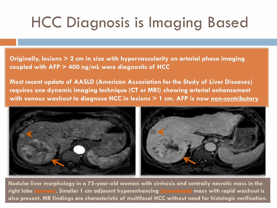
HCC Diagnosis is Imaging Based
Originally, lesions > 2 cm in size with hypervascularity on arterial phase imaging
coupled with AFP > 400 ng/mL were diagnostic of HCC
Most recent update of AASLD (American Association for the Study of Liver Diseases)
requires one dynamic imaging technique (CT or MRI) showing arterial enhancement
with venous washout to diagnose HCC in lesions > 1 cm. AFP is now non-contributory
Nodular liver morphology in a 75-year-old woman with cirrhosis and centrally necrotic mass in the
right lobe (arrows). Smaller 1 cm adjacent hyperenhancing (arrowhead) mass with rapid washout is
also present. MR findings are characteristic of multifocal HCC without need for histologic verification.
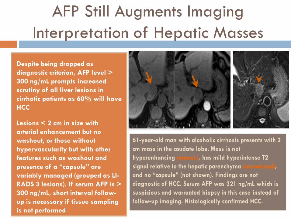
AFP Still Augments Imaging
Interpretation of Hepatic Masses
Despite being dropped as
diagnostic criterion, AFP level >
300 ng/mL prompts increased
scrutiny of all liver lesions in
cirrhotic patients as 60% will have
HCC
Lesions < 2 cm in size with
arterial enhancement but no
washout, or those without
hypervascularity but with other
features such as washout and
presence of a “capsule” are
variably managed (grouped as LI-
RADS 3 lesions). If serum AFP is >
300 ng/mL, short interval follow-
up is necessary if tissue sampling
is not performed

AFP Valuable for Prognosis and
Monitoring After Treatment
Higher concentrations of AFP prior to
treatment indicates a poorer
prognosis (as does increased size and
number of tumors and higher
histologic grade)
AFP > 400 ng/mL portends portal
vein tumor thrombus; distal thrombus
difficult to detect on imaging as it
does not result in expansion, unlike
thrombi in main portal vein
If AFP was elevated prior to
treatment and returned to normal
after therapy, a subsequent rise
heralds recurrence. AFP monitoring
does not replace imaging surveillance
Large infiltrative HCC within the right and left
hepatic lobes (arrow) in a 56-year-old male with
cirrhosis. There is involvement of the hepatic
hilum, where there is portal vein thrombus
extending to the portal vein confluence
(arrowhead). AFP was elevated at 5304 ng/mL.

Prostate Cancer
Most common cancer in men and 2nd
leading cause of cancer related death
in the United States
Prostate specific antigen (PSA) is not
specific for cancer as it is also
elevated in BPH and prostatitis
Guidelines for screening with PSA +/-
rectal exam are not uniform
Serum PSA level > 4 ng/mL most
common threshold used
If screening PSA is abnormal, US-
guided transrectal biopsy required;
PSA has limited role in diagnosis
Nuclear medicine bone scan demonstrates
diffuse osteoblastic metastatic throughout the
axial and appendicular skeleton (superscan
appearance given diminished renal and soft
tissue activity). Patient was newly diagnosed
with prostate cancer and PSA was 1409
ng/mL. See subsequent slide for bone scan
recommendations based on PSA values.
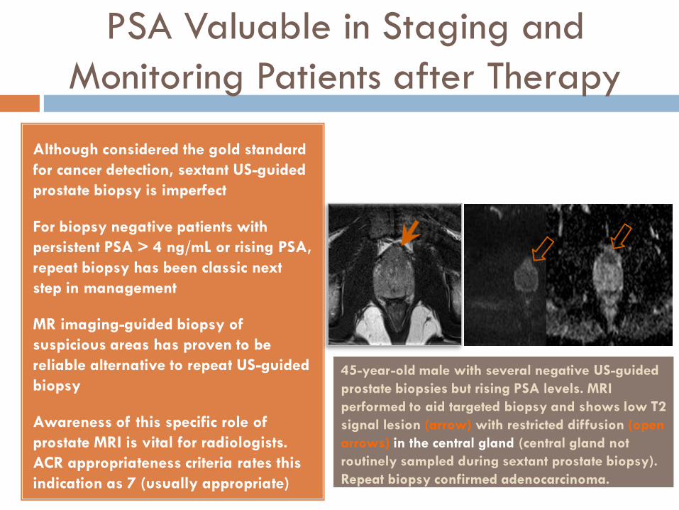
PSA Valuable in Staging and
Monitoring Patients after Therapy
Although considered the gold standard
for cancer detection, sextant US-guided
prostate biopsy is imperfect
For biopsy negative patients with
persistent PSA > 4 ng/mL or rising PSA,
repeat biopsy has been classic next
step in management
MR imaging-guided biopsy of
suspicious areas has proven to be
reliable alternative to repeat US-guided
biopsy
Awareness of this specific role of
prostate MRI is vital for radiologists.
ACR appropriateness criteria rates this
indication as 7 (usually appropriate)
45-year-old male with several negative US-guided
prostate biopsies but rising PSA levels. MRI
performed to aid targeted biopsy and shows low T2
signal lesion (arrow) with restricted diffusion (open
arrows) in the central gland (central gland not
routinely sampled during sextant prostate biopsy).
Repeat biopsy confirmed adenocarcinoma.

PSA Valuable in Staging and
Monitoring Patients after Therapy
Bone scan, CT and/or MRI may be
employed in staging
Bone scans only necessary for patients
with PSA levels > 10 ng/mL (or
Gleason score > 7 on biopsy). There
are similar guidelines for CT given low
likelihood of positive findings in
patients with lower values
Prostate MRI aids risk stratification and
predicts organ-confined disease
Following radical retropubic
prostatectomy (RRP), PSA levels should
be undetectable. Persistent/rising PSA
is evidence of residual or recurrent
disease
81-year-old male with tumor in the left
peripheral zone (open arrow) extending
from the apex to the base on prostate MRI.
Extracapsular extension evident as bulging
of the capsule and obliteration of the
rectoprostatic angle
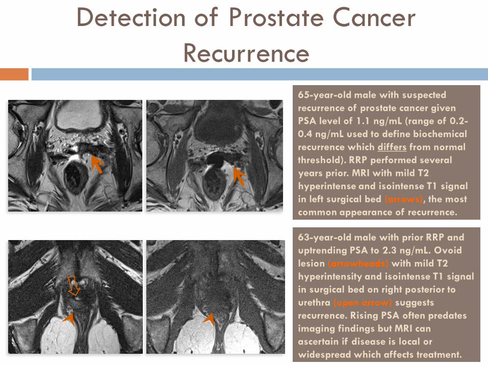
Detection of Prostate Cancer
Recurrence
65-year-old male with suspected
recurrence of prostate cancer given
PSA level of 1.1 ng/mL (range of 0.2-
0.4 ng/mL used to define biochemical
recurrence which differs from normal
threshold). RRP performed several
years prior. MRI with mild T2
hyperintense and isointense T1 signal
in left surgical bed (arrows), the most
common appearance of recurrence.
63-year-old male with prior RRP and
uptrending PSA to 2.3 ng/mL. Ovoid
lesion (arrowheads) with mild T2
hyperintensity and isointense T1 signal
in surgical bed on right posterior to
urethra (open arrow) suggests
recurrence. Rising PSA often predates
imaging findings but MRI can
ascertain if disease is local or
widespread which affects treatment.

Colorectal Cancer (CRC)
3rd most common malignancy
worldwide
Carcinoembryonic antigen (CEA) is
tumor marker associated with CRC
Nonspecific; elevated levels seen with
other malignancies (pancreatic and
gastric cancer) and other entities
(cirrhosis, gastritis, IBD, diverticulitis)
No role for CEA in CRC screening or
diagnosis
Screening colonoscopy recommended
at age 50. CT colonoscopy is an
alternative option for CRC screening
50-year-old woman with non-specific
systemic symptoms and elevated serum CEA
of 15.2 ng/mL (0-4 ng/mL is normal range).
PET imaging reveals focal intense FDG
avidity within the gastric antrum (arrows),
consistent with gastritis or gastric neoplasm.
Biopsy confirmed adenocarcinoma.

CRC Prognosis and Treatment Response
Increased preoperative CEA level (>5
ng/mL) correlates with decreased
survival
New chemotherapies have improved
survival in metastatic CRC but are
potentially toxic and expensive
Therefore, it is prudent to discontinue
ineffective treatments quickly
CEA increase (two successive
elevated levels above baseline)
suggests progressive disease, even in
absence of radiologic findings
Patient with known metastatic colon cancer
and uptrending CEA >50% baseline value.
CT abdomen pelvis shows progression of
metastatic liver disease (arrows), new
mesenteric lesions (not shown), and a new
vertebral body metastasis (arrowhead).

Colorectal Cancer Surveillance
CEA should be measured every 3
months in patients with stage II or III
CRC for 3 years following diagnosis
and treatment
Elevated postoperative CEA level has
a higher likelihood of representing
metastatic disease as opposed to
isolated local recurrence
Normal CEA concentrations do not
exclude disease progression
PET/CT is useful in detecting CRC
recurrence, particularly in patients
who do not present with elevated
serum CEA levels
40-year-old female with history of stage IIIb
colon cancer and uptrending CEA 2 years
after diagnosis. PET imaging reveals a new
FDG avid lesion within the right mesorectal
fat (arrows) consistent with early disease
recurrence.

Pancreatic Cancer
4th leading cause of cancer death in
the United States
Precise role of serum carbohydrate
antigen 19.9 (CA 19-9) is not defined.
It is the most validated tumor marker
but with several limitations
CA 19-9 can be elevated with non-
pancreas cancers as well as with
pancreatitis and cirrhosis. False
negatives for pancreatic cancer occur
in 5-10% of population
No role for CA 19-9 in pancreatic
cancer screening
Double duct sign evident as both common duct
(arrow) and pancreatic duct (arrowhead) are
dilated. Obstructing pancreatic head mass (not
shown) confirmed to be adenocarcinoma.
Patient had normal serum CA 19-9 level of 8
U/mL (0-36 U/mL is normal range) at time of
diagnosis, highlighting case of false negative
tumor marker for pancreatic cancer.

CA 19-9 Helpful for Diagnosis
Normal blood levels of CA 19-9 are
below 37 U/mL
Not accurate enough to detect cancer
in isolation
In conjunction with imaging, elevated
CA 19-9 helps differentiate pancreatic
cancer from focal pancreatitis
Reported specificity of CA19-9 for
pancreatic cancer when > 100 U/mL
is 98%
Addition of CEA level increases
specificity for pancreatic carcinoma if
both markers are elevated
Ill-defined pancreatic head mass (arrow) in a
69-year-old male with epigastric pain.
Findings initially favored to represent focal
pancreatitis, given adjacent fat stranding
(arrowhead) and elevated lipase. However,
CA 19-9 was markedly elevated at 524 U/mL
and CEA was 87 ng/mL, highly suggestive of
malignancy. Surgical biopsy confirmed
pancreatic adenocarcinoma.
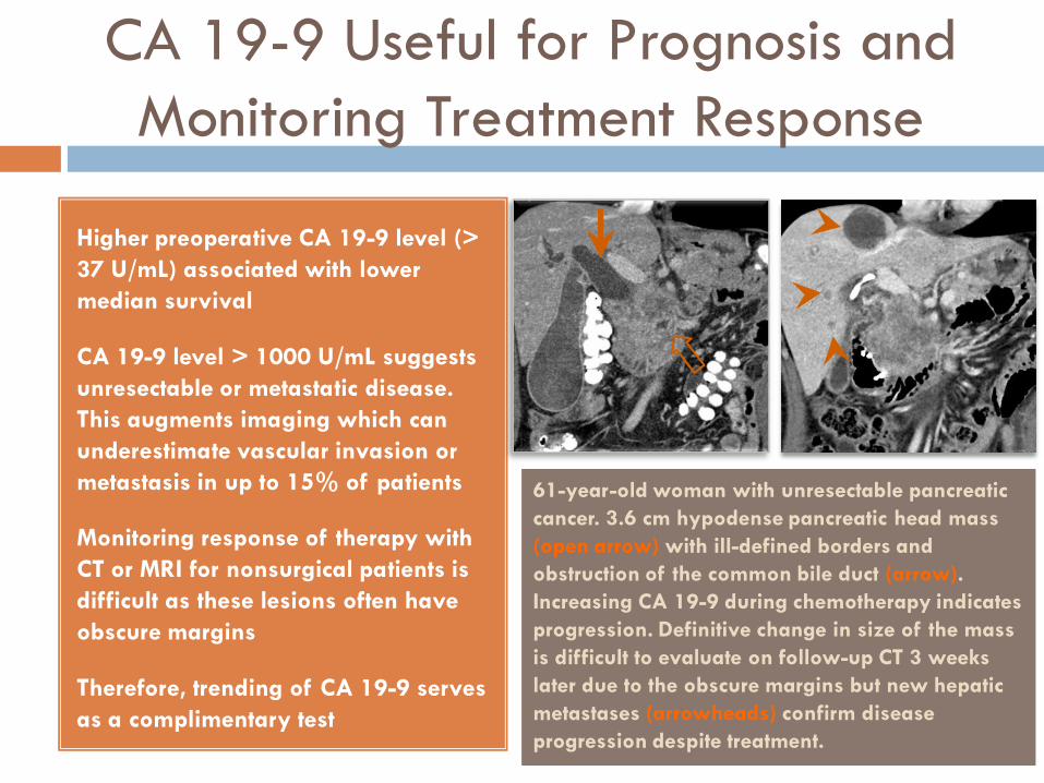
CA 19-9 Useful for Prognosis and
Monitoring Treatment Response
Higher preoperative CA 19-9 level (>
37 U/mL) associated with lower
median survival
CA 19-9 level > 1000 U/mL suggests
unresectable or metastatic disease.
This augments imaging which can
underestimate vascular invasion or
metastasis in up to 15% of patients
Monitoring response of therapy with
CT or MRI for nonsurgical patients is
difficult as these lesions often have
obscure margins
Therefore, trending of CA 19-9 serves
as a complimentary test
61-year-old woman with unresectable pancreatic
cancer. 3.6 cm hypodense pancreatic head mass
(open arrow) with ill-defined borders and
obstruction of the common bile duct (arrow).
Increasing CA 19-9 during chemotherapy indicates
progression. Definitive change in size of the mass
is difficult to evaluate on follow-up CT 3 weeks
later due to the obscure margins but new hepatic
metastases (arrowheads) confirm disease
progression despite treatment.
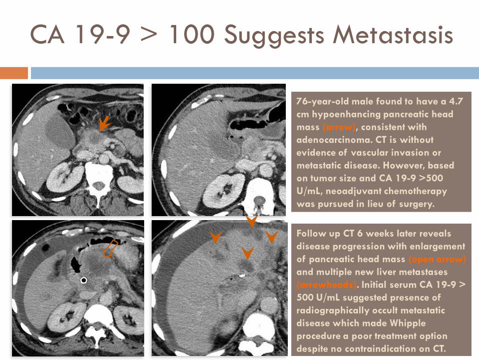
CA 19-9 > 100 Suggests Metastasis
76-year-old male found to have a 4.7
cm hypoenhancing pancreatic head
mass (arrow), consistent with
adenocarcinoma. CT is without
evidence of vascular invasion or
metastatic disease. However, based
on tumor size and CA 19-9 >500
U/mL, neoadjuvant chemotherapy
was pursued in lieu of surgery.
Follow up CT 6 weeks later reveals
disease progression with enlargement
of pancreatic head mass (open arrow)
and multiple new liver metastases
(arrowheads). Initial serum CA 19-9 >
500 U/mL suggested presence of
radiographically occult metastatic
disease which made Whipple
procedure a poor treatment option
despite no contraindication on CT.

Testicular Cancer
Most common in men age 15-35
AFP, lactate dehydrogenase (LDH), and
human chorionic gonadotropin (hCG)
are established tumor markers
No role in screening
Work-up of testicular mass includes
physical exam, US, and serum tumor
markers
Lower levels of hCG (<500 mIU/mL)
suggest seminoma. Non-seminoma
germ cell tumors (NSGCTs) are
associated with levels >1000 mIU/mL
Pure seminomas do not produce AFP
Left testicular NSGCT (arrows). A tumor
histologically classified as a seminoma will
be reclassified as NSGCT if serum AFP is
elevated, indicating the importance of tumor
markers as treatment varies significantly
between the two. Seminomas are also
typically homogenous in appearance on US.
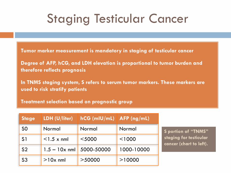
Staging Testicular Cancer
Tumor marker measurement is mandatory in staging of testicular cancer
Degree of AFP, hCG, and LDH elevation is proportional to tumor burden and
therefore reflects prognosis
In TNMS staging system, S refers to serum tumor markers. These markers are
used to risk stratify patients
Treatment selection based on prognostic group
S portion of “TNMS”
staging for testicular
cancer (chart to left).
Stage LDH (U/liter) hCG (mIU/mL) AFP (ng/mL)
S0 Normal Normal Normal
S1 <1.5 x nml <5000 <1000
S2 1.5 – 10x nml 5000-50000 1000-10000
S3 >10x nml >50000 >10000
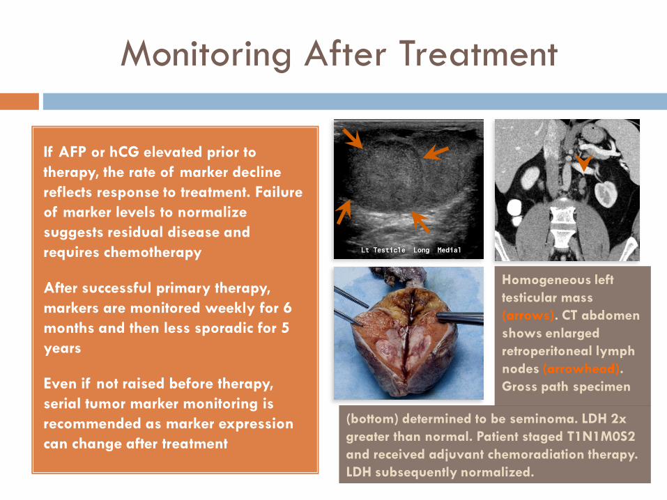
Monitoring After Treatment
If AFP or hCG elevated prior to
therapy, the rate of marker decline
reflects response to treatment. Failure
of marker levels to normalize
suggests residual disease and
requires chemotherapy
After successful primary therapy,
markers are monitored weekly for 6
months and then less sporadic for 5
years
Even if not raised before therapy,
serial tumor marker monitoring is
recommended as marker expression
can change after treatment
(bottom) determined to be seminoma. LDH 2x
greater than normal. Patient staged T1N1M0S2
and received adjuvant chemoradiation therapy.
LDH subsequently normalized.
Homogeneous left
testicular mass
(arrows). CT abdomen
shows enlarged
retroperitoneal lymph
nodes (arrowhead).
Gross path specimen

Ovarian Cancer
Highest mortality of gynecologic
tumors
CA 125 most reliable tumor marker,
but is effective for epithelial ovarian
malignancy only
Not recommended for screening due
to lack of sensitivity and specificity
CA 125 elevated in only 50%-60%
of patients with stage I ovarian
cancer
Pelvic inflammatory disease,
endometriosis, and pregnancy can
falsely elevate CA 125
Left ovarian mass with homogeneous low
level echos (arrows) and no internal flow on
US, suggestive of an endometrioma. Patient
had elevated CA 125 of 87 U/mL (normal
range is 0.6-35 U/mL) and endometrioma
was confirmed at surgery.

Discrimination of Pelvic Masses
Threshold CA 125 value is 35 U/mL
CA 125 used as an adjunct in
distinguishing benign from malignant
ovarian masses
Risk of malignancy index (RMI)
incorporates CA 125 levels in
addition to sonographic features of
an adnexal mass and menopausal
status of patient
A single CA 125 measurement is not
as effective as trends. Benign masses
have stable levels, and progressively
increasing levels indicate ovarian
cancer
Risk of Malignancy Index
Ultrasound Components (>1 = higher risk):
1. Multilocular
2. Solid component
3. Evidence for metastasis
4. Ascites
5. Bilateral ovarian lesions
US score (maximum of 3) is multiplied by
menopausal status (expressed as 1 if
premenopausal and 3 if postmenopausal) and
CA 125 level to derive a RMI value. RMI greater
than 200 indicates malignancy.
If there is suspicion for a germ cell
tumor (more common in women
younger than 40), AFP and hCG are
important adjunct tumor markers

Discrimination of Pelvic Masses
Cystic left ovarian mass with irregular solid
component (arrow) in a 32-year-old woman.
There was no ascites and the serum CA 125
was 20 U/mL. RMI calculated to be 20 which
favors a benign entity. A mature cystic
teratoma was confirmed.
Purely solid right adnexal mass (arrow) with
moderate pelvic free fluid (arrowheads) and
a serum CA 125 of 256 U/mL in a 63-year-
old woman. RMI calculated to be 2304 which
suggests a malignant lesion. A serous
cystadenocarcinoma was confirmed.
Ovarian Disease Recurrence
Declining CA 125 levels correlate
with response to chemotherapy
Rising CA 125 values after definitive
treatment predict relapse. Normal
levels, however, do not exclude
presence of disease
CA 125 is monitored every 2-4
months for at least 2 years
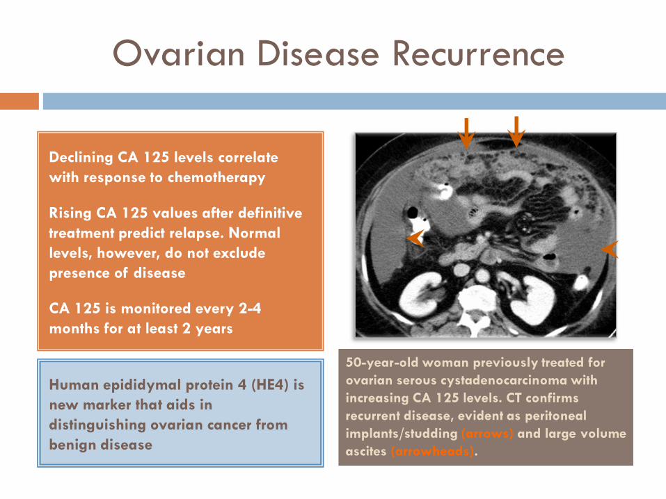
50-year-old woman previously treated for
ovarian serous cystadenocarcinoma with
increasing CA 125 levels. CT confirms
recurrent disease, evident as peritoneal
implants/studding (arrows) and large volume
ascites (arrowheads).
Human epididymal protein 4 (HE4) is
new marker that aids in
distinguishing ovarian cancer from
benign disease

The radiologist often interprets oncologic imaging examinations with only a portion of the complete clinical picture which can yield indeterminate reports
Knowledge of serum tumor markers at the time of imaging interpretation can increase diagnostic accuracy and improve the quality of radiology reports by highlighting information that is germane to the referring clinician
Serum tumor markers are particularly useful in diagnosing pancreatic and ovarian cancer, staging testicular cancer, and predicting advanced disease in numerous cancers
Conclusion

CORRESPONDENCE