clinical anatomy surgical anatomy of the hip: a review of ... · deep to, and along the posterior...
TRANSCRIPT

Page 1 of 8
Review
Licensee OA Publishing London 2013. Creative Commons Attribution License (CC-BY)
For citation purposes: Frank RM, Gandhi S, Cross MB, Haughom B, Rosenberg AG, Levine BR . Surgical anatomy of the hip: a review of the common open approaches. OA Anatomy 2013 Aug 01;1(3):21. Co
mpe
ting
inte
rest
s: n
one
decl
ared
. Con
flict
of i
nter
ests
: non
e de
clar
ed.
All
auth
ors
cont
ribut
ed to
con
cepti
on a
nd d
esig
n, m
anus
crip
t pre
para
tion,
read
and
app
rove
d th
e fin
al m
anus
crip
t.A
ll au
thor
s ab
ide
by th
e A
ssoc
iatio
n fo
r Med
ical
Eth
ics
(AM
E) e
thic
al ru
les
of d
iscl
osur
e.
Clin
ical
Ana
tom
y
Surgical anatomy of the hip: a review of the common open approaches
RM Frank1*, S Gandhi1, MB Cross2, B Haughom1, AG Rosenberg1, BR Levine1
AbstractIntroduction Hip pathology requiring surgical intervention varies from osteoar-thritis to oncologic diseases. An un-derstanding of hip anatomy and, in particular, the potentially dangerous neurovascular structures that are avoided in most exposures, must be understood to help avoid complica-tions that may thwart an otherwise safe surgical procedure. The purpose of this review is to discuss the most common open exposures to the hip, with a focus on surgically relevant hip anatomy of each approach.Conclusion A thorough understanding of pelvic and hip anatomy is critical to per-form these approaches safely and efficiently.
IntroductionOsteoarthritis (OA) is an extreme-ly common condition in the adult patient population1. While non-operative treatment may relieve symptoms, the natural history of the disease is progressive and many patients eventually require a to-tal hip arthroplasty (THA). With an appreciation for the relevant anatomy, each technique can be performed with comparable clini-cal outcomes2–8. Regardless of the specific technique chosen to expose the joint, a complete understanding of the anatomy of each approach is necessary to perform the operation
safely. The four major approaches to the hip9,10 (based on the direction of the respective exposure) that will be discussed include: posterior, lateral, anterior, and antero-lateral.
DiscussionPosterior approach (Moore or Southern approach)The posterior approach to the hip joint was first popularized by Moore and is the most-commonly used ap-proach used in hip replacement sur-gery11. This approach can be easily extended and, as such, is useful in revision cases when increased surgi-cal exposure may be critical. In ad-dition to arthroplasty, the posterior approach is also extremely useful for posterior acetabular fracture repair, surgical management of deep infec-tion, and removal of intra-articular loose bodies.
Important surgical anatomy and landmarksThe posterior approach has no true internervous plane, but does utilize an intramuscular plane, as the glu-teus maximus (GMax) is split dur-ing the superficial dissection (Table 1). The GMax is innervated by the inferior gluteal nerve; however, den-ervation of the muscle does not usu-ally occur with this approach, as the muscle is typically split well lateral of the medially-based nerve supply. Blood supply to the proximal third of the GMax comes from the supe-rior gluteal artery, while the distal two-thirds is supplied by the inferior gluteal artery. The most important superficial landmark in this approach is the greater trochanter (GT), which is used to guide incision location and, ultimately, the arthrotomy.
Surgical approachThe posterior approach is performed with the patient in the lateral decu-bitus position, with the operative leg up (Figure 1). Based on surgeon’s preference, patient factors (e.g. size of the patient), as well as the need for an extensile exposure, the inci-sion starts approximately 6–8 cm proximal and posterior to the GT, and extends distally in a curvilinear fash-ion centred on the posterior aspect of the GT following the direction of the femoral shaft, for a total length of ap-proximately 10–15 cm (or as long as needed for exposure) (Figure 2). Dis-section continues through the subcu-taneous adipose tissue until the fascia lata is exposed (Figure 3). An incision through the fascia lata is made in line with the skin incision, exposing the GMax. The fibres of the GMax are th- en split by blunt dissection beginning distally and proceeding proximally. As noted above, the superior and inferi-or gluteal arteries (and their branch-es), which supply the muscle, may be injured during blunt dissection. Self-retaining retractors are placed on the margins of the split maximus to reveal the posterior aspect of the hip which includes the gluteus medius (GMed) and the underlying short external rotators of the hip: the piriformis, superior gemellus, obturator inter-nus, and inferior gemellus (Figure 4). The latter three muscles merge to form the conjoined tendon, while the piriformis can typically be visualized and palpated as a separate structure just proximal to the conjoined tendon deep to, and along the posterior bor-der of, the GMed.12 These ‘rotators’ are cut, as close as possible to their femoral insertion to preserve ten-don length and allow for an eventual
* Corresponding Author E-mail: [email protected] Department of Orthopaedic Surgery, Rush
University Medical Center, Chicago, IL, USA 2 Department of Adult Reconstruction, Hospital
for Special Surgery, New York, NY, USA

Page 2 of 8
Review
Licensee OA Publishing London 2013. Creative Commons Attribution License (CC-BY)
For citation purposes: Frank RM, Gandhi S, Cross MB, Haughom B, Rosenberg AG, Levine BR . Surgical anatomy of the hip: a review of the common open approaches. OA Anatomy 2013 Aug 01;1(3):21. Co
mpe
ting
inte
rest
s: n
one
decl
ared
. Con
flict
of i
nter
ests
: non
e de
clar
ed.
All
auth
ors
cont
ribut
ed to
con
cepti
on a
nd d
esig
n, m
anus
crip
t pre
para
tion,
read
and
app
rove
d th
e fin
al m
anus
crip
t.A
ll au
thor
s ab
ide
by th
e A
ssoc
iatio
n fo
r Med
ical
Eth
ics
(AM
E) e
thic
al ru
les
of d
iscl
osur
e.
can lead to injury to the GMed and/or minimus, although usually not clini-cally relevant), and thus patients are unlikely to experience the abductor deficient gait often seen with other exposures. However, because the pos-terior capsule and short external rota-tors are incised in this approach, there may be a potential increased risk for hip dislocation following surgery13–15. As such, meticulous soft tissue repair is critical following the posterior ap-proach to the hip. Further, larger head sizes have been implemented in THA to decrease the dislocation rate after a posterior approach16–18.
Structures at risk The sciatic nerve and the inferior glu-teal artery are the structures most at risk during the posterior approach to the hip (Table 2). Errant dissec-tion, excessive limb lengthening, and improper retractor placement are often implicated in cases of sciatic nerve injury following the posterior approach.
Direct lateral approach (Hardinge approach)The direct lateral approach to the hip was first described by Hardinge
In severely contracted hips (or based on surgeon’s preference), a portion of or the entire GMax tendon insertion into the postero-lateral aspect of the femur is released to decrease the risk of compression of the sciatic nerve, as it runs deep to the tendon and is susceptible to compression when the femur is adducted and internally rotated.
Unlike other approaches to the hip, the posterior approach does not typi-cally compromise the abductor mech-anism (aggressive superior retraction
tension-free repair. They are then tagged, and retracted posteriorly, ex-posing the underlying posterior joint capsule. Of note, the sciatic nerve exits the pelvis through the greater sciatic notch and courses distally down the posterior thigh (Figure 4). The nerve usually travels under the piriformis and over the medial border of the short external rotators, though there can be substantial variability in this anatomy and the clinician must be cognizant of the nerve’s position at all times during this approach. Internal rotation of the hip during exposure of the short external rotators places the incision of the rotators well lateral to the nerve, following which gentle retraction of the rotators after they are released is critical in protecting the sciatic nerve. The posterior hip capsule can now be visualized and capsulotomy performed with either a longitudinal, T- or H-shaped inci-sion. To expose the femoral head and acetabulum, the hip is dislocated by gentle traction accompanied by inter-nal rotation, flexion, and adduction of the hip. The superior fibres of quad-ratrus femoris, located just distal to the conjoined tendon, may need to be released to expose the capsule; how-ever, caution should be taken to avoid significant bleeding from the branch-es of the medial femoral circumflex artery which run through the muscle.
Table 1 Approaches and associated internervous/intermuscular planes
Approach Intervernous Intra/Intermuscular
Posterior (Southern) None GMax (IGN)
Lateral (Hardinge) None GMed (SGN)Vastus lateralis (Femoral)
Anterior (Smith-Peterson) SuperficialSartorius (Femoral)TFL (SGN)DeepRectus femoris (Femoral)GMed (SGN)
None
Antero-lateral (Watson-Jones)
None TFL (SGN)GMed (SGN)
IGN, inferior gluteal nerve; SGN, superior gluteal nerve
Figure 1: Intraoperative photograph of a patient undergoing total hip arthroplasty placed in the lateral decubitus position in preparation for a posterior approach to the hip with the operative hip (right) facing up.

Page 3 of 8
Review
Licensee OA Publishing London 2013. Creative Commons Attribution License (CC-BY)
For citation purposes: Frank RM, Gandhi S, Cross MB, Haughom B, Rosenberg AG, Levine BR . Surgical anatomy of the hip: a review of the common open approaches. OA Anatomy 2013 Aug 01;1(3):21. Co
mpe
ting
inte
rest
s: n
one
decl
ared
. Con
flict
of i
nter
ests
: non
e de
clar
ed.
All
auth
ors
cont
ribut
ed to
con
cepti
on a
nd d
esig
n, m
anus
crip
t pre
para
tion,
read
and
app
rove
d th
e fin
al m
anus
crip
t.A
ll au
thor
s ab
ide
by th
e A
ssoc
iatio
n fo
r Med
ical
Eth
ics
(AM
E) e
thic
al ru
les
of d
iscl
osur
e.
Surgical approachThe direct lateral approach can be performed with the patient in the lateral decubitus or the supine posi-tion (which allows both the gluteal musculature and fat to fall posteri-orly off the table and away from sur-gical field). The incision is started approximately 5 cm proximal to the tip of the GT and is continued in a linear fashion distally, centred over the tip of the GT and in line with the femur, for approximately 8–12 cm (or as long as needed to safely complete the exposure). Superficial dissection is continued through the subcutaneous adipose tissue to the underlying fascia lata, which is split in line with the incision. Retraction of the fascia lata anteriorly and the GMax posteriorly exposes the un-derling GMed and vastus lateralis. The anterior one-fifth to one-third (varying percentages have been de-scribed) of the GMed is subsequently split proximal to the trochanter in line with the muscle fibres (at 45 degrees between straight superior and straight anterior) and continues distally along the muscle’s insertion
tip of the GT, which is used to guide incision location and to divide the GMed tendon.
in 198219. This approach provides adequate exposure to the hip joint for primary arthroplasty as well as revision procedures. It is considered to have a low post-operative disloca-tion rate, and does not require tro-chanteric osteotomy to gain broad exposure. Nevertheless, this ap-proach does require release of the anterior portion of the GMed, which may predispose the patient to an abductor lurch (i.e. Trendelenberg limp) following surgery.
Important surgical anatomy and landmarksThe direct lateral approach has no true intervernous plane, but does utilize an intermuscular plane, as a portion of the GMed and a portion of the vastus lateralis is released during the superficial dissection (Table 1). The GMed is innervated by the supe-rior gluteal nerve, while the vastus lateralis is innervated by the femoral nerve. The most important superfi-cial landmark in this approach is the
Figure 3: Intraoperative photograph of a patient undergoing revision total hip arthroplasty via posterior approach (right hip); dissection continues through the subcutaneous adipose tissue until the fascia lata is exposed.
Figure 2: Intraoperative photograph of a patient undergoing total hip arthroplasty via posterior approach (right hip); the incision starts approximately 6–8 cm proximal and posterior to the greater trochanter, and extends distally in a curvilinear fashion centred on the posterior aspect of the trochanter following the direction of the femoral shaft, for a total length of approximately 10–15 cm (or as long as needed for exposure).

Page 4 of 8
Review
Licensee OA Publishing London 2013. Creative Commons Attribution License (CC-BY)
For citation purposes: Frank RM, Gandhi S, Cross MB, Haughom B, Rosenberg AG, Levine BR . Surgical anatomy of the hip: a review of the common open approaches. OA Anatomy 2013 Aug 01;1(3):21. Co
mpe
ting
inte
rest
s: n
one
decl
ared
. Con
flict
of i
nter
ests
: non
e de
clar
ed.
All
auth
ors
cont
ribut
ed to
con
cepti
on a
nd d
esig
n, m
anus
crip
t pre
para
tion,
read
and
app
rove
d th
e fin
al m
anus
crip
t.A
ll au
thor
s ab
ide
by th
e A
ssoc
iatio
n fo
r Med
ical
Eth
ics
(AM
E) e
thic
al ru
les
of d
iscl
osur
e.
must be taken to avoid extending the incision too far proximally in order to prevent injury to the superior gluteal nerve as it runs between the medius and minimus muscle bellies 3–5 cm proximal to the tip of the GT.
The femoral nerve, artery, and vein are vulnerable to errantly placed re-tractors or retracting too vigorously, as they course anteriorly in the thigh.
Anterior approach (Smith-Peterson)The anterior approach to the hip joint, first described by Smith-Peter-son20, 21, allows for safe access to the anterior aspect of the hip, and can also be extended to gain access to the ilium, and thus is useful for pelvic osteotomies. However, this approach is not extensile posteriorly, as it does not allow for complete exposure of the posterior acetabulum and ac-cess to the proximal femur can also be limited. Compared to other tech-niques, the direct anterior approach can be more time-consuming, and some authors advocate for the use of intraoperative fluoroscopy to aid in visualization of landmarks as well as component position22, 23.
Important surgical anatomy and landmarksThe anterior approach to the hip utilizes the internervous plane be-tween the sartorius (femoral nerve) and the tensor fascia lata (superior gluteal nerve) superficially (Figure 5), as well as the internervous plane between the rectus femoris (femoral nerve) and the GMed (superior glu-teal nerve) at a deeper level (Table 1). Superficial bony landmarks that should be identified prior to plan-ning the incision include the anterior superior iliac spine (ASIS) as well as the iliac crest and greater trochanter.
Surgical approachWith the patient supine, the inci-sion starts 3–4 finger-breadths dis-tal to the ASIS and is angled for 8–12 cm towards the lateral aspect of the
vessel when encountered. This vessel originates from the lateral circumflex femoral artery and passes laterally over the vastus intermedius, piercing the vastus lateralis, and ultimately winds around the femur, just below the GT. It anastomoses on the back of the thigh with the medial femoral circumflex artery, inferior gluteal ar-tery, and perforating branches of the profunda femoris artery. Once ad-equate exposure of the anterior hip capsule is obtained, capsulotomy (or capsulectomy) can be achieved using a T-shaped incision (or by removing the anterior capsule). As opposed to the posterior approach, the hip is dislocated by traction, flexion, adduc-tion, and external rotation.
Structures at risk Neurovascular structures at risk (Ta-ble 2) during the direct lateral ap-proach include the superior gluteal nerve as well as the femoral neuro-vascular bundle. The superior gluteal nerve innervates the GMed, minimus, and tensor fascia. It leaves the pelvis through the greater sciatic foramen above the piriformis, accompanied by the superior gluteal artery and vein running deep to the GMax3. Care
on the greater trochanter. Care must be taken not to split the medius more than 3–5 cm proximal the upper bor-der of the GT as at this level is the superior gluteal nerve. The GMed becomes confluent with the proximal portion of the vastus lateralis, which may also be split in line with its fi-bres to provide distal exposure. The gluteus minimus (GMin) tendon lies deep to the medius and its insertion on the trochanter lies just anterior to the medius insertion along the an-terior border of the trochanter. It is incised proximal to the trochanter along the same line as the incision in the medius. Distal to the anterior su-perior tip of the trochanter, it can be released just anterior to its insertion on the greater trochanter leaving a small cuff of tendon on the proxi-mal femur to allow for subsequent repair. To gain exposure of the joint capsule, the anterior aspect of the split GMed, the underlying GMin, and the split vastus lateralis are elevated from the GT, off the anterior capsule as a single flap. During mobilization of the vastus lateralis, the transverse branch of the lateral circumflex ar-tery can be injured, and so care must be taken to identify and cauterize the
Figure 4: Intraoperative photograph of a patient undergoing total hip arthroplasty via posterior approach (right hip); visualized are the short external rotators of the hip including the piriformis, superior gemellus, obturator internus, and inferior gemellus. Note the sciatic nerve (asterisk) exiting the pelvis, travelling under the piriformis and over the medial border of the short external rotators as it courses distally.

Page 5 of 8
Review
Licensee OA Publishing London 2013. Creative Commons Attribution License (CC-BY)
For citation purposes: Frank RM, Gandhi S, Cross MB, Haughom B, Rosenberg AG, Levine BR . Surgical anatomy of the hip: a review of the common open approaches. OA Anatomy 2013 Aug 01;1(3):21. Co
mpe
ting
inte
rest
s: n
one
decl
ared
. Con
flict
of i
nter
ests
: non
e de
clar
ed.
All
auth
ors
cont
ribut
ed to
con
cepti
on a
nd d
esig
n, m
anus
crip
t pre
para
tion,
read
and
app
rove
d th
e fin
al m
anus
crip
t.A
ll au
thor
s ab
ide
by th
e A
ssoc
iatio
n fo
r Med
ical
Eth
ics
(AM
E) e
thic
al ru
les
of d
iscl
osur
e.
Antero-lateral approach (Watson-Jones)The antero-lateral approach, also known as the Watson-Jones ap-proach, is now a common approach for THA24, 25. This approach provides excellent exposure to both the proxi-mal femur and acetabulum, and is useful for patients who are at higher risk for hip dislocation as the posteri-or soft tissue and capsular structures remain mostly intact. However, this approach may require partial disrup-tion of the abductor mechanism and thus, may be associated with a post-operative limp.
Important surgical anatomy and landmarksThe antero-lateral approach has no true internervous plane, but does utilize the intermuscular plane be-tween the TFL and the GMed (Figure 6), both of which are innervated by the superior gluteal nerve (Table 1). The most important superficial land-marks in this approach are the GT, the anterior border of the femur, and ASIS. The interval between the TFL and GMed can be palpated in most patients who are not obese.
Surgical approachSimilar to the direct lateral approach, the antero-lateral approach is per-formed with the patient in either the lateral or supine position. In the lat-eral position, the operating table can be tilted posteriorly so that the surgi-cal site is more easily accessible. De-pending on the surgeon’s preference and patient’s body habitus, an 8–15 cm incision begins 2.5 cm poste-rior and distal to the ASIS and is di-rected towards the tip of the greater trochanter. As it passes toward the posterior third of the trochanter, the incision may be extended distally centred on the anterior border of the greater trochanter and the proximal femur.
Superficial dissection is continued through the subcutaneous adipose tissue to the underlying fascia, which
nerve) and the GMed (superior glu-teal nerve). The two heads of the rectus, the direct and reflected heads, attach to the ASIS and the superior lip of the acetabulum (as well as the an-terior hip capsule). At this point, ac-etabular (and occasionally the ASIS) origins of the rectus are released and the rectus femoris is retracted me-dially, while the GMed is retracted laterally. Once this deep exposure is obtained, the anterior hip joint cap-sule is visible and can be widely ex-posed with retractors placed along the supero-lateral capsule, the ante-ro-superior acetabular rim, and the infero-medial capsule. The capsule is then incised or removed allowing dis-location via extension and external rotation. To safely expose the proxi-mal femur for placement of a femoral THA component, it may be necessary to release additional posterior and superior capsules as well as the short external rotators.
Structures at risk Structures at risk (Table 2) during this approach include the lateral cu-taneous femoral nerve and the as-cending branch of the lateral femoral circumflex artery, as described above. Additionally, the femoral nerve and artery or vein injuries can occur if the rectus femoris is inadvertently retracted laterally instead of medi-ally during the surgical approach.
patella. If more proximal exposure is required, a curvilinear incision can be made along the border of the iliac crest, running as far posteri-orly as the posterior superior iliac spine (PSIS). Superficial dissection is continued anteriorly through the subcutaneous adipose tissue and the underlying fascia. The internervous plane between the sartorius (femo-ral nerve) medially and the TFL (su-perior gluteal nerve) laterally should be palpable. Care must be taken to protect the lateral cutaneous femo-ral nerve, which can travel through, posterior, or superficial to the sar-torius muscle. To avoid injury to the nerve the fascia on the medial side of the TFL is sharply incised, in order to expose the interval between the TFL and sartorius, which are separated by gentle blunt dissection. In the in-terval between sartorius and TFL, the ascending branch of the lateral femo-ral circumflex artery will be visible, crossing transversely across the ante-rior aspect of the hip joint, as it passes upwards, beneath the tensor fasciae lata anastomozing with the terminal branches of the superior gluteal and deep circumflex iliac artery at the lat-eral aspect of the hip. Care should be taken to identify and ligate or coagu-late this vessel. Retraction of the TFL laterally and the sartorius medially, exposes the deep internervous plane between the rectus femoris (femoral
Table 2 Approaches and associated potential dangers
Approach Dangers
Posterior (Southern) Sciatic nerveInferior gluteal arteryFemoral vesselsSuperior gluteal nerve
Lateral (Hardinge) Femoral nerveSuperior gluteal nerve
Anterior (Smith-Peterson) Lateral femoral cutaneous nerveFemoral nerveAscending branch of lateral femoral circumflex artery
Antero-lateral (Watson-Jones)
Femoral nerveFemoral artery/vein
Page 6 of 8
Review
Licensee OA Publishing London 2013. Creative Commons Attribution License (CC-BY)
For citation purposes: Frank RM, Gandhi S, Cross MB, Haughom B, Rosenberg AG, Levine BR . Surgical anatomy of the hip: a review of the common open approaches. OA Anatomy 2013 Aug 01;1(3):21. Co
mpe
ting
inte
rest
s: n
one
decl
ared
. Con
flict
of i
nter
ests
: non
e de
clar
ed.
All
auth
ors
cont
ribut
ed to
con
cepti
on a
nd d
esig
n, m
anus
crip
t pre
para
tion,
read
and
app
rove
d th
e fin
al m
anus
crip
t.A
ll au
thor
s ab
ide
by th
e A
ssoc
iatio
n fo
r Med
ical
Eth
ics
(AM
E) e
thic
al ru
les
of d
iscl
osur
e.
the rectus femoris (anteriorly), and along the rim of the acetabular pos-teriorly and superiorly.
Capsulectomy removes a portion of the capsule aiding in exposure, especially in extremely degenerative hips with significant contractures, while capsulotomy cuts the capsule without removing it, making is avail-able for repair. There is evidence to suggest increased stability with cap-sulotomy and subsequent repair26; however, in patients with severe deformity or contracture, the cap-sule may be adherent to the femoral neck and prevent dislocation without complete capsulectomy and the tech-nique must be tailored to the specific patient.
Structures at risk Structures particularly at risk (Table 2) during this approach include the contents of the femoral triangle, in-cluding the femoral artery, vein, and nerve. Compression injury can be caused by medial retraction of the anterior structures, which are also at risk when acetabular retractors are placed, penetrating the iliopsoas instead of retracting it. This compli-cation can be easily avoided by en-suring that the tip of the retractor rests directly on the anterior rim of the acetabulum. More proximally, release and lateral retraction of the medius and minimus must be per-formed carefully to avoid tension on the superior gluteal neurovascular bundle, which runs from posterior to anterior 3–5 cm proximal to the greater trochanter.
ConclusionExposure to the hip joint can be safely achieved through a variety of surgi-cal approaches. A thorough under-standing of pelvic and hip anatomy is critical to perform these approaches safely and efficiently. Regardless of the specific approach utilized, all ex-posures that require the use of peri-acetabular lever type retractors have the potential to cause significant
minimus muscles proximally (or can convert to a direct lateral approach). The vastus lateralis ridge is used as a landmark as the distal osteotomy site is just proximal to this location. Alternatively, partial detachment of the GMed and minimus tendons from their proximal origins on the iliac crest may allow for easier retraction and better exposure. Now, the ante-rior joint capsule is exposed, and the reflected head of the rectus femoris should be separated from the joint capsule to expose the anterior rim of the acetabulum. Capsulotomy or capsulectomy of the anterior capsule is performed along the intertrochan-teric line (distally), just posterior to
is incised between the GMed and TFL in line with the proximal limb of the skin incision. The fascial flap, still at-tached to the TFL, is retracted anteri-orly and medially exposing the GMed. The interval between the TFL and the GMed is then developed with blunt dissection, taking care to identify and ligate any crossing vessels. The me-dius and minimus are then retracted posteriorly, while vastus lateralis origin at the vastus ridge is retracted inferiorly to fully expose the anterior capsule. In rare cases in which ad-equate exposure cannot be obtained, a greater trochanteric osteotomy can be performed with an oscillating saw, bringing the attached GMed and
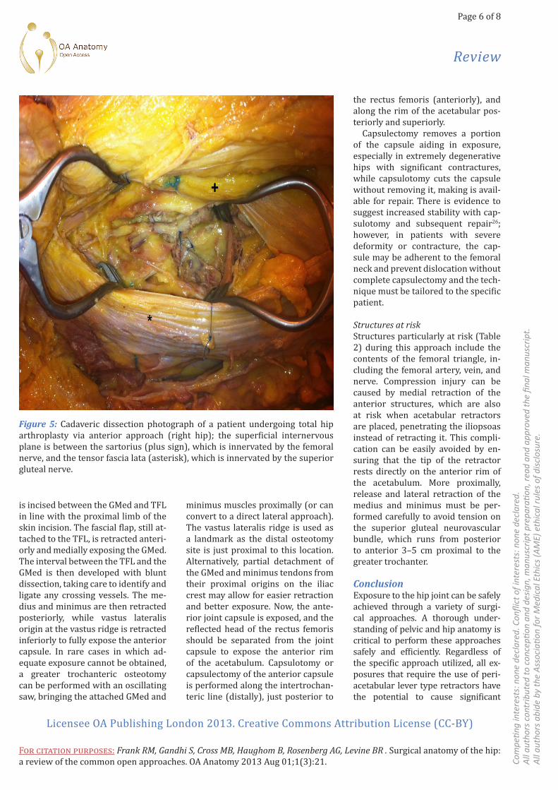
Figure 5: Cadaveric dissection photograph of a patient undergoing total hip arthroplasty via anterior approach (right hip); the superficial internervous plane is between the sartorius (plus sign), which is innervated by the femoral nerve, and the tensor fascia lata (asterisk), which is innervated by the superior gluteal nerve.

Page 7 of 8
Review
Licensee OA Publishing London 2013. Creative Commons Attribution License (CC-BY)
For citation purposes: Frank RM, Gandhi S, Cross MB, Haughom B, Rosenberg AG, Levine BR . Surgical anatomy of the hip: a review of the common open approaches. OA Anatomy 2013 Aug 01;1(3):21. Co
mpe
ting
inte
rest
s: n
one
decl
ared
. Con
flict
of i
nter
ests
: non
e de
clar
ed.
All
auth
ors
cont
ribut
ed to
con
cepti
on a
nd d
esig
n, m
anus
crip
t pre
para
tion,
read
and
app
rove
d th
e fin
al m
anus
crip
t.A
ll au
thor
s ab
ide
by th
e A
ssoc
iatio
n fo
r Med
ical
Eth
ics
(AM
E) e
thic
al ru
les
of d
iscl
osur
e.
5. Witzleb WC, Stephan L, Krummenauer F, Neuke A, Günther KP. Short-term out-come after posterior versus lateral surgi-cal approach for total hip arthroplasty - A randomized clinical trial. Eur J Med Res. 2009 Jun;14(6):256–63.6. Jolles BM, Bogoch ER. Posterior ver-sus lateral surgical approach for total hip arthroplasty in adults with osteoarthri-tis. Cochrane Database Syst Rev. 2006 Jul;(3):CD003828.7. Jolles BM, Bogoch ER. Posterior ver-sus lateral surgical approach for total hip arthroplasty in adults with osteoarthri-tis. Cochrane Database Syst Rev. 2006 Jul;(3):CD003828.8. Zimmerma S, Hawkes WG, Hudson JI, Magaziner J, Hebel JR, Towheed T, et al. Outcomes of surgical management of total HIP replacement in patients aged 65 years and older: cemented versus cementless femoral components and lateral or anterolateral versus posterior anatomical approach. J Orthop Res. 2002 Mar;20(2):182–91.9. Callaghan JJ, Rosenberg AG, Rubash HE. The Adult Hip. Philadelphia: Lippincott Williams & Wilkins; 2007.10. Hoppenfeld S, deBoer P, Buckley R. Surgical Exposures in Orthopaedics: The Anatomic Approach, 4th Edition. Phila-delphia: Lippincott Williams & Wilkins; 2009.11. Moore AT. Metal hip joint; a new self-locking vitallium prosthesis. South Med J. 1952 Nov;45(11):1015–19.12. Bottner F, Pellicci PM. Review: poste-rior soft tissue repair in primary total hip arthroplasty. HSS J. 2006 Feb;2(1):7–11.13. Hummel MT, Malkani AL, Yakkanti MR, Baker DL. Decreased dislocation after revision total hip arthroplasty us-ing larger femoral head size and poste-rior capsular repair. Arthroplasty. 2009 Sep;24(6 Suppl):73–6.14. Masonis JL, Bourne RB. Surgical ap-proach, abductor function, and total hip arthroplasty dislocation. Clinical Orthop Relat Res. 2002 Dec;(405):46–53.15. Mahoney CR, Pellicci PM. Complica-tions in primary total hip arthroplasty: avoidance and management of disloca-tions. Instr Course Lect. 2003;52:247–55.16. Wang L, Trousdale RT, Ai S, An KN, Dai K, Morrey BF. Dislocation after total hip arthroplasty among patients with devel-opmental dysplasia of the hip. J Arthro-plasty. 2012 May;27(5):764–9.
gluteal nerve; OA, osteoarthritis; PSIS, posterior superior iliac spine; SGN, superior gluteal nerve; THA, to-tal hip arthroplasty
References1. Hoaglund FT, Steinbach LS. Primary os-teoarthritis of the hip: etiology and epi-demiology. J Am Acad Orthop Surg. 2001 Sep–Oct;9(5):320–7.2. Barber TC, Roger DJ, Goodman SB, Schurman DJ. Early outcome of total hip arthroplasty using the direct lateral vs the posterior surgical approach. Ortho-pedics. 1996 Oct;19(10):873–5.3. Della Valle CJ, Dittle E, Moric M, Sporer SM, Buvanendran A. A prospec-tive randomized trial of mini-incision posterior and two-incision total hip ar-throplasty. Clin Orthop Relat Res. 2010 Dec;468(12):3348–54.4. Schleicher I, Haas H, Adams TS, Szalay G, Klein H, Kordelle J. Minimal-invasive posterior approach for total hip arthro-plasty versus standard lateral approach. Acta Orthop Belg. 2011 Aug;77(4):480–7.
injury to structures at the acetabular periphery, either via direct injury or via leverage on ‘hidden’ structures. Careless retractor placement may result in neurovascular compression and ultimately, neurovascular com-promise, which can lead to devastat-ing clinical outcomes. With attention to detail and a fundamental knowl-edge of peri-acetabular anatomy, these complications can usually be avoided. Overall, the open surgical ap-proaches to the hip described above, including the posterior, lateral, anteri-or, and antero-lateral, as well as other less common approaches (i.e. medial approach, Ganz surgical-dislocation approach), are all effective ways to approach the hip joint.
Abbreviations listGMax, gluteus maximus; GMed, glu-teus medius; GMin, gluteus minimus; GT, greater trochanter; IGN, inferior
Figure 6: Cadaveric dissection photograph of a patient undergoing total hip arthroplasty via antero-lateral approach (right hip). A) This approach has no true internervous plane, but does utilize the intermuscular plane between the gluteus medius (asterisk) and the tensor fascia lata (plus sign), both of which are innervated by the superior gluteal nerve. B) Deep dissection reveals the femoral head (arrow head); the gluteus medius (asterisk) is seen as it is retracted anteriorly.

Page 8 of 8
Review
Licensee OA Publishing London 2013. Creative Commons Attribution License (CC-BY)
For citation purposes: Frank RM, Gandhi S, Cross MB, Haughom B, Rosenberg AG, Levine BR . Surgical anatomy of the hip: a review of the common open approaches. OA Anatomy 2013 Aug 01;1(3):21. Co
mpe
ting
inte
rest
s: n
one
decl
ared
. Con
flict
of i
nter
ests
: non
e de
clar
ed.
All
auth
ors
cont
ribut
ed to
con
cepti
on a
nd d
esig
n, m
anus
crip
t pre
para
tion,
read
and
app
rove
d th
e fin
al m
anus
crip
t.A
ll au
thor
s ab
ide
by th
e A
ssoc
iatio
n fo
r Med
ical
Eth
ics
(AM
E) e
thic
al ru
les
of d
iscl
osur
e.
the fracture table. Curr Rev Musculoskel-et Med. 2011 Sep;4(3):139–45.24. Harris WH. A new lateral approach to the hip joint. J Bone Joint Surg Am. 1967 Jul;49(5):891–8.25. Charnley J. Low friction arthroplasty of the hip: theory and practice. New York: Springer-Verlag; 1979.26. Goldstein WM, Gleason TF, Kopplin M, Branson JJ. Prevalence of dislocation af-ter total hip arthroplasty through a pos-terolateral approach with partial capsu-lotomy and capsulorrhaphy. J Bone Joint Surg Am. 2001;83-A Suppl 2(Pt 1):2–7.
19. Hardinge K. The direct lateral approach to the hip. J Bone Joint Surg Br. 1982;64(1):17–9.20. Smith-Peterson M. A new supra-ar-ticular subperiosteal approach to the hip joint. Am J Orthop Surg. 1917;15.21. Smith-Peterson MN. Approach to and exposure of the hip joint for mold arthro-plasty. J Bone Joint Surg Am. 1949 Jan;31 A(1):40-6.22. Sculco TP. Anterior approach in THA improves outcomes: opposes. Orthope-dics. 2011 Sep;34(9):e459–61.23. Horne PH, Olson SA. Direct anterior approach for total hip arthroplasty using
17. Jameson SS, Lees D, James P, Serra-no-Pedraza I, Partington PF, Muller SD, et al. Lower rates of dislocation with increased femoral head size after pri-mary total hip replacement: a five-year analysis of NHS patients in England. J Bone Joint Surg Br. 2011 Jul;93(7): 876–80.18. Lombardi AV Jr, Skeels MD, Berend KR, Adams JB, Franchi OJ. Do large heads enhance stability and restore native anat-omy in primary total hip arthroplasty? Clin Orthop Relat Res. 2011 Jun;469(6): 1547–53.