clinical and experimental insights into the use ... - hlr.nu¥ngvarig hlr med mekaniska...
TRANSCRIPT

1
Clinical and experimental insights into the use of mechanical chest
compressions during prolonged resuscitation in the coronary
catheterization laboratory
Henrik Wagner
DOCTORAL DISSERTATION by due permission of the Faculty of Medicine, Lund University, Sweden.
To be defended in Auditorium 5, Centralblocket, Skåne University Hospital, Lund, 17 April 2015 at 13.00.
Faculty Opponent
Professor Jan Erik Nordrehaug, Klinisk Institut II, Haukelands University Hospital, Bergen, Norway.

2
Organization
LUND UNIVERSITY
Document name
DOCTORAL DISSERTATION
Department of Cardiology, Clinical Sciences, Lund, Faculty of Medicine, Lund University, Sweden.
Date of issue: 17 April 2015
Author Henrik Wagner Sponsoring organization
Title and subtitle: Clinical and experimental insights into the use of mechanical chest compressions during prolonged resuscitation in the coronary catheterization laboratory
INTRODUCTION. Prolonged cardiopulmonary resuscitation (CPR) with manual chest compressions (CC) during simultaneous percutaneous coronary intervention (PCI) is exceedingly difficult, with high mortality rates. The use of a mechanical CC (MCC) device can overcome the ordeal of manual CC. The aims of this thesis were to investigate the impact of the introduction of the LUCAS™ MCC device in the cath-lab (Papers I and II); to develop a structured approach in advanced CPR during simultaneous PCI (Paper III); to study myocardial perfusion and blood flow during MCC with and without EPI (Papers IV and V).
MATERIAL and METHODS. A retrospective analysis (5 years) and a prospective follow up study (4 years) with patients treated with MCC during simultaneous PCI were performed. Circumstances leading to the cardiac arrest, and patient and PCI outcomes were investigated (Papers I and II). A structured physiology-guided CPR approach during simultaneous PCI was developed (Paper III). In both animal studies (Papers IV and V) circulation was maintained with MCC during ventricular fibrillation. Coronary blood flow (APV) and coronary perfusion pressure (CPP) were analysed (Papers IV and V), with the addition of amplitude spectrum area (AMSA) in Paper V. The animals in Paper V were randomised to four injections of EPI or saline (control) during the MCC period.
RESULTS. Forty-three patients were included in Paper I and 32 patients in Paper II. Twenty-five percent were discharged from hospital in good neurological condition in each study. Seventy-six percent (Paper I) and 81% (Paper II) were successfully treated with PCI. In Paper III, the development of a structured physiology-guided CPR approach in the cath-lab led to better CPR teamwork during the CPR effort. Coronary artery APV was good throughout the MCC period with a good correlation to CPP (Paper IV). In Paper V, epinephrine significantly increased CPP in 3/4 injections; APV was increased only after the first injection, and no increase was seen in AMSA.
CONCLUSIONS. The use of MCC during prolonged CPR has been shown to be feasible, safe, with good PCI results, and can save lives. Mechanical chest compressions can maintain normal coronary blood flow in the experimental laboratory. Epinephrine decreases myocardial circulation despite increased CPP.
Key words: Cardiac arrest, mechanical chest compressions, PCI, survival, coronary artery blood flow, epinephrine
Classification system and/or index terms (if any)
Supplementary bibliographical information Language: English
ISSN and key title: 1652-8220 ISBN: 978-91-7619-114-9
Recipient’s notes Number of pages Price
Security classification
I, the undersigned, being the copyright owner of the abstract of the above-mentioned dissertation, hereby grant to all reference sources
permission to publish and disseminate the abstract of the above-mentioned dissertation.
Signature Date

3
Clinical and experimental insights into the use of mechanical chest
compressions during prolonged resuscitation in the coronary
catheterization laboratory
Henrik Wagner, MD Department of Cardiology Lund University, Sweden

4
Copyright Henrik Wagner
Department of Clinical Sciences Lund
Cardiology
Faculty of Medicine
Lund University
Sweden
Lund University, Faculty of Medicine Doctoral Dissertation Series 2015:35
ISBN 978-91-7619-114-9
ISSN 1652-8220
Printed in Sweden by Media-Tryck, Lund University
Lund 2015

5
To my wife Sophia, our son Christopher, my children Hannah and Max

6
Contents
Abstract 9 Papers 11 List of abbreviations 13 Introduction 15
Background 16 Treatment of acute myocardial infarction 16 Principles of PCI 17 Cardiogenic shock (CS) 17
Cardiac arrest 17 Cardiac arrest (CA) 17 Training CPR 18 Quality of CPR 18 Mechanical CPR 18 Cardiac arrest in the coronary catheterization laboratory 19 Mechanical CPR in the cath-lab 20 PCI during mechanical chest compressions 21 Cerebral performance category (CPC) 22
Physiology 22 Arterial blood pressure (ABP) 22 Central venous blood pressure (CVP) 23 Coronary perfusion pressure (CPP) 23 Blood flow velocity in the coronary arteries 24 TIMI flow 26 Blood gases in normal circulation and during CPR 27 Cerebral oximetry (SctO2) 27 Pulsewave oxygenation (SpO2) 28 End tidal carbon dioxide (ETCO2) 29 Amplitude spectrum area (AMSA) 30 Epinephrine (EPI) 31
Aim of the study 33 Material and Methods 35
Patient selection in Papers I and II 35 Chest compressions 36
Patient selection and methods in Paper III 36 Animal studies 37

7
Animal preparation 37 Methods 37 Experimental Protocol (Paper V) 41
Measurements 43 Statistical methods 45 Ethical considerations 46
Results 47 Paper I 47 Paper II 51 Paper III 56
Specific recommendations 59 Paper IV 64
ROSC 67 Paper V 67
CPP 69 APV 70 AMSA 72 Time to maximum peak of CPP 77 ROSC 78
Discussion 79 Physiological parameters in cardiopulmonary resuscitation 80 Coronary artery blood flow velocity and coronary perfusion pressure 81 Epinephrine in cardiopulmonary resuscitation 81 Treatment perspective of delayed defibrillation and physiology-guided resuscitation 83
Future perspectives 83 Conclusions 85 Populärvetenskaplig sammanfattning 87
Bakgrund 87 Metoder 88 Resultat 88 Slutsatser 89
Acknowledgements 91 Sources of support 93 References 95 Appendix 109
Original papers I – V 109

8

9
Abstract
INTRODUCTION. Cardiac arrest (CA) in the coronary catheterization laboratory (cath-lab) is not uncommon and in most cases is solved with defibrillation or with a shorter period of chest compressions (CC). However, prolonged, high quality cardiopulmonary resuscitation (CPR) with manual CC during simultaneous percutaneous coronary intervention (PCI) is exceedingly difficult to perform and mortality rates are high. In 2003 the LUCAS™ mechanical CC (MCC) device was introduced and was brought to the cath-lab. Vital parameters are routinely monitored in the cath-lab. However, in CPR efforts during simultaneous PCI, little notice was taken of those vital parameters and the situations were often characterized by unstructured CPR teamwork with suboptimal CPR efforts. When CA occurred, patients’ circulation was maintained with MCC during the simultaneous coronary angiogram, and it was noticed that the coronary artery blood flow was visually almost normal in several cases. On the contrary, when epinephrine (EPI) was administered according to current CPR guidelines, the angiographic visualization and the assessment of the coronary anatomy were impaired. Moreover, several human studies have showed that EPI might even be harmful. Therefore, the aims of this thesis were to investigate the impact of the introduction of the LUCAS™ MCC system in the cath-lab during simultaneous coronary/cardiac intervention (Papers I and II); to organize and optimize the advanced CPR effort during simultaneous PCI (Paper III); to study coronary artery blood flow correlated to coronary perfusion pressure (CPP) (Paper IV) and to study the impact of repeated administrations of EPI on coronary artery blood flow, CPP and bioelectrical activity, measured by amplitude spectrum area (AMSA) (Paper V).
MATERIAL and METHODS. A retrospective analysis was performed consisting of patients who suffered CA in the cath-lab at the Skåne University Hospital, Lund, Sweden, and who were in the need of prolonged CPR with MCC during simultaneous PCI between 2004 and 2008. A prospective follow up study with the same inclusion criteria was performed between 2009 and 2013. Circumstances leading to the CA, different resuscitation parameters, and patient and PCI outcomes were investigated. The survival rate six months after hospital discharge was evaluated in a mixture consisting of the survivors from Papers I and II. For comparison, a group of patients suffering CA in the cath-lab, who required prolonged CPR with manual CC, were evaluated. A detailed educational program took place, based on the experience of the retrospective analysis and ten consecutive patients in the prospective study, literature studies of vital resuscitation parameters and CPR teamwork. A structured, physiology-guided CPR team approach during simultaneous PCI was developed (Paper III). In both animal studies (Papers IV and V), VF was induced and left untreated for one

10
minute. Circulation was maintained with MCC for 10 (Paper IV) and 15 minutes (Paper V). Measurements of coronary blood flow in the left anterior descending coronary artery (LAD) were made at baseline and during VF with a catheter-based Doppler flow wire measuring average peak velocity (APV), and CPP was calculated over the same period (Paper IV). In the second animal study (Paper V), pigs were randomised 1:1:1 to EPI 0.02 mg/kg/dose, EPI 0.03 mg/kg/dose or saline (control). Four EPI/saline-injections were administered, and the effects on CPP, APV and AMSA were recorded. Comparisons were made between the controls and the two EPI groups, a combination of the two EPI groups, and EPI-all.
RESULTS. Forty-three patients were included in Paper I (33 patients with ST-elevation myocardial infarction (STEMI), seven non-STEMI, two planned PCI and one referred for pericardiocentesis). Seventy-six percent were successfully treated with PCI. Eleven patients (25%) were discharged from hospital in good neurological condition. In Paper II, 32 patients were included: 24 STEMI, four non-STEMI, two planned PCI, one angiogram and one intra-aortic counter pulsation balloon pump insertion. Eighty-four percent were successfully treated with PCI. Eight patients (25%) were discharged alive from hospital in good neurological condition. Survival in the merged group after six months was 84%. Ten patients were included in the group treated with manual CC, with one survivor. In Paper III, improved personnel education and the development of a structured, physiology-guided advanced CPR approach led to better teamwork with critical evaluation of vital parameters during the CPR effort. Coronary artery APV (Paper IV) was higher when circulation was maintained by MCC compared to normal circulation. There was a good correlation between APV and CPP. In Paper V, compared to the control group, maximum peak of CPP (Pmax) after injections one and two was significantly increased in the EPI-all group; after injections two and three in the EPI 0.02 group and after injection one in the EPI 0.03 group. Coronary artery APV increased only after the first injection in both the EPI-all and the EPI 0.03 group compared to the control group. No increase of AMSA was seen after any injection of EPI. Seven out of 12 animals (58%) in each EPI group versus 10 out of 12 (83%) achieved spontaneous circulation after defibrillation.
CONCLUSIONS. The use of the LUCAS™ MCC device in the cath-lab during CPR in conjunction with cardiac /coronary intervention has proven to be feasible, safe to use, without impairment of the PCI result and can save lives. The visualized normal coronary artery blood flow seen in CA patients when circulation was maintained by MCC during a simultaneous coronary angiogram was objectively confirmed in the first animal study. Thus the coronary artery blood flow was significantly increased during MCC compared to normal circulation and the correlation between APV and CPP was good. Epinephrine, when administered according to current CPR guidelines, only increases coronary artery blood flow after the first out of four injections and did not improve AMSA despite increased CPP.
11
Papers
This thesis is based on the following papers, referred to in the text by their Roman numerals:
I. Cardiac arrest in the catheterization laboratory: A 5-year experience of using mechanical chest compressions to facilitate PCI during prolonged resuscitation efforts. Resuscitation 2010, 81:4. 383 - 387
II. Mechanical Chest Compressions in the Coronary Catheterization Laboratory to Facilitate Coronary Intervention and Survival in Patients Requiring Prolonged Resuscitation Efforts. Manuscript. Submitted American Heart Journal
III. A Structured Approach for Treatment of Prolonged Cardiac Arrest Cases in the Coronary Catheterization Laboratory using Mechanical Chest Compressions. Int J Cardiovasc Res 2013, 2:4.
IV. Evaluation of Coronary Blood Flow Velocity during Cardiac Arrest with Circulation Maintained through Mechanical Chest Compressions in a Porcine Model. BMC Cardiovascular Disorders 2011, 11:73
V. Repeated epinephrine doses during prolonged cardiopulmonary resuscitation have limited effects on myocardial blood flow: a randomized porcine study. BMC Cardiovascular Disorders 2014, 14:199

12

13
List of abbreviations
ABP Arterial blood pressure (mmHg)
AMI Acute myocardial infarction
AMSA Amplitude spectrum area (mV·Hz)
APV Average peak velocity (cm/s)
BP Blood pressure
CA Cardiac arrest
Cath-lab Coronary catheterization laboratory
CC Chest compression
CO2 Carbon dioxide
CPC Cerebral performance category
CPP Coronary perfusion pressure (mmHg)
CPR Cardiopulmonary resuscitation
CS Cardiogenic shock
CVP Central venous pressure (mmHg)
ECG Electrocardiography
ETCO2 End tidal carbon dioxide (kPa)
EPI Epinephrine
kPa Kilopascal
MCC Mechanical chest compression
MI Myocardial infarction
mmHg Millimetre mercury
mmol/l Millimol per litre
PCI Percutaneous coronary intervention
PEA Pulseless electrical activity
pH Hydrogen ion activity

14
SctO2 Cerebral oximetry (%)
SpO2 Pulse wave oxygenation (%)
TIMI flow Thrombolysis in myocardial infarction flow
VF Ventricular fibrillation
VT Ventricular tachycardia
15
Introduction
Modern treatment of coronary artery disease such as an AMI, consists primarily of mechanical restoration of the blood flow of the occluded coronary artery by PCI. Occasionally the situation is complicated by a sudden CA. Ideally the situation is solved with a short period of CC or a defibrillation with a limited interruption of the PCI. Cardiac arrest due to an AMI in the cath-lab is not uncommon, approximately 1.3% in a study from the late 1990s [1] and in overall CA during any procedure, the incidence is 0.22% [2]. According to the Swedish CPR register, the survival rate for CA victims in the cath-lab is 65% [3], but many of these CA cases are thought to be VF or pulseless VT, treated with either a single or a few defibrillation attempts or a shorter period of CC. However, when this initial CPR treatment fails, and turns in to a prolonged advanced CPR treatment with manual CC, mortality rates are high [4]. There are several contributory factors. Firstly, to perform good manual CC with the cath-lab table extended and elevated is exceedingly difficult. Secondly, the extended table causes a trampoline effect, and thirdly, the fluorescence tubes block the area for the CC provider. To perform high quality CC it is necessary to retract and lower the table and retract the fluorescence tubes and these actions interrupt the potentially lifesaving intervention. With the advent of the LUCAS™ MCC device in 2003, which performs CC according to current guidelines [5], it became possible to treat patients with refractory CA not responding to normal advanced CPR treatment, and to continue with the PCI during MCC without interruptions [6, 7]. We therefore developed a retrospective registry covering the period 1 January 2004 - 31 December 2008 to study the incidence and outcomes of prolonged CA in the cath-lab where patients needed prolonged advanced CPR using MCC during simultaneous PCI (Paper I). A prospective follow-up study (9 April 2009 – 9 April 2013) was performed to re-evaluate outcomes, six month survival and PCI results, among patients who suffered a CA in the cath-lab and needed prolonged A-CPR including MCC during simultaneous PCI (Manuscript Paper II). Initially, the CPR situation using MCC in the cath-lab during simultaneous PCI was notable for the lack of a structured approach concerning teamwork, familiarity with the cath-lab environment, knowledge of the monitoring possibilities of vital physiological parameters such as ABP, ETCO2 and SpO2 and their value for predicting ROSC. Therefore, a protocol was developed describing a physiological approach in CA situations with prolonged advanced CPR including MCC and simultaneous PCI (Paper III). When assessing flow in coronary arteries during coronary angiogram and PCI, the visual TIMI flow scale is often used, where TIMI-0 means no flow and TIMI-III flow represents normal flow [8]. During interventions in the CA patients whose circulation was maintained with MCC, it was

16
observed that the LUCAS™ device could perform a visual TIMI-III flow in the coronary arteries [9], (Paper I). But to translate this indirect information to actual measurements in humans with CA treated with MCC is extremely difficult. We therefore conducted an animal CA study where the relationship between coronary artery blood flow velocity was correlated to CPP during prolonged CPR with MCC (Paper IV). In advanced CPR guidelines the use of EPI has a Class IIb (level of evidence C) immediately if a non-shockable rhythm (asystole or PEA) is present and after four minutes after first defibrillation if there is a shockable rhythm (VF or pulseless VT), and thereafter every fourth minute [5, 10]. However, a CA in the cath-lab is instantaneously attended and the use of EPI according to current guidelines can be questioned. Based upon the knowledge that the LUCAS™ device produces sufficient values of CPP [11-13], (Paper IV), cerebral cortical blood flow [14] and TIMI flow [9], (Paper I) the use of EPI according to current guidelines seems redundant. Further, when EPI was administered to patients with prolonged CA in the cath-lab, there was indeed a significant peak in blood pressure but a clearly visible constriction of the coronary arteries which blurred any assessment of them. Epinephrine also impairs cardiac output measured by ETCO2 [15]. Furthermore, there is evidence of a positive relationship between cumulative dose of EPI administered and a worse neurological outcome [16, 17]. Randomised out-of-hospital CA trials do not support the use of EPI in terms of increased survival at discharge from hospital [18, 19]. With this knowledge it was necessary to conduct a study where the use of guideline administered EPI in the setting of a CA in the cath-lab was evaluated.
Background
Treatment of acute myocardial infarction
An MI is typically caused by a plaque rupture in the coronary artery where a thrombus formation of platelets, red and white blood cells and fibrin causes a coronary artery occlusion which leads to an interruption of blood to the affected myocardium. Originally, treatment consisted mainly of symptomatic care with strict bed rest with careful mobilization after several weeks [20]. Later the use of fibrinolytic agents was introduced and gave the attending cardiologist an effective treatment option which dramatically reduced mortality and morbidity [21]. Despite the good results, there were still a large number of patients where the fibrinolytic result was unsuccessful.
With the development of peripheral percutaneous artery interventions in the 1960s, Dr. Andreas Gruentzig performed the first PCI on coronary arteries in conscious human patients in 1977 [22]. During the 1980s the mechanical restoration of coronary blood flow in patients with AMI using this technique was further developed by Meier

17
and Hartzler [23, 24] and studies showed that this technique was superior to fibrinolytic therapy [25]. The evolution of treatment with this technique took major steps forward, with increasing numbers of patients with AMI being brought to the cath-lab, when studies could show superior results even outside high expertise centres, and also when patients had to be transferred from a hospital without PCI-facilities to a PCI centre [26, 27].
Principles of PCI
An introducer sheath is inserted in either the radial or the femoral artery. A guide wire is inserted through the sheath up to the aortic valve, upon which a catheter is advanced to the ostium of the coronary artery (left or right ostium). To visualize the coronary arteries, contrast is injected though the catheter during simultaneous fluoroscopy. A thin metal wire is inserted into the artery through the catheter, passing the occlusion. A folded balloon or a stent is placed on the wire and introduced into the artery and then inflated at the site of the occlusion. In order to avoid acute thrombus formation and new cardiac events, anti-thrombotic therapy is administered prior to, during and after the intervention [28]. Normally, these interventions restore blood flow in more than 90% of patients when treated with primary PCI for an AMI [29].
Cardiogenic shock (CS)
Depending on the size of the affected area of the myocardium, typically caused by a proximal occlusion in the LAD or in the LM, the damage in the acute phase can cause a CS in five to eight per cent of patients with AMI [30] and is associated with high mortality rates which have been estimated between 60.9% and 65.4% [31, 32]. The condition is caused by decreased cardiac output, systemic perfusion and tissue hypoxia despite the presence of sufficient intravascular volume leading to the heart’s inability to adequately perfuse the tissues.
Cardiac arrest
Cardiac arrest (CA)
One of the most feared complications in an AMI is the development of a CA. This can happen in the acute phase, prior to or during the intervention, as well in the later phase. A CA can also be induced by iatrogenic causes, for example wedging of the catheter in the coronary ostium, coronary artery dissection or a rupture induced by the inflation of a balloon or stent which can cause an acute pericardial tamponade. The rhythm when the CA occurs can be either pulseless VT or VF, asystole or PEA. If a CA occurs,

18
the most important treatment is a rapid start of effective and accurate CC to sustain circulation to the brain and heart, and to perform early defibrillation when the rhythm of the CA is VF or pulseless VT and then to treat the cause of the CA. The first known defibrillation was performed on humans in early 1948 [33] and treatment with external CC and ventilation as we know it today was first developed by Kouwenhoven and Knickerbocker in 1960 [34].
Training CPR
During CPR treatment, the outcome is not only dependent on the cause of the CA and the patient´s co-morbidity. Cohesive teamwork by the medical emergency team [35, 36], technical skills and the presence of an unequivocal team leader [35-38] are also important. But leadership practice is just as important. [39]. However, a CA situation in the cath-lab is very different from a CA in an ordinary ward. Hence it is important to practice CPR in the cath-lab, both for familiarity with the environment and for teamwork training in tandem with the cath-lab personnel. Another advantage of training in the cath-lab is the opportunity to learn the advanced technologies needed to succeed during such stressful emergent circumstances.
Quality of CPR
Current CPR guidelines presented in 2010 recommend CC at 50 - 60 mm depth with a frequency of 100 – 120 per minute. Ventilation rates are two ventilations every 30 CC cycle, and when intubated, ten inflations during continuous CC [5]. Even though the provider performs excellent CC, the quality of both depth and frequency will be reduced during extended CPR due to rescuer fatigue, and impaired circulation is a consequence [40]. Further, frequent or long-lasting pauses in manual CC are associated with a worse outcome [41-43].
Mechanical CPR
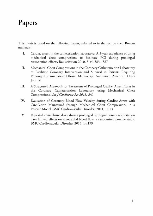
In an effort to overcome the shortcomings of reduced depth and frequency, and also in the hope of a better outcome, the development of MCC devices made its contribution in the early 1960s [44]. The LUCAS™ device (Physio-Control Inc./Jolife AB Sweden), which was introduced in 2002, performs CC and active decompressions with a compression depth of 53±2 mm at a rate of 102 CCs per minute in a 50/50 ratio (Figure 1a). In animal studies MCC with the LUCAS™ device has been proven to create higher cerebral cortical blood flow [14] and CPP compared to manual CC [11, 45]. The other commercial devices available on the market include the band-loading distribution device Autopulse (Zoll Medical, Chelmsford, WA) which came onto the market in 2004 (Figure 1b). In 2003 the Skåne Regional Council placed the LUCAS™ device in all the county’s ambulances. Initially, concerns were raised about increased
19
injuries due to MCC [46, 47], but this has not been seen in other autopsy studies [48, 49]. A large randomised multi-centre study of out-of-hospital CA patients showed that MCC with the LUCAS™ device was as good as manual CC in terms of survival and there was no difference in injuries in either group [50]. The same result was also seen in a randomised out-of-hospital CA study with the Autopulse device in an adjusted cohort [51].
Figure 1 Left (a): The electrical driven LUCAS™ device (Jolife AB, Lund, Sweden). Right (b), Autopulse (Zoll Medical, Chelmsford, WA, USA).
Cardiac arrest in the coronary catheterization laboratory
One consequence of the movement of patients with AMI into the cath-lab for primary PCI due to new treatment results [27, 52, 53] was an increasing amount of CA in the cath-lab. Ideally the situation is solved with a defibrillation or a short period of CC. It is quite the reverse when initial CPR efforts fail and the situation turns into a prolonged CPR operation: the demands are suddenly different. To perform high quality manual CC when the patient is on the cath-lab table is exceedingly difficult. Firstly, when the table is elevated the force from the CC provider is not optimal. Secondly, when the table is fully extended the CCs will be substandard due to the trampoline effect. Thirdly, the CC provider is exposed to an unacceptably high amount of radiation and also hampers the view of the coronary arteries for the interventionist. In order to overcome these obstacles, it is necessary to lower and retract the table, at the cost of interrupting the potentially lifesaving intervention. (Figure 2)

20
Figure 2 shows the difficulties with manual CC on the cath-lab table. Note the retracted and lowered table, the retracted fluoroscopyfluoroscopy tubes, the angle of the arms of the chest compression provider which reduces force to the chest. (Arranged picture courtesy of Jolife AB.)
Mechanical CPR in the cath-lab
The LUCAS™ device was introduced into the cath-lab in 2004 and this led to several advantages. First of all, when a patient suffered CA during a PCI and the situation was not solved in a few minutes, the use of MCC made it possible to continue the intervention without interruption of CCs. Secondly, the MCC device delivers CCs according to current guidelines without interruptions. Thirdly, the design of the LUCAS™ device, where most parts are radio-lucent (Figure 3), allows the interventionist to continue the intervention and use the fluoroscopy angels normally used with only small adjustments (Figure 4). Several case reports and case series have proven the feasibility of this approach [6, 7, 9].

21
Figure 3 The LUCAS™ device put into a fluoroscopy field. Note the radio-lucent parts. The back plate is specially designed and manufactured in carbon fibre for use in cath-labs for optimal sight during fluoroscopy.
PCI during mechanical chest compressions
PCI is normally performed on a beating heart with the patient conscious. However, when the patient suffers a CA, it is essential to perform high quality CC without interruptions, ideally with the LUCAS™ device. To wire an occluded vessel during MCC is more difficult than with the patient consciousand there are minor limitations in views during MCC. To obtain exact deployment of a stent, it is often necessary to stop the MCC device for a few seconds and then start again during or immediately after balloon/stent inflation.
Figure 4 Left (a) shows the fluoroscopyfluoroscopy tube in left anterior cranial oblique position during MCC. Right (b) shows the fluoroscopyfluoroscopy tube in the left anterior caudal oblique position (spider view). Arranged picture, courtesy of Jolife AB.
22
Cerebral performance category (CPC)
The cerebral performance category scale (CPC) describes the patient’s neurological status in five categories and was adopted from the Glasgow outcome coma scale [54]. This scale can be used in the assessment of neurological function in patients who have survived a CA.
CPC 1: Good cerebral performance (normal life). Able to work and lead a normal life. May have minor psychological or neurological deficits (mild dysphasia, non-incapacitating hemiparesis, or minor cranial nerve abnormalities)
CPC 2: Moderate cerebral disability (disabled but independent). Conscious. Sufficient cerebral function for part-time work in a sheltered environment or independent daily-life activities (dressing, travelling by public transportation, food preparation). May have hemiplegia seizures, ataxia, dysarthria, dysphasia or permanent mental or memory changes.
CPC 3: Severe cerebral disability. (Conscious but disabled and dependent). Conscious but dependent on others for daily support (in an institution or at home with exceptional family effort). Has at least limited cognition. This category includes a wide range of cerebral abnormalities from patients who are ambulatory but have severe memory disturbances or dementia precluding independent existence, to those who are paralyzed and can communicate only with their eyes as in the locked-in syndrome.
CPC 4: Coma vegetative state (unconscious). Unconscious, unaware of surroundings, no cognition. No verbal or psychological interaction with environment.
CPC 5: Brain dead. Certified brain dead or dead by traditional criteria.
Physiology
Arterial blood pressure (ABP)
Normal systolic ABP values range from 100 – 130 mmHg with 60 – 90 mmHg for diastolic values. In a CA situation, ABP falls dramatically. In CPR situations the level of systolic ABP depends on the CC quality, e.g. rate, depth and minimized interruptions [55]. Currently there are no recommendations of the optimal level of systolic ABP that can predict ROSC. However, in some animal studies, values of 70 – 80 mmHg in systolic ABP and diastolic values > 25 – 40 mmHg have been proposed [11, 56, 57]. Figures 5 and 6 show the intra-aortic BP in a patient who is suffering a CA. The cath-lab personnel initiate manual CC (Figure 5). The situation is not solved in due time, and thus the LUCAS™ device is deployed and started, giving a more stable compression depth and rate leading to a consistent and better ABP (Figure 6).

23
Figure 5 shows the development of intra-aortic blood pressure during 44 seconds of manual chest compressions in a patient suffering cardiac arrest on the cath-lab table. Note the narrow spikes, the inconcistency in the systolic values, and the interruptions for ventilation. mmHg = millimetre mercury.
Figure 6 shows the development of intra-aortic blood pressure during 16 seconds after initiation of mechanical chest compressions in a patient suffering cardiac arrest on the cath-lab table. mmHg = millimetre mercury.
Central venous blood pressure (CVP)
In normal physiology the mean CVP measured in the right atrium is generally low, between 0 – 8 mmHg. However, in a CA situation CVP is dramatically elevated because of the no flow situation where the right atrium and right ventricle are filled with returning blood and eventually distended [58, 59].
Coronary perfusion pressure (CPP)
Coronary perfusion pressure is thought to represent the perfusion pressure in the myocardium. The calculated value is the pressure difference between the aortic end diastolic pressure and the right atrial end diastolic pressure (a – b = c) (Figure 7) [60].
Arterial Pressure
10
20
30
40
50
60
70
80
90
[44 sec]
[mmHg]
Arterial pressure
0
20
40
60
80
100
120
[16 sec]
[mmHg]

24
Closed chest animal CA models are frequently used in CPR studies and the methods for calculating CPP in these studies are well established [45, 61, 62]. Studies in humans and in animals have demonstrated a positive correlation between CPP and ROSC [11, 45, 62-64]. In humans the lower cut-off threshold for the possibility of attaining ROSC following a defibrillation is 15 mmHg, as shown by Paradis and co-workers [63]. Recently one study has suggested that it is the total dose of perfusion over time which may be more important than a threshold in CPP for the possibility of attaining ROSC [64].
Figure 7 shows the intra-aortic blood pressure curve (red curve) and the right atrial blood pressure (blue curve) during MCC in a patient suffering CA on the cath-lab table. Arrows indicate (a) arterial and (b) right atrial end diastolic values. The clammer represents the difference between the intra-aortic diastolic value and the right atrial end diastolic value. (c) = coronary perfusion pressure (CPP), which is 32 mmHg in this example.
Blood flow velocity in the coronary arteries
Measurements of intracoronary blood flow velocity can be performed by using a Doppler flow wire. A Doppler transducer is placed at the tip of a 0.014 inch wire (Figure 8). The appearance of the Doppler curve during normal sinus rhythm and during MCC is shown in Figure 9. The preferred variable is APV which is the averaged value of the instantaneous peak velocity blood flow samples over the last two cardiac cycles in cm/s. This method has previously been used to measure coronary blood flow

25
before and after PCI in patients with stable circulation [65-68] or when correlating other modalities for measuring coronary blood flow [69, 70]. Figure 10 shows the principle of the placement of a Doppler wire in the LAD.
Figure 8 Volcano Doppler FloWire (Volcano Corp., Rancho Cordova, CA, USA)
Figure 9 Left (a) Doppler flow velocity curve in the left descendent coronary artery (LAD) during sinus rhythm. Right (b) Doppler flow velocity curve in the LAD during MCC. Velocity is shown in cm/s.

26
Figure 10 shows a drawing of a guiding catheter with the tip at the left coronary main artery ostium with a Doppler wire in the left anterior descending artery.
TIMI flow
Assessment of TIMI flow is basically a visual estimate of the filling of the coronary arteries with contrast media [8]. It is graded from TIMI 0 (no flow) to TIMI III, normal coronary artery flow. TIMI flow estimation is used routinely when performing a coronary angiogram or PCI. Mechanical chest compressions performed with the LUCAS™ device have been shown to produce a TIMI III flow in coronary arteries in patients who suffer a CA treated with MCC during simultaneous PCI [9, 13, 40] (Paper I). TIMI flow has also been shown to be a surrogate marker for CPP [13].

27
Blood gases in normal circulation and during CPR
In normal circulation, pH is between 7.37 and 7.47 and reflects the relationship between acid and base in the body. In CA victims and during CPR, pH is decreased, reflecting the anaerobic metabolism and accumulation of H+ -ions and hence lower pH.
Cerebral oximetry (SctO2)
Cerebral oximetry (SctO2) measures the mix of oxyhaemoglobin and deoxyhaemoglobin in the regional space of microvasculature - arterioles, venules and capillaries in the brain [71, 72]. Absolute oximetry with fibre optic laser spectroscopy uses four different wavelengths at a depth of 2.5 cm in the brain excluding skin and bone perfusion (FORE-SIGHT, Casmed, Brandford, CT, USA) (Figures 11 and 12). The lower safe threshold for FORE-SIGHT SctO2 values has been estimated at 55% and normal values range from 65 to 70% [73, 74]. Cerebral oximetry is routinely used in vascular surgery, cardiac surgery [74, 75], carotid endarterectomy [76], and in neonatal intensive care [77]. However, information on the use of SctO2 during CA is limited [78-82].
Figure 11 Left (a) Principle of penetration of Near Infrared Spectroscopy (NIRS) and the absorbtion of NIRS from skin and bone obtained at 15 mm from the light souce, and skin, bone and gray matter of the brain at 50 mm from the light source. Right (b) Principle of the placement of the NIRS electrodes on the forehead. (Used with permission from Casmed, WN, USA.)

28
Figure 12 The FORE-SIGHT Cerebral oximetry device (Casmed, WN, USA.) (Used with permission from Casmed, WN, USA.)
Pulsewave oxygenation (SpO2)
Non-invasive SpO2 provides a continuous real-time estimation of the oxygen saturation of haemoglobin within arteries proximal to the precapillary sphincters. This monitoring equipment is widely used in several wards such as coronary care units, intensive care units, emergency rooms, surgery wards, etc. (Figure 13) Normal SpO2 varies between 96% and 99%. In patients with CA, SpO2 is generally low and there are a limited amount of studies using SpO2 in the monitoring of resuscitation attempts in CA. One study using pulse oximetry in CPR found it equivocal [83] while another found it feasible [84].

29
Figure 13 This figure shows the utilization of a pulse oximeter.
End tidal carbon dioxide (ETCO2)
In the lungs the deoxygenated blood exchanges CO2 as a bicarbonate ion which is the by-product of cellular metabolism. The value of exhaled CO2 varies with the physical and chemical demands on the cellular metabolism reflecting changes in pCO2, pH and pO2 with the goal of normalizing these parameters. For many years, the measurement of ETCO2 has been a method used by anaesthesiologists to confirm correct placement of the endotracheal tube [85]. In an animal CA study and in an animal shock study (haemorrhagic, septic and cardiogenic), ETCO2 has been shown to correlate to cardiac output [86, 87]. Cardiac output correlated to ETCO2 has also been studied in humans [88]. Further, in both human and animal studies, ETCO2 has been shown to be a useful predictor of the possibility to obtain ROSC during CPR [89-91]. This finding also led to the ability to assess the quality of CC in terms of the value of ETCO2 [92, 93]. Figure 14 shows examples on the ETCO2 curve.

30
Figure 14 Left (a) End tidal carbon dioxide (ETCO2) curve during normal ventilation. Right (b) ETCO2 curve during mechanical chest compressions. kPa = Kilopascal.
Amplitude spectrum area (AMSA)
AMSA represents the sum of the products of the individual frequencies and the corresponding amplitude (mV·Hz) of the VF wavelets derived from the ECG (Figure 15) [94]. AMSA has proven to be a strong predictor both for successful defibrillation as well as a strong negative predictor for defibrillation failure [94, 95]. Further, one animal study has shown that there is a positive correlation between AMSA and CPP [96] which is a known predictor for the possibility of attaining ROSC following a defibrillation [63]. In humans, several studies show the usefulness of AMSA in predicting successful defibrillation attempts in VF CA victims [97-100].
Figure 15 A representative example of amplitude frequency relationship. The area under the curve defines the amplitude spectrum area, AMSA. (Marn-Pernat et al. Crit Care Med 2001 Vol 29, No 12).

31
Epinephrine (EPI)
Epinephrine is a hormone that is endogenously produced by the adreno-medullary glands and other chromaffin tissue and which acts like a circulating hormone. Epinephrine stimulates α1 and α2, β1 and β2 receptors. Via its α1-adrenergic effect on the arteries and arterioli it causes vasoconstriction in the skin, mucosa, abdominal organs and veins [101]. Further, when stimulating both α1 and α2 adrenoreceptors in the myocardium, predominantly α2adrenoreceptors in the arterioli, it causes vasoconstriction [102]. Epinephrine has been recommended in cardiac arrest CPR situations since 1962, when Redding and Pearson were able to show improved survival in dogs resuscitated from CA using EPI, [103] and ever since that, it has been established in resuscitation guidelines (Class 2b LOE A) [5, 10, 104-106]. Due to the α1 adrenoreceptor vascular effects, EPI has been proven to increase CPP during CPR and as a result of that, it has improved the possibility of attaining ROSC in animal studies [15, 107] as well as in human studies, where more CA victims attain ROSC and are admitted to hospital [18, 19]. On the other hand, when stimulating the β adrenoreceptors, oxygen consumption rises and reduces sub-endocardial perfusion [108]. Furthermore, in animal studies EPI causes constriction in the cortical capillaries in the brain during CPR as well as in the microcirculatory blood flow in gingival mucosa [109, 110]. Epinephrine also impaired ETCO2 and cardiac output in an animal study [15, 108]. Several studies have failed to show an increased survival rate to discharge from hospital in patients who received EPI during the resuscitation effort [18, 19], and in survivors an even worse neurological outcome has been observed in the EPI groups [16, 17]. In one large registry study, Warren et al. concluded that CA victims of in-hospital CA resuscitated with less frequent doses of EPI than recommended had a more favourable outcome [111]. Despite the conflicting data, the use of EPI in current resuscitation guidelines is still recommended [5].

32

33
Aim of the study
The aim of this study was to evaluate the effect of the introduction of the LUCAS™ MCC in the cath-lab in CA situations where the patients required prolonged CPR including MCC during simultaneous cardiac or coronary intervention. In 2003 the LUCAS™ device was introduced into clinical operation without any randomised trial in this particular field. It was therefore important to evaluate feasibility, safety, interventional results during simultaneous MCC, and both short and long term survival in a larger cohort than previously published [6, 7, 9]. Moreover, the CPR situation in the cath-lab was marked to a certain extent by a lack of teamwork structure, a lack of awareness of options for monitoring vital parameters, and limited knowledge of vital parameter physiological cut-off values during CPR. Hence there was a need for a structured approach in CPR situations in the cath-lab during simultaneous PCI. When CA patients had their blood circulated with MCC during a simultaneous coronary angiogram, it was noticed that there was a TIMI-III flow in the coronary arteries of several patients which has also been seen in other studies [9, 13, 40]. Moreover, when EPI was administrated to patients who suffered a CA in the cath-lab and who required prolonged CPR including MCC, it was noticed that repeated injections of EPI gave a significant but short-lasting peak in ABP, constricting coronary arteries and aggravating both the assessment of the anatomical structure and the progress of the intervention. In addition, several studies revealed that EPI could even be harmful [16-19]. Based on these observations and studies, the use of EPI according to current guidelines was questioned. Two exploratory animal CA studies were conducted to study the physiological effect of MCC on APV and CPP and the effects of EPI on CPP, APV and AMSA during MCC. As a result, five papers have been produced describing:
• a retrospective analysis including patients between 1 January 2004 and 31 December 2008 where the frequency and survival rate in patients who were referred to the cath-lab with sustained circulation and who at some point during the intervention suffered a CA, thus requiring prolonged CPR including MCC during simultaneous PCI, were studied (Paper I).
• a second study with prospectively enrolled patients with the same inclusion criteria as those above between 9 April 2009 and 9 April 2013. In this cohort, circumstances leading to CA, resuscitation parameters and outcomes were evaluated. Specifically survival rate at discharge from hospital and 6-month survival in a mixture of the survivors from Paper I and II were assessed. We also investigated duration of MCC in the CA situation (Paper II).

34
• proceedings, teamwork, comprehension and assessment of vital parameters in prolonged CPR with MCC during simultaneous PCI. This was based on the insights and experience obtained from the first study, an analysis of 10 consecutive patients from the prospective study, a case report on five patients suffering CA in the cath-lab where we also measured SctO2 along with studies of articles investigating CPR training and leadership and the impact on survival of examining vital parameters during CPR (Paper III).
• a closed-chest animal CA model where the correlation between CPP and APV in the coronary artery during prolonged CPR with MCC was studied (Paper IV).
• a closed-chest animal CA model where the impact of guideline administered EPI during prolonged CPR with MCC on CPP, APV and AMSA was studied (Paper V).

35
Material and Methods
Patient selection in Papers I and II
Patients recruited for the retrospective analysis and the prospective study were treated at the cath-lab at Skåne University Hospital in Lund, which is a tertiary centre in southern Sweden that performs PCIs 24 hours a day, seven days a week, and serves a population of 1.2 million inhabitants. Patients in the retrospective registry (1 January 2004 - 31 December 2008) were localized through the local hospital CA registry and in the study covering the time period 9 April 2009 – 9 April 2013, patients were prospectively recruited. Among those who suffered a CA in the cath-lab, patients were included if immediate resuscitation efforts (prompt defibrillation/manual CCs) failed and there was consensus among the attending cardiologist, anaesthesiologist and the PCI operator that MCCs were indicated. The reason for referral to the cath-lab for the included patients were elective coronary angiogram, primary PCI in STEMI patients, sub-acute PCI in non-STEMI patients, planned PCI, insertion of an intra-aortic balloon counter pulsation pump or treatment of cardiac tamponade. In the retrospective analysis similar patients (requiring >one minute of manual CC and tracheal intubation) in which no MCC device was used in the same time span, were evaluated. For comparison, patients who suffered CA in the cath-lab and who were treated with manual CC ≥ ten minutes, between 1999 and 2003 (prior to the introduction of MCC devices in the cath-lab) were analysed using the local hospital CA registry. Inclusion criteria were the same as for those treated with MCC. In the prospective study informed consent was obtained either from survivors or from relatives.
Prolonged CPR was defined as an episode of CA necessitating a period of several minutes of manual CC, followed by the use of MCC.
All patient charts and medical files were examined in both studies. Autopsy reports were examined in the retrospective analysis. Analysis of deaths of patients who were discharged alive from hospital was found in the Swedish registry for cause of death.
The patients included in the retrospective analysis were divided into two outcome groups (Group 1: in-hospital death; Group 2: discharged from hospital alive). In the prospective study, patients were divided into four outcome groups (Group 1: whole group; Group 2: expired in the cath-lab; Group 3: discharged from the cath-lab with circulation and Group 4: discharged alive from hospital in good neurological condition).

36
In both studies, patient demography, indication for admission to the cath-lab, culprit lesion and cardiac rhythm at the time of the CA were assessed. In the prospective study, circulatory state at the time of arrival in the cath-lab was assessed. The predefined endpoints in both studies were mortality status on departure from the cath-lab; successful PCI, assessed by TIMI flow, or <50% residual stenosis and discharge from hospital in CPC 1 or 2 representing a good neurological outcome. The MCC time was calculated for the two groups in Paper I and for all our groups in Paper II.
In the prospective study the use of vasoactive drugs was assessed.
The six-month survival rate was analysed in a merged group that consisted of patients discharged from hospital in good neurological condition from Paper I and Paper II.
Chest compressions
In Paper I the LUCAS™ V1 MCC device (European version) was used between 1 January 2004 and 31 December 2007. Between 1 January 2008 and 31 December 2008, the LUCAS™ V2 MCC device (US version) was used. It has the same operating parameters as LUCAS™ V1 except for the decompression force, which is set to a maximum of 13 N.
In Paper II, the LUCAS™ V2 (US version), and later the electrically driven LUCAS™ 2 chest compression system (Physio-Control/Jolife AB, Lund, Sweden) was used.
Patient selection and methods in Paper III
Ten consecutive patients with prolonged CA in the cath-lab were analysed [12] together with five other patients where SctO2 was added to the other vital parameters (ABP, CPP, ETCO2, SpO2) [112]. Several deficiencies in monitoring and teamwork were noticed. Four areas which needed improvement were identified from this data: (1) the understanding of the importance of teamwork and leadership within the unique circumstances of a prolonged CPR effort in the cath-lab. (2) the importance of practical simulations within the cath-lab setting. (3) an understanding of the vital physiological parameters for the successful restoration of spontaneous circulation. (4) familiarity with the advanced technology needed to succeed during such emergent and stressful circumstances. Several studies addressing teamwork [35-37], leadership [35, 39], technical skills [38], vital parameters such as ABP [11, 56, 57], CPP [11, 45, 62, 63, 96], SpO2 [83, 84], ETCO2 [89-91, 113] and SctO2 [78-82, 114, 115] were studied. In addition, the importance of correct positioning over the heart during CC [116-119] and ventilation rate were brought to light [120].

37
Mechanical chest compressions were performed using a LUCAS™ V2 (US version) and a LUCASTM2 device. Parameters of ECG, ABP, CVP, SpO2 and ETCO2 were monitored on an IntelliVue MP90 monitoring system (Philips, Eindhoven, The Netherlands), and SctO2 was monitored using the FORE-SIGHT monitoring system (CAS Medical Systems Inc., Branford, CT, US). TIMI flow in non-occluded vessels was assessed as done routinely during PCI [8]. The hemodynamic parameters were recorded every 2nd to 5th millisecond on an external PC computer using custom made software and evaluated using LabChart 7 (AD Instruments Corp., Colorado Springs, CO, US). Coronary perfusion pressure was calculated as described earlier [60].
Animal studies
Two animal studies were carried out. In both studies Swedish Landrace pigs were used. The pig has a coronary anatomy similar to humans which makes it suitable for coronary cardiac experiments [121].The ideal size of the pig is 20 – 30 kg because at this weight the size of the heart is identical to that of an adult human heart [122]. However, occasionally at this size, the foramen ovale has not been closed, which can cause unexpected results during the experiment, which in turn may lead to exclusion from the study.
Animal preparation
Methods
In Paper IV (n = 11) and Paper V (n = 36), Swedish Landrace pigs were used with a mean weight of 31 ±1.5 kg (range 28 – 31kg) in paper IV and a mean weight of 38 ±4.1 kg (range 32 – 46 kg) in paper V.
Anaesthesia The animals in both studies were fasted over night with free access to water. In Paper IV, the animals were anaesthetized with an induction dose of intramuscular ketamine (30 mg/kg) (Ketamine 100mg/ml, Intervet, Danderyd, Sweden). Sodium thiopental (5-8 mg/kg) (Pentothal 100 mg/ml, Abbott, Stockholm, Sweden) and atropine (0.015mg/kg) were given intravenously before tracheotomy. Anaesthesia and muscular paralysis were maintained with a continuous infusion of 10 ml/h of a 0.9% saline solution containing ketamine (16 mg/ml) and pancuronium (0.6 mg/ml).

38
In Paper V, the animals were pre-medicated with Ketaminol 15 mg/kg (Ketamine 100mg/ml, Intervet, Danderyd, Sweden), and Rompun 0.1ml/kg (Xylazin 20 mg/ml, Bayer AG, Leverkusen, Germany), intramuscularly. The anaesthetic induction was accomplished with thiopental 12.5 mg/kg (Pentothal 100 mg/ml, Abbott, Stockholm, Sweden). The animals were orally intubated with cuffed endotracheal tubes. For maintenance of anaesthesia a slow infusion of 1μg/ml fentanyl (Fentanyl, Pharmalink AB, Stockholm, Sweden) in buffered glucose (25 mg/ml) was started at a rate of 2 ml/min and adjusted as needed. Meprobamat (Mebumal DAK, Copenhagen, Denmark) and thiopental was titrated if needed in small bolus doses.
Ventilator settings In the first animal study (Paper IV), a Boussignac endotracheal tube, 7 mm internal diameter (Laboratoires Pharmaceutiques VYGON, Ecouen, France) was used as an ordinary endotracheal tube for ventilation. After tracheotomy it was connected to a Servo Ventilator 300 (Siemens, Solna, Sweden) using pressure-regulated (max 30 cm H2O = 23 mmHg) and volume-controlled intermittent positive pressure ventilation (IPPV). Normoventilation ETCO2 (around 5.3 kPa = 40 mmHg) was obtained by using a tidal volume of 8 ml/kg body weight, 20 breaths/min, a PEEP of 5 cm H2O (6 mmHg) and a FiO2 of 0.21. End tidal carbon dioxide was measured by CO2SMO Plus Respiratory Profile Monitor Model 8100 (Novametrix Medical Systems Inc., Wallingsford, CT, USA) with a CO2 sensor (REF 6719) connected to the proximal end of the Boussignac tube.
In the second animal study (Paper V), the animals were orally intubated with cuffed 7 mm inner lumen endotracheal tubes. Mechanical ventilation was established with a Siemens-Elema 900B ventilator (Siemens, Solna, Sweden) in the volume-controlled mode, adjusted in order to obtain normoventilation (4.5 – 5.5 kPa). The animals were ventilated with a mixture of nitrous oxide (70%), oxygen (30%) and normal air. End tidal carbon dioxide was monitored by a Circuit ETCO2 Sensor (Philips Medical Systems, Andover, MA, USA) connected to an IntelliVue MP90 monitoring system (Philips, Eindhoven, The Netherlands).
Catheterization of LAD In both animal experiments, a 6 F introducer sheath (Boston Scientific Scimed, Maple Grove, MN, USA) was inserted using the Seldinger technique into the surgically exposed left carotid artery and a 6F JL 3.5 Wiseguide™ (Boston Scientific Scimed, Maple Grove, MN, USA) catheter was then inserted through the introducer with the tip at the ostium of the left main coronary artery. The catheter was used to place a 0.014 inch, 12 MHz pulsed Doppler flow velocity transducer (FloWire® Volcano Inc., San Diego, CA, USA) into the mid-portion of the LAD. Continuous coronary flow velocity profiles were displayed and recorded using the Doppler flow wire connected to a FloMap monitor (Cardio Metrics, Mountain View, CA, USA).

39
General catheterizations and monitoring In the first animal study (Paper IV), two catheters (Secalon-T-over-needle catheter, 16G/1.70/130 mm) for monitoring the aortic pressure and CVP were introduced via direct puncture of the right carotid artery and the right jugular vein respectively, in order to avoid ligation of the artery. The tip of the arterial catheter was inserted into the thoracic aorta and the central venous catheter was placed with the tip in the right atrium. The fluid-filled catheters were connected via short tubes to pressure transducers.
A temperature probe was placed in the oesophagus and ECG was obtained by three electrodes glued to the chest. The following variables were continuously sampled (100 – 500 Hz) to a computer supplied with a data acquisition system (Testpoint, Capital Equipment Corporation, Billerica, Massachusetts, USA): body temperature, ECG, intra thoracic AP, CVP, CPP, APV, ETCO2.
All radiological procedures were performed in an experimental cath-lab (GE Healthcare, Chalfont St Giles, UK).
In the second animal study (Paper V), the pigs were continuously monitored with ECG, ETCO2, SpO2 on an IntelliVue MP90 monitoring system (Philips, Eindhoven, The Netherlands). Unfractioned heparin (10 000 IU) (LEO Pharma AB, Malmoe, Sweden) was given intravenously at the start of the catheterization. A 6 Fr pigtail catheter was placed with the tip in the proximal thoracic aorta for obtaining ABP. A 9 F introducer sheath (Boston Scientific Scimed, Maple Grove, MN, USA) was inserted into the surgically exposed right jugular vein. A 7.5 F Continuous Cardiac Output Pulmonary Artery Catheter™ (Edwards Life Sciences, Irvine, CA, USA) was inserted into a pulmonary artery. Arterial blood pressure and CVP were continuously measured using separate transducers (AD Instruments Inc., Colorado Springs, CO, USA). Arterial BP, CVP and APV were digitally recorded using Chart v4.2 (AD Instruments Inc., Colorado Springs, CO, USA). The procedures were performed in an experimental cath-lab (Shimadzu Corp., Kyoto, Japan).
Induction of VF In the first animal study (Paper IV), VF was induced with a 5-20 mA, 6 Hz and 30 V alternating current delivered to the epicardial surface via a needle electrode. In the second animal study (Paper V), VF induction was executed with a 9 V direct current battery (Duracell, Procter & Gamble, Cincinnati, OH, USA) with one pole connected to a needle inserted into the skin surface and the other pole connected to a needle which was inserted to the epicardial surface with a stimulation time of 5 – 10 seconds. Circulatory arrest was confirmed by an instant loss of ABP, flow velocity in LAD and an ECG showing VF.

40
CPR during the experiments Chest compressions in both experiments were performed with the LUCAS™2 MCC device (Physio-Control Inc./Jolife AB, Sweden).
Defibrillation Defibrillations in both animal studies were performed with a Lifepack™ 12 (Physio-Control, Redmond, WA, USA).
Blood gas analysis Blood gas, haemoglobin and electrolytes were analysed directly after a sample had been obtained, using a blood gas analyser (ABL 505, Radiometer, Copenhagen Brönshöj, Denmark).
Experimental protocol (Paper IV) A flow chart of the experiment is presented in Figure 16. At baseline when all parameters were stable, VF was induced, upon which ventilation was stopped. Following 60 seconds of VF, CPR was started using the LUCASTM2 and ventilation was initiated at a rate of ten manual inflations per minute. Continuous measurements of ECG, body temperature, ABP, CVP and CPP were performed. APV was documented by both a VHS recorder and by digital recording. After ten minutes of CPR the pigs were defibrillated. If ROSC was not obtained after the first defibrillation, EPI 0.01 mg/kg was given in the central venous catheter and another defibrillation attempt was made after two minutes, if VF persisted. Repeated doses of EPI and defibrillation attempts were performed as needed for a total of three times, with two-minute intervals of CC between each dose. After the third dose of EPI, CPR was continued for two minutes and then terminated. When ROSC was obtained, measurements continued for 15 minutes with a ventilation rate of 20 breaths per minute with 100% oxygen in the respirator setting described above. Blood gas was obtained at baseline and after 9 minutes of MCC.

41
Figure 16 Experimental timeline. MCC= mechanical chest compressions. VF = ventricular fibrillation. CPR = cardiopulmonary resuscitation. EPI = epinephrine. min = minutes. ROSC = return of spontaneous circulation.
Experimental Protocol (Paper V)
A flow chart of the experiment is presented in Figure 17. When baseline was obtained, the pigs were randomised in a 1:1:1 ratio by one researcher to receive either EPI 0.02 mg/kg/dose, EPI 0.03mg/kg/dose or NaCl as a control group diluted to 10 ml. The remainder of the researchers were blinded to the randomization result. Ventricular fibrillation was induced and ventilation was stopped. After one minute of VF the LUCASTM device was started with manual ventilation at a rate of eight to ten inflations per minute with 100% oxygen. At five, eight, 11 and 14 minutes after VF induction, an injection of EPI 0.02 mg/kg/dose or 0.03 mg/kg/dose or NaCl was administered via a cannula in a vein in the ear. The chosen time interval for administration of EPI/NaCl was according to current CPR guidelines [5]. The rationale behind a 16-minute VF period (1 minute of untreated VF followed by 15 minutes of MCC) was an attempt to reflect a CA situation in the human cath-lab with prolonged advanced CPR in connection with PCI (Paper I).

42
Figu
re 1
7 Ex
perim
enta
l tim
elin
e. M
CC
= m
echa
nica
l che
st co
mpr
essio
ns. V
F =
vent
ricul
ar fi
brill
atio
n. C
PR =
car
diop
ulm
onar
y re
susc
itatio
n. E
PI =
epi
neph
rine.
min
=
min
utes
. RO
SC =
ret
urn
of sp
onta
neou
s circ
ulat
ion.
Dru
g ad
min
istra
tion
eith
er 0
.02
mg
EPI/
kg/d
ose
or 0
.03
mg
EPI/
kg/d
ose
or N
aCl ,
was
giv
en a
fter
4 m
inut
es o
f MC
C a
nd th
en e
very
3rd
min
ute
durin
g th
e 15
min
utes
of M
CC
. Thr
ee d
efib
rilla
tion
atte
mpt
s wer
e m
ade
if ne
cess
ary.

43
After 16 minutes of MCC, the first defibrillation was attempted. If ROSC was not obtained, MCC was continued for two minutes and then briefly stopped in order to analyse the rhythm. After 17 minutes, a fifth dose of EPI or NaCl according to the randomization was administered. If VF persisted, a second defibrillation attempt was performed or MCC was continued if it was a non-defibrillatable rhythm. If ROSC was not obtained after the second defibrillation MCC was continued for two more minutes, followed by a short stop for rhythm analysis and then MCC was continued with defibrillation if appropriate. If the animals did not obtain ROSC after three cycles, the CPR effort was stopped. Return of spontaneous circulation was defined as a systolic BP > 60 mmHg for 15 minutes after regaining spontaneous circulation. Blood gas was obtained at baseline.
Measurements
In both animal studies maximum, mean and minimum ABP and CVP were continuously measured. In both animal studies CPP was calculated as the difference between the thoracic intra-aortic pressure and the right atrial pressure in the end-decompression phase which was defined between 0.1 and 0.05 seconds before the start of next compression [60].
In the first animal study, Doppler time periods which were visually free from noise and had a typical Doppler curve-like shape on the VHS recording tape where used for analysis of APV measurement (Figure 18). Time periods that were obviously artefacts were excluded. Arterial blood pressure, CVP and APV signals were sampled 50 times/second and the mean values were recorded every fifth second during the whole experiment, using a computer supplied with a data acquisition system (Test Point, Capital Equipment Corporation, Bilerica, MA, USA).

44
Figure 18 Doppler flow measurement from which the APV is calculated, shown for all periods of the experiments and each pig (P n), baseline sinus rhythm (Baseline), untreated VF (VF without CC), VF during chest compressions (VF with CC) and post return of spontaneous circulation (Post ROSC). Note the difference in scale on the y-axis, which is due to automatic adjustments made by the FloMap monitor.
In the second animal study, ABP and CVP were continuously measured using a sampling rate at 1000 Hz (AD Instruments Inc., Colorado Springs, CO, USA). Hemodynamic parameters were digitally recorded using Chart v4.2 (AD Instruments Inc., Colorado Springs, CO, USA). Baseline values of ABP, CVP, APV, heart rate and ETCO2 were obtained as a median value of 10 seconds, at 20 and 10 seconds prior to VF. The maximum peak of CPP (Pmax) was depicted at 20, 60, 120, and 180 seconds after initiation of MCC, at the time of every EPI injection, and when CPP reached the highest value after each injection. In the control group, CPP was depicted 90 seconds after each NaCl injection. Continuous coronary APV was displayed and recorded using the Doppler flow wire connected to the FloMap monitor transmitted into Chart v4.2 (AD Instruments Inc., Colorado Springs, CO, USA). The APV was analysed in visual artefact free zones concomitantly with Pmax .
Analogue ECG signals were digitized and converted from a time to a frequency domain by fast Fourier transformation at a sampling rate of 250 Hz. The amplitude spectrum
45
area was calculated as the sum of the products of individual frequencies between eight and 48 Hz at Pmax. The measurements were performed throughout the 16 minutes of VF as median values of every ten-second period.
Times to Pmax were analysed in the EPI groups. Survival was defined as stable ROSC for 15 minutes after successful defibrillation and was assessed in each group.
All analyses of these parameters were performed on the three groups and on a merged group including the two EPI groups (EPI-all).
Statistical methods
In all statistical comparisons, P-values < 0.05 were considered to be significant.
1. In the retrospective analysis (Paper I), continuous data are given as mean and standard error of the mean (SEM). Categorical variables are given as numbers or percentages. No statistical comparisons were made due to the limited amount of patients.
2. In the prospective study of patients suffering CA in the cath-lab who required prolonged CPR (Paper II), continuous data are given as mean±SD, and median and range when appropriate. Categorical variables are given as numbers or percentages. For non-parametric statistics, the Mann-Whitney U-test was used for comparing age and MCC time between the outcome groups.
3. In Paper III, hemodynamic parameters, SpO2 and ETCO2 are presented as mean ±SD of different time intervals of the MCC period for each of the ten patients.
4. In the first animal study (Paper IV), all values are presented as mean ± standard deviation (SD). The Mann-Whitney U-test was used to compare unpaired independent continuous variables between baseline measurements of APV and APV during MCCs as well as for analysing differences between blood gases. To test the null-hypothesis for correlation between APV and CPP, a correlation z-test was used. Multiple continuous statistical comparisons were made between baseline APV, each two-minute period and in the blood gas analysis. We therefore used the Bonferroni correction on all p-values.

46
5. In the second animal study (Paper V), quantitative data are given as mean±SD. Categorical variables are given as numbers or percentages. Fischer’s exact test was used to compare categorical variables. Continuous variables are presented as median, 25th and 75th percentile. The Mann-Whitney U-test was used for comparing unpaired independent continuous variables in ABP, CVP, ETCO2, APV, heart rate and blood gas analyses at baseline. During the MCC period the Mann-Whitney U-test was used to compare APV, CPP and AMSA between the control group, EPI-all, EPI-0.02 mg/kg and EPI-0.03 mg/kg. The Kruskal-Wallis test was used when comparing multiple median values in time to Pmax after each EPI administration between the different EPI groups.
Ethical considerations
In the prospective study (Paper II), the local ethics Review Board (667/2009) accepted the study. Written informed consent had to be accepted and signed, either by survivors or by relatives if the patient was diseased. In the case of patients who did not survive their CA, their relatives were contacted by telephone some weeks to some months after the patient had expired. The purpose was to inform and ask for permission to include the deceased family member. Practically all the relatives found the telephone call positive. In many of those telephone calls they felt that they had an opportunity to ask questions and get explanations, which they had possibly already received at the hospital but had forgotten or never heard due to shock. Thereafter they received the written information release form to sign and return by mail. For those who survived, the information was given primarily in the coronary care unit. Coronary artery blood flow measured with a Doppler flow wire during CPR with MCC and during the influence of EPI administered according to current CPR guidelines has not been studied further. To perform such an experiment in humans was considered unethical. Hence animal studies were the only option for these studies. The institutional Review Board for animal experimentation at Lund University, Sweden, approved the experimental protocols, M 184-06, for the study in Paper IV, and M 192-10 for the study in Paper V. The animals in both studies received humane care in compliance with The Guide for the Care and Use of Laboratory Animals, published by the National Institutes of Health (NIH publication 85 – 23, revised 1985) and the European Convention for the Protection of Vertebrate Animals used for Experimental and Other Scientific Purposes (1986).

47
Results
Paper I
During the 5-year period of the retrospective analysis, more than 6300 PCIs were performed. Forty-three patients were included in the registry fulfilling the inclusion criteria. Patient demographics are presented in Table 1. The vast majority were admitted to the cath-lab because of an ongoing STEMI, followed by non-STEMI, elective PCI and cardiac tamponade. The majority of the patients had their culprit lesion in LM and LAD (n = 34, 81%). In most cases the presenting rhythm at the time of the CA was a non-shockable rhythm (PEA and asystole, n = 37, 86%) (Table 1). In five of the patients a myocardial rupture was revealed at the intervention. These patients died and were considered to be beyond saving from the start.

48
Table 1 Patient characteristics and outcomes Background information on the 43 patients, the indication for the procedure in the cath-lab, the culprit coronary artery and the initial rhythm of the cardiac arrest. AMI: acute myocardial infarction. PCI: percutaneous coronary intervention. CABG: coronary artery by-pass grafting. STEMI: ST-elevation myocardial infarction. NSTEMI: non-ST-elevation myocardial infarction. LM: left main artery. LAD: left anterior descending artery. Cx: left circumflex artery. RCA: right coronary artery. VF: ventricular fibrillation. VT: ventricular tachycardia. PEA: pulseless electrical activity.
n (%) In-hospital death Discharged alive
Patient history Hypertension 24 (56%) 16 8 Diabetes 11 (25.5%) 10 4 Hyperlipidaemia 15 (35%) 10 7 Smoker/ex-smoker 22 (51%) 16 3 Previous MI 12 (28%) 8 4 Previous PCI 5 (11.5%) 5 0 Previous CABG 6 (14%) 5 1 Indication for cath-lab procedure STEMI 33 (77%) 27 6 NSTEMI 7 (16.1%) 3 4 Elective PCI 2 (4.6%) 0 2 Tamponade 1 (2.3%) 1 0 Culprit lesion in coronary patients (n=42) LM 9 (21%) 6 3 LAD 25 (60%) 20 5 LCx 2 (4.7%) 0 2 RCA 6 (14.3%) 5 1 Initial rhythm at cardiac arrest (n=43) VF/VT 6 (14%) 2 4 PEA 28 (65%) 25 3 Asystole 9 (21%) 4 5
49
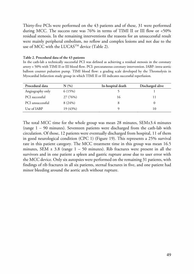
Thirty-five PCIs were performed on the 43 patients and of these, 31 were performed during MCC. The success rate was 76% in terms of TIMI II or III flow or <50% residual stenosis. In the remaining interventions the reasons for an unsuccessful result were mainly peripheral embolism, no reflow and complex lesions and not due to the use of MCC with the LUCASTM device (Table 2).
Table 2. Procedural data of the 43 patients In the cath-lab a technically successful PCI was defined as achieving a residual stenosis in the coronary artery < 50% with TIMI II or III blood flow. PCI: percutaneous coronary intervention. IABP: intra-aortic balloon counter pulsation pump. TIMI blood flow: a grading scale developed by the Thromolysis in Myocardial Infarction study group in which TIMI II or III indicates successful reperfusion.
Procedural data N (%) In-hospital death Discharged alive
Angiography only 6 (15%) 5 1
PCI successful 27 (76%) 16 11
PCI unsuccessful 8 (24%) 8 0
Use of IABP 19 (43%) 9 10
The total MCC time for the whole group was mean 28 minutes, SEM±3.4 minutes (range 1 – 90 minutes). Seventeen patients were discharged from the cath-lab with circulation. Of those, 12 patients were eventually discharged from hospital, 11 of them in good neurological condition (CPC 1) (Figure 19). This represents a 25% survival rate in this patient category. The MCC treatment time in this group was mean 16.5 minutes, SEM ± 3.8 (range 1 – 50 minutes). Rib fractures were present in all the survivors and in one patient a spleen and gastric rupture arose due to user error with the MCC device. Only six autopsies were performed on the remaining 31 patients, with findings of rib fractures in all six patients, sternal fractures in five, and one patient had minor bleeding around the aortic arch without rupture.

50
Figure 19 Flow chart illustrating the outcomes of the study patients. * Cerebral performance category 1.

51
Paper II
Thirty-two patients were included during the study period. For patient demographics see Table 3. Patient characteristics such as the indications for the cath-lab procedure, culprit lesion, circulatory state upon arrival in the cath-lab, and rhythm when the CA occurred, are presented in Table 4.
Table 3. Patient demographics. Cath-lab = coronary catheterization laboratory. MI = myocardial infarction. PCI = percutaneous coronary intervention. CABG = coronary artery by-pass grafting.
All patients Expired Cath-lab
Discharged Cath-lab
Discharged Hospital
n=32 (%) 17 (53) 15 (47) 8 (25)
Patient History
Age 70.9±12.9 73±10 68.3±15.2 68.1±18.8
Gender (male) 20 (63%) 11(65) 9 (60) 4 (50)
Hypertension 18 (56%) 9 (53) 9 (60) 7 (86)
Diabetes 8 (25%) 6 (35) 2 (13) 2 (25)
Hyperlipidaemia 9 (28 %) 7 (41) 2 (13) 2 (25)
Smoker/Ex-smoker 14 (44%) 7 (41) 7 (47) 4 (50)
Previous MI 9 (28%) 4 (24) 5 (33) 4 (50)
Previous PCI 3 (9%) 1 (6) 2 (13) 2 (25)
Previous CABG 4 (13%) 3 (18) 1 (7) 1 (13)
In one specific patient, the reason for referral for the intra-aortic balloon counter pulsation insertion was therapy-resistant VT with CS. In the patients referred for planned PCI and non-STEMI, complications such as, for example, thrombus formation, vessel rupture and dissection, caused the CA. One of the patients with non-STEMI was in CS at the time of arrival in the cath-lab. The patient, who was referred for an elective pre-operative coronary angiogram for surgery on the aortic valve, deteriorated into PEA due to aortic stenosis and reduced systolic left ventricular function.
Seventeen patients expired in the cath-lab. Fifteen patients left the cath-lab with circulation, of whom eight were discharged from hospital in CPC 1 - 2. During the study period (9 April 2009 – 9 April 2013), 8738 patients were admitted to the cath-lab for an invasive cardiac or coronary procedure. In total, 3368 patients were evaluated with a coronary angiogram only and 5370 patients were treated with PCI (acute or

52
elective) whereof 2728 were treated for STEMI. Of these, 116 patients were in CS when admitted to the cath-lab. There was no statistical age difference between the patients who expired in the cath-lab and those who were discharged from the cath-lab with circulation (p = 0.37) or those discharged from hospital (p = 0.64). Successful PCI defined as TIMI-II-III or <50% residual stenosis, PCI during mechanical CC, and treatment time with mechanical CC are presented in Table 5. There was a statistically significant difference in duration of mechanical CC when comparing patients who expired in the cath-lab to those discharged from the cath-lab with circulation (p = 0.02) and to those discharged from hospital (p = 0.004). At least one vasoactive drug (norepinephrine, EPI or dobutamine) was administered either intermittently or as a continuous infusion to 29 patients, and the majority received a combination of these drugs during the procedure.

53
Table 4. Indication for referral to the coronary catheterization laboratory, culprit lesion, circulatory state upon arrival in the cath-lab, rhythm at the time of the cardiac arrest. Cath-lab = coronary catheterization laboratory. STEMI = ST elevation myocardial infarction. NSTEMI = non-ST elevation myocardial infarction. PCI = percutaneous coronary intervention. LM = left main coronary artery. LAD = left anterior descendent coronary artery. LCx = left circumflex coronary artery. RCA = right coronary artery. VT = ventricular tachycardia. VF = ventricular fibrillation. PEA = pulseless electrical activity.
All patients
n = 32 (%)
Expired
Cath-lab
Discharged
Cath-lab
Discharged
Hospital
17 (53) 15 (47) 8 (25)
Indication for cath-lab procedure
STEMI 24 (75) 15 (88) 9 (60) 4 (50)
Non-STEMI 4 (13) 1 (6) 3 (20) 2 (25)
Elective PCI 2 (6) 1 (6) 1 (7) 1 (13)
Other 1 (3) 0 1 (7) 1 (13)
Angiogram 1 (3) 0 1 (7) 0 (0)
Culprit lesion in coronary patients
LM 10 (31) 6 (35) 4 (27) 2 (25)
LAD 12 (38) 7 (41) 4 (27) 2 (25)
LCx 2 (6) 0 3 (20) 2 (25)
RCA 6 (19) 4 (24) 2 (13) 1 (13)
Other 2 (6) 0 2 (13) 1 (13)
Circulatory state upon arrival in the cath-lab
Cardiogenic shock 20 (62) 12 (71) 8 (53) 2 (25)
Initial rhythm at cardiac arrest
VT/VF 5 (16) 1 (6) 4 (27) 2 (25)
PEA 22 (69) 14 (82) 8 (53) 4 (50)
Asystole 5 (16) 2 (12) 3 (20) 2 (25)

54
The manual CC group
Ten patients (eight men) with a mean age of 67.9 ±6.3 years suffered CA and required prolonged advanced resuscitation efforts with manual CC in the cath-lab between 1 January 1999 and 31 December 2003. Eight of these were referred due to a STEMI, one patient had a non-STEMI, and one patient had developed a ventricular septum defect due to a STEMI a few days earlier. Seven patients were in CS when admitted to the cath-lab. Two patients had a shockable rhythm at the time of the CA. Six patients were treated with PCI during manual CC, with 50% PCI success rate. The median time with manual CC for the whole group (n = 10) was 20 minutes (range 15 – 75) for those who expired in the cath-lab (n = 6); 25 minutes (range 15 – 60) for those discharged from the cath-lab (n = 4), and 15 minutes for the patient discharged from hospital in CPC 1 (n = 1).
Six-month survival rate
In the merged group with survivors discharged from hospital in CPC 1 from the previous study (n = 11) (Paper I) and the current study (n = 8), there was an 84% survival rate at six months (n = 16) (Figure 19).
Table 5. Cath-lab procedural data. Cath-lab = coronary catheterization laboratory. PCI = percutaneous coronary intervention. MCC = mechanical chest compression. IABP = intra-aortic counter pulsation pump. CC = chest compression. CC times are presented as median minutes (range).
All patients Expired Cath-lab
Discharged Cath-lab
Discharged Hospital
n=32 (%) 17 (53) 15 (47) 8 (25)
Procedural data
Angiography during MCC 5 (16) 2 (12) 3 (20) 2 (25)
PCI during MCC 27 (87) 16 (94) 11 (73) 6 (75)
PCI successful 25 (81) 12 (71) 13 (87) 6 (75)
PCI unsuccessful 6 (20) 5 (29) 1 (7) 1 (12.5)
Use of concomitant IABP 12 (38) 3 (18) 9 (60) 4 (50)
CC time 34 (5-90) 42.5(10-75) 15 (5-90) 10 (5-52)
Thoracic surgery 4 (13) 0 4 (27) 2 (25)

55
Figure 20. Flow-chart showing the included patients (Paper I left, Paper II right) requiring prolonged advanced resuscitation including mechanical chest compressions during percutaneous coronary/cardiac interventions. Cath-lab = coronary catheterization laboratory. CPC = cerebral performance category.
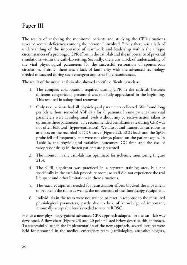
56
Paper III
The results of analysing the monitored patients and studying the CPR situations revealed several deficiencies among the personnel involved. Firstly there was a lack of understanding of the importance of teamwork and leadership within the unique circumstances of a prolonged CPR effort in the cath-lab and the importance of practical simulations within the cath-lab setting. Secondly, there was a lack of understanding of the vital physiological parameters for the successful restoration of spontaneous circulation. Thirdly, there was a lack of familiarity with the advanced technology needed to succeed during such emergent and stressful circumstances.
The result of the initial analysis also showed specific difficulties such as:
1. The complex collaboration required during CPR in the cath-lab between different categories of personnel was not fully appreciated in the beginning. This resulted in suboptimal teamwork.
2. Only two patients had all physiological parameters collected. We found long periods without recorded ABP data for all patients. In one patient three vital parameters were at suboptimal levels without any corrective action taken to optimize these parameters. The recommended ventilation rate during CPR was not often followed (hyperventilation). We also found numerous variations in artefacts on the recorded ETCO2 curve (Figure 22). ECG leads and the SpO2
probe fell off frequently and were not always placed on the patient again. In Table 6, the physiological variables, outcomes, CC time and the use of vasopressor drugs in the ten patients are presented
3. The monitor in the cath-lab was optimized for ischemic monitoring (Figure 21b).
4. The CPR algorithm was practiced in a separate training area, but not specifically in the cath-lab procedure room, so staff did not experience the real life space and other limitations in these situations.
5. The extra equipment needed for resuscitation efforts blocked the movement of people in the room as well as the movements of the fluoroscopy equipment.
6. Individuals in the team were not trained to react in response to the measured physiological parameters, partly due to lack of knowledge of important, minimally acceptable levels needed to secure ROSC.
Hence a new physiology-guided advanced CPR approach adapted for the cath-lab was developed. A flow chart (Figure 23) and 20 points listed below describe this approach. To successfully launch the implementation of the new approach, several lectures were held for personnel in the medical emergency team (cardiologists, anaesthesiologists,

57
anaesthetic nurses, interventionists, cath-lab nurses and assistant nurses) which included >200 people. Focus was put on training CA scenarios in the cath-lab, teamwork and an understanding of the minimally acceptable levels of vital parameters needed for successful resuscitation.

58
Tab
le 6
V
ital p
aram
eter
s, ou
tcom
es, C
PR ti
me,
the
use
of v
asop
ress
or su
bsta
nces
in te
n pa
tient
s suf
ferin
g C
A in
the
cath
-lab
durin
g sim
ulta
neou
s PC
I. AB
P =
arte
rial
bloo
d pr
essu
re. C
VP
= ce
ntra
l ven
ous p
ress
ure.
SpO
2 = in
dex
finge
r sat
urat
ion.
ET
CO
2 = e
nd ti
dal c
arbo
n di
oxid
e. n
d =
not d
eter
min
ed d
ue to
art
efac
ts. n
r =
not r
egist
ered
or l
ost d
urin
g pr
oced
ure.
RO
SC =
retu
rn o
f spo
ntan
eous
circ
ulat
ion.
CPC
= c
ereb
ral p
erfo
rman
ce c
ateg
ory.
Pa
tient
1
Patie
nt
2 Pa
tient
3
Patie
nt
4 Pa
tient
5
Patie
nt
6 Pa
tient
7
Patie
nt
8 Pa
tient
9
Patie
nt
10
M
ean
SD
Mea
nSD
M
ean
SD
Mea
n SD
M
ean
SD
Mea
n SD
M
ean
SD
Mea
n SD
M
ean
SD
Mea
n SD
ABP
Systo
lic [m
mH
g]
114±
18
133±
4 63
±2
50±1
8 87
±7
85±6
34
±3
139±
19
117±
15
81±1
0 AB
P D
iasto
lic [m
mH
g]
2±12
10
±4
39v3
22
±8
59±4
7±
6 20
±2
nd
31±1
8 12
±10
ABP
Mea
n [m
mH
g]
40±4
51
±3
46±4
31
±10
68±7
33
±6
25±2
43
±9
60±1
6 35
±9
CV
P Sy
stolic
[mm
Hg]
12
3±10
95
±4
nr
nr
nr
nr
46±2
16
9±17
70
±10
nr
CV
P D
iasto
lic [m
mH
g]
15±1
1 -1
2±3
nr
nr
nr
nr
6±9
nd
13±6
nr
C
VP
Mea
n [m
mH
g]
52±1
3 24
±3
nr
nr
nr
nr
19±1
36
±8
32±7
nr
Sp
O2 [
%]
82±8
81
±0
95±0
77
±6
78±4
nr
64
±3
nr
nr
nr
ETC
O2 [
kPa]
2,
4±0,
4 3.
5±0,
3 2,
8±0,
5 nr
nr
nr
nr
nr
3,
1±0,
7 nr
Out
com
e R
OSC
(3
12 h
) D
ead
in
cath
-lab
RO
SC
(216
h)
Dea
d in
ca
th-la
b R
OSC
(1
3 h)
C
PC 1
D
ead
in
cath
-lab
Dea
d in
ca
th-la
b C
PC 1
D
ead
in
cath
-lab
CPR
tim
e (m
inut
es)
45
50
15
75
35
10
50
30
45
70
Nor
ephi
neph
rine
YES
YES
YES
YES
YES
YES
YES
YES
YES
YES
Epin
ephr
ine
YES
NO
N
o YE
S YE
S N
O
YES
YES
No
YES
Dob
utam
ine
YES
YES
YES
YES
YES
NO
N
O
NO
N
O
NO

59
Figure 21 Left (a) Monitor screen designed for CPR-CA arrest situations displaying vital physiological parameters. Green line = ECG, red line = intra-aortic arterial blood pressure, dark blue line = central venous pressure (CVP), light blue line = pulse oximetry (SpO2), white line = end tidal carbon dioxide (ETCO2), green figures bottom = cerebral oximetry SctO2 values.
Right (b) Monitor screen primarily designed for ischemic monitoring in the cath-lab. Green line = ECG, red line = intra-aortic arterial blood pressure, dark blue line = pulse oximetry (SpO2), green circles = ischemic monitoring.
Specific recommendations
• SpO2, ECG and ABP should be measured from the start of all interventions in the cath-lab.
• In case of a CA, defibrillate as early as possible if indicated by rhythm. All these cases are instantaneously attended and do not have a high volume load on the venous side [58, 123]. After defibrillation, start CC immediately if the patient does not obtain instantaneous ROSC.
• Alert the medical emergency team, whose swift arrival should be in 60 - 90 seconds.
• Select a team leader (most often the cardiologist).
• If the CA situation has not been solved in a few minutes with manual CC and defibrillations, apply the MCC device and start CCs (30 CC followed by two ventilations) for patients not intubated. The MCC device should be placed in the cath-lab for quick access and deployment.
• When the patient is intubated, switch to continuous MCCs and a ventilation rate of ten per minute. Ventilation has to be modified to avoid high intra-thoracic pressures. Intermittent manual ventilation between compressions is usually possible. When blood gases are available, ventilation should aim at normoventilation. Blood gases should also be used for adjustment of acidosis if needed.

60
• To simplify an instant overview of the vital parameters, use a specially designed monitor screen showing one continuous ECG lead, two ABP curves, one CVP curve (optional), one ETCO2 curve, one SpO2 curve and values for SctO2 (Figure 21a).
• As soon as the patient is intubated start monitoring ETCO2. However, be aware of compression artefacts in the ETCO2 curve during MCC (Figure 22). To correctly assess the curve, stop the MCC device, perform two inflations and assess the ETCO2 curve.
• If the patient has a shock-resistant VF, continue MCC and proceed with PCI in order to open the occlusion, rather than continue with further defibrillation attempts while the culprit coronary vessel remains occluded.
• Place the equipment brought by the medical emergency team in dedicated zones marked on the floor, so as not to interfere with the needed fluoroscopic projections.
• Optimize physiological parameters according to Table 7. Since CPP cannot be calculated instantaneously, TIMI flow might be used as a surrogate marker. If systolic ABP is below 70 mmHg, rule out cardiac tamponade, reposition the LUCAS™ device, consider changing the ventilation rate, or administer norepinephrine [124] or an infusion of EPI [125]. Be careful with the latter to avoid high dose EPI.

61
Figure 22 When analysing ETCO2 during CCs, be aware that MCC can cause compression artefacts and also avoid hyper-ventilation. A: Shows two examples from the analysed patients where hyperventilation was seen during CC. B: Shows some examples of the artefacts that were noticed on the ETCO2 registration. C: Shows an example of the difference between ETCO2 levels (decreasing from ~5 kPa to ~3 kPa) in an
ETCO2 curve with and without CC artefacts.

62
Table 7 Target values for vital physiological parameters during the resucitation in the cath-lab. AoD = aortic diastolic pressure. RaD = right atrial diastolic pressure. TIMI = thrombolysis in myocardial infarction. mmHg = millimetre mercury. kPa = kilopascal.
• Apply the electrodes for SctO2 on the patient’s forehead as soon as possible if available.
• At the interventionist´s discretion, an introducer could be inserted in the femoral vein, both in order to place a pigtail catheter in the right atria for measuring CVP and to serve as a central venous line for infusions and drug administration.
• Collaboration between the anaesthesiologist and cardiologist is key in all efforts to optimize vital parameters.
• In the relatively rare cases of pulmonary oedema, an adjustable PEEP valve could be tried. When ROSC has been attained, intermittent positive pressure ventilation using a conventional intensive care unit ventilator can be started.
• Record physiology parameters and communicate them to the team leader and anaesthesiologist repeatedly or when dramatic change occurs. Every person in the cath-lab has the responsibility to react to unsatisfactory physiology parameters.
• Consider initiation of therapeutic hypothermia/target temperature management as soon as possible.
• If successful PCI is accomplished and the patient has a shockable rhythm, defibrillate during MCC. If unsuccessful, consider one bolus injection of EPI (1 mg) followed by defibrillation.

63
• If successful PCI is accomplished and the patient has PEA or asystole, continue MCC for 15 – 20 minutes followed by an infusion of EPI for approximately two to three minutes. Then administer a bolus dose of 1 mg of EPI in the right atria. Continue MCC for at least ten minutes after last drug delivery before termination of resuscitation efforts.
• If successful PCI is accomplished and the patient has obtained ROSC, deploy a left ventricular assist device if needed. Prepare for further post-resuscitation treatment in the intensive care unit according to guideline recommendations [126] or local directive.
These points, together with the flow chart shown in Figure 23, were put into a pocket folder and given to all personnel involved in the medical emergency team and to all cath-lab personnel.
Due to this education (theoretical and practical), personnel felt more secure and comfortable in their roles when real CA situations arose in the cath-lab and they experienced a more success-oriented result.
Figure 23 shows a flow chart visualizing the important steps and decision points in the physiology-guided CPR approach in CA situations in the cath-lab during simultaneous PCI. PEA = pulseless electrical activity. CC = chest compression. CPR = cardiopulmonary resuscitation. ABP = intra-aortic arterial blood pressure.

64
Paper IV
The baseline variables for the ten pigs analysed are shown in Table 8. The blood pressure and APV (APV noise-free period was s: 0 – 2 min: six pigs = 66 ±47 s, 2 – 4 min: seven pigs = 114 ±11 s, 4 – 6 min: seven pigs = 99 ±38 s, 6 – 8 min: nine pigs = 68 ±46 s, 8 – 10 min: seven pigs = 84 ±40 s) are shown in Table 10. The statistical comparison revealed a significant (P <0.0005) increase in coronary blood flow with MCC compared to baseline (sinus rhythm, at a frequency of 97±16) at each time interval, ranging between a 12 and 39% increase (Table 8).
Table 8 Measurements of intra-aortic arterial blood pressure, central venous pressure, coronary perfusion pressure and average peak blood flow velocity. Mean values ±SD during baseline, during 0 – 2 minutes, 2 – 4 minutes, 4 – 6 minutes, 6 – 8 minutes and 8 – 10 minutes of mechanical chest compressions. Min = minimum. AP = intra-aortic arterial blood pressure. Max = maximum. CVP = central venous blood pressure. CPP = coronary perfusion pressure. APV = average peak blood flow velocity. mmHg = millimetre mercury. cm/s = centimetre per second.
Variable/time Baseline 0-2 min 2-4 min 4-6 min 6-8 min 8-10 min
Min AP [mmHg] 101.1 ±1.1 16.9 ±2.3 14.1 ±1.1 11.9 ±1.2 8.5 ±1.2 5.6 ±1.3 Mean AP [mmHg] 115.7 ±1.2 35.7 ±3.3 38.0 ±0.7 36.3 ±1.4 33.8 ±0.8 37.8 ±1.3 Max AP [mmHg] 129.3 ±1.5 65.9 ±6.4 69.9 ±0.8 68.0 ±1.1 66.2 ±0.9 65.6 ±0.9 Min CVP [mmHg] 1.7 ±0.5 1.9 ±1.3 1.2 ±0.2 1.1 ±0.2 8.1 ±0.2 1.0 ±0.3 Mean CVP [mmHg] 3.5 ±0.2 23.2 ±1.7 23.7 ±0.4 23.8 ±0.8 24.8 ±0.4 24.2 ±0.6 Max CVP [mmHg] 5.2 ±1.8 59.5 ±8.4 67.9 ±1.1 66.7 ±1.7 69.7 ±0.9 70.1 ±1.2 CPP [mmHg] 98.0 ±2.0 24.3 ±1.4 24.1 ±0.5 23.6 ±1.3 21.5 ±1.2 20.6 ±1.5 APV [ml/min] 14.3 ±1.0 20.0 ±1.2 18.9 ±0.5 17.8 ±0.9 17.3 ±0.9 16.0 ±1.1
Arterial pressure and CVP over the total study period are presented in Figure 24. The progress of calculated CPP and the measured intracoronary APV are shown during the experimental period in Figure 24. APV values could not be calculated correctly in the ROSC period due to technical disturbances. However, there is a hyperemic period evidenced by increased AP and CPP that is shown in Figures 24 and 25 indicating that increased Doppler flow is evident. However, this cannot be proven due to the technical problems we experienced.

65
Figure 24 The development of minimum, mean and max (Min, Mean and Max) intra-thoracic aortic pressure and right atrial pressure during the total experimental period (28 minutes). Data are presented as the mean value of the 30-second periods of analysed data, and each individual pig’s data are time-adjusted to the same length for each period of the experiments.
Figure 25 The development of the calculated mean coronary perfusion pressure (CPP in mmHg) over the total experimental period (28 minutes) and the development of coronary artery flow velocity (cm/s) during the experimental period. Coronary flow is not shown after ROSC due to technical problems after defibrillation. Data are presented as the mean value of the 30-second periods of analysed data, and each individual pig’s data are time-adjusted to the same length for each period of the experiments.

66
Correlation analysis between calculated CPP and APV was performed for the entire ten minute period of MCC. A significant correlation (R = 0.761, R2 = 0.6 and P-value <0.0001) between the calculated CPP and APV during MCC was seen (Figure 26).
Figure 26 Correlation of coronary perfusion pressure (CPP) and average blood flow peak velocity (APV). Shows the correlation of calculated CPP and APV during the 10 minutes of mechanical chest compressions. To test the null-hypothesis for CPP and APV correlation, the Z-test was used.
Arterial blood gas values are shown in Table 9. There was a significant fall between base line and after ten minutes of MCC in pH and BE, but lactate was significantly elevated as well as glucose. The significant difference in pH, lactate and glucose also persisted following 20 minutes of ROSC, but BE stabilized and PO2 increased.

67
Table 9 Measurements of blood gas, haemoglobin, lactate, electrolytes and glucose at baseline and after ten minutes of MCC. Data are presented as mean ±SD, n= 10. ns= not significant. pH = hydrogen ions. pCO2 = partial pressure carbon dioxide. pO2 = partial pressure oxygen. ABE = arterial base excess. Hb = haemoglobin. Na = sodium. K = potassium. Ca = calcium.
Variable Baseline 10 min P-value ROSC 20 min P-value Arterial Arterial Arterial
Mean ±SD Mean ±SD Mean ±SD
pH 7.350 ±0.026 7.257 ±0.060 0.012 7.287 ±0.050 0.014
PCO2 5.8 ±0.63 6.5 ±1.30 ns 6.4 ±0.83 ns
PO2 14.3 ±6.9 27.5 ±18.6 ns 48.75 ±17.7 0.042 ABE -1.7 ±1.4 -6.0 ±1.4 0.002 -3.97 ±1.6 ns Lactate 1.42 ±0.47 4.35 ±1.33 0.02 3.86 ±1.02 0.002 Hb 108 ±10 120 ±11 ns 114 ±10 ns Na 137 ±2,1 136 ±2.7 ns 136 ±2.8 ns K 3.96 ±0.24 4.59 ±0.89 ns 3.86 ±0.34 ns Ca 1.33 ±0.04 1.35 ±0.06 ns 1.27 ±0.05 ns Glucose 6.6 ±1.4 11.5 ±2.2 0.002 11.79 ±3.15 0.008
ROSC
Seven animals regained ROSC following the first defibrillation attempt; two pigs required three defibrillation attempts to attain ROSC and two pigs did not obtain ROSC following defibrillation at the end of the ten-minute episode of VF, one of which was excluded due to misplacement of the device before start of CC.
Paper V
No difference was seen during the baseline period regarding analysed measurements between any groups (Table 10).

68
Tab
le 1
0.
Base
line
para
met
ers.
NaC
l = S
alin
e. E
PI =
epi
neph
rine.
Sys
t = sy
stolic
. ABP
= a
rter
ial b
lood
pre
ssur
e (m
mH
g). D
iast
= di
asto
lic. M
ax =
max
imum
. CV
P =
cent
ral v
enou
s pre
ssur
e (m
mH
g). M
in =
min
imum
. ET
CO
2 =
end
tidal
car
bon
diox
ide
(kPa
). AP
V =
ave
rage
pea
k ve
loci
ty (c
m/s
). H
eart
rate
(bea
ts/m
inut
e).
PCO
2 = p
artia
l pre
ssur
e ca
rbon
dio
xide
(kPa
). PO
2 = p
artia
l pre
ssur
e ox
ygen
(kPa
). AB
E =
arte
rial b
ase
exce
ss (m
mol
/l). A
ll va
lues
are
exp
ress
ed a
s med
ian,
25
th a
nd 7
5th
inte
rqua
rtile
.
N
aCl (
n=12
) EP
I al
l (n=
24)
p-va
lue
EPI
0.02
m
g/kg
/dos
e (n
=12)
p-
valu
e EP
I 0.
03 m
g/kg
/dos
e (n
=12)
p-
valu
e
Syst
ABP
15
2 (1
37 -
167)
15
1 (1
36 -
163)
0.
5347
14
9 (1
36 -
172)
0.
4357
15
2 (1
36 -
172)
0.
7950
Dia
st A
BP
96
(79
- 122
) 96
(8
9 - 1
06)
0.69
96
93
(89
- 99)
0.
7075
10
0 (7
8 - 1
07)
0.79
50
Mea
n AB
P 11
5 ( 9
9 - 1
39)
114
(105
- 12
4)
0.53
47
113
(105
- 11
8)
0.50
67
116
(94
- 126
) 0.
7075
Max
CV
P 12
(1
0 - 1
3)
11
(10
-13)
0.
6505
11
(1
0 - 1
3)
0.54
44
11
(10
-13)
0.
8852
Min
CV
P 8
(5 -
10)
7 (6
- 8)
0.
4502
7
(4 -
8)
0.28
55
7 (6
- 9)
0.
8399
Mea
n C
VP
10
(8 -
11)
9 (8
- 11
) 0.
7944
9
(8 -
11)
0.73
50
9 (8
- 11
) 0.
9310
ETC
O2
4.9
(4.3
- 5.
0)
4.5
(4
.1 -
4.9)
0.
5642
4.
8
(4.4
- 5.
0)
0.86
91
4.3
(4
.0 -4
.7)
0.27
27
APV
17
(1
2 - 2
8)
17
(12
- 24)
0.
7677
20
(1
3 - 2
6)
0.83
99
15
(11
- 21)
0.
4417
Hea
rt ra
te
88
(58
- 97)
75
(6
4 - 9
1)
0.74
55
77
(71
- 93)
0.
8955
71
(5
6 - 9
0)
0.47
01
pH
7.47
3 (7
.420
-7.5
05)
7.48
4 (7
.402
-7.5
10)
0.93
31
7.46
8 7.
414-
7.52
9)
0.79
50
7.48
5 (7
.387
5-7.
503)
0.
6650
PC
O2
5.3
(5.0
-6.5
) 5.
4 (5
.1-5
.9)
0.72
46
5.5
(5.1
-5.9
) 0.
9310
5.
2 (4
.9-5
.9)
0.62
36
PO2
27.2
(1
6.6-
37.1
) 20
.2
(17.
2-33
.9)
0.60
30
23.0
(1
7.3-
33.2
) 0.
8174
20
.2
(16.
4-33
.9)
0.52
54
ABE
6.0
(4.6
-7.3
) 5.
9 (2
.8-7
.8)
0.90
65
6.8
(3.2
-8.7
) 0.
7075
5.
5 (2
.5-6
.1)
0.28
55

69
CP
P
Dur
ing
the f
irst f
our m
inut
es o
f MC
C an
d at
the t
ime o
f the
firs
t EPI
inje
ctio
n, th
ere w
ere n
o sig
nific
ant d
iffer
ence
s in
CPP
be
twee
n th
e co
ntro
l gro
up (
n=12
), EP
I-al
l gro
up (
n=24
), EP
I 0.
02 m
g/kg
/dos
e (n
=12)
or
EPI
0.03
mg/
kg/d
ose
(n=1
2)
(Tab
le 1
1, F
igur
es 2
7a –
27c
). D
urin
g th
e su
bseq
uent
per
iod
of M
CC
ther
e w
as a
sign
ifica
nt in
crea
se in
CPP
at P
max a
fter
EPI
inje
ctio
ns o
ne a
nd t
wo
in t
he E
PI-a
ll gr
oup
(p =
0.0
22, p
= 0
.016
), co
mpa
red
to t
he c
ontr
ol g
roup
but
not
afte
r in
ject
ions
thre
e an
d fo
ur (T
able
11,
Fig
ure
27a)
. The
re w
as a
sign
ifica
nt in
crea
se in
CPP
afte
r inj
ectio
ns tw
o an
d th
ree
in
EPI 0
.02m
g/kg
/dos
e co
mpa
red
to th
e co
ntro
l gro
up (p
= 0
.023
, p =
0.0
27) (
Tab
le 1
1, F
igur
e 27
b). W
hen
com
parin
g th
e EP
I 0.0
3 m
g/kg
/dos
e to
the
cont
rol g
roup
, the
re w
as a
stat
istic
al si
gnifi
cant
diff
eren
ce a
t Pm
ax fo
llow
ing
inje
ctio
n on
e (p
=
0.01
3) b
ut n
ot a
t Pm
ax fo
llow
ing
inje
ctio
ns tw
o, th
ree
and
four
(Tab
le 1
1, 2
7c).
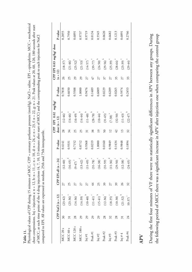
70
Tab
le 1
1.
Phys
iolo
gica
l val
ues o
f CPP
dur
ing
15 m
inut
es o
f MC
C. C
PP =
cor
onar
y pe
rfusio
n pr
essu
re (m
mH
g). N
aCl =
salin
e. E
PI =
epi
neph
rine.
MC
C =
mec
hani
cal
ches
t com
pres
sions
. Inj
= in
ject
ion
a: n
= 1
2, b
: n =
11,
c: n
= 1
0, d
: n =
24,
e: n
= 2
3, f:
n =
22,
g: n
= 2
1. P
eak
valu
es a
t 20,
60,
120
, 180
seco
nds a
fter s
tart
of
MC
C, a
t eac
h tim
e po
int o
f the
4 d
rug
inje
ctio
ns (4
, 7, 1
0, 1
3 m
inut
es a
fter s
tart
of M
CC
) and
the
corr
espo
ndin
g pe
ak to
eac
h in
ject
ion
for N
aCl
com
pare
d to
EPI
. All
valu
es a
re e
xpre
ssed
as m
edia
n, 2
5th
and
75th
inte
rqua
rtile
.
C
PP
NaC
l (n
= 12
) C
PP
EP
I-al
l (n
= 24
) P
-val
ue
CP
P
EPI
0.02
m
g/kg
/ do
se (n
= 1
2)
P-v
alue
C
PP
EP
I 0.
03 m
g/kg
/ do
se
(n =
12)
P
-val
ue
MC
C 2
0 s
30
(23-
42) a
27
(12-
40) e
0.41
41
28
(12-
46) b
0.78
18
26
(18-
37) a
0.28
55
MC
C 6
0 s
31
(23-
36) a
25
(16-
45) d
0.93
31
34
(14-
46) a
0.66
50
25
(21-
46) a
0.79
50
MC
C 1
20 s
28
(14-
36) a
27
(9-4
7) e
0.71
52
25
(16-
49) b
0.55
88
29
(25-
52)b
0.68
91
MC
C 1
80 s
29
(14-
39) a
27
(13-
42) f
0.87
12
25
(14-
44) b
1.00
00
29
(21-
53)c
0.97
37
Inj #
1 29
(1
0-40
) b 27
(1
1-49
) n 0.
9368
31
(1
1-48
) b 0.
9476
27
(1
4-57
) c 0.
9719
Peak
#1
29
(11-
41) c
44
(31-
78) e
0.02
19
38
(28-
78) b
0.14
89
47
(39-
77) a
0.01
34
Inj #
2 29
(1
5-41
) b 25
(1
6-38
) e 1.
0000
19
(1
0-44
) a 0.
6009
27
(2
4-50
) b 0.
5545
Peak
#2
28
(12-
36) c
39
(31-
59) e
0.01
60
50
(31-
65) a
0.02
29
37
(29-
56) b
0.06
20
Inj #
3 32
(1
8-35
) c 27
(1
1-36
) d 0.
9849
12
(7
-38)
a 0.
6209
27
(2
5-39
) a 0.
6682
Peak
#3
28
(10-
38) c
39
(29-
50) e
0.14
34
42
(32-
50) a
0.02
65
35
(28-
64) a
0.12
13
Inj #
4
31
(21-
32) b
26
(12-
38) f
0.98
48
13
(11-
43) c
0.59
74
27
(23-
39) a
0.68
91
Peak
#4
24
10-3
7) c
32
(24-
45) e
0.18
94
32
(22-
47) b
0.24
53
33
(29-
44) a
0.27
66
AP
V
Dur
ing
the
first
four
min
utes
of V
F th
ere
wer
e no
stat
istic
ally
sign
ifica
nt d
iffer
ence
s in
APV
bet
wee
n an
y gr
oups
. Dur
ing
the
follo
win
g pe
riod
of M
CC
ther
e w
as a
sign
ifica
nt in
crea
se in
APV
afte
r inj
ectio
n on
e w
hen
com
parin
g th
e co
ntro
l gro
up
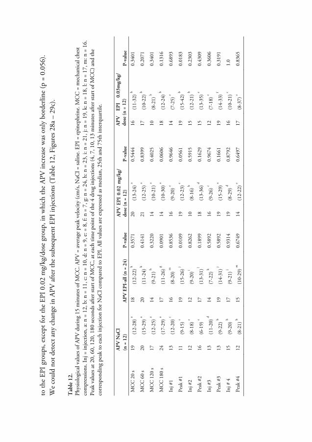
71
to th
e EP
I gro
ups,
exce
pt fo
r the
EPI
0.0
2 m
g/kg
/dos
e gr
oup,
in w
hich
the
APV
incr
ease
was
onl
y bo
rder
line
(p =
0.0
56).
We
coul
d no
t det
ect a
ny c
hang
e in
APV
afte
r the
subs
eque
nt E
PI in
ject
ions
(Tab
le 1
2, F
igur
es 2
8a –
29c
).
Tab
le 1
2.
Phys
iolo
gica
l val
ues o
f APV
dur
ing
15 m
inut
es o
f MC
C. A
PV =
ave
rage
pea
k ve
loci
ty (c
m/s
, NaC
l = sa
line.
EPI
= e
pine
phrin
e. M
CC
= m
echa
nica
l che
st co
mpr
essio
ns. I
nj =
inje
ctio
n, a
: n =
12,
b: n
= 1
1, c
: n =
10,
d: n
= 9
, e: =
8, f
: n =
7, g
: n =
24,
h: n
= 2
3, i:
n =
21,
j: n
= 1
9, k
: n =
18,
l: n
= 1
7, m
: n =
16.
Pe
ak v
alue
s at 2
0, 6
0, 1
20, 1
80 se
cond
s afte
r sta
rt o
f MC
C, a
t eac
h tim
e po
int o
f the
4 d
rug
inje
ctio
ns (4
, 7, 1
0, 1
3 m
inut
es a
fter s
tart
of M
CC
) and
the
corr
espo
ndin
g pe
ak to
eac
h in
ject
ion
for N
aCl c
ompa
red
to E
PI. A
ll va
lues
are
exp
ress
ed a
s med
ian,
25t
h an
d 75
th in
terq
uarti
le.
A
PV
NaC
l (n
= 1
2)
AP
V E
PI-
all (
n =
24)
P-v
alue
A
PV
EP
I 0.
02 m
g/kg
/ do
se (n
= 1
2)
P-v
alue
A
PV
EP
I 0.
03m
g/kg
/ do
se (n
= 1
2)
P-v
alue
MC
C 2
0 s
19
(12-
28) a
18
(12-
22) h
0.35
71
20
(13-
24) a
0.54
44
16
(11-
32) b
0.34
01
MC
C 6
0 s
20
(15-
29) a
20
(11-
24) h
0.41
41
21
(12-
25) a
0.83
99
17
(10-
22) b
0.20
71
MC
C 1
20 s
17
(12-
25) a
14
(9-2
1) h
0.32
20
14
(10-
21) a
0.40
25
10
(8-2
1) b
0.34
01
MC
C 1
80 s
24
(17-
29) a
17
(11-
26) g
0.09
01
14
(10-
30) a
0.06
06
18
(12-
24) b
0.13
16
Inj #
1 13
(1
2-20
) c 16
(8
-20)
m
0.85
36
16
(9-2
0) e
0.96
46
14
(7-2
5) e
0.68
93
Peak
#1
11
(9-1
5) c
19
(15-
26) j
0.01
09
19
(12-
23) e
0.05
61
19
(15-
42) b
0.01
83
Inj #
2 12
(8
-18)
e 12
(9
-20)
i 0.
8262
10
(8
-16)
b 0.
5915
15
(1
2-21
) b 0.
2303
Peak
#2
16
(6-1
9) e
17
(13-
31) l
0.18
99
18
(13-
36) d
0.16
29
15
(13-
35) e
0.43
09
Inj #
3 13
(1
1-20
) d 14
(7
-22)
k 0.
5892
16
(9
-26)
c 0.
9674
12
(7
-18)
e 0.
3606
Peak
#3
13
(9-2
2) b
19
(14-
31) j
0.58
92
19
(15-
29) a
0.16
61
19
(14-
33) f
0.31
91
Inj #
4
15
(9-2
0) b
17
(9-2
1) j
0.93
14
19
(8-2
9) d
0.87
92
16
(10-
21) c
1.0
Peak
#4
12
(8-2
1) b
15
(10-
29) m
0.
6749
14
(1
2-22
) e 0.
6497
17
(8
-37)
e 0.
8365
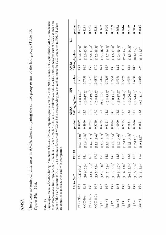
72
AM
SA
The
re w
ere
no s
tatis
tical
diff
eren
ces
in A
MSA
, whe
n co
mpa
ring
the
cont
rol g
roup
to
any
of t
he E
PI g
roup
s. (T
able
13,
Fi
gure
s 29a
– 2
9c).
Tab
le 1
3.
Phys
iolo
gica
l val
ues o
f AM
SA d
urin
g 15
min
utes
of M
CC
. AM
SA=
ampl
itude
spec
tral
are
a (m
V·H
z). N
aCl =
salin
e. E
PI =
epi
neph
rine.
MC
C =
mec
hani
cal
ches
t com
pres
sions
. Inj
= in
ject
ion,
a: n
= 1
2, b
: n =
10,
c: n
= 9
, d: n
= 2
1, e
: n =
6. P
eak
valu
es a
t 20,
60,
120
, 180
seco
nds a
fter s
tart
of M
CC
, at e
ach
time
poin
t of t
he 4
dru
g in
ject
ions
(4, 7
, 10,
13
min
utes
afte
r sta
rt o
f MC
C) a
nd th
e co
rres
pond
ing
peak
to e
ach
inje
ctio
n fo
r NaC
l com
pare
d to
EPI
. All
valu
es
are
expr
esse
d as
med
ian,
25t
h an
d 75
th in
terq
uarti
le.
A
MSA
NaC
l EP
I A
ll p-
valu
e A
MSA
EP
I 0.
02m
g/kg
/dos
e p-
valu
e A
MSA
EP
I 0.
02m
g/kg
/dos
e p-
valu
e
MC
C 2
0 s
12.1
(9
.6-1
4.0)
b 13
.0
(10.
9-16
.0)d
0.40
99
13.0
(1
1.8-
13.0
)c 0.
3913
13
.1
(10.
6-15
.0)a
0.57
52
MC
C 6
0 s
12.4
(1
0.8-
15.3
)b 14
.4
(11.
4-16
.8)d
0.49
90
13.7
(1
0.8-
17.6
)c 0.
7751
14
.6
(13.
0-15
.8)a
0.42
88
MC
C 1
20 s
13.8
(1
2.1-
16.3
)b 16
.0
(14.
2-18
.7)d
0.19
74
16.0
(1
4.2-
19.8
)c 0.
1779
15
.5
(13.
8-17
.6)a
0.37
34
MC
C 1
80 s
14.7
(1
3.3-
17.9
)b 17
.0
(12.
8-19
.0)d
0.37
49
17.0
(1
2.8-
19.3
)c 0.
4877
17
.0
(15.
3-18
.3)a
0.42
88
Inj #
1 14
.3
(12.
7-16
.8)b
15.9
(1
3.9-
16.7
)d 0.
4725
15
.4
(9.7
-16.
7)c
0.41
42
16.1
(1
3.3-
16.7
)a 0.
6682
Peak
#1
14.7
(1
1.5-
15.9
)b 14
.6
(13.
0-16
.2)d
0.61
21
14.6
(1
3.8-
15.9
)c 0.
7133
14
.7
(12.
7-16
.2)a
0.64
44
Inj #
2 13
.9
(11.
0-15
.4)b
14.0
(1
1.6-
14.5
)d 0.
8824
14
.3
(13.
0-14
.9)c
0.77
51
13.8
(1
1.6-
14.3
)a 0.
6209
Peak
#2
13.5
(1
0.4-
14.7
)b 12
.5
(11.
0-14
.6)d
1.00
00
13.1
(1
1.9-
17.5
)c 0.
5956
12
.2
(10.
4-13
.9)a
0.66
82
Inj #
3 13
.7
(11.
4-14
.8)b
11.5
(9
.7-1
3.8)
d 0.
2285
11
.5
(10.
2-20
.9)c
0.56
76
11.2
(9
.5-1
1.7)
a 0.
1616
Peak
#3
12.7
(1
0.6-
15.5
)b 12
.5
(11.
1-14
.5)d
0.98
31
13.1
(1
0.2-
20.9
)c 0.
5954
12
.2
(11.
3-14
.3)a
0.71
69
Inj #
4
13.9
(1
1.3-
15.3
)b 11
.0
(9.7
-13.
5)d
0.16
96
11.8
(1
0.5-
14.3
)c 0.
6534
10
.6
(8.8
-12.
2)a
0.08
06
Peak
#4
14.0
(1
1.4-
15.3
)e 11
.0
(8.9
-15.
9)d
0.88
41
16.8
(9
.9-1
1.1)
c 0.
3165
9.
9 (8
.8-1
4.3)
a 0.
2814

73
Figure 27a Development of CPP in the combined group – EPI-all (0.02 mg EPI/kg/dose and 0.03 mg EPI/kg/dose) compared to control group (NaCl) during the MCC period (15 minutes). Significant difference between the EPI group compared to control group, * = p <0.05. CPP = coronary perfusion pressure. EPI = epinephrine. MCC = mechanical chest compressions. Inj = injection.
Figure 27b Development of CPP in 0.02 mg EPI/kg/dose compared to control group during the MCC period (15 minutes). Significant difference between the EPI group compared to control group, * = p <0.05. CPP = coronary perfusion pressure. EPI = epinephrine. MCC = mechanical chest compressions. Inj = injection.
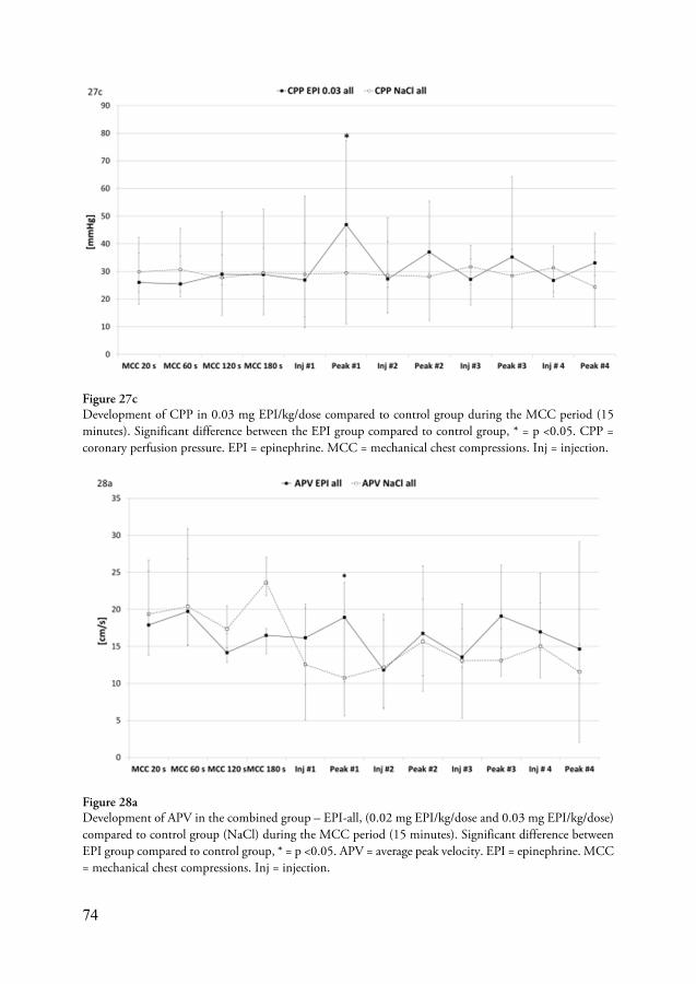
74
Figure 27c Development of CPP in 0.03 mg EPI/kg/dose compared to control group during the MCC period (15 minutes). Significant difference between the EPI group compared to control group, * = p <0.05. CPP = coronary perfusion pressure. EPI = epinephrine. MCC = mechanical chest compressions. Inj = injection.
Figure 28a Development of APV in the combined group – EPI-all, (0.02 mg EPI/kg/dose and 0.03 mg EPI/kg/dose) compared to control group (NaCl) during the MCC period (15 minutes). Significant difference between EPI group compared to control group, * = p <0.05. APV = average peak velocity. EPI = epinephrine. MCC = mechanical chest compressions. Inj = injection.

75
Figure 28b Development of APV in 0.02 mg EPI/kg/dose compared to control group during the MCC period (15 minutes). APV = average peak velocity. EPI = epinephrine. MCC = mechanical chest compressions. Inj = injection.
Figure 28c Development of APV in 0.03 mg EPI/kg/dose compared to control group during the MCC period (15 minutes). Significant difference between EPI group compared to control group, * = p <0.05. APV = average peak velocity. EPI = epinephrine. MCC = mechanical chest compressions. Inj = injection.

76
Figure 29a Development of AMSA in the combined group – EPI-all, (0.02 mg EPI/kg/dose and 0.03 mg EPI/kg/dose) compared to NaCl during the MCC period (15 minutes). AMSA = amplitude spectrum area. EPI = epinephrine. MCC = mechanical chest compressions. Inj = injection.
Figure 29b Development of AMSA in 0.02 mg EPI/kg/dose compared to control group during the MCC period (15 minutes). AMSA = amplitude spectrum area. EPI = epinephrin., MCC = mechanical chest compressions. Inj = injection.

77
Figure 29c Development of AMSA in 0.03 mg EPI/kg/dose compared to control group during the MCC period (15 minutes). AMSA = amplitude spectrum area. EPI = epinephrine. MCC = mechanical chest compressions. Inj = injection.
Time to maximum peak of CPP
The median time to Pmax following the EPI injections was 50 seconds (interquartile range 48 to 52) seconds (Table 3). There were no significant differences in time to Pmax between the EPI groups following injections one to four (Table 14).
Table 14 Time duration from drug administration to peak time (seconds) /Pmax of CPP for the 4 different drug injections. Inj = injection. mg = milligram. kg = kilogram. a; n = 12, b; n = 11, c; n = 23. All values are expressed as median, 25th and 75th interquartile.
Inj #1 Inj #2 Inj #3 Inj #4 P-value
Epinephrine all 51 (32-66)c 53 (41-58)c 49 (41-70)c 56 (44-78)c 0.392
Epinephrine 0.02 mg/kg/dose
48 (39-49)b 48 (40-52)a 52 (50-60)a 47 (43-59)b 0.44
Epinephrine 0.03 mg/kg/dose
48 (38-60)a 51 (40-56)b 51 (46-62)b 49(44-65)a 0.44

78
ROSC
Return of spontaneous circulation was achieved in 10 out of 12 animals (84%) in the control group compared with seven out of 12 (58%) in each EPI group (p=0.37).

79
Discussion
The use of MCC in the cath-lab during simultaneous PCI has been shown to be feasible, safe and can save lives. Over the nine years (2004 – 2013) covered by Papers I and II, 11 720 patients underwent PCI (elective, sub-acute or primary PCI) and of those 75 patients (0.64%) suffered a protracted CA in the cath-lab. The LUCAS™ mechanical CC system offers a valuable tool in these cases. The device performs high quality CC according to current CPR guidelines, without interruptions, and is easy to deploy. The radio-lucent design of most parts allows for fluoroscopy in all preferred angles during an intervention.
The majority of coronary interventions were performed during MCC with a success rate of 76% (Paper I) and 80% (Paper II), defined as TIMI II-III flow, as compared to PCI results in primary PCI with a 90% success rate [29]. This result indicates that the use of MCC is feasible and does not impair the progress of the coronary or cardiac intervention or substantially reduce the PCI result.
Questions have been raised regarding severe injuries caused by MCC compared to manual CC [46, 47] while other studies were unable to confirm this [48, 49]. In autopsy reports in Paper I (n=6), all had rib fractures and five had sternal fractures. In one autopsy there was minor bleeding around the aortic arch without rupture. Of the survivors, all suffered rib fractures and possibly sternal fractures. In one of the survivors, there was a gastric and spleen rupture due to mal-apposition of the MCC device, but the patient was successfully surgically treated. Thus, with the device correctly deployed, its utilization in the cath-lab is safe.
Survival rates at discharge from hospital after in-hospital CA vary widely, from 17 to 65% [3, 127-131], as do the causes of the CA [3, 127-131]. In some studies, subgroups such as patients < 65 years of age [129] or patients suffering a CA during a procedure or experiencing their CA in the emergency department [127] were excluded. Other factors that differ are a high amount of initial shockable rhythm (49% and 39%) [130, 131]. Herlitz et al. (data from the Swedish CPR registry) showed a 65% survival rate in patients who had their CA in the cath-lab [3]. In a newly published study by Demidova et al., the incidence of reperfusion VF associated with primary PCI was found to be 1.9% yearly with a survival rate of 81.7% at hospital discharge [132].
In Papers I and II, the survival rate was 25%. All these patients were discharged from hospital in good neurological condition (CPC 1 – 2). In the merged group including the survivors at hospital discharge, 84% were alive after 6 months. Thus, the use of the MCC device has probably increased survival in this group.

80
The population in Papers I and II consisted of patients with CA due to cardiac ischemic causes and there were no restrictions in upper age or procedure. There was a low amount of initial shockable rhythm causing the CA, which might be explained by the fact that all cases that were solved with one or a few defibrillation attempts, such as reperfusion VF, were excluded. The latter circumstance is probably an explanation for the difference in survival rate presented by Herlitz et al. [3] compared to the survival rate in Papers I and II. Since no other studies have described a similar population, these differences make comparisons delicate.
The DANAMI-2 trial in 2003 showed that primary PCI was superior to fibrinolytic therapy in STEMI patients [27]. Hochman et al. showed a survival benefit in patients in CS treated with early invasive strategy, both in the short- and long-term perspective [52, 53]. The results from these studies contributed to new treatment recommendations, which may have increased referrals to the cath-lab of patients in a more severe cardio-vascular circulatory condition with a higher risk of developing CA. When comparing historical data to current data, indications for referral to the cath-lab may have changed over time. Over a four year period (1999 – 2003) prior to the introduction of the LUCASTM device, only 10 patients with one survivor were treated with prolonged CPR with manual CC in the cath-lab. The introduction of the new treatment recommendations [133] corresponds to the introduction of the use of MCC in the cath-lab. This may explain the difference in the numbers of patients in the pre-MCC era (n=10) compared to the MCC era (n=75) suffering CA requiring prolonged CPR.
Physiological parameters in cardiopulmonary resuscitation
The cath-lab offers a unique opportunity to treat and monitor physiological response during prolonged CPR in CA. The personnel involved can react immediately to changes in vital parameters and evaluate given therapy in tandem with continued cardiac/coronary intervention. Though the absolute minimum values in vital parameters for obtaining ROSC are unknown, some reasonable recommendations are possible. Animal studies have suggested a systolic ABP of at least 70 – 80 mmHg, diastolic values > 25 to around 40 mmHg and mean ABP > 40 mmHg [11, 56, 57]. In human studies a diastolic ABP > 25 mmHg [55] and a CPP > 15 mmHg [63] have been suggested. Values of ETCO2 > 2.0 kPa have been proposed in several studies [87, 90] and SpO2 > 80% has been proposed in a few reports [83, 84]. Hence, to achieve these values, high quality CPR as well as the position of the CC pressure point [93, 116-119], and the ventilation rate might be important [120]. When analysing ETCO2 during CCs, one should be aware that CC can cause compression artefacts. Positive pressure ventilation during MCC can result in baro-trauma, so if possible, experienced personnel should be assigned this task. Other reasons for suboptimal values in

81
physiological parameters such as cardiac tamponade must also be ruled out simultaneously.
The use of cerebral oximetry (SctO2) in CA situations has been found to be feasible [134] and able to predict ROSC [135]. Yet an editorial ask if the increase of SctO2 during CPR is instead an early indicator of ROSC [136]. In a small pilot study (n=5), the monitoring of SctO2 in CA situations in the cath-lab was found to be feasible. However, due to the small number of patients further conclusions could not be drawn [112].
Coronary artery blood flow velocity and coronary perfusion pressure
In Paper IV there was a significant correlation between CPP and APV during VF with circulation maintained by MCC. During ongoing MCCs, APV was equal or higher than baseline levels. Simultaneously, the CPP was well above 20 mmHg.
After ROSC, CPP approaches the baseline value. In APV there is an initial increase but this gradually decreases to the baseline values. One can speculate that the APV value is primarily driven by hyperaemia caused by a post-ischemic state and probably a release of a number of endogenous substances such as adenosine tri-phosphate and catecholamines, which is known to be extremely high in this situation in human and animal resuscitation [137-139]. Coronary perfusion pressure, however, is a theoretical calculation which does not take into account a dilatation or constriction of the capillary bed of the myocardium, which is of course of great importance for the actual coronary flow when measuring APV.
Some animal studies have shown evidence of good coronary flow measured by CPP [11, 45] and case studies have documented visually normalized coronary blood flow (TIMI III) in CA patients with circulation maintained with MCC [7, 9] and Paper I. Thus the findings in Paper IV support the findings in these studies, namely that effective circulation ca be maintained by MCC during prolonged CA.
Epinephrine in cardiopulmonary resuscitation
Human and experimental studies have shown that a CPP above 15 mmHg increases the possibility of attaining ROSC following defibrillation [62-64, 94, 140]. Hence, to strive towards a CPP greater than 15 mmHg during CPR seems logical. Several experimental studies have shown that an EPI-induced increase of CPP leads to a higher probability of subsequent ROSC [15, 62, 64]. However, it is important to stress that in these studies only one injection of EPI was administered. The use of repetitive doses of one mg EPI every fourth to fifth minute as recommended in current guidelines for CPR [5] has been shown to have a positive effect on attaining ROSC but to have a

82
worse neurological/survival outcome at discharge from hospital [16, 19]. This has also been observed when using higher cumulative doses of EPI [17].
The goal in Paper V was to simulate a CA in the cath-lab, with a short period of untreated VF, followed by repeated administrations of EPI during CPR in conjunction with defibrillation-resistant VF. Repeated EPI injections increased CPP in three out of four injections with a subsiding effect after the third injection. On the contrary, in the control group CPP remains stable throughout the VF period. The increase in CPP only transfers the first injection into a significant increase in APV. Thus, the increase in CPP after EPI injections two and three indicate a rise in local vascular resistance and a lesser amount of oxygenated blood reaching the myocardium. These findings correspond to the findings of Brown et al., using standard doses of EPI [141]. In studies investigating repeated EPI injections, Bar-Joseph et al. showed a significant increase in CPP only after the first injection of repeated doses of high dose EPI (0.1mg/kg) [142]. Cairns et al. showed a significant increase in CPP only in the animals that attained ROSC after the first EPI injection followed by defibrillation [143]. The studies by Bar-Joseph et al. and Cairns et al. used longer periods of untreated VF simulating out-of-hospital CA, higher doses of EPI and defibrillation attempts after each injection of EPI [142, 143].
In regard to coronary pressures and their influence on bioelectrical activity during VF, a high CPP correlates to a high AMSA in some studies [94, 96]. Similar to the findings in Paper V, Achleitner et al. showed an elevated mean fibrillation frequency and VF mean amplitude during basic life support, i.e. without the influence of administered EPI [144]. On the contrary, AMSA showed an insignificant tendency to decline in the EPI groups in Paper V despite the increase in CPP, which is similar to previous findings [144]. Despite different CPR methods and EPI dosages, the results in AMSA were similar in a previous study [144] compared to the findings in Paper V. This may have contributed to a successful defibrillation in only 7 out of 12 animals in each EPI group. A possible explanation for this result may be an increased vascular resistance induced by EPI resulting in a decreased myocardial microcirculatory blood flow, which has also been described in capillaries of the brain [109].
Thus, the lack of effects on APV and AMSA following the repeated doses of EPI may be a result of a successively diminished myocardial tissue perfusion caused by EPI. The initial injection of EPI may serve a purpose, but repeated injections may not be beneficial and may, in fact, be detrimental in subjects with VF whose circulation is reasonable with CC.
According to the physiology-guided CPR approach in the cath-lab described in Paper III, restrained use of indiscriminate injections of bolus doses of EPI should be considered when patients are sufficiently circulated by MCC. The findings in Papers IV and V, and the negative effects of EPI found in other studies [15-17, 19, 109], support this approach. However, in special circumstances, the addition of

83
norepinephrine [28, 124] or a low dose infusion of EPI might be necessary to preserve acceptable physiology [125].
Treatment perspective of delayed defibrillation and physiology-guided resuscitation
When patients deteriorate into refractory VF in the cath-lab, it is mainly due to an occluded coronary artery. The approach with delayed defibrillation until the vessel is opened, described in Paper III and previously suggested by Kern et al. [123], has been successful to some extent [145-147] and may offer further evidence for sufficient circulation in defibrillation-resistant VF. If the patient is in PEA/asystole, PCI should be continued during MCC. During the PCI procedure, the focus is on optimizing each vital physiological parameter during the intervention. When successful reperfusion with PCI is accomplished, additional CPR efforts are continued including norepinephrine and/or an infusion of EPI if needed and as described in Paper III.
Future perspectives
Another approach to further increasing survival in the cath-lab might be the introduction of extracorporeal membranous oxygenation, which has been used in CA treatment situations with some success [148-150]. If implementing this strategy, the challenge will be to choose the “right” patient, namely one who is already in CA. In a Japanese study of patients suffering CA who were treated with extracorporeal membranous oxygenation, the authors found that patients with refractory VF or pulseless VT without any evidence of developing signs of multiple organ dysfunction, had a favourable outcome [151]. Nevertheless, to improve the chances of successful results in this scenario, it is again important to optimize the prerequisites such as teamwork, knowledge of vital parameters and optimal quality of CC and ventilation prior to the initiation of the extracorporeal membranous oxygenation.
Another option is to prevent the patient’s CA. In the group in our study where the patients expired, most of them were in CS at the time of admission and suffered CA during the intervention. A possible tool for selection might be blood lactate in STEMI patients with CS, which has been shown to be a predictor of mortality [152, 153]. It might be possible in this group that patients could be selected for extracorporeal membranous oxygenation [154, 155] or Impella® [156] before the CA occurs. Both the strategy with Impella® in patients with acute MI complicated by CS [157] as well as with extracorporeal membranous oxygenation in the setting of OHCA [158] are currently under evaluation in two randomised studies. A novel percutaneous axial catheter pump, the Reitan catheter pump (CardioBridge GmbH, Hechingen,

84
Germany) has been proven to reduce afterload and increase organ perfusion in animal studies [159], and offer hemodynamic and renal improvement in patients with acutely decompensated severe cardiac failure [160]. Furthermore, the Reitan catheter pump has been proven safe for use during high-risk PCIs [161]. This device might also be suitable in the setting of CS but further studies are necessary.

85
Conclusions
This thesis included both human and animal CA studies in the setting of prolonged CPR efforts including MCC during simultaneous percutaneous cardiac and coronary intervention in the cath-lab. The use of the LUCAS™ device in this setting has been shown to be feasible without impairment of PCI result; it is safe and can save lives. In the two human studies (Papers I and II) the survival rate at discharge from hospital in good neurological condition was 25% and survival six months after hospital discharge was 84% in a composite of the survivors in Papers I and II. The visualized normal coronary artery blood flow in CA patients when circulation was maintained by MCC was objectively confirmed in Paper IV. When circulation was maintained by MCC, the coronary artery APV was significantly increased compared to baseline values during normal circulation. Thus, MCC can, at minimum, re-establish coronary blood flow in non-diseased coronary arteries during CA. Repeated administration of EPI during CPR when pigs are adequately circulated by means of MCCs does not increase coronary artery APV or improve AMSA despite increased CPP.

86

87
Populärvetenskaplig sammanfattning
Bakgrund
Akut hjärtinfarkt orsakas av att en blodådra (kranskärl) i hjärtat täpps till. Tillståndet behandlas vanligtvis med kateterburen teknik med så kallad ballongsprängning av det tilltäppta kranskärlet och anläggande av stent med hjälp av röntgengenomlysning. Detta görs på sektioner oftast kallade kranskärlsröntgen laboratorier. Hjärtstopp i samband med akut hjärtinfarkt på kranskärlsröntgen är inte ovanligt och i de flesta fall behandlas hjärtstoppet med en eller ett par strömstötar eller en kortare tids manuella bröstkompressioner. Däremot är långvarig hjärtlungräddning (HLR) med manuella bröstkompressioner och samtidig ballongvidgning mycket svårt att utföra med god kvalitet och dödligheten är hög i denna patientgrupp. Dels på grund av att det blir en så kallad trampolineffekt vid manuella bröstkompressioner på ett röntgenbord, men även att sikten försämras för den som utför ballongvidgningen eftersom den som utför bröstkompressionerna hindrar röntgenstrålarna och blir själv utsatt för en oacceptabel hög stråldos. År 2003 introducerades en mekanisk brötkompressions apparat (LUCAS™) i Region Skånes alla ambulanser. LUCAS™-apparaten utför bröstkompressioner enligt aktuella HLR riktlinjer. I djurförsök har den visat sig upprätthålla en god cirkulation till hjärtat och till hjärnan. Sedan 2004 har LUCAS™-apparaten varit i bruk på kranskärlsröntgen i Lund. Det medförde att man kunde utföra långvarig HLR med mekaniska bröstkompressioner och samtidig ballongvidgning utan att behöva avbryta för vare sig den ena eller andra åtgärden.
I samband med undersökningar på kranskärlsröntgen övervakas rutinmässigt patientens hjärtrytm, syremättnad och blodtryck. Om patienten är medvetslös och får andningshjälp kan man även mäta koldioxid i utandningsluften. Utandad koldioxid kan användas som mått på hur väl patienten är cirkulerad. I de fall där patienten drabbas av hjärtstopp, ger övervakningen en unik möjlighet att följa HLR-kvaliteten genom att bedöma nivåerna på syremättnad, blodtryck och utandad koldioxid (vitala parametrar). För de flesta i personalstyrkan som ingår i larmgruppen var det en ny och okänd situation med HLR med LUCAS™-apparaten och samtidig ballongvidgning. Situationen var inte sällan ostrukturerad och man tog ingen större hänsyn till möjligheten att använda övervakningen för att bedöma HLR-kvaliteten.
När patienter som fick HLR med LUCAS™-apparaten kunde man i många fall se ett i det närmaste normaliserat blodflöde i kranskärlen. Omvänt såg man, att i de fall där patienterna fick adrenalin direkt i blodet i samband med HLR, försämrades möjligheten att göra en korrekt bedömning av kranskärlen eftersom dessa drog ihop sig

88
och blev mycket smalare än normalt. Det finns visserligen studier som visar att adrenalin som ges i samband med HLR ökar antalet återupplivade patienter som skrivs in på sjukhuset, men färre skrivs ut levande från sjukhus från denna grupp. Av de som överlever som fått adrenalin är det däremot ingen skillnad jämfört med de som inte fått adrenalin i samband med HLR när de skrivs ut från sjukhus.
Därför syftar denna avhandling (bestående av 5 delarbeten) till att utvärdera effekten av den mekaniska bröstkompressionsapparaten LUCAS™ i samband med långvarig HLR och samtidig behandling av hjärtats kranskärl på människa, att bedöma cirkulationen i hjärtmuskeln när cirkulation upprätthålls av mekaniska bröstkompressioner utan- och med adrenalin, att organisera och optimera HLR-situationen anpassat till kranskärlsröntgen.
Metoder
Utvärderingen har skett i två perioder, 2004 – 2008 och 2009 – 2013. Patienter som drabbades av hjärtstopp på kranskärlsröntgen och krävde långvarig HLR med mekaniska bröstkompressioner och samtidig ballongvidgning inkluderades. Orsak till hjärtstoppet, antal lyckade ballongvidgningar och överlevnad studerades.
Ett strukturerat HLR-protokoll utvecklades, grundat på erfarenheterna från den första utvärderingsstudien och på en del patienter i den andra utvärderingsstudien. Arbeten studerades där man undersökt HLR-kvalitet, och vitalparametrars betydelse i samband med HLR.
Två djur-hjärtstoppstudier utfördes. I den första studien undersöktes betydelsen av mekaniska bröstkompressioner på blodflödet i hjärtats kranskärl. I den andra studien undersöktes effekten av adrenalin (givet enligt dagens riktlinjer för HLR), på blodflödet i hjärtats kranskärl, det beräknade genomströmningstrycket i hjärtmuskeln och den bioelektriska aktiviteten i hjärtmuskelns celler.
Resultat
Första utvärderingsstudien (arbete I) inkluderade 43 patienter. Utav dessa kunde 11 patienter (25%) skrivas ut från sjukhuset med välbevarad hjärnfunktion. Sjuttiofem procent hade akut hjärtinfarkt och 75% av behandlingarna bedömdes som tekniskt lyckade under pågående HLR. Medelbehandlingstiden med bröstkompressioner var 28 minuter för hela gruppen och för de som kunde skrivas ut från sjukhuset var det 16.5 minuter.

89
Andra utvärderingsstudien (arbete II) inkluderade 32 patienter där 8 patienter (25%) kunde skrivas ut från sjukhuset med välbevarad hjärnfunktion. Även i denna studie var det 75% som hade akut hjärtinfarkt och 81% av behandlingarna under pågående HLR bedömdes som tekniskt lyckade. I en sammanslagen grupp bestående av överlevarna från båda utvärderingsstudierna var överlevnaden 6 månader efter utskrivning 84%.
I arbete III identifierades ett behov av utbildning rörande HLR på kranskärlsröntgen med dess möjligheter att övervaka och optimera patientens cirkulation och syresättning vid pågående HLR med mekaniska bröstkompressioner, kunskap om vitala parametrar; förståelsen av teamarbete i den komplicerade HLR-situationen med samtidig ballongvidgning, och HLR-träning på kranskärlsröntgen. Ett HLR-protokoll utarbetades anpassat för kranskärlsröntgen samtidigt som hela personalstyrkan som bemannar hjärtlarmgruppen undervisades och tränades i HLR på kranskärlsröntgen.
I första djurförsöket (arbete IV) ökade blodflödets hastighet i hjärtats kranskärl markant när cirkulation upprätthölls med mekaniska bröstkompressioner jämfört med normal cirkulation, med en hög överensstämmelse med genomströmningstrycket i hjärtmuskeln.
I andra djurförsöket (arbete V) steg genomströmningstrycket i hjärtmuskeln markant efter injektion av adrenalin i 3 av 4 injektioner jämfört med en kontrollgrupp som fick koksalt. Däremot ökade blodflödets hastighet i kranskärlet markant endast efter första adrenalininjektionen. Den bioelektriska aktiveten ökade inte, men en tendens till försämring sågs. I kontrollgruppen återfick 83% egen cirkulation. Endast 58% återfick egen bärande cirkulation i adrenalingruppen.
Slutsatser
Tack vare sin röntgen-genomsläpplighet och förmåga att upprätthålla en god cirkulation gör MCC med LUCAS det möjligt att på ett säkert sätt fortsätta med ballongvidgningen trots samtidig långvarig HLR. Behandlingsresultatet är inte nämnvärt försämrat jämfört med då patienten har normal cirkulation. Överlevnaden med välbevarad hjärnfunktion är hög i denna grupp. Sannolikt hade få, om ens några överlevt utan tillgång till mekaniska bröstkompressioner under pågående HLR och samtidig ballongvidgning. Det anpassade protokollet för HLR på kranskärlsröntgen upplevs ge en ökad säkerhet och framgångskänsla hos personalen i HLR-situationer på kranskärlsröntgen Det visuellt normaliserade blodflödet i hjärtats kranskärl som sågs vid HLR med MCC blev objektivt bekräftat i den första djurstudien med en god samstämmighet med genomströmningstrycket i hjärtmuskeln. Den andra djurstudien visar att om individen är tillfredsställande cirkulerad med hjälp av mekaniska

90
bröstkompressioner under pågående HLR tillför upprepade injektioner adrenalin enligt dagens riktlinjer ingen förbättring av blodflödet i hjärtmuskeln.

91
Acknowledgements
I would like to thank all the people who have contributed to this thesis, and who have encouraged and supported my research.
I would also specifically like to thank:
My supervisor, David Erlinge, for excellent mentorship and encouragement. Your enthusiasm and experience in research is truly inspiring. You have the natural ability to solve complex research issues and convert them into clear comprehensive sentences, figures and tables. It has been a privilege to have you as my supervisor, not only in research but also in teaching interventional cardiology.
My co-supervisor, Bjarne Madsen Härdig, for your never-ending enthusiasm, support and expertise in resuscitation research. Your teaching of the mysteries of statistics is impressive. I have appreciated all the interesting discussions and fun during lab work and summits in Europe and the United States, where your natural skill in connecting with other researchers shines. I am grateful to have had the opportunity to become friends with you and your wife, Ylva.
My co-supervisor Göran Olivecrona, for friendship with you and your family. Thank you for giving me the opportunity to present an abstract at the ERC summit in 2008. The topic? Prolonged resuscitation in the cath-lab with mechanical chest compressions during simultaneous PCI. This was the first step towards the fulfilment of this thesis. I have really enjoyed all the scientific discussions we have had and thank you for instructing me in interventional cardiology. I value your ability always to see the possibilities: no difficulty is too difficult to overcome.
My fellow researcher Malin Rundgren, Department of Anaesthesia and Intensive Care. It has been a true pleasure to work with you. Your enthusiasm and your comments on the manuscripts are always encouraging.
Michael Götberg, for the time spent in the research lab, with catheterizations, calibrations and recordings. For all the fun, laughter and support.
Matthias Götberg, my former roommate when I worked in Lund, for interesting discussions, and great fun during work and research in the research lab.
My other two roommates Jesper van der Pals and Gustav Smith for all fun.
Jan Harnek, for encouragement and willingness to teach interventional cardiology and your valuable contributions to the manuscripts.

92
My colleagues in the Lund cath-lab, Per Bondesson, Anders Lundin, Thomas Molund and Fredrik Scherstén, for struggling day and night with the cardiac arrest patients in the cath-lab and for your encouragement.
The staff of the Cardiac Intensive Care Unit and the Cath-Lab staff in Lund.
David Zughaft, for your enthusiasm and effort in the implementation of the cath-lab adapted CPR approach during simultaneous PCI.
Anders Roijer, who once hired me for the Department of Cardiology.
My other co-writers Jonas Carlson, Hans Friberg, Karl Kern, Jens Flensted Lassen, Trygve Sjöberg, Stig Steen and Christian Juul Terkelsen, for valuable input into the manuscripts.
Monica Magnusson, for your positive attitude, support, for paving the way and guiding me through all the administrative labyrinths on the way to becoming a PhD.
Lena Lindén, for your support and your ability to always find a solution that fits all.
Sven-Erik Olsson, Director of the Cardiology Unit, Helsingborg Hospital, for your support and encouragement, and for enabling me to do research in Helsingborg too.
My colleagues in the Cardiology Unit and in the Cath-Lab, Helsingborg Hospital, for your support and for your patience during my absences from daily clinical work.
My sister Marianne Wagner-Kelbaek and brother Michael Wagner, for your support and interest in my work.
My brother Ole Wagner, for having the privilege of working with you in the cath-lab in Helsingborg and for your support in research and clinical studies.
My beloved children Hannah and Max, for showing interest in my work and for encouraging me while I was sitting at my desk, reading, writing, creating posters and lectures.
Finally and above all: to the love of my life, my wife and best friend, Sophia, for your unconditional love, support and interest in my research, despite the heavy burden you have carried during this period with our wonderful son, Christopher, the light of our lives.

93
Sources of support
This thesis was funded by the Swedish Heart and Lung Foundation, Thelma Zoega’s Foundation, the Laerdal Foundation, Skåne County Council´s Research and Development Foundation, the Swedish Research Council, the Marianne and Marcus Wallenberg Foundation, the Swedish Medical Society and SUMMIT (part of the Innovative Medicines Initiative within the European Commission’s Seventh Framework Program).

94

95
References
1. Webb JG, Solankhi NK, Chugh SK, Amin H, Buller CE, Ricci DR, Humphries K, Penn IM, Carere R: Incidence, correlates, and outcome of cardiac arrest associated with percutaneous coronary intervention. Am J Cardiol 2002, 90(11):1252-1254.
2. Sprung J, Ritter MJ, Rihal CS, Warner ME, Wilson GA, Williams BA, Stevens SR, Schroeder DR, Bourke DL, Warner DO: Outcomes of cardiopulmonary resuscitation and predictors of survival in patients undergoing coronary angiography including percutaneous coronary interventions. Anesth Analg 2006, 102(1):217-224.
3. Herlitz J: Swedish Cardiopulmonary resuscitation Registry. In.; 2013: Yearly report of cardiac arrest reported to the Swedish cardiopulmonary resucitation registry.
4. Noc M BB, Kern KB.: Percutaneous coronary intervention (PCI) after successful reestablishment of spontaneous circulation and during cardiopulmonary resuscitation., 2nd edn. New York: Cambridge University Press; 2007.
5. Nolan JP, Soar J, Zideman DA, Biarent D, Bossaert LL, Deakin C, Koster RW, Wyllie J, Bottiger B: European Resuscitation Council Guidelines for Resuscitation 2010 Section 1. Executive summary. Resuscitation 2010, 81(10):1219-1276.
6. Grogaard HK, Wik L, Eriksen M, Brekke M, Sunde K: Continuous mechanical chest compressions during cardiac arrest to facilitate restoration of coronary circulation with percutaneous coronary intervention. J Am Coll Cardiol 2007, 50(11):1093-1094.
7. Agostoni P, Cornelis K, Vermeersch P: Successful percutaneous treatment of an intraprocedural left main stent thrombosis with the support of an automatic mechanical chest compression device. Int J Cardiol 2008, 124(2):e19-21.
8. Nicklas JM, Diltz EA, O'Neill WW, Bourdillon PD, Walton JA, Jr., Pitt B: Quantitative measurement of coronary flow during medical revascularization (thrombolysis or angioplasty) in patients with acute infarction. J Am Coll Cardiol 1987, 10(2):284-289.
9. Larsen AI, Hjornevik AS, Ellingsen CL, Nilsen DW: Cardiac arrest with continuous mechanical chest compression during percutaneous coronary intervention. A report on the use of the LUCAS device. Resuscitation 2007, 75(3):454-459.
10. Neumar RW, Otto CW, Link MS, Kronick SL, Shuster M, Callaway CW, Kudenchuk PJ, Ornato JP, McNally B, Silvers SM et al: Part 8: adult advanced cardiovascular life support: 2010 American Heart Association Guidelines for Cardiopulmonary Resuscitation and Emergency Cardiovascular Care. Circulation 2010, 122(18 Suppl 3):S729-767.

96
11. Steen S, Liao Q, Pierre L, Paskevicius A, Sjoberg T: Evaluation of LUCAS, a new device for automatic mechanical compression and active decompression resuscitation. Resuscitation 2002, 55(3):285-299.
12. Wagner H, Madsen Hardig B, Gotberg M, Harnek J, Olivecrona GK: Abstract 91: Aspects on Resuscitation in the Coronary Interventional Catheter Laboratory. In: AHA ReSS. vol. 122. Chicago Illinois USA: Circulation; 2010: A91
13. Larsen AI, Hjornevik A, Bonarjee V, Barvik S, Melberg T, Nilsen DW: Coronary blood flow and perfusion pressure during coronary angiography in patients with ongoing mechanical chest compression: a report on 6 cases. Resuscitation 2010, 81(4):493-497.
14. Rubertsson S, Karlsten R: Increased cortical cerebral blood flow with LUCAS; a new device for mechanical chest compressions compared to standard external compressions during experimental cardiopulmonary resuscitation. Resuscitation 2005, 65(3):357-363.
15. Lindberg L, Liao Q, Steen S: The effects of epinephrine/norepinephrine on end-tidal carbon dioxide concentration, coronary perfusion pressure and pulmonary arterial blood flow during cardiopulmonary resuscitation. Resuscitation 2000, 43(2):129-140.
16. Olasveengen TM, Wik L, Sunde K, Steen PA: Outcome when adrenaline (epinephrine) was actually given vs. not given - post hoc analysis of a randomized clinical trial. Resuscitation 2012, 83(3):327-332.
17. Behringer W, Kittler H, Sterz F, Domanovits H, Schoerkhuber W, Holzer M, Mullner M, Laggner AN: Cumulative epinephrine dose during cardiopulmonary resuscitation and neurologic outcome. Ann Intern Med 1998, 129(6):450-456.
18. Olasveengen TM, Sunde K, Brunborg C, Thowsen J, Steen PA, Wik L: Intravenous drug administration during out-of-hospital cardiac arrest: a randomized trial. JAMA 2009, 302(20):2222-2229.
19. Jacobs IG, Finn JC, Jelinek GA, Oxer HF, Thompson PL: Effect of adrenaline on survival in out-of-hospital cardiac arrest: A randomised double-blind placebo-controlled trial. Resuscitation 2011, 82(9):1138-1143.
20. H. H: Rehabilitation of patients with coronary heart diesease., vol. Volume 4: McGraw-Hill; 1959.
21. Intravenous streptokinase given within 0-4 hours of onset of myocardial infarction reduced mortality in ISIS-2. Lancet 1987, 1(8531):502.
22. Gruntzig A: Transluminal dilatation of coronary-artery stenosis. Lancet 1978, 1(8058):263.
23. Meier B, Gruentzig AR: Learning curve for percutaneous transluminal coronary angioplasty: skill, technology or patient selection. Am J Cardiol 1984, 53(12):65C-68C.
24. Hartzler GO, Rutherford BD, McConahay DR: Percutaneous transluminal coronary angioplasty: application for acute myocardial infarction. Am J Cardiol 1984, 53(12):117C-121C.

97
25. Grines CL, Browne KF, Marco J, Rothbaum D, Stone GW, O'Keefe J, Overlie P, Donohue B, Chelliah N, Timmis GC et al: A comparison of immediate angioplasty with thrombolytic therapy for acute myocardial infarction. The Primary Angioplasty in Myocardial Infarction Study Group. N Engl J Med 1993, 328(10):673-679.
26. Keeley EC, Boura JA, Grines CL: Primary angioplasty versus intravenous thrombolytic therapy for acute myocardial infarction: a quantitative review of 23 randomised trials. Lancet 2003, 361(9351):13-20.
27. Andersen HR, Nielsen TT, Rasmussen K, Thuesen L, Kelbaek H, Thayssen P, Abildgaard U, Pedersen F, Madsen JK, Grande P et al: A comparison of coronary angioplasty with fibrinolytic therapy in acute myocardial infarction. N Engl J Med 2003, 349(8):733-742.
28. Steg PG, James SK, Atar D, Badano LP, Blomstrom-Lundqvist C, Borger MA, Di Mario C, Dickstein K, Ducrocq G, Fernandez-Aviles F et al: ESC Guidelines for the management of acute myocardial infarction in patients presenting with ST-segment elevation. Eur Heart J 2012, 33(20):2569-2619.
29. Tadel-Kocjancic S, Zorman S, Jazbec A, Gorjup V, Zorman D, Noc M: Effectiveness of primary percutaneous coronary intervention for acute ST-elevation myocardial infarction from a 5-year single-center experience. Am J Cardiol 2008, 101(2):162-168.
30. Thiele H, Schuler G: Cardiogenic shock: to pump or not to pump? Eur Heart J 2009, 30(4):389-390.
31. Aissaoui N, Puymirat E, Tabone X, Charbonnier B, Schiele F, Lefevre T, Durand E, Blanchard D, Simon T, Cambou JP et al: Improved outcome of cardiogenic shock at the acute stage of myocardial infarction: a report from the USIK 1995, USIC 2000, and FAST-MI French nationwide registries. Eur Heart J 2012, 33(20):2535-2543.
32. Goldberg RJ, Spencer FA, Gore JM, Lessard D, Yarzebski J: Thirty-year trends (1975 to 2005) in the magnitude of, management of, and hospital death rates associated with cardiogenic shock in patients with acute myocardial infarction: a population-based perspective. Circulation 2009, 119(9):1211-1219.
33. Beck CS, Weckesser EC, Barry FM: Fatal heart attack and successful defibrillation; new concepts in coronary artery disease. Journal of the American Medical Association 1956, 161(5):434-436.
34. Kouwenhoven WB, Jude JR, Knickerbocker GG: Closed-chest cardiac massage. JAMA 1960, 173:1064-1067.
35. Hunziker S, Tschan F, Semmer NK, Zobrist R, Spychiger M, Breuer M, Hunziker PR, Marsch SC: Hands-on time during cardiopulmonary resuscitation is affected by the process of teambuilding: a prospective randomised simulator-based trial. BMC Emerg Med 2009, 9:3.
36. Marsch SC, Muller C, Marquardt K, Conrad G, Tschan F, Hunziker PR: Human factors affect the quality of cardiopulmonary resuscitation in simulated cardiac arrests. Resuscitation 2004, 60(1):51-56.

98
37. Andersen PO, Jensen MK, Lippert A, Ostergaard D: Identifying non-technical skills and barriers for improvement of teamwork in cardiac arrest teams. Resuscitation 2010, 81(6):695-702.
38. Oermann MH, Kardong-Edgren SE, Odom-Maryon T: Effects of monthly practice on nursing students' CPR psychomotor skill performance. Resuscitation 2011, 82(4):447-453.
39. Hunziker S, Buhlmann C, Tschan F, Balestra G, Legeret C, Schumacher C, Semmer NK, Hunziker P, Marsch S: Brief leadership instructions improve cardiopulmonary resuscitation in a high-fidelity simulation: a randomized controlled trial. Crit Care Med 2010, 38(4):1086-1091.
40. Bonnemeier H, Olivecrona G, Simonis G, Gotberg M, Weitz G, Iblher P, Gerling I, Schunkert H: Automated continuous chest compression for in-hospital cardiopulmonary resuscitation of patients with pulseless electrical activity: a report of five cases. Int J Cardiol 2009, 136(2):e39-50.
41. Berg RA, Hilwig RW, Kern KB, Sanders AB, Xavier LC, Ewy GA: Automated external defibrillation versus manual defibrillation for prolonged ventricular fibrillation: lethal delays of chest compressions before and after countershocks. Ann Emerg Med 2003, 42(4):458-467.
42. Yu T, Weil MH, Tang W, Sun S, Klouche K, Povoas H, Bisera J: Adverse outcomes of interrupted precordial compression during automated defibrillation. Circulation 2002, 106(3):368-372.
43. Christenson J, Andrusiek D, Everson-Stewart S, Kudenchuk P, Hostler D, Powell J, Callaway CW, Bishop D, Vaillancourt C, Davis D et al: Chest compression fraction determines survival in patients with out-of-hospital ventricular fibrillation. Circulation 2009, 120(13):1241-1247.
44. Harkins GA, Bramson ML: Mechanized external cardiac massage for cardiac arrest and for support of the failing heart. A preliminary communication. J Surg Res 1961, 1:197-200.
45. Liao Q, Sjoberg T, Paskevicius A, Wohlfart B, Steen S: Manual versus mechanical cardiopulmonary resuscitation. An experimental study in pigs. BMC Cardiovasc Disord 2010, 10:53.
46. Englund E, Kongstad PC: Active compression-decompression CPR necessitates follow-up post mortem. Resuscitation 2006, 68(1):161-162.
47. Englund E, Silferstolpe J, Halvarsson B, Löfberg H, Wather C: Injuries after cardiopulmonary resuscitation: A comparison between LUCAS mechanical CPR and standard CPR. Resuscitation 2008, 77(Supplement: S13 - S4).
48. Smekal D, Johansson J, Huzevka T, Rubertsson S: No difference in autopsy detected injuries in cardiac arrest patients treated with manual chest compressions compared with mechanical compressions with the LUCAS device--a pilot study. Resuscitation 2009, 80(10):1104-1107.

99
49. Menzies D, Barton D, Darcy C, Nolan JP: Does the LUCAS device increase trauma during CPR? Resuscitation 2008, 77(Supplement S13-S4).
50. Rubertsson S, Lindgren E, Smekal D, Ostlund O, Silfverstolpe J, Lichtveld RA, Boomars R, Ahlstedt B, Skoog G, Kastberg R et al: Mechanical chest compressions and simultaneous defibrillation vs conventional cardiopulmonary resuscitation in out-of-hospital cardiac arrest: the LINC randomized trial. JAMA 2014, 311(1):53-61.
51. Wik L, Olsen JA, Persse D, Sterz F, Lozano M, Jr., Brouwer MA, Westfall M, Souders CM, Malzer R, van Grunsven PM et al: Manual vs. integrated automatic load-distributing band CPR with equal survival after out of hospital cardiac arrest. The randomized CIRC trial. Resuscitation 2014, 85(6):741-748.
52. Hochman JS, Sleeper LA, Webb JG, Sanborn TA, White HD, Talley JD, Buller CE, Jacobs AK, Slater JN, Col J et al: Early revascularization in acute myocardial infarction complicated by cardiogenic shock. SHOCK Investigators. Should We Emergently Revascularize Occluded Coronaries for Cardiogenic Shock. N Engl J Med 1999, 341(9):625-634.
53. Hochman JS, Sleeper LA, White HD, Dzavik V, Wong SC, Menon V, Webb JG, Steingart R, Picard MH, Menegus MA et al: One-year survival following early revascularization for cardiogenic shock. JAMA 2001, 285(2):190-192.
54. Jennett B, Bond M: Assessment of outcome after severe brain damage. Lancet 1975, 1(7905):480-484.
55. Meaney PA, Bobrow BJ, Mancini ME, Christenson J, de Caen AR, Bhanji F, Abella BS, Kleinman ME, Edelson DP, Berg RA et al: Cardiopulmonary resuscitation quality: [corrected] improving cardiac resuscitation outcomes both inside and outside the hospital: a consensus statement from the American Heart Association. Circulation 2013, 128(4):417-435.
56. Redding JS: Abdominal compression in cardiopulmonary resuscitation. Anesth Analg 1971, 50(4):668-675.
57. Sanders AB, Ewy GA, Taft TV: Prognostic and therapeutic importance of the aortic diastolic pressure in resuscitation from cardiac arrest. Crit Care Med 1984, 12(10):871-873.
58. Steen S, Liao Q, Pierre L, Paskevicius A, Sjoberg T: The critical importance of minimal delay between chest compressions and subsequent defibrillation: a haemodynamic explanation. Resuscitation 2003, 58(3):249-258.
59. Sorrell VL, Bhatt RD, Berg RA, Squire S, Kudithipudi V, Hilwig RW, Altbach MI, Kern KB, Ewy GA: Cardiac magnetic resonance imaging investigation of sustained ventricular fibrillation in a swine model--with a focus on the electrical phase. Resuscitation 2007, 73(2):279-286.
60. Otlewski MP, Geddes LA, Pargett M, Babbs CF: Methods for calculating coronary perfusion pressure during CPR. Cardiovasc Eng 2009, 9(3):98-103.

100
61. Ditchey RV, Winkler JV, Rhodes CA: Relative lack of coronary blood flow during closed-chest resuscitation in dogs. Circulation 1982, 66(2):297-302.
62. Kern KB, Ewy GA, Voorhees WD, Babbs CF, Tacker WA: Myocardial perfusion pressure: a predictor of 24-hour survival during prolonged cardiac arrest in dogs. Resuscitation 1988, 16(4):241-250.
63. Paradis NA, Martin GB, Rivers EP, Goetting MG, Appleton TJ, Feingold M, Nowak RM: Coronary perfusion pressure and the return of spontaneous circulation in human cardiopulmonary resuscitation. JAMA 1990, 263(8):1106-1113.
64. Reynolds JC, Salcido DD, Menegazzi JJ: Coronary perfusion pressure and return of spontaneous circulation after prolonged cardiac arrest. Prehosp Emerg Care 2010, 14(1):78-84.
65. Segal J: Applications of coronary flow velocity during angioplasty and other coronary interventional procedures. Am J Cardiol 1993, 71(14):17D-25D.
66. Ninomiya Y, Hamasaki S, Saihara K, Ishida S, Kataoka T, Ogawa M, Orihara K, Oketani N, Fukudome T, Okui H et al: Comparison of effect between nitrates and calcium channel antagonist on vascular function in patients with normal or mildly diseased coronary arteries. Heart Vessels 2008, 23(2):83-90.
67. Ofili EO, Labovitz AJ, Kern MJ: Coronary flow velocity dynamics in normal and diseased arteries. Am J Cardiol 1993, 71(14):3D-9D.
68. Gaster AL, Korsholm L, Thayssen P, Pedersen KE, Haghfelt TH: Reproducibility of intravascular ultrasound and intracoronary Doppler measurements. Catheterization and cardiovascular interventions : official journal of the Society for Cardiac Angiography & Interventions 2001, 53(4):449-458.
69. Kurita T, Sakuma H, Onishi K, Ishida M, Kitagawa K, Yamanaka T, Tanigawa T, Kitamura T, Takeda K, Ito M: Regional myocardial perfusion reserve determined using myocardial perfusion magnetic resonance imaging showed a direct correlation with coronary flow velocity reserve by Doppler flow wire. Eur Heart J 2009, 30(4):444-452.
70. Kessler W, Moshage W, Galland A, Zink D, Achenbach S, Nitz W, Laub G, Bachmann K: Assessment of coronary blood flow in humans using phase difference MR imaging. Comparison with intracoronary Doppler flow measurement. Int J Card Imaging 1998, 14(3):179-186; discussion 187-179.
71. Benni PB, Chen B, Dykes FD, Wagoner SF, Heard M, Tanner AJ, Young TL, Rais-Bahrami K, Rivera O, Short BL: Validation of the CAS neonatal NIRS system by monitoring vv-ECMO patients: preliminary results. Advances in experimental medicine and biology 2005, 566:195-201.
72. Watzman HM, Kurth CD, Montenegro LM, Rome J, Steven JM, Nicolson SC: Arterial and venous contributions to near-infrared cerebral oximetry. Anesthesiology 2000, 93(4):947-953.

101
73. Fischer GW, Lin HM, Krol M, Galati MF, Di Luozzo G, Griepp RB, Reich DL: Noninvasive cerebral oxygenation may predict outcome in patients undergoing aortic arch surgery. J Thorac Cardiovasc Surg 2011, 141(3):815-821.
74. Fischer GW: Recent advances in application of cerebral oximetry in adult cardiovascular surgery. Seminars in cardiothoracic and vascular anesthesia 2008, 12(1):60-69.
75. Faulkner JT, Hartley M, Tang A: Using cerebral oximetry to prevent adverse outcomes during cardiac surgery. Perfusion 2011, 26(2):79-81.
76. Espenell AE, McIntyre IW, Gulati H, Girling LG, Wilkinson MF, Silvaggio JA, Koulack J, West M, Harding GE, Kaufmann AM et al: Lactate flux during carotid endarterectomy under general anesthesia: correlation with various point-of-care monitors. Can J Anaesth 2010, 57(10):903-912.
77. Mittnacht AJ: Near infrared spectroscopy in children at high risk of low perfusion. Current opinion in anaesthesiology 2010, 23(3):342-347.
78. Kamarainen A, Sainio M, Olkkola KT, Huhtala H, Tenhunen J, Hoppu S: Quality controlled manual chest compressions and cerebral oxygenation during in-hospital cardiac arrest. Resuscitation 2011.
79. Martens PR, Dhaese HL, Van den Brande FG, Van Laecke SM: External cardiac massage improved cerebral tissue oxygenation shown by near-infrared spectroscopy during transcatheter aortic valve implantation. Resuscitation 2010, 81(11):1590-1591.
80. Nagdyman N, Fleck TP, Ewert P, Abdul-Khaliq H, Redlin M, Lange PE: Cerebral oxygenation measured by near-infrared spectroscopy during circulatory arrest and cardiopulmonary resuscitation. Br J Anaesth 2003, 91(3):438-442.
81. Newman DH, Callaway CW, Greenwald IB, Freed J: Cerebral oximetry in out-of-hospital cardiac arrest: standard CPR rarely provides detectable hemoglobin-oxygen saturation to the frontal cortex. Resuscitation 2004, 63(2):189-194.
82. Ramakers F, De Deyne C, Jans F, Vandermeersch E, Heylen R: New technology of non-invasive cerebral oximetry (Fore-Sight Technology) to monitor cerebral perfusion during resuscitation from cardiac arrest (CPR). In: Euroanaesthesia 2010. vol. Volume 27, Supplement 47, 2010. Helsinki Finland June 12 - 15; 2010.
83. Spittal MJ: Evaluation of pulse oximetry during cardiopulmonary resuscitation. Anaesthesia 1993, 48(8):701-703.
84. Griffin M, Cooney C: Pulse oximetry during cardiopulmonary resuscitation. Anaesthesia 1995, 50(11):1008.
85. Nunn JF: Ventilation and end-tidal carbon dioxide tension; a study during routine anaesthesia. Anaesthesia 1958, 13(2):124-137.
86. Gudipati CV, Weil MH, Bisera J, Deshmukh HG, Rackow EC: Expired carbon dioxide: a noninvasive monitor of cardiopulmonary resuscitation. Circulation 1988, 77(1):234-239.

102
87. Jin X, Weil MH, Tang W, Povoas H, Pernat A, Xie J, Bisera J: End-tidal carbon dioxide as a noninvasive indicator of cardiac index during circulatory shock. Crit Care Med 2000, 28(7):2415-2419.
88. Shibutani K, Muraoka M, Shirasaki S, Kubal K, Sanchala VT, Gupte P: Do changes in end-tidal PCO2 quantitatively reflect changes in cardiac output? Anesth Analg 1994, 79(5):829-833.
89. Sanders AB, Kern KB, Otto CW, Milander MM, Ewy GA: End-tidal carbon dioxide monitoring during cardiopulmonary resuscitation. A prognostic indicator for survival. JAMA 1989, 262(10):1347-1351.
90. Grmec S, Klemen P: Does the end-tidal carbon dioxide (EtCO2) concentration have prognostic value during out-of-hospital cardiac arrest? Eur J Emerg Med 2001, 8(4):263-269.
91. Kolar M, Krizmaric M, Klemen P, Grmec S: Partial pressure of end-tidal carbon dioxide successful predicts cardiopulmonary resuscitation in the field: a prospective observational study. Crit Care 2008, 12(5):R115.
92. Heradstveit BE, Sunde K, Sunde GA, Wentzel-Larsen T, Heltne JK: Factors complicating interpretation of capnography during advanced life support in cardiac arrest--a clinical retrospective study in 575 patients. Resuscitation 2012, 83(7):813-818.
93. Qvigstad E, Kramer-Johansen J, Tomte O, Skalhegg T, Sorensen O, Sunde K, Olasveengen TM: Clinical pilot study of different hand positions during manual chest compressions monitored with capnography. Resuscitation 2013, 84(9):1203-1207.
94. Marn-Pernat A, Weil MH, Tang W, Pernat A, Bisera J: Optimizing timing of ventricular defibrillation. Crit Care Med 2001, 29(12):2360-2365.
95. Povoas HP, Bisera J: Electrocardiographic waveform analysis for predicting the success of defibrillation. Crit Care Med 2000, 28(11 Suppl):N210-211.
96. Reynolds JC, Salcido DD, Menegazzi JJ: Correlation between coronary perfusion pressure and quantitative ECG waveform measures during resuscitation of prolonged ventricular fibrillation. Resuscitation 2012, 83(12):1497-1502.
97. Young C, Bisera J, Gehman S, Snyder D, Tang W, Weil MH: Amplitude spectrum area: measuring the probability of successful defibrillation as applied to human data. Crit Care Med 2004, 32(9 Suppl):S356-358.
98. Ristagno G, Gullo A, Berlot G, Lucangelo U, Geheb E, Bisera J: Prediction of successful defibrillation in human victims of out-of-hospital cardiac arrest: a retrospective electrocardiographic analysis. Anaesth Intensive Care 2008, 36(1):46-50.
99. Nakagawa Y, Sato Y, Kojima T, Wakabayashi T, Morita S, Amino M, Inokuchi S: Amplitude spectral area: predicting the success of electric shock delivered by defibrillators with different waveforms. The Tokai journal of experimental and clinical medicine 2013, 38(2):71-76.

103
100. Ristagno G, Li Y, Fumagalli F, Finzi A, Quan W: Amplitude spectrum area to guide resuscitation-a retrospective analysis during out-of-hospital cardiopulmonary resuscitation in 609 patients with ventricular fibrillation cardiac arrest. Resuscitation 2013, 84(12):1697-1703.
101. Otto CW, Yakaitis RW: The role of epinephrine in CPR: a reappraisal. Ann Emerg Med 1984, 13(9 Pt 2):840-843.
102. Heusch G, Baumgart D, Camici P, Chilian W, Gregorini L, Hess O, Indolfi C, Rimoldi O: alpha-adrenergic coronary vasoconstriction and myocardial ischemia in humans. Circulation 2000, 101(6):689-694.
103. Redding JS, Pearson JW: Resuscitation from asphyxia. JAMA 1962, 182:283-286. 104. The 1998 European Resuscitation Council guidelines for adult advanced life support.
Advanced Life Support Working Group of the European Resuscitation Council. BMJ 1998, 316(7148):1863-1869.
105. Guidelines 2000 for Cardiopulmonary Resuscitation and Emergency Cardiovascular Care. Part 2: ethical aspects of CPR and ECC. Circulation 2000, 102(8 Suppl):I12-21.
106. 2005 International Consensus on Cardiopulmonary Resuscitation and Emergency Cardiovascular Care Science with Treatment Recommendations. Part 6: Paediatric basic and advanced life support. Resuscitation 2005, 67(2-3):271-291.
107. Angelos MG, Butke RL, Panchal AR, Torres CA, Blumberg A, Schneider JE, Aune SE: Cardiovascular response to epinephrine varies with increasing duration of cardiac arrest. Resuscitation 2008, 77(1):101-110.
108. Klouche K, Weil MH, Tang W, Povoas H, Kamohara T, Bisera J: A selective alpha(2)-adrenergic agonist for cardiac resuscitation. The Journal of laboratory and clinical medicine 2002, 140(1):27-34.
109. Ristagno G, Tang W, Huang L, Fymat A, Chang YT, Sun S, Castillo C, Weil MH: Epinephrine reduces cerebral perfusion during cardiopulmonary resuscitation. Crit Care Med 2009, 37(4):1408-1415.
110. Fries M, Weil MH, Chang YT, Castillo C, Tang W: Microcirculation during cardiac arrest and resuscitation. Crit Care Med 2006, 34(12 Suppl):S454-457.
111. Warren SA, Huszti E, Bradley SM, Chan PS, Bryson CL, Fitzpatrick AL, Nichol G: Adrenaline (epinephrine) dosing period and survival after in-hospital cardiac arrest: a retrospective review of prospectively collected data. Resuscitation 2014, 85(3):350-358.
112. Wagner H, Madsen-Hardig B, Rundgren M, Harnek J, Gotberg M, Olivecrona K, G.: Cerebral Oximetry during Prolonged Cardiac Arrest and Percutaneous Coronary Intervention – A report on 5 cases. ICU-Director 2013, 4(1):22 – 32.
113. Kern KB, Sanders AB, Voorhees WD, Babbs CF, Tacker WA, Ewy GA: Changes in expired end-tidal carbon dioxide during cardiopulmonary resuscitation in dogs: a prognostic guide for resuscitation efforts. J Am Coll Cardiol 1989, 13(5):1184-1189.

104
114. Ito H, Yokoyama I, Iida H, Kinoshita T, Hatazawa J, Shimosegawa E, Okudera T, Kanno I: Regional differences in cerebral vascular response to PaCO2 changes in humans measured by positron emission tomography. Journal of cerebral blood flow and metabolism : official journal of the International Society of Cerebral Blood Flow and Metabolism 2000, 20(8):1264-1270.
115. Lathouwers K, De Deyne CS, Jans F, Crits T, Vundelinckx G, Heylen R: New technology of non-invasive cerebral oximetry (Fore-Sight technology) to assess cerebral perfusion during resuscitation from cardiac arrest (CPR) In: The fifth Mediterranian emergency medicine congress. 14 – 17 September 2009 Valencia Spain; 2009.
116. Cha KC, Kim YJ, Shin HJ, Cha YS, Kim H, Lee KH, Kwon W, Hwang SO: Optimal position for external chest compression during cardiopulmonary resuscitation: an analysis based on chest CT in patients resuscitated from cardiac arrest. Emerg Med J 2012.
117. Qvigstad E, Tømte Ø, Sunde K, Skålhegg T, Sørensen Ø, Kramer-Johansen J, Olasveengen TM: Changing Hand Position During Manual Chest Compressions in Cardiac Arrest Affects the Hemodynamic Response: A Clinical Pilot Study In: AHA ReSS. Orlando; 2011.
118. Xanthos T, Bassiakou E, Dontas I, Pantazopoulos I, Lelovas P, Kouskouni E, Papadimitriou L: Abdominal compressions do not achieve similar survival rates compared with chest compressions: an experimental study. Am J Emerg Med 2010.
119. de Rooij PP, Wiendels DR, Snellen JP: Fatal complication secondary to mechanical chest compression device. Resuscitation 2009, 80(10):1214-1215.
120. Aufderheide TP, Sigurdsson G, Pirrallo RG, Yannopoulos D, McKnite S, von Briesen C, Sparks CW, Conrad CJ, Provo TA, Lurie KG: Hyperventilation-induced hypotension during cardiopulmonary resuscitation. Circulation 2004, 109(16):1960-1965.
121. Sahni D, Kaur GD, Jit H, Jit I: Anatomy & distribution of coronary arteries in pig in comparison with man. The Indian journal of medical research 2008, 127(6):564-570.
122. Hughes HC: Swine in cardiovascular research. Laboratory animal science 1986, 36(4):348-350.
123. Kern K, Thai H: Cardiac arrest and percutaneous coronary intervnetion. In: Complications of coronary interventions. Edited by SM. B, 1st ed edn: Springer New York; 2005: 141-151.
124. De Backer D, Biston P, Devriendt J, Madl C, Chochrad D, Aldecoa C, Brasseur A, Defrance P, Gottignies P, Vincent JL: Comparison of dopamine and norepinephrine in the treatment of shock. N Engl J Med 2010, 362(9):779-789.
125. Van Herck JL, Claeys MJ, De Paep R, Van Herck PL, Vrints CJ, Jorens PG: Management of cardiogenic shock complicating acute myocardial infarction. European heart journal Acute cardiovascular care 2015.

105
126. Castren M, Silfvast T, Rubertsson S, Niskanen M, Valsson F, Wanscher M, Sunde K: Scandinavian clinical practice guidelines for therapeutic hypothermia and post-resuscitation care after cardiac arrest. Acta Anaesthesiol Scand 2009, 53(3):280-288.
127. Girotra S, Nallamothu BK, Spertus JA, Li Y, Krumholz HM, Chan PS: Trends in survival after in-hospital cardiac arrest. N Engl J Med 2012, 367(20):1912-1920.
128. Nolan JP, Soar J, Smith GB, Gwinnutt C, Parrott F, Power S, Harrison DA, Nixon E, Rowan K: Incidence and outcome of in-hospital cardiac arrest in the United Kingdom National Cardiac Arrest Audit. Resuscitation 2014, 85(8):987-992.
129. Ehlenbach WJ, Barnato AE, Curtis JR, Kreuter W, Koepsell TD, Deyo RA, Stapleton RD: Epidemiologic study of in-hospital cardiopulmonary resuscitation in the elderly. N Engl J Med 2009, 361(1):22-31.
130. Fredriksson M, Aune S, Thoren AB, Herlitz J: In-hospital cardiac arrest--an Utstein style report of seven years experience from the Sahlgrenska University Hospital. Resuscitation 2006, 68(3):351-358.
131. Wallmuller C, Meron G, Kurkciyan I, Schober A, Stratil P, Sterz F: Causes of in-hospital cardiac arrest and influence on outcome. Resuscitation 2012, 83(10):1206-1211.
132. Demidova MM, Carlson J, Erlinge D, Platonov PG: Predictors of Ventricular Fibrillation at Reperfusion in Patients With Acute ST-Elevation Myocardial Infarction Treated by Primary Percutaneous Coronary Intervention. Am J Cardiol 2014.
133. Erhardt L, Herlitz J, Bossaert L, Halinen M, Keltai M, Koster R, Marcassa C, Quinn T, van Weert H: Task force on the management of chest pain. Eur Heart J 2002, 23(15):1153-1176.
134. Parnia S, Nasir A, Ahn A, Malik H, Yang J, Zhu J, Dorazi F, Richman P: A feasibility study of cerebral oximetry during in-hospital mechanical and manual cardiopulmonary resuscitation*. Crit Care Med 2014, 42(4):930-933.
135. Singer AJ, Ahn A, Inigo-Santiago LA, Thode HC, Jr., Henry MC, Parnia S: Cerebral oximetry levels during CPR are associated with return of spontaneous circulation following cardiac arrest: an observational study. Emerg Med J 2014.
136. Nolan JP: Cerebral oximetry during cardiac arrest-feasible, but benefit yet to be determined*. Crit Care Med 2014, 42(4):1001-1002.
137. Lindner KH, Ahnefeld FW, Bowdler IM, Prengel AW: Influence of epinephrine on systemic, myocardial, and cerebral acid-base status during cardiopulmonary resuscitation. Anesthesiology 1991, 74(2):333-339.
138. Wortsman J, Frank S, Cryer PE: Adrenomedullary response to maximal stress in humans. Am J Med 1984, 77(5):779-784.
139. Pytte M, Kramer-Johansen J, Eilevstjonn J, Eriksen M, Stromme TA, Godang K, Wik L, Steen PA, Sunde K: Haemodynamic effects of adrenaline (epinephrine) depend on chest compression quality during cardiopulmonary resuscitation in pigs. Resuscitation 2006, 71(3):369-378.

106
140. Niemann JT, Criley JM, Rosborough JP, Niskanen RA, Alferness C: Predictive indices of successful cardiac resuscitation after prolonged arrest and experimental cardiopulmonary resuscitation. Ann Emerg Med 1985, 14(6):521-528.
141. Brown CG, Werman HA, Davis EA, Hobson J, Hamlin RL: The effects of graded doses of epinephrine on regional myocardial blood flow during cardiopulmonary resuscitation in swine. Circulation 1987, 75(2):491-497.
142. Bar-Joseph G, Weinberger T, Ben-Haim S: Response to repeated equal doses of epinephrine during cardiopulmonary resuscitation in dogs. Ann Emerg Med 2000, 35(1):3-10.
143. Cairns CB, Niemann JT: Hemodynamic effects of repeated doses of epinephrine after prolonged cardiac arrest and CPR: preliminary observations in an animal model. Resuscitation 1998, 36(3):181-185.
144. Achleitner U, Wenzel V, Strohmenger HU, Krismer AC, Lurie KG, Lindner KH, Amann A: The effects of repeated doses of vasopressin or epinephrine on ventricular fibrillation in a porcine model of prolonged cardiopulmonary resuscitation. Anesth Analg 2000, 90(5):1067-1075.
145. Ali A, Hothi SS, Cox D: Coronary intervention on a moving target: a case report and procedural considerations. The Journal of invasive cardiology 2013, 25(8):E178-179.
146. Olivecrona G: Mechanical Chest Compressions in a Patient with LeftMainClosure During PCI. TCTMDcom, Cardiovascular Research Foundation, NY, NY [accessed 23 October, 2006 at http://www.tctmd.com/Show.aspx?id=53760].
147. Libungan B, Dworeck C, Omerovic E: Successful percutaneous coronary intervention during cardiac arrest with use of an automated chest compression device: a case report. Therapeutics and Clinical Risk Management 2014, 10:255-257.
148. Arlt M, Philipp A, Voelkel S, Schopka S, Husser O, Hengstenberg C, Schmid C, Hilker M: Early experiences with miniaturized extracorporeal life-support in the catheterization laboratory. European journal of cardio-thoracic surgery : official journal of the European Association for Cardio-thoracic Surgery 2012, 42(5):858-863.
149. Kagawa E, Dote K, Kato M, Sasaki S, Nakano Y, Kajikawa M, Higashi A, Itakura K, Sera A, Inoue I et al: Should we emergently revascularize occluded coronaries for cardiac arrest?: rapid-response extracorporeal membrane oxygenation and intra-arrest percutaneous coronary intervention. Circulation 2012, 126(13):1605-1613.
150. Mooney MR, D H, Feldman D, Sun B, Rilla D, Wang Y, Chavez I, Burke MN, Henry T: Level 1shock team-early experince in ECMO use as a rescue device in cardiac arrest from STEMI in the cardiac catheterization laboratory. In: Journal of american college of cardiology. vol. 61; 2013.

107
151. Aoyama N, Imai H, Kurosawa T, Fukuda N, Moriguchi M, Nishinari M, Nishii M, Kono K, Soma K, Izumi T: Therapeutic strategy using extracorporeal life support, including appropriate indication, management, limitation and timing of switch to ventricular assist device in patients with acute myocardial infarction. Journal of artificial organs : the official journal of the Japanese Society for Artificial Organs 2013.
152. Cheng JM, Helming AM, van Vark LC, Kardys I, Den Uil CA, Jewbali LS, van Geuns RJ, Zijlstra F, van Domburg RT, Boersma E et al: A simple risk chart for initial risk assessment of 30-day mortality in patients with cardiogenic shock from ST-elevation myocardial infarction. European heart journal Acute cardiovascular care 2015.
153. Ostenfeld S, Lindholm MG, Kjaergaard J, Bro-Jeppesen J, Moller JE, Wanscher M, Hassager C: Prognostic implication of out-of-hospital cardiac arrest in patients with cardiogenic shock and acute myocardial infarction. Resuscitation 2015, 87:57-62.
154. Sheu JJ, Tsai TH, Lee FY, Fang HY, Sun CK, Leu S, Yang CH, Chen SM, Hang CL, Hsieh YK et al: Early extracorporeal membrane oxygenator-assisted primary percutaneous coronary intervention improved 30-day clinical outcomes in patients with ST-segment elevation myocardial infarction complicated with profound cardiogenic shock. Crit Care Med 2010, 38(9):1810-1817.
155. Tsao NW, Shih CM, Yeh JS, Kao YT, Hsieh MH, Ou KL, Chen JW, Shyu KG, Weng ZC, Chang NC et al: Extracorporeal membrane oxygenation-assisted primary percutaneous coronary intervention may improve survival of patients with acute myocardial infarction complicated by profound cardiogenic shock. Journal of critical care 2012, 27(5):530 e531-511.
156. Seyfarth M, Sibbing D, Bauer I, Frohlich G, Bott-Flugel L, Byrne R, Dirschinger J, Kastrati A, Schomig A: A randomized clinical trial to evaluate the safety and efficacy of a percutaneous left ventricular assist device versus intra-aortic balloon pumping for treatment of cardiogenic shock caused by myocardial infarction. J Am Coll Cardiol 2008, 52(19):1584-1588.
157. Danshockinvestigatorerne: Treatment of cardiogenic shock with percutanous mechanical circulatory support. Ugeskr laeger 2013, 175(4):205-208.
158. Belohlavek J, Kucera K, Jarkovsky J, Franek O, Pokorna M, Danda J, Skripsky R, Kandrnal V, Balik M, Kunstyr J et al: Hyperinvasive approach to out-of hospital cardiac arrest using mechanical chest compression device, prehospital intraarrest cooling, extracorporeal life support and early invasive assessment compared to standard of care. A randomized parallel groups comparative study proposal. "Prague OHCA study". Journal of translational medicine 2012, 10:163.
159. Reitan O, Steen S, Ohlin H: Hemodynamic effects of a new percutaneous circulatory support device in a left ventricular failure model. ASAIO J 2003, 49(6):731-736.

108
160. Keeble T, Smith E, Ferrari M, Hullin R, Schersten F, Reitan O, Rothman M: Interim analysis of the Reitan Catheter Pump (RCP) heart failure efficacy study: RCP improves cardiovascular and renal function in acute decompensated heart failure (ADHF) In: J Am Coll Cardiol. vol. 60. Trans catheter terapeutics Miami Beach October 2012; 2012: 17-s.
161. Smith EJ, Reitan O, Keeble T, Dixon K, Rothman MT: A first-in-man study of the Reitan catheter pump for circulatory support in patients undergoing high-risk percutaneous coronary intervention. Catheterization and cardiovascular interventions : official journal of the Society for Cardiac Angiography & Interventions 2009, 73(7):859-865.

109
Appendix
Original papers I – V
I. Cardiac arrest in the catheterization laboratory: A 5-year experience of using mechanical chest compressions to facilitate PCI during prolonged resuscitation efforts. Resuscitation 2010, 81:4. 383 - 387
II. Mechanical Chest Compressions in the Coronary Catheterization Laboratory to Facilitate Coronary Intervention and Survival in Patients Requiring Prolonged Resuscitation Efforts. Manuscript. Submitted.
III. A Structured Approach for Treatment of Prolonged Cardiac Arrest Cases in the Coronary Catheterization Laboratory Using Mechanical Chest Compressions. Int J Cardiovasc Res 2013, 2:4
IV. Evaluation of Coronary Blood Flow Velocity during Cardiac Arrest with Circulation Maintained through Mechanical Chest Compressions in a Porcine Model. BMC Cardiovascular Disorders 2011, 11:73
V. Repeated epinephrine doses during prolonged cardiopulmonary resuscitation have limited effects on myocardial blood flow: a randomized porcine study. BMC Cardiovascular Disorders 2014, 14:199


Paper I


Resuscitation 81 (2010) 383–387
Contents lists available at ScienceDirect
Resuscitation
journa l homepage: www.e lsev ier .com/ locate / resusc i ta t ion
Clinical paper
Cardiac arrest in the catheterisation laboratory: A 5-year experience of usingmechanical chest compressions to facilitate PCI during prolongedresuscitation efforts�
Henrik Wagnera, Christian J. Terkelsenb, Hans Fribergc, Jan Harneka, Karl Kernd,Jens Flensted Lassenb, Goran K. Olivecronaa,∗
a Department of Cardiology, Lund University Hospital, 221 85 Lund, Swedenb Department of Cardiology B, Aarhus University Hospital, Aarhus, Denmarkc Department of Emergency Medicine, Lund University Hospital, Lund, Swedend Sarver Heart Center, University of Arizona, Tucson, AZ, USA
a r t i c l e i n f o
Article history:Received 3 July 2009Received in revised form 14 October 2009Accepted 11 November 2009
Keywords:Mechanical chest compressionsPCICardiac arrestCatheterisation laboratory
a b s t r a c t
Purpose: Lengthy resuscitations in the catheterisation laboratory carry extremely high rates of mortal-ity because it is essentially impossible to perform effective chest compressions during percutaneouscoronary intervention (PCI). The purpose of this study was to evaluate the use of a mechanical chest com-pression device, LUCASTM, in the catheterisation laboratory, in patients who suffered circulatory arrestrequiring prolonged resuscitation.Materials and methods: The study population was comprised of patients who arrived alive to the catheter-isation laboratory and then required mechanical chest compression at some time during the angiogram,PCI or pericardiocentesis between 2004 and 2008 at the Lund University Hospital. This is a retrospectiveregistry analysis.Results: During the study period, a total of 3058 patients were treated with PCI for ST-elevation myocardialinfarction (STEMI) of whom 118 were in cardiogenic shock and 81 required defibrillations. LUCASTM wasused in 43 patients (33 STEMI, 7 non-ST-elevation myocardial infarction (NSTEMI), 2 elective PCIs and1 patient with tamponade). Five patients had tamponade due to myocardial rupture prior to PCI thatwas revealed at the start of the PCI, and all five died. Of the remaining 38 patients, 1 patient underwenta successful pericardiocentesis and 36 were treated with PCI. Eleven of these patients were dischargedalive in good neurological condition.Conclusion: The use of mechanical chest compressions in the catheterisation laboratory allows for con-tinued PCI or pericardiocentesis despite ongoing cardiac or circulatory arrest with artificially sustainedcirculation. It is unlikely that few, if any, of the patients would have survived without the use of mechanicalchest compressions in the catheterisation laboratory.
© 2009 Elsevier Ireland Ltd. All rights reserved.
Cardiac arrest in the catheterisation laboratory during per-cutaneous coronary interventions (PCIs), although a rare event,is often treated rapidly and successfully with cardiopulmonaryresuscitation (CPR) and DC conversions.1 In a minority of thesepatients, the initial resuscitation efforts are not immediately suc-cessful, and extended periods of manual chest compressions areneeded to maintain coronary and cerebral circulation. In patients
� A Spanish translated version of the abstract of this article appears as Appendixin the final online version at doi:10.1016/j.resuscitation.2009.11.006.
∗ Corresponding author. Tel.: +46 46173053; fax: +46 46176011.E-mail addresses: [email protected] (H. Wagner),
christian juhl [email protected] (C.J. Terkelsen), [email protected](H. Friberg), [email protected] (J. Harnek), [email protected] (K. Kern),[email protected] (J.F. Lassen), [email protected] (G.K. Olivecrona).
who require prolonged CPR, mortality rates are very high.2 Fre-quently, a continued uninterrupted PCI procedure of, for example,a suddenly occluded left main artery is often the best option forattaining return of spontaneous circulation (ROSC). However, con-tinued cardiac compressions with simultaneous PCI, which requiresfluoroscopy, is exceedingly difficult to perform effectively.
A novel mechanical chest compression device, LUCASTM (JolifeAB, Lund, Sweden), (Fig. 1), has been shown to sustain both coro-nary and cerebral circulation despite cardiac arrest.3,4 The device ismostly radio translucent, enabling continued PCI with maintainedcirculation despite ongoing cardiac arrest, which has been docu-mented in several reports.5–9 Although LUCASTM has been shownto be successfully used in the catheterisation laboratory to main-tain circulation,5–9 a recent report found abysmal survival ratesin patients who were brought to the catheterisation laboratory
0300-9572/$ – see front matter © 2009 Elsevier Ireland Ltd. All rights reserved.doi:10.1016/j.resuscitation.2009.11.006

384 H. Wagner et al. / Resuscitation 81 (2010) 383–387
Fig. 1. Image of LUCASTM, Jolife AB, Lund, Sweden.
in cardiac arrest but with circulation maintained with mechani-cal chest compressions.8 Our experience since 2004 with the chestcompression device LUCASTM is similar. Essentially, all patientsadmitted to our catheterisation laboratory already in cardiac arrest,but with circulation maintained through mechanical chest com-pressions, do not survive. However, patients who arrive at thecatheterisation laboratory with intact circulation and who then suf-
fer cardiac arrest in the catheterisation laboratory, often during theprocedure of angiography or PCI, may have a benefit of LUCASTM,especially if there is concomitant PCI performed.
1. Methods
The study is a retrospective registry analysis of all patients whosuffered a prolonged resuscitation episode while scheduled for anyprocedure in the coronary catheterisation laboratory at the LundUniversity Hospital during 2004–2008, to which the hospital’s car-diac arrest team was alerted. The database of the cardiac arrestteam was used to find the patients in the study.
Prolonged resuscitation was defined as an episode of car-diac arrest necessitating a period of several minutes of manualchest compressions, followed by the use of a mechanical chestcompressions and then tracheal intubation. The other selectioncriterion was that the patients had to arrive alive to the catheter-isation laboratory before the episode of cardiac arrest. We alsoevaluated similar patients (requiring >1 min of manual chestcompressions and requiring tracheal intubation) in which nomechanical chest compression device was used in the same timespan.
During the period 1 January 2004 to 31 December 2007, themechanical chest compression device used was LUCAS V1 (Euro-pean version).3 During the period 1 January 2008 to 31 December2008, the mechanical chest compression device used was LUCAS V2(US version), which has the same operating parameters as LUCASV1 except for the decompression force which is set to a maximumof 13 N.
All charts and autopsy reports of our cohort of 43 patientswere examined. The predefined endpoints were mortality sta-tus on departure from the catheterisation laboratory, successfulPCI and discharge from hospital in the Cerebral PerformanceCategories (CPCs) 1 or 2,10 representing a good neurologicaloutcome.
The use of LUCASTM was stopped at the discretion of the attend-ing physician either because the patient achieved ROSC or becausefurther treatment with LUCASTM was deemed futile.
Table 1Patient characteristics and outcomes.
n (%) In hospital death Discharged alive
Patient history (n = 43)Hypertension 24 (56%) 16 8Diabetes 11 (25.5%) 10 4Hyperlipidemia 15 (35%) 10 7Smoking/X-smoke 22 (51%) 16 3Previous MI 12 (28%) 8 4Previous PCI 5 (11.5%) 5 0Previous CABG 6 (14%) 5 1
Indication for cath lab procedure (n = 43)STEMI 33 (77%) 27 6NSTEMI 7 (16.1%) 3 4Elective PCI 2 (4.6%) 0 2Tamponade 1 (2.3%) 1 0
Culprit lesion in coronary patients (n = 42)LM 9 (21%) 6 3LAD 25 (60%) 20 5LCx 2 (4.7%) 0 2RCA 6 (14.3%) 5 1
Initial rhythm at cardiac arrest (n = 43)VF/VT 6 (14%) 2 4PEA 28 (65%) 25 3Asystole 9 (21%) 4 5
Background information of the 43 patients, the indication for the procedure in the cath lab, the culprit coronary artery and the initial rhythm at cardiac arrest. AMI: acutemyocardial infarction, PCI: percutaneous coronary intervention, CABG: coronary artery bypass grafting, STEMI: ST elevation myocardial infarction, NSTEMI: non-ST elevationmyocardial infarction, LM: left main artery, LAD: left anterior descending artery, Cx: left circumflex artery, RCA: right coronary artery, VT: ventricular tachycardia, VF:ventricular fibrillation, PEA: pulseless electrical activity.

H. Wagner et al. / Resuscitation 81 (2010) 383–387 385
Table 2Procedural data of the 42 coronary patients.
Procedural data n (%) In hospital death Discharged alive
Angiography only 6 (15%) 5 1PCI successful 27 (76%) 16 11PCI Unsuccessful 8 (24%) 8 0Use of IABP 19 (43%) 9 10
The procedural data in the cath lab a technically successful PCI was defined as achiev-ing a residual stenosis in the coronary artery of <50% with TIMI 2 or 3 blood flow.PCI: percutaneous coronary intervention, IABP: intraaortic balloon pump, TIMI bloodflow: a grading scale developed by the Thrombolysis in Myocardial Infarction studygroup in which TIMI 2 or 3 indicates successful reperfusion.
2. Results
During the study period, a total of 6350 PCIs were performed ofwhich 3058 patients were treated for acute ST-elevation myocar-dial infarctions (STEMI). A total of 118 patients were in cardiogenicshock and 81 required defibrillations due to VF or ventricular tachy-cardia (VT).
Mechanical chest compressions were used in 43 patients ofwhich 31 (72%) were males. Patients were between ages 31 and86 years (mean age: 73.3 years). The patients were admitted forSTEMI, non-ST-elevation myocardial infarct (NSTEMI), elective PCIand pericardiocentesis (Table 1). The culprit artery was the leftanterior descending artery (LAD) or the left main in 81% of the casesand 65% of the patients had PEA as the initial rhythm during cardiacarrest (Table 1).
A total of 36 PCIs were attempted in the 42 patients withcoronary disease, of which 27 PCIs were considered ‘technicallysuccessful’ procedures defined as a residual stenosis <50% at thesite of the target lesion and achieving TIMI 2 or TIMI 3 bloodflow with ongoing mechanical chest compressions (Table 2).11 Rea-sons for unsuccessful PCIs were primarily complex lesions, distalembolisation or no reflow in the coronary arteries and not pri-marily due to the use of LUCASTM. A total of 39 patients weretreated with LUCASTM, and mean treatment time was 28.15 min(SEM ± 3.4, range: 1–90 min). Seventeen patients were dischargedfrom the catheterisation laboratory, one with ongoing treatmentwith mechanical chest compressions and 16 with ROSC. Twenty-six patients were declared dead in the catheterisation laboratory(Fig. 2). In six of the coronary patients, no PCI was performed; in fivecases, the reason was undiagnosed subacute myocardial rupturethat suddenly became a fulminate rupture in the catheterisationlaboratory and in one case, it was global ischaemia without any
obvious culprit lesion. All five patients with myocardial ruptureand tamponade died. Three patients were referred for emergencycardiac surgery, of which two died during surgery and one postop-eratively. In 31 patients, the PCI procedures were performed withongoing mechanical chest compressions and in four patients, onlyan angiogram was performed during mechanical chest compres-sions. Of the 17 patients who were discharged from the catheteri-sation laboratory, 12 were eventually discharged from the hospitalwith 11 of them in CPC 1 (Fig. 2). The initial rhythm in the 12 patientsdischarged alive was VF in four patients, PEA in three patientsand asystole in five patients. One of the discharged patients hadincurred a serious hypoxic brain injury and later died at the referralhospital. One patient, who was successfully treated for cardiac tam-ponade, died at the oncology ward due to cancer, and one patientdied post-procedurally in the ward due to therapy-resistant VF.
Of the 11 patients who were discharged in good condition, allunderwent successful PCIs, and in eight of the cases the PCI was per-formed with ongoing LUCASTM, while in two cases, LUCASTM wasused during the angiography. The mean treatment time of LUCASTM
for the 11 survivors was 16.5 min (SEM ± 3.8, range: 1–50 min).All 12 patients, who were discharged from hospital, suffered
rib fractures and, possibly, sternal fractures. One of the survivorssuffered a ruptured spleen and gastric ventricle due to user error.Six autopsies were performed on the remaining thirty-one patientswho died, and rib fractures were seen in all six patients and sternalfractures in five patients. One autopsy showed a small bleedingaround the aortic arch. No other potential injuries of consequencedue to mechanical chest compressions were found.
During the study period, only four patients with a lengthy car-diac arrest were treated with manual chest compressions alone andall four patients died.
3. Discussion
We have retrospectively analysed the effect of using a mechan-ical chest compression device as an adjunctive treatment forpatients who suffer extended periods of cardiac arrest followingarrival in the catheterisation laboratory and found that >25% ofthe patients could eventually be discharged to their homes in goodneurological condition (CPC 1).
Although approximately 2,000,000 patients undergo PCI yearlyworldwide, during the procedure, only a very small minority ofpatients will suffer a protracted cardiac arrest episode that resultsin death.1 However, performing effective manual chest compres-sions during a PCI in the catheterisation laboratory is linked with
Fig. 2. Flow chart of the treatment and outcomes. Flow chart illustrating the treatment and outcomes of the patients in the study. *Cerebral Performance Categories, CPC 1.

386 H. Wagner et al. / Resuscitation 81 (2010) 383–387
extreme difficulties. Thus, it would be attractive to have a chestcompression device that could deliver effective compressions whilestill allowing for continued PCI due to its radio-translucent design.LUCASTM is one of two available mechanical chest compressionsdevices, the other being AutopulseTM (Zoll Medical Corporation,Chelmsford, MA, USA), which has been used in the catheterisa-tion laboratory. With LUCASTM, due to the anterior–posterior (AP)design of the piston, essentially all views (such as cranial, caudal,RAO and LAO) except the straight AP are available during the timeLUCASTM is in place. Fortunately, the available views are almostalways the preferred angiographic views during PCIs even with-out LUCASTM. Manoeuvring the X-ray detector around LUCASTM iseasily performed with a monoplane catheterisation laboratory andwith more difficulty in a biplane laboratory. Although LUCASTM pro-duces an excessive movement of the chest and is somewhat bulky,it should still always be feasible, although not always easy, to per-form a PCI. But if a high-precision manoeuvre is required, such aspositioning a coronary stent, LUCASTM can be temporarily pausedfor a few seconds, which remedies the situation of excessive chestmovement during stenting.
How well does LUCASTM work? From animal data, LUCASTM
been shown to maintain a positive coronary perfusion pressure andrestore at least 60% of the cerebral blood flow in pigs.3,4 In one par-ticular patient, LUCASTM maintained circulation for 25 min, duringwhich time the patient was in VF. Following PCI and defibrillation,the patient regained consciousness in the catheterisation labora-tory and was later discharged in CPC 1.12 In patients with cardiacarrest and ongoing treatment with LUCASTM, Larsen et al. found thata mean systolic blood pressure of 70 mmHg (range: 60–110 mmHg,n = 11) could be attained,8 which mirrors our own experience.
However, all of the patients in the study by Larsen et al.were already in cardiac arrest before arrival to the catheterisa-tion laboratory and all these patients died. On the contrary, allthe patients in our study arrived to the catheterisation laboratorywith intact circulation and of these, 11 of 43 patients survived todischarge.
The main issue for patients in cardiac arrest is to restore circula-tion to the brain and only thereafter to attempt to remedy the causeof the cardiac arrest. Other means to solve restoration of blood flowin cardiac arrest in the catheterisation laboratory has been throughpercutaneous cardiopulmonary bypass (PCPB) or percutaneous leftventricular assist devices (PLVADs) such as ImpellaTM (AbiomedInc., Danvers, MA, USA) or Tandem HeartTM (CardiacAssist Inc.,Pittsburgh, PA, USA). Several small studies using PCPB in intractablecardiac arrest have been reported with good results.13–15 PCPB andPLVADs seem to be effective but are usually take longer to initiateand can require extra staff with special skills, such as a cardiovas-cular surgeon and a perfusionist. LUCASTM may, on the other hand,be applied quickly (<1 min) and may also be used as a bridge to aPLVAD or PCPB.
There are several limitations to our study. First, we were unableto find any remaining recordings of continuous blood pressuremeasurements or coronary perfusion pressures during the periodsof mechanical chest compressions, but in our experience, the cir-culation attained mirrors that found by Larsen et al. Second, twosurviving patients had use of mechanical chest compressions for alimited time, less than 5 min. However, both patients had under-gone treatment with CPR and manual compressions for severalminutes prior to the application of LUCASTM. Third, this is a ret-rospective registry study and is limited in size. Still, our centre isthe first one to ever use LUCASTM during PCI in the catheterisationlaboratory and we have collected a large cohort of patients, mainlyfrom our STEMI population.
During the analysed period, mechanical chest compressionsduring prolonged CPR were not used in four patients who wereeither very old or suffering from severe co-morbidities.
Another issue of importance is if mechanical chest compres-sions cause more injuries to patients than manually performedchest compressions. Manual chest compressions are well known tocause traumatic complications16,17 and this is also likely to occurwith mechanical chest compression devices to at least a similardegree.18 Reports of injuries caused by manual chest compressionsare, however, difficult to compare due to different methodologiesused,19 but a large ongoing randomised trial comparing manualchest compressions to mechanical chest compressions in out-of-hospital cardiac arrest will address this issue (the LINC study, seewww.clinicaltrials.gov).
Among our patients, only one patient suffered severe injuriesdue to treatment with LUCASTM, but this was due to misapplication,as the LUCASTM device was placed too low and started performingcompressions in the upper abdomen, which caused traumatic rup-ture of both the gastric ventricle and the spleen. Both the gastricbleeding as well as the ruptured spleen was revealed and success-fully treated during abdominal surgery following PCI. All survivorsshowed evidence of likely suffering from non-debilitating rib frac-tures and possibly sternal fractures. Similar injuries were seenin the limited number of autopsies performed on the deceasedpatients.
One patient suffered an anoxic brain injury. It is unclear whythis occurred, but it is likely due to a delay in applying LUCASTM, asthis was one of the early patients that LUCASTM was used in, andthis delay caused an insufficient cerebral perfusion leading to theinjury.
Finally, one great advantage of mechanical chest compressionsduring cardiac arrest emergencies in the catheterisation laboratoryis the ability to provide continuous chest compressions. Any inter-ruption of chest compressions compromises both heart and brainblood flow. Such interruptions are a leading cause of poor outcomefrom cardiac arrest wherever it occurs. In circumstances where PCIor pericardiocentesis can correct the underlying cause of cardiacarrest, the provision of continued perfusion during such emergentprocedures can result in not only lives being saved, but normalfunctioning neurological outcomes being achieved as well.20
4. Conclusions
Mechanical chest compressions devices enable continued chestcompressions during PCI with maintained circulation, which mayreduce mortality in patients with cardiac arrest, requiring lengthyCPR, in the catheterisation laboratory, especially in patients withan initial rhythm of VF.
Conflict of interest statement
Dr. Olivecrona and Dr. Friberg have received lecture honorari-ums from Jolife AB, Lund, Sweden.
Acknowledgments
The authors thank Nurse Hans Olsson, Department of Anaes-thesiology, Lund University Hospital, Lund, Sweden, for help incollection of data.
References
1. Webb JG, Solankhi NK, Chugh SK, et al. Incidence, correlates, and outcome of car-diac arrest associated with percutaneous coronary intervention. The AmericanJournal of Cardiology 2002;90:1252–4.
2. Noc M, Benz B, Kern KB. Percutaneous coronary intervention (PCI) after suc-cessful reestablishment of spontaneous circulation and during cardiopulmonaryresuscitation. In: Paradis NA, Halperin HR, Kern KB, Wenzel V, Chamberlain DA,editors. Cardiac Arrest, the Science and Practice of Resuscitative Medicine. 2nded. New York: Cambridge University Press; 2007. p. 764–71.

H. Wagner et al. / Resuscitation 81 (2010) 383–387 387
3. Steen S, Liao Q, Pierre L, Paskevicius A, Sjoberg T. Evaluation of LUCAS, a newdevice for automatic mechanical compression and active decompression resus-citation. Resuscitation 2002;55:285–99.
4. Rubertsson S, Karlsten R. Increased cortical cerebral blood flow with LUCAS; anew device for mechanical chest compressions compared to standard externalcompressions during experimental cardiopulmonary resuscitation. Resuscita-tion 2005;65:357–63.
5. Grogaard HK, Wik L, Eriksen M, Brekke M, Sunde K. Continuous mechanicalchest compressions during cardiac arrest to facilitate restoration of coronarycirculation with percutaneous coronary intervention. Journal of the AmericanCollege of Cardiology 2007;50:1093–4.
6. Agostoni P, Cornelis K, Vermeersch P. Successful percutaneous treatment ofan intraprocedural left main stent thrombosis with the support of an auto-matic mechanical chest compression device. International Journal of Cardiology2008;124:e19–21.
7. Bonnemeier H, Olivecrona G, Simonis G, et al. Automated continuous chestcompression for in-hospital cardiopulmonary resuscitation of patients withpulseless electrical activity: A report of five cases. International Journal of Car-diology 2008. August (Epub ahead of print).
8. Larsen AI, Hjornevik AS, Ellingsen CL, Nilsen DW. Cardiac arrest with continu-ous mechanical chest compression during percutaneous coronary intervention.A report on the use of the LUCAS device. Resuscitation 2007;75:454–9.
9. Nielsen N, Sandhall L, Schersten F, Friberg H, Olsson SE. Successful resuscitationwith mechanical CPR, therapeutic hypothermia and coronary interven-tion during manual CPR after out-of-hospital cardiac arrest. Resuscitation2005;65:111–3.
10. Jennett B, Bond M. Assessment of outcome after severe brain damage. Lancet1975;305:480–4, 7905.
11. Nicklas JM, Diltz EA, O’Neill WW, Bourdillon PD, Walton Jr JA, Pitt B. Quantitativemeasurement of coronary flow during medical revascularization (thrombolysisor angioplasty) in patients with acute infarction. Journal of the American Collegeof Cardiology 1987;10:284–9.
12. Olivecrona G. Mechanical Chest Compressions in a Patient with Left Main ClosureDuring PCI. TCTMD.com, Cardiovascular Research Foundation, NY, NY [accessed23 October, 2006 at http://www.tctmd.com/Show.aspx?id=53760].
13. Shawl FA, Domanski MJ, Wish MH, Davis M, Punja S, Hernandez TJ. Emergencycardiopulmonary bypass support in patients with cardiac arrest in the catheter-ization laboratory. Catheterization and Cardiovascular Diagnosis 1990;19:8–12.
14. Overlie PA. Emergency use of portable cardiopulmonary bypass. Catheterizationand Cardiovascular Diagnosis 1990;20:27–31.
15. Mooney MR, Arom KV, Joyce LD</ET-AL>. Emergency cardiopulmonary bypasssupport in patients with cardiac arrest. The Journal of Thoracic and Cardiovas-cular Surgery 1991;101:450–4.
16. Hoke RS, Chamberlain D. Skeletal chest injuries secondary to cardiopulmonaryresuscitation. Resuscitation 2004;63:327–38.
17. Baubin M, Rabl W, Pfeiffer KP, Benzer A, Gilly H. Chest injuries afteractive compression-decompression cardiopulmonary resuscitation (ACD-CPR)in cadavers. Resuscitation 1999;43:9–15.
18. Smekal D, Johansson J, Huzevka T, Rubertsson S. No difference in autopsydetected injuries in cardiac arrest patients treated with manual chest compres-sions compared with mechanical compressions with the LUCAS device: a pilotstudy. Resuscitation 2009;80:1104–7.
19. Wininger KL. Chest compressions: biomechanics and injury. Radiologic Tech-nology 2007;78:269–74, 4.
20. Kern KB, Thai HM. Cardiac arrest and resuscitation during percutaneous coro-nary intervention. In: Butman SM, editor. Complications of PercutaneousCoronary Interventions. 1st ed. New York: Springer; 2005. p. 141–51.


Paper II


1
Mechanical Chest Compressions in the Coronary Catheterization Laboratory to Facilitate Coronary
Intervention and Survival in Patients Requiring Prolonged Resuscitation Efforts
Henrik Wagner MD1§, Bjarne Madsen Hardig RN, PhD
2, Malin Rundgren MD, PhD
3, David Zughaft
RN1, Jan Harnek MD, PhD
1, Matthias Götberg MD, PhD
1 and Göran K. Olivecrona MD, PhD
1
1Department of Cardiology, Lund University, Lund, Sweden
2Physio-Control Sweden/Jolife AB, Lund, Sweden
3Department of Anesthesiology and Intensive Care, Lund University, Lund, Sweden
§Corresponding author
Henrik Wagner MD
E-mail: [email protected]
Department of Cardiology, Helsingborg Hospital
S Vallgatan 5
SE 251 87 Helsingborg
Sweden
Phone: +46424063319
Fax: +46424063597

2
Key words:
Cardiac arrest, resuscitation, mechanical chest compressions, percutaneous coronary intervention,
survival
Word count:
Abstract: 249
Manuscript: 3769

3
Abstract
Background
Resuscitation after cardiac arrest (CA) in the catheterization laboratory (cath-lab) using mechanical
chest compressions (CC) during simultaneous percutaneous coronary intervention (PCI) is a class IIa
recommendation in the 2010 AHA guidelines. This study aimed to re-evaluate survival to hospital
discharge and assess 6-month outcomes in this patient population.
Methods
Patients presenting at the cath lab with spontaneous circulation, suffering CA, requiring prolonged
mechanical CC during procedures from 2009-2013 were included. Circumstances leading to CA,
resuscitation parameters and outcomes were evaluated within this cohort. For comparison, patients
needing prolonged manual CC in the cath lab in the pre-mechanical CC era were evaluated. Six-
month survival with a mechanical CC treatment strategy from 2004-2013 was evaluated.
Results
Thirty-two patients were included between 2009-2013 (24 ST-elevation myocardial infarction
(STEMI), 4 non-STEMI, 2 planned PCI, 1 angiogram and 1 intra-aortic counter pulsation balloon pump
insertion). Twenty were in cardiogenic shock prior to inclusion. Twenty-five were successfully treated
with PCI. Median mechanical CC duration for the total cohort (n=32) was 34 min (range 5-90), for the
15 patients with circulation discharged from the cath-lab, 15 min (range 5-90), and for the eight
discharged alive from hospital, 10 min (range 5-52). Twenty-five percent survived with good
neurological outcome at hospital discharge. Ten patients treated with manual CC were included with
one survivor.
Conclusions
Among patients suffering CA treated with mechanical CC in the cath-lab, 25% had a good
neurological outcome at hospital discharge compared to 10% treated with manual CC. Six-month
survival in patients discharged from hospital was 84%.

4
Introduction
Since 2004, we have routinely used a mechanical chest compression (CC) device in cardiac arrest (CA)
situations in the coronary catheterization laboratory (cath-lab) when initial advanced resuscitation
efforts have failed to obtain return of spontaneous circulation. A mechanical CC device can
successfully be used to overcome the difficulties of performing manual CC during simultaneous
percutaneous coronary intervention (PCI) [1]. There have been an increasing number of publications
describing favorable outcomes with this treatment option, mostly small cohort studies and case
reports [2-7]. We have previously documented a 25% survival rate in cerebral performance category
1 or 2 at hospital discharge using this treatment strategy [8]. Since 2010, the use of mechanical CC in
CA situations in the cath-lab during simultaneous PCI has been incorporated into the American Heart
Association’s guidelines with a class-IIa level of evidence C [9].
When implementing a new treatment strategy, it is important to evaluate the results over a long
period of time. One of the difficulties with new treatment options is that they are often introduced
without critical evaluation such as a randomized trial and without an organized implementation [10].
Over the years of using mechanical CC in the cath-lab in resuscitation efforts in combination with
simultaneous PCI, we have noted several practical short-comings. This has led to the development of
a more structured and more tightly conducted approach, which has been described in detail
elsewhere [11]. We have therefore continued to evaluate both short and long term outcomes in
patients suffering CA and treated with mechanical CC during an invasive cardiac/coronary procedure.
However, little is known about survival to hospital discharge and long term survival after CA in the
cath-lab when the patient is in the need of prolonged advanced resuscitation efforts during an
invasive cardiac/coronary procedure. Ehlenbach et al. analyzed the outcomes of CA, in individuals
>65 years of age suffering in-hospital CA, where the survival to discharge was 18.3% [12]. Girotra et
al. studied in-hospital CA with an overall survival rate of 17% to discharge from hospital, with survival
rates increasing from 13.7% to 22.3% at the end of the study [13]. However, CA cases occurring
during a procedure in the operating room, in procedural suites or in the emergency department,

5
were excluded [13]. A recently published study on in-hospital CA found a survival rate to discharge
from hospital of 18.4% [14]. Further, in a Swedish study, the survival rate to discharge in in-hospital
CA was found to be 37% and one-year survival among discharged patients was 84% [15]. Another
study focusing on in-hospital CA, in an elderly cohort >65 years of age, concluded that among the
patients discharged from hospital, 59% of were alive 1 year after discharge [16].
Therefore the aim of this prospective study was to analyze circumstances leading to CA and
resuscitation parameters, to re-evaluate survival to hospital discharge and to assess 6-month
outcomes in this patient population who suffer CA and require prolonged resuscitation with
mechanical CC in combination with an invasive cardiac/coronary procedure.

6
Material and methods
This prospective study was performed between 9 April 2009 and 9 April 2013 at the cath-lab at Skane
University Hospital, Lund, Sweden. This is a tertiary center in southern Sweden that performs PCIs 24
hours a day, 7 days a week, and serves a population of 1.2 million. The study was approved by the
local ethics review board (667/2009). Informed consent was obtained from survivors or from family
members.
Among those who suffered a CA in the cath-lab, patients were included if immediate resuscitation
efforts failed and there was consensus among the attending cardiologist, anesthesiologist and the
interventionist that mechanical CC was indicated. The reason for referral to the cath-lab for those
who were included in the study was either a diagnostic coronary angiogram in a coronary stable
state, non-ST-elevation myocardial infarction (non-STEMI), elective planned PCI, insertion of an intra-
aortic balloon counter pulsation device and for primary PCI in patients suffering a STEMI.
Cardiac arrest treatment was performed according to the structural approach described elsewhere
[11]. For mechanical CC, the LUCAS™2 chest compression system (Physio-Control/Jolife AB, Lund,
Sweden) was used. The patient cohort was evaluated in four outcome groups: patient characteristics
for the whole group, for patients who expired in the cath-lab, for patients discharged from the cath-
lab and for patients discharged from hospital. The predefined endpoints were spontaneous
circulation when leaving the cath-lab, and hospital discharge in cerebral performance category 1 or 2
[17]. The cause of the referral to the cath-lab, culprit lesion, circulatory state at the time of arrival to
the cath-lab and rhythm at the time of the occurrence of the CA, were assessed in the 4 outcome
groups. The number of PCIs during mechanical CC was assessed and successful PCI was defined
according to the thrombolysis in myocardial infarction (TIMI) flow [18]. Treatment times with
mechanical CC were calculated for all groups and compared across the four outcome groups. The use
of vasoactive drugs was assessed.

7
As a comparison outcomes for 10 consecutive patients suffering CA who needed prolonged
resuscitation with manual CC in the cath lab (from 1999 – 2003) (the time period prior to the start of
using mechanical CC in our lab) were analyzed using the local hospital CA registry and medical files.
Inclusion criteria were the same as for those treated with mechanical CC (i.e. patients suffering CA
where initial resuscitation efforts failed).
The 6-month survival rate was analyzed in a merged group that consisted of 11 patients discharged
from hospital in cerebral performance category 1 – 2 from a previous retrospective registry (1
January 2004 to 31 December 2008) [8] and the eight patients discharged from hospital in the
current study.
Statistical methods
Continuous data are presented as mean ±SD and median and range as appropriate. Categorical
variables are presented as numbers or percentages. For non-parametric statistics, the Mann-Whitney
U-test was used for calculating differences between the outcome groups, in age and mechanical CC
time. A p-value < 0.05 was considered significant.

8
Results
Thirty-two patients were included during the study period. For patient demographics see Table 1.
Patient characteristics such as the indications for cath-lab procedure, culprit lesion, circulatory state
upon arrival at the cath-lab, and rhythm when the CA occurred, are presented in Table 2.
Table 1
Patient demographics, concomitant diseases, smoking habits and previous coronary interventions in
included patients.
All patients
Expired
Cath-lab
Discharged
Cath-lab
Discharged
Hospital
Patient History n=32 (%) 17 (53) 15 (47) 8 (25)
Age 70.9±12.9 73±10 68.3±15.2 68.1±18.8
Gender (male) 20 (63)
11(65) 9 (60) 4 (50)
Hypertension 18 (56%)
9 (53) 9 (60) 7 (86)
Diabetes 8 (25%)
6 (35) 2 (13) 2 (25)
Hyperlipidemia 9 (28 %)
7 (41) 2 (13) 2 (25)
Smoking/X-smoker 14 (44%)
7 (41) 7 (47) 4 (50)
Previous MI 9 (28%)
4 (24) 5 (33) 4 (50)
Previous PCI 3 (9%)
1 (6) 2 (13) 2 (25)
Previous CABG 4 (13%) 3 (18) 1 (7) 1 (13)
Cath-lab = coronary catheterization laboratory, MI = myocardial infarction, PCI = percutaneous
coronary intervention, CABG = coronary artery by-pass grafting.
In one specific patient, the reason for referral for the intra-aortic balloon counter pulsation insertion
was therapy-resistant ventricular tachycardia with cardiogenic shock. In the patients referred for
planned PCI and non-STEMI, complications such as, for example, thrombus formation, vessel rupture
and dissection caused the CA. One of the patients with non-STEMI was in cardiogenic shock at the
time of arrival at the cath-lab. The patient, who was referred for an elective pre-operative coronary
angiogram for surgery on the aortic valve, deteriorated into pulseless electrical activity due to aortic
stenosis and reduced systolic left ventricular function.

9
Seventeen patients expired in the cath-lab. Fifteen left the cath-lab with circulation, of whom eight
were discharged from hospital in cerebral performance category 1 - 2. During the study period (9
April 2009 – 9 April 2013), 8738 patients were admitted to the cath-lab for an invasive
cardiac/coronary procedure. In total, 3368 patients were evaluated with a coronary angiogram only
and 5370 patents were treated with PCI (acute or elective) whereof 2728 were treated for STEMI. Of
these, 116 patients were in cardiogenic shock when admitted to the cath-lab. There was no statistical
age difference between the patients who expired in the cath-lab and those who were discharged
from the cath-lab with circulation (p = 0.37) and those discharged from hospital (p = 0.64). Successful
PCI defined as TIMI-II-III or <50% residual stenosis, PCI during mechanical CC, and treatment time
with mechanical CC are presented in Table 3. There was a statistically significant difference in time
with mechanical CC when comparing patients who expired in the cath-lab to those discharged from
the cath-lab with circulation (p = 0.02) and to those discharged from hospital (p = 0.004). At least one
vasoactive drug (norepinephrine, epinephrine or dobutamine) was administered either intermittently
or as a continuous infusion) to 29 patients, and the majority received a combination of these drugs
during the procedure.
Table 2
Indication for referral to the coronary catheterization laboratory, culprit lesion, circulatory state at
arrival in the coronary catheterization laboratory, rhythm at the time of the cardiac arrest.
All patients Expired
Cath-lab Discharged
Cath-lab Discharged
Hospital n = 32 (%) 17 (53) 15 (47) 8 (25)
Indication for cath-lab procedure STEMI 24 (75) 15 (88) 9 (60) 4 (50) non-STEMI 4 (13) 1 (6) 3 (20) 2 (25) Elective PCI 2 (6) 1 (6) 1 (7) 1 (13) Other 1 (3) 0 1 (7) 1 (13) Angiogram 1 (3) 0 1 (7) 0 (0) Culprit lesion in coronary patients LM 10 (31) 6 (35) 4 (27) 2 (25) LAD 12 (38) 7 (41) 4 (27) 2 (25)

10
LCx 2 (6) 0 3 (20) 2 (25) RCA 6 (19) 4 (24) 2 (13) 1 (13) Other 2 (6) 0 2 (13) 1 (13) Circulatory state at the arrival to the cath-lab Cardiogenic shock 20 (62) 12 (71) 8 (53) 2 (25) Initial rhythm at cardiac arrest VT/VF 5 (16) 1 (6) 4 (27) 2 (25) PEA 22 (69) 14 (82) 8 (53) 4 (50) Asystole 5 (16) 2 (12) 3 (20) 2 (25) Cath-lab = coronary catheterization laboratory, STEMI = ST-elevation myocardial infarction, non-
STEMI-non-ST-elevation myocardial infarction, PCI = percutaneous coronary intervention, LM = left
main coronary artery, LAD = left anterior descendent coronary artery, LCx = left circumflex coronary
artery, RCA = right coronary artery, VT = ventricular tachycardia, VF = ventricular fibrillation, PEA =
pulseless electrical activity.
Table 3
Coronary catheterization laboratory procedural data.
All patients Expired Cath-lab
Discharged
Cath-lab Discharged
Hospital Procedural data n=32 (%) 17 (53) 15 (47) 8 (25) Angiography during MCC 5 (16) 2 (12) 3 (20) 2 (25) PCI during MCC 27 (87) 16 (94) 11 (73) 6 (75) PCI successful 25 (81) 12 (71) 13 (87) 6 (75) PCI unsuccessful 6 (20) 5 (29) 1 (7) 1 (12.5) Use of concomitant IABP 12 (38) 3 (18) 9 (60) 4 (50) CC- time 34 (5-90) 42 (10-75) 15 (5-90) 10 (5-52) Thoracic surgery 4 (13) 0 4 (27) 2 (25) Cath-lab = coronary catheterization laboratory, PCI = percutaneous coronary intervention, MCC =
mechanical chest compression, IABP = intra-aortic counter pulsation pump, CC = chest compression
times are presented as median minutes (range).
The manual CC treated group
Ten patients (eight men) with a mean age of 67.9 ±6.3 years suffered CA and required prolonged
advanced resuscitation efforts with manual CC in the cath-lab between 1 January 1999 and 31

11
December 2003. Eight of these were referred due to a STEMI; one patient had a non-STEMI, and one
patient had developed a ventricular septum defect due to a STEMI a few days earlier. Seven patients
were in cardiogenic shock when admitted to the cath-lab. Two patients had a shockable rhythm at
the time of the CA. Six patients were treated with PCI during manual CC, with 50% PCI success rate.
The median time with manual CC for the whole group (n = 10) was 20 min (range 15 – 75min), 20 min
(range 15 – 75) for those who expired in the cath-lab (n = 6), 25 min (range 15 – 60) for those
discharged from the cath-lab (n = 4), and 15 min for the patient discharged from hospital in cerebral
performance category 1 (n = 1).
6-month survival rate
In the merged group with survivors discharged from hospital in cerebral performance category 1 – 2
from the previous study (n = 11) [8] and current study (n= 8) there was an 84% survival rate at six
months (n = 16) (Figure 1).

12
Figure 1.
Flow-chart showing the included patients requiring prolonged advanced resuscitation including
mechanical chest compressions during percutaneous coronary/cardiac interventions (Cath-lab =
coronary catheterization laboratory, CPC = cerebral performance category)

13
Discussion
In this prospective study evaluating the outcomes of patients treated with mechanical CC during a
simultaneous cardiac/coronary procedure due to CA where normal advanced resuscitation efforts
had failed, we found a 25% survival rate in cerebral performance category 1 or 2 at hospital
discharge. These results verify our previous retrospective study [8] and lend further support to the
current AHA guideline [9].
In-hospital CA could depend on a broad range of underlying conditions [12-15, 19, 20]. Reported
survival rates to discharge from hospital after in-hospital CA vary widely, from 17 to 36% [12-15, 19,
20] and differences in inclusion and exclusion criteria are important when interpreting the results. In
some studies, subgroups such as patients suffering a CA during a medical procedure or in the
emergency department [13], or patients <65 years of age, have been excluded [12]. There are also
important differences in background factors such as a high rate of initial shockable rhythm (49% and
39%) [15, 20]. As a comparison, the study presented here included patients without age restriction
suffering CA in the cath lab, who required prolonged advanced resuscitation efforts including
mechanical CC during an intervention, and only 16% had an initial shockable rhythm. Thus,
comparisons with the referred studies are delicate because of the important population differences.
In one registry report covering survival to hospital discharge after a CA in the cath lab, survival was as
high as 65% [19]. The survival difference compared to our study may be caused by different inclusion
criteria. In the report by Herlitz et al. [19], a large proportion of patients suffering a CA may have
received one or two defibrillations and/or a few moments of manual CC. The incidence of
reperfusion ventricular fibrillation in patients with STEMI treated by primary PCI in the cath-lab in our
institution is 1.9% annually, with a survival rate of 81.7% at discharge from hospital when
defibrillated early [21]. In the current study we have excluded these specific patients: hence making
direct comparisons with the study by Herlitz et al. [19] and the study by Demidova et al. [21] in terms
of survival are difficult.

14
In the current study, 27 patients (84%) had a non-shockable rhythm at the time of the CA. This
indicates that coronary ischemic-driven CA in this setting has a large proportion of patients
presenting with a rhythm not treatable with defibrillation. A similar percentage was seen in our
previous study [8] and in our historical group treated with manual CC. However, these percentages
differ from other in-hospital studies, where 51% and 61% had an initial non-shockable rhythm [15,
20]. Again the cohorts studied in these papers differ, because all patients suffering a CA by any cause
are included [15, 20] compared to the highly selected cohort in our current study. However, in the
study by Nolan et al. there was a high amount of non-shockable rhythm (72.9%) and only 16.9% had
an initial shockable rhythm [14]. The cause is unclear, but one explanation might be that 56.6% of the
CAs occurred in a general ward, likely without monitoring [14]. Thus the occurrence of the CA may
not be instantly noticed, which may lead to the conversion of an initial ventricular fibrillation or a
pulseless ventricular tachycardia to a non-shockable rhythm, but this remains speculative.
Twenty-five out of 31 (80%) interventions were successful. Considering that 87% of the interventions
were performed during mechanical CC, an 80% success rate is reasonable compared to the expected
90% success rate in primary PCI for STEMI [22]. In the previous study, there was a 76% PCI success
rate [8]. Thus, the use of mechanical CC during simultaneous PCI does not appear to reduce PCI
results substantially compared to primary PCI for STEMI.
In the 10 patients from the pre-mechanical CC era treated with manual CC, 10% survived to hospital
discharge. One problem comparing historical data is that indications for referral to the cath-lab may
have changed over time. This may be reflected by the fact that only 10 patients in four years needed
prolonged resuscitation. The DANAMI-2 trial in 2003 showed superiority for primary PCI compared to
fibrinolytic therapy in STEMI-patients [23], and Hochman et al. showed a survival benefit in patients
in cardiogenic shock treated with early invasive strategy both in the short and long term perspective
[24, 25]. The results of these studies may have increased referrals to the cath-lab of patients in a
more severe cardio-vascular circulatory condition at greater risk of developing CA.

15
In the group treated with mechanical CC, only two (25%) of the patients discharged alive from
hospital were in cardiogenic shock compared to 12 (60%) who expired in the cath-lab. This mortality
rate is higher compared to what was reported by Minha et al. where 29% with cardiogenic shock
expired in the cath-lab [26]. In another large registry (patients suffering CA and resuscitated prior to
PCI), CA was more common in patients with cardiogenic shock presenting with and without STEMI,
where 82% of the deceased patients in the STEMI-group and 78% in the non-STEMI group were in
cardiogenic shock [27]. One explanation for the high mortality rate in the group with cardiogenic
shock might be that when a CA has occurred, the hemodynamic status prior to the CA reflects the
severity of the disease, which may be important for achieving the return of spontaneous circulation.
In the merged group (2004–2013), the 6-month survival rate for patients discharged from the
hospital in cerebral performance category 1 – 2 was 84%. This is higher in comparison to a large
retrospective study of elderly patients where the survival rate after in-hospital CA was found to be
58% at one year [16] but similar to the study from Fredriksson et al. [15]. However, these studies
included different causes of CA and survival was analyzed at different time points, which makes
comparison difficult.
When implementing new technologies in medical settings, it is important that this be done
methodologically and with thorough follow-up [10]. In the case of the implementation of mechanical
CC in the cath-lab, there have been no randomized trials. Over the past ten years, we have included
75 patients and show a survival rate of 25% at discharge from hospital. With this number of patients
and reproducible findings of successful interventions, we find the use of mechanical CC devices in the
cath lab to be a reliable, safe and a valuable tool in the treatment of CA during PCI.
In an effort to further increase the survival rate for these patients, extracorporeal membranous
oxygenation has been used in CA treatment situations with some success [28-30]. If implementing
this strategy, the challenge will be to choose the “right” patients. In a Japanese study, the authors
found that patients with refractory ventricular fibrillation and pulseless ventricular tachycardia

16
without any evidence of developing signs of multi-organ dysfunction had a favorable outcome when
treated with extracorporeal membranous oxygenation [31]. Most of those who died in our current
study were in cardiogenic shock at the time of admission to the cath-lab. A reasonable thought is
that the patients with cardiogenic shock should be selected for extracorporeal membranous
oxygenation or Impella®, perhaps prior to the occurrence of the CA. Both the strategy with Impella®
as well with extracorporeal membranous oxygenation for prolonged resuscitation efforts are
currently under evaluation in two randomized studies [32, 33].
Limitations
The study has some limitations. Firstly, it was not a randomized, controlled study. However,
performing a randomized study in this setting and comparing mechanical CC to manual CC during
simultaneous PCI might be controversial, since it is exceedingly difficult to perform manual CC during
simultaneous PCI and the CC provider would be exposed to unacceptably high amounts of X-ray
radiation. The numbers of patients treated with manual CC during prolonged CA in the cath-lab are
small, which make comparison difficult. The time period covering the period prior to the start of
using mechanical CC could not be extended since registration started in 1999 and treatment
recommendations differ in the two time periods. Despite the limitation of being a small study (only
0.09% of the patients who were referred for primary PCI suffered CA and required prolonged
resuscitation including mechanical CC), this is the second-largest prospective single-center case series
(n = 32) describing the use of mechanical CC devices in the cath-lab.
Conclusion
This study confirms the results of our earlier study, which showed a survival rate at hospital
discharge of 25% in patients treated with mechanical CC during PCI and who arrived at the cath-lab
with spontaneous circulation. Furthermore, there was an 84% 6-month survival rate with good
neurological outcome among patients discharged from the hospital.

17
Contributions
HW has drafted the concept and design of the study, acquisition and evaluation of data, drafted the
manuscript and was the main writer and has participated in the treatment of the patients.
BMH participated in the concept and design of the study, statistical analysis, of data as well as critical
evaluation of the manuscript.
MR has participated in the treatment of the patients, critical evaluated the manuscript.
MG has participated in the treatment of the patients and critically evaluated the manuscript.
DZ has participated in the treatment of the patients and critically evaluated the manuscript.
JH has participated in the treatment of the patients and critically evaluated the manuscript.
GOA has drafted the concept and design of the study and has participated in the treatment of the
patients and critically evaluated the manuscript.
Conflict of interest
Henrik Wagner has received lecture honoraria from Physio-Control/Jolife AB, Lund, Sweden.
Bjarne Madsen Hardig is an employee of Physio-Control/Jolife AB, Lund, Sweden.
Göran K. Olivecrona has received lecture honoraria from Physio-Control/Jolife AB, Lund, Sweden.
No other authors have any conflict of interest.
Acknowledgement
The authors thank Registered Nurse Hans Olson, Department of Anesthesiology and Intensive Care,
Lund University, Lund, Sweden, for help in collecting data.
Funding
This study has been funded by the Swedish Heart and Lung Foundation, Skane County Council´s
Research and Development Foundation and the Laerdal Foundation.

18

19
References
1. Azman, K.J., V. Gorjup, and M. Noc, Rescue percutaneous coronary intervention during
cardiopulmonary resuscitation. Resuscitation, 2004. 61(2): p. 231-6.
2. Nielsen, N., L. Sandhall, F. Schersten, H. Friberg, and S.E. Olsson, Successful resuscitation with
mechanical CPR, therapeutic hypothermia and coronary intervention during manual CPR after
out-of-hospital cardiac arrest. Resuscitation, 2005. 65(1): p. 111-3.
3. Grogaard, H.K., L. Wik, M. Eriksen, M. Brekke, and K. Sunde, Continuous mechanical chest
compressions during cardiac arrest to facilitate restoration of coronary circulation with
percutaneous coronary intervention. J Am Coll Cardiol, 2007. 50(11): p. 1093-4.
4. Larsen, A.I., A.S. Hjornevik, C.L. Ellingsen, and D.W. Nilsen, Cardiac arrest with continuous
mechanical chest compression during percutaneous coronary intervention. A report on the
use of the LUCAS device. Resuscitation, 2007. 75(3): p. 454-9.
5. Agostoni, P., K. Cornelis, and P. Vermeersch, Successful percutaneous treatment of an
intraprocedural left main stent thrombosis with the support of an automatic mechanical
chest compression device. Int J Cardiol, 2008. 124(2): p. e19-21.
6. Bonnemeier, H., G. Simonis, G. Olivecrona, B. Weidtmann, M. Gotberg, G. Weitz, I. Gerling, R.
Strasser, and N. Frey, Continuous mechanical chest compression during in-hospital
cardiopulmonary resuscitation of patients with pulseless electrical activity. Resuscitation,
2011. 82(2): p. 155-9.
7. Azadi, N., J.T. Niemann, and J.L. Thomas, Coronary imaging and intervention during
cardiovascular collapse: use of the LUCAS mechanical CPR device in the cardiac
catheterization laboratory. J Invasive Cardiol, 2012. 24(2): p. 79-83.
8. Wagner, H., C.J. Terkelsen, H. Friberg, J. Harnek, K. Kern, J.F. Lassen, and G.K. Olivecrona,
Cardiac arrest in the catheterisation laboratory: a 5-year experience of using mechanical
chest compressions to facilitate PCI during prolonged resuscitation efforts. Resuscitation,
2010. 81(4): p. 383-7.
9. Vanden Hoek, T.L., L.J. Morrison, M. Shuster, M. Donnino, E. Sinz, E.J. Lavonas, F.M.
Jeejeebhoy, and A. Gabrielli, Part 12: cardiac arrest in special situations: 2010 American
Heart Association Guidelines for Cardiopulmonary Resuscitation and Emergency
Cardiovascular Care. Circulation, 2010. 122(18 Suppl 3): p. S829-61.
10. Grol, R. and J. Grimshaw, From best evidence to best practice: effective implementation of
change in patients' care. Lancet, 2003. 362(9391): p. 1225-30.
11. Wagner, H., M. Rundgren, B. Madsen Hardig, K. Kern, D. Zughaft, J. Harnek, M. Gotberg, and
G.K. Olivecrona, A structured approach for treatment of prolonged cardiac arrest cases in the
coronary cathetrization laboratory using mechanical chest compressions. Int.J Cardiovasc Res,
2013. 2(4): p. 1-7.
12. Ehlenbach, W.J., A.E. Barnato, J.R. Curtis, W. Kreuter, T.D. Koepsell, R.A. Deyo, and R.D.
Stapleton, Epidemiologic study of in-hospital cardiopulmonary resuscitation in the elderly. N
Engl J Med, 2009. 361(1): p. 22-31.
13. Girotra, S., B.K. Nallamothu, J.A. Spertus, Y. Li, H.M. Krumholz, and P.S. Chan, Trends in
survival after in-hospital cardiac arrest. N Engl J Med, 2012. 367(20): p. 1912-20.
14. Nolan, J.P., J. Soar, G.B. Smith, C. Gwinnutt, F. Parrott, S. Power, D.A. Harrison, E. Nixon, and
K. Rowan, Incidence and outcome of in-hospital cardiac arrest in the United Kingdom
National Cardiac Arrest Audit. Resuscitation, 2014. 85(8): p. 987-92.
15. Fredriksson, M., S. Aune, A.B. Thoren, and J. Herlitz, In-hospital cardiac arrest--an Utstein
style report of seven years' experience from the Sahlgrenska University Hospital.
Resuscitation, 2006. 68(3): p. 351-8.
16. Chan, P.S., J.A. Spertus, and B.K. Nallamothu, Long-term outcomes in elderly survivors of
cardiac arrest. N Engl J Med, 2013. 368(25): p. 2438-9.

20
17. Jennett, B. and M. Bond, Assessment of outcome after severe brain damage. Lancet, 1975.
1(7905): p. 480-4.
18. Nicklas, J.M., E.A. Diltz, W.W. O'Neill, P.D. Bourdillon, J.A. Walton, Jr., and B. Pitt,
Quantitative measurement of coronary flow during medical revascularization (thrombolysis
or angioplasty) in patients with acute infarction. J Am Coll Cardiol, 1987. 10(2): p. 284-9.
19. Herlitz, J., Swedish Cardiopulmonary Resuscitation Registry, 2013. p. Yearly report of cardiac
arrest reported to the Swedish cardiopulmonary resucitation registry.
20. Wallmuller, C., G. Meron, I. Kurkciyan, A. Schober, P. Stratil, and F. Sterz, Causes of in-
hospital cardiac arrest and influence on outcome. Resuscitation, 2012. 83(10): p. 1206-11.
21. Demidova, M.M., J. Carlson, D. Erlinge, and P.G. Platonov, Predictors of Ventricular
Fibrillation at Reperfusion in Patients With Acute ST-Elevation Myocardial Infarction Treated
by Primary Percutaneous Coronary Intervention. Am J Cardiol, 2014.
22. Tadel-Kocjancic, S., S. Zorman, A. Jazbec, V. Gorjup, D. Zorman, and M. Noc, Effectiveness of
primary percutaneous coronary intervention for acute ST-elevation myocardial infarction
from a 5-year single-center experience. Am J Cardiol, 2008. 101(2): p. 162-8.
23. Andersen, H.R., T.T. Nielsen, K. Rasmussen, L. Thuesen, H. Kelbaek, P. Thayssen, U.
Abildgaard, F. Pedersen, J.K. Madsen, P. Grande, A.B. Villadsen, L.R. Krusell, T. Haghfelt, P.
Lomholt, S.E. Husted, E. Vigholt, H.K. Kjaergard, and L.S. Mortensen, A comparison of
coronary angioplasty with fibrinolytic therapy in acute myocardial infarction. N Engl J Med,
2003. 349(8): p. 733-42.
24. Hochman, J.S., L.A. Sleeper, J.G. Webb, T.A. Sanborn, H.D. White, J.D. Talley, C.E. Buller, A.K.
Jacobs, J.N. Slater, J. Col, S.M. McKinlay, and T.H. LeJemtel, Early revascularization in acute
myocardial infarction complicated by cardiogenic shock. SHOCK Investigators. Should We
Emergently Revascularize Occluded Coronaries for Cardiogenic Shock? N Engl J Med, 1999.
341(9): p. 625-34.
25. Hochman, J.S., L.A. Sleeper, H.D. White, V. Dzavik, S.C. Wong, V. Menon, J.G. Webb, R.
Steingart, M.H. Picard, M.A. Menegus, J. Boland, T. Sanborn, C.E. Buller, S. Modur, R. Forman,
P. Desvigne-Nickens, A.K. Jacobs, J.N. Slater, and T.H. LeJemtel, One-year survival following
early revascularization for cardiogenic shock. JAMA, 2001. 285(2): p. 190-2.
26. Minha, S., I.M. Barbash, D. Dvir, I. Ben-Dor, J.P. Loh, L.K. Pendyala, L.F. Satler, A.D. Pichard, R.
Torguson, and R. Waksman, Correlates for mortality in patients presented with acute
myocardial infarct complicated by cardiogenic shock. Cardiovasc Revasc Med, 2014. 15(1): p.
13-7.
27. Gupta, N., M.C. Kontos, A. Gupta, D. Dai, G.W. Vetrovec, M.T. Roe, and J. Messenger,
Characteristics and outcomes in patients undergoing percutaneous coronary intervention
following cardiac arrest (from the NCDR). Am J Cardiol, 2014. 113(7): p. 1087-92.
28. Arlt, M., A. Philipp, S. Voelkel, S. Schopka, O. Husser, C. Hengstenberg, C. Schmid, and M.
Hilker, Early experiences with miniaturized extracorporeal life-support in the catheterization
laboratory. Eur J Cardiothorac Surg, 2012. 42(5): p. 858-63.
29. Kagawa, E., K. Dote, M. Kato, S. Sasaki, Y. Nakano, M. Kajikawa, A. Higashi, K. Itakura, A. Sera,
I. Inoue, T. Kawagoe, M. Ishihara, Y. Shimatani, and S. Kurisu, Should we emergently
revascularize occluded coronaries for cardiac arrest? Rapid-response extracorporeal
membrane oxygenation and intra-arrest percutaneous coronary intervention. Circulation,
2012. 126(13): p. 1605-13.
30. Mooney, M.R., H. D, D. Feldman, B. Sun, D. Rilla, Y. Wang, I. Chavez, M.N. Burke, and T.
Henry, Level 1shock team-early experince in ECMO use as a rescue device in cardiac arrest
from STEMI in the cardiac catheterization laboratory. Journal of American College of
Cardiology2013.
31. Aoyama, N., H. Imai, T. Kurosawa, N. Fukuda, M. Moriguchi, M. Nishinari, M. Nishii, K. Kono,
K. Soma, and T. Izumi, Therapeutic strategy using extracorporeal life support, including
appropriate indication, management, limitation and timing of switch to ventricular assist
device in patients with acute myocardial infarction. J Artif Organs, 2013.

21
32. Danshockinvestigatorerne, Treatment of cardiogenic shock with percutanous mechanical
circulatory support. Ugeskr laeger, 2013. 175(4): p. 205-8.
33. Belohlavek, J., K. Kucera, J. Jarkovsky, O. Franek, M. Pokorna, J. Danda, R. Skripsky, V.
Kandrnal, M. Balik, J. Kunstyr, J. Horak, O. Smid, J. Valasek, V. Mrazek, Z. Schwarz, and A.
Linhart, Hyperinvasive approach to out-of hospital cardiac arrest using mechanical chest
compression device, prehospital intraarrest cooling, extracorporeal life support and early
invasive assessment compared to standard of care. A randomized parallel groups
comparative study proposal. "Prague OHCA study". J Transl Med, 2012. 10: p. 163.


Paper III


a S c i T e c h n o l j o u r n a lResearch Article
Wagner et al., Int J Cardiovasc Res 2013, 2:4http://dx.doi.org/10.4172/2324-8602.1000135
International Publisher of Science, Technology and Medicine
International Journal of Cardiovascular Research
All articles published in International Journal of Cardiovascular Research are the property of SciTechnol, and is protected by copyright laws. Copyright © 2013, SciTechnol, All Rights Reserved.
A Structured Approach for Treatment of Prolonged Cardiac Arrest Cases in the Coronary Catheterization Laboratory Using Mechanical Chest CompressionsHenrik Wagner1*, Malin Rundgren2, Bjarne Madsen Hardig3, Karl B Kern4, David Zughaft1, Jan Harnek1, Matthias Götberg1 and Goran K Olivecrona1
Abstract
Background: This article aims at describing a logistic approach for prolonged resuscitation efforts in the cath-lab using mechanical chest compressions (MCC) during simultaneous percutaneous coronary intervention (PCI).
Methods: When analysing physiological measurements and logistics in 10 patients experiencing prolonged CA in the coronary intervention laboratory (cath-lab), critical areas for improvement were identified. 1. Understanding and commitment to team work with designated individual roles. 2. Practical simulations within the cath-lab setting. 3. Knowledge of the physiological parameters limitations for successful restoration of spontaneous circulation (ROSC). 4. Familiarity with the advanced technology needed.
Results: The medical emergency team and the cath-lab team were trained as one team. A structured approach was developed: In patients not obtaining ROSC following a few minutes of advanced life support according to guidelines, perform MCC during simultaneous PCI and optimize physiological parameters; arterial blood pressure ≥70/40 mmHg, end-tidal carbon dioxide ≥15 mmHg/2.0 kPa, pulse oximetry >80%, thrombolysis in myocardial infarction-3-flow in open vessels and cerebral oximetry ~45%. Optimization can be done by repositioning the MCC-device, changing the ventilation rate, by use of vasoactive drugs and correction of acidosis. In shock resistant ventricular fibrillation, maintain circulation by MCC until restoration of coronary flow prior to further defibrillation attempts. Consider therapeutic hypothermia.
Conclusion: Implementing a structured resuscitation approach during prolonged resuscitation efforts in the cath-lab, might improve team work and physiological parameters, which may result in a more calm and success-oriented setting.
Keywords
Resuscitation; CPR; Mechanical chest compressions; PCI; Cardiac arrest
*Corresponding author: Henrik Wagner, Department of Cardiology, Lund University, Lund Sweden, Tel: +46424063319; Fax: +46424063597; E-mail: [email protected]
Received: June 30, 2013 Accepted: July 22, 2013 Published: July 28, 2013
Introduction Cardiac arrest (CA) in the coronary intervention laboratory
(cath-lab) is commonly resolved with defibrillation and a short period of chest compressions (CC). However, in some cases, the patient is in need of prolonged resuscitation efforts and does not obtain return to spontaneous circulation (ROSC) by advanced life support (ALS) according to current guidelines. In a previously study, we have shown a 25% survival rate among patients needing prolonged resuscitation efforts after suffering CA in the cath-lab [1,2]. This was achieved using mechanical chest compressions (MCC) provided by the LUCAS–device (Physio-Control Sweden/Jolife AB, Lund, Sweden) and simultaneously performing emergent percutaneous coronary intervention (PCI). The use of MCC has since been elevated to Class IIa in the AHA guidelines for use during CA in the cath-lab with simultaneous PCI [3].
Through our early experience we identified several points that were complex in this resuscitation situation: Familiarity with the working environment in the cath-lab, team work among the multiple staff involved, recognising the cause of the CA (typically a catastrophic acute closure of a coronary artery), and the opportunity for continuous monitoring of vital physiological parameters during the resuscitation effort. Available physiological parameters in most cath-labs include ECG, arterial blood pressure (ABP), end tidal carbon dioxide (ETCO2) and pulse oximetry (SpO2). These parameters have previously been correlated with successful return of spontaneous circulation (ROSC) in several studies [4-11]. This approach to monitor resuscitation efforts by physiology in cath-lab has to some extent already been described by Kern and co-workers [12], but has not been clinically tested or refined. Furthermore, our cath-lab setting (one interventionist, two registered nurses and one assistant nurse) and the staffing for medical emergencies such as CA (one anaesthesiologist, one registered anaesthetic nurse, one assistant nurse and one cardiologist) differ from day to day, which makes this approach challenging. Since the resuscitation situation is different in the cath-lab compared to a CA in an ordinary hospital ward, the demands for highly regimented team work, with specific assignments for individual personnel are important.
This article aims therefore to describe important changes in the standard advanced life support algorithm when implementing prolonged resuscitation efforts in the cath-lab combining MCC and PCI.
Material and MethodsMechanical chest compressions were performed using a
LUCASTM2 device. Parameters of ECG, ABP, central venous pressure (CVP), SpO2 and ETCO2 were monitored on an IntelliVue MP90 monitoring system (Philips, Eindhoven, The Netherlands), and cerebral oximetry (SctO2) was monitored using the FORE-SIGHT (CAS Medical Systems, Inc. Branford, CT, US) monitoring system. Thrombolysis in myocardial infarction (TIMI) flow in non-occluded vessels was assessed as done routinely during PCI [13]. The hemodynamic parameters were recorded every 2nd to 5thms on an external PC-computer using custom made software and evaluated using Lab Chart 7 (AD Instruments Corp., Colorado Springs, CO,

Citation: Wagner H, Rundgren M, Hardig BM, Kern KB, Zughaft D, et al. (2013) A Structured Approach for Treatment of Prolonged Cardiac Arrest Cases in the Coronary Catheterization Laboratory Using Mechanical Chest Compressions. Int J Cardiovasc Res 2:4.
doi:http://dx.doi.org/10.4172/2324-8602.1000135
• Page 2 of 7 •Volume 2 • Issue 4 • 1000135
US). Coronary perfusion pressure (CPP) was calculated as described earlier [13]. Hemodynamic parameters collected are presented as mean ±SD of different time intervals of the MCC period for each patient.
Deficiencies found in the early experience
When analysing the physiologic parameters in the first 10 patients with prolonged CA in the cath lab [2] we noticed several deficiencies regarding monitoring and team work. Individual hemodynamic data, treatment and outcome for each patient are shown in Table 1. Four areas needing improvement were identified from this data. First, an understanding of the importance of team work within the unique circumstances of a prolonged resuscitation effort in the cath-lab. Second, the importance of practical simulations within the cath- lab setting. Third, an understanding of the vital physiological parameters for the successful restoration of spontaneous circulation. Fourth, familiarity with the advanced technology needed to succeed during such emergent and stressful circumstances.
Specific problems addressed following the initial evaluation
1. The complex collaboration required during resuscitation in the cath-lab between different categories of personnel was not fully appreciated in the beginning. This resulted in suboptimal team work.
2. Only two patients had all physiological parameters collected; we found long periods without recording of ABP data in all patients. In one patient three vital parameters were at suboptimal levels without any corrective action to optimize these parameters (Figure 1). The recommended ventilation rate [14] during resuscitation was not often followed (hyperventilation). We also found numerous variations in artefacts on the recorded ETCO2 curve (Figure 2), ECG-leads and the SpO2 probes fell off frequently and were not always placed on the patient again.
3. The monitor in the cath-lab was optimized for ischemic monitoring (Figure 3a).
4. The resuscitation algorithm was practiced in a separate training area, but not specifically in the cath-lab procedure room, thereby not experiencing the real life space and other limitations in these situations.
5. The extra equipment needed for resuscitation efforts blocked the movement of people in the room as wells as the movements of the fluoroscopy equipment.
6. Individual persons in the team were not trained to react in response to the measured physiological parameters, partly due to lack of knowledge of important minimally acceptable levels needed to secure return of spontaneous circulation.
Results Training and teamwork
When launching the new approach, several lectures for cardiologists, anaesthesiologists, interventionists, nurses and assistant nurses, were held with focus on training CA-scenarios, team-work and the knowledge of minimally acceptable levels of vital parameters needed for successful resuscitation.
Monitoring equipment
In the cath-lab there are two web cameras, these are turned-on at the beginning of any CA situation which gives the possibility to record the resuscitation situation and serve as feedback and debriefing instrument. All hemodynamic measurements important for resuscitation should be recorded on the monitoring equipment in the cath-lab to simplify an instant overview of the vital parameters.
Other specific recommendations
• SpO2, ECG and ABP should be measured from the start of all interventions in the cath-lab.
• IncaseofaCA,defibrillateasearlyaspossibleifindicatedbyrhythm. All these cases are witnessed and do not have a high volume load on the venous side [12,15]. After defibrillation, start immediately CC if the patient does not obtain instantaneous ROSC.
Patient 1 Patient 2 Patient 3 Patient 4 Patient 5 Patient 6 Patient 7 Patient 8 Patient 9 Patient 10
ABP Systolic [mmHg] 114 ± 18 133 ± 4 63 ± 2 50 ± 18 87 ± 7 85 ± 6 34 ± 3 139 ± 19 117 ± 15 81 ± 10
ABP Diastolic [mmHg] 2 ± 12 10 ± 4 39 ± 3 22 ± 8 59 ± 4 7 ± 6 20 ± 2 nd 31 ± 18 12 ± 10
ABP Mean [mmHg] 40 ± 4 51 ± 3 46 ± 4 31 ± 10 68 ± 7 33 ± 6 25 ± 2 43 ± 9 60 ± 16 35 ± 9
CVP Systolic [mmHg] 123 ± 10 95 ± 4 nr nr nr nr 46 ± 2 169 ± 17 70 ± 10 nr
CVP Diastolic [mmHg] 15± 11 -12 ± 3 nr nr nr nr 6 ± 9 nd 13 ± 6 nr
CVP Mean [mmHg] 52 ± 13 24 ± 3 nr nr nr nr 19 ± 1 36 ± 8 32 ± 7 nr
SpO2 [%] 82 ± 8 81 ± 0 95 ± 0 77 ± 6 78 ± 4 nr 64 ± 3 nr nr nr
ETCO2 [mmHg] 18 ± 3 26 ± 2 21 ± 4 nr nr nr nr nr 23 ± 5 nr
OutcomeROSC
Dead in labROSC
Dead in labROSC
CPC 1 Dead in lab Dead in lab CPC 1 Dead in lab(312 h) (216h) (13 h)
CPR time (min) 45 50 15 75 35 10 50 30 45 70
Nor-ephinephrine YES YES YES YES YES YES YES YES YES YES
Epinephrine YES NO No YES YES NO YES YES No YES
Dobutamin YES YES YES YES YES NO NO NO NO NO
Table 1: Physiologic parameters, CPR time, outcome and drug treatments data in the 10 first patients evaluated, data is presented as Mean ± SD.
ABP = Arterial Blood pressure, CVP = Central Venous Pressure, SpO2 = Index finger saturation, ETCO2 = End Tidal carbon Dioxid, nd = not determint due to artifacts, nr = not registered or lost during procedure. ROSC = Return Of Spontaneous Circulation, CPC = Cerebral Performance Category

Citation: Wagner H, Rundgren M, Hardig BM, Kern KB, Zughaft D, et al. (2013) A Structured Approach for Treatment of Prolonged Cardiac Arrest Cases in the Coronary Catheterization Laboratory Using Mechanical Chest Compressions. Int J Cardiovasc Res 2:4.
doi:http://dx.doi.org/10.4172/2324-8602.1000135
• Page 3 of 7 •Volume 2 • Issue 4 • 1000135
Figure 1: Deficiency in physiological parameters.
Figure 2: ETCO2. When analysing ETCO2 during CCs, be aware that MCC can cause compression artefacts and also avoid hyper-ventilation. A: Shows two examples from the analysed patients where hyperventilation during CC was seen. B: Shows some examples of the artefacts on the ETCO2 registration that were noticed. C: Shows an example of the difference between ETCO2 levels (decreasing from ~37.5 mmHg to 22.5 mmHg (5 kPa to ~3 kPa)) in an ETCO2 curve with and without CCs artefact.
Figure 3: Monitoring settings. 3a shows the monitoring screen equipment optimized for ischemic monitoring normally used during cardiac interventions. 3b shows the monitoring screen equipment optimized for physiologic monitoring during resuscitation efforts.
• IftheCAsituationhasnotbeensolvedinafewminuteswithmanual CC and defibrillations, apply the MCC-device and start CC’s in 30:2 mode (30 CC followed by two ventilations) for patients not intubated. The MCC-device should be placed in the cath-lab for quick access and deployment (Figure 5).
• Whenthepatient is intubated, switch tocontinuousMCCsand a ventilation rate of 10/min, ventilation has to be modified to avoid high intra-thoracic pressures. Intermittent manual ventilation “between compressions” is usually possible. When blood gases are available, ventilation should aim at normo ventilation. Blood gases should also be used for adjustment of acidosis if needed.
• Alertthemedicalemergencyteamwhoseswiftarrivalshouldbe in 60-90 seconds.
• Selectateamleader(mostoftenthecardiologist).
• If thepatienthas a shock resistantVF, continueMCCandprecede with PCI in order to open the occlusion, rather than continue with further defibrillation attempts while the culprit coronary vessel remains occluded.
• Placetheequipmentbroughtbythemedicalemergencyteamin dedicated zones marked on the floor, so as not to interfere with the needed fluoroscopic projections; Left anterior

Citation: Wagner H, Rundgren M, Hardig BM, Kern KB, Zughaft D, et al. (2013) A Structured Approach for Treatment of Prolonged Cardiac Arrest Cases in the Coronary Catheterization Laboratory Using Mechanical Chest Compressions. Int J Cardiovasc Res 2:4.
doi:http://dx.doi.org/10.4172/2324-8602.1000135
• Page 4 of 7 •Volume 2 • Issue 4 • 1000135
oblique (LAO) Cranial/Caudal Oblique, RAO Cranial/Caudal Oblique, Straight Caudal, Straight Lateral and Straight Cranial in monoplane (Figure 6).
• AssoonasthepatientisintubatedstartmonitoringETCO2.
• Tosimplifyaninstantoverviewofthevitalparameters,usea special designed monitor-screen showing one continuous ECG-lead, two ABP-curves, one CVP-curve (optional), one ETCO2-curve, one SpO2-curve and values for SctO2 (Figure 3b).
• Optimize physiological parameters according to Table 2. Since coronary perfusion pressure (CPP) cannot be calculated instantaneously, TIMI might be used as a surrogate marker since it has been shown to correlate to CPP [16,17].
• If systolic ABP is below 70 mmHg, rule out cardiactamponade, reposition the LUCAS-device (Figure 4), consider change in ventilation rate, or administer inotropic/vasoactive medications. Be careful with the latter to avoid high dose adrenaline (ADR).
• ApplytheelectrodesofSctO2 on the fore head of the patient as soon as possible if available.
• On the interventionist´s discretion, an introducer in thefemoral vein could be inserted, both in order to place a pig tail catheter in the right atria for measuring CVP and to serve as a central venous line for infusions and drug administration.
• Collaborationbetweentheanaesthesiologistandcardiologistis key in all efforts to optimize vital parameters.
• In the relatively rare cases of pulmonary oedema, anadjustable PEEP-valve could be tried. When ROSC has been attained, intermittent positive pressure ventilation using a conventional ICU ventilator can be started.
• Recordphysiologyparametersandcommunicatetotheteamleader and anaesthesiologist repeatedly or when dramatic changes occurs, each and every one in the cath-lab has the responsibility to react on unsatisfactory physiology parameters.
• Consider initiation of therapeutic hypothermia as soon aspossible.
• If successful PCI is accomplished and the patient has a
shockable rhythm, defibrillate during MCC, if no success, consider one bolus injection of ADR (1 mg) followed by defibrillation.
• If successful PCI and the patient has pulseless electricalactivity (PEA) or a systole, continue MCC for 15-20 minutes followed by an infusion of ADR for approximately 2-3 minutes, then administering a bolus dose of 1 mg of ADR. Continue MCC for at least 10 minutes after last drug delivery before termination of resuscitation efforts.
• IfsuccessfulPCIandthepatienthasobtainedROSC,deployaleft ventricular assist device if needed. Prepare for further post resuscitation treatment in the intensive care unit according to recommendation by guidelines [18] or local directive.
DiscussionIn the cath-lab setting of prolonged resuscitation efforts, it is
possible to monitor a number of important physiological parameters that has been predictive of resuscitation success in earlier studies [4-10]. On-going responses during the resuscitation to optimize such parameters might improve outcome, but this could probably only be accomplished when specific roles are assigned in the crucial team work dynamics.
Figure 4: Chest compressions point. Shows the increase in systolic blood pressure and ETCO2 due to change of compression point for the MCC device since low blood pressure registrations were noted (in two patients). During the same period we also noted changes in the systolic and mean arterial pressure.
Parameter Target value
Arterial blood pressure (ABP) Systolic >70 mmHg
Diastolic >40 mmHg
Central venous pressure (CVP) Diastolic <25 mmHg
Coronary perfusion pressure (CPP) (AoD-RaD) >15 mmHg
End Tidal Carbon Dioxide (ETCO2)
Mean >10 mmHg or >2 kPa
Pulse oximetry (SpO2) >80 %
Cerebral oximetry (SctO2) >45 %
Coronary flow TIMI-3
Table 2: Target values for vital physiological parameters during the resuscitation in the cath-lab setting. Aortic diastolic pressure (AoD), Right atrial diastolic pressure (RaD), Thrombolysis in myocardial infarction (TIMI).

Citation: Wagner H, Rundgren M, Hardig BM, Kern KB, Zughaft D, et al. (2013) A Structured Approach for Treatment of Prolonged Cardiac Arrest Cases in the Coronary Catheterization Laboratory Using Mechanical Chest Compressions. Int J Cardiovasc Res 2:4.
doi:http://dx.doi.org/10.4172/2324-8602.1000135
• Page 5 of 7 •Volume 2 • Issue 4 • 1000135
Figure 5: Description of the placement of the mechanical device. The mechanical chest compression-device should be placed in the coronary catheterisation laboratory for quick access and deployment. Follow steps 1-7 when applying the MCC device when in the coronary catheterisation laboratory.
Figure 6: Fluoroscopic projections. The interventionist should be able to use the following projections; Left anterior oblique (LAO) Cranial/Caudal Oblique, Right anterior oblique (RAO) Cranial/Caudal Oblique, Straight Caudal, Straight Lateral and Straight Cranial in monoplane during mechanical chest compressions, according to the arranged pictures below.
Physiologic parameters
The cath-lab environment offers a unique opportunity to treat prolonged CA since one can immediately react to changes in parameters and evaluate given therapy while the interventionist is attempting revascularisation with PCI. Though the absolute minimal requirements for resuscitation physiological parameters are not known, some reasonable recommendations are possible. Pre-clinical
translational studies has suggested a Systolic ABPof at least 70-80 mmHg, a Diastolic ABP> 40 mmHg, a Mean ABP > 40 mmHg [4,9] and other vital parameters as shown in Table 2 are needed to obtain ROSC [5-8,10,11].The position over the heart of the MCC pressure point might be important to achieve this ABP values [19-21]. If the compression point is inappropriate it can impair circulation, as well as cause injury [21,22]. It is important to assess the quality of ventilation and ventilation rate, since it is well known that hyperventilation has

Citation: Wagner H, Rundgren M, Hardig BM, Kern KB, Zughaft D, et al. (2013) A Structured Approach for Treatment of Prolonged Cardiac Arrest Cases in the Coronary Catheterization Laboratory Using Mechanical Chest Compressions. Int J Cardiovasc Res 2:4.
doi:http://dx.doi.org/10.4172/2324-8602.1000135
• Page 6 of 7 •Volume 2 • Issue 4 • 1000135
a negative influence on chest compression generated blood pressure [14]. When analysing ETCO2 during CCs, be aware that MCC can cause compression artefacts (Figure 2). Providing positive pressure ventilation during MCC can result in barotrauma, hence if possible, experienced personnel should be assigned this task.
Vasoactive drugs
The use of bolus doses of ADR recommended in current guidelines for CPR [23] is not used routinely in our CPR approach for prolonged CA in the cath-lab. Though the general use of ADR during resuscitation efforts remains controversial, it has been shown that high doses of ADR are detrimental [24,25]. In an effort to avoid cumulative high doses of ADR during these prolonged resuscitations using MCC and concurrent PCI, all efforts are made to improve hemodynamic support without repeated dosing with ADR. Noradrenaline (NA) might be a substitute, but it has the same potential for producing “stone heart” in prolonged resuscitations. If the MCC-generated blood pressure is still considered to be insufficient despite changes in placement of the MCC-device, changes in ventilation and rule out cardiac tamponade, we start with an infusion of NA which does not constrict cerebral vessels to the same extent as ADR and provides a higher cerebral oxygen consumption compared to ADR [26]. In an open chest model in cardiopulmonary resuscitation NA improves the 02-extraction ratio and eases defibrillation in obtaining ROSC compared to ADR [27]. In another study, Brown et al showed significantly higher cerebral blood flow with NA compared to ADR [28]. Thus in our approach we try to preserve acceptable physiology but avoid peak levels with ADR which appear to be harmful.
Treatment perspective
A major difference in this treatment approach proposed for refractory VF in the cath-lab is delaying defibrillation until the culprit coronary artery has been re-perfused. The rationale behind this decision once initial defibrillation proves ineffective is that reperfusion of the myocardium is required for successful restoration of spontaneous circulation. Indeed, the cause of such shock resistant ventricular fibrillation is almost certainly the acutely occluded vessel. Once the vessel is re-opened, defibrillation is usually easily accomplished. Systemic blood pressure and organ perfusion is provided by MCC and followed carefully to ensure adequacy. Our experience has shown that this approach, previously suggested by Kern et al. [12] indeed works. If the patient is in PEA/asystole we continue with PCI during MCC. During the PCI procedure, focus for the non-interventional staff is on optimizing each vital physiological parameter to levels that are predictive of attaining ROSC. When successful reperfusion with PCI is accomplished, additional resuscitation efforts are continued including defibrillation attempts if needed, as well as ADR. We have experienced success in beginning a low dose infusion of ADR to increase ABP 20-40 mmHg or administering a bolus of ADR into the right atria as was done in the early studies [29]. We recommend that resuscitation efforts with MCC, including the use of ADR, continue for at least an additional 20-30 minutes after the termination of the intervention.
Training and team work
An essential part of this modified ALS approach for prolonged CA during PCI is coordinated team work. Due to the large number of different individuals who could be responding with the CA emergency response team, continuous education and practical simulations
are performed in the cath-lab with personnel from the CA team as well as from the cath-lab staff. Similar training has been shown to be beneficial in other studies with respect to retain and improve psychomotor skills [30]. The cardiologist and anaesthesiologist have clearly defined roles in the team dynamics. Multiple studies have shown that teamwork and leadership are an important issue in all CPR - performance [31-35]. Studies have shown that deviation from treatment algorithms can result in poorer outcomes [36-38]. This is the reason why the implementation of customized approaches suitable for the cath-lab must be followed with extensive education and practice. Since we have the opportunity to observe all CAs in the cath-lab, all personnel involved in these CA-situations can be debriefed after every case which has been shown beneficial for quality improvement in these critical clinical events [39,40].
ConclusionWhen implementing a structured resuscitation approach during
prolonged resuscitation efforts in the cath-lab, the improvement in team work and physiological parameters may result in a more calm and success-oriented setting.Acknowledgement
This evaluation has received grants from Skane Regional Council and Swedish Heart and Lung foundation.
References
1. Wagner H, Terkelsen CJ, Friberg H, Harnek J, Kern K, et al. (2010) Cardiac arrest in the catheterisation laboratory: a 5-year experience of using mechanical chest compressions to facilitate PCI during prolonged resuscitation efforts. Resuscitation 81: 383-387.
2. Hardig BM, Gotberg M, Harnek J, Olivecrona GK, et al. (2010) Aspects on Resuscitation in the Coronary Interventional Catheter Laboratory. Circulation: Chicago Illinois USA. p. A91.
3. Vanden Hoek TL, Morrison LJ, Shuster M, Donnino M, Sinz E, et al. (2010) Part 12: cardiac arrest in special situations: 2010 American Heart Association Guidelines for Cardiopulmonary Resuscitation and Emergency Cardiovascular Care. Circulation 122: S829-S861.
4. Steen S, Liao Q, Pierre L, Paskevicius A, Sjöberg T (2002) Evaluation of LUCAS, a new device for automatic mechanical compression and active decompression resuscitation. Resuscitation 55: 285-299.
5. Jin X, Weil MH, Tang W, Povoas H, Pernat A, et al. (2000) End-tidal carbon dioxide as a noninvasive indicator of cardiac index during circulatory shock. Crit Care Med 28: 2415-2419.
6. Paradis NA, Martin GB, Rivers EP, Goetting MG, Appleton TJ, et al. (1990) Coronary perfusion pressure and the return of spontaneous circulation in human cardiopulmonary resuscitation. JAMA 263: 1106-1113.
7. Moon SW, Lee SW, Choi SH, Hong YS, Kim SJ, et al. (2007) Arterial minus end-tidal CO2 as a prognostic factor of hospital survival in patients resuscitated from cardiac arrest. Resuscitation 72: 219-225.
8. Grmec S, Klemen P (2001) Does the end-tidal carbon dioxide (EtCO2) concentration have prognostic value during out-of-hospital cardiac arrest? Eur J Emerg Med 8: 263-269.
9. Redding JS (1971) Abdominal compression in cardiopulmonary resuscitation. Anesth Analg 50: 668-675.
10. Spittal MJ (1993) Evaluation of pulse oximetry during cardiopulmonary resuscitation. Anaesthesia 48: 701-703.
11. Griffin M, Cooney C (1995) Pulse oximetry during cardiopulmonary resuscitation. Anaesthesia 50: 1008.
12. Kern K, Thai B (2005) Cardiac arrest and percutaneous coronary intervnetion, in Complications of coronary interventions. Springer New York. p.141-151.
13. Nicklas JM, Diltz EA, O’Neill WW, Bourdillon PD, Walton JA Jr, et al. (1987) Quantitative measurement of coronary flow during medical revascularization

Citation: Wagner H, Rundgren M, Hardig BM, Kern KB, Zughaft D, et al. (2013) A Structured Approach for Treatment of Prolonged Cardiac Arrest Cases in the Coronary Catheterization Laboratory Using Mechanical Chest Compressions. Int J Cardiovasc Res 2:4.
doi:http://dx.doi.org/10.4172/2324-8602.1000135
• Page 7 of 7 •Volume 2 • Issue 4 • 1000135
(thrombolysis or angioplasty) in patients with acute infarction. J Am Coll Cardiol 10: 284-289.
14. Aufderheide TP, Sigurdsson G, Pirrallo RG, Yannopoulos D, McKnite S, et al. (2004) Hyperventilation-induced hypotension during cardiopulmonary resuscitation. Circulation 109: 1960-1965.
15. Steen S, Liao Q, Pierre L, Paskevicius A, Sjöberg T (2003) The critical importance of minimal delay between chest compressions and subsequent defibrillation: a haemodynamic explanation. Resuscitation 58: 249-258.
16. Wagner H, Madsen Hardig B, Steen S, Sjoberg T, Harnek J, et al. (2011) Evaluation of coronary blood flow velocity during cardiac arrest with circulation maintained through mechanical chest compressions in a porcine model. BMC Cardiovasc Disord 11: 73.
17. Larsen AI, Hjørnevik A, Bonarjee V, Barvik S, Melberg T, et al. (2010) Coronary blood flow and perfusion pressure during coronary angiography in patients with ongoing mechanical chest compression: a report on 6 cases. Resuscitation 81: 493-497.
18. Castrén M, Silfvast T, Rubertsson S, Niskanen M, Valsson F, et al. (2009) Scandinavian clinical practice guidelines for therapeutic hypothermia and post-resuscitation care after cardiac arrest. Acta Anaesthesiol Scand 53: 280-288.
19. Cha KC, Kim YJ, Shin HJ, Cha YS, Kim H, et al. (2013) Optimal position for external chest compression during cardiopulmonary resuscitation: an analysis based on chest CT in patients resuscitated from cardiac arrest. Emerg Med J 30: 615-619.
20. Qvigstad E, Tømte O, Sunde K, Skålhegg T, Sørensen O, et al. (2011) Changing Hand Position During Manual Chest Compressions in Cardiac Arrest Affects the Hemodynamic Response: A Clinical Pilot Study. AHA ReSS2011: Orlando.
21. Xanthos T, Bassiakou E, Dontas I, Pantazopoulos I, Lelovas P, et al. (2011) Abdominal compressions do not achieve similar survival rates compared with chest compressions: an experimental study. Am J Emerg Med 29: 665-669.
22. de Rooij PP, Wiendels DR, Snellen JP (2009) Fatal complication secondary to mechanical chest compression device. Resuscitation 80: 1214-1215.
23. Nolan JP, Soar J, Zideman DA, Biarent D, Bossaert LL, et al. (2010) European Resuscitation Council Guidelines for Resuscitation 2010 Section 1. Executive summary. Resuscitation 81: 1219-1276.
24. Behringer W, Kittler H, Sterz F, Domanovits H, Schoerkhuber W, et al. (1998) Cumulative epinephrine dose during cardiopulmonary resuscitation and neurologic outcome. Ann Intern Med 129: 450-456.
25. Olasveengen TM, Sunde K, Brunborg C, Thowsen J, Steen PA, et al. (2009) Intravenous drug administration during out-of-hospital cardiac arrest: a randomized trial. JAMA 302: 2222-2229.
26. Pfenninger E, et al (1994) The effects of Vasopressin, Epinephrine and Norepinephrine on cerebral blood flow metabolism in a pig model of cardiopulmonary resuscitation. Annals of emergency medicine 23.
27. Lindner KH, et al. (1988) A comparison of epinephrine and norepinephrine on myocardial oxygen delivery and consumption during cardiopulmonary resuscitation. Anesthesiology 69: 1.
28. Brown CG, Robinson LA, Jenkins J, Bowman K, Schlaifer J, et al. (1989) The effect of norepinephrine versus epinephrine on regional cerebral blood flow during cardiopulmonary resuscitation. Am J Emerg Med 7: 278-282.
29. Pearson JW, Redding JS (1963) The role of epinephrine in cardiac resuscitation. Anesth Analg 42: 599-606.
30. Oermann MH, Kardong-Edgren SE, Odom-Maryon T (2011) Effects of monthly practice on nursing students’ CPR psychomotor skill performance. Resuscitation 82: 447-453.
31. Andersen PO, Jensen MK, Lippert A, Østergaard D (2010) Identifying non-technical skills and barriers for improvement of teamwork in cardiac arrest teams. Resuscitation 81: 695-702.
32. Hunziker S, Tschan F, Semmer NK, Zobrist R, Spychiger M, et al. (2009) Hands-on time during cardiopulmonary resuscitation is affected by the process of teambuilding: a prospective randomised simulator-based trial. BMC Emerg Med 9: 3.
33. Hunziker S, Bühlmann C, Tschan F, Balestra G, Legeret C, et al. (2010) Brief leadership instructions improve cardiopulmonary resuscitation in a high-fidelity simulation: a randomized controlled trial. Crit Care Med 38: 1086-1091.
34. Marsch SC, Müller C, Marquardt K, Conrad G, Tschan F, et al. (2004) Human factors affect the quality of cardiopulmonary resuscitation in simulated cardiac arrests. Resuscitation 60: 51-56.
35. Thomas EJ, Taggart B, Crandell S, Lasky RE, Williams AL, et al. (2007) Teaching teamwork during the Neonatal Resuscitation Program: a randomized trial. J Perinatol 27: 409-414.
36. Abella BS, Alvarado JP, Myklebust H, Edelson DP, Barry A, et al. (2005) Quality of cardiopulmonary resuscitation during in-hospital cardiac arrest. JAMA 293: 305-310.
37. Wik L, Kramer-Johansen J, Myklebust H, Sørebø H, Svensson L, et al. (2005) Quality of cardiopulmonary resuscitation during out-of-hospital cardiac arrest. JAMA 293: 299-304.
38. Chan PS, Krumholz HM, Nichol G, Nallamothu BK; American Heart Association National Registry of Cardiopulmonary Resuscitation Investigators (2008) Delayed time to defibrillation after in-hospital cardiac arrest. N Engl J Med 358: 9-17.
39. Pringle M, Stewart-Evans C (1990) Does awareness of being video recorded affect doctors’ consultation behaviour? Br J Gen Pract 40: 455-458.
40. Coleman T (2000) Using video-recorded consultations for research in primary care: advantages and limitations. Fam Pract 17: 422-427.
Author Affiliations Top
1Department of Cardiology, Lund University, Lund, Sweden 2Department of Anaesthesiology and Intensive Care, Lund University, Lund Sweden3Physio-Control Sweden/Jolife AB, Lund, Sweden4Sarver Heart Center, University of Arizona, Tucson, AZ, USA
Submit your next manuscript and get advantages of SciTechnol submissions
� 50 Journals � 21 Day rapid review process � 1000 Editorial team � 2 Million readers � More than 5000 � Publication immediately after acceptance � Quality and quick editorial, review processing
Submit your next manuscript at ● www.scitechnol.com/submission


Paper IV


RESEARCH ARTICLE Open Access
Evaluation of coronary blood flow velocity duringcardiac arrest with circulation maintainedthrough mechanical chest compressions in aporcine modelHenrik Wagner1, Bjarne Madsen Hardig2, Stig Steen3, Trygve Sjoberg3, Jan Harnek1 and Goran K Olivecrona1*
Abstract
Background: Mechanical chest compressions (CCs) have been shown capable of maintaining circulation inhumans suffering cardiac arrest for extensive periods of time. Reports have documented a visually normalizedcoronary blood flow during angiography in such cases (TIMI III flow), but it has never been actually measured. Onlyindirect measurements of the coronary circulation during cardiac arrest with on-going mechanical CCs have beenperformed previously through measurement of the coronary perfusion pressure (CPP). In this study our aim was tocorrelate average peak coronary flow velocity (APV) to CPP during mechanical CCs.
Methods: In a closed chest porcine model, cardiac arrest was established through electrically induced ventricularfibrillation (VF) in eleven pigs. After one minute, mechanical chest compressions were initiated and thenmaintained for 10 minutes upon which the pigs were defibrillated. Measurements of coronary blood flow in theleft anterior descending artery were made at baseline and during VF with a catheter based Doppler flow firemeasuring APV. Furthermore measurements of central (thoracic) venous and arterial pressures were also made inorder to calculate the theoretical CPP.
Results: Average peak coronary flow velocity was significantly higher compared to baseline during mechanical chestscompressions and this was observed during the entire period of mechanical chest compressions (12 - 39% abovebaseline). The APV slowly declined during the 10 min period of mechanical chest compressions, but was still higherthan baseline at the end of mechanical chest compressions. CPP was simultaneously maintained at > 20 mmHg duringthe 10 minute episode of cardiac arrest.
Conclusion: Our study showed good correlation between CPP and APV which was highly significant, duringcardiac arrest with on-going mechanical CCs in a closed chest porcine model. In addition APV was even higherduring mechanical CCs compared to baseline. Mechanical CCs can, at minimum, re-establish coronary blood flowin non-diseased coronary arteries during cardiac arrest.
BackgroundMechanical chest compression (CC) devices used duringcardiac arrest in the cardiac catheterization laboratory(cath-lab) have been shown capable of producing adequatecoronary perfusion pressures (CPP) which is well corre-lated to normal coronary blood flow, Thrombolysis In
Myocardial Infarction-flow (TIMI-flow) III [1]. TIMI IIIflow in conjunction with mechanical CCs during cardiacarrest in patients has also been reported by Bonnemeier etal. [2,3]. TIMI-flow is commonly used in the cardiac cath-lab to evaluate coronary blood flow, however intracoron-ary Doppler blood flow measurements were not performedin these studies to correlate with the visual TIMI-flow[1-3]. Cardiac arrest (CA) due to ventricular fibrillation(VF) has frequently been used in animal cardiopulmonaryresuscitation (CPR) models for measuring CPP, which is
* Correspondence: [email protected] of Cardiology, Skane University Hospital, Lund, Lund University,SE-221 85 Lund, SwedenFull list of author information is available at the end of the article
Wagner et al. BMC Cardiovascular Disorders 2011, 11:73http://www.biomedcentral.com/1471-2261/11/73
© 2011 Wagner et al; licensee BioMed Central Ltd. This is an Open Access article distributed under the terms of the Creative CommonsAttribution License (http://creativecommons.org/licenses/by/2.0), which permits unrestricted use, distribution, and reproduction inany medium, provided the original work is properly cited.

done by calculating the pressure gradient between theaorta and the right atrium during the end of the decom-pression phase [4-6]. Studies in animals and in humanshave demonstrated a positive correlation between CPPand return of spontaneous circulation (ROSC) [5-9], how-ever, little is known about the relationship between CPPand directly measured intracoronary blood flow velocityduring mechanical CCs or manual CCs. Measurements ofintracoronary blood flow is performed with a Dopplerflow wire and the method has previously been used tomeasure coronary blood flow before and after percuta-neous coronary intervention (PCI) in patients with stablecirculation [10-12] or when correlating other modalitiesfor measuring coronary blood flow [13,14]. One studyused a cardio pulmonary bypass system to mimic a lowflow state and an ultrasonic flow probe inserted in the leftanterior descending artery (LAD) in a porcine open chestmodel [15], thus this method is well documented.LUCAS™ (Jolife AB, Lund, Sweden) is a mechanical
CCs device that delivers compressions according to inter-national guidelines for resuscitation [16,17] and it hasbeen shown to produce significantly higher mean arterialblood pressure, coronary perfusion pressure [9] andincreases cerebral blood flow compared to manual CCsover extended periods of CA in pig models [18]. Duringcardiac/circulatory arrest in human as well as pig studies,using manual CCs over extended periods of time, bloodpressure has been shown to successively decline and finallyresult in a low flow or no flow situation most often due toimpaired manual CCs or development of stone heart [3,6].This negative downward spiral will further reduce the pos-sibility to obtain return of ROSC [8].The aim of this study was to explore the correlation
between CPP and Doppler blood flow in the LAD dur-ing mechanical CCs in an experimental animal model.To our knowledge this has not been done before.
MethodsAnimal careEleven Swedish-bred specific (Swedish Landrace) pathogenfree pigs with a mean weight of 31 kilo (range 28 - 31 kg)were used. Temperature of the pigs at VF was 37.9°C ± 0.7°C. The animals received humane care in compliance withThe Guide for the Care and Use of Laboratory Animals,published by the national institute of health (NIH publica-tion 85 - 23, revised 1985) and the European conventionfor the Protection of Vertebrate Animals used for experi-mental and Other Scientific Purposes (1986). The institu-tional Review Board for animal experimentation at LundUniversity, Sweden, approved the experimental protocols.
Anesthesia and preparationThe pigs had free access to water but were not allowed toeat on the day of experiment. They were anaesthetized
with an induction dose of intramuscular ketamine(30 mg/kg). Sodium thiopental (5-8 mg/kg) and atropine(0.015 mg/kg) were given intravenously before tracheot-omy. Anesthesia and muscular paralysis were maintainedwith continuous infusion of 10 ml/h of a 0.9% saline solu-tion containing ketamine (16 mg/ml) and pancuronium(0.6 mg/ml).
Ventilator settings and instrumentationA Boussignac ET tube, 7 mm internal diameter (Labora-tories Pharamceutiques VYGON, Ecouen, France) wasused as an ordinary endotracheal tube for ventilation.After tracheothomy it was connected to a Servo Ventilator300 (Servo Ventilator 300, Siemens, Solna, Sweden) usingpressure-regulated (max 30 cmH2O = 23 mmHg) andvolume controlled intermittent positive pressure ventila-tion (IPPV). Normo-ventilation (end-tidal CO2 around 5.3kPa = 40 mmHg) was obtained by using a tidal volume of8 ml/kg body weight, 20 breaths/min, a PEEP of 5 cmH2O(6 mmHg) and a FiO2 of 0.21. End tidal CO2 was mea-sured by CO2SMO Plus respiratory Profile Monitor(Model 8100; Novametrix Medical Systems Inc., Wallings-ford, CT, USA) with a CO2 sensor (REF 6719) connectedto the proximal end of the Boussignac tube.For monitoring of aortic pressure and central venous
pressure, two catheters (Secalon-T-over-needle catheter,16G/1.70/130 mm) were introduced via direct puncture ofthe right carotid artery and the right jugular vein respec-tively, in order to avoid ligation of the artery. The tip ofthe arterial catheter was inserted into the thoracic aortaand the central venous catheter was placed with the tip inthe right atrium. The fluid filled catheters were connectedvia short tubes to pressure transducers.A temperature probe was placed in the esophagus and
electrocardiogram (ECG) was obtained by three electrodesglued to the chest. The following variables were continu-ously sampled (100 - 500 Hz) to a computer supplied witha data acquisition system (Testpoint, Capital EquipmentCorporation, Billerica, Massachusetts): body temperature,ECG, intra thoracic arterial pressure (AP), right atrial pres-sure (CVP), CPP, End Tidal CO2 (ETCO2).A 6 F introducer sheath (Boston Scientific Scimed,
Maple Grove, MN, USA) was inserted into the surgicallyexposed left carotid artery and a 6F JL 3.5 Wiseguide™(Boston Scientific Scimed, Maple Grove, MN, USA) cathe-ter was then inserted through the introducer for place-ment of the tip in the left main coronary artery and 10,000IU of Heparin was administered. The catheter was used toplace a 0.014-inch, 12 MHz pulsed Doppler flow velocitytransducer (FloWire® Volcano Inc., San Diego CA) intothe mid-portion of the LAD. Continuous coronary velocityflow profiles were displayed and recorded using the Dop-pler flow wire connected to a FloMap monitor (Cardiometrics, Mountain View, CA). Coronary flow is in this
Wagner et al. BMC Cardiovascular Disorders 2011, 11:73http://www.biomedcentral.com/1471-2261/11/73
Page 2 of 9

system measured as the average peak velocity (APV), inwhich APV is the time-averaged value of the instanta-neous peak velocity samples over the last 2 cardiac cyclesin centimeters per second. All radiological procedureswere performed in an experimental catheterization labora-tory, (GE Healthcare, Chalfont St Giles, UK).
Experimental protocolAfter baseline when all parameters were stable, especiallyflow velocity in the LAD, VF was induced with a 5-20 mA,6 Hz and 30 V alternating current delivered to the epicar-dial surface via a needle electrode. Circulatory arrest wasconfirmed by a fall in AP, fall in Doppler flow velocity andan ECG showing VF upon which ventilation was stopped(Figure 1, 2 and 3). Following 60 seconds of VF, CPR wasstarted using the electrically driven mechanical chest com-pression/decompression device LUCAS™2 (Jolife AB,Lund, Sweden) and ventilation was initiated at a rate of 10inflations per minute. Continuous measurements of ECG,body temperature, AP, CVP and CPP, were performed.APV was documented by both a VHS-recorder and bydigital recording. After ten minutes of CPR the pigs weredefibrillated. If return of spontaneous circulation (ROSC)was not obtained after the first defibrillation, epinephrine0.01 mg/kg was given in the central venous catheter andanother defibrillation attempt made after 2 minutes, if per-sistent VF. Repeat doses of adrenaline and defibrillationattempts were performed as needed for a total of 3 times,with 2-minute intervals of chest compressions betweeneach dose. After the third dose of epinephrine, CPR wascontinued for 2 minutes and then terminated. WhenROSC was obtained, measurements continued 15 minuteswith a ventilation rate of 20 breaths per minute with 100%oxygen in the respirator setting described above.
MeasurementsArterial pressure and CVP were measured and the maxi-mum, mean and minimum pressures were depicted. CPPwas calculated as the difference between the thoracicintra-aortic pressure and the right atrial pressure in theend-decompression phase. The end of the decompressionphase was defined between 0.1 - 0.05 seconds before thestart of next compression [9,19]. Blood gases were drawnat baseline and after ten minutes of VF. APV, in the LADwas continuously measured and to evaluate the measure-ments, time periods which were visually free from noiseand had a typical Doppler-curve like shape on the VHS-recording-tape where used for analysis (Figure 1) Timeperiods that were obviously artifacts were excluded. Arter-ial pressure, CVP and blood flow velocity signals weresampled 50 times/second and the mean value wererecorded every 5 second during the whole experiment,using a computer supplied with a data acquisition system(Test Point, Capital Equipment Corporation, Bilerica,
MA). Blood gas and electrolytes were analyzed directlyafter a sample had been obtained using a blood gas analy-zer (ABL 505, Radiometer, Copenhagen, Denmark).
StatisticsAll data are presented as Mean ± standard deviation (SD).When analyzing and calculating the blood pressure curvesand APV curves, these were analyzed as Mean ± SD of 30second registration periods which then was divided in to2-minute-intervals (e.g. 0-2 min, 2-4 min, 4-6 min, 6-8min, 8-10 min) and a mean value for each period was cal-culated for each animal during the mechanical CC period.The difference in pressure and blood flow curves betweenbaseline measurements during the VF-period and mea-surements during mechanical CCs were analyzed. TheMann-Whitney U test was used to determine differencesbetween baseline measurements of APV and APV duringmechanical CCs as well as for analyzing differencesbetween blood gases. To test the null-hypothesis for corre-lation between APV and CPP, correlation z-test was used.Multiple continuous statistical comparisons were madebetween base line APV in each two minute periods as wellas in the blood gas analysis; therefore we used the Bonfer-roni correction on all p-values. All analyses were carriedout using StatView for Windows, Version 5.01 (SAS Insti-tute Inc., SAS Campus Drive, Cary, NC, USA). A p-value< 0.05 was considered significant.
ResultsSeven animal regained ROSC following the first defibrilla-tion attempt, two pigs required three defibrillationattempts to attain ROSC and two pigs did not obtainROSC following defibrillation at the end of the 10 minuteepisode of VF of which one was excluded due to misplace-ment of the device before start of CCs. The baseline vari-ables for the 10 pigs analyzed are shown in Table 1. Theblood pressure and APV (APV noise free period was s: 0 -2 min: 6 pigs = 66 ± 47 s, 2 - 4 min: 7 pigs = 114 ± 11 s,4 - 6 min: 7 pigs = 99 ± 38 s, 6 - 8 min: 9 pigs = 68 ± 46 s,8 - 10 min: 7 pigs = 84 ± 40 s) are shown in table 1, thestatistical comparison revealed a significant (P < 0.0005)increase in coronary flow with mechanical CCs (CC-fre-quency 100 ± 1) compared to baseline (sinus rhythm, at afrequency of 97 ± 16) at each time interval, rangingbetween 12 and 39% increase (Table 1). Figure 1 showsthe original Doppler curves from which APV was calcu-lated during baseline, during the VF-period without CC,during the VF with mechanical CC and during the ROSCperiod indicating not only an instant increase in peak velo-city but Doppler flow through the whole cyclic period ofmechanical CCs. Arterial pressure and CVP during thetotal study period are presented in Figure 2. The progressof calculated CPP and the measured intracoronary APV isshown during the experimental period in Figure 3. Actual
Wagner et al. BMC Cardiovascular Disorders 2011, 11:73http://www.biomedcentral.com/1471-2261/11/73
Page 3 of 9

Figure 1 Doppler curves from each of the four different experimental periods. Doppler flow measurement from which the APV iscalculated shown for all periods of the experiments and each pig (P n), baseline sinus rhythm (Baseline), untreated VF (VF without CC), VFduring chest compressions (VF with CC) and post return of spontaneous circulation (Post ROSC). Note the difference in scale on the y-axis,which is due to automatic adjustments made by the FloMap monitor.
Wagner et al. BMC Cardiovascular Disorders 2011, 11:73http://www.biomedcentral.com/1471-2261/11/73
Page 4 of 9

APV values from the velocity time integral could not bemeasured correctly during ROSC-period by the softwarein the Flomap machine which were likely caused by largerepeated Doppler scale changes in response to a largereactive hyperemia combined with aliasing. However,there is a hyperemic period evident by increased APV andCPP that are shown in Figures 2 and 3 indicating thatincreased Doppler flow is evident (Figure 1).Correlation analysis between calculated CPP and APV
was performed for the entire 10 min period of mechanicalCCs. A significant correlation (R = 0.761, R2 = 0.6 andP-value < 0.0001) between the calculated CPP and APVduring mechanical CCs was seen (Figure 4). Arterial bloodgas values are shown in Table 2. There was a significantfall between base line and after ten minutes of mechanicalCC in pH and Base Excess (BE) but Lactate was signifi-cantly elevated as well as glucose. The significant differencein pH, Lactate and glucose was persisting also following20 min of ROSC, but BE stabilized and PO2 increased.
DiscussionWe found a significant correlation between CPP and APVduring VF with circulation dependent on mechanical CCs.
During ongoing mechanical CCs, APV was quantitativelyequal or greater than baseline levels despite cardiac arrest,indicating that a mean CPP well above 20 mmHg mightresult in restored coronary blood flow.Prolonged cardiac arrest in the cath-lab represents one
of the biggest challenges to an interventional cardiologist.The recent introduction of mechanical chest compressiondevices may offer means of securing the initial circulationdespite cardiac arrest. Several experiments have shownindirect evidence of good coronary circulation as mea-sured by CPP and case studies have subjectively documen-ted normalized coronary blood flow (TIMI III) duringcardiac arrest during the use of mechanical chest compres-sions [1-3,20,21]. However, actual measurements of thecoronary blood flow during cardiac arrest and ongoingmechanical chest compressions has been lacking. Reactivehyperemia may also play a role when assessing coronaryblood flow with a Doppler flow wire, something whichcannot be measured through evaluation TIMI flow duringcoronary angiograms or through CPP.The initial measurements of APV and CPP at baseline
were within the normal range for pigs. During VF withoutcompressions both CPP and APV severely decreased.
Figure 2 Central Venous Pressure vs. Arterial pressure. The development of minimum, mean and max (Min, Mean and Max) intra-thoracicaortic pressure and right atrial pressure during the total experimental period (28 min). Data are presented as the mean value of the 30 secondsperiods of analyzed data, and each individual pigs data are time adjusted to same length for each period of the experiments.
Wagner et al. BMC Cardiovascular Disorders 2011, 11:73http://www.biomedcentral.com/1471-2261/11/73
Page 5 of 9

Then almost immediately following initiation of chestcompressions both CPP and APV increased. APVincreased to above baseline while CPP only increased toabove 20 mmHg. Interestingly, APV and CPP correlatequite well during the VF phase although CPP is muchlower than at baseline. During mechanical CCs arterialblood pressure was significantly lower than baseline result-ing in an expected fall in CPP. However, APV was signifi-cantly higher compared to baseline during mechanical
CCs and this was observed during the entire period ofmechanical CCs. Following successful ROSC, CPP onceagain approaches baseline values while APV, after aninitial increase, gradually decreases to return to the base-line values, but was still significantly higher than baselineat the end of mechanical CCs. This is a new finding asCPP has generally been assumed to correlate with coron-ary blood flow. How can APV be maintained close to base-line values while CPP remains significantly lower
Figure 3 Mean coronary perfusion pressure vs. Average Peak Velocity. The development of the calculated mean coronary perfusionpressure (CPP in mmHg) during the total experimental period (28 min) and the development of coronary flow velocity (cm/s) during theexperimental period. Coronary flow is not shown after ROSC due to technical problems after defibrillation. Data are presented as the mean valueof the 30 seconds periods of analyzed data, and each individual pigs data are time adjusted to same length for each period of the experiments.
Table 1 Measurements of central venous and arterial pressures, CPP and APV
Variable/time Baseline 0-2 min 2-4 min 4-6 min 6-8 min 8-10 min
Min AP [mmHg] 101.1 ± 1.1 16.9 ± 2.3 14.1 ± 1.1 11.9 ± 1.2 8.5 ± 1.2 5.6 ± 1.3
Mean AP [mmHg] 115.7 ± 1.2 35.7 ± 3.3 38.0 ± 0.7 36.3 ± 1.4 33.8 ± 0.8 37.8 ± 1.3
Max AP [mmHg] 129.3 ± 1.5 65.9 ± 6.4 69.9 ± 0.8 68.0 ± 1.1 66.2 ± 0.9 65.6 ± 0.9
Min CVP [mmHg] 1.7 ± 0.5 1.9 ± 1.3 1.2 ± 0.2 1.1 ± 0.2 8.1 ± 0.2 1.0 ± 0.3
Mean CVP [mmHg] 3.5 ± 0.2 23.2 ± 1.7 23.7 ± 0.4 23.8 ± 0.8 24.8 ± 0.4 24.2 ± 0.6
Max CVP [mmHg] 5.2 ± 1.8 59.5 ± 8.4 67.9 ± 1.1 66.7 ± 1.7 69.7 ± 0.9 70.1 ± 1.2
CPP [mmHg] 98.0 ± 2.0 24.3 ± 1.4 24.1 ± 0.5 23.6 ± 1.3 21.5 ± 1.2 20.6 ± 1.5
APV [cm/s] 14.3 ± 1.0 20.0 ± 1.2 18.9 ± 0.5 17.8 ± 0.9 17.3 ± 0.9 16.0 ± 1.1
Baseline measurements and measurements during 0 - 2 min, 2 - 4 min, 4 - 6 min, 6 - 8 min and 8 - 10 min of mechanical chest compressions. AP = intrathoracicaortic pressure, CVP = Central Venous Pressure (right atrial pressure), CPP = calculated coronary perfusion pressure, APV = Average peak velocity. Data arepresented as mean ± SD, n = 10.
Wagner et al. BMC Cardiovascular Disorders 2011, 11:73http://www.biomedcentral.com/1471-2261/11/73
Page 6 of 9

compared to baseline until after ROSC? One can speculatethat the APV value is primarily driven by hyperemiacaused by a post ischemic state and probably a release of anumber of endogenous substances such as ATP and cate-cholamine’s which is known to be extremely high in thissituation in humans and animal resuscitation [22-24]. CPPhowever only correlates with a theoretical calculation ofthe coronary perfusion pressure which does not take intoaccount a dilatation or constriction of the capillary bed ofthe myocardium which is of course of great importancefor the actual coronary flow when measuring APV. Mea-surements of the proximal diameter of coronary arteriesduring angiograms indicate that there is very little
difference in diameter of the coronary vessels during thebaseline, VF and ROSC phases.There was also an expected slight fall in pH as well as
an increase in pCO2 tension, Base Excess, and lactate,probably because of the impaired circulation after tenminutes with ongoing VF and mechanical CCs. The ele-vation of CO2 may be the result of elevated CO2 pro-duction during ischemia and/or lowered excretion fromthe lungs. The elevation of blood glucose could be theresult of endogenous produced epinephrine. The slightelevation in K+ could be due to metabolic acidosis; how-ever this remains speculative and needs to be furtherevaluated in future studies.
Figure 4 Correlation of Coronary Perfusion Pressure and Average Peak Velocity. Shows the correlation between calculated coronaryperfusion pressure (CPP) and average peak velocity (APV) during the 10 min of mechanical chest compressions. To test the null-hypothesis forcorrelation between APV and CPP, correlation Z-test was used.
Table 2 Arterial blood gas measurements
Variable Baseline 10 min P-value ROSC 20 min P-value
Arterial Arterial Arterial
Mean ± SD Mean ± SD Mean ± SD
pH 7.350 ± 0.026 7.257 ± 0.060 0.012 7.287 ± 0.050 0.014
PCO2 5.8 ± 0.63 6.5 ± 1.30 ns 6.4 ± 0.83 ns
PO2 14.3 ± 6.9 27.5 ± 18.6 ns 48.75 ± 17.7 0.042
ABE -1.7 ± 1.4 -6.0 ± 1.4 0.002 -3.97 ± 1.6 ns
Lactate 1.42 ± 0.47 4.35 ± 1.33 0.02 3.86 ± 1.02 0.002
Hb 108 ± 10 120 ± 11 ns 114 ± 10 ns
Na 137 ± 2,1 136 ± 2.7 ns 136 ± 2.8 ns
K 3.96 ± 0.24 4.59 ± 0.89 ns 3.86 ± 0.34 ns
Ca 1.33 ± 0.04 1.35 ± 0.06 ns 1.27 ± 0.05 ns
Glucos 6.6 ± 1.4 11.5 ± 2.2 0.002 11.79 ± 3.15 0.008
Measurements of blood gas, electrolytes, hemoglobin, hematocrit, lactate and glucose at base line and after ten minutes of mechanical chest compressions. Dataare presented as Mean ± SD, n = 10. ns = not significant.
Wagner et al. BMC Cardiovascular Disorders 2011, 11:73http://www.biomedcentral.com/1471-2261/11/73
Page 7 of 9

In this study we found CPP values and other physiologi-cal parameters that were comparable to earlier publisheddata using a mechanical LUCAS device on pigs [6]. TheCPP was high in this study as well. Judging from thevisually observed TIMI-flow in the cath-lab, the mechani-cal chest compression device LUCAS seems capable ofproducing a TIMI-III flow in the cath-lab setting and cor-roborates earlier clinical findings [1-3,20,21].
LimitationsIn this study there are some limitations. First of all weused healthy juvenile pigs. As in all animal studies it is dif-ficult to extrapolate these findings into human medicine inwhich cardiac arrest is commonly caused by an underlyingheart disease such as an acute myocardial infarction orheart failure.The length of the period with circulation performed
with mechanical CCs in our study was limited to ten min-utes which might be a limitation compared to clinical car-diac arrest cases. However, in our recently published studyon humans with prolonged cardiac arrest in the cath-labtreated with mechanical CCs, mean treatment time amongthe survivors was 16 minutes (range 1 - 50 minutes) andin all patients 28 minutes (range 1 - 90 minutes) [2]. Thisindicates that coronary flow velocity is sufficient forextended periods of time in humans, when circulated withmechanical CCs.Thus, as a resuscitation model this study does not
mirror the true life situation for out of hospital cardiacarrest since we have optimal conditions with anaesthe-tized, intubated and otherwise fully controlled pigs inwhich we induced VF. However, the model can be morereadily applied to, and conclusions drawn from scenar-ios in which patients suffer cardiac arrest in the cath-lab.During ongoing mechanical chest compressions when
evaluating coronary flow with APV, the measured curveswere prone to movement artifacts from the mechanicalCCs and the curves had to a large part be manually vali-dated frame by frame from VHS recordings. During thehyperemia phase in the ROSC-period, it was not possi-ble to perform reliable calculations of APV due to tech-nical disturbances but there was a clear elevation ofDoppler flow (Figure 1) indicating that APV reacted onthe hyperemic phase as well. Furthermore, during themechanical CCs, the coronary catheter had a tendencyto further intubate the Left Main and LAD somethingwhich we have not seen in real patients during mechani-cal CCs. This was probably due to the angle of thecatheters entry to the circulation from the left carotidartery; however this complication was continuouslychecked for and avoided but caused disturbances in themeasurements of the APV.
ConclusionsWe conclude that there was a good correlation betweenCPP and APV which was highly significant, duringmechanical CCs using an electrical driven LUCASTM2device. In addition APV was even higher during mechan-ical CCs compared to baseline flow which also could beattributed to a combination of reactive hyperemia andendogenously produced epinephrine in the early phase ofcardiac arrest. Thus, we have verified that mechanicalCCs can, at minimum, re-establish coronary blood flowin non-diseased coronary arteries during cardiac arrest.
Acknowledgements and fundingThe study has been has received research grants from Lund University andthe Municipality of Skåne, Sweden. The study has also received a researchgrants from Jolife AB, Lund, Sweden.
Author details1Department of Cardiology, Skane University Hospital, Lund, Lund University,SE-221 85 Lund, Sweden. 2Jolife AB, Ideon Science Park, SE-223 70, Lund,Sweden. 3Department of Thoracic Surgery, Skane University Hospital, Lund,Lund University, SE-221 85 Lund, Sweden.
Authors’ contributionsHW drafted the manuscript and has partaken in the animal experiments aswell as acquisition and evaluation of data. BMH has partaken in the animalexperiments and acquisition of data as well as critical evaluation of themanuscript. SS has partaken in the animal experiments and critical analysisof the manuscript. TS has partaken in the animal experiments as well asacquisition and evaluation of data he has also made critical evaluation ofthe manuscript and also helped with statistics. JH has been essential forcritical evaluation of the manuscript. GO is responsible for concept anddesign as well as critical evaluation of the manuscript.All authors have read and approved the final version of the manuscript.
Authors’ informationHW is an interventional cardiologist and physician on staff at theDepartment of Cardiology, Skåne University Hospital-Lund at the time of theconduction of the study. BMH is a former RN and PhD at the Department ofCardiology, Skåne University Hospital-Lund who is now employed by JolifeAB, Lund. SS is the Professor at the Department of Thoracic Surgery, LundUniversity. TS is an Associate Professor at the Department of ThoracicSurgery, Lund University. JH is an interventional radiologist and AssociateProfessor at Lund University. He is also a Consultant at the Department ofCardiology, Skåne University Hospital-Lund. GO is an interventionalcardiologist and a Consultant at the Department of Cardiology, SkåneUniversity Hospital-Lund.
Competing interestsGöran Olivecrona has received honorariums from Jolife AB and MedtronicInc. for presenting case based lectures. Henrik Wagner has receivedhonorariums from Jolife AB for presenting case based lectures. BjarneMadsen-Härdig is an Employee of the Jolife AB Company. Stig Steen hasreceived economical support for the research in cardiopulmonaryresuscitation from Jolife AB. There are no other competing financial or non-financial interests.
Received: 3 June 2011 Accepted: 19 December 2011Published: 19 December 2011
References1. Larsen AI, Hjornevik A, Bonarjee V, Barvik S, Melberg T, Nilsen DW: Coronary
blood flow and perfusion pressure during coronary angiography inpatients with ongoing mechanical chest compression: a report on 6cases. Resuscitation 2010, 81(4):493-497.
Wagner et al. BMC Cardiovascular Disorders 2011, 11:73http://www.biomedcentral.com/1471-2261/11/73
Page 8 of 9

2. Wagner H, Terkelsen CJ, Friberg H, Harnek J, Kern K, Lassen JF,Olivecrona GK: Cardiac arrest in the catheterisation laboratory: a 5-yearexperience of using mechanical chest compressions to facilitate PCIduring prolonged resuscitation efforts. Resuscitation 2010, 81(4):383-387.
3. Bonnemeier H, Olivecrona G, Simonis G, Gotberg M, Weitz G, Iblher P,Gerling I, Schunkert H: Automated continuous chest compression for in-hospital cardiopulmonary resuscitation of patients with pulselesselectrical activity: a report of five cases. Int J Cardiol 2009, 136(2):e39-50.
4. Ditchey RV, Winkler JV, Rhodes CA: Relative lack of coronary blood flowduring closed-chest resuscitation in dogs. Circulation 1982, 66(2):297-302.
5. Kern KB, Ewy GA, Voorhees WD, Babbs CF, Tacker WA: Myocardialperfusion pressure: a predictor of 24-hour survival during prolongedcardiac arrest in dogs. Resuscitation 1988, 16(4):241-250.
6. Liao Q, Sjoberg T, Paskevicius A, Wohlfart B, Steen S: Manual versusmechanical cardiopulmonary resuscitation. An experimental study inpigs. BMC Cardiovasc Disord 2010, 10:53.
7. Reynolds JC, Salcido DD, Menegazzi JJ: Coronary perfusion pressure andreturn of spontaneous circulation after prolonged cardiac arrest. PrehospEmerg Care 2010, 14(1):78-84.
8. Paradis NA, Martin GB, Rivers EP, Goetting MG, Appleton TJ, Feingold M,Nowak RM: Coronary perfusion pressure and the return of spontaneouscirculation in human cardiopulmonary resuscitation. JAMA 1990,263(8):1106-1113.
9. Steen S, Liao Q, Pierre L, Paskevicius A, Sjoberg T: Evaluation of LUCAS, anew device for automatic mechanical compression and activedecompression resuscitation. Resuscitation 2002, 55(3):285-299.
10. Segal J: Applications of coronary flow velocity during angioplasty andother coronary interventional procedures. Am J Cardiol 1993,71(14):17D-25D.
11. Ninomiya Y, Hamasaki S, Saihara K, Ishida S, Kataoka T, Ogawa M, Orihara K,Oketani N, Fukudome T, Okui H, et al: Comparison of effect betweennitrates and calcium channel antagonist on vascular function in patientswith normal or mildly diseased coronary arteries. Heart Vessels 2008,23(2):83-90.
12. Ofili EO, Kern MJ, Labovitz AJ, St Vrain JA, Segal J, Aguirre FV, Castello R:Analysis of coronary blood flow velocity dynamics in angiographicallynormal and stenosed arteries before and after endolumen enlargementby angioplasty. J Am Coll Cardiol 1993, 21(2):308-316.
13. Kurita T, Sakuma H, Onishi K, Ishida M, Kitagawa K, Yamanaka T, Tanigawa T,Kitamura T, Takeda K, Ito M: Regional myocardial perfusion reservedetermined using myocardial perfusion magnetic resonance imagingshowed a direct correlation with coronary flow velocity reserve byDoppler flow wire. Eur Heart J 2009, 30(4):444-452.
14. Kessler W, Moshage W, Galland A, Zink D, Achenbach S, Nitz W, Laub G,Bachmann K: Assessment of coronary blood flow in humans using phasedifference MR imaging. Comparison with intracoronary Doppler flowmeasurement. Int J Card Imaging 1998, 14(3):179-186, discussion 187-179.
15. Mayr VD, Wenzel V, Muller T, Antretter H, Rheinberger K, Lindner KH,Strohmenger HU: Effects of vasopressin on left anterior descendingcoronary artery blood flow during extremely low cardiac output.Resuscitation 2004, 62(2):229-235.
16. Handley AJ, Koster R, Monsieurs K, Perkins GD, Davies S, Bossaert L:European Resuscitation Council guidelines for resuscitation 2005.Section 2. Adult basic life support and use of automated externaldefibrillators. Resuscitation 2005, 67(Suppl 1):S7-23.
17. 2005 International Consensus on Cardiopulmonary Resuscitation andEmergency Cardiovascular Care Science with Treatment Recommendations:Part 2: Adult basic life support. Resuscitation 2005, 67(2-3):187-201.
18. Rubertsson S, Karlsten R: Increased cortical cerebral blood flow withLUCAS; a new device for mechanical chest compressions compared tostandard external compressions during experimental cardiopulmonaryresuscitation. Resuscitation 2005, 65(3):357-363.
19. Lindberg L, Liao Q, Steen S: The effects of epinephrine/norepinephrineon end-tidal carbon dioxide concentration, coronary perfusion pressureand pulmonary arterial blood flow during cardiopulmonaryresuscitation. Resuscitation 2000, 43(2):129-140.
20. Larsen AI, Hjornevik AS, Ellingsen CL, Nilsen DW: Cardiac arrest withcontinuous mechanical chest compression during percutaneouscoronary intervention. A report on the use of the LUCAS device.Resuscitation 2007, 75(3):454-459.
21. Wagner H, Gotberg M, Harnek J, Olivecrona GK: Aspects on Resuscitationin the Coronary Interventional Catheter Laboratory. Circulation 2010, 122:A91.
22. Lindner KH, Ahnefeld FW, Bowdler IM, Prengel AW: Influence ofepinephrine on systemic, myocardial, and cerebral acid-base statusduring cardiopulmonary resuscitation. Anesthesiology 1991, 74(2):333-339.
23. Wortsman J, Frank S, Cryer PE: Adrenomedullary response to maximalstress in humans. Am J Med 1984, 77(5):779-784.
24. Pytte M, Kramer-Johansen J, Eilevstjonn J, Eriksen M, Stromme TA,Godang K, Wik L, Steen PA, Sunde K: Haemodynamic effects of adrenaline(epinephrine) depend on chest compression quality duringcardiopulmonary resuscitation in pigs. Resuscitation 2006, 71(3):369-378.
Pre-publication historyThe pre-publication history for this paper can be accessed here:http://www.biomedcentral.com/1471-2261/11/73/prepub
doi:10.1186/1471-2261-11-73Cite this article as: Wagner et al.: Evaluation of coronary blood flowvelocity during cardiac arrest with circulation maintained throughmechanical chest compressions in a porcine model. BMC CardiovascularDisorders 2011 11:73.
Submit your next manuscript to BioMed Centraland take full advantage of:
• Convenient online submission
• Thorough peer review
• No space constraints or color figure charges
• Immediate publication on acceptance
• Inclusion in PubMed, CAS, Scopus and Google Scholar
• Research which is freely available for redistribution
Submit your manuscript at www.biomedcentral.com/submit
Wagner et al. BMC Cardiovascular Disorders 2011, 11:73http://www.biomedcentral.com/1471-2261/11/73
Page 9 of 9


Paper V


RESEARCH ARTICLE Open Access
Repeated epinephrine doses during prolongedcardiopulmonary resuscitation have limitedeffects on myocardial blood flow: a randomizedporcine studyHenrik Wagner1*, Michael Götberg1, Bjarne Madsen Hardig2, Malin Rundgren3, Jonas Carlson1, Matthias Götberg1,David Zughaft1, David Erlinge1 and Göran K Olivecrona1
Abstract
Background: In current guidelines, prolonged cardiopulmonary resuscitation (CPR) mandates administration ofrepeated intravenous epinephrine (EPI) doses. This porcine study simulating a prolonged CPR-situation in thecoronary catheterisation laboratory, explores the effect of EPI-administrations on coronary perfusion pressure (CPP),continuous coronary artery flow average peak velocity (APV) and amplitude spectrum area (AMSA).
Methods: Thirty-six pigs were randomized 1:1:1 to EPI 0.02 mg/kg/dose, EPI 0.03 mg/kg/dose or saline (control)in an experimental cardiac arrest (CA) model. During 15 minutes of mechanical chest compressions, fourEPI/saline-injections were administered, and the effect on CPP, APV and AMSA were recorded. Comparisons wereperformed between the control and the two EPI-groups and a combination of the two EPI-groups, EPI-all.
Result: Compared to the control group, maximum peak of CPP (Pmax) after injection 1 and 2 was significantlyincreased in the EPI-all group (p = 0.022, p = 0.016), in EPI 0.02-group after injection 2 and 3 (p = 0.023, p = 0.027)and in EPI 0.03-group after injection 1 (p = 0.013). At Pmax, APV increased only after first injection in both the EPI-alland the EPI 0.03-group compared with the control group (p = 0.011, p = 0.018). There was no statistical difference ofAMSA at any Pmax. Seven out of 12 animals (58%) in each EPI-group versus 10 out of 12 (83%) achieved spontaneouscirculation after CA.
Conclusion: In an experimental CA-CPR pig model repeated doses of intravenous EPI results in a significant increase inAPV only after the first injection despite increments in CPP also during the following 2 injections indicating inappropriatechanges in coronary vascular resistance during subsequent EPI administration.
Keywords: Cardiac arrest, Mechanical chest compressions, Epinephrine
BackgroundRoutine use of epinephrine (EPI) during cardiopulmonaryresuscitation (CPR) was first described in the early 1960-ies [1] and has ever since been recommended in guidelinesduring advanced CPR. Current guidelines recommendadministration of 1 mg of EPI given intravenously every3 – 5 minute Class IIb (LOE A) [2,3]. Several clinicalstudies and randomized trials have reported an increasedfrequency of return of spontaneous circulation (ROSC) in
out-of-hospital cardiac arrest (OHCA) after various dos-ages of repeated EPI administrations [4-7]. However, theincreased frequency of ROSC does not convey improve-ments in discharge from hospital in good neurologicalcondition or long term survival [4,7,8].Both experimental and clinical studies have shown a
higher frequency of ROSC when the coronary perfusionpressure (CPP) can be brought to a level > 15 mmHgbefore defibrillation during CPR-treatment [9-13]. Bothincrements in CPP and myocardial creatine phosphate areassociated with altered ventricular fibrillation (VF) waveletsthat in turn increase the possibility of a successful CPR
* Correspondence: [email protected] of Cardiology, Lund University, Lund, SwedenFull list of author information is available at the end of the article
© 2014 Wagner et al.; licensee BioMed Central. This is an Open Access article distributed under the terms of the CreativeCommons Attribution License (http://creativecommons.org/licenses/by/4.0), which permits unrestricted use, distribution, andreproduction in any medium, provided the original work is properly credited. The Creative Commons Public DomainDedication waiver (http://creativecommons.org/publicdomain/zero/1.0/) applies to the data made available in this article,unless otherwise stated.
Wagner et al. BMC Cardiovascular Disorders 2014, 14:199http://www.biomedcentral.com/1471-2261/14/199

[14]. Amplitude spectrum area (AMSA) represents aquantitative combined measure of the electrical activityof VF wavelets, that seems to be a predictor of defibril-lation success [15] and other experimental studies haveshown a correlation between CPP, AMSA, and defibril-lation success [13,16].A Doppler flow wire can be used to measure coronary
blood flow in patients with a stable circulation [17-19],and experimentally a good correlation has been demon-strated between CPP and the average blood flow (APV)in a coronary vessel assessed by a Doppler flow wire duringmechanical chest compressions (MCC) [20].Previous experimental studies have evaluated the
effects of repeated doses of EPI on CPP [21,22], VF-amplitude [23] and myocardial blood flow [24] duringCPR, but all these studies have been designed to reflectthe OHCA situation. The aim of this study was to assessCPP, coronary artery APV reflecting myocardial pressureand perfusion in addition to AMSA (reflecting bioelec-trical activity) during CA caused by VF in an experimentalsituation adopted to the catheterisation laboratory setting,which includes a shorter untreated VF time and prolongedneed for CPR while performing PCI [25].
MethodsThe study conformed to the guide for the care and use oflaboratory animals, US National Institute of Health (NIHPublication No. 85-23, revised 1996) and was approved bythe Malmö/Lund Committee for Animal ExperimentEthics, Dnr M 192-10.
Animal preparation and monitoringThirty-six Swedish-bred (Swedish Landrace) pathogen freepigs with a mean weight of 38 kg (SD ± 4.1, range 32 -46 kg) were included. The pigs were fasted overnight withfree access to water. At the day of the experimentthey were pre-medicated with Ketaminol 150 mg/10 kg(Ketamine 100 mg/ml, Intervet, Danderyd, Sweden) andRompun 20 mg/10 kg intramuscularly (Xylazin 20 mg/ml,Bayer AG, Leverkusen, Germany). After induction ofanaesthesia with thiopental 12.5 mg/kg/dose (Pentothal100 mg/ml Abbott, Stockholm, Sweden) the animalswere orally intubated with cuffed endotracheal tubes.To maintain anaesthesia, a slow infusion of (Fentanyl1.25 μg/ml (Fentanyl Pharmalink AB, Stockholm, Sweden)in buffered glucose (25 mg/ml) was started at a rate of2 ml/min and adjusted as needed. During the anaesthe-sia, meprobamat (Mebumal DAK Copenhagen Denmark)and/or thiopental was titrated if needed in small bolusdoses. The animals were ventilated with a mixture of ni-trous oxide (70%) and oxygen (30%) and normal air usinga mechanical Siemens-Elema 900B ventilator (SiemensElema Solna, Sweden) in the volume-controlled mode,adjusted in order to obtain normoventilation maintaining
end tidal carbon dioxide at 4.5 - 5.5 kPa. In addition, theanimals were monitored with a three lead electrocar-diogram using an IntelliVue MP90 monitoring system(Philips, Eindhoven, The Netherlands). A 6 F FL3.5 diag-nostic catheter (Boston Scientific Scimed, Maple Grove,MN, USA) was introduced through the left carotid arteryinto the ostium of the left main coronary artery. Thiscatheter was used to place a 0.014-inch, 12 MHz pulsedDoppler flow velocity transducer (FloWire® Volcano Inc.,San Diego, CA, USA) into the mid-portion of the leftanterior descending artery (LAD). A 7.5 F ContinuousCardiac Output Pulmonary Artery Catheter™ (EdwardsLifesciences, Irvine, CA, USA) was inserted through thesurgically exposed right jugular vein. Central venous pres-sure (CVP) was measured in the right atria via a separatetransducer. A 6 F pig-tail catheter was placed in the as-cending aorta for arterial blood pressure (ABP) measure-ment. Ten-thousand units of un-fractioned heparin (LEOPharma AB, Malmoe Sweden) was given intravenously atthe start of the catheterisation. The procedures were per-formed in an experimental catheterization fluoroscopylaboratory (Shimadzu Corp., Kyoto, Japan).
Experimental protocolA flow chart of the experiment is presented in Figure 1.Pigs were randomized using sealed envelopes. The personopening the envelope prepared the prescribed drug whilethe remainder of the researchers were blinded to the drugadministered during the experiment. The animals receivedEPI 0.02 mg/kg/dose as previously described in a study byPytte et al [26] or 0.03 mg/kg/dose as described in a studyby Ristagno et al [27] or saline (control). Each syringe wasdiluted to a total of 10 ml. After preparation and a base-line period, VF was induced using a 9 V direct current(Duracell Battery, Procter & Gamble, Cincinnati OH,USA) between a skin electrode and an intracardiac needleinserted to the epicardium with a stimulation time be-tween 5 – 10 seconds, resulting in an instant loss of ABPand LAD flow. After one minute of untreated VF, MCCand manual ventilation at a rate of 8 – 10 inflations/mi-nute with 100% of oxygen was started and continued for15 min prior to defibrillation. For standardized chest com-pressions an electrically driven MCC device was used(LUCAS™2, Physio-Control/Jolife AB, Lund, Sweden). At5, 8, 11 and 14 minutes after VF-induction an injection ofthe allocated drug/saline was administered followed bya flush of 10 ml NaCl via a peripheral cannula in a veinin the ear. The rationale behind a 16 minute VF period(1 minute of untreated VF followed by 15 minutes ofMCC) was an attempt to reflect a CA situation in thehuman cardiac catheterisation laboratory with prolongedadvanced CPR in connection with PCI [25].After 15 minutes of VF and MCC, the first defibrilla-
tion (LIFEPAK 12 Defibrillator/Monitor, Physio-Control,
Wagner et al. BMC Cardiovascular Disorders 2014, 14:199 Page 2 of 9http://www.biomedcentral.com/1471-2261/14/199

Redmond, Wa, USA) was attempted. If ROSC was notobtained, a fifth dose of EPI was given and repeated defi-brillations were performed as needed for a total of 3times, with 2-minute intervals during MCC. ROSC wasdefined as a stable circulation with an ABP > 60 mmHgfor 15 minutes after defibrillation. After 15 minutes ofROSC the animals were euthanized with 40 mmol potas-sium injected into the pulmonary artery catheter.
Measurements and analysisArterial blood pressure and CVP were continuouslymeasured using a sampling rate at 1000 Hz (AD Instru-ments Inc, Colorado Springs, CO, USA). Hemodynamicparameters were digitally recorded using Chart v4.2 (ADInstruments Inc, Colorado Springs, CO, USA). Coronaryperfusion pressure was calculated as the difference be-tween the arterial-end diastolic pressure and the venousright atrial-end diastolic pressure [28]. The maximumpeak of CPP (Pmax) was depicted at 20, 60, 120, 180 sec-onds after initiation of MCC, at the time of every EPIinjection and when CPP reached the highest value aftereach injection. In the control group CPP was depicted90 seconds after each saline injection. Continuous coron-ary APV were displayed and recorded using the Dopplerflow wire connected to a FloMap monitor (Cardiometrics,Mountain View, CA). The APV was analysed in visualartefact free zones concomitantly with Pmax.Analog ECG signals were digitized and converted
from a time to a frequency domain by fast Fouriertransformation at a sampling rate of 250 Hz. Ampli-tude spectrum area was calculated as the sum of theproducts of individual frequencies between 8 and 48 Hz atPmax. The measurements were performed throughoutthe 16 minutes of VF as median values of every10 second period. Time to Pmax were analyzed in theEPI-groups. Survival was defined as stable ROSC for15 min post successful defibrillation and assessed ineach group.All analyses of these parameters were performed on
the three groups and on a merged group including thetwo EPI-groups (EPI-all).
StatisticsAnalyses were performed comparing data in the controlgroup with those of each EPI dose group and a combin-ation of the 2 EPI-groups. Continuous variables are pre-sented as their mean ± SD or median and 25th to 75th
interquartile. Categorical values are presented as numbersand percentages. Fisher’s exact test was used for compar-ing categorical variables. The Mann-Whitney U test wasused for comparing unpaired continuous variables. TheKruskal – Wallis test was used to compare multiple me-dian values when time to peak maximum was compared.
ResultsNo difference was seen during the baseline period regard-ing analysed measurements between any groups (Table 1).
CPPDuring the first 4 minutes of MCC and at the time of thefirst EPI injection, there were no significant differences inCPP between the control group (n = 12), EPI-all group(n = 24), EPI 0.02 mg/kg/dose (n = 12) or EPI 0.03 mg/kg/dose (n = 12). During the following period of MCCthere was a significant increase in CPP at Pmax after EPIinjection 1 and 2 in the EPI-all group (p = 0.022, p = 0.016),compared to the control group but not after injection 3and 4. We found a significant increase in CPP after in-jection 2 and 3 in EPI 0.02 mg/kg/dose compared to thecontrol group (p = 0.023, p = 0.027). When comparingEPI 0.03 mg/kg/dose to the control group there was astatistical significant difference at Pmax following injec-tion 1 (p = 0.013) but not at Pmax following injection 2, 3and 4 (Table 2, Figure 2a).
APVDuring the first 4 minutes of VF there were no statisti-cally significant differences in APV between any groups.During the following period of MCC there was a signifi-cant increase in APV after injection 1when comparingthe control group to the EPI-groups except for EPI0.02 mg/kg/dose group, in which the APV increase wasonly borderline (p = 0.056). We could not detect any
Figure 1 Experimental time line. MCC =mechanical chest compressions, VF = ventricular fibrillation, CPR = cardiopulmonary resuscitation,EPI = epinephrine, defib = defibrillation, min =minutes, ROSC = return of spontaneous circulation, Drug administration; either 0.02 mg EPI/kg/doseor 0.03 mg EPI/kg/dose or NaCl , was administrated after 4 minutes of MCC and then every 3rd minute during the 15 minutes of MCC.
Wagner et al. BMC Cardiovascular Disorders 2014, 14:199 Page 3 of 9http://www.biomedcentral.com/1471-2261/14/199

change in APV after the subsequent EPI injections(Table 3, Figure 2b).
AMSAThere were no statistical differences in AMSA, whencomparing the control group to any of the EPI-groups.(Table 4, Figure 2c).
Time to maximum peak of CPPThe median time to Pmax following the EPI injectionswas 50 (interquartile range 48 to 52) seconds (Table 5).There were no significant difference in time to Pmax
between the EPI-groups following injection 1 to 4(Table 5).
ROSCReturn of spontaneous circulation was achieved in 10/12(84%) animals in the control group compared with 7/12(58%) in each EPI-group (p = 0.37).
DiscussionThis study shows that intravenous administration ofrecommended doses of EPI during prolonged advancedCPR with MCC increases the perfusion pressure in the
Table 2 Physiological values of CPP during 15 minutes of MCC-time
CPP NaCl (n = 12) CPP EPI all (n = 24) P-value CPP EPI 0.02 mg/kg/dose (n = 12)
P-value CPP EPI 0.03 mg/dose (n = 12)
P-value
MCC 20 s 30 (23-42)a 27 (12-40)e 0.4141 28 (12-46)b 0.7818 26 (18-37)a 0.2855
MCC 60 s 31 (23-36)a 25 (16-45)d 0.9331 34 (14-46)a 0.6650 25 (21-46)a 0.7950
MCC 120 s 28 (14-36)a 27 (9-47)e 0.7152 25 (16-49)b 0.5588 29 (25-52)b 0.6891
MCC 180 s 29 (14-39)a 27 (13-42)f 0.8712 25 (14-44)b 1.0000 29 (21-53)c 0.9737
Inj #1 29 (10-40)b 27 (11-49)n 0.9368 31 (11-48)b 0.9476 27 (14-57)c 0.9719
Peak #1 29 (11-41)c 44 (31-78)e 0.0219 38 (28-78)b 0.1489 47 (39-77)a 0.0134
Inj #2 29 (15-41)b 25 (16-38)e 1.0000 19 (10-44)a 0.6009 27 (24-50)b 0.5545
Peak #2 28 (12-36)c 39 (31-59)e 0.0160 50 (31-65)a 0.0229 37 (29-56)b 0.0620
Inj #3 32 (18-35)c 27 (11-36)d 0.9849 12 (7-38)a 0.6209 27 (25-39)a 0.6682
Peak #3 28 (10-38)c 39 (29-50)e 0.1434 42 (32-50)a 0.0265 35 (28-64)a 0.1213
Inj # 4 31 (21-32)b 26 (12-38)f 0.9848 13 (11-43)c 0.5974 27 (23-39)a 0.6891
Peak #4 24 10-37)c 32 (24-45)e 0.1894 32 (22-47)b 0.2453 33 (29-44)a 0.2766
CPP = Coronary perfusion pressure (mmHg), NaCl = Saline, EPI = epinephrine, MCC =Mechanical chest compressions, Inj = injection, a: n = 12, b: n = 11, c: n = 10,d: n = 24, e: n = 23, f: n = 22, g: n = 21. Peak values at 20 , 60, 120, 180 seconds after start of MCC, at each time point of the 4 drug injections (4, 7, 10, 13 minutesafter start of MCC) and the corresponding peak to each injection for NaCl compared to EPI. All values are expressed as median, 25th and 75th interquartile.
Table 1 Baseline parameters
NaCl (n = 12) EPI all (n = 24) P-value EPI 0.02 mg/kg/dose(n = 12)
P-value EPI 0.03 mg/kg/dose(n = 12)
P-value
Syst ABP 152 (137 - 167) 151 (136 - 163) 0.5347 149 (136 - 172) 0.4357 152 (136 - 172) 0.7950
Diast ABP 96 (79 - 122) 96 (89 - 106) 0.6996 93 (89 - 99) 0.7075 100 (78 - 107) 0.7950
Mean ABP 115 ( 99 - 139) 114 (105 - 124) 0.5347 113 (105 - 118) 0.5067 116 (954 - 126) 0.7075
Max CVP 12 (10 - 13) 11 (10 -13) 0.6505 11 (10 - 13) 0.5444 11 (10 -13) 0.8852
Min CVP 8 (5 - 10) 7 (6 - 8) 0.4502 7 (4 - 8) 0.2855 7 (6 - 9) 0.8399
Mean CVP 10 (8 - 11) 9 (8 - 11) 0.7944 9 (8 - 11) 0.7350 9 (8 - 11) 0.9310
ETCO2 4.9 (4.3 - 5.0) 4.5 (4.1 - 4.9) 0.5642 4.8 (4.4 - 5.0) 0.8691 4.3 (4.0 -4.7) 0.2727
APV 17 (12 - 28) 17 (12 - 24) 0.7677 20 (13 - 26) 0.8399 15 (11 - 21) 0.4417
Heart rate 88 (58 - 97) 75 (64 - 91) 0.7455 77 (71 - 93) 0.8955 71 (56 - 90) 0.4701
pH 7.473 (7.420-7.505) 7.484 (7.402-7.510) 0.9331 7.468 7.414-7.529) 0.7950 7.485 (7.3875-7.503) 0.6650
PCO2 5.3 (5.0-6.5) 5.4 (5.1-5.9) 0.7246 5.5 (5.1-5.9) 0.9310 5.2 (4.9-5.9) 0.6236
PO2 27.2 (16.6-37.1) 20.2 (17.2-33.9) 0.6030 23.0 (17.3-33.2) 0.8174 20.2 (16.4-33.9) 0.5254
ABE 6.0 (4.6-7.3) 5.9 (2.8-7.8) 0.9065 6.8 (3.2-8.7) 0.7075 5.5 (2.5-6.1) 0.2855
NaCl = Saline, EPI = epinephrine, Syst = Systolic, ABP = arterial blood pressure (mmHg), Diast = Diastolic, Max =maximum.CVP = central venous pressure (mmHg), Min =minimum, ETCO2 = end tidal carbon dioxide (kPa), APV = average peak velocity (cm/s).Heart rate (beats/minute), PCO2 = partial pressure carbon dioxide, (kPa), PO2 = partial pressure oxygen (kPa).ABE = arterial base excess (mmol/l). All values are expressed as median, 25th and 75th interquartile.
Wagner et al. BMC Cardiovascular Disorders 2014, 14:199 Page 4 of 9http://www.biomedcentral.com/1471-2261/14/199

coronary vessel as expected. However, the effect of EPIseems to be weakened after three out of four injectionswhen compared to controls. We also found, that theincrease in CPP only transfers into an increase in APVafter the first injection of EPI and no effect could be detec-ted in AMSA. Human studies have shown that adminis-tration of EPI according to guidelines recommendationshas a positive effect to attain ROSC but a worse neuro-logical/survival outcome at discharge from hospital [8,29].This has also been observed when using higher cumula-tive doses of EPI [4]. In the present study, numericallyfewer animals receiving EPI obtained ROSC compared tothe control group, although the difference was not signifi-cant. Larger studies are needed to demonstrate a possiblenegative effect on survival of repeated intravenousEPI-doses during CPR.In this study median time to Pmax was 50 (48 – 52)
seconds. Pytte et al showed a median time to peak after
53 seconds with MCC with a CC-depth at 45 mm andafter 83 seconds with manual CC according to guidelinesfrom 2005 [26]. Thus both studies support a better circu-lation in terms of time to peak of CPP created by MCC.In addition to an adequate CPP, myocardial perfusion
is dependent on coronary artery flow velocity. In a previ-ous study it was shown that APV was highly correlatedto CPP during prolonged CPR with MCC [20], a studythat was conducted without administration of EPI. Inanother study Mayr et al demonstrated that repeatedinjections of vasopressin resulted in an increased coron-ary blood flow in pigs with induced VF circulated with acardio-pulmonary bypass technique in a low flow state[30]. Brown et al showed that high doses of EPI elevatedmyocardial blood flow to a higher extent compared withstandard doses [24]. In our study we could only detect asignificant rise in APV after the first EPI injection. Thus,the increase in CPP after EPI-injection 2 and 3 was not
Figure 2 Peak maximum development of coronary perfusion pressure, coronary artery average peak blood flow velocity and amplitudespectrum area. a = Development of CPP in the combined group, (0.02 mg EPI/kg/dose and 0.03 mg EPI/kg/dose) compared to control group (NaCl)during the MCC-period (15 min). Significant difference between EPI-group compared to control group, * = p <0.05. b = Development of APV in thecombined group, (0.02 mg EPI/kg/dose and 0.03 mg EPI/kg/dose) compared to control group (NaCl) during the MCC-period (15 min). Significantdifference between EPI-group compared to control group, * = p <0.05. c = Development of AMSA in the combined group, (0.02 mg EPI/kg/dose and0.03 mg EPI/kg/dose) compared to NaCl during the MCC-period (15 min). EPI = epinephrine, NaCl = saline, CPP = coronary perfusion pressure (mmHg),inj = injection number 1- 4, Peak 1 – 4 corresponding to injection 1 - 4, APV = average peak velocity (cm/s), AMSA = amplitude spectrum area (mV · Hz).
Wagner et al. BMC Cardiovascular Disorders 2014, 14:199 Page 5 of 9http://www.biomedcentral.com/1471-2261/14/199

accompanied by a concomitant elevation in APV indicat-ing a rise in local vascular resistance and a lesser amountof oxygenated blood reaching the myocardium, corre-sponding to the findings of Brown et al using standarddoses of EPI. The different circulation techniques andventilation rates used in these studies may have signi-ficant impact on differences in their results. Still, theimportance to keep CPP at a high level may be ques-tioned, since the elevated pressure values caused byEPI in 3 out of 4 injections was not accompanied by anincrease in APV.Amplitude spectrum area has been associated with a
high positive and negative predictive value to obtain a
successful defibrillation in both experimental and humanstudies [31,32]. In one study the analyses were performedonly on data from defibrillators used in CA-cases, noother information about CPR-time or administered EPIduring the resuscitation were recorded [32], renderingfurther comparisons difficult to our experimental study.Furthermore, human and experimental studies have
shown that a CPP above 15 mmHg markedly increasesthe possibility to attain ROSC following defibrillation[9,10]. Hence, to strive towards a CPP above 15 mmHgduring CPR in order to attain ROSC seems logical. Severalexperimental studies have shown that an EPI-induced ele-vation of CPP leads to a higher probability of subsequent
Table 3 Physiological values of APV during 15 minutes of MCC-time
APV NaCl (n = 12) APV EPI all (n = 24) P-value APV EPI 0.02 mg/kg/dose(n = 12)
P-value APV EPI 0.03 mg/kg/EPI(n = 12)
P-value
MCC 20 s 19 (12-28)a 18 (12-22)h 0.3571 20 (13-24)a 0.5444 16 (11-32)b 0.3401
MCC 60 s 20 (15-29)a 20 (11-24)h 0.4141 21 (12-25)a 0.8399 17 (10-22)b 0.2071
MCC 120 s 17 (12-25)a 14 (9-21)h 0.3220 14 (10-21)a 0.4025 10 (8-21)b 0.3401
MCC 180 s 24 (17-29)a 17 (11-26)g 0.0901 14 (10-30)a 0.0606 18 (12-24)b 0.1316
Inj #1 13 (12-20)c 16 (8-20)m 0.8536 16 (9-20)e 0.9646 14 (7-25)e 0.6893
Peak #1 11 (9-15)c 19 (15-26)j 0.0109 19 (12-23)e 0.0561 19 (15-42)b 0.0183
Inj #2 12 (8-18)e 12 (9-20)i 0.8262 10 (8-16)b 0.5915 15 (12-21)b 0.2303
Peak #2 16 (6-19)e 17 (13-31)l 0.1899 18 (13-36)d 0.1629 15 (13-35)e 0.4309
Inj #3 13 (11-20)d 14 (7-22)k 0.5892 16 (9-26)c 0.9674 12 (7-18)e 0.3606
Peak #3 13 (9-22)b 19 (14-31)j 0.5892 19 (15-29)a 0.1661 19 (14-33)f 0.3191
Inj # 4 15 (9-20)b 17 (9-21)j 0.9314 19 (8-29)d 0.8792 16 (10-21)c 1.0
Peak #4 12 (8-21)b 15 (10-29)m 0.6749 14 (12-22)e 0.6497 17 (8-37)e 0.8365
APV = Average peak velocity (cm/s), NaCl = Saline, EPI = epinephrine, MCC =mechanical chest compressions, Inj = injection, a: n = 12, b: n = 11, c: n = 10, d: n = 9,e: = 8, f: n = 7, g: n = 24, h: n = 23, i: n = 21, j: n = 19, k: n = 18, l: n = 17, m: n = 16. Peak values at 20 , 60, 120, 180 seconds after start of MCC, at each time point ofthe 4 drug injections (4, 7, 10, 13 minutes after start of MCC) and the corresponding peak to each injection for NaCl compared to EPI. All values are expressed asmedian, 25th and 75th interquartile.
Table 4 Physiological values of AMSA during 15 minutes of MCC-time
AMSA NaCl AMSA EPI All P-value AMSA EPI 0.02 mg/kg/dose P-value AMSA EPI 0.03 mg/kg/dose P-value
MCC 20 s 12.1 (9.6-14.0)b 13.0 (10.9-16.0)d 0.4099 13.0 (11.8-13.0)c 0.3913 13.1 (10.6-15.0)a 0.5752
MCC 60 s 12.4 (10.8-15.3)b 14.4 (11.4-16.8)d 0.4990 13.7 (10.8-17.6)c 0.7751 14.6 (13.0-15.8)a 0.4288
MCC 120 s 13.8 (12.1-16.3)b 16.0 (14.2-18.7)d 0.1974 16.0 (14.2-19.8)c 0.1779 15.5 (13.8-17.6)a 0.3734
MCC 180 s 14.7 (13.3-17.9)b 17.0 (12.8-19.0)d 0.3749 17.0 (12.8-19.3)c 0.4877 17.0 (15.3-18.3)a 0.4288
Inj #1 14.3 (12.7-16.8)b 15.9 (13.9-16.7)d 0.4725 15.4 (9.7-16.7)c 0.4142 16.1 (13.3-16.7)a 0.6682
Peak #1 14.7 (11.5-15.9)b 14.6 (13.0-16.2)d 0.6121 14.6 (13.8-15.9)c 0.7133 14.7 (12.7-16.2)a 0.6444
Inj #2 13.9 (11.0-15.4)b 14.0 (11.6-14.5)d 0.8824 14.3 (13.0-14.9)c 0.7751 13.8 (11.6-14.3)a 0.6209
Peak #2 13.5 (10.4-14.7)b 12.5 (11.0-14.6)d 1.0000 13.1 (11.9-17.5)c 0.5956 12.2 (10.4-13.9)a 0.6682
Inj #3 13.7 (11.4-14.8)b 11.5 (9.7-13.8)d 0.2285 11.5 (10.2-20.9)c 0.5676 11.2 (9.5-11.7)a 0.1616
Peak #3 12.7 (10.6-15.5)b 12.5 (11.1-14.5)d 0.9831 13.1 (10.2-20.9)c 0.5954 12.2 (11.3-14.3)a 0.7169
Inj # 4 13.9 (11.3-15.3)b 11.0 (9.7-13.5)d 0.1696 11.8 (10.5-14.3)c 0.6534 10.6 (8.8-12.2)a 0.0806
Peak #4 14.0 (11.4-15.3)e 11.0 (8.9-15.9)d 0.8841 16.8 (9.9-11.1)c 0.3165 9.9 (8.8-14.3)a 0.2814
AMSA= amplitude spectral area (mV · Hz), NaCl = saline, EPI = epinephrine, MCC =Mechanical chest compressions, Inj = injection, a: n = 12, b: n = 10, c: n = 9, d: n = 21,e: n = 6. Peak values at 20 , 60, 120, 180 seconds after start of MCC, at each time point of the 4 drug injections (4, 7, 10, 13 minutes after start of MCC) and thecorresponding peak to each injection for NaCl compared to EPI. All values are expressed as median, 25th and 75th interquartile.
Wagner et al. BMC Cardiovascular Disorders 2014, 14:199 Page 6 of 9http://www.biomedcentral.com/1471-2261/14/199
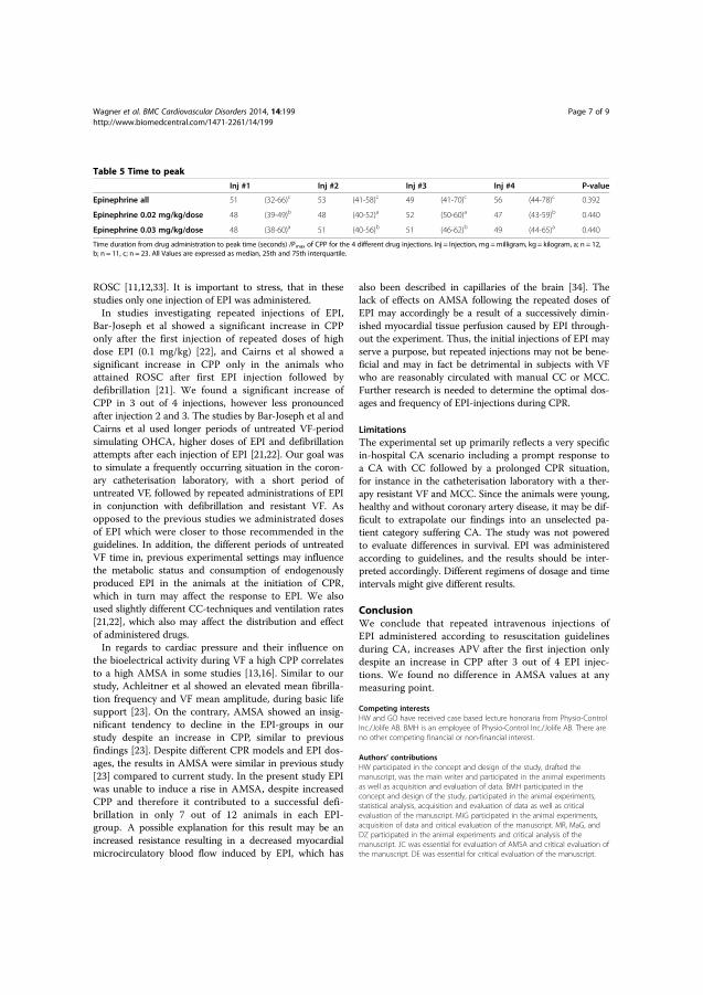
ROSC [11,12,33]. It is important to stress, that in thesestudies only one injection of EPI was administered.In studies investigating repeated injections of EPI,
Bar-Joseph et al showed a significant increase in CPPonly after the first injection of repeated doses of highdose EPI (0.1 mg/kg) [22], and Cairns et al showed asignificant increase in CPP only in the animals whoattained ROSC after first EPI injection followed bydefibrillation [21]. We found a significant increase ofCPP in 3 out of 4 injections, however less pronouncedafter injection 2 and 3. The studies by Bar-Joseph et al andCairns et al used longer periods of untreated VF-periodsimulating OHCA, higher doses of EPI and defibrillationattempts after each injection of EPI [21,22]. Our goal wasto simulate a frequently occurring situation in the coron-ary catheterisation laboratory, with a short period ofuntreated VF, followed by repeated administrations of EPIin conjunction with defibrillation and resistant VF. Asopposed to the previous studies we administrated dosesof EPI which were closer to those recommended in theguidelines. In addition, the different periods of untreatedVF time in, previous experimental settings may influencethe metabolic status and consumption of endogenouslyproduced EPI in the animals at the initiation of CPR,which in turn may affect the response to EPI. We alsoused slightly different CC-techniques and ventilation rates[21,22], which also may affect the distribution and effectof administered drugs.In regards to cardiac pressure and their influence on
the bioelectrical activity during VF a high CPP correlatesto a high AMSA in some studies [13,16]. Similar to ourstudy, Achleitner et al showed an elevated mean fibrilla-tion frequency and VF mean amplitude, during basic lifesupport [23]. On the contrary, AMSA showed an insig-nificant tendency to decline in the EPI-groups in ourstudy despite an increase in CPP, similar to previousfindings [23]. Despite different CPR models and EPI dos-ages, the results in AMSA were similar in previous study[23] compared to current study. In the present study EPIwas unable to induce a rise in AMSA, despite increasedCPP and therefore it contributed to a successful defi-brillation in only 7 out of 12 animals in each EPI-group. A possible explanation for this result may be anincreased resistance resulting in a decreased myocardialmicrocirculatory blood flow induced by EPI, which has
also been described in capillaries of the brain [34]. Thelack of effects on AMSA following the repeated doses ofEPI may accordingly be a result of a successively dimin-ished myocardial tissue perfusion caused by EPI through-out the experiment. Thus, the initial injections of EPI mayserve a purpose, but repeated injections may not be bene-ficial and may in fact be detrimental in subjects with VFwho are reasonably circulated with manual CC or MCC.Further research is needed to determine the optimal dos-ages and frequency of EPI-injections during CPR.
LimitationsThe experimental set up primarily reflects a very specificin-hospital CA scenario including a prompt response toa CA with CC followed by a prolonged CPR situation,for instance in the catheterisation laboratory with a ther-apy resistant VF and MCC. Since the animals were young,healthy and without coronary artery disease, it may be dif-ficult to extrapolate our findings into an unselected pa-tient category suffering CA. The study was not poweredto evaluate differences in survival. EPI was administeredaccording to guidelines, and the results should be inter-preted accordingly. Different regimens of dosage and timeintervals might give different results.
ConclusionWe conclude that repeated intravenous injections ofEPI administered according to resuscitation guidelinesduring CA, increases APV after the first injection onlydespite an increase in CPP after 3 out of 4 EPI injec-tions. We found no difference in AMSA values at anymeasuring point.
Competing interestsHW and GO have received case based lecture honoraria from Physio-ControlInc./Jolife AB. BMH is an employee of Physio-Control Inc./Jolife AB. There areno other competing financial or non-financial interest.
Authors’ contributionsHW participated in the concept and design of the study, drafted themanuscript, was the main writer and participated in the animal experimentsas well as acquisition and evaluation of data. BMH participated in theconcept and design of the study, participated in the animal experiments,statistical analysis, acquisition and evaluation of data as well as criticalevaluation of the manuscript. MiG participated in the animal experiments,acquisition of data and critical evaluation of the manuscript. MR, MaG, andDZ participated in the animal experiments and critical analysis of themanuscript. JC was essential for evaluation of AMSA and critical evaluation ofthe manuscript. DE was essential for critical evaluation of the manuscript.
Table 5 Time to peak
Inj #1 Inj #2 Inj #3 Inj #4 P-value
Epinephrine all 51 (32-66)c 53 (41-58)c 49 (41-70)c 56 (44-78)c 0.392
Epinephrine 0.02 mg/kg/dose 48 (39-49)b 48 (40-52)a 52 (50-60)a 47 (43-59)b 0.440
Epinephrine 0.03 mg/kg/dose 48 (38-60)a 51 (40-56)b 51 (46-62)b 49 (44-65)a 0.440
Time duration from drug administration to peak time (seconds) /Pmax of CPP for the 4 different drug injections. Inj = Injection, mg=milligram, kg = kilogram, a; n = 12,b; n = 11, c; n = 23. All Values are expressed as median, 25th and 75th interquartile.
Wagner et al. BMC Cardiovascular Disorders 2014, 14:199 Page 7 of 9http://www.biomedcentral.com/1471-2261/14/199

GKO participated in the concept and design of the study as well as criticalevaluation of the manuscript. All authors read and approved the final versionof the manuscript.
AcknowledgementsWe thank Dr Giuseppe Ristagno for valuable help and contribution inanalysing AMSA.
FundingThe study was funded by the Swedish Heart and Lung Foundation, ThelmaZoega’s Foundation, the Laerdal Foundation, Skane County Council´sResearch and Development Foundation, the Swedish Research Council,Marianne and Marcus Wallenberg´s Foundation, the Swedish Medical Societyand SUMMIT (part of the Innovative Medicines Initiative within the EuropeanCommission’s Seventh Framework Program).
Author details1Department of Cardiology, Lund University, Lund, Sweden. 2Physio-Control/Jolife AB, Ideon Science Park, Lund, Sweden. 3Department of Anesthesiologyand Intensive Care, Lund University, Lund, Sweden.
Received: 20 October 2014 Accepted: 16 December 2014Published: 20 December 2014
References1. Redding JS, Pearson JW: Resuscitation from asphyxia. JAMA 1962,
182:283–286.2. Nolan JP, Soar J, Zideman DA, Biarent D, Bossaert LL, Deakin C, Koster RW,
Wyllie J, Bottiger B: European Resuscitation Council Guidelines forResuscitation 2010 Section 1. Executive summary. Resuscitation 2010,81(10):1219–1276.
3. Neumar RW, Otto CW, Link MS, Kronick SL, Shuster M, Callaway CW,Kudenchuk PJ, Ornato JP, McNally B, Silvers SM, Passman RS, White RD, HessEP, Tang W, Davis D, Sinz E, Morrison LJ: Part 8: adult advancedcardiovascular life support: 2010 American Heart Association Guidelinesfor Cardiopulmonary Resuscitation and Emergency Cardiovascular Care.Circulation 2010, 122(18 Suppl 3):S729–S767.
4. Behringer W, Kittler H, Sterz F, Domanovits H, Schoerkhuber W, Holzer M,Mullner M, Laggner AN: Cumulative epinephrine dose duringcardiopulmonary resuscitation and neurologic outcome. Ann Intern Med1998, 129(6):450–456.
5. Gueugniaud PY, Mols P, Goldstein P, Pham E, Dubien PY, Deweerdt C,Vergnion M, Petit P, Carli P: A comparison of repeated high doses andrepeated standard doses of epinephrine for cardiac arrest outside thehospital. European Epinephrine Study Group. N Engl J Med 1998,339(22):1595–1601.
6. Mukoyama T, Kinoshita K, Nagao K, Tanjoh K: Reduced effectivenessof vasopressin in repeated doses for patients undergoingprolonged cardiopulmonary resuscitation. Resuscitation 2009,80(7):755–761.
7. Olasveengen TM, Sunde K, Brunborg C, Thowsen J, Steen PA, Wik L:Intravenous drug administration during out-of-hospital cardiac arrest:a randomized trial. JAMA 2009, 302(20):2222–2229.
8. Jacobs IG, Finn JC, Jelinek GA, Oxer HF, Thompson PL: Effect ofadrenaline on survival in out-of-hospital cardiac arrest: Arandomised double-blind placebo-controlled trial. Resuscitation 2011,82(9):1138–1143.
9. Paradis NA, Martin GB, Rivers EP, Goetting MG, Appleton TJ, Feingold M,Nowak RM: Coronary perfusion pressure and the return of spontaneouscirculation in human cardiopulmonary resuscitation. JAMA 1990,263(8):1106–1113.
10. Niemann JT, Criley JM, Rosborough JP, Niskanen RA, Alferness C: Predictiveindices of successful cardiac resuscitation after prolonged arrest andexperimental cardiopulmonary resuscitation. Ann Emerg Med 1985,14(6):521–528.
11. Kern KB, Ewy GA, Voorhees WD, Babbs CF, Tacker WA: Myocardialperfusion pressure: a predictor of 24-hour survival during prolongedcardiac arrest in dogs. Resuscitation 1988, 16(4):241–250.
12. Reynolds JC, Salcido DD, Menegazzi JJ: Coronary perfusion pressure andreturn of spontaneous circulation after prolonged cardiac arrest.Prehosp Emerg Care 2010, 14(1):78–84.
13. Marn-Pernat A, Weil MH, Tang W, Pernat A, Bisera J: Optimizingtiming of ventricular defibrillation. Crit Care Med 2001, 29(12):2360–2365.
14. Noc M, Weil MH, Gazmuri RJ, Sun S, Biscera J, Tang W: Ventricularfibrillation voltage as a monitor of the effectiveness of cardiopulmonaryresuscitation. J Lab Clin Med 1994, 124(3):421–426.
15. Povoas HP, Bisera J: Electrocardiographic waveform analysis for predictingthe success of defibrillation. Crit Care Med 2000, 28(11 Suppl):N210–N211.
16. Reynolds JC, Salcido DD, Menegazzi JJ: Correlation between coronaryperfusion pressure and quantitative ECG waveform measures duringresuscitation of prolonged ventricular fibrillation. Resuscitation 2012,83(12):1497–1502.
17. Segal J: Applications of coronary flow velocity during angioplastyand other coronary interventional procedures. Am J Cardiol 1993,71(14):17D–25D.
18. Ofili EO, Labovitz AJ, Kern MJ: Coronary flow velocity dynamics in normaland diseased arteries. Am J Cardiol 1993, 71(14):3D–9D.
19. Ninomiya Y, Hamasaki S, Saihara K, Ishida S, Kataoka T, Ogawa M, Orihara K,Oketani N, Fukudome T, Okui H, Ichiki T, Shinsato T, Kubozono T, Mizoguchi E,Ichiki H, Tei C: Comparison of effect between nitrates and calcium channelantagonist on vascular function in patients with normal or mildly diseasedcoronary arteries. Heart Vessels 2008, 23(2):83–90.
20. Wagner H, Madsen Hardig B, Steen S, Sjoberg T, Harnek J, Olivecrona GK:Evaluation of coronary blood flow velocity during cardiac arrest withcirculation maintained through mechanical chest compressions in aporcine model. BMC Cardiovasc Disord 2011, 11(1):73.
21. Cairns CB, Niemann JT: Hemodynamic effects of repeated dosesof epinephrine after prolonged cardiac arrest and CPR:preliminary observations in an animal model. Resuscitation 1998,36(3):181–185.
22. Bar-Joseph G, Weinberger T, Ben-Haim S: Response to repeated equaldoses of epinephrine during cardiopulmonary resuscitation in dogs.Ann Emerg Med 2000, 35(1):3–10.
23. Achleitner U, Wenzel V, Strohmenger HU, Krismer AC, Lurie KG, Lindner KH,Amann A: The effects of repeated doses of vasopressin or epinephrineon ventricular fibrillation in a porcine model of prolongedcardiopulmonary resuscitation. Anesth Analg 2000, 90(5):1067–1075.
24. Brown CG, Werman HA, Davis EA, Hobson J, Hamlin RL: The effects ofgraded doses of epinephrine on regional myocardial blood flowduring cardiopulmonary resuscitation in swine. Circulation 1987,75(2):491–497.
25. Wagner H, Terkelsen CJ, Friberg H, Harnek J, Kern K, Lassen JF,Olivecrona GK: Cardiac arrest in the catheterisation laboratory: A5-year experience of using mechanical chest compressions tofacilitate PCI during prolonged resuscitation efforts. Resuscitation2010, 81(4):383–387.
26. Pytte M, Kramer-Johansen J, Eilevstjonn J, Eriksen M, Stromme TA,Godang K, Wik L, Steen PA, Sunde K: Haemodynamic effectsof adrenaline (epinephrine) depend on chest compression qualityduring cardiopulmonary resuscitation in pigs. Resuscitation 2006,71(3):369–378.
27. Ristagno G, Tang W, Huang L, Fymat A, Chang YT, Sun S, Castillo C, WeilMH: Epinephrine reduces cerebral perfusion during cardiopulmonaryresuscitation. Crit Care Med 2009, 37(4):1408–1415.
28. Otlewski MP, Geddes LA, Pargett M, Babbs CF: Methods for calculatingcoronary perfusion pressure during CPR. Cardiovasc Eng 2009,9(3):98–103.
29. Olasveengen TM, Wik L, Sunde K, Steen PA: Outcome when adrenaline(epinephrine) was actually given vs. not given - post hoc analysis of arandomized clinical trial. Resuscitation 2012, 83(3):327–332.
30. Mayr VD, Wenzel V, Muller T, Antretter H, Rheinberger K, Lindner KH,Strohmenger HU: Effects of vasopressin on left anterior descendingcoronary artery blood flow during extremely low cardiac output.Resuscitation 2004, 62(2):229–235.
31. Ristagno G, Gullo A, Berlot G, Lucangelo U, Geheb E, Bisera J: Prediction ofsuccessful defibrillation in human victims of out-of-hospital cardiacarrest: a retrospective electrocardiographic analysis. Anaesth Intensive Care2008, 36(1):46–50.
32. Ristagno G, Li Y, Fumagalli F, Finzi A, Quan W: Amplitude spectrum areato guide resuscitation-a retrospective analysis during out-of-hospitalcardiopulmonary resuscitation in 609 patients with ventricular fibrillationcardiac arrest. Resuscitation 2013, 84(12):1697–1703.
Wagner et al. BMC Cardiovascular Disorders 2014, 14:199 Page 8 of 9http://www.biomedcentral.com/1471-2261/14/199

33. Lindberg L, Liao Q, Steen S: The effects of epinephrine/norepinephrine onend-tidal carbon dioxide concentration, coronary perfusion pressure andpulmonary arterial blood flow during cardiopulmonary resuscitation.Resuscitation 2000, 43(2):129–140.
34. Ristagno G, Sun S, Tang W, Castillo C, Weil MH: Effects of epinephrineand vasopressin on cerebral microcirculatory flows during andafter cardiopulmonary resuscitation. Crit Care Med 2007,35(9):2145–2149.
doi:10.1186/1471-2261-14-199Cite this article as: Wagner et al.: Repeated epinephrine doses duringprolonged cardiopulmonary resuscitation have limited effects onmyocardial blood flow: a randomized porcine study. BMC CardiovascularDisorders 2014 14:199.
Submit your next manuscript to BioMed Centraland take full advantage of:
• Convenient online submission
• Thorough peer review
• No space constraints or color figure charges
• Immediate publication on acceptance
• Inclusion in PubMed, CAS, Scopus and Google Scholar
• Research which is freely available for redistribution
Submit your manuscript at www.biomedcentral.com/submit
Wagner et al. BMC Cardiovascular Disorders 2014, 14:199 Page 9 of 9http://www.biomedcentral.com/1471-2261/14/199
