clinical article removal of thermafil root canal filling ...€¦ · removal showed no deformation...
TRANSCRIPT

0099-2399/97/2301-0054503.00/0 JOURNAL OF ENDODONTICS Copyright © 1997 by The American Association of Endodontists
CLINICAL ARTICLE
Printed in U.S.A. VOL. 23, No. 1, JANUARY 1997
Removal of Thermafil Root Canal Filling Material
Marie-France Bertrand, DCD, Jean-Claude Pellegrino, DCD, Jean-Paul Rocca, DCD, DSO, DEBH, Ann Klinghofer, DCD, MS, and Marc Bolla, DCD, DSO
The aim of this study was to evaluate if removal of Thermafil plastic carriers and reestablishment of working length were possible in single rooted teeth filled with the Thermafil system. Twenty freshly extracted maxillary central incisors were prepared and filled with #30 Thermafil plastic obturators ac- cording to manufacturer's recommendations. Teeth were divided in two groups:
Group I: 10 teeth were retreated using dimeth- ylformamide as a solvent
Group Ih 10 teeth were retreated using chloro- form as a solvent
Removal of filling material was performed man- ually using K files and H files alternately between carrier and dentinal walls. The average time needed to remove the plastic carrier was 7 min for group I and approximately 61/2 min for group II. No deformation of the plastic carrier was observed after removal from the canal.
Removal of plastic carriers and measurement of the proper working length were easily performed using manual endodontic instruments. Chloroform greatly enhanced this procedure.
The goal of endodontic therapy is to achieve a three dimensional seal of the root canal system. One of the requirements of endodon- tic filling materials is biocompatibility. Gutta-percha associated with a root canal sealer is a commonly accepted material, and many different techniques using gutta-percha have previously been de- scribed.
The Thermafil technique described by Dr. Wm. Ben Johnson and popularized in France during the past 2 yr (t) has already been studied regarding sealing ability of the apex. Effectiveness of the apical seal has been tested and compared with other filling tech- niques using dye penetration. Beatty et al. (2) found less penetra- tion of the dye using Thermafil, compared to the lateral conden- sation and single cone techniques. On the other hand, Lares and Eldeeb (3) obtained better results with lateral condensation. A
comparable study by Chohayeb (4) seems to confirm Lares' re- sults. Baker and Oguntebi (5) found no significant difference of dye penetration on teeth obturated with the Thermafil system without apical root resection and after apical root resection and retrograde filling with amalgam. McMurtrey et al. (6), using teeth with severe root canal curvatures (angle > 30°), found no signif- icant differences in comparing lateral condensation and Thermafil. The authors observed that sealer was often extruded into the apical constriction and that at other times both gutta-percha and sealer were extruded. Other studies have been based on the relationship between canal walls, gutta-percha, sealer, and carrier. Juhlin et al. (7) studied this relationship on resin blocks simulating severe canal curvatures. They frequently observed contact between the metal carrier and the canal walls in the coronal third and the middle third as well as asymmetrical distribution of components in the apical third. The metal carrier was never completely surrounded with gutta-percha, and sealer was often missing. The authors theorized that the results were due to the lack of flexibility of the metal carrier. Fabra-Campos (8) studied the apical seal on human teeth. Contact between metal carrier and canal walls in the apical third was often seen. In a previous study on human teeth with simple anatomy (9), it was shown that the plastic carder never reached the cementodentinal junction.
Due to complex root canal anatomy, all obturation techniques are subject to retreatment. It might be of interest to evaluate retreatment cases when a "foreign body" has been left for a long period of time within the bulk of the filling. The goal of this study was to verify if removal of Thermafil plastic carrier and reestab- lishment of working length were possible in single rooted canals filled with the Thermafil system.
54
MATERIALS AND METHODS
Twenty human maxillary central incisors (freshly extracted for periodontal reasons) were selected for this study. Each tooth was numbered, measured, and two radiographs (buccal and mesial views) were taken to demonstrate the complexity of the root canal system.
Each tooth was accessed with a high-speed #2 diamond round bur and safety fissure bur (Endo Z #152 Maillefer) to remove all coronal interferences. Canal preparation was performed according to manufacturer's recommendations. Initial penetration was made

Vol. 23, No. 1, January 1997
using a #08 MMC file (Micromega) inserted into each canal to the apical constriction. Then #08, #10, and #15 MMC and MME files (Micromega, ProFile Hedstrom) were used alternately. Radiograph was taken with the #15 MME file at the working length. The coronal and the middle thirds were shaped using Gates-Glidden drills from #5 to #1 in the straight portion of the canal. The apical third was shaped with #20, #25, and #30 K-type and Hedstrom files. Next a #35 K-type file, 1 mm short of the working length, and a #40 K-type file, 2 mm short of the working length, were used. All canal preparation was done under copious irrigation with 3% sodium hypochlorite using Monoject-type syringe. Canals were dried with sterile standardized paper points corresponding to a #30 file. A #30 Thermafil plastic obturator corresponding to the #30 file was heated in the Thermaprep for 5 minutes. The root canal sealer (Endobtur, Laboratoire Septodont, France) was mixed ac- cording to manufacturer's recommendations. A small amount of sealer was carried to the working length with a #15 file. The obturator was then inserted into the canal to the working length using a firm vertical motion. The plastic carrier was cut at the canal orifice using a high-speed round bur. The gutta-percha, still in the thermoplasticized phase, was vertically compacted around the car- rier with a plugger. Access cavity was filled with IRM (Caulk). Control radiographs (buccal and mesial views) were then taken.
After cement was set (24 hours later), the IRM filling was removed with a slow-speed #06 round bur, and teeth were divided in two groups. In group I, 10 teeth were retreated using dimeth- ylformamide as a solvent. The teeth in group II were retreated using chloroform as a solvent. The access cavity was completely filled with the solvent. The removal of the material was done using a reverse sequence of endodontic hand instruments, alternately K and H files #40 to #15. K files were used to reach the working length and H files to remove gutta-percha debris on the surface. The solvent was renewed after each file. Measurements were done after 1, 2, 3, and 4 minutes. The necessary time to reach the working length was recorded and a radiograph was taken when the working length was reached.
Canals were then observed after a longitudinal splitting of all specimens. A groove was made using a slow-speed carborundum disk to canal proximity, and chisels were used to complete the fracture of the specimens.
The fractured teeth were observed under an Olympus WMZ 1-4 stereozoom microscope (Olympus Optical Co., Hamburg, Germa- ny). Illumination was improved with two fiber optic light sources and specimens were observed to magnification x 6 to 50. Photo- graphs were taken with an Olympus camera and a 100 ASA Ektachrome film, used at 1/60 second.
Some specimens were studied with a scanning electron micro- scope (JEOL 35, Jeol LTD, Tokyo, Japan). The two fractured parts of each specimen were fixed on a Balzer-Union cartier with a double-sided adhesive tape and Emerton silver glue. The whole system was coated with gold. The specimens were introduced in the SEM column under 35kV power and observed under magni- fication X 15 to 100. Photographs were taken on Polaroid 55 film.
RESULTS
Data collected for groups I and II are shown in Tables 1 and 2. The average time required to remove the plastic carrier was 7 rain for group I and 6V2 min for group II (Table 3). Complete removal of filling material was successful in 18 instances. The whole carrier
Thermafil Removal 55
TABLE 1. Dimethylformamide solvent--Specimens 1 to 10
N ° W.L. L1 L2 L3 L4 T
1 20 mm 1 2 m m 1 7 m m 1 8 m m 1 9 m m 7' 2 22 mm 1 4 m m 1 8 m m 2 2 m m 2 2 m m 5' 3 26 mm 11 mm 1 6 m m 2 3 m m 2 3 m m 7' 30" 4 29 mm 1 3 m m 2 0 m m 2 5 m m 2 5 m m 7' 5 19 mm 1 2 m m 1 6 m m 1 8 m m 1 8 m m 8' 6 21 mm 11ram 1 4 m m 18 ram 2 0 m m 7' 7 22 mm 1 6 m m 2 0 m m 2 0 m m 2 0 m m 4' 8 20.5 mm 1 0 m m 1 4 m m 15ram 1 5 m m 5' 30" 9 21 mm 1 6 m m 1 9 m m 1 9 m m 19mrn 7'
10 21 mm 1 4 m m 1 6 m m 1 6 m m 1 8 m m 12'
W.L.: working length; L1, L2, L3, L4: measurement of files penetration after 1, 2, 3, 4 minutes; T: Time needed to remove the plastic carrier and to get back to the W.L
TABLE 2: Chloroform solvent--Specimens 11 to 20
N ° W.L. L1 L2 L3 L4 T
11 2 4 m m 1 3 m m 1 7 m m 1 8 m m 1 8 m m 4' 30" 12 2 4 m m 1 0 m m 15ram 1 9 m m 1 9 m m 4' 30" 13 21 rnm 1 5 m m 1 7 m m 1 9 m m 1 9 m m 5' 30" 14 2 4 m m 1 4 m m 1 9 m m 2 0 r a m 2 0 m m F 15 2 4 m m 11 mm 1 3 m m 1 6 m m 1 8 m m 10' 40" 16 2 2 m m 1 3 m m 16ram 1 8 m m 1 8 m m 6' 30" 17 24 rnm 12ram 1 5 m m 1 7 m m 1 7 m m 9' 45" 18 2 2 r a m 1 2 m m 1 8 m m 21 mm 21 mm 4' 19 2 3 m m 6 m m 1 0 m m 1 3 m m 1 5 m m 6' 20 2 2 m m 1 0 m m 1 3 m m 1 7 m m 2 0 m m 6' 30"
W.L: working length L1, 1.2, L3, L4: measurement of files penetration after 1,2, 3, 4 minutes T: Time needed to remove the plastic carrier and to get back to the W.L F: 40 H file fractured
TABLE 3. Time needed to remove the Thermafil plastic carrier.
DMF SPECIMENS T CHL SPECIMENS T
1 270" 11 420" 2 275" 12 300" 3 330" 13 450" 4 380" 14 420" 5 640" 15 480" 6 390" 16 420" 7 585" 17 240" 8 240" 18 330" 9 360" 19 420"
10 390" 20 720" AVERAGE TIME 386" AVERAGE TIME 420"
was extracted by use of the H file. Upon removal of the carrier, proper working length was recovered.
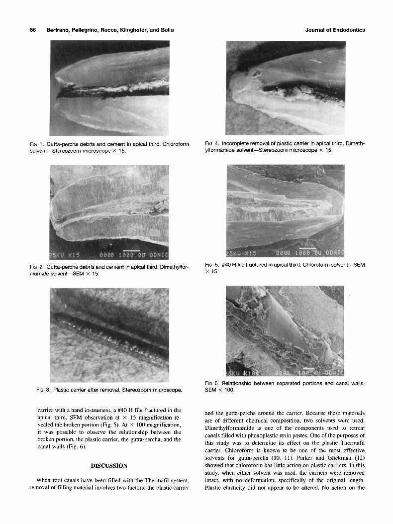
The study of the specimens by stereozoom microscope and SEM showed canal walls with gutta-percha debris and cement in the apical third (Figs. 1 & 2).
Observation of the carrier with the stereozoom microscope after removal showed no deformation of the plastic carrier with either chloroform or dimethylformamide used as solvents (Fig. 3). In two instances, complete removal of filling material was unsuccessful:
• Incomplete removal of the plastic carrier: the remainder was imbedded in the canal walls, as shown in Figure 4. However, it was possible to bypass the cartier, reaching the working length with the file.
• #40 H file fractured: during removal of the plastic
56 Bertrand, Pellegrino, Rocca, Klinghofer, and Bolla Journal of Endodontics
i:,[~ ~ ~
FIG 1. Gutta-percha debris and cement in apical third. Chloroform solvent--Stereozoom microscope × 15.
FIG 4. Incomplete removal of plastic carrier in apical third. Dimeth- ylformamide solvent--Stereozoom microscope × 15.
FIG 2. Gutta-percha debris and cement in apical third. Dimethylfor- mamide sotvent--SEM × 15.
FiG 5. #40 H file fractured in apical third. Chloroform solvent--SEM x15 .
FIG 3. Plastic carrier after removal. Stereozoom microscope. FIG 6. Relationship between separated portions and canal walls. SEM x 100.
carrier with a hand instrument, a #40 H file fractured in the apical third. SEM observation at × 15 magnification re- vealed the broken portion (Fig. 5). At × 100 magnification, it was possible to observe the relationship between the broken portion, the plastic carrier, the gutta-percha, and the canal walls (Fig. 6).
DISCUSSION
When root canals have been filled with the Thermafil system, removal of filling material involves two factors: the plastic carrier
and the gutta-percha around the carrier. Because these materials are of different chemical composition, two solvents were used. Dimethylfonnamide is one of the components used to retreat canals filled with phenoplastic resin pastes. One of the purposes of this study was to determine its effect on the plastic Thermafil carrier. Chloroform is known to be one of the most effective solvents for gutta-percha (10, 11). Parker and Glickman (12) showed that chloroform has little action on plastic carriers. In this study, when either solvent was used, the carriers were removed intact, with no deformation, specifically of the original length. Plastic elasticity did not appear to be altered. No action on the

Vol. 23, No. 1, January 1997
plastic carrier was demonstrated when using either dimethylform- amide or chloroform.
Removal of filling material was performed manually using K files and H files alternately between plastic carrier and dentinal wall. Removal of the gutta-percha around carriers was facilitated by the use of solvents and manual endodontic instruments. File progression was easily accomplished until the apical third was encountered. This was because there was a homogeneous bulk of gutta-percha around the carrier in the middle third and coronal third. When the apical third was reached, progress was slowed due to narrowing of the canal, leaving only a thin layer of gutta-percha between the wall and the carrier. Thus two failures were observed:
• A #40 H file fractured: #40 H file was probably used in a vertical apical direction instead of a pulling motion.
• A plastic carder fractured: probably due to a preoper- ative torque motion during insertion of the cartier within the root canal.
• Further studies using teeth with severe root canal cur- vatures should be performed to complete these results.
Dr. Bertrand is an Assistant, Department of Restorative Dentistry and Endodontics, UFR of Odontology, University of Nice. Dr. Pellegrino is an Associate Researcher, Laboratory of Biomatedals and Experimental Odontol- ogy, UFR of Odontology, University of Nice. Dr. Rocca is a Professor and Chairman, Department of Restorative Dentistry and Endodontics, UFR of Odontology, University of Nice. Dr. Klinghofer is an Endodontist in private practice and Associate Researcher, Laboratory of Biomaterials and Experi- mental Odontology, UFR of Odontology, University of Nice. Dr. BoUa is an
Thermafil Removal 57
Assistant Professor, Department of Dental Biomaterials, UFR of Odontology, University of Nice.
Address requests for reprints to Dr. M. F. Bertrand, UFR d'Odontologie, Universit~ de Nice-Sophia Antipolis, Parc Valrose, Avenue Joseph Vallot, 06108 Nice Cedex 2, France.
References
1. De Jaureguiberry M. L'obturateur Thermafil. Endo 1992;4:29-39. 2. Beatty RG, Baker PS, Haddix J, Hart F. The efficacy of four root canal
obturation techniques in preventing apical dye penetration. J Am Dent Assoc 1989;119:633-7.
3. Lares C, Eldeeb M. The sealing ability of the Thermafil obturation technique. J Endodon 1990;10:474-9.
4. Chohayeb A. Comparison of conventional root canal obturation tech- niques with Thermafil obturators. J Endodon 1992;1:10-2.
5. Baker PS, Oguntebi BR. Effect of apical resections and reserve fillings on Thermafil root canal obturations. J Endodon 1990;5:227-9.
6. McMurtrey LG, Krell KV, Wilcox LR. A comparison between Thermaffi and lateral condensation in highly curved canals. J Endodon 1992;2:68-71.
7. Juhlin J, Walton R, Dovgan J. Adaptation of Thermafil components to canal walls. J Endodon 1993;3:130-5.
8. Fabra-Campos H. Experimental apical sealing with a new canal obtu- ration system. J Endodon 1993;2:71-5.
9. Bertrand MF, Larousse JM, Rocca JP. Scellement apical et syst~eme Thermafil. Endo 1993;3:17-31.
10. Tamse A, Unger V, Metzger Z, Rosenberg M. Gutta percha solvents, a comparative study. J Endodon 1986;8:337-9.
11. Wourms D, Campbell D, Hicks L, Pelleu G. Alternative solvents to chloroform of gutta percha removal. J Endodon 1990;5:224-6.
12. Parker H, Glickman GN. Solubility of plastic Thermafil carriers. [Ab- stract 680] IADR 1992.
A Word for the Wise
Rarely, a great insight is expressed. Consider Alfred North Whitehead's suggestion that "Civilization ad- vances by extending the number of important operations which we can perform without thinking about them." I take it that "without thinking about them" in this context does not mean "mindlessly" but rather "without laboriously planning and considering all of the alternatives for each step in the process."
Taken thus, civilization might be deduced tO have progressed from building a fire to trading derivatives.
A. Smith