clinical characteristics and epidemiology of pulmonary ... · clinical characteristics and...
TRANSCRIPT

R
C
Aa
b
c
a
ARAA
KFPPPS
PBPPIS
1d
Rev Iberoam Micol. 2012;29(1):1–13
Revista Iberoamericanade Micología
www.elsev ier .es / rev iberoammicol
eview
linical characteristics and epidemiology of pulmonary pseudallescheriasis
yse Serda Kantarcioglua,∗, Gerritis Sybren de Hoogb, Josep Guarroc
Department of Microbiology and Clinical Microbiology, Cerrahpasa Medical Faculty, 34303 Cerrahpasa, Istanbul, TurkeyCentraalbureau voor Schimmelcultures, Utrecht, and Institute for Biodiversity and Ecosystem Dynamics, University of Amsterdam, Amsterdam, The NetherlandsUnitat de Microbiologia, Facultat de Medicina i Ciències de la Salut, IISPV, Universitat Rovira i Virgili, E-43201 Reus, Spain
r t i c l e i n f o
rticle history:eceived 31 December 2010ccepted 1 April 2011vailable online 7 May 2011
eywords:ungus ballseudallescheriaseudallescheriomaulmonary fungal infectionscedosporium
a b s t r a c t
Background: Some members of the Pseudallescheria (anamorph Scedosporium) have emerged as an impor-tant cause of life-threatening infections in humans. These fungi may reach the lungs and bronchial treecausing a wide range of manifestations, from colonization of airways to deep pulmonary infections.Frequently, they may also disseminate to other organs, with a predilection for the brain. In otherwisehealthy patients, the infection is characterized by non-invasive type involvement, while invasive and/ordisseminated infections were mostly seen in immunocompromised patients.Aims: We reviewed all the available reports on Pseudallescheria/Scedosporium pulmonary infections,focusing on the geographical distribution, immune status of infected individuals, type of infections,clinical manifestations, treatment and outcome.Results and conclusions: The main clinical manifestations of the 189 cases of pulmonary pseudallescheri-asis reviewed were pneumonia (89), followed by fungus ball (26), and chest abscess (18). Some patientshad more than one type of invasive pulmonary manifestations. Among patients with pneumonia, severalcases of pneumonia associated with near-drowning (10/89, 11.2%) have also been reported in immuno-competent hosts. Major underlying conditions for non-invasive pulmonary infection were preexistinglung cavities and medical immunosuppression for invasive pulmonary infection. Saprobic airway col-onization was mostly seen in patients with mucosal dysfunction, i.e. patients with cystic fibrosis. Themortality rate was closely related to the infection type, being 26.8% in non-invasive type (fungus balls)and 57.2% in invasive type.
© 2010 Revista Iberoamericana de Micología. Published by Elsevier España, S.L. All rights reserved.
Características clínicas y epidemiología de la pseudalescueriasis pulmonar
alabras clave:ala fúngicaseudallescheriaseudalesqueriomanfecciones fúngicas pulmonarescedosporium
r e s u m e n
Antecedentes: Algunos miembros del género Pseudallescheria (anamorfo Scedosporium) están emergiendocomo causantes de infecciones humanas graves. Estos hongos pueden alcanzar los pulmones y el árbolbronquial causando una amplia variedad de manifestaciones clínicas, desde colonizaciones de las víasaéreas hasta infecciones pulmonares profundas. Frecuentemente estos hongos pueden diseminarse aotros órganos, mostrando una marcada predilección por el cerebro. En pacientes por otra parte sanos lainfección no suele ser invasora, mientras que en el paciente inmunocomprometido se caracteriza por sucarácter invasor.Objetivos: Se ha llevado a cabo una revisón de los artículos disponibles sobre infecciones pulmonares porPseudallescheria/Scedosporium, destacando la distribución geográfica de las mismas, el estado inmunitariode los pacientes, el tipo de infección, las manifestaciones clínicas, el tratamiento y curso clínico de laenfermedad.
Resultados y conclusiones: La principal manifestación clínica de los 189 casos de pseudalescheri-asis pulmonar revisados fue neumonía (89), seguido por la presencia de bola fúngica (46),y absceso pulmonar (18). En algunos casos de sujetos inmunocompetentes la neumonía fuedebida a aspiración con agua contaminada (10/89, 11,2%). Los principales factores de riesgopara las infecciones pulmonares no invasoras fueron la preexistencia de cavidades pulmonaresy el tratamiento inmunosupresor para infecciones pulmonares invasoras. La colonizaciónsaprofítica de vías aéras se observó principalmente en pacientes con alteraciones de la mucosa,∗ Corresponding author.E-mail address: [email protected] (A. Serda Kantarcioglu).
130-1406/$ – see front matter © 2010 Revista Iberoamericana de Micología. Published by Elsevier España, S.L. All rights reserved.oi:10.1016/j.riam.2011.04.002

2 A. Serda Kantarcioglu et al. / Rev Iberoam Micol. 2012;29(1):1–13
como aquellos con fibrosis quística. La tasa de mortalidad estuvo estrechamente relacionada con el tipode infección, siendo del 26,8% en las infecciones no invasoras (bola fúngica) y del 57,2% en las invasoras.
amer
oructimtpadmmarimctipmtd
M
L
Lrc“u“n“atmnawcvIu
D
tpulai
© 2010 Revista Ibero
Currently, Pseudallescheria/Scedosporium infections are somef the most prevalent mould infections in humans, being theespiratory tract the most commonly infected site.52 Recent molec-lar studies have demonstrated that Pseudallescheria/Scedosporiumomplex (PSC) includes several phylogenetic species,46 but sincehe degree of involvement of each individual species in humannfections has not been determined, the present review will
aintain the name PSC in all disease entities. The species ofhat complex and relatives recovered so far from clinical sam-les are: Scedosporium apiospermum (teleomorph Pseudallescheriapiosperma), Scedosporium aurantiacum, Scedosporium boydii (Pseu-allescheria boydii), Pseudallescheria angusta and Pseudallescheriainutispora.46,47 Several types of respiratory system involve-ents of PSC have been described in both immunocompromised
nd immunocompetent individuals. Three general reviews, on aange of PSC infections29,52 and central nervous system (CNS)nfections,68 have recently been published. In addition, a few
ore, shorter, reviews each covering a small number of previousases6,9,14,82,109,113,115 have also been published. The clinical spec-rum of the disease associated with PSC was examined by Rippon,n 1980.112 In the present study, the available case reports of PSCulmonary infections have been reviewed chronologically to clarifyany aspects associated particularly with these illnesses, including
he risk factors and underlying conditions, clinical manifestations,iagnostic factors, treatment and outcome.
ethods
iterature search
A computerized search of the MEDLINE database (Nationalibrary of Medicine, Bethesda, Maryland, USA) was made for caseseported in the literature between 1955 and mid-2009, with (byross-referencing) the terms: “P. boydii” and “S. apiospermum”,pulmonary”, “pneumonitis”, “lung abscess”, “pulmonary nod-les”, “mycetomas”, “fungomas”, “respiratory system infection”,disseminated” and “near-drowning”, “respiratory system colo-ization”, “Pseudallescherial colonization”, “fungal colonization”cystic fibrosis”, “allergic bronchopulmonary pseudallescheri-sis”, “scedosporiosis” and “pseudallescheriasis”. Additional searcherms included were “Allescheria boydii”, “Monosporium apiosper-um”, and “Petriellidium boydii” as referring to prior or otheromenclature for this fungus. These key words were used alonend/or in combination with an “and” statement. Additional casesere found by scanning the references cited in the original arti-
les. Original full texts of all the relevant articles were foundia MEDLINE, TUBITAK-ULAKBIM (Turkish Academic Network andnformation Center), and/or other international libraries and weresed for the analysis or personal communication of the authors.
efinitions
A case was considered an invasive pulmonary infection whenhe presence of lesion and clinical syndrome consistent withulmonary infection (involvement of lung parenchyma) was doc-
mented and any species of the PSC was recovered from theesion, usually from lung tissue, mucosal biopsy, aspirate from anbscess or bronchoalveolar lavage fluid (BAL). Cases were includedn the study as non-invasive involvement when the fungus grew in
icana de Micología. Publicado por Elsevier España, S.L. Todos los derechosreservados.
pre-existing lung cavities from a previous disease, i.e. tuberculo-sis or sarcoidosis, without invading the cavity wall. The mass maymove within the cavity but does not invade the cavity wall.
Infection types that refer to a saprobic involvement, such as fun-gus ball or mycetoma were evaluated and categorized as reportedby the authors. Duplicate publications were excluded and followup reports were regarded as associated with a single case togetherwith the previous report. The following data were recorded for eachpatient, if stated: age and sex, geographical location, predispos-ing factors (including underlying diseases and associated medicalconditions), clinical symptoms, mode and time to diagnosis, otherpathogens isolated or observed in specimens if any, antimicrobialagents administered, regimens and duration of antifungal therapy,invasive or surgical procedures, duration of hospitalization, andpatient outcome.
Results
There were 231 case reports and records of isolation of PSCfrom pulmonary specimens identified from 1955 to end-2010.PSC was first reported as a cause of pulmonary disease in 1955by Creitz and Harris,30 although the organism was probablya secondary invader, being inhaled from the soil. Four caseswere described twice,22,67,74,84,85,109,134,145 due to the progres-sion or reactivation of the disease. No details of the patients’histories were available in two case reports.36,38 One case wassummarized in a general report on brain abscesses followingbone marrow transplantation,36 and the presence of the fun-gus in sputum was mentioned in an environmental study.31 Inanother case with bronchiectasis, in spite of PSC being repeat-edly isolated from the patient’s sputum it was not obtained inculture from the intercavitary mass, in which many Aspergillusfumigatus conidiophores were histologically observed.109 Of these231 published cases, 56 involved patients with cystic fibrosis(CF).23,26,27,32,53,59,63,83,86,94,105,119,127,129,135,144,148 Twelve of theseCF patients were reported to have invasive and two non-invasivepulmonary infection.53,83,86,94,127,135,144 A total of 189 cases wereinvasive or non-invasive infections with isolation of PSC from lowerrespiratory tract specimens.
Overall demographic and geographic features
The majority of those 189 pulmonary pseudallescheriasis werereported from the USA (78 cases), followed by Australia (40 cases),Japan (14 cases), France (14 cases) and Germany (7 cases). Occa-sionally, there were cases reported from Argentina, Belgium, Brazil,Canada, China, Congo, Croatia, Finland, India, Spain, Mexico, TheNetherlands, Poland, Spain, Taiwan and the UK [total = 80 femalepatients and 101 male, age range = 2–90 years].
Portals of entry and route of dissemination
Pulmonary involvement, which mainly affectedfarmers, probably resulted from inhalation of theconidia.33,53,64,67,72,83,86,94,120,123,127,135,152 A case was reported in
an immunocompetent patient who was working in a thermal bath,being in charge of scrubbing off the sedimented filth at the bottomof the pools after draining the water. S. apiospermum was isolatedfrom several samples of the thermal water and the sediment filled
ev Ibe
taasmotafbittiic
C
bmeiwtecowctmafpsrewccfasstrh
C
trubtttelfio
A. Serda Kantarcioglu et al. / R
he patient’s working place.138 In one case, the patient suffered lymphatic and haematogenous dissemination of the fungus via
skin injury while gardening and developed a lymphocutaneousyndrome, similar to sporotrichosis, along with a lung mass.74 Pul-onary involvement may have been secondary from septic emboli
riginated from lymphangitis or phlebitis in the left arm. Aspira-ion of polluted water was reported in 17 patients who developed
CNS infection. It is likely that after an invasive pneumonitis, theungus can reach the CNS by haematogenous spread facilitatedy the immunosuppression.52 The fungus could also be directly
noculated through a perforated chest wound, or inhaled,134,145 orransferred from an infected donor to an organ recipient.143 Pat-erson et al.,101 reported a case of nosocomial pseudallescheriasisn a liver transplant patient who was probably not colonized ornfected as he was immunocompetent on admission but developedavitary lung and brain lesions on day 25 post-transplant.
olonization of bronchial lumen or intracavitary colonization
PSC can grow within poorly draining bronchi, causing an endo-ronchial saprobic colonization without tissue invasion. The fungusay colonize the respiratory tract of people exposed to a high
nvironmental inoculum in the absence of anatomical or phys-ological abnormalities of the respiratory tract. This colonization
ould most likely be transient once the patient is removed fromhe environmental source. Transient colonization without appar-nt invasion has been recorded secondary to other diseases oronditions.67,109,111,126 Rippon and Carmichael111 reported a casef transient colonization of bronchial lumen in which the patientas on prednisone for 15 years for rheumatoid arthritis, and had
oughing, wheezing, and pulmonary congestion. Direct examina-ion of several sputum specimens revealed intertwined hyphal
asses and PSC was cultured from all samples. Reddy et al.109
nd Jung et al.67 described another transient colonization in aarmer’s wife with chronic bronchiectasis and chronic obstructiveulmonary disease. Castiglioni et al.22 reported the case of threeolid-organ transplant patients who had airway colonization andeceived itraconazole (ITZ) prophylaxis, without evidence of dis-ase. Similarly, in an allogenic bone marrow transplant patientith acute lymphocytic leukemia, treated with chemotherapy,
yclosporin and corticosteroids for graft-versus-disease compli-ation, A. fumigatus was isolated from sputum culture 5 monthsollowing the transplantation. The patient was treated with ITZ and
follow up sputum culture revealed a heavy growth of PSC. Lipo-omal amphotericin B was added to the treatment and repeatedputum cultures and a bronchoalveolar lavage fluid were nega-ive for PSC.12 Endobronchial chronic colonization by PSC has beeneported in CF patients, often without pathological effects for theost.
linical presentations
The role of PSC in producing pulmonary lesions and some ofheir relevant conditions has already been discussed in earliereports. However, pseudallescherial lung infections have contin-ed to be reported and consequently their clinical spectrum haseen considerably enlarged. The most relevant clinical manifesta-ions of infection are outlined in Table 1. Of those, pneumonitis washe most common clinical manifestation (94/189, 49.7%). Althoughhe chest X-rays were not specific, they were usually helpful instablishing the diagnosis. A dense infiltrate first appears, followed
ater by cavitation and in some cases by the development of aungus ball, mostly in the upper lobes. Fulminant spread withnvasion through the lung parenchyma and the pleura and devel-pment of pleural effusion has commonly been described. Caseroam Micol. 2012;29(1):1–13 3
reports that have based the diagnosis of pulmonary disease on theisolation of PSC from sputum are contradictory.75 Most patientswith this fungus in the sputum do not appear to have invasiveinfection.72 Cases of pulmonary pseudallescheriasis appear similarto pulmonary aspergillosis, clinically, radiologically, histologically,and in terms of severity. Macroscopically, pulmonary pseu-dallescherial infections produce inflammatory cystic or cavitarylesions. Regarding the data obtained from the above-mentionedcases, pulmonary pseudallescheriasis can be subdivided into threecategories:
(i) Pulmonary mycetomas and fungus balls (pseudallescheriomas)Forty-six case reports of non-invasive involvement of intratho-
racic cavities, which can be divided into two groups as pulmonarymycetoma (18/46, 39.1%) and pseudallescherioma (28/46, 60.9%),were identified. The terminology used here is based on the spe-cific descriptions made in the different case reports. Pulmonarymycetomas were reported to contain many small, greyish-yellowand white granules, measuring 1–2 mm in diameter, within thick,brownish, semi-fluid, odourless exudate. The granules of pul-monary mycetoma consist of closely intertwined hyphal massesand occasional swollen cyst-like chlamydospores. In rare instances,white or yellow lobulated granules of up to 4 mm in diameter havebeen observed.5,8,11,14,21,30,38,53,57,58,81,91,109,141 There has been noevidence of any cementing substance between the hyphae or pro-duction of conidia on the periphery of the granules.10,53,111
Intercavitary colonization may typically lead to the formationof a mass consisting of loose hyphal strands or conglomerationof intertwined fungal hyphae admixed with mucus and cellulardebris within a preexisting pulmonary cavity or ectatic bronchus.A patient with this type of infection may have a chronic pulmonaryinfiltrate from a previously existing disease, such as sarcoidosisor tuberculosis.86,111,112 People who have pre-existing lung prob-lems, especially with cavities typically affected by tuberculosis,92
sarcoidosis etc. are at risk of developing non-invasive amorphousfungal masses, called fungomas, fungus balls or in this case pseu-dallescheriomas. The fungus settles in a cavity and is able to growfree from interference because the immune system is unable topenetrate the cavity. As the fungus multiplies, it forms a ball whichincorporates dead tissue from the surrounding lung, mucus, andother debris.
Pseudallescherioma of the lung is the extremeconsequence of intercavitary colonization, where themass of fungus reaches sufficient size to be visibleradiologically.3,9,14,22,44,69,87,109,111,119,121,123,125,152,154 Radio-graphs of the pseudallescheriomas show the presence of a solid,round or oval mass with soft tissue opacity within a lung cavity.
Pseudallescherioma may be different in its morphological fea-tures; concentric rings of hyphae radiating from a central area weremainly noted.121 In addition, conidia occur on the surface where themass is in contact with an air space, generally on the periphery ofthe pseudallescherioma.69,72,75,121 Similarly to that which occursin aspergillosis, pseudallescherioma are found in the upper lobeof patients with pre-existing lung disease and are often associatedwith a thickening of the cavity wall and adjacent pleura.140
In non-invasive type cases, these fungi did not invade the tissues,their presence as a mass within cavities stimulated chronic activeinflammation and a markedly vascular granulation tissue response.Based on two case reports,107,140 Przyjemski108 hypothesized thatfungus balls may begin as “tissue balls” infiltrated by fungus. Inthe first case,107 the radiological progression from normal lungthrough poorly defined infiltrate to fungus ball occurred within
two weeks and coincided with recovery from granulocytopeniaand derived from infected lung sequestra with inflammatory infil-trate. Since surgically removed fungus balls usually fail to grow onlaboratory media,3,5,69,110,115,140,141 the author concluded that the
4 A. Serda Kantarcioglu et al. / Rev Iberoam Micol. 2012;29(1):1–13
Table 1Overall clinical manifestations of 189 respiratory involvement by Pseudallescheria/Scedosporium complex.
Clinical manifestations
Allergic bronchopulmonarypseudallescheriasis
Non-invasive types Invasive types*
Number ofpatients
References Type Number ofpatients
References Type Number of patients References
5 12, 76, 90 (2 p), 111 Pulmonarymycetoma
18 8, 11, 14, 21, 33, 40, 57,58, 67 (& 109 s), 73, 81,91, 100, 125 (2 p), 125,131, 141
Bronchopneumonia 4 34, 44, 144, 151
Fungus ball 28 3 (&11 s), 5, 9, 10, 22,25, 30 (&139 s), 32, 39,44, 60, 67, 69, 87, 92,109, 111, 119, 120, 123,125 (3 p), 133, 139, 152(2 p), 154
Pneumonia 94 1, 4, 5, 7, 12, 15, 16,17, 18, 22 (3 p), 24,28, 37 (4 p), 43 (5p), 45, 53, 56, 65 (3p), 66, 71, 77, 78,79, 83, 86 (5 p), 88,94, 95, 97, 103 (7p), 104, 106,110,117 (3 p), 122, 127,128, 130, 134, 135,136, 137 (7 p), 138,143, 146 (3 p), 147(18 p), 149
Necrotising pneumoniaassociated with abscess
18 6, 7, 13, 34, 39, 49,51, 55, 61, 62, 66,70, 90, 96, 116,132,142, 150
Cavitary necrotizatingpneumonia
10 35, 48, 64, 84 (2 p),98, 99, 114, 118,149
Nodular pneumonia 8 9, 17, 22, 43, 54, 72,101, 124
Cystic mass formation 3 22, 74, 98Intrabronchial polipoidlesions
2 96, 153
Invasion of pulmonaryvessels
2 149, 150
A
ph
Drm(r
ocic(t4ap
ShcicTiCc
bbrevations: p: patients; s: the same patient.* Total of patients is not 189 because some IPP patients had more than one type.
seudallescherioma formation might be associated with improvingost resistance.
emographic and geographic features. Most cases have beeneported from the USA, with occasional cases from the UK, Ger-any, France, Poland, India, Japan, Canada, Brazil and Australia
female, n = 24, male, n = 20, gender was not indicated in the othereports, age range = 11–81).
Predisposing factors and underlying conditions. Twenty sevenf these 44 patients had associated diseases, which could haveontributed to the occurrence and progression of the disease,.e. tuberculosis and/or tuberculosis cavity (16), sarcoidosis (4),avitary bronchiectasis (1), chronic bronchitis (1), secular bronch1), anaplastic cavity in lung (1), lung transplantation (1), sys-emic lupus erythematosus (1) and alcoholism (1). Four of these6 patients were otherwise healthy. Pulmonary involvement prob-bly resulted from inhalation of the conidia or ascospores. Sevenatients were long time rural residents, or worked closely with soil.
igns and symptoms. Clinical symptoms varied from none toaemoptysis and general debilitation. Other symptoms includedough, purulent expectoration, malaise, weight loss, respiratorynsufficiency, fatigue, and dyspnea. Haemoptysis was the mostommon, being noted in 16 cases. One patient was asymptomatic.81
uberculin skin test was positive in 5 patients. Precipitat-ng antibodies to PSC were found in 15/56 patients with CF.omplement-fixing antibodies to A. fumigatus were present in onease.3,115
Radiology. Radiological examination may show a moon-shapedradiolucent sign which caps the fungus ball like the one seen inaspergilloma.5 In some cases, the mass is separated from the wallof the cavity by an airspace of variable size and shape, resulting inthe “air-crescent” sign which is believed to indicate invasive pul-monary aspergillosis.5,25 Radiographs of one of the cases presentedas a solitary round lesion proved to be related to cancer on patho-logical examination.33 In three cases, the pseudallescherioma wasbilateral, in 13 it was localized in the right upper lobe, and in 3 inthe left upper lobe.
Laboratory diagnosis. In most cases, the fungus was isolated fromsputum cultures. In 17 cases, it was isolated from surgical speci-mens. In the case reported by Rosen et al.,115 PSC was repeatedlyisolated from the sputum and intracavitary exudate of a man withcavitary bronchiectasis, A. fumigatus also being found in the lungsat autopsy. In a case reported by McCarthy et al.,87 the diagnosiswas made by precipitin test, which gave a strong reaction to theextract of PSC and a weak reaction to Aspergillus versicolor. Nei-ther fungus was cultured from the sputum, possibly because ofa lack of free communication of the mycetoma with the bronchi.PSC and A. versicolor were isolated from cavity contents obtainedby thoracotomy. Although repeated sputum cultures and serumimmunoprecipitin tests may be helpful,14,53 surgical excision was
often needed to make the diagnosis.Treatment and outcome. Of the 46 patients, 20 were man-aged surgically. Lobectomy was performed in three cases and

A. Serda Kantarcioglu et al. / Rev Iberoam Micol. 2012;29(1):1–13 5
4
1
5
11
27
8
5
10
6
12
10
11
11
5
6
4
0
311
1
5
0
5
10
15
20
25
30
35
Nu
mb
er o
f p
atie
nts
NS0-9 10-19 20-29 30-39 40-49 50-59 60-69 70-79 80-89 90-99
Years
Male
Female
of 138
pch(sFc
FC
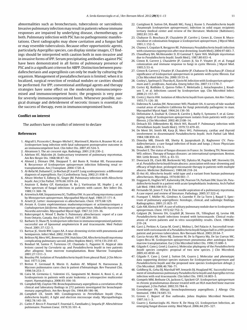
Fig. 1. Age and sex distribution
neumonectomy in two. Twelve patients were managed medi-ally. Twelve cases were fatal (26.8%), and 22 patients (47.5%) whoad undergone surgery (15) or had been treated with miconazole
MCZ),119 ITZ,23,32 voriconazole (VRZ)44,154 or had no therapy,67,131urvived. In one case, sputum cultures continued to be positive.ollowing a course of amphotericin B (AMB), the patient remainedlinically well without any symptoms.11 Outcome was not reported
0
5
10
15
20
25
30
35
AML
ALL
Lung
canc
er
Other
org
an ca
ncer
s
Non-H
odgk
in’s l
ymph
oma
Mye
loma
Hystio
cytic
-lym
phoc
ytic l
ymph
oma
CGD
Gliobla
stom
a m
ultifo
rme
BMT
SOT
GVHD CF
Acute
rena
l failu
re
Cortic
oste
roid
t
Underlyin
Nu
mb
er o
f p
atie
nts
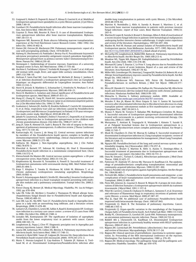
ig. 2. Frequency of underlying conditions reported in 138 cases of IPP. ALL: acute lymphoGD: chronic granulomatous disease; CMV: cytomegalovirus;DM: diabetes mellitus; GVH
patients with IPP, 1955–2010.
in the other cases. Regarding these data, in suitable patients surgeryappears a successful treatment choice for a cavitary lesion contain-ing a fungus ball.
(ii) Allergic bronchopulmonary pseudallescheriasis (ABPP)Although most allergic bronchopulmonary mycoses have been
attributed to Aspergillus species, this syndrome has been reported in
reat
men
tDM
Antifu
ngal
prop
hylax
is
Bacte
rial p
neum
onia
CMV p
neum
onia CF
Other
bro
nchia
l diso
erde
rs
Inte
rstiti
al lun
g dis
ease
HIV in
fecti
on
Cigare
tte sm
oking
Near d
rownin
g
Previo
usly
healt
hy NS
g conditions
cytic leukemia; AML: acute myeloid leukemia; BMT: bone marrow transplantation;D: graft versus host disease; NS: non-stated; SOT: solid organ transplantation.

6 ev Iberoam Micol. 2012;29(1):1–13
Pcoa
i2trtg
AgoarA
taai(rwisgenRhasswte
sotid
Awbpgwb
wdi
(
tinona
Table 2Other sites of involvement in 29 of 138 patients with invasive pulmonary infection.
Body site(s) Number of patients
Central nervous system 23Kidney 12Thyroid 5Heart 5Spleen 4Liver 5Wall of vessels 1Blood 4Bone 1Gastrointestinal system 3Eye 3Subcutaneous tissue 1
Signs and symptoms. Clinical symptoms are often insidious andnonspecific, such as chronic cough, sputum production, fever,
Table 3Treatment and outcome of 138 patients with IPP, 1955–2010.
Treatment types Outcome (Number of patients)
Total Death Survived Not stated
Surgery 6 2 3 1
A. Serda Kantarcioglu et al. / R
SC as well.23,76,90,144 Allergic bronchopulmonary fungal disease isharacterized by asthma, peripheral blood eosinophilia, infiltratesn the chest radiograph, raised IgE levels, precipitating antibodiesnd immediate cutaneous reactivity to the casual fungus.50
Lake et al.76 first suggested allergic bronchopulmonary man-festations induced by PSC. The authors described a case of a4-year-old woman with asthma and clinical symptoms similaro allergic bronchopulmonary aspergillosis (ABPA) who, on chestoentgenogram, was found to have infiltrates, an elevated serumotal IgE, skin prick test reactivity and precipitins against this fun-us. Hyphae were seen on direct examination of sputum.
Five ABPP cases were reported from Canada, andustralia.23,76,131,144 Three of the subjects were female, whileender was not mentioned in the remaining reports; the agef the patients ranged from 18 to 48 years. Five of them hadssociated diseases such as asthma and CF and two had previouslyeceived prednisone therapy for rheumatoid arthritis or previousBPA.
Little is known about the immunological and allergic fea-ures of pulmonary pseudallescheriasis. Precipitating antibodiesre frequently present in Aspergillus mycetoma, but skin testsre usually negative, in contrast to ABPA, in which typically anntermediate (type I) cutaneous reaction occurs, and a delayedArthus type III) reaction frequently follows, giving a dualesponse.19,20,50,80,102 Likewise, precipitating antibodies to PSCere reported in several cases in which the fungus proliferated
n the airway lumen21,27,57,69,87,91,111 and failed to continue afterurgery in those who underwent resection69,81 or after antifun-al treatment.91 In most of them, no reaction was detected withxtracts of other fungi, including A. fumigatus. Eosinophilia wasoted in only one patient.57 Of the three patients reported byeddy et al.,109 one was skin tested with an extract of PSC, butad no response. Rippon and Carmichael111 reported a case of
patient with transient endobronchial colonization with severalputum specimens positive for PSC. Chest radiograph examinationhowed diffuse interstitial infiltrates; and precipitins against PSCere positive. Although the disease was somewhat similar to ABPP,
here was no eosinophilia recorded and skin test sensitivity was notstablished.
Cimon et al.,27 reported two cases of ABPP in a prospectivetudy in 128 CF patients, both chronically colonized by PSC andne with previous ABPA treated with a combination of corticos-eroids and ITZ, leading to a remission of symptoms. In most casesn this study, colonization with PSC was not associated with allergicisease.
Mixed allergic bronchopulmonary disease due to PSC andspergillus was also described by Lake et al.76 in an asthmaticoman without CF, and in two additional cases by Miller et al.,90
ut mixed infections seem uncommon. In the second report, twoatients with probable diagnosis of ABPA also presented the fun-us in sputum and strongly positive pseudallescheriasis serology,hich suggests a contributory role of this fungus in the allergic
ronchopulmonary disease.90
ABPP was seen in patients with long-standing asthma76,90 orith CF.27 On pathological analysis, this form of pulmonary pseu-allescheriasis was characterized by the presence of obvious plugs
n sputum containing PSC cells and eosinophilia.
iii) Invasive pulmonary pseudallescheriasis (IPP)Before the 1980s, PSC was rarely reported as a cause of sys-
emic disease. We have retrospectively examined 138 cases ofnvasive pulmonary pseudallescheriasis (IPP) including pneumo-
ia, pulmonary abscess, pleuritis and other manifestations. Asutlined in Table 1, IPP can vary from nodular pneumonia toecrotizing pneumonias, lung abscess,6,51,66,89,141 empyema,16,137nd pleurisy.16,22,34,35,45,72,74,82,103,136,138,150 Asymptomatic coin
Skin 8
lesion,151 cystic mass98 and polypoid lesions96,153 were alsooccasionally reported. Of these, nodular pneumonia and pleu-ral effusion15,16,22,34,35,45,54,72,82,103,136 were the most common.One case was also reported of simultaneous pulmonary infec-tion with Aspergillus terreus and PSC64 and a pulmonary infectionby Mycobacterium avium concomittant with a polypoid bronchiallesion by PSC.149 Similarly, Morales et al. reported A. fumigatus andPSC isolation from sputum of a patient with CF and Mycobacteriumabscessus infection after lung transplantation.43
Demographic and geographical features. The majority of the 138invasive pulmonary infections by PSC were reported from the USA,with occasional cases from UK, France, Finland, Germany, Spain,Netherlands, Brazil, Congo, Australia, Japan and Taiwan. Of those,78 were female and 52 male. The age of the patients ranged from 2to 90 years, although age was not reported in 8 cases (Fig. 1).
Predisposing factors, underlying conditions. The most frequentunderlying conditions reported were corticosteroid treatments,solid organ transplantations (lung 194, heart 5, liver 1, kidney 3) andhaematological malignancies. Most patients showed a history ofunderlying chronic lung disease, cigarette smoking or occupationalexposure. Ten patients who suffered near-drowning but who hadpreviously been healthy and two further patients were occupation-ally exposed to fungal conidia; one patient was immunocompetentwith a perforated chest wound; one case occurred in a liver trans-plant patient following a skull fracture in an accident; and nopredisposing conditions or underlying disease were stated in sixpatients. Fig. 2 shows the frequency of any underlying conditionsreported in the reviewed cases, such as cellular immunity and, inparticular, neutrophils that might have an important role in thepathogenesis of IPP. Tables 4 and 5 list the underlying conditionsand predisposing factors, respectively, found in 44 patients whosurvived and 75 who died. In the other cases, patient outcome wasnot reported.
Antifungal 73 40 28 5Surgery and antifungal 10 4 4 2None 7 7Not stated 42 6 1 22

A. Serda Kantarcioglu et al. / Rev Iberoam Micol. 2012;29(1):1–13 7
Table 4Demographic characteristics, other sites of infection, and therapy given to survivors of IPP (N = 44).
Reference Age/sex Type of infection Surgery Antifungal therapya
Bousley16 39/M Empyema ThoracentesisJung et al.67 60/F Right upper lobectomy
81/MSaadah and
Dixon11632/F Necrotizing pneumonia Antibiotics (5 m),
thoracotomyWoodard151 70/M Asymptomatic pulmonary coin lesion Thoracotomy with wedge
resectionGalgiani et al.43 50/F Pulmonary nodule KTZ (400 mg, 3 m)
70/F Progressive diffuse peribronchialthickening
KTZ (200 mg, 4 m)
50/M Right upper lobe infiltrate KTZ (400 mg, 8 m)59/F Right middle lobe cavity Right middle lobectomy KTZ (400 mg, 1 m)55/F Right upper lobe infiltrate Thoracotomy (the lesion
had not been excisedcompletely)
KTZ (200 mg, 2 m; 400 mg,5 m)
Plus and Opal106 74/F Antibacterials (ineffective),KTZ (400 mg/d)
Seale andHudson122
59/M IPP MCZ (i.v. 300 mg every 8 h,30 d)
Travis et al.142 39/F Lung abscess SurgeryDworzack et al.37 2/F Lung abscess AMB (a total of 56 mg),
changed to MCZ (a total of481.8 g i.v. + 2700 mgintrathecally)
Mesnard et al.89 17/F AMB, KTZ (400 mg/d, 8 ws)Walsh et al.146 28/M Partial lobectomy AMB (progressively
cavitated)Goldberg et al.48 21/M Simultaneous pulmonary infection
with Aspergillus terreusThoracotomy AMB (1 mg/kg/d), i.v. MCZ
(800 mg t.i.d.)(progression) debridementof necrotic material, ITZ(200 mg po b.i.d., 2 m)
Nomdedéu et al.99 39/M AMB <81 mg/kg/d),stopped after diagnosis, ITZ(600 mg/d)
Stolk-Engelaar andCox134; Verweijet al.145
28/M ITZ (200 mg b.i.d. orally3 m), relapse (after 20 mtherapy, despite adequateserum concentrations) oralTRB (500 mg/d, a total of9 m) (after 4 m therapybronchoscopy showed noevidence of fungalinfection)
Hung et al.64 69/M Laminectomy Anti TBC, AMB(0.5 mg/kg/d),
Martino et al.84 15/M An alveolar infiltrate in the ALL Surgical resection AMB (1 mg/kg/d,cumulative dose 2 g)
Abbrevations: ALL: acute lymphoblastic leukemia; AMB: amphotericin; b.i.d.: bis in die (twice in day); d: day; F: female; IPP: invasive pulmonary pseudallescheriasis;i.v.: intravenous injection; ITZ: itraconazole; KTZ: ketoconazole; LAMB: lyposomal amphotericin B; M: male; m: month; MCZ: miconazole; t.i.d.: tree times a day; TBC:t
ks (w
nostpa
RspphPeoro
uberculosis; TRB: terbinafine.a In parentheses, regimen in mg per day (d) and duration of the treatment in wee
ight sweats, chest pain and shortness of breath. Patientsften complained of weakness and malaise. Other local andystemic symptoms include pleuritic pain, chills, fever, easyiredness, anorexia, and weight loss. One patient with a poly-oid bronchial lesion had no complaint,96 and two others weresymptomatic.142,151
adiology. Radiologically, IPP might have manifested itself as con-olidation (in 7 patients), nodules (in 13 patients), necrotizingneumonia (in 9 patients), pulmonary abscess (in 15 patients), andleural effusions (in 10 patients). Radiological examination mightave been less specific, with diffuse infiltration and pneumonia.5
SC pneumonia, i.e. lobar pneumonia56,62,77,84,99,114,136 and bilat-
ral consolidation22,138 were seen in several cases; two of them43ccurred in patients with no predisposing conditions. Chestadiographs and CT scan images may show ill-defined nodularpacities.22,84 The opacity with a peripheral rim of ground glass,
s) or months (m) are indicated.
known as the “halo sign”, was reported in one case.149 Nodulessurrounded by a halo of ground-glass is often considered to beevidence of haemorrhagic infarcts and believed to represent theperipheral rim of haemorrhagic infarction, described in the angioin-vasive fungal diseases, aspergillosis, zygomycosis and describedas well in PSC infections.41 Angioinvasive pseudallescheriasis wascharacterized histologically by the invasion and occlusion of smallto medium-sized pulmonary arteries by fungal hyphae.39,79
The “air crescent sign”5,45 can be seen in a pulmonary cavi-tary process, which is caused by air surrounded by radiopaquematerial along both its inner and outer margins. The air cres-cent may transform into a cavity space, filled with necrotic debris,including neutrophils, and fungal elements. However, a similar
appearance has been described in a number of infections, includ-ing mucorales, Candida, herpes simplex or cytomegalovirus, orother conditions such as Wegener granulomatosis, Kaposi sarcoma,and haemorrhagic metastasis. The “air crescent sign” is considered
8 A. Serda Kantarcioglu et al. / Rev Iberoam Micol. 2012;29(1):1–13
Table 5Demographic characteristics, other sites of infection, and therapy given to non survivors of IPP (N = 79).
Reference Age/sex Type of infection Other locations Surgery Antifungal therapy
Alture-Werber et al.6 66/F Fungal abscess, partlypneumonia
+ Steroids, antibiotics, anti-TBC
Lutwick et al.82 66/F Multiple pulmonary Renal and brain abscess AMBWinston et al.150 57/F Lung abscess NS
37/M Antibiotics, AMB (a total of576 mg)
Van der Vliet et al.143 15/M NSMeadow et al.88 15/F Antibiotics, methylprednisolone
(1 g/d), then reduced (to 10 mg/dover 10 d), MCZ (i.v. 1200 mgevery 8 h)
Gumbart54 39/M Nodular bilateral pneumonia NSDe Ment et al.34 60/F Necrotizing
bronchopneumonia, pleuritisEmpirically antibiotics and MCZ,changed to AMB (0.3 mg/kg/d)
Enggano et al.39 16/M Lung abscess Antibiotics, empirically AMB(i.v.0.75 mg/kg/d), 5-FC
Shih and Lee128 22/M Lung abscess Brain, thyroid, kidney, lumen andwall of vessels
None
Smith et al.130 41/M Lung abscess Brain, skin, liver, thyroid AMBGuyotat et al.55 26/M Fungal abscess AMB (1 mg/kg) (worsened with
diffuse infiltrates)Anaissie et al.7 7/F Lung abscess Heart, blood, kidney, brain abscess NoneDworzack et al.37 22/F Lung abscess Brain, skin Surgery MCZ (10 mg every 12 h) (a total of
52 g parenterally and 250 mgintrathecally)
20/F Lung abscess Brain, skin AMB (a total of 86 mg)Schawrtz121 43/M Lung abscess Kidney, skin, cerebral fungus ball NonePatterson et al.101 22/M Cavitary lesions in both lungs,
likely nosocomial infectionAMB + KTZ (400 mg/d, 12 d), MCZ(600 mg every 8 h)
Piens et al.104 33/F AMB (400 mg/kg/d)Steens et al.132 28/F Atypical pneumonia Antibiotics (initially erythromycin
and then doxycycline, 1 m); broadspectrum antibiotics
Walsh et al.146 13/M Pneumonia AMB41/M Right upper lobe consolidation Peritonitis on day 86 AMB (70 mg/d)
Hofman et al.61 47/M NSAnaissie (1989) 7/F NoneSevero et al.124 41/F Solitary pulmonary nodule KTZ (400 mg/d), prednisone
(20 mg/d) and insulinKhurshid et al.70 61/F Lung abscess Liver, spleen, kidney, pancreas,
right and left ventriclesAMB
Kusne et al.74;Castiglioni et al.22
67/M Lung mass ITZ (oral, 1 m), VRZ (6 mg/kgevery 12 h the first day, 4 mg/kgevery 12 h thereafter)
Bonduel et al.15 18/F Pneumonia, pleural effusion ITZ (200 mg/d, 90 d), L-AMB(2 mg/kg/d, a total dose of30 mg/kg)
Breton et al.17 61/M Pneumonia Rightpneumonectomy
ITZ (400 mg/d),
Dinesha et al.35 36/M Brain Anti-tuberculose treatment,frontal craniotomy and excisionof the lesion in the left frontallobe, AMB
Nguyen98 78/F ITZ (oral), AMB (i.v.), L vitrectomy,(after identification) changed toITZ
Tamm et al.137 42/F ITZ, FLZ22/F ITZ, FLZ49/M ITZ, FLZ38/F ITZ, FLZ
Bartzacet al.13 50/M Llıung abscess Brain abscess AMB + FLZCastiglioni et al.22 30/M Pneumonia, pleuritis Pericarditis ITZ prophylaxis; AMB + MCZ
37/F ITZ prophylaxis (beginig 11 mafter transplant); broad spectrumantibiotics +MCZ
36/M Pneumonia Brain abscess ITZ, MCZKleinschmidt-De
Masters7141/M Lung abscess Cerebritis, multiple small
haemorrhagic infarctions, heart,thyroid
NS
Horre et al.62 72/F Pneumonia A purulent ulceration on her leftlittle toe
ITZ (100 mg/d) (radiologicallyprogression); (200 mg/d)
Klopfenstein et al.72 14/F A 4 cm nodule Left lunglobectomy
ITZ (200 mg i.v. 3 times daily,AMB empirically, MCZ (600 mgi.v. every 8 h) + 5-FC, VRZ (270 mgi.v. twice a day), (200 mg orallytwice a day, 7 m)

A. Serda Kantarcioglu et al. / Rev Iberoam Micol. 2012;29(1):1–13 9
Table 5 (Continued)
Reference Age/sex Type of infection Other locations Surgery Antifungal therapy
Riddell et al.110 33/F Lungs abscess Thyroid, heart, kidneys, bloodinfection, brain
NS
Symoens et al.135 26/F Lung Eye, subcutaneous nodules, CNS VRZAbgrall et al.1 68/M Cavernous lesion and
paranchymatous consolidationVRZ
Cooley et al.28 NS/F Pulmonary abscess Brain AMB + ITZMorales et al.94 NS/NS LungSahi et al.117 43/M Recurrent pan-lobar Fungal
pneumonia (18 mpost-transplant), mediastinitis,pleuritis
Osteomyelitis and a knee abscess L-AMB (for 8 weeks), after initialclinical improvement, hedeveloped a pulmonary nodule,necrotizing granulomas, ITZ,recurrence
57/F Lung abscess Brain abscess, skin nodules, eye VRZ high dose + TRB + later PSZ19/F Chest wall cellulitis,
mediastinitis, yellow-whiteendobronchial plaques
Endophthalmitis (4 weeks after T),multiple skin nodules, pansinusitis,vertebral osteomyelitis, and septicarthritis,
VRZ, (eye) CAS + TRB, intravitrealinjections of VRZ, oral PSZ(200 mg, 4 times daily with mealsto improve absorption) as salvagetherapy, granulocyte macrophagecolony-stimulating factor as animmunoadjuvant, L-AMB wasadded, oral PSZ was increased to1200 mg/d (400 mg 3 times dailywith meals)
Caira et al.18 NS/NS Lung abscess L-AMB (3 mg/kg, 10 d)NS/NS Lung abscess L-AMB (3 mg/kg, 10 dNS/NS Lung abscess Blood, skin L-AMB (3 mg/kg, 10 dNS/NS Lung abscess Blood D-AMB (1 mg/kg, 14 d)
Sheu et al.127 NS/NS Lung, pleuritisNS/NS Lung, pleuritisNS/NS Lung, pleuritis
Mario et al.83 37/F Lung Skin nodules, CNS, blood VRZ (250 mg, twice a day), CAS(70 mg/kg of body weight/dloading dose, then 50 mg/kg), TRB(250 mg/d)
Maslen and Peel86 60/F Lung45/F Lung58/M Lung43M Lung19/F Lung
A ytosinv d, i.v.:n
cic
CntiiolSptsiatrrshrrhe
bbrevations: AMB: amphotericin B; L-AMB: liposomal amphotericin B; 5-FC: flucoriconazole; PSZ: posaconazole; TRB: terbinafine; CAS: caspofungin; NS: not stateervous system.
haracteristic of invasive pulmonary aspergillosis (IPA) when seenn the appropriate clinical setting.2 Therefore, it is important not toonfuse IPP with IPA.
linical manifestations. In several cases, the existence ofecrotizing pneumonia was detected histologically, charac-erized by the presence of tissue necrosis and granulomatousnflammation.22,34,116,138 IPP is characterized by haemorrhagicnfarction of lung tissue, secondary to vascular invasion by fungalrganisms, causing thrombosis of small arterioles and, sometimes,arger pulmonary vessels, as seen in IPA and invasive fusariosis.aadah and Dixon116 described a truly invasive PSC, necrotizingneumonia in an apparently normal host. The disease was rela-ively destructive, traversing multiple pulmonary segments, theurrounding pleura, and the recurrent laryngeal nerve. Smears ofntrabronchial pus obtained from the surgical specimen had anbundance of septate branching hyphae, while in the tissue sec-ions hyphae were very rare. Based on this finding and a literatureeview, the authors suggested that actual tissue invasion by PSC isare and most of the tissue damage in the lung is secondary to theevere inflammatory reaction of the host incited. This hypothesisas been put forward previously to explain the severe tissue
eaction present in chronic pulmonary histoplasmosis with theelative absence of the organism in the inflamed tissue. Thereave been several other cases reported of an absence of fungallements in lung tissue sections but with positive cultures fore; ITZ: itraconazole; FLZ: fluconazole; KTZ: ketoconazole; MCZ: miconazole; VRZ: intravenous injection; h: hours; d: day; m: month; TBC: tuberculosis; CNS: central
PSC.33,62 Another typical presentation, described in several cases,is pulmonary abscess.4,17,30,49,66,82,136,140,141
A coin lesion is a less frequent presentation of the IPP, definedas a single, discrete pulmonary opacity smaller than 3 cm in diam-eter surrounded by normal lung tissue, and not associated withadenopathy or athelectasis.93 Although the fungal solitary pul-monary nodules are usually caused by pathogenic dimorphic fungiand usually the result of a self-limiting Woodard151reported acase of a solitary pulmonary nodule due to PSC. Histopathologicalexamination of the patient’s lesion revealed a fibrosis encapsulatedgranulomatous nodule with central necrosis and grey granules.151
Cystic mass98 and polypoid lesions96,153 due to PSC observedin fiberoptic bronchoscopy have been reported in two cases. Yanoet al.153 described a bronchus completely obscured by a dark greynecrotizing lesion after the whitish polypoid lesion by a biopsyforceps. Murayama et al.96 reported a case in combination withM. avium pulmonary disease. Loosely formed grains have also beenreported within sinus tracts in lungs in a pediatric patient with dis-seminated disease.88 Pleurisy was commonly found in several IPPcases.16,22,34,45,54,82,86,103,136 Disseminated infection was reportedin 29 patients. Table 2 outlines other sites of involvement.
Laboratory diagnosis. Diagnosis was made through histological
examination and culture (in 17 cases), or only culture (in 28 cases)of the excised lesion or other respiratory tract samples (sputum,bronchial secretions, endobronchial brushings). Fungi from tissuesamples grew in 11 cases, but failed to grow in six. Respiratory
10 A. Serda Kantarcioglu et al. / Rev Iberoam Micol. 2012;29(1):1–13
0
10
20
30
40
50
60
70
80
90
Nu
mb
er o
f ca
ses
Intrabronchialpolypoid lesion
Infection type
Total
Death
Survival
Not stated
Pneumonia Bronchopneumonia Pulmonary abscess Pulmonary nodules Pleuritis Empyeme
s with
taipwilmqcgsrwtt
Toaa(itdwnwwt
Mpwdew
Fig. 3. Mortality differences among 138 patient
ract samples gave negative results. In one patient a thoracic needlespiration was performed and the diagnosis was made by examin-ng a stained smear specimen and culture124; diagnosis was madeostmortem in nine cases. A histopathological study of nodulesas made on some patients, and revealed a round pulmonary
schemic infarction due to arterial invasion by the fungus,42 granu-oma with central necrosis,151 fibrosis mixed with granuloma and
icroabscess or an abscess.6,22,39,49,62,64,82,116,136,142 Regarding theuestionable significance of isolating PSC, Jung et al.,67 establishedriteria for diagnosis as follows: (i) repeated isolation of the fun-us, at least four positive cultures per patient being considered to beignificant; (ii) growth of the fungus from the excised surgical mate-ial; (iii) positive cultures from samples obtained from bronchialashings or selective brushing from the pulmonary lesions through
he fiberoptic bronchoscope; and (iv) evidence of tissue invasion inissue sections.
reatment and outcome. Table 3 shows the treatment and outcomef those patients analyzed with IPP. Of 138 patients, 5 were man-ged surgically, and 73 were managed medically with systemicntifungal agents, such as AMB, liposomal AMB, MCZ, ketoconazoleKTZ), ITZ, fluconazole (FLZ), VRZ and terbinafine (TRB). A youngmmunocompetent patient with previous trauma and having beenreated with ITZ suffered a relapse after 20 months of therapyespite adequate serum concentrations.134 The patient was treatedith oral TRB (500 mg/d) and after 4 months bronchoscopy showedo evidence of fungal infection.145 Seventy nine of the 138 patientsith IPP died (57.2%) and 43 (31.1%) survived, while the outcomeas not reported in the remaining cases. Tables 4 and 5 summarize
he data on survivors and no survivors.Thirty-two patients had a history of corticosteroid treatment.
urayama et al.96 diagnosed a bronchial polypoid lesion in aatient with rheumatoid arthritis. In this case, surgical treatment
as not undertaken because of extensive M. avium pulmonaryisease, but methylprednisolone was discontinued soon afterstablishing the definitive diagnosis and there was no evidence oforsening during a two-year follow up. Similarly, in a report byIPP by Pseudallescheria/Scedosporium complex.
Rippon and Carmichael,111 the patient’s bronchial lesions disap-peared when steroid therapy was discontinued. Horre et al.62
reported a fatal pneumonia in a patient who had a long historyof corticosteroid therapy. Lionakis and Kontoyiannis79 suggestedthat the use of steroids, although necessary, could have facili-tated opportunistic mould infections in cancer patients. The useof steroids may render the patient susceptible to opportunis-tic mycoses. Despite having a normal neutrophil count, affectedpatients have functional neutropenia because the function of theneutrophils is inhibited by the use of high-dose steroids. Basedon that data, discontinuation of steroids and immunomodulationof neutrophyl functions, if needed, may be an optional treatmentapproach.
Tamm et al.137 analysed risk factors, and the clinical course andoutcome of seven lung transplant recipients who had developedIPP infection diagnosed through BAL specimens. The fungus wasdetected 9–58 months after transplantation. Five patients had beentreated for several months with ITZ because of previous detection ofAspergillus in BAL. S. prolificans was first cultured in three cases anda few months later S. apiospermum was found. All seven patientsshowed airway problems. Combined treatment with ITZ and FLZwas not able to eradicate PSC. Four of the seven patients died 3–35months after the diagnosis of IPP. The authors concluded that IPPwas seen in lung transplant recipients with structurally abnormalairways and under long term therapy with ITZ. Eradication of thefungus proved difficult, but under combined treatment with ITZand FLZ this infection did not disseminate. Although the role ofboth drugs in the control of the infections is difficult to understand,ITZ has demonstrated in general poor efficacy against these fungiand FLZ is not usually used for treatment of mycoses caused by fila-mentous fungi. Differences in mortality rates are outlined in Fig. 3.
Conclusion
In most instances non-invasive forms of pulmonary pseu-dallescheriasis have been superimposed on some structural
ev Ibe
aIrbtopiihPdolbsmfgt
C
R
A. Serda Kantarcioglu et al. / R
bnormalities such as bronchectasis, tuberculosis or sarcoidosis.nvasive pulmonary infection may result in patients whose immuneesponses are impaired by underlying disease, chemotherapy, oroth. Pulmonary infection with PSC has no pathogenomic manifes-ations. Chest radiographs may show cavitation and a fungus ballr may resemble tuberculosis. Because other opportunistic agents,articularly Aspergillus species, can display similar images, CT find-
ngs should be interpreted with caution both in non-invasive andn invasive forms of IPP. Serum precipitating antibodies against PSCave been demonstrated in all forms of pulmonary presence ofSC and is a significant criteron for ABPP. Distinction between pseu-allescheriasis and aspergillosis can only be made by culturing therganism. Management of pseudallescheriasis is limited; when it isocalized, surgical resection of residual nodules or cavities shoulde performed. For IPP, conventional antifungal agents and therapytrategies have some effect on the moderately immunocompro-ised and immunocompetent hosts; the prognosis is very poor
or severely immunocompromised hosts. Whenever possible, sur-ical drainage and debridement of necrotic tissues is essential tohe success of therapy, even in immunocompromised hosts.
onflict on interest
The authors have no conflict of interest to declare
eferences
1. Abgrall S, Pizzocolo C, Bouges-Michel C, Martinod E, Martin A, Brauner M, et al.Scedosporium lung infection with fatal subsequent postoperative outcome inan immunocompetent host. Clin Infect Dis. 2007;45:524–5.
2. Abramson S. The air crescent sign. Radiology. 2001;218:230–2.3. Adelson HT, Malcolm JA. Endocavitary treatment of pulmonary mycetomas.
Am Rev Respir Dis. 1968;98:87–92.4. Ahmed J, Ditmars DM, Sheppard T, del Busto R, Venkat KK, Parasuraman
R. Recurrence of Scedosporium apiospermum infection following renal re-transplantation. Am J Transplant. 2004:1720–4.
5. Al-Refai M, Duhamel C, Le Rochais JP, Icard P. Lung scedosporiosis: a differentialdiagnosis of aspergillosis. Eur J Cardiothorac Surg. 2002;21:938–9.
6. Alture-Werber E, Edberg SC, Singer JM. Pulmonary infection with Allesscheriaboydii. Am J Clin Pathol. 1976;66:1019–24.
7. Anaissie E, Bodey GP, Kantarjian H, Ro J, Vartivarian SE, Hopfer J, et al.New spectrum of fungal infections in patients with cancer. Rev Infect Dis.1989;11:369–78.
8. Ariewitsch AM, Stepaniszewa SG, Tiufilina OW. A case of lung mycetoma causedby Monosporium apiospermum. Mycopathol Mycol Appl. 1969;37:171–8.
9. Arnett JC. Letter: monosporosis vs allescheriasis. Chest. 1975;68:129.10. Avram A. Grains expérimentaux maduromycosiques et actinomycosiques a
Cephalosporium falciforme, Monosporium apiospermum, Madurella mycetomi etNocardia asteroides. Mycopathol Mycol Appl. 1967;32:319–36.
11. Bakerspiegel A, Wood T, Burke S. Pulmonary allescheriasis: report of a casefrom Ontario, Canada. Am J Clin Pathol. 1977;68:299–303.
12. Barbaric D, Shaw PJ. Scedosporium infection in immunocompromised patients:successful use of liposomal amphotericin B and itraconazole. Med PediatrOncol. 2001;37:122–5.
13. Bartzac JC, Steele RW, Lopez AA. A near-drowning victim with pneumonia andhemiparesis. Infect Med. 2002;19:98–103.
14. Belitsos NJ, Merz WG, Bowersox DW, Hutchins GM. Allescheria boydii mycetomacomplicating pulmonary sarcoid. Johns Hopkins Med J. 1974;135:259–67.
15. Bonduel M, Santos P, Turienzos CF, Chantada G, Paganini H. Atypical skinlesions caused by Curvularia sp. and Pseudallescheria boydii in two patientsafter allogenic bone morrow transplantation. Bone Marrow Transplant.2001;27:1311–3.
16. Bousley PH. Isolation of Pseudallescheria boydii from pleural fluid. J Clin Micro-biol. 1977;5:244.
17. Breton P, Germaud B, Morin O, Audoin AF, Milpied N, Harousseau JL.Mycoses pulmonaires rares chez le patient d’hématologie. Rev Pneumol Clin.1998;54:253–7.
18. Caira M, Girmenia C, Valentini CG, Sanguinetti M, Bonini A, Rossi G, et al.Scedosporiosis in patients with acute leukemia: a retrospective multicenterreport. Haematologica. 2008;93:104–10.
19. Campbell MJ, Clayton YM. Bronchopulmonary aspergillosis a correlation of theclinical and laboratory findings in 272 patients investigated for bronchopul-monary aspergillosis. Am Rev Respir Dis. 1964;89:186–96.
20. Campbell CK, Smith MD. Conidiogenesis in Petriellidium boydii (Pseu-dallescheria boydii). A light and electron microscope study. Mycopathologia.1982;78:145–50.
21. Carles P, Recco P, Fournial F, Fournial G, Familiades J, Sequela JP. Alleschériosepulmonaire. Poumon Coeur. 1979;35:101–4.
roam Micol. 2012;29(1):1–13 11
22. Castiglioni B, Sutton DA, Rinaldi MG, Fung J, Kusne S. Pseudallescheria boydii(Anamorph Scedosporium apiospermum). Infection in solid organ recipientstertiary medical center and review of the literature. Medicine (Baltimore).2002;81:333–48.
23. Chabasse D, Bouchara JP, Chazalette JP, Carrére J, Genes JL, Cimon B. Muco-viscidose et colonisation fongique à Scedosporium apiospermum. J Mycol Med.1991;1:152–5.
24. Chaney S, Gopalan R, Berggren RE. Pulmonary Pesudallescheria boydii infectionwith cutaneous zygomycosis after near drowning. South Med J. 2004;97:683–7.
25. Chaudhary BA, McAlexander D, El Gammal T, Speir WA. Multiple mycetomasdue to Pseudallescheria boydii. South Med J. 1987;80:653–4.
26. Cimon B, Carrere J, Chazalette JP, Guines JI, Six P, Vinater JF, et al. Fungalcolonization and immune response to fungi in cystic fibrosis. J Mycol Med.1995;5:53–6.
27. Cimon B, Carrere J, Vinatier JF, Chazalette JP, Chabasse D, Bouchara JP. Clinicalsignificance of Scedosporium apiospermum in patients with cystic fibrosis. EurJ Clin Microbiol Infect Dis. 2000;19:53–6.
28. Cooley L, Spelman D, Thursky K, Slavin M. Infection with Scedosporium apiosper-mum and S. prolificans, Australia. Emerg Infect Dis. 2007;13:1170–7.
29. Cortez KJ, Roillides E, Quiros-Telles F, Meletiadis J, Antachopoulos J, Knud-sen T, et al. Infections caused by Scedosporium spp. Clin Microbiol Infect.2008;21:157–97.
30. Creitz J, Harris HW. Isolation of Allescheria boydii from sputum. Am Rev Tuberc.1955;71:126–30.
31. Dabrowa N, Landau JW, Newcomer WD, Plunkett OA. A survey of tide-washedcoastal areas of southern California for fungi potentially pathogenic to man.Mycopathol Mycol Appl. 1964;24:136–50.
32. Defontaine A, Zouhair R, Cimon B, Carrére J, Bailly E, Symoens F, et al. Geno-typing study of Scedosporium apiospermum isolates from patients with cysticfibrosis. J Clin Microbiol. 2002;40:2108–14.
33. Deloach ED, DiBenedetto RJ, Hitch WS, Russell P. Pulmonary infection withPetriellidium boydii. South Med J. 1979;72:479–81.
34. De Ment SH, Smith RR, Karp JE, Merz WG. Pulmonary, cardiac and thyroidinvolvement in disseminated Pseudallescheria boydii. Arch Pathol Lab Med.1984;108:859–61.
35. Dinesha MR, Dinesh KR, Shetty R, Jayaprakash JS, Raghuveer CV. Pseu-dallescheriasis: a rare fungal infection of brain and lungs. J Assoc PhysiciansIndia. 2001;49:574–5.
36. Drouhet E. The status of fungus diseases in France. In: Stenberg TH, NewcomerWD, editors. Therapy of fungus diseases: an international symposium. Boston,MA: Little Brown; 1955. p. 43–53.
37. Dworzack DL, Clark RB, Borkowski WJ, Dykstra M, Pugsley MP, Horowitz EA,et al. Pseudallescheria boydii brain abscess: association with near-drowning andefficacy of high dose, prolonged miconazole therapy in patients with multipleabscess. Medicine (Baltimore). 1989;68:218–24.
38. El-Ani AS. Allescheria boydii: wild type and a variant from human pulmonaryallescheriasis. Mycologia. 1974;66:661–7.
39. Enggano IL, Hughes WT, Kalwinsky DK, Pearson TA, Parham DM, Stass SA. Pseu-dallescheria boydii in a patient with acute lymphoblastic leukemia. Arch PatholLab Med. 1984;108:619–22.
40. Fernando SE, Jones P, Vaz R. Fine needle aspiration of a pulmonary mycetoma.A case report and review of literature. Pathology. 2005;37:322–4.
41. Franquet T, Müller NL, Giménez A, Guembe P, De La Torre J, Baqué S. Spec-trum of pulmonary aspergillosis: histologic, clinical, and radiologic findings.Radiographics. 2001;21:825–37.
42. Gale AM, Kleitsch WP. A case of solitary pulmonary nodule due to Scedosporiumapiospermum. Chest. 1972;62:752–5.
43. Galgiani JN, Stevens DA, Graybill JR, Stevens DL, Tillinghast AJ, Levine HB.Pseudallescheria boydii infections treated with ketoconazole. Clinical evalu-ations of seven patients and in vitro susceptibility results. Chest. 1984;86:219–24.
44. Garci J, Perkins A, Garau M, Gené J, Molina L, Del Palacio A. Successfull treat-ment with voriconazole of a Pseudallescheria boydii fungus ball in a HIV positivepatient and previous tuberculosis. Rev Iberoam Micol. 2003;20:64–7.
45. Garcia-Arrata MI, Otero MJ, Zomeno M, De la Figuera Ma, De las Cuevas MC,Lopez-Brca M. Scedosporium apiospermum pneumonia after autologus bone-marrow transplantation. Eur J Clin Microbiol Infect Dis. 1996;15:600–3.
46. Gilgado F, Cano J, Gené J, Guarro J. Molecular phylogeny of the Pseudallescheriaboydii species complex: proposal of two new species. J Clin Microbiol.2005;43:4930–42.
47. Gilgado F, Cano J, Gené J, Sutton DA, Guarro J. Molecular and phenotypicdata supporting distinct species statuses for Scedosporium spiospermum andPseudallescheria boydii and the proposed new species Scedosporium dehogii. JClin Microbiol. 2008;46:766–71.
48. Goldberg SL, Geha DJ, Marshall WF, Inwards DJ, Hoagland HC. Successful treat-ment of simultaneous pulmonary Pseudallescheria boydii and Aspergillus terreusinfection with oral itraconazole. Clin Infect Dis. 1993;16:803–5.
49. Gompels MM, Bethune CA, Jackson G, Spickett GP. Scedosporium apiospermumin chronic granulomatous disease treated with an HLA matched bone marrowtransplant. J Clin Pathol. 2002;55:784–6.
50. Greenberger PA. Allergic bronchopulmonary aspergillosis. J Allergy Clin
Immunol. 1984;74:645–53.51. Groves C. Report of five outbreaks. Johns Hopkins Microbiol Newslett.1997;16:1–3.
52. Guarro J, Kantarcioglu AS, Horre R, De Hoog GS. Scedosporium Infection, anemerging fungal disease entity. Med Mycol. 2006;44:295–327.
1 ev Ibe
1
1
1
1
1
1
1
1
1
1
1
1
2 A. Serda Kantarcioglu et al. / R
53. Guignard S, Hubert D, Dupont B, Anract P, Alioua D, Guerini H, et al. MultifocalScedosporium apiospermum spondylitis in a cystic fibrosis patient. J Cyst Fibros.2008;7:89–91.
54. Gumbart CH. Pseudallescheria boydii infection after bone marrow transplanta-tion. Ann Intern Med. 1983;99:193–4.
55. Guyotat D, Piens MA, Bouvier R, Fiere D. A case of disseminated Scedospo-rium apiospermum infection after bone marrow transplantation. Mykosen.1987;30:151–4.
56. Hagensee ME, Bauwens JE. Brain abscess following marrow transplantation:experience at the Fred Hutchinson Cancer Research Center, 1982–1992. ClinInfect Dis. 1994;19:402–8.
57. Hainer JW, Ostrow JH, Mackenzie DW. Pulmonary monosporiosis: report of acase with precipitating antibody. Chest. 1974;66:601–3.
58. Halweg H, Olechnowics B, Podoladio I, Bestry-Fus K. Poszukiwanie kryzowichodczynow precypitacji pomiedzy antigenami rozynch gatunkow Aspergillus IMonosporium apiospermum za pomoca surowic ludzi I immunizowanych kro-likow. Pneum Pol. 1988;4:239–45.
59. Hennequin C. Epidemiology of invasive mycoses. Experience of a universityhospital center in Paris. Rev Med Interne. 1996;17:754–60.
60. Hochino S, Tachibana I, Kijima T, Yoshida M, Kumagai T, Osaki T, et al. A 60 yearold woman with cough, fever, and upper-lobe cavitary consolidation. Chest.2007;132:708–10.
61. Hofman P, Saint-Paul MC, Gari-Toussaint M, Michiels JF, Boissy C, Jambou P,et al. Infection disséminée a Scedosporium apiospermum chez un transplantéhépatique: un diagnostic differential de l’aspergillose invasive. Ann Pathol.1993;13:332–5.
62. Horre R, Jovanic B, Marklein G, Schumacher G, Friedrichs N, Neuhaus T, et al.Fatal pulmonary scedosporiosis. Mycoses. 2003;46:418–21.
63. Horre R, Marklein G. Isolation and clinical significance of Pseudallescheria andScedosporium species. Med Mycol. 2009;47:415–21.
64. Hung CC, Chang SC, Yang PC, Hsieh WC. Invasive pulmonary pseudallescheri-asis with direct invasion of the thoracic spine in an immunocompetent patient.Eur J Clin Microbiol Infect Dis. 1994;13:749–51.
65. Issakainen J, Salonen JH, Antilla VJ, Koukila-Kahkola P, Castrén M, LiimatainenO, et al. Deep, respiratory tract and ear infections caused by Pseudallescheria(Scedosporium) and Microascus (Scopulariopsis) in Finland. A 10-year retrospec-tive multi-center study. Med Mycol. 2010;48:458–65.
66. Jabado N, Casanova JL, Haddad E, Dulieu F, Fournet J-C, Dupond B, et al. Invasivepulmonary infection due to Scedosporium apiospermum in two children withchronic granulomatous disease. Clin Infect Dis. 1998;27:1437–41.
67. Jung JY, Salas R, Almond CH, Saab S, Reyna R. The role of surgery in the man-agement of pulmonary monosporosis. A collective review. J Thorac CardiovascSurg. 1977;73:139–44.
68. Kantarcioglu AS, Guarro J, de Hoog GS. Central nervous system infectionsby members of the Pseudallescheria boydii species complex in healthy andimmunocompromised hosts: epidemiology, clinical characteristics and out-come. Mycoses. 2008;51:275–90.
69. Kathuria SK, Rippon J. Non-Aspergillus aspergilloma. Am J Clin Pathol.1982;78:870–3.
70. Khurshid A, Barnett VT, Sekosan M, Ginzburg AS, Onal E. DisseminatedPseudallescheria boydii infection in a nonimmuno compromised host. Chest.1999;116:572–4.
71. Kleinschmidt-De Masters BK. Central nervous system aspergillosis: a 20-yearretrospective series. Hum Pathol. 2002;33:116–24.
72. Klopfenstein KJ, Rosselet R, Termuhlen A, Powell D. Succesful treatment ofScedosporium pneumonia with voriconazole during AML. Med Pediatr Oncol.2003;41:494–5.
73. Koga T, Kitajima T, Tanaka R, Hirokawa M, Ichiki M, Rikimaru T, et al.Chronic pulmonary scedosporiosis simulating aspergillosis. Respirology.2005;10:682–4.
74. Kusne S, Ariyanayagam-Baksh S, Strollo DC, Abernethy J. Invasive Scedosporiumapiospermum infection in a heart transplant recipient presenting with multi-ple skin nodules and a pulmonary consolidation. Transpl Infect Dis. 2000;2:194–6.
75. Kwon-Chung KJ, Bennet JE. Medical Mycology. Filadelfia, PA: Lea & Febiger;1992. pp. 678–694.
76. Lake FR, Tribe AE, McAleer J, Froudist J, Thompson PJ. Mixed allergic bron-chopulmonary fungal disease due to Pseudallescheria boydii and Aspergillus.Thorax. 1990;45:489–91.
77. Lam SM, Lau AC, Ma MW, Yam LY. Pseudallescheria boydii or Aspergillus fumi-gatus in a lady with an unresolving lung infiltrate, and a literature review.Respirology. 2008;13:478–80.
78. Lamaris GA, Chamilos G, Lewis RE, Safdar A, Raad II, Kontoyiannis DP. Scedospo-rium infection in a tertiary care cancer center: a review of 25 cases from 1989to 2006. Clin Infect Dis. 2006;43:1580–4.
79. Lionakis MS, Kontoyiannis DP. The significance of isolation of saprophyticmolds from the lower respiratory tract in patients with cancer. Cancer.2004;100:165–72.
80. Longbottom JL, Pepys J, Clive FT. Diagnostic precipitin test in Aspergillus pul-monary mycetoma. Lancet. 1964;1:588–9.
81. Louria DB, Lieberman PH, Collins HS, Blevins A. Pulmonary mycetoma due to
Allecheria boydii. Arch Intern Med. 1966;117:748–51.82. Lutwick LI, Rytel MW, Yanez JP, Galgiani JN, Stevens DA. Deep infections fromPetriellidium boydii treated with miconazole. JAMA. 1979;241:272–3.
83. Mario F, Horeau-Langlard D, Gay-Andrieu F, Talamin JP, Haloun A, Treil-haud M, et al. Disseminated Scedosporium/Pseudallescheria infection after
1
1
roam Micol. 2012;29(1):1–13
double-lung transplantation in patients with cystic fibrosis. J Clin Microbiol.2010;48:1978–82.
84. Martino R, Nomdedéu J, Altés A, Sureda A, Brunet S, Martinez C, et al.Successful bone marrow transplantation in patients with previous invasivefungal infections: report of four cases. Bone Marrow Transplant. 1994;33:265–9.
85. Martino R, Lopez R, Sureda A, Brunet S, Domingo-Albos A. Risk of reactivation ofa recent invasive fungal infection in patients with hematological malignanciesundergoing further intensive chemo-radiotherapy. A single center experienceand review of the literature. Haematologica. 1997;82:297–304.
86. Maslen M, Peel M. Human and animal isolates of Pseudallescheria boydii andScedosporium species, from Melbourne, Australia, 1977–1995. Mycoses. 2010,doi:10.1111/j.1439-0507.2010.01875.x [Epub ahead of print].
87. McCarthy DS, Longbottom JL, Riddel RW, Batten JC. Pulmonary mycetoma dueto Allescheria boydii. Am Rev Respir Dis. 1969;100:213–6.
88. Meadow WL, Tippie MA, Rippon JW. Endophthalmitis caused by Petriellidiumboydii. Am J Dis Child. 1981;135:378–80.
89. Mesnard R, Lamy T, Dauriac C, Le Prise Y. Lung abscess due to Pseudallescheriaboydii in the course of acute leukemia. Report of a case and review of theliterature. Acta Haematol. 1992;87:78–82.
90. Miller MA, Greenberger PA, Amerian J, Toogood JH, Noskin GA, Roberts M, et al.Allergic bronchopulmonary mycosis caused by Pseudallescheria boydii. Am RevRespir Dis. 1993;148:811–2.
91. Milne LJ, McKerrow WS, Paterson WD, Petrie GR, Postlethwaite R.Pseudallescheriasis in northern Britain. J Med Vet Mycol. 1986;24:377–82.
92. Misra SP, Shende GY, Yerwadekar SN, Padhye AA, Thirumalachar MJ. Allescheriaboydii and Emmonsia ciferrina isolated from patients with chronic pulmonaryinfections. Hindustan Antibiot Bull. 1966;9:99–103.
93. Monacha S, Sharma S. Solitary pulmonary nodule. 2009: Available at:http://emedicine.medscape.com/article/362787-overview.
94. Morales P, Ros JA, Blanes M, Pérez Enquix D, Saiz V, Santos M. Successfulrecovery after disseminated infection due to Mycobacterium abscessus in a lungtransplant patient: subcutaneous nodule as first manifestation-a case report.Transplant Proc. 2007;39:2413–5.
95. Munoz P, Marin M, Tornero P, Rabadan PM, Rodriguez-Creixéms M, BouzaE. Successful outcome of Scedosporium apiospermum disseminated infectiontreated with voriconazole in a patient receiving corticosteroid therapy. ClinInfect Dis. 2000;31:1499–501.
96. Murayama T, Amitani R, Tsuyuguchi K, Watanabe I, Kimoto T, Suzuki K,et al. Polypoid bronchial lesions due to Scedosporium apiospermum in apatient with Mycobacterium avium complex pulmonary disease. Eur Respir J.1998;12:745–7.
97. Musk M, Chambers D, Chin W, Murray R, Gabbay E. Successful treatment ofdisseminated Scedosporium infection in 2 lung transplant recipients: review ofthe literature and recommendations for management. J Heart Lung Transplant.2006;25:1268–72.
98. Nguyen BD. Pseudallescheriasis of the lung and central nervous system: mul-timodality imaging. Am J Neuroradiol. 2001;176:257–8.
99. Nomdedéu J, Brunet S, Martino R, Altés A, Ausins V, Domingo-Albos A.Successful treatment of pneumonia due to Scedosporium apiospermum withitraconazole: case report. Clin Infect Dis. 1993;16:731–3.
00. Oury M, Simard C, Tuchais E, Cokaid J. Allescheriase pulmonaire. J Med ChestThorax. 1968;22:425–37.
01. Patterson TF, Andriole VT, Zervos MJ, Therasse D, Kauffman CA. The epidemi-ology of pseudallescheriasis complicating transplantation: nosocomial andcommunity-acquired infection. Mycoses. 1990;33:297–302.
02. Pepys J. Possible role of precipitins against Aspergillus fumigatus. Am Rev RespirDis. 1964;90:465–7.
03. Perlroth MG, Miller J. Pseudallescheria boydii pneumonia and empyeme: a rarecomplication of heart transplantation cured with voriconazole. J Heart LungTransplant. 2004;23:469–647.
04. Piens MA, Jimenez JL, Guyotat D, Bouver R, Mojon M. A propos de trois obser-vations d’infection humaine a Scedosporium apiospermum intérêt du traitmentitraconazole. J Mycol Med. 1991;1:157–8.
05. Pihet M, Carrere J, Cimon B, Chabasse D, Delhaes L, Symoens F, et al. Ocurrenceand relevance of filamentous fungi in respiratory secretions of patients withcystic fibrosis: a review. Med Mycol. 2009;47:387–97.
06. Plus JL, Opal SM. An additional case of pulmonary Pseudallescheria boydiiimproved with ketaconazole therapy. Chest. 1985;87:843.
07. Przyjemski CJ, Matti R. The formation of pulmonary mycetoma. Cancer.1980;46:1703–4.
08. Przyjemski CJ. Organ-specific variation in the morphology of the fungomas(fungus balls) of Pseudallescheria boydii. Arch Pathol Lab Med. 1989;113:1324.
09. Reddy PC, Christianson CS, Gorelick DF, Larsh HW. Pulmonary monosporosis:an uncommon pulmonary mycotic infection. Thorax. 1969;24:722–8.
10. Riddell 4th J, Chenoweth CE, Kauffman CA. Disseminated Scedosporiumapiospermum infection in a previously healthy woman with HELLP syndrome.Mycoses. 2004;47:442–6.
11. Rippon JW, Carmichael JW. Petriellidiosis (allescheriosis): four unusual casesand review of literature. Mycopathologia. 1976;58:117–24.
12. Rippon JW. Clinical spectrum of petriellidiosis: mycetoma to systemic oppor-tunist. In: Proceeding of the fifth international conference on the mycoses.Washington: Pan American Health Organisation; 1980. p. 276–95.
13. Rippon JW. Medical mycology. The pathogenic fungi and the pathogenic acti-nomycetes. Filadelfia: Saunders; 1988. pp. 650–676.

ev Ibe
1
1
1
1
11
1
1
1
1
1
1
1
1
1
1
1
1
1
1
1
1
1
1
1
1
1
1
1
1
1
1
1
1
1
1
1
1
1
153. Yano S, Sishido S, Toritani T, Yoshida K, Nakano H. Intrabronchial
A. Serda Kantarcioglu et al. / R
14. Rollot F, Blanche P, Richaud-Thiriez B, Le Pimphec-Barthes F, Riquet M, DusserD, et al. Pneumonia due to Scedosporium apiospermum in a patient with HIVinfection. Scand J Infect Dis. 2000;32:439.
15. Rosen P, Adelson HT, Burleigh E. Bronchiectasis complicated by the pres-ence of Monosporium apiospermum and Aspergillus fumigatus. Am J Clin Pathol.1969;52:182–7.
16. Saadah HA, Dixon T. Petriellidium boydii (Allescheria boydii). Necrotizing pneu-monia in a normal host. JAMA. 1981;245:605–6.
17. Sahi H, Avery RK, Minai OA, Hall G, Mehta AC, Raina P, et al. Scedosporiumapiospermum (Pseudallescheria boydii) infection in lung transplant recipients. JHeart Lung Transplant. 2007;26:350–6.
18. Saubolle MA. Fungal pneumonias. Semin Respir Infect. 2000;15:162–7.19. Sawada M, Isogai S, Miyake S, Kubota T, Yoshizawa Y. Pulmonary pseu-
dallescherioma associated with systemic lupus erythematosus. Intern Med.1998;37:1046–9.
20. Scharyj M, Levene N, Gordon H. Primary pulmonary infection with Monospo-rium apiospermum. Report of a case with clinical, pathologic and mycologicdata. J Infect Dis. 1960;106:141–8.
21. Schawrtz DA. Organ-specific variation in the morphology of the fungomas (fun-gus balls) of Pseudallescheria boydii. Development within necrotic host tissue.Arch Pathol Lab Med. 1989;113:476–80.
22. Seale JP, Hudson JA. Successfull medically treatment of pulmonary petriellid-iosis. South Med J. 1985;78:473–5.
23. Severo DA, Londero AI, Piero PD, Rizzon RC, Taraconi LC. Petriellidium boydii ina patient with active tuberculosis. Mycopathologia. 1982;77:15–7.
24. Severo LC, Porto NS, Londero AT. Pulmonary scedosporiosis. Rev Inst Med TropSao Paulo. 1998;4:241–3.
25. Severo LC, Oliviera Fde, Irion K. Respiratory tract intracavitary colonizationdue to Scedosporium apiospermum: report of four cases. Rev Inst Med Trop SaoPaulo. 2004;46:43–6.
26. Shear CL. Life history of an undescribed ascomycete isolated from a granularmycetoma of man. Mycologia. 1922;14:239–43.
27. Sheu R, Bricker AO, Sahi H, Mohammed TL. Pseudallescheria boydii (Scedospo-rium species) in 3 lung transplant recipients: computed tomography findingsand literature review. J Comput Assist Tomogr. 2009;33:247–52.
28. Shih J, Lee N. Disseminated petriellidiosis (allescheriasis) in a patient withrefractory acute lymphoblastic leukemia. Clin Pathol. 1984;37:82–4.
29. Simmonds EJ, Littlewood LM, Evans EG. Cystic fibrosis and allergic bronchopul-monary aspergillosis. Arch Dis Child. 1990;65:507–11.
30. Smith AG, Crain SM, Dejongh C, Thomas GM, Vigorito RD. Systemic pseu-dallescheriasis in a patient with acute myelocytic leukemia. Mycopathologia.1985;90:85–9.
31. Stanley MW, Deike M, Knoedler J, Iber C. Pulmonary mycetomas in immuno-competent patients: diagnosis by fine needle aspiration. Diagn Cytopathol.1992;8:577–9.
32. Steens RD, Summers QA, Tarala RA. Pulmonary alveolar proteinosis in associ-
ation with Fanconi’s anemia and psoriasis. Chest. 1992;102:636–8.33. Stoeckel H, Ehmer CH. Ein fall von Monosporium mycetom der lunge. BeitrKlinik Tuberk. 1960;122:37–8.
34. Stolk-Engelaar MV, Cox NJ. Successful treatment of pulmonary pseu-dallescheriasis with itraconazole. Eur J Clin Microbiol Infect Dis. 1993;12:142.
1
roam Micol. 2012;29(1):1–13 13
35. Symoens F, Knoop C, Schrooyen M, Denis O, Estenne M, Nolard N, et al. Dis-seminated Scedosporium apiospermum infection in a cystic fibrosis patient afterdouble-lung transplantation. J Heart Lung Transplant. 2006;25:603–7.
36. Talbot TR, Hatcher J, Davis SF, Pierson RN, Barton R, Dummer S. Scedosporiumapiospermum pneumonia and internal wound infection in a heart transplantrecipient. Transplantation. 2002;74:1645–7.
37. Tamm M, Malouf M, Glanville A. Pulmonary Scedosporium infection followinglung transplantation. Transplant Infect Dis. 2001;3:189–94.
38. Tekavec J, Mlinaric-Missoni E, Babic-Vazic V. Pulmonary tuberculosis associ-ated with invasive pseudallescheriasis. Chest. 1997;111:508–11.
39. Thirumalachar MJ, Shende GY. Hamycin in pulmonary mycoses-complicatedtuberculosis. Hindustan Antibiot Bull. 1973;15:141–4.
40. Tong JL, Valentine EH, Durrance JR, Wilson GM, Fischer DA. Pulmonary infec-tion with Allescheria boydii; report of a fatal case. Am Rev Tuberc. 1958;78:604–9.
41. Travis RE, Ulrich EW, Phillips S. Pulmonary pseudallescheriasis. Ann InternMed. 1961;54:141–52.
42. Travis LB, Roberts GD, Wilson WR. Clinical significance of Pseudallescheria boy-dii: a review of 10 years’ experience. Mayo Clin Proc. 1985;60:531–7.
43. Van der Vliet JA, Tidow G, Kootstra G, Van Saene HFK, Krom RAF, SloofMJH, et al. Transplantation of contaminated organs. Br J Surg. 1980;67:596–8.
44. Vazquez-Tsuji O, Campos Rivera T, Rodan Zarate A, Mirabal Garcia M. Endo-bronchitis by Scedosporium apiospermum in a child with cystic fibrosis. RevIberoam Micol. 2006;23:245–8.
45. Verweij PE, Cox NJM, Meis JFG. Oral terbinafine for treatment of pulmonaryPseudallescheria boydii infection refractory to itraconazole therapy. Eur J ClinMicrobiol Infect Dis. 1997;16:26–8.
46. Walsh M, Atkinson K, White J, Erno A. Fungal Pseudallescheria boydii lung infil-trates unresponsive to amphotericin B in a leukemic patient. Ann NZ J Med.1992;22:265–8.
47. Wheat LJ, Goldman M, Sarosi G. State-of-the-art review of pulmonary fungalinfections. Semin Respir Infect. 2002;17:158–81.
48. Williamson ECM, Speers D, Arthur IH, Harnett G, Ryan G, Inglis TJJ. Molecu-lar epidemiology of Scedosporium apiospermum infection determined by PCRamplification of ribosomal intergenic spacer sequences in patients with chroniclung disease. J Clin Microbiol. 2001;39:47–50.
49. Winer-Muram HT, Vargas S, Slobod K. Cavitary lung lesions in an immunosup-pressed child. Chest. 1994;106:937–8.
50. Winston DJ, Jordan MC, Rhodes J. Allescheria boydii infections in immunosup-pressed host. Am J Med. 1977;63:830–5.
51. Woodard BH. Asymptomatic pulmonary coin lesion with Petriellidium boydii.South Med. 1982;75:229–30.
52. Yamashita H, Moriyama R. The operated cases of pulmonary Monosporidiosis.Nippon Kyobu Shikkan Gakkai Zasshi. 1975;13:221–4.
pseudallescheriasis in an immunocompetent woman. Clin Infect Dis.1997;24:735–6.
54. Zaas D. Scedosporium apiospermum mycetoma of the lung. Am J Med.2002;113:760–2.