clinical decisions in glaucoma glaucoma: an evidence based ... · glaucoma: an evidence based...
TRANSCRIPT

5/3/12
1
Clinical Decisions in Glaucoma:
An Evidence Based Approach Mark T. Dunbar, O.D., F.A.A.O.
Bascom Palmer Eye Institute University of Miami, Miller School of Medicine
Miami, FL 33136 [email protected]
Glaucoma Important topic Numbers of patients with glaucoma
Current over 2 million Projected 3.36 million by 2020 Estimates ½ not aware
Identification of patients is critical Optometrists capable of diagnosis and
management
Glaucoma Optic nerve condition with
characteristic structural changes and associated vision loss
Commonly encountered in practice Advances in understanding
pathophysiology, diagnostic abilities and therapeutic options
Where does intraocular pressure fit in the diagnosis and management?
Does pressure matter? Historically IOP always linked with
glaucoma Chandler, 1960 – 20 may prove to be
too high for a damaged nerve Armaly et al 1980 – increased risk of
glaucoma damage over range of IOP from low teens to mid-20’s
David et al increased risk with increased IOP above normal range
Does Pressure Matter? Collective experience tells us yes Experience shows us higher IOP more damage
Unilateral cases or monocular pressure elevation
Asymmetric IOP with asymmetric damage
Literature shows more stability with lower and more stable IOP control; more progression with higher IOP
IOP not the best tool to diagnose but is a consideration

5/3/12
2
Trial N Dx Randomization Follow-up
EMGT1 255 pts OAG Tx (ALT + betaxolol) vs observation 4-9 years
OHTS2 1636 pts OHT Medical Tx vs
observation 5 years
CIGTS3 607 pts OAG Medical Tx vs surgery 5 years
AGIS4 738 eyes OAG ALT vs surgery 8 years
CNTGS5 140 eyes NTG Medical Tx and/or
surgery vs observation 7 years
3376 Patients Observed in NEI and GRF Clinical Trials
1. Kass et al. Arch Ophthalmol. 2002. 2. Heijl et al. Arch Ophthalmol. 2002. 3. CNTG Study Group. Am J Ophthalmol. 1998. 4. Lichter et al. Ophthalmology. 2001. 5. AGIS Investigators: 7. Am J Ophthalmol. 2000.
Glaucoma Update
IOP is still the primary risk factor in the effective treatment of Glaucoma
Lowering IOP decreases the risk of glaucoma progression
How Often Does Glaucoma Progress?
OHTS 1636 Pts 5 y 4.4% 9.5%
EGPS 1081 pts 5 y 13.4% 14.1%
DIGS 126 pts 7 Y N/A 25%
EMGT 255 pts 6Y 45% 62%
CNTGS 140 Eyes 7 Y 12% 35%
CIGTS 607 pts 5Y 10.7%-13% N/A
AIGS 747 Eyes 8-13 Y 28.1%-32.5% N/A
Study N Follow Up Progression
Treat Non Tx Clinic Based
Population Based
St. Lucia 205 Pts 10 Y N/A 52%-73%
Glaucoma Progression
It is common
Tania: 44 y/o Hispanic Female
Has been seen several times over the yrs for routine eye care
1998: TA 20/22 09/05: TA 18/20 12/07: 19/20
Tania: 44 y/o Hispanic Female 12/08: TA: 25/21
Pach: 610/620 µ OCT done 1/5/08 –
for review
4/20/09: TA 23/24 4/19/10: TA 23/25 10/11/2010: TA 22/23
2009

5/3/12
3
Tania
Ocular HTN No treatment Is there a reason to justify treating her?
What is her risk for developing glaucoma? 5 yrs vs. lifetime?
Ocular Hypertension Treatment Study (OHTS)
Long-term randomized, multicentered controlled, clinical trial
Evaluate safety and efficacy of topical meds in delaying or preventing the onset of VF loss and/or nerve damage Can we identify patients at greater risk?
Issues Relevant to Tania What is his risk of actually developing
glaucoma in 5 yrs? Observation group: 9.5% Treatment group: 4.4%
From OHTS: Depends mostly on corneal thickness…? IOP of 25.75 mmHg
• Ave Corneal thickness < 556 µ: 36% Risk
• Corneal thickness 565 to 588 µ: 13%
Baseline IOP (mmHg)
Central Corneal Thickness (microns)
< 23.75
>23.75 to < 25.75
>25.75
< 555
>555 to < 588
>588
17%
9%
2%
12%
10%
7%
36%
13%
6%
POAG Risk Over 5 Years by Central Corneal Thickness and Baseline IOP
in Observation Group
Vertical C/D Ratio
Central Corneal Thickness (microns)
< 0.30
>0.30 to <0.50
>0.50
< 555 >555 to < 588 >588
15% 1% 4%
26% 16% 4%
22% 16% 8%
POAG Risk Over 5 Years by Corneal Thickness and Baseline Vertical C/D Ratio in Observation Group OHTS
Arch Ophthalmol June 2002;120:701-713
55% of POAG endpoints involved ON changes in the absence of VF endpoint
EMGT: < 10% progressed based on ON > 90% progressed based on VF
5/3/12
4
Risk Factors POAG Arch Ophthal June 2002:;120:714-720
Thin corneas Age Cup-disc ratio IOP Race – but African Americans had thinner
corneas and greater vertical C/D ratios Sig in Univariate analyses (59% greater
risk), not sig in multivariate analysis Reduced PSD at baseline (need multiple VF’s)
OHTS
Careful consideration of individual patients
Not everyone requires treatment Some risk factors development of
glaucoma are vertical cup: disc, level of IOP; central corneal thickness
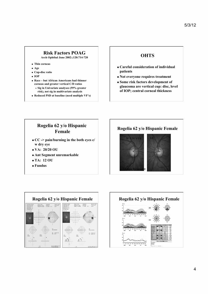
Rogelia 62 y/o Hispanic Female
CC -> pain/burning in the both eyes c/w dry eye
VA: 20/20 OU Ant Segment unremarkable TA: 12 OU Fundus
Rogelia 62 y/o Hispanic Female
? Inferior thinning RE
H: 0.5X V 0.6 0.6
Rogelia 62 y/o Hispanic Female Rogelia 62 y/o Hispanic Female

5/3/12
5
Rogelia - Summary 63 y/o Hispanic Female presents with
dry eye complaints Suspicious Cups RE inferior thinning
and a superior nasal field defect RE (Normal LE), consistent with OCT RNFL findings, IOP 12
Diagnosis -> NTG RE, No GL LE Management…Tx vs NoTx
Normal Tension Glaucoma Chronic optic neuropathy exhibiting
characteristic optic disc cupping and visual field loss
Untreated IOP in statistically normal range
No secondary causes for optic neuropathy No accepted level of IOP below which the
diagnosis becomes “Normal Tension” 20 to 50% of patients with POAG have
IOP within statistically normal range
NTG: The Big Question?
What is the relationship between IOP and visual field loss?
Is there a benefit of lowering IOP in patients with normal tension glaucoma?
Collaborative Normal Tension Glaucoma Study
Collaborative effort of 24 research and medical centers around North American and Europe
Study conceived in 1984 out of the Glaucoma Research Foundation meeting
Enrolled 230 patients 140 eyes of 140 patients met randomization
criteria. 90 excluded (38%)
NTG Study Criteria Only patients with progressive disease
were enrolled, or fixation was threatened 20 and 90 years old No previous recorded IOP of >24 mmHg 4 week washout period from previous
meds 10 baseline IOPs, 6 between 8 am - 6pm in
1 day, 4 reading other days Median IOP had to be < 20
3 Baseline VFs

5/3/12
6
NTG Study Criteria Enrolled 1 eye per patient Used better eye and excluded advanced disease Randomize threats to fix immediately Patients randomized:
Untreated control group Tx group with a 30% IOP reduction
Beta adrenergic blockers, adrenergic agonists excluded b/c cardiovascular and crossover affects
Endpoint defined as progression Progressive VF loss or ON changes
CNTGS Am J Ophthalmol 1998;126:487-497
230 enrolled: 140 eyes met criteria, were randomized
• 90/230 (39%) excluded - never progressed
61 Treatment group, 79 observation group Untreated group: 35% progressed, 65% no
progression Treatment group: 12% progressed Longer time to progression in tx’d eyes
NTG Study: Natural History Ophthalmology 2001;108:247-253
160 subjects (of the original 260) that were initially not treated
1/3 showed localized progression in 3 yrs Rate of progression was variable
50% showed progression 5-7 yrs > 50% not treated show no progression Conclusion: Rate progression highly
variable
NTG General Considerations How should we manage NTG? Because many pts showed no progression
Wait to treat, until rate of the disease can established
For those in high risk groups, watch more closely Women with history migraine, and disc
hemorrhages are at the highest risk for progression
Asian and men had the least risk for progression For those with advanced disease: Treat
more aggressively
How Low Does IOP Need to Be?
Advanced Glaucoma Intervention Study (AGIS)
To assess long-range outcome in sequence of interventions in Trab vs ALT in eyes who have failed initial med therapy
Study being done b/c varying degrees of success with either procedure.
Eyes randomized: trab followed by ALT followed by trab (TAT) ALT - trab - 2nd trab (ATT)
– May use antifibrotic agents on 2nd surgery

5/3/12
7
AGIS Multicenter randomized clinical trial 789 eyes of 591 patients inadequately
controlled on med therapy Randomized to 2 treatment sequences
ATT or TAT Additional sequences offered after
failure Antifibrotic agents later in study
AGIS: IOP and Field Loss
789 eyes followed for 6-11 years
4 analysis groups based on how often IOP < 18 100% visits 75- 99% 50-74% < 50% of visits Report # 7 AJO Oct 2000
12.3 % of time IOP < 18
20.2
14.7
12.3
14.7 16.9
20.2
% of time IOP < 18
AGIS: IOP and Field Loss, Report # 7 AJO Oct 2000
AGIS Long-term IOP fluctuation was
associated with progression
Low Mean IOP (Lower tercile) N = 100 Mean IOP = 10.8
Higher Mean IOP (Upper tercile) N = 100 (Mean IOP = 20.6
Low Fluctuation
N = 33
High fluctuation N = 33
Low Fluctuation N = 33
High Fluctuation
N = 33
9.1%
30.3%
24.2%
30.3%
% pts with VF Worsening
Reanalysis AGIS: IOP Fluctuation Important in Pts With Low Mean IOP
Caprioli, Colean Ophthalmology 2008
P=.03
P=.58
Which IOP is Most Important?
Mean IOP Pressure during the night IOP over 24 hours Peak IOP Fluctuation in IOP Overall variability

5/3/12
8
Forest vs. Trees I don’t really care The challenge:
Diagnosing early Making sure patients
understand the disease Make sure patients are
compliant Follow closely to
determine the rate of progression
Luisa: Hispanic Female Initial Presentation
Presented for routine exam VA: 20/20 OU TA: 26 OD; 27 OS Gonioscopy – CBB 360 OU, No PAS ON: 0.55 – 0.6 OU Inferior notch
Louisa Hispanic Female Initial Presentation
Luisa: IOP: 26/27 mmHg What should her initial management be? How low does the pressure need to go?
What is the basis for starting medical therapy? Is there any argument that could be made
for not treating? What is the risk of blindness? If we don’t treat her – will she go blind? At what rate will she lose visual field?
Early Manifest Glaucoma Trial
Randomized clinical trial to assess the effect of immediate tx on progression compared to no initial or delayed tx
Identify factors related to progression
Study natural history

5/3/12
9
Early Manifest Glaucoma Treatment Study
(EMGT) NEI supported clinical trial
performed in Sweden Does early treatment alter the natural
course of the disease in OAG? Early POAG, PDG, PXF, NTG Randomized:
ALT and Betaxolol vs. Careful Observation
EMGT 255 OAG patients (POAG, NTG, EFG) 129 randomized to 360 ALT &
betaxolol 126 randomized to observation Mean age 68 years old 66% women Mean baseline IOP 20.6
EMGT Follow up visit with VF q 3 mos; disc
photos q 6 mos Progression monitored with
Full threshold VF with Glaucoma Change Probability (using pattern deviation values)
Flicker chronoscopy of nerve photos, side by side comparison for suspected change
EMGT Results
Median f/u 6 years Average decrease IOP in Tx’d group
25% or 5.1mmHg 53% progressed -> 47% did not progress Progression slower in the Tx group
Longer time to progression in the tx’d group Median time to progression 18 mos
longer in tx’d group 45% (58/129) Tx’d v. 62% (78/126)
control
Arch Ophthalmol 2002;120:1268-1279
EMGT Progression
Treated Untreated
5 Yr Progression 44% 66%
8 Yr Progression 59% 76%
EMGT Results Baseline characteristics and
progression Initial IOP Exfoliation
Follow-up findings associated with progression Higher IOP % of visits with a disc hemorrhage

5/3/12
10
EMGT Baseline factors that predicted progression on a multivariate analysis:
Higher IOP Exfoliation Worse MD Older age Frequent disc hemorrhages
Leske MC, et al. Factors for glaucoma progression and the effect of treatment. The Early Manifest Glaucoma Trial. Arch Ophthalmol 2003;121:48-56.
Lessons from the EMGT Treatment works… Average rate of progression was 2.3
dB over 10 yrs Rate of progression was decreased by
10% for every 1 mmHg reduction of IOP
NonTx Group: 1/3 of pts 6 yrs out still have no progression
Recommendations for Management from the EMGT
Newly Dx pts should be followed often Take more VF’s early –establish rate
of progression – up to 7 VF over 2 yrs Pts with rapid progression should be
vigorously treated Tx should be tailored for each patient
Lessons from the EMGT Average age of defect discovered -> 72 yo
Most pts at this age will not go blind or get any disability from blindness
Average 70 yo patient diagnosed with glaucoma is expected to live 12 years
He/she will loose ~ 4.2 dB during his remaining lifetime
This patient will likely not “get in trouble” unless he starts with MD of < 10 dB
Lessons from the EMGT
Low Risk Patients Patients with early stage disease…and Low IOP’s
Is at low risk for rapid progression Tx affect is rather small
May leave room for recommending close follow up with no treatment
Elderly w/ unilateral Dz also considered low risk
Glaucoma Treatment Once diagnosis is made, decision to lower IOP
follows
Selection of target IOP
Consider extent and rapidity of damage
Consider IOP and other risk factors
Gain insight from recent studies
Treatment is arbitrary and individualized
Medications, laser therapy, surgery
Follow - up

5/3/12
11
Target IOP
Individualized to patient Initial IOP, extent, rapidity of damage Existing damage is clue to risk of future
damage Aggressive decrease if worsening at one
level
Target IOP
Advanced damage strive for low normal range
Consider family history, age and fellow eye
IOP not great for identifying those with glaucoma but is important in management and follow up care.
There is a benefit to IOP reduction in glaucoma management
More advanced damage and/or more rapid onset or progression calls for more aggressive IOP reduction
Monocular trials when possible Consider switch before adding med If not at goal move on Be aggressive with IOP reduction in
advanced cases or when there is evidence of progression
Baseline info is critical Pre-tx IOP, Level of IOP at times of
progression VF and nerve appearance
Chop or Drop
Several studies have questioned traditional beliefs regarding treatment:
"Are we in fact harming our patients by delaying surgery until there is evidence of further field loss, and/or deterioration while utilizing medical and laser regimens."
Does medical therapy/ALT provide as good longterm control of IOP as surgery in preventing continued field loss?
Is medical therapy truly benign? Is the overall “quality of life” better
with standard medical therapy or with surgical intervention?

5/3/12
12
168 newly diagnosed untreated patients with POAG (1983-84) followed 4 yrs Standard medical therapy ALT Trabeculectomy
Consistently lower IOP in Surgical group Surgery: 13.3 Medical: 16.8 Laser: 17.8
Better IOP Control with Surgery
Better IOP Control with Surgery 1st
Success of IOP control: Surgery: 98% Medical: 80% Laser: 60%
VA worse in surgery group by 1/2 line
Incidence of cat = 4%
Migdal CS, Hitchings RA: Long-term functional outcome of treatment POAG Ophthalmology 1994; 101:1651-1656
Surgery 1st Argument Better Long-term IOP Control
52 POAG patients Randomized to medical Tx
vs Scheis trabeculectomy Subdivided medical group
Well controlled on simple meds Need for surgical intervention Follow up 6-8 yrs
Smith RJH: The Lang lecture 1986. The enigma of primary open angle glaucoma. Trans Ophthalmol Soc UK 105:618-633 1986 (Moorfields Eye Hospital, London)
Surgery 1st Better Long-term IOP Control
Better IOP control with surgery
Less field deterioration
Similar VA Poor results for
patients who needed surgery after medical therapy failed
Smith RJH: The Lang lecture 1986. The enigma of primary open angle glaucoma. Trans Ophthalmol Soc UK 105:618-633 1986 (Moorfields Eye
Hospital, London)
Surgery as Initial Treatment for POAG: Better IOP Control
99 pts randomized to Surgery vs Medical therapy Medical therapy further randomized:
Successful IOP control with medicine Those who failed medical therapy and needed surgery
Better IOP control in surgery group (15 vs 20.8) Surgery 1st eyes were less likely to lose field Many of med Tx eyes ultimately needed further
surgery – 53% Jay JL, Murry SB: Early trabeculectomy versus conventional management in
POAG BJO. 1988; 72: 881-889.
Surgery 1st Argument
Medical therapy followed by trab Primary trab followed by supplemental
med tx Follow up 4.6 years Lower and better IOP control Sig. more field loss with medical therapy No significant difference in VA Jay JL, Allen D: The benefit of early trabeculectomy versus conventional
management in POAG related to severity of disease. Eye. 3: 528-535, 1988

5/3/12
13
Can medical Tx be Harmful?
Loss of visual field while waiting for IOP control
Jay/Allen, Smith showed worse field preservation in patients who failed medical therapy and later went on to need surgery
Is Medical Therapy Harmful?
Less success with filter after medical therapy fails (Lavin et al)
98% success with primary trab 79% success with trab when
medical therapy fails
Is Medical Therapy Harmful? Direct adverse reaction to drops
9/78-12/85 32 deaths directly from T5 Other Reactions
• Aplastic anemia • Respiratory effects • CNS problems • Loss of labido • Lathargy
CAI’s: Renal and GI effects
Quality of life Compliance
76% objective yet subjective report 98%
Older patients are on more multiple meds for other conditions. More complicated regimens lead to less compliance GLT showed only 30% of patients were
controlled on a single beta blocker
More visits result in worse quality of life
Summary: Surgery 1st Safe Better IOP control Good if not better field
preservation than med Tx VA is at least as good Cost effective
Collaborative Initial Glaucoma Treatment Study (CIGTS)
Purpose: To compare the long‑term effect of treating newly diagnosed POAG with standard medical vs. filtration surgery
607 pts w/ mild to moderate glaucoma randomized b/w Oct 93 – April 97
IOP lowering goal: 35% decrease from baseline
5 year follow-up

5/3/12
14
CIGTS Results
Both groups had substantial/sustained ↓ in IOP
Baseline mean IOP = 27 mmHg Medical therapy IOP reduced to 17.6 mmHg
37% reduction Surgical therapy: IOP reduced to 14mmHg
52% reduction
CIGTS and IOP VF loss did not differ
by Tx Surgery 2-3 mmHg
less than Medical Surgery group had
> VF loss and > VA loss in 1st 3 yrs, but equal by yrs 4-5
Lichter PR, et al Ophthalmology 2001 Nov;108(11):1943-53
CIGTS: Visual Fields and Cataract
10-12% of subjects in both groups progressed over 5 yrs
Rate of cataract development greater surgery group
Lichter PR, et al Ophthalmology 2001 Nov;108(11):1943-53
CIGTS: Quality of Life Both groups satisfied
Surgery: more local eye symptoms, irritation Most disappeared by yrs 4-5
Medical: variety of systemic symptoms, but not consistent over time Clearly different from surgery Sx
Janz NK, Wren PA Ophthalmology 2001 Nov;108(11):1954-65
CIGTS: Conclusion Both surgery and medicine as initial Tx
result in the same VF outcome at 5 yrs An IOP lowering of 35% seems to be
optimal in mild glaucoma with no net decline in visual function
The greater IOP lowering in the surgery group was of no further benefit
CIGTS: Conclusion
Investigators do not recommend changes to current approaches of management
Longer follow up is needed as this is a chronic disease 5 yrs is not adequate time to draw
conclusions

5/3/12
15
CIGTS Bottom Line
At 5 yrs, no difference between surgery and medicine for control of IOP
The study legitimized surgery as a primary procedure for treating newly diagnosed GL When all was said and done – surgery resulted
in lower IOP vs. Medicine Safe Overall in the long-term – may prove be better