clinical guideline title rib fractures in major trauma: a
TRANSCRIPT

V2.1 Rib Fractures in Major Trauma: a guideline for management version Page 1 of 16
CLINICAL GUIDELINE TITLE Rib Fractures in Major Trauma: a guideline for management in
adults (version 2.1)
1) SUMMARY This revised guideline provides a framework for the assessment and management of adult patients with rib fractures at Imperial College Healthcare NHS Trust. The guideline has been updated to take account of new evidence in patient risk assessment, analgesic management strategies and physiotherapy techniques. Additionally, criteria for accepting referrals for tertiary level care of rib fractures are provided.
2) INTRODUCTION 2.1 Incidence
Blunt chest-wall trauma accounts for 10-15% of all trauma admissions to Emergency Departments (EDs) globally1,2. Rib fractures may complicate up to two thirds of these injuries2. North West London Trauma Network treats approximately 450 patients with rib fractures per year, of whom approximately two thirds receive tertiary care at St Mary’s.
2.2 Importance
Rib fractures are markers of severe injury and are associated with significant morbidity and mortality. Patients with these injuries are at greater risk of complications and poor outcomes2-4. Associated injuries occur in 94% of patients, typically concomitant thoracic trauma, but also injuries to the head, abdomen and limbs3. Mortality associated with rib fractures is hard to calculate, as death often happens indirectly, however it has been estimated as between 10–13%3,4, with one article reporting up to 30%5. Common immediate thoracic sequelae of rib fractures include pneumothorax, haemothorax, haemopneumothorax, pneumatocoele, pulmonary contusions. Solid organs, such as the liver, kidneys and spleen, may also sustain lacerations from broken ribs1. Pain is the most common symptom from rib fractures and a key component in pulmonary complications. Pain restricts tidal volume, leading to hypoventilation, and impairs coughing ability, leading to sputum retention; these combine to cause atelectasis and predispose to pneumonia. Additionally, injured lung tissue underlying the fractures has impaired ability to exchange gasses (leading to shunt and VQ mismatch) and reduced compliance. Compensatory increases in respiratory rate may increase oxygen consumption. Pneumonia occurs in up to 30% of cases, with or without sepsis, causing further respiratory compromise. The combination of hypoventilation, atelectasis and/or lobar collapse and impaired gas exchange results in hypoxaemia, respiratory failure and, in some cases, a need for mechanical ventilation. Respiratory complications typically develop at 48 – 72 hours post injury. Other respiratory complications include pulmonary embolus, pulmonary effusions, empyema and acute respiratory distress syndrome (ARDS)1-6. Patients with rib fractures often require hospital admission, and in more significant injuries, to level 2 or 3 care. The associated incremental costs have not been fully evaluated, but can be considered in terms of length of inpatient stay, ‘ICU bed days’ and ‘ventilator days’ and are likely to be significant. Studies evaluating longer term outcomes have demonstrated high rates of chronic disability and chronic pain3,6,7,10. The severity of acute pain predicts chronic pain whilst disability is predicted by acute pain intensity and the presence of bilateral fractures10. Elderly patients (aged 65 years or older), have been consistently shown to have worse outcomes, higher complication rates and greater mortality after rib fractures than younger patients3,7.

V2.1 Rib Fractures in Major Trauma: a guideline for management version Page 2 of 16
3) DEFINITIONS
Rib fracture: a break in a bone making up the rib cage.
Flail chest: at least 2 fractures per rib in at least 2 adjacent ribs are needed to produce a flail segment. Flail segments cause paradoxical inspiratory movements, compromise breathing and may be life threatening.
Verbal Rating Scale: a method for assessing pain on an alphanumeric scale. ICHNHST recommends mild, moderate and severe, whilst in the ED the numerical scale 0-10 is used.
Thoracic Epidural (TE): a fine bore catheter placed into the thoracic epidural space which is used to give analgesic drugs.
Paravertebral block (PVB): regional anaesthetic technique providing analgesia to a segment of one hemithorax.
Patient Controlled Analgesia (PCA): a method of allowing a patient to administer their own analgesia intravenously, usually opioid based.
Non-invasive ventilation (NIV): facial Continuous Positive Airways Pressure (CPAP) or Bi-level Positive Airways Pressure (BIPAP) ventilation
Morphine Immediate Release (IR): a morphine immediate release preparation (available as a liquid - commonly known as Oramorph - and tablets)
Oxycodone Immediate Release (IR): a oxycodone immediate release preparation (available as a liquid and capsules)
4) SCOPE
These guidelines are for all staff involved in the care of adult trauma patients with rib fractures but are of particular relevance to those working in the Emergency Department, Theatres, Anaesthesia, Major Trauma Ward and other wards receiving trauma patients. They may also be adopted more widely within the North West London Trauma Network.
V2.1 Rib Fractures in Major Trauma: a guideline for management version Page 3 of 16
5) FULL GUIDELINE The use of multidisciplinary bundled care pathways is associated with improved outcomes in patients with rib fractures/ blunt chest injury3,24.
5.1 Identifying the high-risk patient
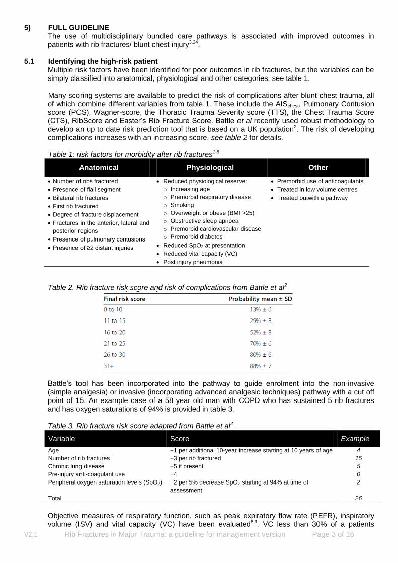
Multiple risk factors have been identified for poor outcomes in rib fractures, but the variables can be simply classified into anatomical, physiological and other categories, see table 1. Many scoring systems are available to predict the risk of complications after blunt chest trauma, all of which combine different variables from table 1. These include the AISchest, Pulmonary Contusion score (PCS), Wagner-score, the Thoracic Trauma Severity score (TTS), the Chest Trauma Score (CTS), RibScore and Easter’s Rib Fracture Score. Battle et al recently used robust methodology to develop an up to date risk prediction tool that is based on a UK population2. The risk of developing complications increases with an increasing score, see table 2 for details. Table 1: risk factors for morbidity after rib fractures1-8
Anatomical Physiological Other
Number of ribs fractured
Presence of flail segment
Bilateral rib fractures
First rib fractured
Degree of fracture displacement
Fractures in the anterior, lateral and
posterior regions
Presence of pulmonary contusions
Presence of ≥2 distant injuries
Reduced physiological reserve:
o Increasing age
o Premorbid respiratory disease
o Smoking
o Overweight or obese (BMI >25)
o Obstructive sleep apnoea
o Premorbid cardiovascular disease
o Premorbid diabetes
Reduced SpO2 at presentation
Reduced vital capacity (VC)
Post injury pneumonia
Premorbid use of anticoagulants
Treated in low volume centres
Treated outwith a pathway
Table 2. Rib fracture risk score and risk of complications from Battle et al2
Battle’s tool has been incorporated into the pathway to guide enrolment into the non-invasive (simple analgesia) or invasive (incorporating advanced analgesic techniques) pathway with a cut off point of 15. An example case of a 58 year old man with COPD who has sustained 5 rib fractures and has oxygen saturations of 94% is provided in table 3.
Table 3. Rib fracture risk score adapted from Battle et al2
Variable Score Example
Age +1 per additional 10-year increase starting at 10 years of age 4
Number of rib fractures +3 per rib fractured 15
Chronic lung disease +5 if present 5
Pre-injury anti-coagulant use +4 0
Peripheral oxygen saturation levels (SpO2) +2 per 5% decrease SpO2 starting at 94% at time of
assessment
2
Total 26
Objective measures of respiratory function, such as peak expiratory flow rate (PEFR), inspiratory volume (ISV) and vital capacity (VC) have been evaluated8,9. VC less than 30% of a patients
V2.1 Rib Fractures in Major Trauma: a guideline for management version Page 4 of 16
predicted value predicts pulmonary complications and each 10% increase in VC is associated with a reduced chance of pulmonary complications (odds ratio 0.64)9. The risk of complications can be reduced by adhering to the key principles of management: optimal analgesia, respiratory support, early mobilisation and appropriate surgical intervention.
5.2 Analgesic options
As pain is a significant contributor to the morbidity arising from rib fractures, optimisation of analgesia is key to preventing complications. There are many options for managing pain from rib fractures including multimodal oral therapy, intravenous analgesia, topical treatments and a variety of regional anaesthetic blocks. Acute pain should be assessed according to the Imperial College Healthcare NHS Trust Acute Pain Guideline. In particular for rib fracture patients, assess pain at rest and on coughing/ deep inspiration and ask about features of neuropathic pain for example tingling, burning and electric shock like sensations. Multimodal oral analgesia should be started on admission and include regular paracetamol, ibuprofen and opioids. Morphine should be prescribed both regularly and for breakthrough (PRN). In patients aged 65 years or older, ibuprofen should be omitted and the regular opioid changed to oxycodone, which has a better pharmacokinetic profile for elderly patients. In patients with renal impairment, defined as eGFR <30mls/min, ibuprofen should be omitted and the oxycodone dose reduced. All patients should receive adjunctive treatments for opioid analgesia including laxatives and anti-emetics. See figure 1 for details. Opioids may be administered via a patient controlled analgesia (PCA) device if the pain requires more frequent opioid dosing than the standard prescription allows. Patients with features of neuropathic pain such as burning, tingling, electric shock like sensations or numbness should be prescribed a gabapentinoid. Ketamine may be trialled intravenously (up to 10mg) and continued orally or as an infusion, following the Imperial College Healthcare NHS Trust guidelines, in patients with difficult to manage pain. In any patient with pain that is difficult to control using conventional measures, conversion to the invasive pathway should be considered. (Note the use of ketamine in pain management is unlicensed). All regional anaesthetic techniques for rib fracture analgesia impair impulse transmission at various points along the intercostal nerve. Thoracic Epidural analgesia remains the gold standard analgesic modality. They are the most widely studied mode of analgesia, and their use in rib fractures is endorsed by several systematic reviews and international bodies1,3,7. Expertise at St Mary’s allows for timely epidural insertion by senior anaesthetists and appropriate management once sited. Despite this, epidurals have a low but significant complication rate11 and in some patients may be contra-indicated (see table 4); there is also an inevitable failure rate and their benefit has been questioned12. Thoracic epidurals and blocks should only be inserted, doses titrated and drugs bolused by appropriately trained members of healthcare staff. The first line epidural infusion should be mixed levobupivacaine 0.125% with fentanyl 2micrograms/mL. In patients with rib fractures and other injuries arising from the same incident, a plain 0.125% levobupivacaine epidural infusion can be started in conjunction with an opioid PCA. Commencing and caring for a patient with an epidural infusion should follow the clinical guideline Epidural Analgesia: Continuous Infusions Clinical Guidelines for Adult Patients. Table 4: Contra-indications to epidural analgesia. Starred items apply to regional blocks
Contraindications to epidural analgesia
Absolute Relative
1. Patient refusal* 2. Spinal cord injury or haematoma 3. Epidural haematoma 4. Thoracic vertebral body fracture at
level of insertion 5. Local or generalised sepsis* 6. Open wound at site of insertion*
1. Unable to position patient* 2. Traumatic Brain Injury with
uncontrolled Intracranial Pressure 3. Incomplete spinal evaluation 4. Previous thoracic spinal surgery
5. Coagulopathy: INR >1.4 or platelets <80 x 10
9/L*
6. Active Anticoagulant therapy. AAGBI RAPAC guide.*
7. Extubation not anticipated within 5 days (ICU patients)*

V2.1 Rib Fractures in Major Trauma: a guideline for management version Page 5 of 16
Alternatives to epidurals include intercostal, interpleural and paravertebral blocks, but all have significant drawbacks and lack evidence of benefit over epidurals13. Newer techniques, including serratus anterior plane and erector spinae plane blocks, have not yet been robustly evaluated14,15,23 in the rib fracture population but are used in some trauma units and are the subject of much research interest. They may be of use in patients with unilateral injuries, when thoracic epidurals are contra-indicated or are not possible to insert. In addition to trauma specific cautions and contra-indications (see table 4), standard cautions should be applied prior to inserting an epidural or regional anaesthetic block. Invasive monitoring and critical care should be considered in patients with pre-existing comorbid disease that may be affected by an epidural or regional block e.g. stenotic valvular heart lesions.
5.3 Ventilation Management
Rib fractures are commonly associated with underlying pulmonary contusions and pleural injuries such as pneumothorax and haemothorax. These, combined with pain, can lead to respiratory failure. To prevent complications and ensure a timely recovery, all patients admitted with rib fractures should receive respiratory support titrated to their individual needs. Patients should be managed on wards with nursing staff familiar with the injury. Supplemental oxygen should be prescribed and administered at the lowest concentration required to achieve peripheral oxygen saturations (SpO2) of 94-98%, or 88-92% in patients at risk of carbon dioxide retention. If more than 2-4L/min via nasal cannulae is required, administered oxygen should be humidified to loosen secretions. Sodium chloride (NaCL) 0.9% nebulisers may be prescribed as required or regularly to assist expectoration. Salbutamol nebulisers can be prescribed for bronchospasm. Patients should be mobilised where possible and when in bed, be nursed sitting as upright as possible with attention to pressure area care. All rib fracture patients should receive physiotherapy input at least once a day (see below) until respiratory function normalises and mobility restored to baseline. Advanced respiratory support including Continuous Positive Airways Pressure (CPAP), Non-Invasive Ventilation (NIV) and Nasal high flow oxygen (Optiflow/ Airvo etc) can be considered, in liaison with critical care staff, for patients with anticipated or deteriorating respiratory failure. Decisions regarding which of these treatment modalities is appropriate will be specific to individual patients and determined by the clinical judgement of the attending trauma, anaesthetic and critical care teams. Patients with significant flail segments in particular should be referred early for critical care review regarding advanced respiratory support, even without evidence of respiratory failure, with the goal of maintaining lung volume and effective cough. A proactive approach to Chest X-Rays should be adopted for patients with flail segments; changes such as atelectasis warrant prompt referral to critical care. Preventative ventilatory support is a key strategy that must be adopted.
5.4 Physiotherapy
Physiotherapy should be started within 24 hours of admission in all patients to support ventilation and prevent complications. The ability of patients to participate in physiotherapy is dependent on adequate analgesia using non-sedative doses. All patients should be taught to perform a simple active cycle breathing technique (ACBT), huffing and encouraged to cough. This can be initiated by nursing staff if necessary, for example at weekends and in the evenings. ACBT consists of taking 3-5 deep breaths with an inspiratory hold of 2-3 seconds followed by 3 relaxed breaths. Patients should be asked to complete ACBT hourly when awake. Instructions for patients (adapted from the Association of Chartered Physiotherapists in Respiratory Care leaflet GL-05): 1. Please take a long, slow, deep breath in, ideally through your nose. 2. Hold your breath for 2-3 seconds. 3. Breathe out gently, like a sigh. 4. Repeat this technique for 3-5 breaths approximately every hour.

V2.1 Rib Fractures in Major Trauma: a guideline for management version Page 6 of 16
Huffing is a rapid exhalation of air through an open mouth and throat, as if trying to mist up a mirror. It helps to move sputum up the airways from where it can be coughed out. Huffing should be followed by the deep breathing cycle described above. In patients who are able to mobilise, early and regular mobilisation should be encouraged. Exercise bikes/floor pedal exercisers can be provided to assist patients who are only able to move into a chair. Shoulder exercises should be taught in appropriate patients to prevent movement restrictions post injury which can limit return to work.
5.5 Surgery
Surgical fixation and stabilisation of flail chest injuries is associated with reductions in duration of mechanical ventilation, ICU stay, total hospital stay, hospital acquired pneumonia and mortality rates16-20. In the long term patients return to work sooner and have a reduced incidence of chronic pain and analgesic dependence18-22. Studies have also shown similar beneficial outcomes in patients with multiple rib fractures but without a flail segment21. A multidisciplinary approach to patient selection for surgery is essential. The National Institute of Clinical Health and Excellence has approved and issued guidance on surgical fixation of flail chest injuries22.
5.6 Referral criteria for tertiary care in North West London Trauma Network Many patients with rib fractures will present to trauma units within the North West London Network. Some may require tertiary level services, however, the majority can receive care and pain relief at their local trauma unit following local rib fracture pain management guidelines. Indications for referral to St Mary’s Major Trauma Centre are detailed below. Acute referrals should be made to the Trauma Team Leader (TTL) on duty via the TTL mobile or bleep 1328 via switchboard. Internal referrals for consideration of rib fixation should be made to the rib fracture team, via e-mail ([email protected]). 1. Rib fractures associated with:
a. Significant pneumothorax b. Haemothorax c. Flail segment d. Moderate/ significant pulmonary contusions
2. Rib fractures in a polytrauma patient. 3. Consideration for rib fracture fixation. Indications (adapted from NICE IPG 361)22 include:
a. Clinical evidence of flail chest (visible paradoxical chest movements) associated with respiratory failure
b. ≥3 displaced rib fractures not responding to adequate analgesia c. Significant chest wall deformity or ≥25% lung volume loss on CXR d. Bilateral rib fractures e. Non-invasive ventilation/ invasive ventilation dependent f. Presence of clavicle or scapular fracture
5.7 Placement of patients at St Mary’s Hospital
Most patients will be admitted onto the major trauma, orthopaedic or general surgical wards, but some patients can be admitted elsewhere. Patients with thoracic epidurals should be admitted to major trauma ward or the critical care complex. Clinical Decision Unit Patients with rib fractures can be considered for admission to CDU only if they meet certain criteria:
Patients 65 years or older: less than or equal to 2 rib fractures.
Patients aged under 65 years: less than or equal to 3 rib fractures.
Exclusion criteria for all age groups: Injury includes: flail segment, significant pneumothorax or haemothorax. Pain relief: requires PCA, epidural or block.

V2.1 Rib Fractures in Major Trauma: a guideline for management version Page 7 of 16
Patients with isolated rib fractures, meeting CDU criteria, who are well pre-injury and who have adequate social support can be considered for immediate or early discharge. Such patients should be counselled to seek medical advice if symptoms change or deteriorate. Intensive Care Unit Patients in ICU receiving level 2 care should follow the standard non-invasive or invasive pathway as appropriate. Patients who would benefit from non-invasive ventilation should also be considered for arterial line insertion and regular blood gases to assess respiratory function. In patients who are receiving level 3 care (i.e. sedated, intubated and ventilated), the following principles should be considered:
Ventilation:
o Use lung protective ventilation strategies.
o Nurse with the head of the bed elevated to ≥30 degrees if not contraindicated by other
injuries.
o Regular subglottic suctioning.
Analgesia:
o Assess pain regularly and treat with multimodal analgesic therapy.
o Contact 1201 to arrange for a thoracic epidural:
In patients showing signs of impaired respiratory effort e.g. tachypnoea, poor tidal
volumes, signs of pain or the patient indicates they are in pain.
In the 24 hours prior to planned extubation in patients meeting the invasive pathway
criteria.
Consider referral for rib fixation (and concurrent epidural) if:
o Flail chest or paradoxical chest movement during weaning from a ventilator o > 6 ribs fractured o Bilateral rib fractures o Hypoxia and/or hypercarbia under 40% inspired oxygen inhalation o Repeated atelectasis o Significant chest wall deformity o Not responding to thoracic epidural and do not require intubation and ventilation otherwise
5.8 Discharge
Prior to discharge, patients should be given information, for example the NHS choices rib injury sheet, to ensure their progress after discharge is optimal. Pain should be adequately controlled such that patients are discharged with weak opioids (as well as other multimodal agents) in the TTO pack. If stronger opioids, e.g. morphine, are still in use, a clear weaning plan should be in place for the GP. Patients should be advised to see their GP if the pain isn’t responding to prescribed analgesics or if they develop features of chest infection. Other simple interventions that can be recommended for completion at home include continued use of the active cycle breathing technique, use of ice packs, use of splints when coughing (e.g. a rolled-up towel), encouragement of mobilisation but avoidance of heavy strenuous exercise or work. Patients who have received surgical rib fracture fixation should be followed up in fracture clinic at 6 weeks after discharge.

V2.1 Rib Fractures in Major Trauma: a guideline for management version Page 8 of 16
Figure 1: Overview of Imperial rib fracture pathway
R i b F r a c t u r e P a t h w a y O v e r v i e w
1 Rib fractures confirmed on CXR / CT
2 Start multimodal analgesia and adjuncts
Adult patients
Patients aged 65yrs or older
Patients with renal impairment (eGFR <30)
Paracetamol 1g PO/ IV 6 hourly (if weight <50kg dose at 15mg/kg IV)
Paracetamol 1g PO/ IV 6 hourly (if weight <50kg dose at 15mg/kg IV)
Paracetamol 1g PO/ IV 6 hourly (if weight <50kg dose at 15mg/kg IV)
Morphine IR* 10 - 20mg PO 4 hourly +
Morphine IR* 5 – 10mg PO 4 hourly PRN
Oxycodone IR* 2.5 – 5mg PO 4 hourly +
Oxycodone IR* 2.5 – 5mg PO 4 hourly PRN
Oxycodone IR* 1.25 – 2.5mg PO 4 hourly +
Oxycodone IR* 1.25 – 2.5mg PO 4 hourly PRN
Ibuprofen 400mg PO QDS Avoid NSAIDs Avoid NSAIDs
Laxatives: macrogol 1 sachet BD and senna 15mg ON
Laxatives: macrogol 1 sachet BD and senna 15mg ON
Laxatives: macrogol 1 sachet BD and senna 15mg ON
Anti-emetics
Anti-emetics
Anti-emetics
*IR = Immediate Release
3 Assess Risk (Battle’s score)
Variable Score
Age +1 per additional 10-year increase starting at 10 years of age Number of rib fractures +3 per rib fractured Chronic lung disease +5 if present Pre-injury anti-coagulant use +4 Peripheral oxygen saturation levels (SpO2) +2 per 5% decrease SpO2 starting at 94% at time of assessment Total
Please document Battle score on Cerner Consider other risk factors for morbidity and the need for fib fixation:
Risk factors for morbidity Early review for surgical rib fixation Pulmonary contusion Current smoker Flail chest Chest wall deformity Cardiovascular disease Frailty ≥3 displaced rib fractures CXR: ≥25% lung volume loss Presence of ≥2 distant injuries Obesity & OSA ≥65 years old NIV/ ventilator dependent
4 Start on appropriate arm of pathway
Battle score 0 – 15 Battle Score ≥16
Non Invasive Invasive
If score 0 – 10: consider CDU if meets criteria or discharge if appropriate
If score ≥30: consider early liaison with critical care team
If score 11 – 15 or other risk factors for morbidity present: consider more advanced analgesia & escalation to invasive pathway
V2.1 Rib Fractures in Major Trauma: a guideline for management version Page 9 of 16
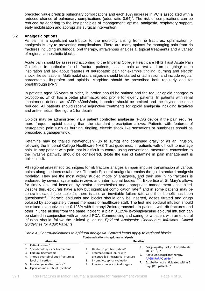
Figure 2: Non-Invasive pathway for rib fracture management
N o n – I n v a s i v e P a t h w a y
1 Admit to appropriate ward and start regular monitoring Nursing care provided by staff familiar with major trauma patients Monitor oxygen saturation (SpO2) and Vital Capacity Assess pain severity using verbal rating scale and ask about features of neuropathic pain
2 Start respiratory support Encourage to sit upright and mobilise early where possible Provide supplemental oxygen at the lowest concentration to achieve appropriate SpO2 Oxygen should be humidified Prescribe NaCL 0.9% 10ml nebulisers 4 hourly regularly or as required to assist expectoration Consider prescribing salbutamol 2.5 – 5mg nebulised 4 hourly as required
3 Confirm analgesia is prescribed and titrate as required
Adult patients
Patients aged 65yrs or older
Patients with renal impairment (eGFR <30)
Paracetamol 1g PO/ IV 6 hourly (if weight <50kg dose at 15mg/kg IV)
Paracetamol 1g PO/ IV 6 hourly (if weight <50kg dose at 15mg/kg IV)
Paracetamol 1g PO/ IV 6 hourly (if weight <50kg dose at 15mg/kg IV)
Morphine IR* 10 – 20 PO 4 hourly +
Morphine IR* 5 – 10mg PO 4 hourly PRN
Oxycodone IR* 2.5 – 5mg PO 4 hourly +
Oxycodone IR* 2.5 – 5mg PO 4 hourly PRN
Oxycodone IR* 1.25 – 2.5mg PO 4 hourly +
Oxycodone IR* 1.25 – 2.5mg PO 4 hourly PRN
Ibuprofen 400mg PO QDS Avoid NSAIDs Avoid NSAIDs
Laxatives: macrogol 1 sachet BD and senna 15mg ON
Laxatives: macrogol 1 sachet BD and senna 15mg ON
Laxatives: macrogol 1 sachet BD and senna 15mg ON
Anti-emetics
Anti-emetics
Anti-emetics
Consider IV opioid PCA: Morphine 1st choice, Fentanyl 2nd choice (unless renal impairment). Give pregabalin if features of neuropathic pain. Consider PO ketamine after successful IV trial. Contact pain team (bleep 1043) or anaesthetic team (bleep 1213) for advice
*IR = Immediate Release
4 Start regular physiotherapy within 24 hours of admission Active Cycle Breathing Technique including “huffing” hourly when awake Encourage early and regular mobilisation for those able Pedal exercisers for patients unable to mobilise beyond a chair Teach and encourage regular shoulder exercises
5 Regular reassessment and titration of therapy
SpO2/ PaO2/ Vital Capacity reducing SpO2/ PaO2/ Vital Capacity improving/ stable O2 needs increasing O2 needs reducing/ stable
Pain score/ Analgesia needs increasing Pain score improving/ stable
Invasive Pathway Continue regular reassessment and titration

V2.1 Rib Fractures in Major Trauma: a guideline for management version Page 10 of 16
Figure 3: invasive pathway for rib fracture management
I n v a s i v e P a t h w a y
1 Admit to Major Trauma Ward and start regular monitoring Nursing care provided by staff familiar with major trauma patients Monitor oxygen saturation (SpO2) and Vital Capacity Assess pain severity using verbal rating scale and ask about features of neuropathic pain
2 Start regular analgesia, ventilation support and physiotherapy As per items 2-4 on the Non-Invasive Pathway
3 Anaesthetic Review for Thoracic Epidural (TE) – bleep 1201/1213 TE is the 1st choice block for rib fractures. If there are contraindications, consider a block. Both should be booked under “Emergency NCEPOD” on Cerner and completed within 6 hours of admission. Insertion should take place in a monitored area
No contraindications
TE contraindicated
Thoracic Epidural Serratus Anterior or Erector Spinae Plane Block
Initial bolus of 10 – 15mls levobupivacaine 0.125% Initial bolus of 40mls levobupivacaine 0.125% +
1:400000 adrenaline Prescribe mixed bag levobupivacaine 0.125% + 2mcg/ml fentanyl at 15mls/hr + rescue bolus 10mls 4 hourly PRN
Prescribe plain levobupivacaine 0.125% at 15mls/hr + rescue bolus 20mls 4 hourly PRN
4 Regular reassessment and titration of therapy
SpO2/ PaO2 reducing SpO2/ PaO2/ Vital Capacity improving/ stable Vital Capacity deteriorating O2 needs reducing/ stable
O2 needs increasing Pain controlled
Pain score worse Pain controlled Continue regular
reassessment and titration
Call 1213 for epidural/ Contact ICU/ outreach Consider step down to non block trouble shooting Consider Chest X-Ray invasive pathway if Re-site if necessary Consider NIV/ nasal high flow sustained improvements
Candidate for surgical rib fixation

V2.1 Rib Fractures in Major Trauma: a guideline for management version Page 11 of 16
Figure 4: Rib fixation pathway
S u r g i c a l R i b F i x a t i o n P a t h w a y
Decision making for potential candidates
Review by member of rib fracture fixation team.
Discussion & Decisions MDT may include: rib fracture fixation team, MTW consultant, anaesthetist,
intensive care consultant, physiotherapist, occupational therapy & nursing staff.
Decision for surgery made by 2 consultants.
Liaise with thoracic/ trauma surgeons if suspicion of large air leak or visceral injury.
If ipsilateral clavicle fractured, have low threshold for ORIF clavicle.
Pre-operative
Book Half day theatre session.
Drugs Give prophylactic LMWH ≥12 hours before planned surgery.
Equipment MatrixRib fixation kit is sterile and available.
Imaging 3D CT reconstruction thorax complete.
Blood results FBC, renal profile (U&E & creatinine), clotting screen/ TEG.
Blood products 2 units of packed red blood cells cross matched.
Intra-operative
Anaesthetic team
1. Insert thoracic epidural prior to anaesthetic induction if not already in situ OR ensure the dressings are distant to the operative site.
2. Use standard single lumen endotracheal tube. 3. Insert arterial line if anticipating NIV or inability to extubate.
All theatre team Position patient supine or laterally after discussion with surgeons
Surgical team
1. Plan surgical incision(s) by identifying fractures clinically or with ultrasound. 2. If thoracic epidural not possible or contraindicated, insert paravertebral block/
catheter under direct vision at the end of the procedure. 3. Clean and debride original thoracostomy site to prevent infective complications.
Chest drains
1. Replace any pre-existing chest drains prior to operative fixation through a new thoracostomy
2. Insert a minimum of 1 chest drain per operated hemi-thorax at the end of the procedure and connect it to an underwater seal.
Mobile Chest X Ray at end of procedure in recovery or HDU to confirm drain position.
Postoperative
Post theatre destination 1. Aim to extubate at end of procedure. Arrange Major Trauma Ward/ Level 2 bed. 2. If intubated pre-op or significant respiratory compromise, to remain intubated and
transfer back to ICU.
Analgesia
1. Give epidural levobupivacaine 0.125%/ fentanyl 2 mcg/ml mixture at 15 mls/hr. 2. Remove epidural 3-5 days after insertion (7 days maximum, if tunnelled, at
discretion of anaesthetist & pain team). 3. Follow the invasive pathway
Rehabilitate 1. Regular ACBT and physiotherapy 2. Sit up and ambulate ASAP.
Discharge planning 1. Continue ACBT at home 4 hourly. 2. Wound review at 2 weeks. 3. Clinic review at 6 weeks.

V2.1 Rib Fractures in Major Trauma: a guideline for management version Page 12 of 16
Figure 5: Rib fracture tertiary referral form
NORTH WEST LONDON TRAUMA NETWORK RIB FRACTURE TERTIARY REFERRAL FORM
It is the policy of the North West London Major Trauma Centre to repatriate patients within 72 hours of rib fracture surgery if deemed medically fit
Referral details
Referring clinician
Responsible Consultant:
Contact telephone (mobile):
Contact e-mail address:
Hospital
Ward
Patient
Name:
Date of birth:
NHS number:
Home address:
General Practitioner:
Injury
Mechanism:
Rib fractures:
Pneumothorax? Yes No Haemothorax? Yes No Pulmonary contusion? Yes No
Associated injuries:
Interventions
Intercostal drain? Yes No Date of insertion: …… / …… / …… Persistent air leak? Yes No
Imaging and findings
Chest XR + CT chest (including 3D reconstruction rendered images) transferred by IEP: Yes No
Thoracic epidural in situ: Yes No
Ventilation: Self-ventilating Non-invasive ventilation Intubated
FiO2 ABG results
Details of any other treatment/surgery received:
Pre-morbid state
Co-morbidities:
Previous thoracic/ cardiac/ thoracic spinal surgery:
Anticoagulant history including VTE prophylaxis:
Please e-mail referral to [email protected]

V2.1 Rib Fractures in Major Trauma: a guideline for management version Page 13 of 16
6) IMPLEMENTATION
Training required for staff ☑ Yes ☐ No
If yes, who will provide training: Anaesthetic department: Epidurals and RA techniques Major Trauma Department: overall management
When will training be provided? At induction of junior doctors, new consultants and regularly at nursing updates.
Date for implementation of guideline:
1/3/2019
7) MONITORING / AUDIT
When will this guideline be audited? Ongoing currently (clinical governance reference number 1850)
Who will be responsible for auditing this guideline? Dr Alex Wickham, Consultant Anaesthetist
Mr Ian Sinha, Consultant Orthopaedic Surgeon
Are there any other specific recommendations for audit?
None
8) REVIEW
Frequency of review Please indicate frequency of review: 3 years Person and post responsible for the review: Mr Ian Sinha, Consultant Orthopaedic Surgeon Dr Alex Wickham, Consultant Anaesthetist
9) REFERENCES 1. EAST Practice Management Guidelines Work Group. Pain management in Blunt Thoracic Trauma. J
Trauma 2005; 59(5):1256-1267.
2. Battle C et al. Predicting outcomes after blunt chest wall trauma: development and external validation of
a new prognostic model. Crit Care 2014, 18:R98.
3. Witt C & Bulger E. Comprehensive approach to the management of the patient with multiple rib
fractures: a review and introduction of a bundled rib fracture management protocol. Trauma Surg Acute
Care Open 2017; 2:1–7.
4. Galvagno SM Jr, Smith CE, Varon AJ, Hasenboehler EA, Sultan S, et al. Pain management for blunt
thoracic trauma: a joint practice management guideline from the Eastern Association for the Surgery of
Trauma and Trauma Anesthesiology Society. J Trauma Acute Care Surg 2016; 81:936–51.
5. Jones KM, Reed RL, 2nd, Luchette FA. The ribs or not the ribs: which influences mortality? Am J Surg.
2011; 202(5):598Y604.
6. May L, Hillermann C & Patil S. Rib Fracture Management. BJA Education 2016; 16(1):26-32 7. Unsworth A, Curtis K & Asha SE. Treatments for blunt chest trauma and their impact on patient
outcomes and health service delivery. Scandinavian Journal of Trauma, Resuscitation and Emergency
Medicine, 2015; 23:17
8. Butts C et al. Do simple beside lung function tests predict morbidity after rib fractures? Am J Surg 2017;
213(3):473-477.
9. Carver T, Milia D, Somberg C, Brasel K & Paul J. Vital capacity helps predict pulmonary complications
after rib fractures. J Trauma Acute Care Surg 2015; 79(3):413-6.
10. Gordy S et al. The contribution of rib fractures to chronic pain and disability. Am J Surg 2014; 207(5):
659-62.
11. Royal College Of Anaesthetists. National Audit Project 3: Major Complications of Central Neuraxial
Block in the United Kingdom. Report and Findings. January 2009. RCOA; London.
12. Carrier F, Turgeon A, Nicole P et al. Effect of epidural analgesia in patients with traumatic rib fractures:
a systematic review and meta-analysis of randomized controlled trials. Can J Anesth 2009; 56:230–242.
V2.1 Rib Fractures in Major Trauma: a guideline for management version Page 14 of 16
13. Jarvis AM, Cook CH, Lindsey DE, Reilley TE, Steinberg SM, Beery RM, Whitmill ML, Papadimos TJ,
Stawicki SP. Comparison of epidural versus parenteral analgesia for traumatic rib fractures: A meta-
analysis. OPUS 12 Scientist 2009; 3(3): 50-57.
14. Kunhabdulla NP, Agarwal A. Serratus Anterior Plane Block for Multiple Rib Fractures. Pain Physician
2014; 17: E651-662
15. Fu P, Weyker PD, Webb CAJ. Case Report of Serratus Plane Catheter for Pain Management in a
Patient with Multiple Rib Fractures and an Inferior Scapular Fracture. Anesth Analg 2017; 8(6):132-135.
16. Marasco SF, Davies AR, Cooper J, Varma D, Bennett V, Nevill R, Lee G, Bailey M, Fitzgerald M.
Prospective randomised controlled trial of operative rib fixation in traumatic flail chest. J Am Coll Surg
2013; 216(5):924-32.
17. Ahmed Z, Mohyuddin Z. Management of flail chest injury: internal fixation versus endotracheal
intubation and ventilation. J Thorac Cardiovasc Surg. 1995; 110:1676-80.
18. Tanaka H, Yukioka T, Yamaguti Y, et al. Surgical stabilization or internal pneumatic stabilization? A
prospective randomized study of management of severe flail chest patients. J Trauma. 2002; 52:727–
732.
19. Lafferty PM, Anavian J, Will RE, et al. Operative treatment of chest wall injuries: indications, technique,
and outcomes. J Bone Joint Surg Am 2011; 93:97–110.
20. Marasco SF, Davies AR, Cooper J, Varma D, Bennett V, Nevill R, Lee G, Bailey M & Fitzgerald M.
Prospective Randomized Controlled Trial of Operative Rib Fixation in Traumatic Flail Chest. J Am Coll
Surg 2013;216:924-932.
21. Bottlang M, Walleser S, Noll M, et al. Biomechanical rationale and evaluation of an implant system for
rib fracture fixation. European Journal of Trauma and Emergency Surgery 2010; 36(5): 417–426.
22. National Institute for Health and Care Excellence (NICE). Interventional procedure guidance 361:
Insertion of metal rib reinforcements to stabilise a flail chest wall. 2010. NICE: London. Available from:
https://www.nice.org.uk/guidance/ipg361
23. Adhikary S, Liu W, Fuller E, Cruz-Eng H and Chin K. The effect of erector spinae plane block on
respiratory and analgesic outcomes in multiple rib fractures: a retrospective cohort study. Anaesthesia
2019; published online https://doi.org/10.1111/anae.14579
24. Todd S et al. A multidisciplinary clinical pathway decreases rib fracture–associated infectious morbidity
and mortality in high-risk trauma patients. Am. J Surg 2006; 192: 806–811.
V2.1 Rib Fractures in Major Trauma: a guideline for management version Page 15 of 16
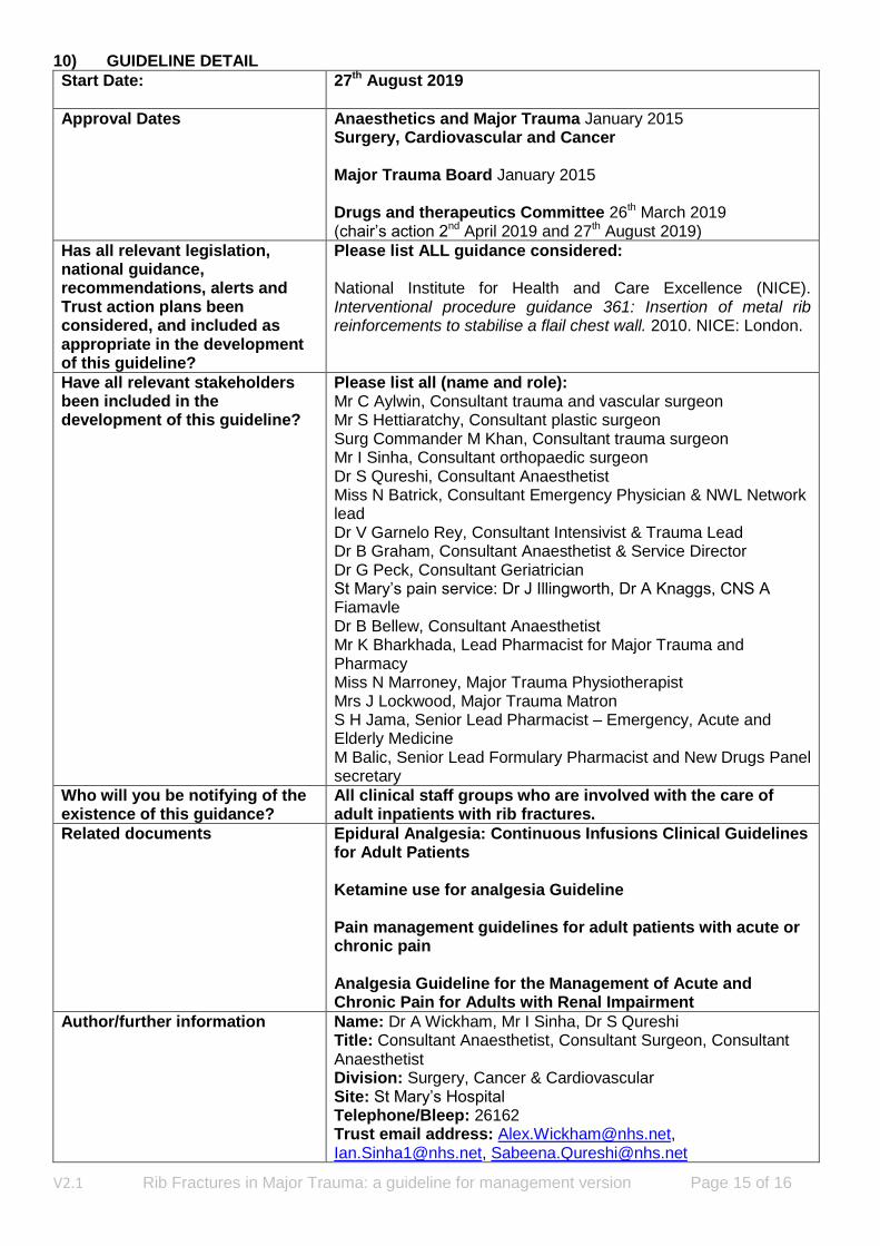
10) GUIDELINE DETAIL
Start Date:
27th August 2019
Approval Dates Anaesthetics and Major Trauma January 2015 Surgery, Cardiovascular and Cancer Major Trauma Board January 2015 Drugs and therapeutics Committee 26th March 2019 (chair’s action 2nd April 2019 and 27th August 2019)
Has all relevant legislation, national guidance, recommendations, alerts and Trust action plans been considered, and included as appropriate in the development of this guideline?
Please list ALL guidance considered: National Institute for Health and Care Excellence (NICE). Interventional procedure guidance 361: Insertion of metal rib reinforcements to stabilise a flail chest wall. 2010. NICE: London.
Have all relevant stakeholders been included in the development of this guideline?
Please list all (name and role): Mr C Aylwin, Consultant trauma and vascular surgeon Mr S Hettiaratchy, Consultant plastic surgeon Surg Commander M Khan, Consultant trauma surgeon Mr I Sinha, Consultant orthopaedic surgeon Dr S Qureshi, Consultant Anaesthetist Miss N Batrick, Consultant Emergency Physician & NWL Network lead Dr V Garnelo Rey, Consultant Intensivist & Trauma Lead Dr B Graham, Consultant Anaesthetist & Service Director Dr G Peck, Consultant Geriatrician St Mary’s pain service: Dr J Illingworth, Dr A Knaggs, CNS A Fiamavle Dr B Bellew, Consultant Anaesthetist Mr K Bharkhada, Lead Pharmacist for Major Trauma and Pharmacy Miss N Marroney, Major Trauma Physiotherapist Mrs J Lockwood, Major Trauma Matron S H Jama, Senior Lead Pharmacist – Emergency, Acute and Elderly Medicine M Balic, Senior Lead Formulary Pharmacist and New Drugs Panel secretary
Who will you be notifying of the existence of this guidance?
All clinical staff groups who are involved with the care of adult inpatients with rib fractures.
Related documents Epidural Analgesia: Continuous Infusions Clinical Guidelines for Adult Patients Ketamine use for analgesia Guideline Pain management guidelines for adult patients with acute or chronic pain Analgesia Guideline for the Management of Acute and Chronic Pain for Adults with Renal Impairment
Author/further information
Name: Dr A Wickham, Mr I Sinha, Dr S Qureshi Title: Consultant Anaesthetist, Consultant Surgeon, Consultant Anaesthetist Division: Surgery, Cancer & Cardiovascular Site: St Mary’s Hospital Telephone/Bleep: 26162 Trust email address: [email protected], [email protected], [email protected]

V2.1 Rib Fractures in Major Trauma: a guideline for management version Page 16 of 16
Document review history Next review due: 2nd April 2022 Version 1: Major Trauma Board review July 2015 Version 2: DTC review and recommendations 26/3/19 Version 2.1: Update to CDU criteria
THIS GUIDELINE REPLACES: Rib Fractures in Major Trauma: a guideline for management version 1, July 2015
11) INTRANET HOUSEKEEPING
Key words Major Trauma, Rib Fracture, Epidural, Rib fixation
Which Division/Directorate category does this belong to?
Surgery, Cancer & Cardiovascular
Which specialty should this belong to when appearing on the Source?
Major Trauma
12) EQUALITY IMPACT OF GUIDELINE Is this guideline anticipated to have any significant equality-related impact on patients, carers or staff?
No ☑