clinical pathology, haematology and biochemistry
TRANSCRIPT
Aeromonas Causes Severe Skin Lesions in Rainbow Trout (Oncorhynchus mykiss):Clinical Pathology, Haematology and Biochemistry
J. ¤EHULKA
University of South Bohemia, âeské Budûjovice, Research Institute of Fish Culture and Hydrobiology,VodÀany, Department of Aquatic Toxicology and Fish Diseases, Laboratory Opava, Czech Republic
Received March 23, 2002Accepted June 19, 2002
Abstract
¤ehulka J . : Aeromonas Causes Severe Skin Lesions in Rainbow Trout (Oncorhynchus mykiss):Clinical Pathology, Haematology and Biochemistry. Acta Vet. Brno 2002, 71: 351-360.
Aeromonas infection caused mass death of rainbow trout, Oncorhynchus mykiss (Walbaum)weighing 394 ± 69 g, at a water temperature of 4 oC in April. In a bioassay, the disease was inducedby an Aeromonas strain whose biochemical characteristics most closely resembled Aeromonassobria and Aeromonas caviae. The development of the skin lesions started as depigmented spotssurrounded by a hyperaemic zone with the formation of ulcers, or the changes on the skin resembledfurunculosis, taking the form of very large prominent bulges filled with clear exudate which, whenbroken, revealed haemorrhagically altered muscle. Some fish showed exophthalmus;inflammation around pectoral fins; hyperaemia of the wall of the swim-bladder and petechialhaemorrhages on the liver were found inside the abdominal cavity. Severe anaemia wascharacterized by a reduced erythrocyte count and lower haematocrit and haemoglobin levels.Clinical chemistry analyses in the diseased fish indicated reduced levels of total protein,cholesterol, triacylglycerol and total calcium and an increase in the urea level. Among the fiveenzymes and isoenzymes analyzed, catalytic concentration reaching multiples of the normal levelwas found in alanine aminotransferase, lactate dehydrogenase, α-hydroxybutyryl dehydrogenaseand γ-glutamyl transferase. Electrophoretic analysis indicated a reduced level of albumin in thediseased fish. These results point out the importance of mesophilic motile Aeromonas as causalagents of severe skin affections in salmonids. The findings encourage efforts to extend theknowledge of clinical haematology for the identification of health disorders and specific responsestypical of the individual diseases.
Aeromonas caviae, Aeromonas sobria, histopathology, red blood count, biochemical indices,blood plasma, electrophoresis
The care of the health of fish stocks in intensive salmonid culture necessitates theparticipation of qualified veterinary supervision. Among the suite of methods forexamination of the physiological health of the fish, an increased awareness of the usefulnessof clinical chemistry is materializing among first pathologists and veterinarians.Appropriate haematological and biochemical assays may identify changes in organfunction, find anomalies in the metabolism, determine additional laboratory procedures andmake prognoses. Synthesis and analysis of the figures obtained for the individual diseasesmay provide valuable information on the specific response or the range and nature of thepathological process. For salmonids this is confirmed by the analyses performed byMulcahy (1969, 1971), Foda (1973), Shieh and MacLean (1976), Harbel l et al.(1979), Hoffmann and Lommel (1984), Bruno (1986), Waagbø et al. (1988) andMøyner (1993).
Skin affections frequently occur in intensive salmonid culture. If they are identifiedin time, the development of the pathological process can be very effectively prevented.This applies, in particular, to cases in which the epizootic breaks out at a low watertemperature when the voracity of the fish is low and the diseased fish are less willing to
ACTA VET. BRNO 2002, 71: 351–360
Address for correspondence:Ing. J. ¤ehulka, CSc.Research Institute of Fish Culture and Hydrobiology NádraÏní okruh 33 746 01 Opava, Czech Republic
Phone: +42 653 628 660E-mail: [email protected]://www.vfu.cz/acta-vet/actavet.htm
take medicated food so that the effectiveness of targeted therapeutic intervention ispoor. In addition to flavobacterial infections, microbiologists have good reasons also tofocus their interest on the motile mesophilic Aeromonas species as causative agentsresponsible for septicaemia, local inflammations and necroses on the skin, muscles andsoft tissues. Apart from Aeromonas hydrophila, the aetiology of Aeromonas-relatedlesions may also involve certain species identified in recent years, including forexample Aeromonas allosaccharophila, described in conger eel by Martinez–Murciaet al. (1992), or also described by us in the clinical material of isolated Aeromonassobria and Aeromonas caviae. The authors of widely recognized identification systems(Ardu ino et al. 1988; Carnahan et al. 1991) assert that the identification of theindividual species is difficult; others prefer identification of pathogenic factors togetherwith biochemical traits rather than giving a strain a specific name (Agger et al. 1985;Burke et al. 1982).
The purpose of this report is to describe changes in the peripheral blood of rainbow troutin mass occurrence of Aeromonas natural infection. The basic characteristics oferythrocytes, certain metabolites of the blood plasma (including activities of the variousenzymes) and changes in the plasma protein spectrum were examined. The findings extend,enhance and refine the knowledge learned from the given case, as described in a contributionto the V. International Ichthyohaematology Conference (¤ehulka 1998).
Materials and MethodsFish
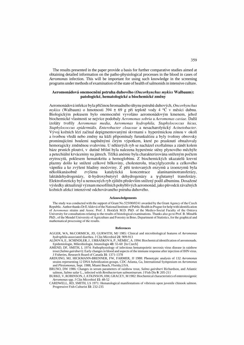
The infection occurred early in April on a trout farm where the fish was kept in flow-through concrete tanks,were fed for the market (average weight was 370 g and standard length 300 mm). The water temperature was 4 oC,dissolved oxygen content 11.8 O2· l-1, oxygen saturation 88%, water hardness 3.4 o N and CODMn 0.8 mg .· l-1. Thedisease took a chronic course, mortality reached 50%. Selected for the investigation were fish with lesions of thesame nature as shown in Figs 1 and 2 (Plate XII), manifested in 90% of the affected fish.
Sampling procedure for his topathologyTissue samples from 8 moribund fish, including muscle, heart, kidney, liver, spleen and intestine, were fixed in
a 10% neutral buffered formalin solution. Histologic sections were stained by haematoxylin-eosin and Gram.
Bacter iologyBacteriological procedures consisted of the preparation and Gram staining of tissue smears, inoculation of
growth media and evaluation of microbial cultures. Tissues from the edges of the skin lesions, exudate from thelesions, and tissues of the internal organs were subjected to these bacteriologic procedures. Blood agar (ColumbiaAgar Base MERCK), cytophaga agar (Pacha and Ordal 1967), tryptone soya agar (TSA, Oxoid), KDM2(Evelyn 1977) and sweet wort agar (Fassat iová 1979) served as the culture.
Experimental infect ion of the f ishInfection trials were performed to identify the causative agent of the disease. Infection assays were conducted
using 5 fish held in 200-litre flow-through tanks. Rainbow trout weighing about 200 g (some transferred from a tankfree of the diseases, some from a laboratory where they had been kept for experimental purposes) were used as theexperimental fish. Fish in one of the experimental groups were infected intramuscularly (0.4 ml) andintraperitoneally (0.8 ml) using exudate from the skin lesion (see Plate xx, Fig. 3). Other experimental groups wereinfected by the same route, using individual bacterial isolates propagated for 24 h in 10 ml of a meat-peptonebouillon six days after isolation from the affected fish. The fish in the control group were inoculated with a liquidmedium free of the bacteria. The fish were examined daily for 14 days, and dead and moribund fish were examinedbacteriologically, as described above. The trials were performed at a water temperature of 7-9 oC, water hardness3.4 oN and dissolved oxygen level of 11 mg · l-1.
Preparat ion of the blood samplesBlood was sampled from the 8 fish with clinical signs of the disease and from another 8 clinical healthy fish with
a negative bacterial finding coming from the site where the affected fish occurred. The sampling was performed 20h after the last feeding.The fish were anesthetized with Menocaine /3-ethoxycarbonylfenyl/ ammonium natriumhydrogensulfuricum in concentration 0.1 g · l-1 and then the blood sample was taken by puncturing the caudal veinsbetween 08.00 and 11.00 h. EDTA and sodium heparin (5000 IU in 1 ml injection) were used as anticoagulants,the former being used for the haematological examination and the latter for the biochemical analyses of the blood
352
plasma. During blood sampling, water temperature was 4 oC, oxygen saturation of water 88%, water hardness 3.4oN and CODMn 0.8 mg O2 · l-1.
Haematology and cl inical chemistryThe red blood cell counts (RBCc T · l-1) were determined with a Bürker heamocytometer and in Hayem solution.
Erythrocytes were counted in 2 x 20 rectangles per sample. Haematocrit (Hct) was analyzed in microhaematocrit-heparinized capillaries, using a microhaematocrit centrifuge (15 300 g for 3 min). Haemoglobin (Hb g · l-1) wasdetermined by the cyanhaemoglobin method, photometrically at a wavelength of 540 nm. The derived blood indicesof mean corpuscular volume (MCV fl), mean corpuscular haemoglobin (MCH pg), and mean corpuscularhaemoglobin concentration (MCHC) were calculated from the haematological data.
The biochemical indices of the blood plasma were determined within 24 h of storage at 4 oC; a Hitachi704C instrument was used for the determinations, involving the C.f.a.s. calibrator for automatic systems (Rocheproduction batch). These included total protein (TP g · l-1), blood urea nitrogen (BUN mmol · l-1), uric acid (UAµmol · l-1), creatinine (CREA µmol · l-1), glucose (GL mmol · l-1), cholesterol (CHOL mmol · l-1), triacylglycerol(TGL mmol · l-1), total bilirubin (T-BIL µmol · l-1), catalytic concentration of alanine aminotransferase (ALT µkat · l-1), aspartate aminotransferase (AST µkat ·l-1), lactate dehydrogenase (LD µkat · l-1), γ-glutamyl transferase(GMT µkat · l-1), and α-hydroxybutyryl dehydrogenase (HBD µkat · l-1). The content of total calcium (Ca mmol · l-1) was determined by flame emission photometry and inorganic phosphate (P mmol·l-1 was determinedspectrophotometrically.
The electrophoretic separation of protein was performed on agarose gel at pH 8.6, at a separation tension of 15to 20 V cm-1, temperature of 10 oC and separation time of 45 min, using the LKB 2117 Bromma Multiphor IIapparatus. The laser densitometer was used for the qualitative and quantitative assessment of the protein fractions,with graphic recording.
Stat is t ical analysisIn compliance with the objective of the study, mathematic-statistical methods were used to describe the selected
sets from the one-dimensional and multidimensional aspects. Data from the healthy and diseased fish werecompared using the F – test and Student´s t- test. All the calculations were made using the statistical packageUNISTAT® for MS WindowsTM. All the used procedures are described user´s guide (1995).
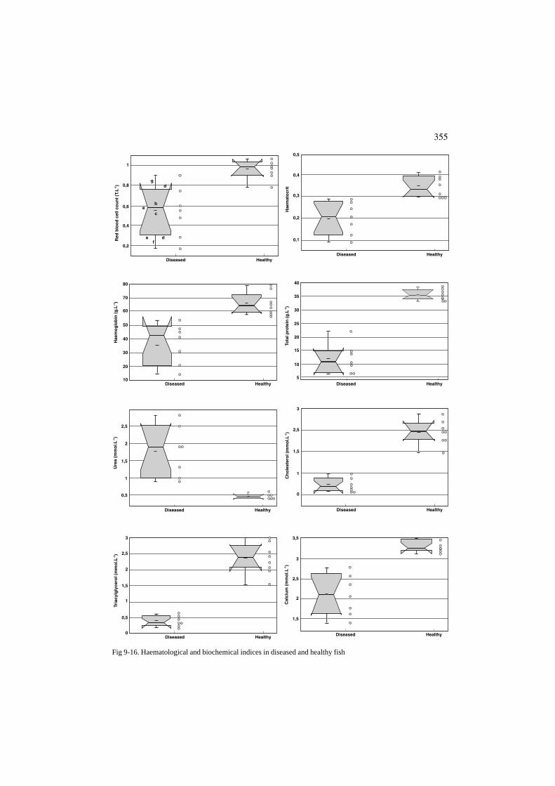
For the graphic representation of the results we used notch box graphs with filaments, where a = width of thebox, indicating the size of the set, b = mid diagonal of the box, representing the position of the median in relationto the y axis, c = mark inside the box showing the position of the arithmetic mean, d = lower and upper edge of thebox indicating successively the position of the lower and upper quartiles, e = width of the notch, corresponding tothe confidence interval around the median, f = the lower filament having a length corresponding to the value of thelower quartile reduced by 1.5 times the span of the quartiles. If this value is lower than the minimum value in theset the length of the filament corresponds to this minimum value. If values lower than those corresponding to thecoordinate of the end point of the lower filament do occur in the set, then these values are signaled as remote, g =the upper filament having a length corresponding to the value of the upper quartile enlarged 1.5 times the span ofthe quartiles. If this value is higher than the maximum value in the set the length of the filament corresponds to thismaximum value. If values higher than those corresponding to the coordinate of the end point of the upper filamentdo occur in the set, then these values are signaled as remote.
Results
Clinical s igns and gross pathology and his topathologyThe gross lesions of the skin varied. The depigmented erosions of various sizes, occurring
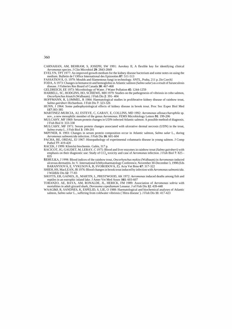
mainly on the sides near the gill covers (Plate XII, Figs 1 and 2) were considered to be thestage of the changes. Enormous prominent bulges developed on the skin of 10 % of the fish(Plate XIII, Fig. 3); the lesions were filled with clear to slightly turbid exudate, in some caseswith a slight tint of blood. On cut section they exposed haemorrhagically altered muscle (Fig.4). As distinct from furunculosis, the muscle does not disintegrate under the skin bulge anddoes not produce crater-like furuncles because the pathological process did not reach deeperlayers. The majority of the fish had pale gills, indicating different degrees of anaemia. Somehad exophthalmus and inflammations around the bases of the pectoral fins. The findingsinside the abdominal cavity included hyperaemia on the wall of the swim-bladder, smallhaaemorrhages in the liver, splenomegalia and dampness throughout the internal organs.
The histological findings of the skin lesions included a necrotizing granulocyticinflammation with no distinct borders and with an abundant exudative component, anddisintegration of the muscle fibers with a marked interstitial inflammatory oedema. The
353
characteristic changes are documented in Figs 5 and 6 (Plate XIV). The kidneys showedsigns of albumin dystrophy and formation of hyaline droplets in the epithelium of proximaltubules. The wall of the macroscopically altered swim-bladder was thickened, congestedand infiltrated eedematous matter.
Bacter iologyThree Aeromonas species/strains (Aeromonas hydrophila, Aeromonas media and strain
A) and also Staphylococcus hicus, Staphylococcus epidermidis and Enterobacter cloacaewere isolated from the edges of the open skin lesions, from the exudate taken from closedlesions and from the internal organs. Two species of fungi, Cladosporium herbarum andCladosporium sphaerospermum, were also isolated from the clear exudate and from theclosed skin lesions. Strain A, which played the most important role in the etiology of thedisease, gave the following biochemical results:
Positive response: lipase; arginine dihydrolase; oxidaseNegative response: H2S production; mannitol, acid; lysine decarboxylase; indole
production; ornithine decarboxylase; citrate, Simmons; urea hydrolysis; ONPG; Voges-Proskauer; inositol, acid; lipase; phenylalanine deaminase; maltose, acid; adonitol, acid;cellobiose, acid; rhammose, acid; sucrose, acid; sorbitol, acid; esculin hydrolysis; trehalose,acid; dulcitol, acid; motility; gelatin hydrolysis; lactose, acid; arabinose, acid; pigment.
According to the bioassays and the scheme by Aldová (1994), the strain most closelyresembled the species Aeromonas sobria (mannitol 98%, sucrose 98%, arabinose 7% +) andAeromonas caviae (mannitol 92%, sucrose 97% +, arabinose 69% +). We excluded ourinitial tentative identification as Aeromonas schubertii (¤ehulka 1998), which shouldhave had a positive lysine, and also Aeromonas trota, which might also have negativemannitol (83% +) and which, however, was only found in southeast Asia.
Experimental infect ionIn the test fish, intraperitoneal infection with an exudate from a closed skin lesion
produced a swelling at the puncture site surrounded by depigmentation (62 h); the infectedfish refused food and kept away from the shoal (86 h); this was followed by agony and death(168 h). The post-mortem examination revealed congestion of the bases of pectoral fins,ascites, petechial haemorrhages in the liver and hyperaemia of the swim-bladder. In trialswith individual bacterial isolates, the only strain with which we succeeded to reproduce thedisease with manifestation of skin lesions was strain A: the disease was so induced withintraepidermal and intramuscular infection and with death after 48, 144 and 312 h. Duringthe post-mortem examination of the fish infected with this strain, which was re-isolated,contact of the culture with the surface tissue of pyloric appendages produced exudativefibrinous purulent inflammation (Plate XV, Figs 7 and 8), which was also found in fishinfected by the exudate.
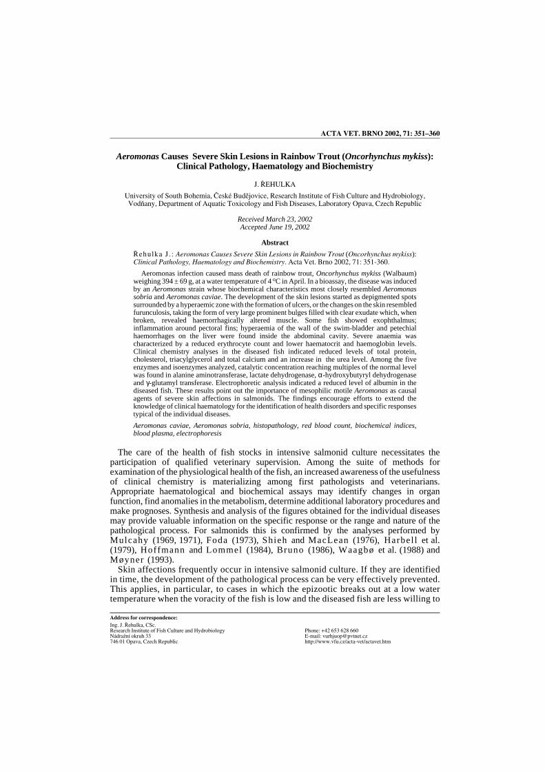
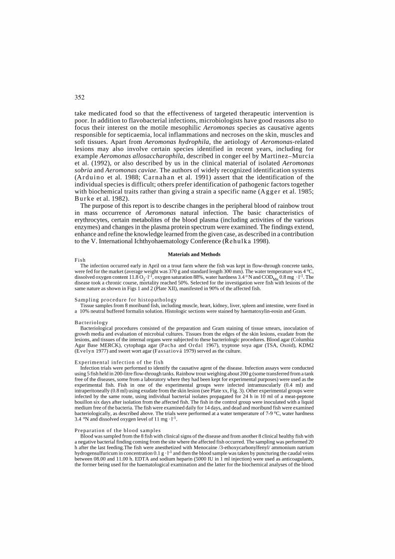
Haematology and cl inical chemistryTo document the haematological and biochemical indices, we submit a series of Figs 9 to
20. The figures indicate that the affected fish had markedly reduced parameterscharacterizing the erythrocytes: their RBCc values ranged from 0.17 to 0.9 (P = 0.004), Hctfrom 0.09 to 0.283 (P = 0.001) and Hb from 14 to 45.7 (P = 0.000). For the healthy fish, onthe other hand, the RBCc range was from 0.78 to 1.06, Hct 0.294 to 0.411 and Hb 57.1 to78.7. As to the biochemical indices, there was a marked decline in TP (6.5-22 vs 33.3-38.1,P = 0.000), CHOL (0.34-1.89 vs 5.1-7.4, P = 0.000)), TGL (0.18-0.62 vs 1.53-2.99, P =0.000) and Ca (1.38-2.78 vs 2.79-3.49, P = 0.001)). An increase was recorded in BUN (0.9-2.8 vs 0.2-1, P = 0.003). As to the five enzymes tested, catalytic concentration reaching
354
355
Diseased Healthy
0,2
0,4
0,6
0,8
1
Red
blo
od
cel
l co
un
t (T
.L-1)
c
dg
e
a df
b
Diseased Healthy10
20
30
40
50
60
70
80
Hae
mo
glo
bin
(g
.L-1)
Diseased Healthy
0,5
1
1,5
2
2,5
Ure
a (m
mo
l.L-1)
Diseased Healthy0
0,5
1
1,5
2
3
2,5
Tria
cylg
lyce
rol (
mm
ol.L
-1)
Diseased Healthy
1,5
2
2,5
3
3,5
Cal
ciu
m (
mm
ol.L
-1)
Diseased Healthy
0
1
1,5
3
2,5
Ch
ole
ster
ol (
mm
ol.L
-1)
Diseased Healthy5
10
15
25
20
40
30
35
Tota
l pro
tein
(g
.L-1)
Diseased Healthy
0,1
0,2
0,3
0,4
0,5
Hae
mat
ocr
it
Fig 9-16. Haematological and biochemical indices in diseased and healthy fish
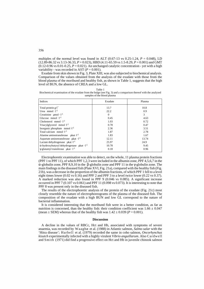
multiples of the normal level was found in ALT (0.67-13 vs 0.25-1.24, P = 0.048), LD(12.89-86.32 vs 3.13-36.33, P = 0.023), HBD (4.11-65.59 vs 1.5-8.29, P = 0.001) and GMT(0.12-0.96 vs 0.01-0.25, P = 0.021). An unchanged catalytic concentration - yet with a highvariability - was recorded in AST (P = 0.001).
Exudate from skin shown in Fig. 3, Plate XIII, was also subjected to biochemical analysis.Comparison of the values obtained from the analysis of the exudate with those from theblood plasma of the moribund and healthy fish, as shown in Table 1, suggests that the highlevel of BUN, the absence of CREA and a low GL.
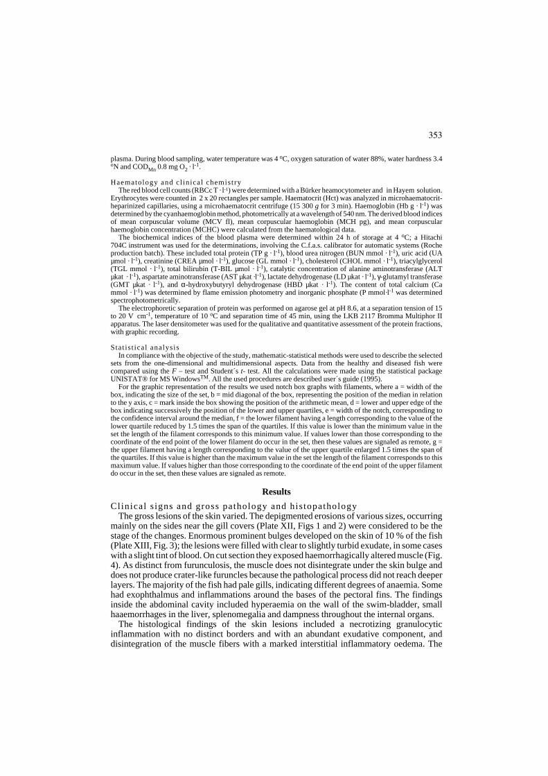
Electrophoretic examination was able to detect, on the whole, 11 plasma protein fractions(PPF 1 to PPF 11), of which PPF 1,2,3 were included in the albumin zone, PPF 4,5,6,7 in theα-globulin zone, PPF 8,9,10 in the β-globulin zone and PPF 11 in the γ-globulin zone. Themain findings in the diseased fish (Plate XVI, Fig. 21a), compared with the healthy fish (Fig.21b), was a decrease in the proportion of the albumin fractions, of which PPF 1 fell to a leveleight times lower (0.02 vs 0.16) and PPF 2 and PPF 3 to a level twice lower (0.22 vs 0.37).A marked reduction was also found in PPF 9 (0.046 vs 0.085). A significant increaseoccurred in PPF 7 (0.107 vs 0.061) and PPF 11 (0.098 vs 0.073). It is interesting to note thatPPF 8 was present only in the diseased fish.
The results of the electrophoretic analysis of the protein of the exudate (Fig. 21c) mostclosely resemble the nature of electrophoreograms of the plasma of the diseased fish. Thecomposition of the exudate with a high BUN and low GL correspond to the nature ofbacterial inflammation.
It is considered interesting that the moribund fish were in a better condition, as far asnutrition is concerned, than the healthy fish: their condition coefficient was 1.66 ± 0.047(mean ± SEM) whereas that of the healthy fish was 1.42 ± 0.039 (P = 0.001).
Discussion
A decline in the values of RBCc, Hct and Hb, associated with symptoms of severeanaemia, was recorded by Waagbø et al. (1988) in Atlantic salmon, Salmo salar with the‘Hitra disease’; Harbel l et al. (1979) recorded the same in coho salmon, Oncorhynchuskisutch experimentally infected with a highly virulent Vibrio anguillarum. Also Cardwelland Smith (1971) did find a progressive effect on Hct and Hb in juvenile chinook salmon
356
Table 1Biochemical examination of the exudate from the bulge (see Fig. 3) and a comparison thereof with the analyzed
samples of the blood plasma
Indices Exudate Plasma
Total protein g·l-1
13.7 10.8Urea mmol · l-1 22.2 0.9Creatinine µmol · l-1 0 3Glucose mmol · l-1 0.45 4.63Cholesterol mmol · l-1 1.03 0.72Triacylglycerol mmol ·l-1 0.78 0.47Inorganic phosphate mmol ·l-1 2.36 3.51Total calcium mmol ·l-1 1.87 2.78Alanine aminotransferase µkat ·l-1 1.83 1.67Aspartate aminotransferase µkat · l-1 12.11 13.74Lactate dehydrogenase µkat ·l-1 25.97 24.9α-hydroxybutyryl dehydrogenase µkat · l-1 10.78 9.45γ-glutamyl transferase µkat · l-1 0.18 0.96
with vibriosis. For a severe Aeromonas infection in Atlantic salmon, Foda (1973) describeda decrease in Hb. Amend and Smith (1974) demonstrated a reduction in RBCc, Hct andHb in IHN virus-infected rainbow trout. A decline in RBCc, Hct and Hb, combined withsigns of anaemia, was also described by Hoffmann and Lommel (1984) in cases ofproliferative kidney disease (PKD). The non-significant differences in MCV, MCH andMCHC between the diseased and healthy fish testify to the fact that the anemia was due, inparticular, to blood losses from the skin lesions; in agreement with the findings of Waagbøet al. (1988), these blood indices indicated active erythropoiesis to compensate for the loss.
We recorded - and so did other authors - a decline in TP in salmonids affected by infectiousdiseases: Mulcahy (1971) did so in the serum of the brown trout and Atlantic salmon withulcerative dermal necrosis (U.D.N.) and single fungal infection (Saprolegnia ferax) and insalmon fingerlings with fin rot and furunculosis (Mulcahy 1969). Hypoproteinaemia wasreported by Harbel l et al. (1979) to occur in this salmon with vibriosis, and Hunn (1964)found it to occur in brook trout with corynebacterial kidney disease. A decrease in TP, TGLand CHOL in the serum of Atlantic salmon suffering from cold-water vibriosis is describedby Waagbø et al. (1988).
Unlike Harbel l et al. (1979) we did not find an increased GL but what we did find wasa reduced Ca level (though the above authors claim that the Ca level is about the same inboth moribund and healthy fish). Waagbø et al. (1988) described a four times lower levelof CREA but in our essays this parameter - though also slightly decreased - were highlybalanced in comparison with the healthy fish. Our evaluation of the catalytic activity ofamino transferases matched the results published by Waagbø et al. (1988) who had alsorecorded an unchanged catalytic concentration of AST in the Atlantic salmon with vibriosis;however, they had recorded an increased ALT activity. Harbel l et al. (1979), in turn, hadfound an increased catalytic AST activity in cases of vibriosis in coho salmon. The increase
357
Diseased Healthy
0
5
10
Ala
nin
e am
ino
tran
sfer
ase
(�ka
t.L
-1)
Diseased Healthy
0
5
10
15
Hyd
roxy
bu
tyry
l deh
ydro
gen
ase
(�ka
t.L
-1)
Diseased Healthy
0
0,2
0,4
0,6
0,8
1�-
glu
tam
yl t
ran
sfer
ase
(�ka
t.L
-1)
Diseased Healthy
0
20
40
Lac
tate
deh
ydro
gen
ase
(�ka
t.L
-1)
Fig 17-20. Haematological and biochemical indices in diseased and healthy fish
in LD found by us is the same as what Harbel l et al. (1979) and also Racicot et al. (1975)had found in rainbow trout with Aeromonas infection.
As to the results of the electrophoretic analysis, the highest importance is attached to thedecline of the albumin fractions which were most significantly involved in thehypoproteinaemia. The low level of albumin may be the result of losses from the skinlesions, an increased catabolism in acute inflammation or reduced synthesis due tohepatopathy, or may be related to a renal damage. What can also be considered important isthe reduction of PPF 9 (the β-globulin zone) and the manifestation of PPF 8, not detected inhealthy fish. A decline in the β2 fraction when disc acrylamide gel system was used wasdescribed by Harbel l et al. (1979) in coho salmon with vibriosis.
The increase in the BUN level is probably associated with an escalated protein catabolismwhich grows with fasting, infection and loss of blood. A greater increase in BUN over CREAmay be indicative of pre-renal uraemia which is caused by hypovolaemia - a condition atwhich glomerular filtering declines and absorption of BUN grows (and so does BUNpresence in the plasma). The enormous and highly significant (P = 0.001) correlation of totalbilirubin with urea (r = 0.949, T-BIL = -1.4336 + 1.4419 BUN or BUN = 1.0700 + 0.6247T-BIL) can probably be ascribed to the accelerated catabolism of albumin and release ofnon-conjugated bilirubin from its link to albumin. As known in homoiotherms (Racek1999), the link to albumin is reduced by acidemia, with accumulation of acid endogenousmetabolites leading to metabolic acidosis which may have the nature of lactic acidosiscombined with insufficient oxygenation of the blood and with tissue hypoxia. This conditioncorresponds to anaemic hypoxia caused by an erythrocyte or haemoglobin defect oranaemia. The considerable decrease in the CHOL levels is in agreement with the findingspublished by Waagbø et al. (1988) and might be related to a damage to liver, uraemia andsepsis. The marked decline in Ca which, in the plasma, is linked to albumin, can be ascribaedto hypoproteinaemia.
The increase in the catalytic activity of ALT is probably related with a damage to themembranes of hepatocytes. The higher levels of LD and HBD correlate with the describedpathological changes in the skin lesions.
The report draws attention to the importance of mesophilic motile Aeromonas strains asthe causal agents responsible for severe skin lesions in salmonids. Compared withfurunculosis, the disease has different clinical manifestations, develops in a different periodof the year and has strange pathological characteristics. All this encourages researchers tocontinue studying the conditions and causal relations underlying the disease. On the basisof differential diagnosis in bioassays, scientists are trying to identify the causative agent ofthe disease among the isolated strains. This was confirmed by our other findings in the brooktrout affected on mass in intensive culture, as well as in eel and chub in dam reservoirs.Toranzo et al. (1989) found Aeromonas sobria to be associated with outbreaks of fataldisease in gizzard shad (Dorosoma cepedianum) and showed it to be pathogenic to rainbowtrout in trhe laboratory.
As to the underlying factors, what can be considered to be related to the rise of the diseasedescribed by us is the occasional water pollution with municipal sewage; in the case of eel andchub the disease may be ascribed to the development of water eutrophication. As asserted byShotts et al. (1972) eutrophication caused by pollution with farm waste supports increasedoccurrence of mesophilic aeromonads and their pathogenicity to the inhabitants of the aquaticenvironment, including fish, frogs and slugs. Increased occurrence of motile aeromonads inwaters with high sewage levels is reported by Geldreich (1973). Unfortunately, the diseasewas recognized too late, so that we were unable to record evidence relating to this type ofpollution. As mentioned, at the time of the investigation the physical and chemical propertiesof the water complied with the requirements for salmonid culture.
358
The results presented in the paper provide a basis for further comparative studies aimed atobtaining detailed information on the patho-physiological processes in the blood in cases ofAeromonas infection. This will be important for using such knowledge in the screeningprograms under methods of examination of the state of health of salmonids in intensive culture.
Aeromonádová onemocnûní pstruha duhového (Oncorhynchus mykiss Walbaum):patologické, hematologické a biochemické zmûny
Aeromonádová infekce byla pfiíãinou hromadného úhynu pstruhÛ duhov˘ch, Oncorhynchusmykiss (Walbaum) o hmotnosti 394 ± 69 g pfii teplotû vody 4 oC v mûsíci dubnu.Biologick˘m pokusem bylo onemocnûní vyvoláno aeromonádov˘m kmenem, jehoÏbiochemické vlastnosti se nejvíce podobaly Aeromonas sobria a Aeromonas caviae. Dal‰íizoláty tvofiily Aeromonas media, Aeromonas hydrophila, Staphylococcus hicus,Staphylococcus epidermidis, Enterobacter cloaceae a nesacharolytick˘ Acinetobacter.V˘voj koÏních lézí zaãínal depigmentovan˘mi skvrnami s hyperémickou zónou v okolía tvorbou vfiedÛ nebo zmûny na kÛÏi pfiipomínaly furunkulózu a byly tvofieny obrovskyprominujícími boulemi naplnûn˘mi ãir˘m v˘potkem, které po prasknutí obnaÏovalyhemoragicky zmûnûnou svalovinu. U nûkter˘ch ryb se nacházel exoftalmus a zánût kolembáze prsních ploutví, v dutinû bfii‰ní byla nalezena hyperémie stûny plynového mûch˘fiea petechiální krváceniny na játrech. TûÏká anémie byla charakterizována sníÏen˘m poãtemerytrocytÛ, poklesem hematokritu a hemoglobinu. Z biochemick˘ch ukazatelÛ krevníplazmy do‰lo ke sníÏení celkové bílkoviny, cholesterolu, triacylglycerolu a celkovéhovápníku a ke zv˘‰ení hladiny moãoviny. Z pûti testovan˘ch enzymÛ a izoenzymÛ bylanûkolikanásobnû zv˘‰ena katalytická koncentrace alaninaminotransferázy,laktátdehydrogenázy, α-hydroxybutyryl dehydrogenázy a γ-glutamyl transferázy.Elektroforeticky byl u nemocn˘ch ryb zji‰tûn pfiedev‰ím sníÏen˘ podíl albuminu. DosaÏenév˘sledky aktualizují v˘znam mesofilních pohybliv˘ch aeromonád, jako pÛvodcÛ závaÏn˘chkoÏních afekcí intenzivnû odchovávaného pstruha duhového.
Acknowledgements
The study was conducted with the support of Grant No.525/00/0241 awarded by the Grant Agency of the CzechRepublic. Author thanks Dr E Aldová of the National Institute of Public Health in Prague for help with identificationof Aeromonas strains and Assoc. Prof. J. Horáãek M.D. PhD. of the Medico-Social Faculty of the OstravaUniversity for consultations relating to the results of histological examinations. Thanks also go to Prof. B. MinafiíkPhD., of the Mendel University of Agriculture and Forestry in Brno, Department of Statistics, for the graphical andmathematical processing of the results.
References
AGGER, WA, McCORMICK, JD, GURWITH, MJ 1985: Clinical and microbiological features of Aeromonashydrophila-associated diarrhea. J Clin Microbiol 21: 909-913
ALDOVÁ, E., SCHINDLER, J., URBÁ·KOVÁ, P., NùMEC, A. 1994: Biochemical identification of aeromonads.Epidemiologie, Mikrobiologie, Imunologie 43: 55-60 [In Czech]
AMEND, DF, SMITH, L 1974: Pathophysiology of infectious hematopoietic necrosis virus disease in rainbowtrout (Salmo gairdneri): Early changes in blood and aspects of the immune response after injection of IHN virus.J Fisheries, Research Board of Canada 31: 1371-1378
ARDUINO, MJ, HICKMANN-BRENNER, FW, FARMER, JJ 1988: Phenotypic analysis of 132 Aeromonasstrains representing 12 DNA hybridization groups. CDC Atlanta, Ga, International Symposium on Aeromonasand Plesiomonas, Sept. 1988, Miami Beach, Florida,USA.
BRUNO, DW 1986: Changes in serum parameters of rainbow trout, Salmo gairdneri Richardson, and Atlanticsalmon, Salmo salar L., infected with Renibacterium salmoninarum. J Fish Dis 9: 205-211
BURKE, V, ROBINSON, J, ATKINSON, HM, GRACEY, M 1982: Biochemical characteristics of enterotoxigenicAeromonas spp. J Clin Microbiol 15: 48-52
CARDWELL, RD, SMITH, LS 1971: Hematological manifestations of vibriosis upon juvenile chinook salmon.Progressive Fish-Culturist 33: 232-235
359
CARNAHAN, AM, BEHRAM, S, JOSEPH, SW 1991: Aerokey II, A flexible key for identifying clinicalAeromonas species. J Clin Microbiol 29: 2843-2849
EVELYN, TPT 1977: An improved growth medium for the kidney disease bacterium and some notes on using themedium. Bulletin de l´Office International des Epizooties 87: 511-513
FASSATIOVÁ, O. 1979: Moulds and filamentous fungi in technology. SNTL, Praha, 211 p. [In Czech]FODA, A 1973: Changes in hematocrit and hemoglobin in Atlantic salmon (Salmo salar) as a result of furunculosis
disease. J Fisheries Res Board of Canada 30: 467-468GELDREICH, EE 1973: Microbiology of Water. J Water Pollution 45: 1244-1259HARBELL, SC, HODGINS, HO, SCHIEWE, MH 1979: Studies on the pathogenesis of vibriosis in coho salmon,
Oncorhynchus kisutch (Walbaum). J Fish Dis 2: 391- 404HOFFMANN, R, LOMMEL, R 1984: Haematological studies in proliferative kidney disease of rainbow trout,
Salmo gairdneri Richardson. J Fish Dis 7: 323-326HUNN, J 1964: Some pathophysiological effects of kidney disease in brook trout. Proc Soc Exper Biol Med
117:383-385MARTINEZ-MURCIA, AJ, ESTEVE, C, GARAY, E, COLLINS, MD 1992: Aeromonas allosaccharophila sp.
nov., a new mesophilic member of the genus Aeromonas. FEMS Microbiology Letters 91: 199-206MULCAHY, MF 1969: Serum protein changes in UDN-infected Atlantic salmon: A possible method of diagnosis.
J Fish Biol 1: 333-338 MULCAHY, MF 1971: Serum protein changes associated with ulcerative dermal necrosis (UDN) in the trout,
Salmo trutta L. J Fish Biol 3: 199-201MØYNER, K 1993: Changes in serum protein composition occur in Atlantic salmon, Salmo salar L., during
Aeromonas salmonicida infection. J Fish Dis 16: 601-604PACHA, RE, ORDAL, EJ 1967: Histopathology of experimental columnaris disease in young salmon. J Comp
Pathol 77: 419-420RACEK, J 1999: Klinická biochemie. Galén, 317 p.RACICOT, JG, GAUDET, M, LERAY, C 1975: Blood and liver enzymes in rainbow trout (Salmo gairdneri) with
emphasis on their diagnostic use: Study of CCl4 toxicity and case of Aeromonas infection. J Fish Biol 7: 825 -835
¤EHULKA, J 1998: Blood indices of the rainbow trout, Oncorhynchus mykiss (Walbaum) in Aeromonas-inducedulcerous dermatitis. In: V. International Ichthyohaematology Conference, November 30-December 3, 1998 (EdsBARANYIOVÁ, E, VYKUSOVÁ, B, SVOBODOVÁ, Z). Acta Vet Brno 67: 317-322
SHIEH, HS, MacLEAN, JR 1976: Blood changes in brook trout induced by infection with Aeromonas salmonicida.J Wildlife Dis 12: 77-83
SHOTTS, EB, GAINES, JL, MARTIN, L, PRESTWOOD, AK 1972: Aeromonas induced deaths among fish andreptiles in an eutrophic inland lake. J Amer Vet Med Assoc 161: 603-607
TORANZO, AE, BAYA, AM, RONALDE, JL, HERICK, FM 1989: Association of Aeromonas sobria withmortalities in adult gizzard shark, Dorosoma cepediamum Lesueur. J of Fish Dis 12: 439-448
WAAGBØ, R, SANDNES, K, ESPELID, S, LIE, O 1988: Haematological and biochemical analyses of Atlanticsalmon, Salmo salar L., suffering from coldwater vibriosis (´Hitra disease´). J Fish Dis 11: 417-423
360
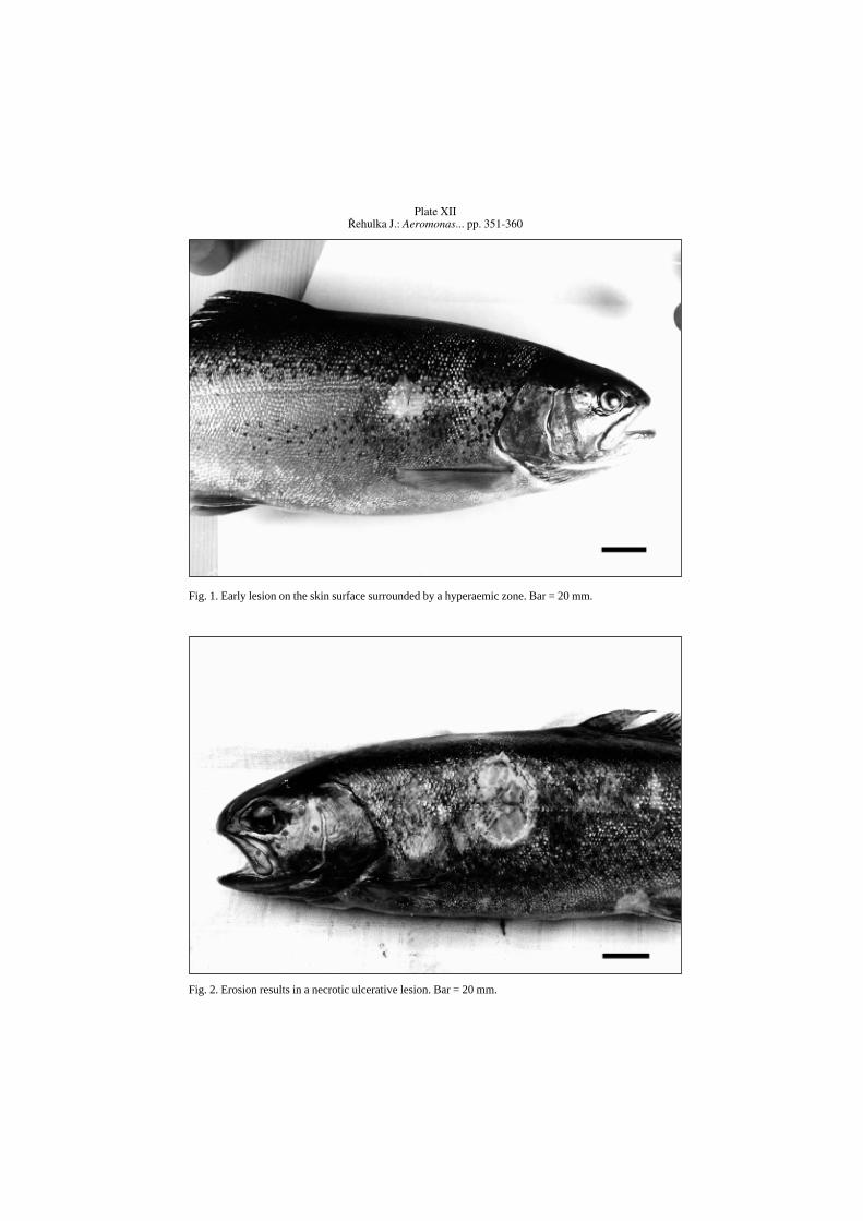
Plate XII¤ehulka J.: Aeromonas... pp. 351-360
Fig. 1. Early lesion on the skin surface surrounded by a hyperaemic zone. Bar = 20 mm.
Fig. 2. Erosion results in a necrotic ulcerative lesion. Bar = 20 mm.
Plate XIII
Fig. 4. Haemorrhagically altered muscle after cutting off the skin (this is the fish illustrated in Figure 3). Bar= 10 mm.
Fig. 3. Enormous subcutaneous bulge filled with a clear to slightly turbid exudate. Bar = 20 mm

Plate XIV
Fig. 5. A more pronounced interstitial inflammation with lipomatous atrophy of the skeletal muscles.Haematoxylin – eosin. Bar = 20 µm.
Fig. 6. Detailed view of an atrophy muscle fibers, involving multinuclear muscle fibers. Haematoxylin –eosin. Bar = 10 µm.
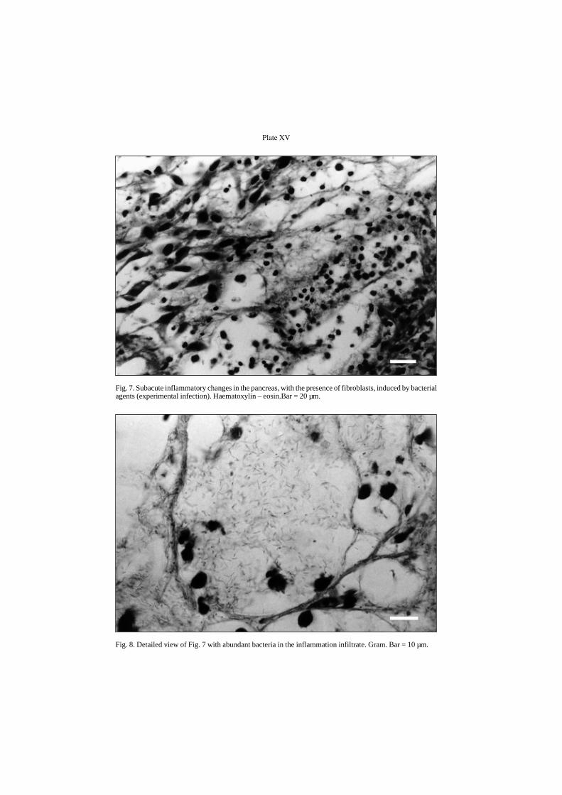
Plate XV
Fig. 7. Subacute inflammatory changes in the pancreas, with the presence of fibroblasts, induced by bacterialagents (experimental infection). Haematoxylin – eosin.Bar = 20 µm.
Fig. 8. Detailed view of Fig. 7 with abundant bacteria in the inflammation infiltrate. Gram. Bar = 10 µm.
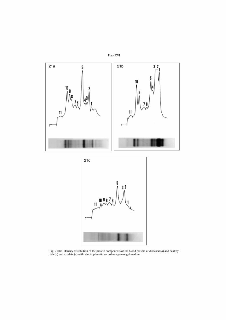
Plate XVI
Fig. 21abc. Density distribution of the protein components of the blood plasma of diseased (a) and healthyfish (b) and exudate (c) with electrophoretic record on agarose gel medium
21a 21b
21c