clinical pharmacokinetics and pharmacodynamics of propofol · clinical pharmacokinetics and...
TRANSCRIPT
University of Groningen
Clinical Pharmacokinetics and Pharmacodynamics of PropofolSahinovic, Marko M; Struys, Michel M R F; Absalom, Anthony R
Published in:Clinical Pharmacokinetics
DOI:10.1007/s40262-018-0672-3
IMPORTANT NOTE: You are advised to consult the publisher's version (publisher's PDF) if you wish to cite fromit. Please check the document version below.
Document VersionPublisher's PDF, also known as Version of record
Publication date:2018
Link to publication in University of Groningen/UMCG research database
Citation for published version (APA):Sahinovic, M. M., Struys, M. M. R. F., & Absalom, A. R. (2018). Clinical Pharmacokinetics andPharmacodynamics of Propofol. Clinical Pharmacokinetics, 57(12), 1539-1558.https://doi.org/10.1007/s40262-018-0672-3
CopyrightOther than for strictly personal use, it is not permitted to download or to forward/distribute the text or part of it without the consent of theauthor(s) and/or copyright holder(s), unless the work is under an open content license (like Creative Commons).
Take-down policyIf you believe that this document breaches copyright please contact us providing details, and we will remove access to the work immediatelyand investigate your claim.
Downloaded from the University of Groningen/UMCG research database (Pure): http://www.rug.nl/research/portal. For technical reasons thenumber of authors shown on this cover page is limited to 10 maximum.
Download date: 15-03-2020
REVIEW ARTICLE
Clinical Pharmacokinetics and Pharmacodynamics of Propofol
Marko M. Sahinovic1,3 • Michel M. R. F. Struys1,2 • Anthony R. Absalom1
� The Author(s) 2018
Abstract Propofol is an intravenous hypnotic drug that is
used for induction and maintenance of sedation and general
anaesthesia. It exerts its effects through potentiation of the
inhibitory neurotransmitter c-aminobutyric acid (GABA) at
the GABAA receptor, and has gained widespread use due to its
favourable drug effect profile. The main adverse effects are
disturbances in cardiopulmonaryphysiology.Due to its narrow
therapeutic margin, propofol should only be administered by
practitioners trained and experienced in providing general
anaesthesia. Many pharmacokinetic (PK) and pharmacody-
namic (PD)models for propofol exist. Some are used to inform
drug dosing guidelines, and some are also implemented in so-
called target-controlled infusion devices, to calculate the
infusion rates required for user-defined target plasma or effect-
site concentrations. Most of the models were designed for use
in a specific and well-defined patient category. However,
models applicable in a more general population have recently
been developed and published. Themost recent example is the
general purpose propofol model developed by Eleveld and
colleagues. Retrospective predictive performance evaluations
show that this model performs as well as, or even better than,
PK models developed for specific populations, such as adults,
children or the obese; however, prospective evaluation of the
model is still required. Propofol undergoes extensive PK and
PD interactionswith both other hypnotic drugs andopioids. PD
interactions are the most clinically significant, and, with other
hypnotics, tend to be additive, whereas interactions with opi-
oids tend to be highly synergistic. Response surface modelling
provides a tool to gain understanding and explore these com-
plex interactions. Visual displays illustrating the effect of these
interactions in real time can aid clinicians in optimal drug
dosing while minimizing adverse effects. In this review, we
provide an overview of the PK and PD of propofol in order to
refresh readers’ knowledge of its clinical applications, while
discussing the main avenues of research where significant
recent advances have been made.
Key Points
Propofol is a potent intravenous hypnotic drug. It
exerts its effects through potentiation of the
inhibitory neurotransmitter, c-Aminobutyric acid
(GABA). Much experience with its clinical use has
been amassed since it was introduced over three
decades ago.
A general purpose pharmacokinetic (PK) and
pharmacodynamic (PD) propofol model recently
published by Eleveld and colleagues might replace
PK/PD models currently used in clinical practice.
Defining the nature of interaction between propofol
and other drugs remains a challenge. Response
surface model studies can help to elucidate these
interactions.
& Marko M. Sahinovic
Michel M. R. F. Struys
Anthony R. Absalom
1 Department of Anaesthesiology, University Medical Center
Groningen, University of Groningen, Hanzeplein 1, 9713 GZ
Groningen, The Netherlands
2 Department of Anaesthesia and Peri-Operative Medicine,
Ghent University, Ghent, Belgium
3 University Medical Center Groningen, Hanzeplein 1,
PO Box 30.001, 9700 RB Groningen, The Netherlands
Clin Pharmacokinet
https://doi.org/10.1007/s40262-018-0672-3
1 Introduction
Propofol (2,6-diisopropylphenol) is a potent intravenous
hypnotic drug that was developed by Imperial Chemical
Industries Limited (London, UK), patented by John (Iain)
Glen and Roger James in 1977 [1], and commercially
launched in 1986 in Europe and 1989 in the US [2].
Like most anaesthetics, propofol is a c-aminobutyric
acid (GABA) receptor agonist. It has a favourable phar-
macokinetic (PK) and pharmacodynamic (PD) profile,
which has resulted in it becoming the most commonly used
intravenous anaesthetic for the past three decades [2–7].
Rapid and smooth induction with nearly no excitation
phenomena, relatively short context-sensitive time, rapid
terminal half-life time and low incidence of postoperative
nausea and vomiting (PONV) make it a very versatile
hypnotic drug. It is used for sedation and anaesthesia for
almost all types of surgery, but is particularly well-suited
for anaesthesia in patients undergoing ambulatory [8] and
neurosurgery where rapid psychomotor recovery are of
upmost importance. Its efficacy and utility has also been
proven for sedation of patients in the intensive care unit
(ICU) [9] and conscious sedation of patients undergoing
diagnostic or invasive procedures [10].
The adverse effects of propofol are well-documented,
with the most common being pain on injection. Other
adverse effects are cardiovascular (bradycardia, hypoten-
sion) and metabolic (hyperlipidaemia secondary to infusion
of lipid formulation) [11].
Although propofol is well-established in clinical prac-
tice, there have been a number of studies published in
recent years on the clinical use and pharmacology of the
drug. The main aim of this narrative review is to refresh
readers’ knowledge of the drug, it’s clinical uses, PK and
PD, and to update the reader on the advances in two main
fields of research, namely compartmental PK/PD mod-
elling and drug interaction modelling of propofol.
2 Methods
A MEDLINE database search was performed using
PubMed. The following ‘propofol’ Medical Subject
Heading (MeSH) terms were used: ‘administration and
dosage’ OR ‘adverse effects’ OR ‘blood’ OR ‘cere-
brospinal fluid’ OR ‘chemical synthesis’ OR ‘chemistry’
OR ‘contraindications’ OR ‘metabolism’ OR ‘pharma-
cokinetics’ OR ‘pharmacology’ OR ‘physiology’ OR
‘poisoning’ OR ‘therapeutic use’ OR ‘toxicity’ OR ‘urine’.
All articles published after 1 January 1985, in English, with
full-text available and relating to human subjects were
evaluated for relevancy. A full-text copy was obtained for
all relevant publications. A further search of the bibli-
ographies of all relevant articles was performed (also
known as the snowball method) to identify any relevant
publications missed during the initial search. Finally the
‘Google Scholar’ web search engine was used, with com-
parable search terms, to complete the literature search.
3 Drug Formulation
Propofol is insoluble in water [4]. It was first marketed in a
formulation containing the surfactant Cremophor EL
(polyethoxylated castor oil); however, the vehicle was soon
changed to a lipid emulsion because of a high incidence of
anaphylactic reactions, thought to be caused by Cre-
mophor� [4]. The Diprivan� (AstraZeneca, London, UK)
formulation of propofol came on to the market in 1989 in
the US and is still available today worldwide. It is for-
mulated as an Intralipid�-based emulsion (Fresenuis Kabi,
Bad Homburg, Germany) and thus contains the same
ingredients, namely soybean oil (100 mg/mL), glycerol
(22.5 mg/mL), and egg lecithin (12 mg/mL), and of course
propofol as the active ingredient [12]. After a series of
reports of bacterial contamination of opened ampoules,
ethylenediaminetetraacetic acid (EDTA; 0.05 mg/mL) was
added to the formulation in 1996 [13], and is still sold in
some but not all countries. The solution is isotonic, and
neutral pH is achieved by adding sodium hydroxide. The
pKa is 11.1 at 20 �C [14]. It has a melting point of 18 �Cand should therefore be stored at room temperature. A
number of different formulations of propofol are currently
available, produced by many different manufacturers.
Most propofol formulations cause pain on injection,
which is thought to be due to direct and indirect irritation
of venous adventitia by free aqueous propofol through an
interaction with TRPV1 and TRPA1 receptors [15, 16].
The free aqueous concentration of propofol is thought to be
reduced when it is prepared in a solution containing more
medium chain triglycerides than in Intralipid�. Therefore,
some manufacturers produced and sold generic propofol
formulations, such as Propoven� (Fresenius-Kabi, Bad
Homburg, Germany) and Propofol-Lipuro� (B-Braun,
Melsungen, Germany), containing more medium and fewer
long chain-length fatty acids [17]. Although this reformu-
lation of propofol has reduced the prevalence of pain on
injection during the induction of anaesthesia, it still
remains a problem in clinical practice [18].
M. M. Sahinovic et al.
4 Indications, Contraindications, Drug DosingRegimen and Adverse Effects
Propofol is used for the induction and maintenance of the
hypnotic component of sedation or general anaesthesia. Its
use is contraindicated in two patient categories: (1) patients
with hypersensitivity to propofol or any of the components
of its formulation, and (2) patients with fat metabolism
disorders. Since a small number of case reports of ana-
phylactic and anaphylactoid reactions have been published,
the Diprivan� package insert also advises against Dipri-
van� use in patients with allergies to eggs, egg products,
soy beans or soy products [19]. However, in two large
retrospective studies, Asserhøj and colleagues were unable
to confirm the link between propofol and food allergies
[20].
Propofol should only be administered by healthcare
professionals trained in the safe care of patients undergoing
general anaesthesia. During any procedure involving
sedation or anaesthesia with propofol, the anaesthetist
should closely monitor, and act on any changes in, the
patient’s physiological parameters according to the Amer-
ican Society of Anesthesiologists (ASA) guidelines on
minimum monitoring standards [21]. Multiple propofol
dosing schemes exist; however, due to considerable vari-
ability in the propofol dose necessary to achieve certain
clinical endpoints, these should only be regarded as very
approximate dosing guidelines. Propofol administration
should always be titrated according to clinical effect, which
is part of the reason why the clinician should closely
monitor the effects and adverse effects of propofol (vide
infra). In healthy adults younger than 55 years of age, the
Diprivan� package insert advises an induction dose of
2–2.5 mg/kg, administered in boluses of 40 mg every 10 s,
titrated to the onset of hypnotic effect, and a maintenance
dose of 6–12 mg/kg/h. This dose should be adjusted (re-
duced) when propofol is administered to less-fit patients
undergoing general anaesthesia, such as those fulfilling the
ASA physical status categories ASA 3 or 4, or when
propofol is used to induce and maintain sedation in criti-
cally ill patients in the ICU. For exact dosing guidelines,
please refer to the package insert.
The main adverse effects of propofol administration
relate to alterations of cardiopulmonary physiology,
including loss of airway reflexes, hypoventilation or even
apnoea, and hypotension. Any provider of anaesthesia
should be trained in managing these adverse effects. A rare
but serious adverse effect of propofol administration is the
‘propofol infusion syndrome’ (PRIS), which comprises
severe metabolic acidosis, rhabdomyolysis, hyperkalaemia
and cardiovascular collapse, and is frequently fatal. PRIS
occurs mostly in the setting of prolonged and high-dose
propofol infusion administration in children. The Dipri-
van� package insert advises against administration of
propofol[ 5 mg/kg/h for more than 48 h. Lastly, there are
strong indications, mainly from animal studies [22] and
retrospective clinical studies [23], that sedation with
GABA agonistic drugs might be responsible for neuro-
toxicity in paediatric patients, resulting in long-term cog-
nitive deficits; however, clinical significance of these
findings is not known and further prospective studies are
currently being conducted.
5 Pharmacokinetics (PK)
5.1 Absorption
Propofol is only suitable for intravenous use. It is not
suitable for enteral or other routes of administration due to
its bitter taste and low oral bioavailability caused by a high
first-pass effect and the high hepatic extraction rate
([ 90%) [24]. Some researchers have tried to increase the
oral bioavailability of propofol, with some success, by
administering it in nanoparticles, but this application
remains experimental [25].
5.2 Distribution
After intravenous administration, propofol is extensively
bound to the plasma proteins (predominantly albumin) and
erythrocytes. The free fraction is only 1.2–1.7%. As up to
50% of propofol is bound to the erythrocytes [26], many
clinical PK investigators measure whole concentrations
rather than plasma propofol concentrations.
Propofol readily crosses the blood–brain barrier (BBB)
and causes rapid loss of consciousness (sometimes within
the time it takes for a drug to pass through the circulation
once [27]). The speed of induction depends on patient-
related factors (cardiac output being one of the most
important factors) and speed of infusion.
Whereas approximately 1% of total plasma propofol is
unbound, free fraction of propofol in the CSF is approxi-
mately 31% [28]. Equilibrium between blood and brain
concentrations is reached after 30 min, resulting in a total
blood to CSF propofol ratio of 0.01–0.02 [29]. Placenta
transfer is also fast and extensive, with venous blood
concentration mother-to-foetus ratios ranging from 0.7 to
0.8 [30]; however, due to its clearance from the neonatal
circulation, it has only minimal and short-lived clinical
effects in unborn neonates [31] and is thus safe for use
during caesarean section [32].
After a single bolus, or short infusion, the time to offset
of clinical effects is short because of the fast initial dis-
tribution. Redistribution to and from a slow compartment
Pharmacokinetics and Pharmacodynamics of Propofol
also occurs and is due to the high lipid solubility of
propofol. This compartment has a large capacity to absorb
propofol, which results in a very large apparent volume of
distribution at steady state (Vdss; three to four multiples of
total body volume), even in non-obese individuals [33].
Nonetheless, even after prolonged administration, the off-
set of clinical effects is still reasonably fast compared with
other intravenous hypnotics because redistribution of drug
from the slow compartment is slow compared with the
rates of metabolism and excretion.
The context-sensitive decrement time [34] for propofol
is thus generally favourable compared with other hyp-
notics. For a short infusion (\ 3 h), the 80% decrement
time is\ 50 min, whereas for longer infusions ([ 12 h) it
increases up to 3.5 h [35].
5.3 Metabolism
The liver is the main site of propofol metabolism. The
majority of propofol (70%) is conjugated to propofol glu-
curonide by uridine 50-diphosphate (UDP) glucuronosyl-
transferase. Approximately 29% of propofol is
hydroxylated to 2,6-diisopropyl-1,4-quinol (4-hydrox-
ypropofol). A number of different cytochrome P450 (CYP)
P450 isoforms are involved in this step. CYP2B6 and, to a
lesser extent, CYP2C9 are the major catalysts. Environ-
mental and genetic influences on the CYP2B6 can, at least
partially, explain the interindividual variability in hydrox-
ylation of propofol in liver microsomes [36]. Propofol
metabolites are subsequently conjugated to form 4-(2,6-
diisopropyl-1,4-quinol)-sulphate, 1-(2,6-diisopropyl-1,4-
quinol)-glucuronide and 4-(2,6-diisopropyl-1,4-quinol)-
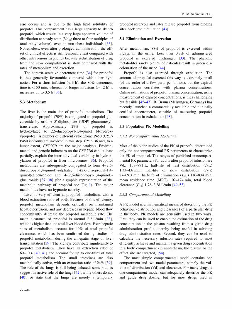
glucuronide [37, 38] (for a graphic representation of the
metabolic pathway of propofol see Fig. 1). The major
metabolites have no hypnotic activity.
Liver is very efficient at propofol metabolism, with a
blood extraction ratio of 90%. Because of this efficiency,
propofol metabolism depends critically on maintained
hepatic perfusion, and any decreases in hepatic blood flow
concomitantly decrease the propofol metabolic rate. The
mean clearance of propofol is around 2.2 L/min [33],
which is higher than the total liver blood flow. Extrahepatic
sites of metabolism account for 40% of total propofol
clearance, which has been confirmed during studies of
propofol metabolism during the anhepatic stage of liver
transplantation [39]. The kidneys contribute significantly to
propofol metabolism. They have an extraction ratio of
60–70% [40, 41] and account for up to one-third of total
propofol metabolism. The small intestines are also
metabolically active, with an extraction ratio of 24% [39].
The role of the lungs is still being debated; some studies
suggest an active role of the lungs [42], while others do not
[40], or state that the lungs are merely a temporary
propofol reservoir and later release propofol from binding
sites back into circulation [43].
5.4 Elimination and Excretion
After metabolism, 88% of propofol is excreted within
5 days in the urine. Less than 0.3% of administered
propofol is excreted unchanged [33]. The phenolic
metabolites rarely (\ 1% of patients) result in green dis-
colouration of the urine [44].
Propofol is also excreted through exhalation. The
amount of propofol excreted this way is extremely small
(of the order of a few parts per billion), but the expired
concentration correlates with plasma concentrations.
Online estimations of propofol plasma concentration, using
measurement of expired concentrations, is thus challenging
but feasible [45–47]. B. Braun (Melsungen, Germany) has
recently launched a commercially available and clinically
certified spectrometer, capable of measuring propofol
concentration in exhaled air [48].
5.5 Population PK Modelling
5.5.1 Noncompartmental Modelling
Most of the older studies of the PK of propofol determined
only the noncompartmental PK parameters to characterize
the PK of propofol. The ranges of published noncompart-
mental PK parameters for adults after propofol infusion are
Vdss 159–771 L, half-life of fast distribution (T�a)
1.33–4.6 min, half-life of slow distribution (T�b)
27–69.3 min, half-life of elimination (T�c) 116–834 min,
mean residence time (MRT) 102–174 min, total blood
clearance (Clb) 1.78–2.28 L/min [49–53]
5.5.2 Compartmental Modelling
A PK model is a mathematical means of describing the PK
behaviour (distribution and clearance) of a particular drug
in the body. PK models are generally used in two ways.
First, they can be used to enable the estimation of the drug
concentration in the plasma resulting from a given drug
administration profile, thereby being useful in advising
drug administration rates. Second, they can be used to
calculate the necessary infusion rates required to most
efficiently achieve and maintain a given drug concentration
in a body compartment (in anaesthesia, the plasma or the
effect site are targeted) [54].
The most simple compartmental model contains one
compartment and two model parameters, namely the vol-
ume of distribution (Vd) and clearance. For many drugs, a
one-compartment model can adequately describe the PK
and guide drug dosing, but for most drugs used in
M. M. Sahinovic et al.
anaesthesia this is not the case. Therefore, most PK
propofol models consist of two or three compartments and
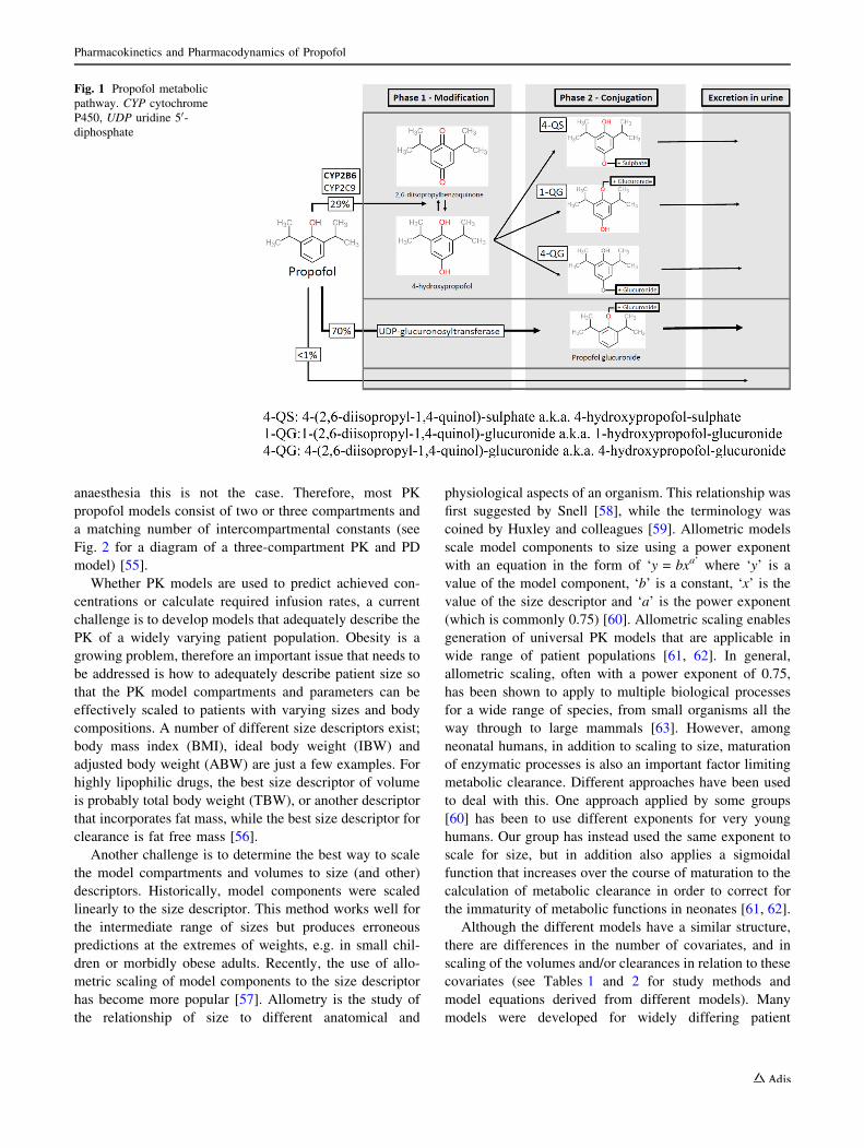
a matching number of intercompartmental constants (see
Fig. 2 for a diagram of a three-compartment PK and PD
model) [55].
Whether PK models are used to predict achieved con-
centrations or calculate required infusion rates, a current
challenge is to develop models that adequately describe the
PK of a widely varying patient population. Obesity is a
growing problem, therefore an important issue that needs to
be addressed is how to adequately describe patient size so
that the PK model compartments and parameters can be
effectively scaled to patients with varying sizes and body
compositions. A number of different size descriptors exist;
body mass index (BMI), ideal body weight (IBW) and
adjusted body weight (ABW) are just a few examples. For
highly lipophilic drugs, the best size descriptor of volume
is probably total body weight (TBW), or another descriptor
that incorporates fat mass, while the best size descriptor for
clearance is fat free mass [56].
Another challenge is to determine the best way to scale
the model compartments and volumes to size (and other)
descriptors. Historically, model components were scaled
linearly to the size descriptor. This method works well for
the intermediate range of sizes but produces erroneous
predictions at the extremes of weights, e.g. in small chil-
dren or morbidly obese adults. Recently, the use of allo-
metric scaling of model components to the size descriptor
has become more popular [57]. Allometry is the study of
the relationship of size to different anatomical and
physiological aspects of an organism. This relationship was
first suggested by Snell [58], while the terminology was
coined by Huxley and colleagues [59]. Allometric models
scale model components to size using a power exponent
with an equation in the form of ‘y = bxa’ where ‘y’ is a
value of the model component, ‘b’ is a constant, ‘x’ is the
value of the size descriptor and ‘a’ is the power exponent
(which is commonly 0.75) [60]. Allometric scaling enables
generation of universal PK models that are applicable in
wide range of patient populations [61, 62]. In general,
allometric scaling, often with a power exponent of 0.75,
has been shown to apply to multiple biological processes
for a wide range of species, from small organisms all the
way through to large mammals [63]. However, among
neonatal humans, in addition to scaling to size, maturation
of enzymatic processes is also an important factor limiting
metabolic clearance. Different approaches have been used
to deal with this. One approach applied by some groups
[60] has been to use different exponents for very young
humans. Our group has instead used the same exponent to
scale for size, but in addition also applies a sigmoidal
function that increases over the course of maturation to the
calculation of metabolic clearance in order to correct for
the immaturity of metabolic functions in neonates [61, 62].
Although the different models have a similar structure,
there are differences in the number of covariates, and in
scaling of the volumes and/or clearances in relation to these
covariates (see Tables 1 and 2 for study methods and
model equations derived from different models). Many
models were developed for widely differing patient
Fig. 1 Propofol metabolic
pathway. CYP cytochrome
P450, UDP uridine 50-diphosphate
Pharmacokinetics and Pharmacodynamics of Propofol
categories, but, to our knowledge, propofol PK have not
been studied in critically ill ICU patients; thus, none of the
existing PK models applies to these patients.
5.5.2.1 Adult Population Many adult PK models for
propofol have been published. Large variability among the
model parameters can be found due to differences in study
design, drug administration profile and study population
used to develop the model [64]. Although numerous mul-
ticompartmental mammillary models exist, only two adult
models are commonly used in clinical practice for target-
controlled infusions (TCIs), namely the Marsh [65] and
Schnider [66] models.
In the Marsh model, the volumes (V1, V2, V3) are a linear
function of patient weight, while the intercompartmental
transfer rates (k12, k21, k13, k31) are constant. In contrast, the
Schnider model has fixed V1, V3, k13 and k31, and uses age
as a covariate in the calculation of V2, k12 and k21, and lean
body weight, sex, total body mass and height as covariates
of the metabolic clearance.
As indicated above, a benefit of the Schnider model over
the Marsh model is that it adjusts the dose and infusion rate
according to the patient’s age. This results in smaller bolus
doses administered by the TCI pump (on starting, or when
the target concentration is increased) in older patients,
which might improve haemodynamic stability. A disad-
vantage of the Schnider model is that the lean body mass
equation incorporated into the calculation of k10 can gen-
erate paradoxical values, resulting in excessive increases in
maintenance infusion rates in the obese [67].
In general, both models have acceptable performance in
daily practice, and the choice of model to use is predom-
inantly made based on user experience and the model
availability.
5.5.2.2 Obese Population Obesity is associated with a
number of physiological and pathophysiological changes
that can have a significant impact on drug PK. The most
important factors affecting PK in obese patients are chan-
ges in body composition, haemodynamics, regional blood
flow, and liver and kidney function [68].
Older propofol models, such as the Marsh [65] and
Schnider [66] models, were developed from studies
involving healthy adults with a limited range of weights.
Some practitioners still use the Marsh model [65] for TCI
propofol administration in obese patients [67], but instead
of inputting the TBW, they input an ABW calculated from
the TBW and IBW, and calculated as follows:
ABW ¼ IBW þ 0:4 � TBW � IBWð Þ:
where IBW is calculated as:
IBW ¼ 45:4 þ 0:89 � HT cm½ � �152:4ð Þþ 4:5 if maleð Þ
or more simply as:
IBW ¼ height cm½ � �100 if maleð Þ or IBW¼ height cm½ � �105 if femaleð Þ:
As mentioned earlier, in severely obese patients, the
LBM equation used in the Schnider model generates
paradoxically large increases in the k10, when the BMI
Fig. 2 Overview of a three-
compartment pharmacokinetic/
pharmacodynamic model
M. M. Sahinovic et al.
is[ 37 in females and[ 42 in males [67]. As a result,
commercially available TCI devices programmed with the
Schnider model do not allow the user to input parameters
that generate BMI values above these limits. Users
attempting to use the Schnider model in severely obese
patients must either change to a different model, enter a
‘false’ combination of parameters (i.e. a greater height or a
lower weight in place of the TBW), or administer propofol
by manually controlled infusion.
There are currently a number of propofol models
specifically developed for obese patients, such as the
Cortınez [69] and van Kralingen [70] models, or a general
purpose model that is probably also suitable for use in the
obese (the Eleveld allometric model) [61] (see Table 1).
Cortınez et al. [71] have investigated the predictive
performance of different PK models in obese patients and
concluded that when models were implemented as pub-
lished (i.e. using the TBW as a weight-scaling measure),
the global performance of the Eleveld model [72] was best
compared with other models. All models tended to
underestimate the propofol plasma concentration. When
the user entered the ABW instead of the TBW, the Sch-
nider and Marsh models had the highest accuracy and
lowest bias [71].
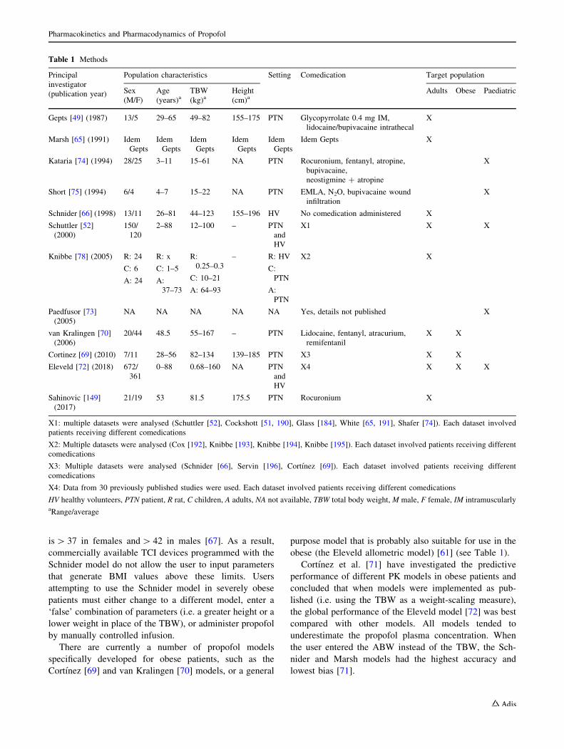
Table 1 Methods
Principal
investigator
(publication year)
Population characteristics Setting Comedication Target population
Sex
(M/F)
Age
(years)aTBW
(kg)aHeight
(cm)aAdults Obese Paediatric
Gepts [49] (1987) 13/5 29–65 49–82 155–175 PTN Glycopyrrolate 0.4 mg IM,
lidocaine/bupivacaine intrathecal
X
Marsh [65] (1991) Idem
Gepts
Idem
Gepts
Idem
Gepts
Idem
Gepts
Idem
Gepts
Idem Gepts X
Kataria [74] (1994) 28/25 3–11 15–61 NA PTN Rocuronium, fentanyl, atropine,
bupivacaine,
neostigmine ? atropine
X
Short [75] (1994) 6/4 4–7 15–22 NA PTN EMLA, N2O, bupivacaine wound
infiltration
X
Schnider [66] (1998) 13/11 26–81 44–123 155–196 HV No comedication administered X
Schuttler [52]
(2000)
150/
120
2–88 12–100 – PTN
and
HV
X1 X X
Knibbe [78] (2005) R: 24
C: 6
A: 24
R: x
C: 1–5
A:
37–73
R:
0.25–0.3
C: 10–21
A: 64–93
– R: HV
C:
PTN
A:
PTN
X2 X
Paedfusor [73]
(2005)
NA NA NA NA NA Yes, details not published X
van Kralingen [70]
(2006)
20/44 48.5 55–167 – PTN Lidocaine, fentanyl, atracurium,
remifentanil
X X
Cortinez [69] (2010) 7/11 28–56 82–134 139–185 PTN X3 X X
Eleveld [72] (2018) 672/
361
0–88 0.68–160 NA PTN
and
HV
X4 X X X
Sahinovic [149]
(2017)
21/19 53 81.5 175.5 PTN Rocuronium X
X1: multiple datasets were analysed (Schuttler [52], Cockshott [51, 190], Glass [184], White [65, 191], Shafer [74]). Each dataset involved
patients receiving different comedications
X2: Multiple datasets were analysed (Cox [192], Knibbe [193], Knibbe [194], Knibbe [195]). Each dataset involved patients receiving different
comedications
X3: Multiple datasets were analysed (Schnider [66], Servin [196], Cortınez [69]). Each dataset involved patients receiving different
comedications
X4: Data from 30 previously published studies were used. Each dataset involved patients receiving different comedications
HV healthy volunteers, PTN patient, R rat, C children, A adults, NA not available, TBW total body weight, M male, F female, IM intramuscularlyaRange/average
Pharmacokinetics and Pharmacodynamics of Propofol
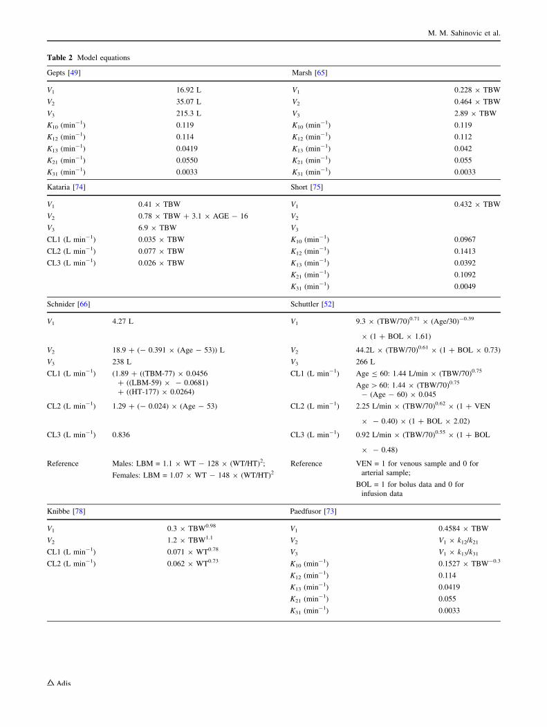
Table 2 Model equations
Gepts [49] Marsh [65]
V1 16.92 L V1 0.228 9 TBW
V2 35.07 L V2 0.464 9 TBW
V3 215.3 L V3 2.89 9 TBW
K10 (min-1) 0.119 K10 (min-1) 0.119
K12 (min-1) 0.114 K12 (min-1) 0.112
K13 (min-1) 0.0419 K13 (min-1) 0.042
K21 (min-1) 0.0550 K21 (min-1) 0.055
K31 (min-1) 0.0033 K31 (min-1) 0.0033
Kataria [74] Short [75]
V1 0.41 9 TBW V1 0.432 9 TBW
V2 0.78 9 TBW ? 3.1 9 AGE - 16 V2
V3 6.9 9 TBW V3
CL1 (L min-1) 0.035 9 TBW K10 (min-1) 0.0967
CL2 (L min-1) 0.077 9 TBW K12 (min-1) 0.1413
CL3 (L min-1) 0.026 9 TBW K13 (min-1) 0.0392
K21 (min-1) 0.1092
K31 (min-1) 0.0049
Schnider [66] Schuttler [52]
V1 4.27 L V1 9.3 9 (TBW/70)0.71 9 (Age/30)-0.39
9 (1 ? BOL 9 1.61)
V2 18.9 ? (- 0.391 9 (Age - 53)) L V2 44.2L 9 (TBW/70)0.61 9 (1 ? BOL 9 0.73)
V3 238 L V3 266 L
CL1 (L min-1) (1.89 ? ((TBM-77) 9 0.0456
? ((LBM-59) 9 - 0.0681)
? ((HT-177) 9 0.0264)
CL1 (L min-1) Age B 60: 1.44 L/min 9 (TBW/70)0.75
Age[ 60: 1.44 9 (TBW/70)0.75
- (Age - 60) 9 0.045
CL2 (L min-1) 1.29 ? (- 0.024) 9 (Age - 53) CL2 (L min-1) 2.25 L/min 9 (TBW/70)0.62 9 (1 ? VEN
9 - 0.40) 9 (1 ? BOL 9 2.02)
CL3 (L min-1) 0.836 CL3 (L min-1) 0.92 L/min 9 (TBW/70)0.55 9 (1 ? BOL
9 - 0.48)
Reference Males: LBM = 1.1 9 WT - 128 9 (WT/HT)2;
Females: LBM = 1.07 9 WT - 148 9 (WT/HT)2Reference VEN = 1 for venous sample and 0 for
arterial sample;
BOL = 1 for bolus data and 0 for
infusion data
Knibbe [78] Paedfusor [73]
V1 0.3 9 TBW0.98 V1 0.4584 9 TBW
V2 1.2 9 TBW1.1 V2 V1 9 k12/k21
CL1 (L min-1) 0.071 9 WT0.78 V3 V1 9 k13/k31
CL2 (L min-1) 0.062 9 WT0.73 K10 (min-1) 0.1527 9 TBW-0.3
K12 (min-1) 0.114
K13 (min-1) 0.0419
K21 (min-1) 0.055
K31 (min-1) 0.0033
M. M. Sahinovic et al.
5.5.2.3 Paediatric Population Develop PK models for
children is challenging, for many reasons. First, there are
ethical challenges with research in children, and, second, as
children grow, their size increases, their body composition
changes and their organ systems mature, making it difficult
to construct a single and accurate PK model.
A number of paediatric PK models for propofol exist,
the most well-known being the Paedfusor model [73],
Kataria model [74], Short model [75], Schuttler model [52]
and the Eleveld ‘general purpose’ model [72]. These
models were developed using a range of methodologies.
Differences in methodology include the age range of the
children enrolled in the study, the study population (healthy
children as opposed to children with comorbidities), the
sampling site (arterial vs. venous) and the administration
regimens used (bolus with or without continuous infusion
thereafter). These differences influence the range of
patients to which the different models are applicable. For
example, the Kataria model was developed from children
3 years of age and older and is thus not validated for use in
children younger than 3 years of age.
Sepulveda et al. [76] and Hara et al. [77] evaluated the
predictive performance of these models during short- and
long-duration infusions, respectively, and concluded that
overall most models overestimate the initial Vd. The rel-
evance is that when they are used to inform dosing schemes
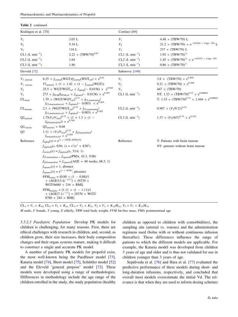
Table 2 continued
Kralingen et al. [70] Cortınez [69]
V1 3.03 L V1 4.48 9 (TBW/70) L
V2 5.34 L V2 21.2 9 (TBW/70) 9 e-0.0164 9 (Age-50) L
V3 116 L V3 237 9 (TBW/70) L
CL1 (L min-1) 2.22 9 (TBW/70)0.67 CL1 (L min-1) 1.92 9 (TBW/70)�
CL2 (L min-1) 1.64 CL2 (L min-1) 1.45 9 (TBW/70)� 9 e-0.0153 9 (Age-50)
CL3 (L min-1) 1.86 CL3 (L min-1) 0.86 9 (TBW/70)�
Eleveld [72] Sahinovic [149]
V1 arterial 6.25 9 fcentral(WGT)/fcentral(WGTref) 9 e0.61 V1 3.8 9 (TBW/70) 9 e0.205
V1 venous V1arterial 9 (1 ? 1.42 9 (1 - fcentral(WGT)) V2 9.21 9 (TBW/70) 9 e0.268
V2 25.5 9 WGT/WGTref 9 faging(- 0.0156) 9 e0.565 V3 447 9 (TBW/70)
V3 273 9 fFFM/fFFM,ref 9 fopiates(- 0.0138) 9 e0.597 CL1 (L min-1) NT: 1.33 9 (TBW/70)0.75 9 e0.00902
CLmale 1.79 9 (WGT/WGTref)0.75 9 fCLmaturation/
fCLmaturation,ref 9 fopiates(- 0.003) 9 e0.265T: 1.33 9 (TBW/70)0.75 9 1.444 9 e0.00902
CLfemale 2.1 9 (WGT/WGTref)0.75 9 fCLmaturation/
fCLmaturation,ref 9 fopiates(- 0.003) 9 e0.265CL2 (L min-1) 0.987 9 (V2/9.21)
0.75
Q2arterial 1.75(V2/V2ref)0.75 9 (1 ? 1.3 9 (1 -
fQ3maturation)) 9 e0.346CL3 (L min-1) 1.37 9 (V3/447)
0.75 9 e0.192
Q2venous Q2arterial 9 0.68
Q3 1.11 9 (V3/V3ref)0.75 9 fQ3maturation/
fmaturation,ref 9 e0.209
Reference faging(x) = e(x 9 (AGE-AGEref))
fsigmoid(x, E50, k) = xk(xk ? E50k)
Reference T: Patients with brain tumour
NT: patients without brain tumour
fcentral(x) = fsigmoid(x, 33.6, 1)
fCLmaturation = fsigmoid(PMA, 42.3, 9.06)
fQ3maturation = fsigmoid(AGE ? 40 weeks, 68.3, 1)
fopiates(x) = 1, absence
fopiates(x) = e(x 9 AGE), presence
FFMmales = [0.88 ? (1 - 0.88)/1
? (AGE/13.4)-12.7] 9 [9270 9
WGT/6680 ? 216 9 BMI]
FFMfemales = [1.11 ? (1 - 1.11)/1
? (AGE/7.1)-1.1] 9 [9270 9 WGT/
8780 ? 244 9 BMI]
CL1 = V1 9 K10; CL2 = V2 9 K21; CL3 = V3 9 K31; V2 = V1 9 K12/K21; V3 = V1 9 K13/K31
M male, F female, Y young, E elderly, TBW total body weight, FFM fat-free mass, PMA postmenstrual age
Pharmacokinetics and Pharmacodynamics of Propofol
designed to achieve a particular plasma concentration, they
result in administration of a larger than necessary induction
bolus. Most paediatric models performed well during
infusions of short duration (up to median of 99 min), while
only the Short [75] and Schuttler [52] models showed
acceptable performance and the least divergence during
infusions of up to 545 min. Overall, the Short model per-
formed the best and the authors concluded that it might be
preferable for use during TCI in children.
5.5.2.4 Unified PK Models A PK model is only appli-
cable in patients in whom the clinical conditions and
patient category match those used in model development.
As soon as these begin to diverge, model predictions can be
erroneous. Using an inappropriate model during a TCI can
lead to unexpected (side) effects. Rational model selection
is of critical importance, but given the large number of
available models, this is not an easy task. Furthermore, an
exact match is often not possible. Even for users knowl-
edgeable in PK, it is difficult to be certain which models to
use in patients with overlapping conditions (e.g. patients
who are elderly and obese), or in grey zones between
conditions (e.g. patients who are moderately obese but
otherwise healthy). These difficulties have stimulated the
development of unified PK models, which are derived from
data collected in a diverse group of patients and clinical
conditions, and are thus designed to provide accurate pre-
dictive performance well in a wide range of patients and
clinical conditions.
The first unified PK model for propofol was published by
Schuttler and Ihmsen [52] in 2000. This group developed a
general PK model for propofol, applicable in both children
and adults, by performing PK analysis on data collected by
five different research groups. This analysis yielded a three-
compartment PK model with weight, age, method of
administration and sampling site as the main covariates.
They concomitantly demonstrated that scaling of model
compartments and clearances to power exponents (between
0.55 and 0.75) of normalized TBW yielded better model
predictions than that of linear scaling. An important obstacle
to a broader clinical application of this model is the fact that
the user must select a mode of administration (bolus vs.
continuous infusion) before using the model, whereas in
clinical practice both administration modes are often used.
In 2005, Knibbe and colleagues [78] published an
interspecies PK model for propofol, which was a two-
compartment model applicable in rats, children and adults.
They achieved this impressive task by allometrically
scaling volumes and clearances to TBW. In other words,
they scaled the volumes and clearances in the model to a
power exponent (between 0.73 and 1.1), which allowed
them to more realistically estimate the volumes and
clearances in a wide range of different bodyweights, from a
small rat weighing 0.25 kg to an adult weighing 93 kg.
The most recently published unified PK model for
propofol is the ‘general purpose model’ constructed by
Eleveld and colleagues [61]. First published in 2014, the PK
model was recently expanded with a PD analysis [72]. In the
updated version of the model, the authors used PK data
derived from 30 previously published studies containing
data collected from children of all ages, adults, obese adults,
and elderly individuals. They constructed a three-compart-
ment model where weight, age, sex and administration of
comedication were covariates. Similar to the work of
Schuttler and Ihmsen [52] and Knibbe et al. [78], they also
used allometric scaling to normalize bodyweight for clear-
ances (three-quarter power exponent), while volumes were
scaled linearly with normalized body weight for V1 and V2,
and with normalized fat-free mass for V3. Retrospective
analysis of this model performance shows that the model
performs at least as good as, or better, than dedicated
population models in all populations. Further model eval-
uation must be performed in prospective studies.
5.5.3 Effects of Patient Characteristics on Propofol
Kinetics
One of the main advantages of analysing a larger set of PK
and PD data from a wide range of patients is the ability to
detect covariate relationships that might remain hidden
when a dataset from patients with a narrower range of
characteristics is analysed. As a result, Eleveld et al. were
able to identify a few, clinically very relevant, correlations.
They found a relationship between age and the volume of
the peripheral compartments and metabolic clearance. The
volume of V2 declines with age, while V3 and metabolic
clearance decline only when other medication are concur-
rently administered. This finding confirms an impression
widely shared by practicing anaesthetists that the mainte-
nance infusion rates required for adequate anaesthesia are
reduced in older individuals, especially when comedication
is administered. Furthermore, they found that the central
compartment scales to an Emax function (in other words
central compartment volume increase follows a sigmoid
function with increasing weight). This suggests that the
induction dose of propofol, per kilogram, should be rela-
tively high in low-weight individuals, and relatively low in
obese patients, compared with individuals with intermedi-
ate weight. This fact is also consistent with the observa-
tions of clinical practitioners. Lastly, they reported a higher
metabolic clearance in women (of all ages) compared with
men, which might explain clinical observations of a more
rapid emergence from propofol anaesthesia in women
compared with men [79].
M. M. Sahinovic et al.
5.5.4 Physiologically-Based PK Models
Contrary to compartmental PK models where compart-
ments have no real physiological meaning (or direct cor-
respondence with body parts), compartments in
physiologically-based PK models (PBPK) are linked to
different body parts connected by the cardiovascular sys-
tem. Typically, all the major tissues, such as adipose tissue,
muscle, brain, heart, kidney, liver, and lung, are repre-
sented [80].
An important advantage of PBPK models over com-
partmental models is the ability of the former to include
more parameters specifying sources of physiological and
biochemical variability in individuals, thereby reducing the
interindividual variability and increasing the individual
prediction accuracy. For example, the incorrect assumption
of instantaneous mixing of a drug in the central compart-
ment used by compartmental models severely limits their
prediction accuracy during the first few minutes of drug
infusion. Adaptations to the compartmental models using
‘administration lag time’ and presystemic compartments
can improve the prediction accuracy [81], however only
PBPK models use cardiac output [82] and blood flow terms
to more accurately describe the drug kinetics in greater
detail. This even potentially enables them to predict the
influence of the effect of changing haemodynamic param-
eters on drug disposition. The same applies to cerebral
blood flow, changes of which can play a major role in
propofol PD.
An important disadvantage of PBPK models is the dif-
ficulty in collecting the extensive physiological data nec-
essary for model development, and often very limited (if
any) improvement in prediction accuracy at a cost of a
much more complex and mathematically elaborate model
compared with compartmental models [83].
The physiologically-based hybrid recirculatory model
published by Upton and Ludbrook [84] is an example of a
PBPK propofol model. It incorporates submodels for the
lung and brain, and a less detailed ‘lumped model’ for the
rest of the body. The prediction accuracy and bias are
comparable with that of the major compartmental models
[83].
6 Pharmacodynamics (PD)
6.1 Central Nervous System
6.1.1 Hypnotic Effects
Propofol exerts its hypnotic effect through potentiation of
the effects of the inhibitory neurotransmitter GABA [85]. It
binds to the b-subunit of the postsynaptic GABAA receptor,
where it causes an inward directed chloride current that
hyperpolarizes the postsynaptic membrane and inhibits
neuronal depolarization. This effect is dose-dependent. At
low concentrations, propofol potentiates GABA-activated
inward chloride currents, while at higher concentrations, it
directly activates the channel opening [86]. GABA recep-
tors are ubiquitous throughout the central nervous system
[87].
Although our understanding of the actions of propofol at
the molecular level is quite extensive, we do not entirely
understand how these molecular effects translate into
alterations in cellular, synaptic and neural network func-
tion, and in turn cause unconsciousness [88]. This knowl-
edge gap is, at least in part, the result of a lack of a
generally accepted theory of consciousness. In recent
years, cognitive neuroscience has seen a resurgence of
interest in this topic, with attempts to integrate anaesthesia
and sleep research in order to address this deficiency. This
resurgence has revealed several brain areas that play a
crucial role in generation of consciousness, and which are
extensively influenced by hypnotic drugs.
In the reticular formation of the brainstem [89], there are
a number of sleep- and wakefulness-promoting cholinergic
and monoaminergic nuclei that exert their effect by influ-
encing higher cortical structures [90]. Their activity and
reciprocal influence changes with the level of wakefulness
[91]. Local inactivation of wakefulness-promoting areas,
such as locus coeruleus and dorsal raphe, enhance anaes-
thesia, while local activation of various other wakefulness-
promoting areas, including pontis oralis and centromedial
thalamus, facilitate emergence from anaesthesia. One of
the sleep-promoting nuclei is the ventrolateral preoptic
area. Lesions in this region enhance wakefulness [92].
These nuclei are extensively affected by clinical concen-
trations of hypnotic drugs.
The thalamus plays a central role in information pro-
cessing with the brain. Increasing concentrations of
propofol, and thereby increasing levels of sedation, cause a
decrease in activity, regional cerebral blood flow and
metabolism in the thalamus [93]. There are a number of
(mutually nonexclusive) hypotheses on how these changes
could lead to or contribute to loss of consciousness [94].
The function of the thalamus as a critical sensory infor-
mation relay from subcortical structures to the cortex could
be impeded by hypnotic drugs [95], although thalamic
depression seen after loss of consciousness could merely
reflect the decrease in global cortical activity [96].
Hyperpolarization of thalamocortical neurons—and the
resulting switching from a tonic firing state in wakefulness
to a bursting firing state in unconsciousness—could be a
final common pathway through which different hypnotics
cause a disruption in thalamocortical and cortico-cortical
loops, thereby causing unconsciousness [97].
Pharmacokinetics and Pharmacodynamics of Propofol
Finally, the cerebral cortex has long been identified as
an important drug effect target for hypnotic drugs. Sleep
and anaesthesia studies have consistently demonstrated
decreases in cortical activity and cerebral blood flow [98];
however, these changes are not homogeneous across the
cortex and across different drugs. The most consistent and
largest changes occur in the frontal and posterior parietal
cortex [99]. These regions form part of a much wider
‘default mode network’, a functional network thought to be
responsible for monitoring of internal environment in
humans. Loss of consciousness in non-REM sleep is
accompanied by increased modularization of brain activity
and reduction in a whole-brain spatiotemporal integration
of information [100–102]. This is concordant with the
information integration theory of consciousness [103].
6.1.2 Amnesia
The amnestic properties of propofol have been extensively
described in the literature [104]. Explicit memory seems to
be most affected, and in a dose-dependent manner. The
most basic form of memory, perceptual priming, seems to
be preserved to some degree [105]. The amnestic effects of
propofol do not seem to be caused by interference with
memory encoding, but a low-dose of propofol has been
shown to induce amnesia without any impairments in
behaviour [106]. The exact neural mechanism of propofol-
induced amnesia in a conscious patient remains to be
elucidated.
6.1.3 Anxiolysis
Propofol produces anxiolysis in subhypnotic doses. This
has been demonstrated in mice [107] and in patients
undergoing propofol sedation while undergoing surgical
procedures under regional anaesthesia [108]. Propofol has
been proposed as an excellent preoperative anxiolytic drug
in day-care surgery, when administered through a patient-
controlled anxiolysis system (a patient-controlled analgesia
system analogue), as a replacement for benzodiazepine
premedication in order to shorten the discharge times
[109]. The exact mechanism of this anxiolysis is still not
known, but inhibition of 5-HT activity in the hippocampus
[110] or nitric oxide synthase in the hypothalamus,
amygdala and hippocampus might be the mechanisms
involved [111].
6.1.4 Analgesia
There are a number of patient studies describing the anal-
gesic effects of subhypnotic doses of propofol [112, 113].
This effect could be caused by an action of propofol at the
spinal level. In animal studies, it has been shown that
propofol produces analgesia in mice when administered
intraperitoneally, through modulation of spinal GABAA
receptors [114], and thereby depresses ventral root poten-
tials in the spinal cord elicited by monosynaptic reflexes or
exposure of the spinal cord to substance P [115]. Further-
more, Nishiyama et al. showed that intrathecal injections of
propofol had effects on pain perception, but they could not
rule out the possibility of neurotoxicity [116].
A recently published meta-analysis comparing postop-
erative pain after propofol versus inhalation-based anaes-
thesia did not demonstrate any significant differences,
mainly because of substantial heterogeneity among studies
published to date [117].
6.1.5 Antiemetic Effect
The antiemetic action of propofol is well known and has
been extensively described [118, 119]. Patients receiving
anaesthesia with propofol experience significantly less
PONV compared with that associated with other hypnotic
drugs, irrespective of the use of adjunct drugs, patient
characteristics or opiate use. The mechanism behind this
effect is not very well understood, but it has been
demonstrated that propofol interacts with dopaminergic
(D2) receptors in the chemoreceptor trigger zone [120],
inhibits the limbic system [121], thereby interacting with
cortical reflexes reaching the vomiting centre, and inhibits
5-HT3 receptors located in the central nervous system in a
noncompetitive and dose-dependent manner, thereby
reducing the incidence and severity of PONV.
6.1.6 Neurophysiological Effects
Propofol decreases cerebral blood flow, intracranial pres-
sure, and cerebral metabolic rate, while maintaining
dynamic and static autoregulation [122] and vascular
responsiveness to carbon dioxide [123]. These favourable
effects on cerebral physiology make propofol an almost
ideal hypnotic for anaesthesia during neurosurgery. The
evidence for neuroprotective effects of propofol during
ischaemia-reperfusion injury is conflicting [124]; however,
its role as part of multimodal neuroprotection has been
established [125].
Propofol has both pro- and anticonvulsive activity. On
the one hand, a number of reports of convulsions and
excitatory events such as myoclonus and tremor, during or
shortly after the start or end of propofol anaesthesia, have
been published [126]; these events might be the result of
preferential depression of subcortical regions. On the other
hand, the role of propofol as a potent treatment of status
epilepticus has been well-established [127, 128].
M. M. Sahinovic et al.
6.2 Cardiovascular System
Propofol has extensive effects on the cardiovascular sys-
tem. The most prominent effect is systemic blood pressure
reduction accompanied by a decrease in cardiac output.
This effect is dose-dependent and even occurs at sedative
doses. It is more pronounced in elderly and physiologically
compromised patients [129]. The effect is, at least partially,
mediated by a significant decrease of sympathetic tone
accompanied by a decrease in vascular resistance. Fur-
thermore, propofol also inhibits the physiological barore-
flex responses, thereby enhancing cardiovascular
depression [130].
Cardiac contractility remains preserved. Propofol only
has direct negative inotropic actions at concentrations
exceeding the clinical range, an effect that is similar in
failing and non-failing human hearts. The negative ino-
tropic effect is mediated through a concentration-depen-
dent decrease in the uptake of Ca2? into the sarcoplasmatic
reticulum, which is simultaneously accompanied by an
increase of myofilament sensitivity to Ca2?, partially
counteracting the effect [131]. The haemodynamic
response lags behind the hypnotic effect of propofol. While
the hypnotic half-time plasma effect site equilibration time
(T�keo) is 2.5 min, independent of age, haemodynamic
half-time plasma effect site equilibration time is 5 min in
young patients, and up to 10 min in elderly patients [132].
Propofol cardioprotective effects in cardiac surgery is a
focus of research. In animal studies, high propofol doses
caused dose-dependent attenuation of ischaemia-reperfu-
sion myocardial injury (IRI) by exerting free radical
scavenging effects and decreasing lipid peroxidase activity
[133]. These cardioprotective effects are less profound than
those caused by sevoflurane [134]. A combination of
inhaled anaesthetic preconditioning and propofol postcon-
ditioning appears to work synergistically in decreasing IRI
[135].
6.3 Respiratory System
Propofol is a potent ventilatory depressant. It interferes
with ventilation in a dose-dependent manner by affecting
central chemoreceptor sensitivity, reducing ventilatory
responses to hypercapnia and hypoxia [136–138]. In higher
doses, propofol causes apnoea. It also changes the pattern
of breathing by decreasing the ribcage contribution to tidal
volume [139] by causing upper airway relaxation and
suppression of upper airway reflexes [140]. Furthermore, it
attenuates vagal- and methacholine-induced bronchocon-
striction [141] and potentiates hypoxic pulmonary vaso-
constriction [142].
6.4 Hepatorenal System
Despite the fact that the liver and kidneys are extensively
involved in metabolism and excretion of propofol, their
function does not appear to be affected by propofol. In
human and animal studies, propofol infusion increased
hepatic perfusion [143] due to higher arterial [144] and
portal venous blood flow [145], while renal perfusion
remained essentially unaltered [146]. However, when car-
diac output is not maintained, organ perfusion, and thus
liver and renal perfusion, could be compromised.
Propofol infusion is known to cause green skin and urine
discolouration caused by production of phenol green
chromophore. Furthermore, urinary uric acid excretion is
increased after propofol infusion, which can result in a
cloudy appearance of the urine.
6.5 PD Modelling
For propofol, the main clinical effect is loss of con-
sciousness, which is difficult to quantify. Most clinicians
consider it to be a continuum of effects, most of which
have a binary nature, i.e. present or absent. However,
several electroencephalogram (EEG)-based methods of
quantifying the depth of anaesthesia have been developed
[147, 148]. The most commonly used is the Bispectral
index (BIS; Medtronic, Dublin, Ireland), a unitless number
between 0 and 100, where 0 represents very high hypnotic
drug effect (i.e. no cerebral electrical activity) and 100
represents total absence of the hypnotic drug effect. The
BIS monitor is one of the best validated EEG-based
monitors for quantifying the drug effect [148]. Several PD
studies have been performed, with most of them using a
sigmoidal Emax model to characterize the relationship
between blood concentration, the concentration in a
hypothetical effect site, and the resultant clinical drug
effect. Published parameters are in the following ranges:
keo, 0.01–0.45 min-1; Ce50, 2.71–3.44 mg L-1; and c,1.47–2.961 [65, 67, 71, 149–152].
7 Drug Interactions
Interactions between drugs occur in the PK, PD, or both
[153]. Alterations in the PD (clinically visible) drug dose–
effect relationship can be secondary to PK and/or PD
interactions; therefore, these interactions are most often not
discussed separately when considering clinical applica-
tions. Drug interactions can be additive, synergistic or
antagonistic [154]. Any anaesthesia provider should be
aware of this and adjust the drug dose accordingly.
Pharmacokinetics and Pharmacodynamics of Propofol
7.1 PK Interactions
PK interactions can result in changes in absorption, dis-
tribution, metabolism and elimination.
7.1.1 Absorption
Propofol is only suitable for intravenous administration and
thus does not undergo any absorption interactions.
Nonetheless, care should be taken when infusing propofol
with other drugs. It is well-described that propofol under-
goes chemical interactions with a number of frequently used
drugs, and should thus not be administered through the
same intravenous line with these particular drugs. Of note
are interactions with certain antibiotics (e.g. ciprofloxacin,
gentamicin, metronidazole) and calcium antagonists (ni-
modipine, verapamil). A more extensive list of interactions
can be found in the summary of product characteristics
(SPC) and in the study by Michaels et al. [155]
7.1.2 Distribution
Propofol is extensively bound to plasma proteins. Con-
comitant infusion with drugs competing for the same
plasma binding sites, or use in patients with low plasma
proteins, could potentially result in high unbound plasma
propofol fraction, causing more profound effects and
adverse effects. The clinical significance of this phe-
nomenon is uncertain [156].
Vd and the rate of metabolism of propofol are affected by
drugs affecting the cardiovascular system. Drugs that
decrease cardiac output and cause a concomitant decrease in
hepatic perfusion alter the distribution and metabolism of
propofol, e.g. esmolol [157] and in propofol itself [158, 159]
7.1.3 Metabolism
As with many other drugs used in anaesthesia [160, 161],
concomitant administration of propofol with other drugs
dependent on metabolism by CYP could interfere with its
metabolism. In vitro studies demonstrate this possibility,
however the results are not directly transposable to the
in vivo situation [162].
7.2 PD Interactions
Propofol interacts with other intravenous and volatile hyp-
notic drugs, as well as with opioids. In order to mathe-
matically delineate the interaction between different drugs,
their combined effectiveness to reach a certain clinically
relevant endpoint is assessed. This combined effectiveness
can be explored in an isobolographic analysis using isoboles
(or ‘iso-effective’ drug effect dose curves) to show all the
dose combinations of drugs resulting in the same clinical
effect [163]. The interaction can also be modelled using a
response surface modelling technique [164].
7.2.1 Interactions with Hypnotic Drugs
7.2.1.1 Midazolam A number of studies have investi-
gated the interaction between propofol and midazolam. All
studies confirm, as can be expected based on clinical
experience, an interaction, however they do not agree on its
nature. The study findings range from an additive to syn-
ergistic interaction [165–171]. This discrepancy might be
due to study methodologies involving inaccurate PK
models, or data collection in a non-steady state.
7.2.1.2 Dexmedetomidine Dexmedetomidine is a potent
a2 agonist. Multiple studies have investigated its effect on
the propofol concentration necessary to reach specific
clinical endpoints. When administered preoperatively,
dexmedetomidine reduced the propofol dose necessary for
sedation and induction of anaesthesia [172, 173]. In adults,
a loading dose of 0.1 lg kg-1 administered during a
10-min period reduced the half maximal effective con-
centration (EC50) of propofol for successful i-gel (Inter-
surgical, Berkshire, UK) airway insertion from 6.75 to
3.18 lg/mL [174]; a comparable effect was not found in
children [175]. The interaction is also evident during and at
the end of anaesthesia. Dexmedetomidine reduced propofol
use during spinal surgery under propofol–remifentanil
anaesthesia, and prolonged recovery times at the end of
propofol anaesthesia if it was not stopped early [176, 177].
As with midazolam, a clear interaction model has yet to be
developed and a well-designed interaction study needs to
be performed, preferably using accurate PK models such as
the Eleveld model [61] for propofol and the Hannivoort
model for dexmedetomidine [35].
7.2.1.3 Volatiles Propofol–sevoflurane interaction stud-
ies show an additive interaction, which was demonstrated
by studies investigating the effects on clinical endpoints
(e.g. loss of consciousness, reaction to laryngoscopy, and
reaction to incision), as well as studies measuring the
effects on EEG-based parameters from a number of depth
of anaesthesia monitors (e.g. Bispectral index, state and
response entropy) [178–181]. This additive interaction is
not surprising as Sebel et al. demonstrated that propofol
and sevoflurane have separate binding sites but converging
pathways of action on the GABAa receptor [181, 182].
7.2.2 Interactions with Opioids
Propofol and opioids interact synergistically. This is more
pronounced for analgesic drug effects (e.g. loss of response
M. M. Sahinovic et al.
to noxious stimuli) than for hypnotic clinical endpoints,
such as loss of responsiveness to verbal commands [154].
Fentanyl significantly decreases the propofol concen-
tration required for loss of consciousness and suppression
of responses to noxious stimuli [183]. A ceiling effect of
this interaction is seen at fentanyl effect concentrations of
3 ng mL-1, where the propofol plasma concentration
necessary for loss of responsiveness to verbal command is
decreased by 40% and the concentration necessary for
suppression of movement on incision is decreased by 90%
compared with when no fentanyl is administered. Further
increases in fentanyl concentrations do not increase inter-
action effects [184].
Sufentanil showed an additive interaction with propofol
with respect to loss of responsiveness to verbal command
during the induction of anaesthesia. It reduced the propofol
concentration necessary for loss of responsiveness in a
dose-dependent manner [185]. Hentgen et al. showed that a
sufentanil effect site concentration (Ce) of 0.3 ng/mL
combined with propofol Ce of 4 ng/mL provides optimal
haemodynamic stability during intubation, while a Ce of
sufentanil 0.2 ng/mL provides an optimal balance between
haemodynamic stability during operation, and a rapid
recovery thereafter [186].
Remifentanil is an ultra-short-acting opiate. Its interac-
tion with propofol has been extensively studied in regard to
multiple clinical endpoints, and it showed a supra-additive
interaction with propofol in regard to hypnotic [187] and
analgesic [188] endpoints.
8 Methods of Drug Delivery
Administration of intravenous hypnotic drugs brings some
unique challenges compared with volatile anaesthetics.
Multiphasic PK and the inability to continuously measure
the achieved drug concentration can present a challenge
when trying to achieve a specific drug effect. In the
majority of countries worldwide (the US being the main
exception), TCI pumps (infusion pumps programmed to
infuse a drug guided by a mathematical PK and PD model
so as to achieve a predefined plasma or effect site drug
concentration expeditiously) are available in order to
simplify the administration of propofol [153, 189]. The
most well-known PK/PD models for propofol have already
been discussed earlier in this article.
Compliance with ethical standards
Funding No funding was received in the preparation of this
manuscript.
Conflict of interest Marko M. Sahinovic reports no conflicts of
interest. Michel M.R.F. Struys reports his research group/department
has received grants and funding from The Medicines Company
(Parsippany, NJ, USA), Masimo (Irvine, CA, USA), Fresenius (Bad
Homburg, Germany), Acacia Design (Maastricht, The Netherlands),
and Medtronic (Dublin, Ireland), as well as honoraria from The
Medicines Company (Parsippany, NJ, USA), Masimo (Irvine, CA,
USA), Fresenius (Bad Homburg, Germany), Baxter (Deerfield, IL,
USA), Medtronic (Dublin, Ireland), and Demed Medical (Temse,
Belgium). A.R. Absalom is an editor of the British Journal of
Anaesthesia. His research group/department has received grants and
funding from The Medicines Company (Parsippany, NJ, USA),
Medtronic (Dublin, Ireland), and BD (Franklin Lakes, NJ, USA). He
has also received honoraria (paid to the institution) from The
Medicines Company (Parsippany, NJ, USA), Janssen Pharmaceutica
NV (Beerse, Belgium) and Ever Pharma (Salzburg, Austria).
Open Access This article is distributed under the terms of the
Creative Commons Attribution-NonCommercial 4.0 International
License (http://creativecommons.org/licenses/by-nc/4.0/), which per-
mits any noncommercial use, distribution, and reproduction in any
medium, provided you give appropriate credit to the original
author(s) and the source, provide a link to the Creative Commons
license, and indicate if changes were made.
References
1. Glen JB, James R. 2,6-Diisopropylphenol as an anaesthetic
agent. London: United States Patent and Trademark Office;
1977. p. 1–10.
2. Thompson KA, Goodale DB. The recent development of
propofol (DIPRIVAN). Intensive Care Med. 2000;26(Suppl
4):S400–4.
3. Schuttler J, Schwilden H, editors. Modern anesthetics (handbook
of experimental pharmacology), vol. 182. Heidelberg: Springer;
2008.
4. Baker MT, Naguib M. Propofol: the challenges of formulation.
Anesthesiology. 2005;103:860–76.
5. Bryson HM, Fulton BR, Faulds D. Propofol. An update of its use
in anaesthesia and conscious sedation. Drugs. 1995;50:513–59.
6. Fulton B, Sorkin EM. Propofol. An overview of its pharma-
cology and a review of its clinical efficacy in intensive care
sedation. Drugs. 1995;50:636–57.
7. Trapani G, Altomare C, Liso G, Sanna E, Biggio G. Propofol in
anesthesia. Mechanism of action, structure-activity relation-
ships, and drug delivery. Curr Med Chem. 2000;7:249–71.
8. Joo HS, Perks WJ. Sevoflurane versus propofol for anesthetic
induction: a meta-analysis. Anesth Analg. 2000;91:213–9.
9. Liu H, Ji F, Peng K, Applegate RL, Fleming N. Sedation after
cardiac surgery: is one drug better than another? Anesth Analg.
2017;124:1061–70.
10. Kochhar GS, Gill A, Vargo JJ. On the horizon: the future of
procedural sedation. Gastrointest Endosc Clin N Am.
2016;26:577–92.
11. Marik PE. Propofol: therapeutic indications and side-effects.
Curr Pharm Des. 2004;10:3639–49.
12. Package insert, Diprivan (Propofol) Injectable Emulsion. Lake
Zurich, IL: Fresenius Kabi; 2014.
13. Hart B. ‘‘Diprivan’’: a change of formulation. Eur J Anaesthe-
siol. 2000;17:71–3.
14. Pubchem. Propofol. 2017. Available at: https://pubchem.ncbi.
nlm.nih.gov/compound/propofol#section=Top.
15. Fischer MJM, Leffler A, Niedermirtl F, Kistner K, Eberhardt M,
Reeh PW, et al. The general anesthetic propofol excites noci-
ceptors by activating TRPV1 and TRPA1 rather than GABAA
receptors. J Biol Chem. 2010;285:34781–92.
Pharmacokinetics and Pharmacodynamics of Propofol
16. Klement W, Arndt JO. Pain on injection of propofol: effects of
concentration and diluent. Br J Anaesth. 1991;67:281–4.
17. Allford MA, Mensah JA. Discomfort on injection. Eur J
Anaesthesiol. 2006;23:971–4.
18. Picard P, Tramer MR. Prevention of pain on injection with
propofol: a quantitative systematic review. Anesth Analg.
2000;90:963–9.
19. Hardman JG, Hopkins PM, Struys MMR, editors. Oxford text-
book of anaesthesia. Oxford: Oxford University Press; 2017.
20. Asserhøj LL, Mosbech H, Krøigaard M, Garvey LH. No evi-
dence for contraindications to the use of propofol in adults
allergic to egg, soy or peanut. Br J Anaesth. 2016;116:77–82.
21. American Society for Anesthesiology; Committee for Standards
and Practice Parameters. Standards for basic anesthetic monitor-
ing. ASA Standard Guidelines; 2015. p. 1–4. Available at: http://
www.asahq.org/quality-and-practice-management/standards-and-
guidelines.
22. Jevtovic-Todorovic V, Hartman RE, Izumi Y, Benshoff ND,
Dikranian K, Zorumski CF, et al. Early exposure to common
anesthetic agents causes widespread neurodegeneration in the
developing rat brain and persistent learning deficits. J Neurosci.
2003;23:876–82.
23. Jevtovic-Todorovic V, Absalom AR, Blomgren K, Brambrink
A, Crosby G, Culley DJ, et al. Anaesthetic neurotoxicity and
neuroplasticity: an expert group report and statement based on
the BJA Salzburg Seminar. Br J Anaesth. 2013;111:143–51.
24. Raoof AA, Augustijns PR, Verbeeck RK. In vivo assessment of
intenstinal, hepatic, and pulmonary first pass metabolism of
propofol in the rat. Pharm Res. 1996;13:891–5.
25. Uchegbu I, Jones M-C, Corrente F, Godfrey L, Laghezza D,
Carafa M, et al. The oral and intranasal delivery of propofol
using chitosan amphiphile nanoparticles. Pharm Nanotechnol.
2014;2:65–74.
26. Mazoit JX, Samii K. Binding of propofol to blood components:
implications for pharmacokinetics and for pharmacodynamics.
Br J Clin Pharmacol. 1999;47:35–42.
27. Tarr L, Oppenheimer B, Sager R. The circulation time in various
clinical conditions determined by the use of sodium dehy-
drochlorate. Am Heart J. 1933;8:766.
28. Dawidowicz AL, Kalitynski R, Fijalkowska A. Free and bound
propofol concentrations in human cerebrospinal fluid. Br J Clin
Pharmacol. 2003;56:545–50.
29. Engdahl O, Abrahams M, Bjornsson A, et al. Cerebrospinal fluid
concentrations of propofol during anaesthesia in humans. Br J
Anaesth 1998;81:957–9.
30. Dailland P, Cockshott ID, Lirzin JD, Jacquinot P, Jorrot JC,
Devery J, et al. Intravenous propofol during cesarean section:
placental transfer, concentrations in breast milk, and neonatal
effects. A preliminary study. Anesthesiology. 1989;71:827–34.
31. Gin T, Yau G, Jong W, Tan P, Leung RKW, Chan K. Dispo-
sition of propofol at caesarina section and in the postpartum
period. Br J Anaesth. 1991;67:49–53.
32. Tumukunde J, Lomangisi DD, Davidson O, Kintu A, Joseph E,
Kwizera A. Effects of propofol versus thiopental on Apgar
scores in newborns and peri-operative outcomes of women
undergoing emergency cesarean section: a randomized clinical
trial. BMC Anesthesiol. 2015;15:63.
33. Simons PJ, Cockshott ID, Douglas EJ, Gordon E a, Hopkins K,
Rowland M. Disposition in male volunteers of a subanaesthetic
intravenous dose of an oil in water emulsion of 14C-propofol.
Xenobiotica. 1988;18:429–40.
34. Hughes MA, Glass PS, Jacobs JR. Context-sensitive half-time in
multicompartment pharmacokinetic models for intravenous
anesthetic drugs. Anesthesiology. 1992;76:334–41.
35. Hannivoort LN, Eleveld DJ, Proost JH, Reyntjens KMEM,
Absalom AR, Vereecke HEM, et al. Development of an
optimized pharmacokinetic model of dexmedetomidine using
target-controlled infusion in healthy volunteers. Anesthesiology.
2015;123:357–67.
36. Court MH, Duan SX, Hesse LM, Venkatakrishnan K, Greenblatt
DJ. Cytochrome P-450 2B6 is responsible for interindividual
variability of propofol hydroxylation by human liver micro-
somes. Anesthesiology. 2001;94:110–9.
37. Shioya N, Ishibe Y, Shibata S, Makabe H, Kan S, Matsumoto N,
et al. Green urine discoloration due to propofol infusion: a case
report. Case Rep Emerg Med. 2011;2011:1–4.
38. Mikstacki A, Skrzypczak-Zielinska M, Tamowicz B, Zakerska-
Banaszak O, Szalata M, Slomski R. The impact of genetic
factors on response to anaesthetics. Adv Med Sci. 2013;58:9–14.
39. Takizawa D, Sato E, Hiraoka H, Tomioka A, Yamamoto K,
Horiuchi R, et al. Changes in apparent systemic clearance of
propofol during transplantation of living related donor liver. Br J
Anaesth. 2005;95:643–7.
40. Hiraoka H, Yamamoto K, Miyoshi S, Morita T, Nakamura K,
Kadoi Y, et al. Kidneys contribute to the extrahepatic clearance
of propofol in humans, but not lungs and brain. Br J Clin
Pharmacol. 2005;60:176–82.
41. Takizawa D, Hiraoka H, Goto F, Yamamoto K, Horiuchi R.
Human kidneys play an important role in the elimination of
propofol. Anesthesiology. 2005;102:327–30.
42. Dawidowicz AL, Fornal E, Mardarowicz M, Fijalkowska A. The
role of human lungs in the biotransformation of propofol.
Anesthesiology. 2000;93:992–7.
43. He YL, Ueyama H, Tashiro C, Mashimo T, Yoshiya I. Pul-
monary disposition of propofol in surgical patients. Anesthesi-
ology. 2000;93:986–91.
44. Bodenham A, Culank LS, Park GR. Propofol infusion and green
urine. Lancet. 1987;2:740.
45. Takita A, Masui K, Kazama T. On-line monitoring of end-tidal
propofol concentration in anesthetized patients. Anesthesiology.
2007;106:659–64.
46. Grossherr M, Hengstenberg A, Meier T, Dibbelt L, Gerlach K,
Gehring H. Discontinuous monitoring of propofol concentra-
tions in expired alveolar gas and in arterial and venous plasma
during artificial ventilation. Anesthesiology. 2006;104:786–90.
47. Colin P, Eleveld DJ, van den Berg JP, Vereecke HEM, Struys
MMRF, Schelling G, et al. Propofol breath monitoring as a
potential tool to improve the prediction of intraoperative plasma
concentrations. Clin Pharmacokinet. 2016;55:849–59.
48. B. Braun. Exhaled propofol concentration monitor, Edmon.
Available at: https://www.bbraun.com/en/products/b1/edmon.
html.
49. Gepts E, Camu F, Cockshott ID, Douglas EJ. Disposition of
propofol administered as constant rate intravenous infusions in
humans. Anesth Analg. 1987;66:1256–63.
50. Shafer A, Doze VA, Shafer SL, White PF. Pharmacokinetics and
pharmacodynamics of propofol infusions during general anes-
thesia. Anesthesiology. 1988;69:348–56.
51. Kirkpatrick T, Cockshott ID, Douglas EJ, Nimmo WS. Phar-
macokinetics of propofol (diprivan) in elderly patients. Br J
Anaesth. 1988;60:146–50.
52. Schuttler J, Ihmsen H. Population pharmacokinetics of propofol:
a multicenter study. Anesthesiology. 2000;92:727–38.
53. Wietasch JKG, Scholz M, Zinserling J, Kiefer N, Frenkel C,
Knufermann P, et al. The performance of a target-controlled
infusion of propofol in combination with remifentanil: a clinical
investigation with two propofol formulations. Anesth Analg.
2006;102:430–7.
54. Struys MMRF, Sahinovic MM, Lichtenbelt BJ, Vereecke HEM,
Absalom AR. Optimizing intravenous drug administration by
applying pharmacokinetic/pharmacodynamic concepts. Br J
Anaesth. 2011;107:38–47.
M. M. Sahinovic et al.
55. Sahinovic MM. Intravenous drug dose optimization and drug
effect monitoring in anaesthesia. Groningen: Rijksuniversiteit
Groningen; 2017.
56. Green B, Duffull SB. What is the best size descriptor to use for
pharmacokinetic studies in the obese? Br J Clin Pharmacol.
2004;58:119–33.
57. Eleveld DJ, Proost JH, Absalom AR, Struys MMRF. Obesity
and allometric scaling of pharmacokinetics. Clin Pharma-
cokinet. 2011;50:751–3.
58. Snell O. Die Abhangigkeit des Hirngewichtes von dem Kor-
pergewicht und den geistigen Fahigkeiten. Arch Psychiatr Ner-
venkr. 1892;23:436–46.
59. Huxley JS, Needham J, Lerner IM. Terminology of relative
growth-rates. Nature. 1941;148:225.
60. Wang C, Allegaert K, Peeters MYM, Tibboel D, Danhof M,
Knibbe CAJ. The allometric exponent for scaling clearance
varies with age: a study on seven propofol datasets ranging from
preterm neonates to adults. Br J Clin Pharmacol.
2014;77:149–59.
61. Eleveld DJ, Colin P, Absalom AR, Struys MMRF. Pharma-
cokinetic-pharmacodynamic model for propofol for broad
application in anaesthesia and sedation. Br J Anaesth.
2018;120(5):942–59.
62. Eleveld DJ, Proost JH, Vereecke H, Absalom AR, Olofsen E,
Vuyk J, et al. An allometric model of remifentanil pharma-
cokinetics and pharmacodynamics. Anesthesiology.
2017;126:1005–18.
63. West GB, et al. A general model for the origin of allometric
scaling laws in biology. Science. 1997;276:122–6.
64. Proost J. Pharmacokinetic-pharmacodynamic modelling of
anesthetic drugs. In: Absalom AR, Mason KP, editors. Total
intravenous anesthesia and target controlled infusions. Cham:
Springer International Publishing; 2017.
65. Marsh B, White M, Morton N, Kenny GN. Pharmacokinetic
model driven infusion of propofol in children. Br J Anaesth.
1991;67:41–8.
66. Schnider TW, Minto CF, Gambus PL, Andresen C, Goodale DB,
Shafer SL, et al. The influence of method of administration and
covariates on the pharmacokinetics of propofol in adult volun-
teers. Anesthesiology. 1998;88:1170–82.
67. Absalom AR, Mani V, De Smet T, Struys MMRF. Pharma-
cokinetic models for propofol: defining and illuminating the
devil in the detail. Br J Anaesth. 2009;103:26–37.
68. De Baerdemaeker LEC, Mortier EP, Struys MMRF. Pharma-
cokinetics in obese patients. Contin Educ Anaesth Crit Care
Pain. 2004;4:152–5.
69. Cortınez LI, Anderson BJ, Penna A, Olivares L, Munoz HR,
Holford NHG, et al. Influence of obesity on propofol pharma-
cokinetics: derivation of a pharmacokinetic model. Br J
Anaesth. 2010;105:448–56.
70. van Kralingen S, Diepstraten J, Peeters MYM, Deneer VHM,
van Ramshorst B, Wiezer RJ, et al. Population pharmacokinetics
and pharmacodynamics of propofol in morbidly obese patients.
Clin Pharmacokinet. 2011;50:739–50.
71. Cortınez LI, De la Fuente N, Eleveld DJ, Oliveros A, Crovari F,
Sepulveda P, et al. Performance of propofol target-controlled
infusion models in the obese: pharmacokinetic and pharmaco-
dynamic analysis. Anesth Analg. 2014;119:302–10.
72. Eleveld DJ, Proost JH, Cortınez LI, Absalom AR, Struys
MMRF. A general purpose pharmacokinetic model for propofol.
Anesth Analg. 2014;118:1221–37.
73. Absalom AR. ‘‘Paedfusor’’ pharmacokinetic data set. Br J
Anaesth. 2005;95:110.
74. Kataria BK, Ved SA, Nicodemus HF, Hoy GR, Lea D, Dubois
MY, et al. The pharmacokinetics of propofol in children using
three different data analysis approaches. Anesthesiology.
1994;80:104–22.
75. Short TG, Aun CS, Tan P, Wong J, Tam YH, Oh TE. A
prospective evaluation of pharmacokinetic model controlled
infusion of propofol in paediatric patients. Br J Anaesth.
1994;72:302–6.
76. Sepulveda P, Cortınez LI, Saez C, Penna A, Solari S, Guerra I,
et al. Performance evaluation of paediatric propofol pharma-
cokinetic models in healthy young children. Br J Anaesth.
2011;107:593–600.
77. Hara M, Masui K, Eleveld DJ, Struys MMRF, Uchida O. Pre-
dictive performance of eleven pharmacokinetic models for
propofol infusion in children for long-duration anaesthesia. Br J
Anaesth. 2017;118:415–23.
78. Knibbe CAJ, Zuideveld KP, Aarts LPHJ, Kuks PFM, Danhof M.
Allometric relationships between the pharmacokinetics of
propofol in rats, children and adults. Br J Clin Pharmacol.
2005;59:705–11.
79. Hoymork SC, Raeder J, Grimsmo B, Steen PA. Bispectral index,
serum drug concentrations and emergence associated with
individually adjusted target-controlled infusions of remifentanil
and propofol for laparoscopic surgery. Br J Anaesth.
2003;91:773–80.
80. Jones H, Rowland-Yeo K. Basic concepts in physiologically
based pharmacokinetic modeling in drug discovery and devel-
opment. CPT Pharmacomet Syst Pharmacol. 2013;2:e63.
81. Masui K, Kira M, Kazama T, Hagihira S, Mortier EP, StruysMMRF. Early phase pharmacokinetics but not pharmacody-
namics are influenced by propofol infusion rate. Anesthesiology.
2009;111:805–17.
82. Upton RN, Ludbrook GL, Grant C, Martinez AM. Cardiac
output is a determinant of the initial concentrations of propofol
after short-infusion administration. Anesth Analg.
1999;89:545–52.
83. Masui K, Upton RN, Doufas AG, Coetzee JF, Kazama T,
Mortier EP, et al. The performance of compartmental and
physiologically based recirculatory pharmacokinetic models for
propofol. Anesth Analg. 2010;111:368–79.
84. Upton RN, Ludbrook G. The performance of compartmental and
physiologically based recirculatory pharmacokinetic models for
propofol: a comparison using bolus, continuous, and target-
controlled infusion data. Anesthesiology. 2005;103:344–52.
85. Rudolph U, Antkowiak B. Molecular and neuronal substrates for
general anaesthetics. Nat Rev Neurosci. 2004;5:709–20.
86. Sieghart W. Structure and pharmacology of gamma-aminobu-
tyric acidA receptor subtypes. Pharmacol Rev.
1995;47:181–234.
87. Bormann J. The, ‘‘ABC’’ of GABA receptors. Trends Pharmacol
Sci. 2000;21:16–9.
88. Adapa RM. Consiousness and anesthesia. In: Absalom AR,
Mason KP, editors. Total intravenous anesthesia and target
controlled infusions. Cham: Springer International Publishing;
2017. p. 63–78.
89. Moruzzi G, Magoun HW. Brain stem reticular formation and
activation of the EEG. Electroencephalogr Clin Neurophysiol.
1949;1:455–73.
90. Saper CB, Fuller PM, Pedersen NP, Lu J, Scammell TE. Sleep
state switching. Neuron. 2010;68:1023–42.
91. Saper CB, Chou TC, Scammell TE. The sleep switch:
hypothalamic control of sleep and wakefulness. Trends Neu-
rosci. 2001;24:726–31.
92. Leung LS, Luo T, Ma J, Herrick I. Brain areas that influence
general anesthesia. Prog Neurobiol. 2014;122:24–44.
93. Fiset P, Paus T, Daloze T, Plourde G, Meuret P, Bonhomme V,
et al. Brain mechanisms of propofol-induced loss of
Pharmacokinetics and Pharmacodynamics of Propofol
consciousness in humans: a positron emission tomographic
study. J Neurosci. 1999;19:5506–13.
94. Mashour GA, Alkire MT. Consciousness, anesthesia, and the
thalamocortical system. Anesthesiology. 2013;118:13–5.
95. Detsch O, Vahle-Hinz C, Kochs E, Siemers M, Bromm B.
Isoflurane induces dose-dependent changes of thalamic
somatosensory information transfer. Brain Res.
1999;829:77–89.
96. Velly LJ, Rey MF, Bruder NJ, Gouvitsos FA, Witjas T, Regis
JM, et al. Differential dynamic of action on cortical and sub-
cortical structures of anesthetic agents during induction of
anesthesia. Anesthesiology. 2007;107:202–12.
97. Alkire MT, Haier RJ, Fallon JH. Toward a unified theory of
narcosis: brain imaging evidence for a thalamocortical switch as
the neurophysiologic basis of anesthetic-induced unconscious-
ness. Conscious Cogn. 2000;9:370–86.
98. Brown EN, Lydic R, Schiff ND. General anesthesia, sleep, and
coma. N Engl J Med. 2010;363:2638–50.
99. Kaisti KK, Langsjo JW, Aalto S, Oikonen V, Sipila H, Teras M,
et al. Effects of sevoflurane, propofol and adjunct nitrous oxide
on regional cerebral blood flow, oxygen consumption, and blood
volume in humans. Anesthesiology. 2003;99:603–13.
100. Boly M, Perlbarg V, Marrelec G, Schabus M, Laureys S, Doyon
J, et al. Hierarchical clustering of brain activity during human
nonrapid eye movement sleep. Proc Natl Acad Sci USA.
2012;109:5856–61.
101. Ferrarelli F, Massimini M, Sarasso S, Casali A, Riedner BA,
Angelini G, et al. Breakdown in cortical effective connectivity
during midazolam-induced loss of consciousness. Proc Natl
Acad Sci USA. 2010;107:2681–6.
102. Massimini M, Ferrarelli F, Huber R, Esser SK, Singh H, Tononi
G. Breakdown of cortical effective connectivity during sleep.
Science. 2005;309:2228–32.
103. Tononi G. An information integration theory of consciousness.
BMC Neurosci. 2004;5:42.
104. Nordstrom O, Sandin R. Recall during intermittent propofol
anaesthesia. Br J Anaesth. 1996;76:699–701.
105. Andrade J, Deeprose C. Unconscious memory formation during
anaesthesia. Best Pract Res Clin Anaesthesiol.
2007;21:385–401.
106. Veselis RA, Pryor KO, Reinsel RA, Mehta M, Pan H, Johnson
R. Low-dose propofol-induced amnesia is not due to a failure of
encoding: left inferior prefrontal cortex is still active. Anesthe-
siology. 2008;109:213–24.
107. Kurt M, Bilge SS, Kukula O, Celik S, Kesim Y. Anxiolytic-like
profile of propofol, a general anesthetic, in the plus-maze test in
mice. Pol J Pharmacol. 2003;55:973–7.
108. Smith I, Monk TG, White PF, Ding Y. Propofol infusion during
regional anesthesia: sedative, amnestic, and anxiolytic proper-
ties. Anesth Analg. 1994;79:313–9.
109. Ure RW, Dwyer SJ, Blogg CE, White AP. Patient-controlled
anxiolysis with propofol. Br J Anaesth. 1991;67:857P–8P.
110. Matsuo M, Ayuse T, Oi K, Kataoka Y. Propofol produces
anticonflict action by inhibiting 5-HT release in rat dorsal hip-
pocampus. NeuroReport. 1997;8:3087–90.
111. Volke V, Koks S, Vasar E, Bourin M, Bradwejn J, Mannisto PT.
Inhibition of nitric oxide synthase causes anxiolytic-like beha-
viour in an elevated plus-maze. NeuroReport. 1995;6:1413–6.
112. Zacny JP, Coalson DW, Young CJ, Klafta JM, Lichtor JL,
Rupani G, et al. Propofol at conscious sedation doses produces
mild analgesia to cold pressor-induced pain in healthy volun-
teers. J Clin Anesth. 1996;8:469–74.
113. Anker-Moller E, Spangsberg N, Arendt-Nielsen L, Schultz P,
Krinstensen M, Bjerring P. Subhypnotic doses of thiopentone
and propofol cause analgesia to experimentally induced acute
pain. Br J Anaesth. 1991;66:185–8.
114. Nadeson R, Goodchild CS. Antinociceptive properties of
propofol: involvement of spinal cord gamma-aminobutyric
acid(A) receptors. J Pharmacol Exp Ther. 1997;282:1181–6.
115. Jewett BA, Gibbs LM, Tarasiuk A, Kendig JJ. Propofol and
barbiturate depression of spinal nociceptive neurotransmission.
Anesthesiology. 1992;77:1148–54.
116. Nishiyama T, Matsukawa T, Hanaoka K. Intrathecal propofol
has analgesic effects on inflammation-induced pain in rats. Can J
Anaesth. 2004;51:899–904.
117. Peng K, Liu H-Y, Wu S-R, Liu H, Zhang Z-C, Ji F-H. Does
propofol anesthesia lead to less postoperative pain compared
with inhalational anesthesia? A systematic review and meta-
analysis. Anesth Analg. 2016;123:846–58.
118. Borgeat A, Stirnemann HR. Antiemetische Wirkung von
propofol. Anaesthesist. 1998;47:918–24.
119. Sneyd JR, Carr A, Byrom WD, Bilski AJ. A meta-analysis of
nausea and vomiting following maintenance of anaesthesia with
propofol or inhalational agents. Eur J Anaesthesiol.
1998;15:433–45.
120. Reddi KK. Nature and possible origin of human serum ribonu-
clease. Biochem Biophys Res Commun. 1975;67:110–8.
121. Cavazzuti M, Porro CA, Barbieri A, Galetti A. Brain and spinal
cord metabolic activity during propofol anaesthesia. Br J
Anaesth. 1991;66:490–5.
122. Strebel S, Lam AM, Matta B, Mayberg TS, Aaslid R, Newell
DW. Dynamic and static cerebral autoregulation during isoflu-
rane, desflurane, and propofol anesthesia. Anesthesiology.
1995;83:66–76.
123. Fox J, Gelb AW, Enns J, Murkin JM, Farrar JK, Manninen PH.
The responsiveness of cerebral blood flow to changes in arterial
carbon dioxide is maintained during propofol-nitrous oxide
anesthesia in humans. Anesthesiology. 1992;77:453–6.
124. Kotani Y, Nakajima Y, Hasegawa T, Satoh M, Nagase H, Shi-
mazawa M, et al. Propofol exerts greater neuroprotection with
disodium edetate than without it. J Cereb Blood Flow Metab.
2008;28:354–66.
125. Fan W, Zhu X, Wu L, Wu Z, Li D, Huang F, et al. Propofol: an
anesthetic possessing neuroprotective effects. Eur Rev Med
Pharmacol Sci. 2015;19:1520–9.
126. Sneyd JR. Excitatory events associated with propofol anaes-
thesia: a review. J R Soc Med. 1992;85:288–91.
127. Rampil IJ. A primer for EEG signal processing in anesthesia.
Anesthesiology. 1998;89:980–1002.
128. Samra SK, Sneyd JR, Ross DA, Henry TR. Effects of propofol
sedation on seizures and intracranially recorded epileptiform
activity in patients with partial epilepsy. Anesthesiology.
1995;82:843–51.
129. Hug CC, McLeskey CH, Nahrwold ML, Roizen MF, Stanley
TH, Thisted RA, et al. Hemodynamic effects of propofol: data
from over 25,000 patients. Anesth Analg. 1993;77:S21–9.
130. Ebert TJ. Sympathetic and hemodynamic effects of moderate
and deep sedation with propofol in humans. Anesthesiology.
2005;103:20–4.
131. Sprung J, Ogletree-Hughes ML, McConnell BK, Zakhary DR,
Smolsky SM, Moravec CS. The effects of propofol on the
contractility of failing and nonfailing human heart muscles.
Anesth Analg. 2001;93:550–9.
132. Kazama T, Ikeda K, Morita K, Kikura M, Doi M, Ikeda T, et al.
Comparison of the effect-site k(eO)s of propofol for blood
pressure and EEG bispectral index in elderly and younger
patients. Anesthesiology. 1999;90:1517–27.
133. Xia Z, Godin DV, Ansley DM. Application of high-dose
propofol during ischemia improves postischemic function of rat
hearts: effects on tissue antioxidant capacity. Can J Physiol
Pharmacol. 2004;82:919–26.
M. M. Sahinovic et al.
134. Li F, Yuan Y. Meta-analysis of the cardioprotective effect of
sevoflurane versus propofol during cardiac surgery. BMC
Anesthesiol. 2015;15:128.
135. Huang Z, Zhong X, Irwin MG, Ji S, Wong GT, Liu Y, et al.
Synergy of isoflurane preconditioning and propofol postcondi-
tioning reduces myocardial reperfusion injury in patients. Clin
Sci. (Lond). 2011;121:57–69.
136. Nieuwenhuijs D, Sarton E, Teppema L, Dahan A. Propofol for
monitored anesthesia care: implications on hypoxic control of
cardiorespiratory responses. Anesthesiology. 2000;92:46–54.
137. Nieuwenhuijs D, Sarton E, Teppema LJ, Kruyt E, Olievier I, van
Kleef J, et al. Respiratory sites of action of propofol: absence of
depression of peripheral chemoreflex loop by low-dose propofol.
Anesthesiology. 2001;95:889–95.
138. Jonsson MM, Lindahl SGE, Eriksson LI. Effect of propofol on
carotid body chemosensitivity and cholinergic chemotransduc-
tion. Anesthesiology. 2005;102:110–6.
139. Yamakage M, Kamada Y, Toriyabe M, Honma Y, Namiki A.
Changes in respiratory pattern and arterial blood gases during
sedation with propofol or midazolam in spinal anesthesia. J Clin
Anesth. 1999;11:375–9.
140. McKeating K, Bali IM, Dundee JW. The effects of thiopentone
and propofol on upper airway integrity. Anaesthesia.
1988;43:638–40.
141. Kabara S, Hirota K, Hashiba E, Yoshioka H, Kudo T, Sato T,
et al. Comparison of relaxant effects of propofol on metha-
choline-induced bronchoconstriction in dogs with and without
vagotomy. Br J Anaesth. 2001;86:249–53.
142. Nakayama M, Murray PA. Ketamine preserves and propofol
potentiates hypoxic pulmonary vasoconstriction compared with
the conscious state in chronically instrumented dogs. Anesthe-
siology. 1999;91:760–71.
143. Meierhenrich R, Gauss A, Muhling B, Bracht H, Radermacher
P, Georgieff M, et al. The effect of propofol and desflurane
anaesthesia on human hepatic blood flow: a pilot study.
Anaesthesia. 2010;65:1085–93.
144. Demeure dit Latte D, Bernard JM, Blanloeil Y, Peltier P,
Francois T, Chatal JF. Induction of anaesthesia by propofol and
hepatic blood flow in the rabbit. Clin. Physiol. 1995;15:515–22.
145. Zhu T, Pang Q, McCluskey SA, Luo C. Effect of propofol on
hepatic blood flow and oxygen balance in rabbits. Can J
Anaesth. 2008;55:364–70.
146. Wouters PF, Van de Velde MA, Marcus MA, Deruyter HA, Van
Aken H. Hemodynamic changes during induction of anesthesia
with eltanolone and propofol in dogs. Anesth Analg.
1995;81:125–31.
147. Palanca BJ, Mashour GA, Avidan MS. Processed electroen-
cephalogram in depth of anesthesia monitoring. Curr Opin
Anaesthesiol. 2009;22:553–9.
148. Bruhn J, Myles PS, Sneyd R, Struys MMRF. Depth of anaes-
thesia monitoring: what’s available, what’s validated and what’s
next? Br J Anaesth. 2006;97:85–94.
149. Sahinovic MM, Eleveld DJ, Miyabe-Nishiwaki T, Struys
MMRF, Absalom AR. Pharmacokinetics and pharmacodynam-
ics of propofol: changes in patients with frontal brain tumours.
Br J Anaesth. 2017;118:901–9.
150. Schnider TW, Minto CF, Shafer SL, Gambus PL, Andresen C,
Goodale DB, et al. The influence of age on propofol pharma-
codynamics. Anesthesiology. 1999;90:1502–16.
151. Billard V, Gambus PL, Chamoun N, Stanski DR, Shafer SL. A
comparison of spectral edge, delta power, and bispectral index
as EEG measures of alfentanil, propofol, and midazolam drug
effect. Clin Pharmacol Ther. 1997;61:45–58.
152. Seo JH, Goo EK, Song IA, Park SH, Park HP, Jeon YT, et al.
Influence of a modified propofol equilibration rate constant
(k(e0)) on the effect-site concentration at loss and recovery of
consciousness with the Marsh model. Anaesthesia.
2013;68:1232–8.
153. Sahinovic MM, Absalom AR, Struys MMRF. Administration
and monitoring of intravenous anesthetics. Curr Opin Anaes-
thesiol. 2010;23:734–40.
154. van den Berg JP, Vereecke HEM, Proost JH, Eleveld DJ,
Wietasch JKG, Absalom AR, et al. Pharmacokinetic and phar-
macodynamic interactions in anaesthesia. A review of current
knowledge and how it can be used to optimize anaesthetic drug
administration. Br J Anaesth. 2017;118:44–57.
155. Michaels MR, Stauffer GL, Haas DP. Propofol compatibility
with other intravenous drug products: two new methods of
evaluating IV emulsion compatibility. Ann Pharmacother.
1996;30:228–32.
156. Schmidt S, Gonzalez D, Derendorf H. Significance of protein
binding in pharmacokinetics and pharmacodynamics. J Pharm
Sci. 2010;99:1107–22.
157. Wilson ES, McKinlay S, Crawford JM, Robb HM. The influence
of esmolol on the dose of propofol required for induction of
anaesthesia. Anaesthesia. 2004;59:122–6.
158. Vuyk J. Pharmacokinetic and pharmacodynamic interactions
between opioids and propofol. J Clin Anesth. 1997;9:23S–6S.
159. Perry S, Whelan E, Shay S, Wood A, Wppd M. Effect if I.V.
anesthesia with propofol on drug distribution and metabolism in
the dog. Br J Anaesth. 1991;66:66–72.
160. Friedericy HJ, Bovill JG. The role of the cytochrome P450
system in drug interactions in anaesthesia. Baillieres Clin
Anaesthesiol. 1998;12:213–28.
161. Vuyk J, Mertens MJ, Olofsen E, Burm AG, Bovill JG. Propofol
anesthesia and rational opioid selection: determination of opti-
mal EC50-EC95 propofol-opioid concentrations that assure
adequate anesthesia and a rapid return of consciousness. Anes-
thesiology. 1997;87:1549–62.
162. Le Guellec C, Lacarelle B, Villard PH, Point H, Catalin J,
Durand A. Glucuronidation of propofol in microsomal fractions
from various tissues and species including humans: effect of
different drugs. Anesth Analg. 1995;81:855–61.
163. Berenbaum MC. What is synergy? Pharmacol Rev.
1989;41:93–141.
164. Minto CF, Schnider TW, Short TG, Gregg KM, Gentilini A,
Shafer SL. Response surface model for anesthetic drug inter-
actions. Anesthesiology. 2000;92:1603–16.
165. Short TG, Plummer JL, Chui PT. Hypnotic and anaesthetic
interactions between midazolam, propofol and alfentanil. Br J
Anaesth. 1992;69:162–7.
166. Short TG, Chui PT. Propofol and midazolam act synergistically
in combination. Br J Anaesth. 1991;67:539–45.
167. Fidler M, Kern SE. Flexible interaction model for complex
interactions of multiple anesthetics. Anesthesiology.
2006;105:286–96.
168. Vinik HR, Bradley EL, Kissin I. Triple anesthetic combination:
propofol-midazolam-alfentanil. Anesth Analg. 1994;78:354–8.
169. Wilder-Smith OH, Ravussin P, Decosterd L, Despland P, Bis-
sonnette B. Midazolam premedication reduces propofol dose
requirements for multiple anesthetic endpoints. Can J Anaesth.
2001;48:439–45.
170. McClune S, McKay AC, Wright PM, Patterson CC, Clarke RS.
Synergistic interaction between midazolam and propofol. Br J
Anaesth. 1992;69:240–5.
171. Lichtenbelt BJ, Olofsen E, Dahan A, van Kleef JW, Struys
MMRF, Vuyk J. Propofol reduces the distribution and clearance
of midazolam. Anesth Analg. 2010;110:1597–606.
172. Dutta S, Karol MD, Cohen T, Jones RM, Mant T. Effect of
dexmedetomidine on propofol requirements in healthy subjects.
J Pharm Sci. 2001;90:172–81.
Pharmacokinetics and Pharmacodynamics of Propofol
173. Peden CJ, Cloote AH, Stratford N, Prys-Roberts C. The effect of
intravenous dexmedetomidine premedication on the dose
requirement of propofol to induce loss of consciousness in
patients receiving alfentanil. Anaesthesia. 2001;56:408–13.
174. Jang Y-E, Kim Y-C, Yoon H-K, Jeon Y-T, Hwang J-W, Kim E,
et al. A randomized controlled trial of the effect of preoperative
dexmedetomidine on the half maximal effective concentration
of propofol for successful i-gel insertion without muscle relax-
ants. J Anesth. 2015;29:338–45.
175. Hammer GB, Sam WJ, Chen MI, Golianu B, Drover DR.
Determination of the pharmacodynamic interaction of propofol
and dexmedetomidine during esophagogastroduodenoscopy in
children. Paediatr Anaesth. 2009;19:138–44.
176. Ngwenyama NE, Anderson J, Hoernschemeyer DG, Tobias JD.
Effects of dexmedetomidine on propofol and remifentanil
infusion rates during total intravenous anesthesia for spine sur-
gery in adolescents. Paediatr Anaesth. 2008;18:1190–5.
177. Ohtani N, Kida K, Shoji K, Yasui Y, Masaki E. Recovery
profiles from dexmedetomidine as a general anesthetic adjuvant
in patients undergoing lower abdominal surgery. Anesth Analg.
2008;107:1871–4.
178. Coppens MJ, Versichelen LFM, Mortier EP. Struys MMRF. Do
we need inhaled anaesthetics to blunt arousal, haemodynamic
responses to intubation after i.v. induction with propofol,
remifentanil, rocuronium? Br J Anaesth. 2006;97:835–41.
179. Diz JC, Del Rıo R, Lamas A, Mendoza M, Duran M, Ferreira
LM. Analysis of pharmacodynamic interaction of sevoflurane
and propofol on Bispectral Index during general anaesthesia
using a response surface model. Br J Anaesth. 2010;104:733–9.
180. Harris RS, Lazar O, Johansen JW, Sebel PS. Interaction of
propofol and sevoflurane on loss of consciousness and move-
ment to skin incision during general anesthesia. Anesthesiology.
2006;104:1170–5.
181. Schumacher PM, Dossche J, Mortier EP, Luginbuehl M,
Bouillon TW, Struys MMRF. Response surface modeling of the
interaction between propofol and sevoflurane. Anesthesiology.
2009;111:790–804.
182. Sebel LE, Richardson JE, Singh SP, Bell SV, Jenkins A.
Additive effects of sevoflurane and propofol on gamma-
aminobutyric acid receptor function. Anesthesiology.
2006;104:1176–83.
183. Kazama T, Ikeda K, Morita K. Reduction by fentanyl of the
Cp50 values of propofol and hemodynamic responses to various
noxious stimuli. Anesthesiology. 1997;87:213–27.
184. Smith C, McEwan AI, Jhaveri R, Wilkinson M, Goodman D,
Smith LR, et al. The interaction of fentanyl on the Cp50 of
propofol for loss of consciousness and skin incision. Anesthe-
siology. 1994;81:820–8 (discussion 26A).
185. Schraag S, Mohl U, Bothner U, Georgieff M. Interaction mod-
eling of propofol and sufentanil on loss of consciousness. J Clin
Anesth. 1999;11:391–6.
186. Hentgen E, Houfani M, Billard V, Capron F, Ropars J-M, Tra-
vagli JP. Propofol-sufentanil anesthesia for thyroid surgery:
optimal concentrations for hemodynamic and electroen-
cephalogram stability, and recovery features. Anesth Analg.
2002;95:597–605.
187. Bouillon TW, Bruhn J, Radulescu L, Andresen C, Shafer TJ,
Cohane C, et al. Pharmacodynamic interaction between propofol
and remifentanil regarding hypnosis, tolerance of laryngoscopy,
bispectral index, and electroencephalographic approximate
entropy. Anesthesiology. 2004;100:1353–72.
188. Kern SE, Xie G, White JL, Egan TD. A response surface
analysis of propofol-remifentanil pharmacodynamic interaction
in volunteers. Anesthesiology. 2004;100:1373–81.
189. Kuizenga MH, Vereecke HEM, Struys MMRF. Model-based
drug administration. Curr Opin Anaesthesiol. 2016;29:475–81.
190. Saint-Maurice C, Cockshott ID, Douglas EJ, Ricjard MO, Har-
mey JL. Pharmacokinetics of propofol in young children after a
single dose. Br J Anaesth. 1989;63:667–70.
191. White M, Kenny GNC. Intravenous propofol anaesthesia using a
computerised infusion system. Anaesthesia. 1990;45:204–9.
192. Cox EH, Knibbe CAJ, Koster VS, Langemeijer MWE, Tukker
EE, Lange R, et al. Influence of different fat emulsion-based
intravenous formulations on the pharmacokinetics and pharma-
codynamics of propofol. Pharm Res. 1998;15:442–8.
193. Knibbe CAJ, Melenhorst-de Jong G, Mestrom M, Rademaker
CMA, Reijnvaan AFA, Zuideveld KP, et al. Pharmacokinetics
and effects of propofol 6% for short-term sedation in paediatric
patients following cardiac surgery. Br J Clin Pharmacol.
2002;54:415–22.
194. Knibbe CA, Aarts LP, Kuks PF, Voortman HJ, Lie-A-Huen L,
Bras LJ, et al. Pharmacokinetics and pharmacodynamics of
propofol 6% SAZN versus propofol 1% SAZN and Diprivan-10
for short-term sedation following coronary artery bypass sur-
gery. Eur J Clin Pharmacol. 2000;56:89–95.
195. Knibbe CAJ, Zuideveld KP, DeJongh J, Kuks PFM, Aarts LPHJ,
Danhof M. Population pharmacokinetic and pharmacodynamic
modeling of propofol for long-term sedation in critically ill
patients: a comparison between propofol 6% and propofol 1%.
Clin Pharmacol Ther. 2002;72:670–84.
196. Servin F, Farinotti R, Haberer JP, Desmonts JM. Propofol
infusion for maintenance of anesthesia in morbidly obese
patients receiving nitrous oxide. A clinical and pharmacokinetic
study. Anesthesiology. 1993;78:657–65.
M. M. Sahinovic et al.