clinical practice guidelines · 02 -dysbarism 22 13 03 -hyperthermia 24 04 -hypothermia 26 16 03...
TRANSCRIPT

Clinical practice guidelines
Version 2.3 June 2013
Page 1

CLINICAL PRACTICE GUIDELINES
01 - Cardiac 3
01 -Acute coronary syndrome (ACS) 4 02 -Brady arrhythmia 8 03 -Broad complex tachycardia (BCT) 10 04 -Cardiogenic shock 12 05 -Narrow complex tachycardia (NCT) 14 06 -Pulmonary embolus (PE) 16 02 - Environmental 19
01 -CBRIE 20 02 -Dysbarism 22 03 -Hyperthermia 24 04 -Hypothermia 26 03 – Medical 29
01 -Abdominal emergencies 30 02 -Anaphylaxis and allergies 32 03 -Hyperglycaemia 34 04 -Hyperkalaemia 36 05 -Hypoglycaemia 38 06 -Meningococcal septicaemia 40 07 -Nausea and vomiting 42 08 -Sepsis and febrile illness 44
04 – Neurological 47
01 -ALOC 48 02 -Autonomic dysreflexia 49 03 -Headache 52 04 -Seizures 54 05 -Stroke 56
05 – Obstetric 59
01 -Breech delivery 60 02 -Cord prolapse 64 03 -Ectopic pregnancy 66 04 -Miscarriage 68 05 -Normal cephalic delivery 70 06 -Placental abruption 74 07 -Placenta previa 76 08 -Pre-eclampsia 78 09 -Primary postpartum haemorrhage 80 10 -Secondary postpartum haemorrhage82 11 -Shoulder dystocia 84 12 -Uterine inversion 88 13 -Uterine rupture 90 06 – Respiratory 93
01 -Acute pulmonary oedema (APO) 94 02 -Airway obstruction (foreign body) 98 03 -Asthma 100 04 -Chronic obstructive pulmonary 104
Disease 05 -Croup/epiglottitis 106 06 -Dyspnoea 108 07 -Hyperventilation 110
07 – Resuscitation 113
01 -Resuscitation – Adult 114 02 -Resuscitation – Newborn 118 03 -Resuscitation – Paediatric 122 04 -Resuscitation – Special 124 circumstances 05 -Resuscitation – Traumatic 126 06 -Return of spontaneous circulation
management (ROSC) 128
08 – Toxicology 131
01 -Alcohol toxidrome 132 02 -Anticholinergic toxidrome 134 03 –Benzodiazepines 136 04 -Beta blocker toxidrome 138 05 -Calcium channel blocker toxicity 140 06 -Carbon monoxide 142 07 -Corrosives 144 08 -Cyanide 146 09 -Gamma-hydroxybutyrate 148 10 -Marine envenomation 150 11 -Opioid toxicity 152 12 -Organophosphate/cholinergic 154 13 -Paraquat 156 14 -Psychostimulant emergencies 158 15 -Serotonin toxidrome 160 16 -Snake bite 162 17 -Spider bite 164 18 -Sympathomimetic toxidrome 166 19 -The poisoned patient 168 20 -Toxic metals 170 21 -Tricyclic antidepressant toxidrome 172 09 – Trauma 175
01 -Abdominal trauma 176 02 -Burns 178 03 -Chest injuries 182 04 -Crush injury 184 05 -Electric shock 186 06 -Eye injury 188 07 -Fluid injection injury 190 08 -Hypovolemic shock 192 09 -Limb injury 194 10 -Pain management 196 11 -Pelvic injury 198 12 -Post submersion 200 13 -Spinal injury 202 14 -Taser® incidents 206 15 -Trauma in pregnancy 208 16 -Traumatic brain injury (TBI) 210
10 – Other 213
01 -Abuse and assault 214 02 -Agitated patient 218 04 -Non SJANT transportation 222 05 -Patient refusal of treatment or
transport 226 06 -Recognition of life extinct (ROLE) 232
and management of a deceased person
07 -Standard cares 238 08 -Suicidal patient 240 09 -Palliative care patients 242

Clinical Practice Guidelines
Uncontrolled when printed Page 3
01 - Cardiac 01-Acute Coronary Syndrome (ACS)
02 -Bradyarrhythmia
03 -Broad Complex Tachycardia (BCT)
04 -Cardiogenic shock
05 -Narrow Complex Tachycardia (NCT)
06 -Pulmonary Embolus (PE)
Home

Clinical practice guidelines Acute Coronary Syndrome (ACS)
Version 2.3 June 2013 Page 1 of 4
Page 4
Acute Coronary Syndrome (ACS) refers to the spectrum of conditions resulting from myocardial ischaemia. It encompasses stable and unstable angina, non-ST elevation myocardial infarction (NSTEMI) and ST elevation myocardial infarction (STEMI).
ACS will usually present with chest pain, however some patients (e.g. DM, elderly, females) may have other symptoms such as shortness of breath.
Complications of ACS include arrhythmia, cardiac failure, acute valvular or septal rupture and cardiogenic shock and death. Early aggressive treatment is vital, including time-critical reperfusion therapy for STEMI patients.
Clinical Features
Certain groups of patients may present atypically (e.g. women, the elderly and those with diabetes or renal failure), leading to missed diagnosis.
Typical features of ischaemic chest pain:
Tightness, discomfort, pressure or squeezing pain felt substernally or in the left side of the chest with radiation to the left arm or up into the jaw.
Chest pain with radiation to right arm is eight times more likely to be cardiac in origin.
Exacerbation with exertion and relief with rest or GTN.
Associated symptoms include pallor, diaphoresis, dyspnoea, lethargy, anxiety and light headedness.
Risk factors:
- advancing age
- smoking
- hypertension
- high cholesterol
- diabetes
- family history.
Clinical Features (continued)
Examination may be unremarkable – signs of cardiac failure or cardiogenic shock may or may not be present.
12-Lead ECG acquisition should be performed and interpreted, where possible, within 10 minutes (prior to moving patient to the ambulance) of attending a patient to select patients for emergency reperfusion.
STEMIs mandate ICP involvement where available and facilitation of early reperfusion therapy.
For evaluation of right ventricular involvement with inferior STEMI consider acquiring a 12-Lead ECG with V4 repositioned to V4R – (if V4R is acquired, the 12-Lead ECG must be annotated to indicate that V4 is V4R).
A normal ECG and vital signs does not rule out ACS. All patients with chest pain should be transported to hospital.
Risk Assessment
High risk features on assessment include:
Repetitive or prolonged (>10 minutes) ischaemic sounding chest pain
ECG changes typical of ischaemia
Haemodynamic compromise
VT
Syncope
Left ventricular dysfunction
Prior pPCI within six months or CABG surgery
Presence of known diabetes or renal impairment
Home

Acute Coronary Syndrome –Page 2 of 4
Uncontrolled when printed Page 5
Patients with right ventricular infarction
Isolated right ventricular infarct is rare and it is usually found in association with an inferior infarction. Approximately one third of patients with inferior wall infarcts have concurrent right ventricular involvement. Right ventricular infarction is diagnosed by the presence of ST elevation in the V4R lead on a right sided ECG. Identifying right ventricular infarction is important as it can contribute to cardiogenic shock, particularly if there is also left ventricular compromise. In these patients the maintenance of preload is very important and volume loading with crystalloid in the prehospital setting is indicated. To maintain appropriate systolic pressures, 250-500mL aliquots of sodium chloride 0.9% should be administered to maintain a systolic blood pressure greater than 90 systolic. Similarly interventions which reduce preload – particularly GTN administration – need to be avoided where possible.
Choice of reperfusion therapy
For patients presenting within six hours of symptom onset and with ECG findings consistent with STEMI, reperfusion should be initiated as soon as possible, independently of the method chosen. In general, where rapid (or timely) 24 hour pPCI is achievable, this is the preferred strategy for patients meeting reperfusion criteria. If this is not available, thrombolysis is indicated. For those STEMI patients presenting in shock, pPCI (or emergency coronary artery bypass surgery) is the clearly preferred reperfusion treatment.
There is evidence to suggest pre-hospital thrombolysis is as efficacious, and perhaps superior, to pPCI in the subset of patients presenting with chest pain less than 60 minutes from onset. Thrombolysis must be completed within 60 minutes of onset of pain in these circumstances. Ideally, the first medical contact to reperfusion time should be less than 90 minutes for all patients meeting reperfusion criteria.
Additional information
pPCI achieves reperfusion in approximately 90-95% of cases, but it is only available in specialised centres and there can be delays in delivery – particularly after hours. Thrombolysis is relatively simple and quick to administer. However it achieves reperfusion in 60-80% of cases and is associated with a 1% risk of potentially fatal intracranial haemorrhage. Myocardial ischaemia as a consequence of cocaine and other sympathomimetics should be treated as per sympathomimetic toxicity.

Clinical practice guidelines Acute coronary syndrome (ACS)
Version 2.3 June 2013 Page 3 of 4
Page 6
Chest pain
Management – SJANT coronary reperfusion guide Patients with STEMI The goal of pre-hospital management is to identify patients with STEMI and expedite their definitive therapy, which is time critical reperfusion, and transfer to a pPCI capable centre where possible. (Includes post-fibrinolysis).
Assessment consistent with ACS?
N Consider other causes
Y
N
Consider:
Serial 12-Lead ECGs
GTN
Morphine
IV fluid Y
Manage as per CPP:
Reperfusion
Transport to hospital
Pre-notify as appropriate
Consider:
12-Lead ECG
Oxygen
GTN
Aspirin
Morphine
ECG consistent with STEMI?
STEMI (fibrinolysis)
Patient meets pre-hospital fibrinolysis criteria
Consent given for enoxaparin, clopidogrel and tenecteplase and form signed by patient.
Tenecteplase
Clopidogrel
Enoxaparin (Subcutaneous)
Pre-notify as appropriate
Code 2 transport unless altered vital signs
Enoxaparin (IV)
Note: Officers are only to perform procedures for which they have received specific training and authorisation by SJANT.
Standard Cares

Acute coronary syndrome –Page 4 of 4
Uncontrolled when printed Page 7
STEMI (pPCI)
Patient meets pPCI criteria according to local protocols
Notify receiving hospital of potential pPCI candidate
Consent given for heparin and clopidogrel and consent form
signed by patient
Heparin
Clopidogrel
Code 1 transport to hospital
Note: Officers are only to perform procedures for which they have received specific training and authorisation by SJANT.

Clinical practice guidelines Bradyarrhythmia
Version 2.3 June 2013 Page 1 of 2
Page 8
Bradyarrhythmia is defined as a heart rate of < 60 bpm in adults and is age dependant in children. It may be normal in some patients and is rarely symptomatic until the heart rate is < 50 bpm. Management is determined by the evidence of poor perfusion. Hypoxia is a common cause of bradyarrhythmia and initial management should focus on respiratory support and implementation of appropriate basic and advanced life support.
Clinical Features
Bradyarrhythmia with poor perfusion including:
Anxiety or altered mental status
Respiratory distress
Skin:
- pale
- cool
- diaphoretic
Hypotension
Chest pain and/or discomfort
Acute heart failure
Nausea and/or vomiting
Risk Assessment
Not applicable
Additional information Goals of management
Restore cardiac output and cerebral perfusion with a target heart rate of 60-80 bpm:
- Pharmacological pacing:
- atropine
- isoprenaline
- consider adrenaline in patients resistant to all other treatment
- Transcutaneous pacing
Correct respiratory compromise.
Correct the underlying cause if possible.
Home

Bradyarrhythmia – Page 2 of 2
Uncontrolled when printed Page 9
Poor perfusion? N Transport to hospital
Pre-notify as appropriate
Y
Need for resuscitation?
Age > 1 year HR < 40 bpm
Age < 1 year HR < 60 bpm
Newborn HR < 100 bpm
Manage as per CPG:
Resuscitation
Consider
Reversible causes
Transport to hospital
Pre-notify as appropriate
Y
N
Consider:
Atropine
Transcutaneous pacing
Adrenaline
Note: Officers are only to perform procedures for which they have received specific training and authorisation by SJANT.
Standard Cares

Clinical practice guidelines Broad Complex Tachycardia
Version 2.3 June 2013 Page 1 of 2
Page 10
Broad Complex Tachycardia (BCT) is defined as a heart rate greater than 100 bpm with a QRS complex greater than 0.12 seconds.
BTC is most commonly ventricular in origin (VT and polymorphic VT, or Torsade de Pointes – TdP), with a ventricular rate of 150-190.
SVT with aberrant conduction can also present as BCT.
BCT should be presumed to be VT until proven otherwise, to reduce the risk of sudden cardiac death.
Differentiation of VT and aberrant SVT Suspect VT if:
QRS > 0.14 seconds
Fusion or capture beats present
Dissociated p-waves present
RBBB with left axis deviation
Concordance in chest leads The priority is treatment of the patient and not definitive interpretation of the ECG. Suspect SVT if:
Consistent onset of tachycardia with PACs
Short PR interval (< 0.1 seconds)
1:1 ventriculoatrial relationship
Regular R-R intervals
Triphasic pattern in V1
Clinical Features
Palpitations
Chest pain
SOB
Light-headedness
Syncope
Haemodynamic compromise
The term haemodynamically unstable refers to a tachycardia associated with hypotension and/or poor tissue perfusion that will lead to cardiac arrest or shock if left untreated.
Risk Assessment
Not applicable
Additional information
Causes of VT include:
ACS, especially if hypoxia or acidosis is present
Cardiomyopathy, CHF, LVH and valvular heart disease
Certain medications, toxins and aberrant conducting pathways.
Causes of TdP include:
QT prolongation due to medications such as phenothiazines, tricyclic antidepressants, quinidine or sotalol
QT prolongation due to electrolyte disturbances, bradyarrythmia and long QT syndrome.
It is possible for a patient with a pacemaker to have a tachycardia that is pacemaker-facilitated. This may appear as a BCT and it is important to check for the presence of a pacing spike using the ZOLL internal pacer detection function.
Home

Broad Complex Tachycardia – Page 2 of 2
Uncontrolled when printed Page 11
Pulse present? N Manage as per CPG:
Resuscitation
Y
Haemodynamically compromised?
N Consider:
Lignocaine 2%
Magnesium for TdP
Y
Synchronised cardioversion
Transport to hospital
Pre-notify as appropriate
Note: Officers are only to perform procedures for which they have received specific training and authorisation by SJANT.
Standard Cares

Clinical practice guidelines Cardiogenic shock
Version 2.3 June 2013 Page 1 of 2
Page 12
Cardiogenic shock is defined as sustained hypotension (systolic BP < 90 mmHg for > 30 minutes) with evidence of tissue hypoperfusion despite adequate LV filling pressure. It is characterised by a decreased pumping ability of the heart most commonly as a result of an AMI.
Other than AMI, causes of cardiogenic shock include:
Drugs:
- -blockers
- calcium channel blockers
- some chemotherapy medications
Electrolyte imbalances:
- hypocalcaemia
- hypophosphatemia
Structural:
- ventricular hypertrophy
- cardiomyopathy
- aortic stenosis
- aortic or mitral regurgitation
Other:
- malignant hypertension
- catecholamine excess
- thyrotoxicosis
Clinical Features
Significant history may include:
Pre-existing cardiac disease
Recent viral infection
Congenital heart disease (children)
Physical examination
Diaphoresis
Cold mottled peripheries
ALOC
Tachycardia (or occasionally bradycardia)
Hypotension (SPB < 90 mmHg)
Respiratory distress (from pulmonary oedema)
- tachypnoea
- hypoxia (SpO2 < 95%)
- wheeze
- crackles
Note: BP should be measured in both arms to detect a differential suggestive of thoracic aortic dissention.
Risk Assessment
Not applicable
Home

Cardiogenic shock – Page 2 of 2
Uncontrolled when printed Page 13
Additional information:
Management focuses on ensuring adequate circulatory and respiratory support.
Judicious fluid boluses may be required to maintain perfusion.
Ventilation support with IPPV may be required in severe pulmonary oedema.
Adrenaline may be required to support perfusion in severe cases.
Due to cardiac dysrrhythmia? Y
N
Due to APO?
N
Y
Manage as per CPG:
Presenting dysrrhythmia
Manage as per CPG:
Acute pulmonary oedema
Consider:
Oxygen
IPPV
IV access
Aspirin
12-Lead ECG
Adrenaline
IV fluid
Coronary artery reperfusion program as appropriate
Transport to hospital
Pre-notify as appropriate Note: Officers are only to perform procedures for which they have received specific training and authorisation by SJANT.
Standard Cares

Clinical practice guidelines Narrow Complex Tachycardia
Version 2.3 June 2013 Page 1 of 2
Page 14
Narrow Complex Tachycardia (NCT) is defined as a heart rate greater than 100 bpm with a QRS width less than 0.12 seconds.
NCT can have a cardiac or non-cardiac aetiology.
Cardiac (Usually atrial or supraventricular in origin):
Supraventricular tachycardia (SVT) – Re-entry mechanism caused by:
- stimulants (drugs, alcohol, coffee, energy drinks)
- increase in sympathetic tone
- electrolyte or acid-base disorders
- hyperventilation
- emotional stress or pre-excitation syndromes such as Wolff-Parkinson-White Syndrome (WPW).
Atrial
- atrial fibrillation (AF)
- multiple atrial ectopics
- atrial flutter.
Non-cardiac (The presence of a p-wave indicates a sinus tachycardia that can result from):
Pain/anxiety
Hyperthermia/fever
Drug induced
Anaemia
Shock
Clinical Features
Palpitations
Chest pain – usually rate related
SOB
Light headedness
Syncope
Haemodynamic compromise
Risk Assessment
Clinical judgement is required when determining sedation requirements prior to synchronised cardioversion of the conscious, haemodynamically-compromised patient. Pre-hospital synchronised cardioversion is RARELY required for NCT.
The haemodynamically compromised patient should have immediate synchronised cardioversion.
Patients with a new (< 24 hours) onset AF are at increased risk of embolism following synchronised cardioversion, and therefore a delayed approach should be considered.
Additional information Valsalva manoeuvre is only to be considered on patients presenting with
NCT of cardiac re-entry mechanism origin (SVT).
Home

Narrow Complex Tachycardia – Page 2 of 2
Uncontrolled when printed Page 15
Suspected cardiac origin? N Manage as per relevant CPG
Y
Haemodynamically compromised?
Suspected AF or atrial flutter?
N
Y
N
Consider:
Valsalva
Oxygen
Aspirin (if suspected myocardial ischaemia)
Y
Consider:
Oxygen
Aspirin (if suspected myocardial ischaemia)
Synchronised cardioversion
Consider:
Oxygen
Aspirin (if suspected myocardial ischaemia)
IV fluid
Transport to hospital
Pre-notify as appropriate
Note: Officers are only to perform procedures for which they have received specific training and authorisation by SJANT.
Standard Cares

Clinical practice guidelines Pulmonary Embolus
Version 2.3 June 2013 Page 1 of 2
Page 16
Pulmonary Embolus (PE) most commonly originates from a deep venous thrombus (DVT) of the lower limbs. Clinical presentation ranges from asymptomatic to sudden death caused by a massive embolus. A significant proportion of patients with PE will present with evidence of DVT, however it should be kept in mind that the condition can be caused by other emboli, such as fat, air and amniotic fluid. Cardiac instability is caused by right ventricular failure due to a massive PE with resultant shock. IV fluid boluses should be administered judiciously (see flowchart), as aggressive fluid resuscitation may cause further overstretching of an already expanded and failing right ventricle.
Clinical Features
The clinical features of PE are varied and non-specific.
Common features:
Dyspnoea
Tachypnoea
Pleuritic, or substernal chest pain
Syncope or near-syncope.
Clinical Features (continued)
Other presentations:
Cough
Haemoptysis
Fever > 38.5C
Signs of DVT - unilateral swelling - redness; localised warmth - tenderness - most often presenting in lower limbs
Signs of right ventricular dysfunction - S1-Q3-T3 - right bundle branch block (RBBB)
Jugular venous distension
Cyanosis
Sinus tachycardia
Shock or hypotension.
Risk Assessment
History of a DVT or PE
Prolonged immobilisation
Recent surgery, trauma, or hospitalisation
Oral contraceptive use
Hormone replacement therapy
Cancer
Pregnancy (the risk is high during the postpartum period, particularly after a caesarean section).
Home

Pulmonary Embolus – Page 2 of 2
Uncontrolled when printed Page 17
Is the patient presenting with cardiovascular instability?
N
Consider:
Oxygen
Differential diagnosis
Analgesia
Y
Anticipate further deterioration and commence resuscitation as required
Oxygen
12-Lead ECG
Consider
Differential diagnosis
IV fluid (adult: 250-500 mL, child: 10 mL/kg)
Adrenaline
Transport to hospital
Pre-notify as appropriate
Note: Officers are only to perform procedures for which they have received specific training and authorisation by SJANT.
Differential diagnoses for a PE include:
AMI
Pneumonia
Pericarditis
CHF
Pleurisy
Pneumothorax
Pericardial tamponade
Standard Cares

Page 18

Page 19
02 - Environmental 01 -CBRIE
02 -Dysbarism
03 -Hyperthermia
04 -Hypothermia
Home

Clinical practice guidelines CBRIE
Version 2.3 June 2013 Page 1 of 2
Page 20
CBRIE stands for chemical, biological, radiological, incendiary and explosive, with incidents being categorised as either acts of terrorism or accidental. Early recognition of a CBRIE event is important to ensure the incident is contained and people are safe. This also ensures the emergency response is appropriate and the incident management is effective.
Chemical
There are over 80,000 materials considered hazardous because of their properties, but there are only three general types deemed suitable for a terrorist attack: vesicants (blister agents such as mustard gas), blood agents (e.g. cyanide), or nerve agents (e.g. sarin).
Radiological
This includes a nuclear explosion, but the more likely possibility is that of a dirty bomb: conventional explosives packed inside radioactive material. The latter has the effect of appearing as a normal bomb blast, but can contaminate large areas with radiation and would pose a serious risk to STJANT paramedics.
Biological
A large number of agents have been tested for use as weapons, but only a small number are serious contenders for use by terrorists: anthrax spores, smallpox virus, plague bacillus and the plant poison, ricin. Using biological agents is technically difficult, but biological weapons remain a dangerous potential threat capable of causing mass casualties.
Incendiary and explosive
These events have the potential to cause mass casualties, however they have less issues with respect to exposure and contamination. One consideration when approaching such an incident is that secondary explosive devices have been used by terrorists to target emergency personnel.
Clinical Features
If suspecting a chemical, biological, or radiological (CBR) incident, use the STEP 1-2-3 (safety triggers for emergency personnel) approach which is the basis of the CBRIE management flowchart.
Risk Assessment
Remember that at all times paramedics will only enter a contaminated zone on authority and under the supervision of the lead agency.
Paramedics are not expected to make decisions about the appropriate level of PPE that is required in the environment.
Paramedics must always follow the instructions and directions of the lead agency incident commander.
If you come into contact with affected or contaminated casualties, you must consider yourself contaminated and therefore a casualty. Remain at the scene, commence self-decontamination and isolate yourself until given further instructions.
Home

CBRIE – Page 2 of 2
Uncontrolled when printed Page 21
Is there one collapsed casualty?
Are there two collapsed casualties?
Are there three or more collapsed casualties?
N
Y CBR contamination unlikely:
Approach using normal procedures
CBR contamination possible:
Approach with caution
Consider all options
N
Y
CBR contamination possible:
DO NOT approach the scene
If possible: withdraw, contain and report
Transmit METHANE information
Send for specialist help
Do not compromise your safety or that of your colleagues or the public
If contaminated, isolate yourself and commence self-decontamination
METHANE:
Major incident confirmation
Exact location
Type of incident
Hazards identified
Access via
Number and priority of injured
Emergency services/resources required Note: Officers are only to perform procedures for which they have received specific training and authorisation by SJANT.
STEP 1
STEP 2
STEP 3 CBR likely?
Y
Y N
Standard Cares

Clinical practice guidelines Dysbarism
Version 2.3 June 2013 Page 1 of 2
Page 22
Decompression Illness (DCI) is caused by bubbles within the blood or tissues that occur during or after a release of ambient pressure, as when surfacing from a SCUBA dive. The term encompasses two conditions:
Decompression sickness (DCS): when breathing air under pressure, body tissues become saturated with the dissolved inert gas component, usually nitrogen. The amount absorbed depends on the time and depth of the dive (among other factors) and, if the ascent is too rapid, the gas comes out of solution and forms bubbles.
Arterial gas embolism (AGE): expanding gas within the alveoli cause pulmonary barotraumas, rupturing the capillary walls and allowing bubbles to enter the arterial circulation. This can result from bolting to the surface, breath holding during an ascent, or airway obstruction from pre-existing pulmonary pathology.
Whatever their source, the bubbles can cause pain and damage by distorting tissues; distal ischaemia by obstructing blood vessels; and initiate biochemical processes, such as platelet activation and leucocyte adhesion. Certain factors can predispose people to DCI, such as:
Repetitive dives with reduced surface intervals
Sawtooth dive profiles or heavy exertion
Inexperience or poor physical condition
Alcohol, drugs, or dehydration
Prior history of DCI or other medical conditions. That said, the incidence of DCI is low, being < 0.1% of dives, even for commercial divers, and the vast majority of these cases are low level DCS, presenting as pain and paraesthesia. AGE is rare, making up < 4% of DCI.
Clinical Features
Differentiation between DCS and AGE can be difficult. AGE is usually of sudden onset presenting with an unconscious surfacing diver or sudden collapse soon after surfacing. Symptoms of DCS are generally slower in onset:
Neurological:
seizure
unilateral motor/sensory deficit
paralysis
unconsciousness
Respiratory:
breathlessness
chest pain
APO
pneumothorax and subcutaneous emphysema
Cardiac:
chest pain
cardiac arrest
Localised symptoms:
skin itch
pain in the joints (the ‘bends’)
tremors
Risk Assessment
Not applicable
Home

Dysbarism – Page 2 of 2
Uncontrolled when printed Page 23
Additional Information
Sinus and ear barotraumas can produce symptoms of vertigo, paraesthesia and facial droop, while water aspiration can cause pulmonary oedema.
Special consideration should be taken for DCI patients requiring aeromedical evacuation due to the fact that symptoms can get worse at altitude.
Arrest?
Unconscious or respiratory distress?
N
N
Y
Y
Manage as per CPG:
Resuscitation
Position patient supine
Consider:
Oxygen
IV fluid
Maintain normothermia
Position patient supine
Consider:
Oxygen
IPPV
IV fluid
LMA/ETT
Maintain normothermia
Transport to hospital
Pre-notify as appropriate
Note: Officers are only to perform procedures for which they have received specific training and authorisation by SJANT.
Standard Cares

Clinical practice guidelines Hyperthermia
Version 2.3 June 2013 Page 1 of 2
Page 24
Hyperthermia is due to failed thermoregulation, with the body core temperature exceeding normal limits required to maintain homeostasis. It can be regarded as a continuum of heat-related conditions, from heat exhaustion progressing to heat stroke. Hyperthermia can be environmental, or secondary to intrinsic heat production. Environmental hyperthermia – high ambient temperatures overwhelm the body’s thermoregulatory mechanisms. Intrinsic hyperthermia – illness or medications that elevate core temperature through increased metabolic activity and/or muscle fasciculation. Heat stroke is a potentially life-threatening condition that can result in multi-organ failure and death.
Heat exhaustion
Heat exhaustion is systemic reaction to heat exposure, where the depletion of body fluids and electrolytes occurs without adequate replacement. Core
temperature is between 37C and 40C.
Heat stroke
Heat stroke occurs at a core temperature above 40.6C, it can be further classified into:
non-exertional (classic) heat stroke which occurs during periods of high environmental temperatures. The elderly, children and patients with underlying illness or co-morbidities are at the highest risk.
Exertional heat stroke occurs during strenuous exercise combined with high environmental and humid temperatures.
Conditions that cause Intrinsic hyperthermia
infection (sepsis)
malignant hyperthermia
serotonin syndrome
neuroleptic malignant syndrome (NMS)
anticholinergic syndrome
central nervous system infection
endocrine disorders (thyroid storm, pheochromocytoma)
drug toxicity or drug withdrawal syndrome.
Clinical Features
Heat exhaustion (< 40C)
severe headache and/or dizziness
diaphoresis, nausea and vomiting
tachypnoea, tachycardia, hypotension
muscle pain, fatigue and cramps.
Heatstroke ( 40C)
CNS dysfunction (bizarre behaviour, seizures)
extreme fatigue, headache, syncope
facial flushing, vomiting and diarrhoea
skin hot, possibly not sweating
dysrrhythmias and hypotension
tachypnoea and ARDS
hypoglycaemia and hyperkalaemia.
Note: Heat exhaustion may progress rapidly to heat stroke if left unmanaged.
Home

Hyperthermia – Page 2 of 2
Uncontrolled when printed Page 25
Risk Assessment
Patients can become hypothermic when cooled. If the patient begins shivering, becomes cool to touch or peripherally shut down, cooling should be discontinued.
The elderly, children and patients with underlying illness or co-morbities are at the highest risk.
Definitive care
Lower core temperature
Maintain adequate cerebral perfusion with judicious IV fluids
Maintain adequate oxygenation
Provide adequate glucose.
Additional information
In the pre-hospital setting it is difficult to accurately measure core temperature (tympanic thermometers lack accuracy at extremes of temperature).
Note: Officers are only to perform procedures for which they have received specific training and authorisation by SJANT.
Unconscious? Y
Consider:
Oxygen
IPPV
LMA/ETT
Remove from heat
Consider:
Rapid cooling if temperature > 39 (cold packs to groin/axilla/neck)
gentle cooling if temperature is < 39
IV fluid
Analgesia
Antiemetic
Paracetamol if infective cause suspected
Transport to hospital
Pre-notify as appropriate
Standard Cares
N

Clinical practice guidelines Hypothermia
Version 2.3 June 2013 Page 1 of 2
Page 26
Hypothermia is defined as a core body temperature of less than 35C, and is caused by excessive cold stress, inadequate body heat production, or both.
Early compensatory mechanisms include shivering, increasing muscle tone, peripheral vasoconstriction, and increased respiratory rate and cardiac output. When these mechanisms no longer compensate for heat loss, body temperature falls.
Despite the Northern Territory’s climate, hypothermia can occur in any season or setting.
Cold injury
Frostbite is a result of localised cooling of an area of the body. The affected area is cooled to a point where crystal formation occurs in the extracellular tissues causing injury.
Management
Wrap the affected parts/area in a blanket and treat appropriately.
Guard against refreezing. (Refreezing significantly increases tissue damage.)
Patients at increased risk of hypothermia include:
elderly and children
drug and alcohol intoxicated
hypoglycaemic
convulsing
stroke
trauma patients
malnutrition
frail
spinal injury
TBI
exhaustion
post submersion
ALOC.
Clinical Features
Hypothermia
Signs and symptoms depend on underlying aetiology and core temperature.
Mild (35-32C) – Increased basal metabolic rate is achieved via shivering. Peripheral vasoconstriction, apathy/lethargy, ataxia and tachycardia occur.
Moderate (32-28C) – Metabolic rate decreases as shivering stops. Confusion, delirium, ALOC, hypotension, muscle rigidity results.
Severe (<28C) – Stupor, coma, diminished or absent signs of life, dilated pupils, reduced/absent reflexes. Dysrrhythmias, initially slow AF (may present with J wave/Osborn wave) then VF, asytole.
The patient can also develop:
blunted catecholamine release, hypoglycaemia
coagulopathy/DIC/thromboembolic disorders
rhabdomyolysis.
Additional information:
Ensure treatable underlying conditions (e.g. overdose, hypoglycaemia, seizure and/or trauma) are managed concurrently.
In the pre-hospital setting it is difficult to accurately measure core temperature (tympanic thermometers lack accuracy at extremes of temperature).
Move hypothermic patients carefully as they are at an increased risk of developing VF because the conduction system is impaired by low temperatures.
Home

Hypothermia – Page 2 of 2
Uncontrolled when printed Page 27
Standard Cares
Signs of life?
Y
N
Manage as per CPG:
Resuscitation – special circumstances
Minimise patient movement
Commence rewarming
o protect against further heat loss and wind chill o gently remove wet clothes, clothes should be cut off
rather than stripped off o dry whole body o cover with blankets and consider insulation with
thermal blanket o warm ambulance
Consider:
Oxygen
LMA/ETT
ECG
IV fluid
BGL
Serial temperature monitoring
Treat concurrent conditions
Transport to hospital
Pre-notify as appropriate
Note: Officers are only to perform procedures for which they have received specific training and authorisation by SJANT.

Page intentionally blank

Clinical Practice Guidelines - Medical
Uncontrolled when printed Page 29
03 -Medical 01 -Abdominal emergencies
02 -Anaphylaxis and allergies
03 -Hyperglycaemia
04 -Hyperkalaemia
05 -Hypoglycaemia
06 -Meningococcal septicaemia
07 -Nausea and vomiting
08 -Sepsis and febrile illness
Home

Clinical practice guidelines Abdominal emergencies
Version 2.3 June 2013 Page 1 of 2
Page 30
There are many conditions which present with abdominal pain and often the exact diagnosis is uncertain. Detecting life-threatening conditions is a priority. This CPG deals with non-traumatic causes of abdominal emergences; for trauma related issues please see the Abdominal injuries CPG.
Clinical Features
Time critical abdominal emergencies include:
Acute coronary syndrome (ACS)
- Cardiac pain can present as indigestion and may occur in the upper abdomen.
Ectopic pregnancy
- consider in any female of child bearing age with lower abdominal pain.
Ruptured abdominal aortic aneurism (AAA).
- Presentation is most often syncope, with sudden onset tearing pain through to the back and signs of shock, with nausea and vomiting.
- AAAs may masquerade as renal colic pain, especially in elderly patients.
- A pulsatile mass may be palpable, although these may be difficult to determine in obese patients.
Clinical Features (continued)
Peritonitis and abdominal sepsis
- Inflammation of the serosal membrane of the abdominal cavity usually due to perforation of a visceral organ.
- Common causes: appendicitis, ulcers, diverticulitis, volvulus, tumours, necrotising pancreatitis, or recent surgical interventions.
- Pain typically is worse with movement (e.g. coughing, road bumps).
Uncontrolled GI haemorrhage
- Sources: ruptured varices, ulcers, or tears
- There may be frank blood, haematemasis, melaena, or the haemorrhage may be occult.
Risk Assessment
Not applicable.
Additional information:
The target of fluid resuscitation in a ruptured AAA or GI bleed, is to maintain perfusion of vital organs, rather than vital signs within normal limits, i.e. permissive hypotension. Radial pulse may be used as a marker of perfusion.
Early ultrasound may improve the diagnosis and treatment of an AAA.
Home

Abdominal Emergencies – Page 2 of 2
Uncontrolled when printed Page 31
Standard Cares
Suspected life-threatening cause?
Y
N
Consider:
IV access
Analgesia
Antiemetic
Transport to hospital
Pre-notify as appropriate
Suspected ACS?
N
Consider:
Oxygen
IV access
Analgesia
Antiemetic
IV fluid
Blood
Transport to hospital
Pre-notify as appropriate
Note:
The assessment of abdominal pain is complex and requires multi-modal investigation.
In general, ALL patients with abdominal pain should be transported for further medical assessment.
Note: Officers are only to perform procedures for which they have received specific training and authorisation by SJANT.
Manage as per CPG:
ACS
Y

Clinical practice guidelines Anaphylaxis and allergy
Version 2.3 June 2013 Page 1 of 2
Page 32
Anaphylaxis is a life-threatening condition, usually involving at least two organ systems (respiratory, cardiovascular, skin and gastrointestinal). An allergic reaction usually only involves the skin. Occasionally, patients can get isolated swelling of the face, lips and tongue (angioedema) due to drug reactions. Although appearing dramatic, these cases rarely progress to anaphylaxis.
Additional information
The aggressiveness of therapy should match the seriousness of the allergic reaction.
Nebulised adrenaline may be administered for isolated minor facial and/or tongue swelling thought to be allergic in origin – if stridor present, parental (IM or IV) adrenaline must be administered.
The use of adrenaline may lead to hypertension, stroke, ACS or dysrrhythmias. Adrenaline is only to be administered to patients suffering anaphylaxis or a severe allergic reaction, not for simple localised reactions such as a rash, itching, or redness.
Be aware that some patients relapse hours after an apparent recovery (biphasic response).
Clinical Features
Often there is a history of a trigger (e.g. an animal sting, ingestion, or drug reaction).
The physical presentation may be localised or generalised, mild to severe and have a gradual or rapid onset. Signs and symptoms may include:
Skin: - urticaria - angioedema
Clinical Features (continued)
Respiratory: - itchy or lump in throat - hoarse voice - inspiratory stridor - bronchospasm - respiratory distress.
Cardiovascular: - tachycardia - hypotension - vasodilation with warm flushed skin.
Gastrointestinal: - abdominal pain/cramping - nausea, vomiting - diarrhoea.
Different patients will have different reactions, some may only have hypotension.
Risk Assessment
Nil in this setting.
Home

Anaphylaxis and allergy – Page 2 of 2
Uncontrolled when printed Page 33
Standard Cares
Life-threatening symptoms?
N Y
Consider:
Oxygen
Salbutamol Neb
Adrenaline Neb
Consider:
Adrenaline
Oxygen
IV fluid
Hydrocortisone
Improvement in symptoms?
N
Y
Transport to hospital
Pre-notify as appropriate Transport to hospital
Pre-notify as appropriate Note: Officers are only to perform procedures for which they have received specific training and authorisation by SJANT.
Clinical practice guidelines Hyperglycaemia
Version 2.3 June 2013 Page 1 of 2
Page 34
Hyperglycaemia is defined as a blood glucose level (BGL) greater than 10 mmol/L. In the diabetic patient hyperglycaemia can present as Diabetic Ketoacidosis (DKA) or Hyperosmolar Hyperglycaemia Syndrome (HHS).
DKA and HHS are predominantly caused by:
Acute illness
Infection
Non-compliance with medication.
DKA is a life threatening complication usually seen in patients with Type 1 Diabetes Mellitus that is characterised by:
Hyperglycaemia
Ketosis
Metabolic acidosis.
It is caused by an absolute insulin deficiency or resistance precipitating a number of physiological changes:
High BGL increases blood osmolarity drawing water out of cells resulting in cellular dehydration, fatigue and polydipsia.
High BGL in the kidney filtrate results in osmotic diuresis and polyuria leading to severe dehydration, hypovolaemia and polydipsia. Fluid deficits typically range from 5-8 litres.
Alternative fuel sources including fatty acids are used, producing organic acids known as ketones, which accumulate causing metabolic acidosis and hyperkalaemia.
HHS is a life-threatening complication of Type 2 Diabetes Mellitus that is characterised by:
Hyperglycaemia
Hyperosmolarity
Severe dehydration. Note – HHS has been known by numerous names, most notably HHNS and HONK, however HHS is more widely accepted now, as coma is not a prerequisite and patients may present with some degree of ketosis.
HHS is caused by a relative insulin deficiency, whereby there is sufficient insulin to limit ketone production preventing metabolic acidosis. It most commonly presents in patients older than 60 and may be the primary presentation of diabetes. Grossly elevated BGLs still initiate the triad of polyuria, polydipsia and polyphagia, and fluid deficits typically range from 8-10 litres. HHS has a greater rate of mortality due to the severity of underlying illness, typically sepsis.
Clinical Features
The clinical features of DKA and HHNS are similar:
Neurological:
Lethargy
ALOC
Seizure
Coma
Cardiovascular:
Signs of hypovolaemia (hypotension, tachycardia)
Pale, cool or clammy or
Flushed, hot if febrile.
The exceptions are:
BGL: DKA (> 10 mmol/L)
HHS: (> 40 mmol/L)
Kussmaul respiration is seen in DKA where there is ketoacidosis.
Risk Assessment
Nil in this setting
Home

Hyperglycaemia – Page 2 of 2
Uncontrolled when printed Page 35
Additional information
Note – Although treatment for severe dehydration may be required, correcting fluid deficits too quickly can cause cerebral oedema – especially in children.
Standard Cares
Evidence of:
Severe dehydration
Altered perfusion status
Y
N
Consider:
IV access
IV fluid
Oxygen
12-Lead ECG
If ECG evidence of life threatening hyperkalaemia, consider:
Calcium gluconate 10%
Sodium bicarbonate 8.4%
Salbutamol
Transport to hospital
Pre-notify as appropriate
Note: Officers are only to perform procedures for which they have received specific training and authorisation by SJANT.

Clinical practice guidelines Hyperkalaemia
Version 2.3 June 2013 Page 1 of 2
Page 36
Hyperkalaemia is defined as a serum potassium of > 4.5 mmol/L.
Severe hyperkalaemia can result in fatal cardiac dysrrhythmias therefore prompt recognition, cardiac protection and promotion of potassium shift into cells is vital. Evidence of a sine wave is pre-terminal and indicates VF or asystole are imminent.
Hyperkalaemia can occur in any condition that causes an increase in extracellular potassium. The most common causes are:
Medical
Renal impairment
DKA
Addisons disease
Metabolic acidosis
Medications
Potassium-sparing diuretics
ACE inhibitors
NSAIDs.
Note – Medication induced hyperkalaemia usually occurs concurrently in patients with some degree of renal impairment.
Cellular injury
Rhabdomyolysis
Crush injury
Burns.
Clinical Features
Nonspecific, including:
Weakness, paraesthesia.
Signs of underlying cause (e.g. renal impairment, burn, metabolic acidosis).
ECG findings in hyperkalaemia
Risk Assessment
Nil in this setting
Home

Hyperkalaemia – Page 2 of 2
Uncontrolled when printed Page 37
Additional information:
Calcium gluconate 10% directly stabilises the myocardium. It does not actually reduce potassium levels, but the effect is immediate.
Sodium bicarbonate 8.4% will reduce serum potassium levels by 0.5-1 mmol/L and provide temporary effect whilst the underlying cause is treated.
Continuous nebulised salbutamol reduces serum potassium levels by 0.5-1 mmol/L within 30 minutes.
Standard Cares
ECG evidence of hyperkalaemia?
Y
N
Treat specific cause (e.g. crush injury)
Consider:
Calcium gluconate 10%
Sodium bicarbonate 8.4%
IV access
Sodium chloride 0.9%
Salbutamol Neb
Transport to hospital
Pre-notify as appropriate
Continue cardiac monitoring
Note: Officers are only to perform procedures for which they have received specific training and authorisation by SJANT.

Clinical practice guidelines Hypoglycaemia
Version 2.3 June 2013 Page 1 of 2
Page 38
Glucose is a necessary metabolic fuel for the brain and a constant supply is critical for normal neurological function. Low blood glucose levels (BGL), known as hypoglycaemia, is defined as < 4.0 mmol/L and this can occur in patients with, or without, a history of diabetes. Intravenous glucose is the recommended first line management strategy in patients unable to swallow oral glucose, and treatment should aim to achieve a BGL of 4.0-8 mmol/L. If there is no improvement in conscious state following such an increase in BGL, then other causes for the ALOC should be considered.
Clinical Features
Autonomic features (warning signs)
Diaphoresis, hunger, tingling around the mouth, tremor, tachycardia, pallor, palpitations and anxiety.
These warning signs may be lost in patients with repeated or prolonged hypoglycaemia.
Neurological features
Consider hypoglycaemia in all patients who have an ALOC.
Lethargy, change in behaviour, headache, visual disturbance, slurred speech, dizziness, ALOC, convulsions, coma.
Chronic, poorly controlled diabetics may be relatively hypoglycaemic despite having a BGL > 4.0 mmol/L.
Signs of hypoglycaemia may be masked in patients taking beta blocker medications.
Patients may present with signs/symptoms mimicking intoxication or stroke.
Risk Assessment
Caution is required if the patient is agitated, aggressive or violent.
Consideration should be given to the possibility of an accidental, or intentional hypoglycaemic agent medication overdose.
Home

Hypoglycaemia – Page 2 of 2
Uncontrolled when printed Page 39
Note: Officers are only to perform procedures for which they have received specific training and authorisation by SJANT.
* If available, advise patient to consume complex carbohydrates (e.g. a sandwich)
Standard Cares
Can patient safely take oral glucose?
N
IV access achieved? Administer glucagon
Administer oral glucose
Y
Y
Administer glucose 10%
Is blood glucose level > 4 mmol/L?
N
Y
Transport to hospital*
Pre-notify as appropriate
N

Clinical practice guidelines Meningococcal septicaemia
Version 2.3 June 2013 Page 1 of 2
Page 40
Meningococcal septicaemia is a life-threatening infection, caused by the meningococcus bacteria Neisseria meningitides. Deterioration can be rapid and irreversible, with treatment becoming less effective as the disease state progresses. Pre-hospital management is aimed at recognition of the condition, antibiotic treatment with ceftriaxone and maintenance of haemodynamics by appropriate fluid resuscitation.
Clinical Features
Non-blanching rash, either:
- petechial (pin-point)
- purpuric (bruises)
Myalgia
Headache
Nausea and/or vomiting
Severe lethargy
Fever
Clinical evidence of shock.
Risk Assessment
Not applicable.
Signs of a seriously ill child
General appearance
Patient looks sicker than usual according to the parents.
‘Floppy’ appearance
‘Grunting’ or ‘head bobbing’ in appearance.
Neurological:
Unusually drowsy or unresponsive.
Bulging or full fontanelles, indicative of raised ICP.
Respiratory
Tiring child with respiratory distress
High pitched cry
Desaturating SpO2.
Cardiovascular:
Early signs of shock in a child include:
- pale, cool and mottled skin
- poor capillary refill
- tachycardia.
Note: A child who is bradycardic or hypotensive is pre-terminal and requires immediate intervention.
Additional information
The definitive non-blanching rash may be difficult to detect in pigmented skin.
Meningococcal septicaemia is not specific to children or young people and can present in healthy people of any age.
The bacteria is shed in droplets from the nose or throat, and close or prolonged contact with a carrier is required to transmit the bacteria.
PPE (face mask) reduces transmission risk, especially during advanced airway management and suctioning.
Post exposure prophylaxis is only indicated in specific circumstances and will be directed by the NT Department of Health Public Health Unit. (See SJANT Infection Control Guidelines).
Home

Meningococcal septicaemia – Page 2 of 2
Uncontrolled when printed Page 41
Standard Cares
Meningococcal septicaemia suspected?
N
Y
Administer:
Ceftriaxone
Consider:
IV fluid
Transport to hospital
Pre-notify as appropriate
Continually reassess for:
Deterioration
Evidence of rash
Signs of shock
Note: Be aware that some children and young adults may require large volumes of fluid over a short period of time to restore their circulating volume.
Fluid resuscitation and initiation of transport should be considered concurrently.
Note: Officers are only to perform procedures for which they have received specific training and authorisation by SJANT.

Clinical practice guidelines Nausea and vomiting
Version 2.3 June 2013 Page 1 of 2
Page 42
Nausea and vomiting is a common presentation that may be associated with life-threatening emergencies. Any cause of ALOC
Potential for airway compromise. Neurological
Traumatic head injury
Stroke
Intracranial haemorrhage (e.g. SAH) Cardiac
Acute coronary syndromes System conditions
Shock
Anaphylaxis
Meningococcal septicaemia GI tract
GI haemorrhage
Bowel obstruction
Appendicitis
Ischaemic bowel
Clinical Features
The clinical presentation of nausea and vomiting is multifaceted and will depend upon the underlying cause.
Risk Assessment
Not applicable.
Additional information
The risk/benefit of antiemetic therapy should be considered for each patient.
Home

Nausea and vomiting – Page 2 of 2
Uncontrolled when printed Page 43
Standard Cares
Investigate and treat underlying causes.
Consider:
Antiemetic
Analgesia
IV fluid
Transport to hospital
Pre-notify as appropriate
Note: Officers are only to perform procedures for which they have received specific training and authorisation by SJANT.

Clinical practice guidelines Sepsis and febrile illness
Version 2.3 June 2013 Page 1 of 2
Page 44
Fever is a normal and common physiological response and is often seen in infection. Unless the body temperature is persistently elevated for prolonged periods of time, fever in itself is rarely dangerous. Fever may also be due to other causes e.g. toxidromes, and treatment should be undertaken as per relevant CPG. Sepsis is infection and systemic inflammatory response syndrome characterised clinically in adults by two or more of:
Temperature > 38 C or < 36 C
Heart rate > 90
Respiratory rate > 20. Septic shock is characterised by sepsis and hypotension. Septic shock can lead to multiple organ dysfunction and death, with mortality increasing significantly every hour prior to appropriate antibiotic delivery and ongoing hypotension. This is considered a medical emergency, and treatment should commence pre-hospital.
Clinical Features
Body temperature < 36 C or > 38 C.
Obvious source of infection with specific features (e.g. pneumonia with cough).
Respiratory distress, pulmonary oedema
Tachycardia, hypotension, mottled skin
Other signs of hypoperfusion (e.g. reduced urine output or ALOC).
Risk Assessment
Not all patients with severe infection are febrile and not all patients with high fevers have severe infections:
- Children will often mount significant febrile responses to infection, including simple viral upper respiratory infections.
- Elderly or immunosuppressed patients will frequently be normo or hypothermic.
Fever in immunosuppressed patients (e.g. elderly or post-chemotherapy) suggests infection which may be rapidly fatal unless appropriate treatment is commenced urgently.
Hypotension in the febrile patient with suspected infection should be considered to be due to septic shock and treated aggressively.
Febrile convulsions may occur in children between six months and six years of age, but other causes of seizure and fever (e.g. meningitis) must be excluded.
Home

Sepsis and febrile illness – Page 2 of 2
Uncontrolled when printed Page 45
Management
The goals of management of a simple febrile illness include symptomatic relief (e.g. antipyretic and analgesic such as paracetamol), and exclusion of more serious illness. The goals of management of sepsis or septic shock include early aggressive fluid resuscitation and inotrope use as needed (refer to shock CPG), early appropriate antibiotics (including ceftriaxone in suspected meningococcal sepsis) and rapid transport to the nearest appropriate facility for ongoing investigation and management.
Additional information
Fluid resuscitation is distinct from simple fluid administration; large volumes of fluid are bolused and patient response is closely monitored to avoid the development of fluid overload.
Fluid overload may be difficult to detect if the patient has sepsis-induced acute lung injury and ARDS. Continue fluid resuscitation to maintain perfusing blood pressure (palpable radial pulse).
A child who is bradycardic and/or hypotensive is pre-arrest and requires immediate intervention.
BGL should be regularly monitored and maintained especially in children.
Standard Cares
Suspected meningococcal sepsis?
N
Y
Manage as per CPG:
Meningococcal septicaemia
Consider:
IV fluid
Antipyretic
Shock? N
Y
Consider:
IV fluid
Adrenaline
Transport to hospital
Pre-notify as appropriate
Note: Officers are only to perform procedures for which they have received specific training and authorisation by SJANT.

Page 46
Page intentionally blank

Clinical Practice Guidelines - Neurological
Uncontrolled when printed Page 47
04 -Neurological 01 -ALOC
02 -Autonomic dysreflexia
03 -Headache
04 -Seizures
05 -Stroke
Home

Clinical practice guidelines Altered Level of Consciousness
Version 2.3 June 2013 Page 1 of 2
Page 48
There are many causes of an Altered Level of Consciousness (ALOC), of which only a limited number are able to be diagnosed pre-hospital. Use of the mnemonic ‘AEIOU TIPS’ will assist in determining easily reversible causes.
ALOC aetiology
A Alcohol
E Endocrine, encephalopathy and electrolyte disorders
I Insulin – hypoglycaemia
O Oxygen (hypoxia) and opiates
U Uraemia (renal failure)
T Trauma, toxidromes and temperature
I Infection
P Psychogenic and pharmacological
S Seizures, syncope, stroke, shock and space-occupying lesions.
Clinical Features
Unable to respond appropriately to environmental stimuli
Drowsy
Confused
Fluctuations in conscious state
The main physiological causes of an ALOC are:
- poor perfusion/shock
- lack of oxygen
- metabolic disorder
- cerebral disorder
The other main causes of an ALOC are:
- trauma
- environmental factors
- alcohol and drug ingestion
- infection
- psychogenic
Risk Assessment
Nil in this setting.
Home

Altered level of consciousness – Page 2 of 2
Uncontrolled when printed Page 49
Signs of life?
Y
N Manage as per CPG:
Relevant resuscitation
Consider:
Oxygen
IPPV
Identify and treat reversible causes o hypoxia o hypovolaemia o hypoglycaemia o overdose o spinal precautions o normothermia
Note: Officers are only to perform procedures for which they have received specific training and authorisation by SJANT.
Transport to hospital
Pre-notify as appropriate
Standard Cares

Clinical practice guidelines Autonomic dysreflexia
Version 2.3 June 2013 Page 1 of 2
Page 50
Autonomic dysreflexia is a serious condition in patients with an existing, non-acute, spinal cord injury above the level of T6. This condition can be caused by a number of different noxious stimuli, including:
Distended bladder due to blocked/kinked catheter
Urinary tract infection
Bowel irritation (constipation/faecal impaction)
Skin irritations (pressure sores, ingrown toenails, burns, sunburn)
Contracting uterus, fractures or any other event that would normally be deemed painful.
Removal of the noxious stimuli is the preferred management, however as this can be often difficult within the pre-hospital environment, symptomatic management and prevention of cerebrovascular catastrophe is the primary goal.
Clinical Features
Flushing of skin above the level of injury – paleness below level of injury
Relative hypertension (BP for quadriplegics and high level paraplegics is typically low when lying and even lower when sitting (90-100/60 mmHg)
Bradycardia
Profuse sweating and goose bumps above the level of injury
Pounding headache (worsening as BP rises)
Blurred vision and headache
Cardiac events including ACS, APO and stroke.
Risk Assessment
Not applicable
Additional Information
All patients with autonomic dysreflexia must be transported to hospital for medical review.
Move the patient gently and slowly, minimise risk of pressure areas developing and maintain normothermia.
Home

Autonomic dysreflexia – Page 2 of 2
Uncontrolled when printed Page 51
Sit patient upright with legs dependent where possible
Ensure indwelling catheter or suprapubic catheter is not kinked or blocked
Remove noxious stimuli if possible
Consider
GTN if BP 160 mmHg
Morphine
Transport to hospital
Pre-notify as appropriate
Note: Officers are only to perform procedures for which they have received specific training and authorisation by SJANT.
Standard Cares
Clinical practice guidelines Headache
Version 2.3 June 2013 Page 1 of 2
Page 52
A headache is a pain anywhere in the head or neck region of a patient. The causes of a headache are categorised into primary and secondary classifications. Primary headaches account for 90% of the headaches people experience and encompass tension-type headaches, cluster headaches, migraines and cervicogenic headaches. Secondary headaches account for the remainder of conditions that cause headaches (10%) and are based on their aetiology and not their symptoms. Secondary headaches encompass intracranial haemorrhages, tumours and infections such as meningitis. It is these types of headache that can be life-threatening or catastrophic if not urgently treated.
Clinical Features
Primary headaches
Tension-type headaches:
Steady ache
Feeling of pressure or tightening
Duration may be minutes to days
Affects both sides of the head
May have photophobia or phonophobia.
Cervicogenic headaches:
Originate from disorders of the neck
It is a referred pain in the head
Exacerbated by awkward head/neck movement
Accompanied by restricted neck movement.
Migraine:
Family or chronic history
Preceded by aura
Throbbing unilateral headache
Visual disturbance
Clinical Features (continued)
Cluster headaches:
Relatively rare and affect about 1% of the population
Predominately affects the male population
Headaches come in groups lasting weeks or months
Extreme pain lasting no more than an hour or two
Pain usually centres around one eye, which may be inflamed and watery
Nasal congestion may also be present on the affected side of the face.
Secondary headaches
Intracranial haemorrhage (e.g. subarachnoid):
severe acute ‘worst ever headache’
neck stiffness
nausea and/or vomiting
seizures
sudden altered or loss of consciousness.
Meningitis:
acute generalised headache
neck stiffness/photophobia
fever
nausea and/or vomiting
ALOC.
Risk Assessment
There are many causes of headache which may be life threatening despite normal vital signs and GCS. ALL patients are to be transported for medical assessment.
Home

Headache – Page 2 of 2
Uncontrolled when printed Page 53
Secondary headache
Sudden catastrophic headache/
intracranial haemorrhage?
Y
N
Consider:
Analgesia
Antiemetic
Secondary headache
Suspected meningococcal septicaemia?
Y
Consider:
Analgesia
Ceftriaxone
IV fluid
Consider:
Analgesia
Antiemetic
IV fluid
N Primary headache
Suspected migraine?
Y
Transport to hospital
Pre-notify as appropriate
Note: Officers are only to perform procedures for which they have received specific training and authorisation by SJANT.
Standard Cares

Clinical practice guidelines Seizures
Version 2.3 June 2013 Page 1 of 2
Page 54
A seizure is defined as a sudden, brief alteration of consciousness caused by abnormal neuronal activity in the brain. Seizure activity may cause motor, sensory, cognitive, psychic, or autonomic disturbances. Patient presentation may range from obvious convulsions to abnormal behaviours.
Epilepsy
Epilepsy describes a number of syndromes which present with recurrent seizures, with diagnosis requiring a history of two or more unprovoked seizures. Seizures can be characterised as partial, generalised or a combination of both:
Partial seizures – where excessive neural activity occurs or begins in one region of the cerebral cortex:
Simple partial – seizure activity that does not impair consciousness.
Complex partial – occurs in one part of the brain however it is characterised by impaired consciousness. These types of seizures may be preceded by simple partial seizures.
Generalised seizures – where the neural activity involves the whole cortex, conscious level is affected:
Absence – characterised by a usually brief lapse in consciousness and pause in activity. Previously called petit mal.
Atonic –is one of two generalised seizures referred to as ‘drop attacks’. Atonic seizures are characterised by sudden loss of consciousness and muscle tone.
Tonic – also referred to as a 'drop attack'. Characterised by loss of consciousness and increased muscle tone.
Clonic – characterised by loss of consciousness with bilateral jerking without stiffening.
Myoclonic – a brief sudden jerking action usually of one muscle or one muscle group in the upper body.
Tonic clonic – the loss of consciousness is concurrent with tonic (stiffening) followed by clonic (jerking) actions. Previously called grand mal.
*Most seizures are a combination of partial and generalised.
Provoked seizures – result from a recognisable cause. Examples include:
Hypoxia
Hypotension
Metabolic (hypoglycaemia, hyponatraemia, hypocalcaemia, hyperthyroidism, uraemia)
Pregnancy – eclampsia
Meningitis/encephalitis
Hyperthermia/febrile convulsions
Drugs/toxins (withdrawal (including alcohol), overdose, poisons)
Cerebral pathology (tumour, stroke, trauma).
Status epilepticus – is a medical emergency. It is defined as seizure activity > 5 minutes or a continuous state of seizures where one seizure follows another and the patient does not recover to a GCS of 15 before the next seizure occurs. Status can occur in almost any seizure type.
Seizure triggers in epilepsy include:
Lack of sleep, stress
Sudden stopping or changing medications
Fever/infection/viruses
Diarrhoea and vomiting, dehydration
Allergies
Alcohol/illicit drug use
Menstruation
Photosensitivity
Extreme temperatures, particularly heat
Electrolyte disturbances.
Pseudoseizures (psychogenic seizures) –the seizure activity is psychological in origin, not from a neuronal disruption. The patient may be unaware of the cause of seizure.
Home

Seizures – Page 2 of 2
Uncontrolled when printed Page 55
Clinical Features
Typical presentations in seizures Visual hallucinations with formed images or flashes of light,
déjà vu, unusual sensory disturbance.
Localised twitching of muscles without impaired consciousness
Localised tingling and numbness
Nonsensical speech
Disoriented movements
Sudden pause in activity or fixed gaze
Nystagmus
Automatism
Increase or loss of tone including trismus
Alternating tonic/clonic posturing
ALOC
Incontinence
Post-ictal: confusion, fatigue, headache, nausea.
Provoked seizures require concurrent treatment of both the seizure and the underlying cause
Focal seizure activity in a patient who is unconscious or has an
ALOC with GCS 12 should be managed as a generalised seizure.
Seizure activity may manifest differently in children, including: - vacant stare - lack of gross muscle tonicity - nystagmus, lateral fixed gaze and/or facial muscle
twitching.
Prolonged seizures or status epilepticus are associated with: Hypoxia, hypercarbia
Progressive lactic and respiratory acidosis
Hyperthermia, hypertension, tachycardia
Hypo/hyperglycemia
Hyperkalaemia.
Risk Assessment
Nil in this setting
Additional information:
Patient history should include any provoking causes, past history, duration of seizure, and whether or not it had a focal onset and that focus type.
Note: Officers are only to perform procedures for which they have received specific training and authorisation by SJANT.
Active seizure? N
Consider:
Reversible causes
Oxygen
Posturing
Y
Protect patient from injury
Consider:
Midazolam
Oxygen
IPPV
Treatment of reversible causes
Transport to hospital
Pre-notify as appropriate
Standard Cares

Clinical practice guidelines Stroke
Version 2.3 June 2013 Page 1 of 2
Page 56
Stroke, or cerebrovascular accident (CVA), damages the brain by brain by haemorrhage or ischaemia. Ischaemic stroke is more common than haemorrhagic (85% versus 15%). If all signs and symptoms disappear within 24 hours, the patient is deemed to have suffered a transient ischaemic attack (TIA), however most TIAs last for a much shorter period. Five to ten per cent of TIA patients will have a stroke within two days. TIA and stroke should be considered the same disease process.
Clinical Features
The clinical presentation of a stroke depends on which part of the brain is injured and the extent of the damage. Strokes may cause:
- confusion
- sudden weakness, loss of sensation or movement in part of the body (hemiparesis/hemiplegia)
- dysphasia
- dysphagia
- visual disturbances
- haemorrhagic strokes typically present as a sudden severe headache, ALOC/unconsciousness, or sudden death.
Differentiating between the two types of stroke cannot be achieved in the pre-hospital setting, as it requires a CT scan.
Risk Assessment
Not applicable
Additional information:
Use of supplemental oxygen and assisted ventilation is not
recommended in patients with SpO2 94% because high levels of oxygen are thought to enhance free radical generation and worsen neuronal damage.
Position patient head-up to maximise cerebral perfusion. Avoid jugular vein compression.
Some hospitals may offer fibrinolysis to certain patients with ischaemic strokes. This is a time-critical intervention, which is assisted by pre-notification of the receiving hospital. The patient must not have had symptoms > 3 hours at first paramedic contact so as to achieve fibrinolysis within 4.5 hours.
Face Arm Speech Test (FAST) screen:
Using the FAST test involves asking three simple questions:
Face – Check their face. Has their mouth drooped?
Arms – Can they lift both arms?
Speech – Is their speech slurred? Do they understand you?
Time – Less than three hours requires urgent transportation
Home

Stroke – Page 2 of 2
Uncontrolled when printed Page 57
Have all the signs/symptoms resolved?
N
Y
Transport to hospital
Pre-notify as appropriate
Suspected TIA
Determine time of symptom onset if possible.
Consider:
Oxygen
IV access
Antiemetic
Analgesia
IV fluid
Glucose
Patient position
Stroke mimics
Note: Officers are only to perform procedures for which they have received specific training and authorisation by SJANT.
Standard Cares

Page 58
Page intentionally blank

Clinical Practice Guidelines - Obstetrics
Uncontrolled when printed Page 59
05 -Obstetrics 01 -Breech delivery 08 -Pre-eclampsia
02 -Cord prolapse 09 -Primary postpartum haemorrhage
03 -Ectopic pregnancy 10 -Secondary postpartum haemorrhage
04 -Miscarriage 11 -Shoulder dystocia
05 -Normal cephalic delivery 12 -Uterine inversion
06 -Placental abruption 13 -Uterine rupture
07 -Placenta praevia
Home

Clinical practice guidelines Breech delivery
Version 2.3 June 2013 Page 1 of 4
Page 60
A breech birth occurs when the foetus enters the birth canal with the buttocks or feet first, with common variations being complete, frank, footling and knee (see Breech birth CPP).
The incidence of breech presentation is around 3-4% with the major risk factors being multiple pregnancy and preterm labour. Breech delivery has an associated high risk of maternal and foetal morbidity and mortality. The primary focus of pre-hospital management is rapid recognition of a breech birth and limiting manipulation of the baby until required, being gentle but timely with the necessary techniques.
Clinical Features
Signs of imminent delivery
Increasing frequency and severity of contractions with an urge to push
Blood stained show – although this may not be an imminent sign
Membrane rupture
Bulging perineum
Appearance of the presenting part.
Home

Breech delivery –Page 2 of 4
Uncontrolled when printed Page 61
Risk Assessment
Complications of breech delivery include:
- foetal hypoxia
- prolapsed cord
- head entrapment
- meconium aspiration
- postpartum haemorrhage
- inversion of the uterus.
Incorrect manoeuvres or rough handling can injure the infant:
- spleen or liver damage
- spinal damage or fractures
- fractured bones or dislocations
- soft tissue injuries
- cerebral haemorrhage, if the delivery is too rapid.
Additional information
Preparation for neonate resuscitation should be made at the earliest sign of breech presentation.
Consideration should be sought to early ICP or obstetric retrieval team backup.
Ensure an aseptic technique with appropriate infection control measures to be taken at all times.
Before performing the Lovesets manoeuvre, ensure that a dry cloth/pad is wrapped around the pelvis of the baby. This will prevent the paramedic’s hands from slipping during the procedure and provide some protection for the baby.

Clinical practice guidelines Breech delivery
Version 2.3 June 2013 Page 3 of 4
Page 62
Cord compressed against pubic arch?
Y
N
Position mother so neonate can hang freely
Gently pull down loop of cord.
Allow neonate to descend freely.
Note: Officers are only to perform procedures for which they have received specific training and authorisation by SJANT.
Gently pull cord around to perineum
Standard Cares

Breech delivery –Page 4 of 4
Uncontrolled when printed Page 63
Arms extended? Y Lovesets manoeuvre
N
Observe descent until occiput visible
Mauriceau-Smellie-Viet (MSV manoeuvre)
Meconium/amniotic fluid present in mouth? Y Clear airway
N
Is neonate breathing or crying with
good muscle tone?
N
Manage as per CPG:
Resuscitation - newborn
Y Dry baby
Maintain warmth
Provide skin to skin contact
Clamp and cut the cord
Apgar score at 1 and 5 min
Conduct postnatal cares
Transport to hospital
Pre-notify as appropriate

Clinical practice guidelines Cord prolapse
Version 2.3 June 2013 Page 1 of 2
Page 64
Cord prolapse is a rare obstetric emergency that is associated with a high perinatal mortality rate. It occurs after the membranes have ruptured, when the umbilical cord slips down in front of the presenting part of the foetus and protrudes into the vagina. Diagnosis is made by visualising the cord at the vaginal opening, which will appear as a bluish white, shiny, pulsating structure. This condition becomes an issue as labour progresses and the presenting part descends, compressing the cord and cutting off the foetal blood supply, leading to hypoxia and eventual foetal demise. The principal of pre-hospital management is to monitor the cord for pulsations and use maternal positioning to prevent compression. If the cord stops pulsating, the pressure from the presenting part will need to be alleviated, either indirectly using gravity (maternal knee-chest position) or directly, by gently pushing the foetus off the cord. Risk factors for cord prolapsed include:
Abnormal foetal presentation
Multiparity
Low birth weight
Prematurity
Polyhydramnios
Spontaneous rupture of membranes.
Clinical Features
Umbilical cord visible at, or external to, the vaginal opening
Evidence of membranes having ruptured
A non-reassuring foetal status:
- change in foetal movement pattern
- meconium in the amniotic fluid (vaginal discharge may be stained green)
- foetal tachycardia > 160 bpm
- foetal bradycardia < 110 bpm (more common).
Risk Assessment
Caution is required if manoeuvring the umbilical cord as pinching can cause vasospasm.
A non-labouring patient has a decreased risk of cord prolapse, but any presentation can compress the cord.
Home

Cord prolapse – Page 2 of 2
Uncontrolled when printed Page 65
Additional Information
A prolapsed umbilical cord requires an immediate delivery, which is usually achieved by Caesarean section.
Assist mother into the Sims position
Ask mother to gently push the cord back into the vagina (this must be done carefully to avoid vasospasm)
Transport to hospital
Pre-notify as appropriate
Note: Officers are only to perform procedures for which they have received specific training and authorisation by SJANT.
Pulsatile cord evident?
Assist mother to assume the knee-chest position
Carefully attempt to push the presenting part off the cord
Use a moist dressing to maintain cord temperature and avoid drying.
N
Y
Standard Cares

Clinical practice guidelines Ectopic pregnancy
Version 2.3 June 2013 Page 1 of 2
Page 66
Ectopic pregnancies occur when the developing embryo implants outside the uterine cavity, with over 98% occurring in the Fallopian tubes. Although this condition can be life-threatening, in the last decade deaths in Australia have been extremely rare. This can be attributed to improved early diagnosis and treatment, despite a worldwide increase of cases from more prevalent risk factors, such as:
In vitro fertilisation and fertility treatments
Sexually transmitted infections (e.g. Chlamydia)
Use of intrauterine devices
Advanced maternal age
Smoking
Previous history of ectopic pregnancy
Tubal damage as a result of surgery. The most significant complication of ectopic pregnancy is tubal rupture, which usually occurs between 6-10 weeks of gestation and can result in significant haemorrhage and shock.
Clinical Features
Unruptured ectopic pregnancy
History of amenorrhoea (usually only one missed period)
Abnormal vaginal bleeding
Pelvic and/or abdominal pain
Nausea
Presyncopal symptoms
Ruptured ectopic pregnancy
Collapse
Shock
Acute severe pelvic and/or abdominal pain
Shoulder tip pain (Kehr’s sign), from free blood irritating the diaphragm when supine
Abdominal distention
Rebound tenderness
Risk Assessment
A high index of suspicion for ectopic pregnancy should be maintained with any female patient of child-bearing age exhibiting any of the associated clinical features.
Home

Ectopic pregnancy – Page 2 of 2
Uncontrolled when printed Page 67
Suspected rupture? Y
N
Consider:
Analgesia
Antiemetic
IV fluid
Patient shocked? Y Manage as per CPG:
Shock
N
Transport to hospital
Pre-notify as appropriate
Note: Officers are only to perform procedures for which they have received specific training and authorisation by SJANT.
Standard Cares

Clinical practice guidelines Miscarriage
Version 2.3 June 2013 Page 1 of 2
Page 68
Miscarriage is defined as the spontaneous loss of pregnancy before 24 weeks gestation, with the aetiology for the majority of cases being unknown. There are several different categories of miscarriage, some of which are more susceptible to intrauterine infection due to retained products of conception (POC), which may lead to septic shock.
Although vaginal bleeding and abdominal pain are characteristics of a miscarriage, about 25% of pregnancies are associated with bleeding in the first 12 weeks. With this in mind, it is important not to make comments that could be interpreted as a diagnosis.
Miscarriage has been associated with significant psychological consequences and patients have been shown to benefit from appropriate counselling and support, which should be initiated in the pre-hospital setting.
Miscarriage is the leading cause of ante-partum haemorrhage. The most significant complications include:
Haemorrhagic shock
Uterine sepsis.
Clinical Features
Clinical presentation includes:
Generalised weakness.
Lower abdominal discomfort
Vaginal bleeding
Hypotension
Tachycardia. Signs suggestive of intrauterine infection include:
Severe uterine pain/rigidity/guarding
Purulent discharge Fever.
Risk Assessment
Pre-hospital diagnosis of miscarriage can be difficult to determine, particularly where the POC are not obvious.
Definitive diagnosis of miscarriage is based on confirmed passage of POC or ultrasound findings consistent with ASUM criteria for miscarriage diagnosis at the receiving facility.
Therefore differential diagnosis of antepartum/PV haemorrhage must include:
Normal early pregnancy bleeding
Ectopic pregnancy
Sexual assault/non accidental-injury.
If possible, all tissue and large clots should be retained and transported to the receiving facility. If termination occurs into the first trimester or later gestation, a foetus may be passed out of the vagina. Often the placenta will not separate. If this occurs:
cut and clamp the cord
wrap the foetus (the mother may or may not wish to hold the foetus).
Some foetuses < 20 weeks will show signs of life (movement/gasp). If the foetus is less than 20 weeks then resuscitation is futile.
European resuscitation guidelines state that it is possible to identify conditions associated with high mortality where withholding resuscitation may be considered reasonable. These include gestational age < 23 weeks and/or birth weight < 400 g.
Home

Miscarriage – Page 2 of 2
Uncontrolled when printed Page 69
Note: Officers are only to perform procedures for which they have received specific training and authorisation by SJANT.
Evidence of foetus?
N
Consider:
Analgesia
Y
Cut and clamp cord
Wrap foetus
Manager as per CPG:
Resuscitation - newborn
Transport to hospital
Pre-notify as appropriate
Suspected sepsis? Y Manage as per CPG:
Sepsis
Consider:
IV fluid
Blood
Significant haemorrhage? Y
N
N
Standard Cares

Clinical practice guidelines Normal cephalic delivery
Version 2.3 June 2013 Page 1 of 4
Page 70
Normal cephalic delivery is defined as the means by which the newborn, placenta and membranes are delivered via the birth canal in which:
A single newborn presents via the vertex
The newborn is born by vaginal delivery at term between 37-42 weeks gestation
The birth is completed spontaneously within 18 hours
No complications occur.
Clinical Features
Signs of imminent delivery
Increasing frequency and severity of contractions with an urge to push
Show of operculum plug – when the cervix dilates, the operculum plug dislodges from the cervical canal
Membrane rupture (this may not occur and active membrane rupture will be required if the head has been delivered without membrane rupture)
Bulging perineum
Appearance of the presenting part at the vulva.
Note – if imminent delivery is initiated due to trauma refer to Trauma in pregnancy CPG.
Risk Assessment
Gaining adequate prenatal history may pre-empt complications associated with delivery and include:
mal-presentation
multiple pregnancy
pre-eclampsia
placenta praevia
substance abuse disorders
history of obstetric or gynaecological disorder or emergency.
Ensure an aseptic technique and always use appropriate infection control measures.
Definitive care:
Note – refer to CPP for normal cephalic delivery procedures.
Home

Normal cephalic delivery – Page 2 of 4
Uncontrolled when printed Page 71
Additional information:
Care of the newborn
Ensure the airway is clear of amniotic fluid/meconium.
Ensure excessive traction is not placed on the cord.
Initiate tactile stimulation and drying of the newborn to encourage breathing.
Assess breathing or crying, muscle tone, heart rate – newborn resuscitation CPG.
Clamp and cut umbilical cord as required. Reassess cord for bleeding. After 15 minutes reclamp if required.
Assess APGAR score at 1 and 5 minutes, then assess mother and newborn continually every five minutes.
Maintain normothermia and warm environment.
Special note – newborn hypothermia can occur quickly and depress breathing. It is very important to keep the newborn warm and skin to skin contact will greatly assist thermoregulation. Pay particular attention to keeping the head covered as heat loss from the newborn head can be substantial.
APGAR Score
0 1 2
Colour Blue/pale Pink with blue extremities
All pink
Respirations Absent Slow, irregular Good cry
Heart Rate Absent < 100 100
Muscle Tone Limp Some flexion/ extension
Active motion
Reflex/ irritability No response Grimace Vigorous cough, cry, sneeze

Clinical practice guidelines Normal cephalic delivery
Version 2.3 June 2013 Page 3 of 4
Page 72
Patient in labour? N
Y
Reassess enroute
Position mother
Prepare equipment
Consider analgesia.
Signs of imminent delivery? N
Y
Y Cord prolapsed?
N
Manage as per CPG:
Cord prolapse
Breech presentation?
Manage as per CPG:
Breech delivery Y
N
Standard Cares

Normal cephalic delivery – Page4 of 4
+-Uncontrolled when printed Page 73
Transport to hospital
Pre-notify as appropriate Note: Officers are only to perform procedures for which they have received specific training and authorisation by SJANT.
Ensure controlled delivery of head.
Cord loop around neck? Y
Unloop cord over head, or if too tight clamp and cut cord.
N
Shoulders obstructed? Manage as per CPG:
Shoulder dystocia
N
Control rate of delivery
Good breathing/crying
Is amniotic fluid clear
Good muscle tone
Post delivery cares.
Y
Manage as per CPG:
Resuscitation - newborn N

Clinical practice guidelines Placental abruption
Version 2.3 June 2013 Page 1 of 2
Page 74
Placental abruption occurs when a normally situated placenta separates either partially or completely from the uterine wall, resulting in haemorrhage prior to the delivery of the foetus. It is an obstetric emergency that is associated with serious maternal complications such as DIC, shock, uterine rupture, or acute renal failure, and also contributes to high rates of foetal perinatal mortality. The incidence of placental abruption is approximately one in 100-200 pregnancies; however the frequency is increasing possibly due to a trend towards later motherhood, or a higher incidence of Caesarean sections. Although blunt trauma can be a factor, the majority of cases are idiopathic, however numerous risk factors have been identified, such as:
Gestational hypertension and pre-eclampsia
Previous history of abruption or Caesarean section
Multiparity and advanced maternal age
Intrauterine infection
Ruptured membranes in the presence of polyhydramnios
Tobacco or cocaine use. Management is based upon early recognition, especially in occult bleeds, and preventing maternal hypotension in order to avoid foetal hypoxia.
Clinical Features
Vaginal bleeding may be profuse and occur as the uterus contracts.
Constant pain in the abdomino-pelvic region.
Bleeding may range from absent to profuse, occurring in waves as the uterus contracts.
Tetanic uterine contractions.
Uterine hypertonicity – feels rigid on palpation
Fundal height may increase due to expanding intrauterine haemorrhage.
Signs of maternal shock.
Non-reassuring foetal heart rate patterns.
Risk Assessment
Diagnosis of placental abruption should be considered in any pregnant woman with abdominal pain, even without evidence of haemorrhage due to the possibility of an occult bleed.
Mild cases may not be clinically obvious.
Home

Placental abruption – Page 2 of 2
Uncontrolled when printed Page 75
Additional information Placental abruption can be classified into three categories:
Marginal, where an edge has separated away
Central, where the centre has detached
Complete, where the whole placenta has come away from the uterine wall.
As can be seen from the diagrams, only a marginal abruption is likely to result in visible PV haemorrhage.
Avoid aortocaval compression.
Evidence of shock? Y Manage as per CPG:
Shock
N
Consider:
IV access
IV fluid
Analgesia
Antiemetics
Transport to hospital
Pre-notify as appropriate
Note: Officers are only to perform procedures for which they have received specific training and authorisation by SJANT.
Note: Officers must be prepared for spontaneous delivery
Standard Cares

Clinical practice guidelines Placenta praevia
Version 2.3 June 2013 Page 1 of 2
Page 76
Placenta praevia occurs when the placenta is situated either partially or wholly in the lower uterine segment. This becomes relevant during the third trimester (28-40 weeks) when the downward and outward thrust of the developing foetus is accommodated by the thinning and stretching of the lower uterine wall. This expansion causes some degree of placental separation and subsequent bleeding. This can worsen during effacement of the cervix, if the placenta is near or over the cervical os. Furthermore, the position of the placenta may physically prevent normal vaginal delivery (see additional notes) and therefore appropriate management relies upon suitable antenatal assessments and monitoring. The condition becomes an obstetric emergency in the presence of antepartum haemorrhage, as initial small bleeds have the potential to develop into profuse blood loss that can threaten both the mother and the foetus. Pre-hospital management is focused on preventing maternal hypotension, as this will result in blood being shunted away from the foetus to maintain maternal blood pressure, which can cause foetal hypoxia.
Clinical Features
Clinical presentation can include:
Several small warning bleeds
Bright red blood
No pain, other than that associated with contractions
A soft, non-tender uterus
Significant blood loss, which may lead to hypovolaemic shock.
Risk Assessment
Note: Under no circumstances perform a digital internal examination or allow anything to be placed into the vagina to control blood loss as this can result in catastrophic haemorrhage.
Additional information:
Grade 1 – There is only a small amount of placenta encroaching on the lower segment which is clear of the cervical os. Vaginal birth is possible.
Grade 2 – The placenta extends to the margin of the os but does not cover it. Vaginal birth may be possible.
Home

Placenta praevia – Page 2 of 2
Uncontrolled when printed Page 77
Grade 3 – The placenta completely covers the internal os, but is not centrally over it. Vaginal birth is not possible as the foetal passage will cause the placenta to separate prematurely, causing catastrophic haemorrhage.
Grade 4 – The placenta completely covers the internal os and is centrally over it. Vaginal birth is not possible because the foetal passage is prevented.
Standard Cares
Avoid aortocaval compression
Signs of shock? Manage as per CPG:
Shock Y
N
Consider:
IV access
IV fluid
Analgesia
Antiemetic
Transport to hospital
Pre-notify as appropriate
Note: Officers must be prepared for spontaneous delivery
Note: Officers are only to perform procedures for which they have received specific training and authorisation by SJANT.

Clinical practice guidelines Pre-eclampsia
Version 2.3 June 2013 Page 1 of 2
Page 78
Pre-eclampsia is defined as a multisystem disorder that only occurs during pregnancy after 20 weeks gestation, and is diagnosed by either a:
systolic blood pressure (SBP) 140 mmHg and/or
diastolic blood pressure (DBP) 90 mmHg
plus one or more of:
neurological problems
proteinuria
renal insufficiency
liver disease
haematological disturbances
foetal growth restriction. Eclampsia and pre-eclampsia are leading causes of perinatal and maternal morbidity and mortality. They can lead to placental abruption, DIC, cerebral haemorrhage, hepatic failure and acute renal failure. The pre-hospital management of eclampsia and pre-eclampsia is supportive care and the prevention of eclampsia. HELELP syndrome is considered a variant of severe pre-eclampsia (Haemolysis, Elevated Liver Enzymes and Low Platelets).
Risk factors for pre-eclampsia:
primagravida
history of pre-eclampsia
extremes of maternal age
renal disease
diabetes
obesity
family history.
The principle pre-hospital management of this condition is supportive care and the prevention of eclampsia, with the latter defined as the occurrence of one or more seizures superimposed on a history of pre-eclampsia. If eclampsia develops, the focus of management is to terminate any seizures in order to prevent maternal and any subsequent foetal hypoxia.
Clinical Features
Clinical presentation can include:
Neurological
- headache
- visual disturbance
- seizure
- hyper reflexia
- clonus
Respiratory
- acute pulmonary oedema
Cardiovascular
- hypertension
- generalised oedema
Gastrointestinal
- epigastric pain
- nausea and vomiting.
Home

Pre-eclampsia – Page 2 of 2
Uncontrolled when printed Page 79
Risk Assessment
Fluid administration should be conservative, due to the high risk of pulmonary oedema.
Patients suspected or diagnosed with severe pre-eclampsia are deemed high risk of eclampsia
Definitive care The only cure for pre-eclampsia is delivery of the placenta, therefore continued gestation post-diagnosis is based on a balance between potential maternal morbidity and continued foetal development, with both patients requiring close surveillance. Drug therapy often includes antihypertensives for the mother and antenatal corticosteroids to accelerate foetal lung maturation.
Standard Cares
Evidence of eclampsia? Y
Consider:
IV fluid
Magnesium sulphate
N
High risk of eclampsia?
CNS dysfunction
Severe pre-eclampsia
Minimise risk of eclampsia:
Maintain quiet environment
Minimise body motion
Attain position of comfort
Transport to hospital
Pre-notify as appropriate
Eclampsia
1st line of treatment is magnesium sulphate
2nd line of treatment is Midazolam if magnesium sulphate is unavailable or seizure prolonged
Note: Officers are only to perform procedures for which they have received specific training and authorisation by SJANT.

Clinical practice guidelines Primary postpartum haemorrhage
Version 2.3 June 2013 Page 1 of 2
Page 80
Primary Postpartum Haemorrhage (PPH) occurs within 24 hours of delivery and constitutes bleeding from, or into, the genital tract of greater than 500 millilitres, or sufficient to cause deterioration of the mother’s condition.
Common causes of PPH (4T’s)
Thrombin – coagulopathy disorders
Tone – uterine hypotonicity
Tissue – retained products
Trauma – ruptured uterus, uterine inversion, tears of upper or lower genital tract
Clinical staff have been shown to significantly underestimate PPH blood loss when making visual assessments, sometimes by more than 50%. The normal signs of haemorrhagic shock are often masked by the physiological changes during pregnancy and so tachycardia may not develop until blood loss exceeds one litre.
Therefore, with any PPH there needs to be a high degree of suspicion, with close monitoring of the patient to ensure early recognition of a poor perfusion status. The principle management of primary PPH is to promote uterine contractions, so that the ‘living ligatures’ can naturally limit blood loss. Active controlled management of the third stage of labour is a leading factor in reducing the risk of PPH. This includes:
controlled rate of delivery
controlled cord traction.
Clinical Features
PV bleed that can be torrential and uncontrolled
Signs of shock
Restlessness
Enlarged and soft uterus on palpation.
Risk Assessment
Nil.
Additional information
In the case of torrential or uncontrolled haemorrhage, apply external aortic compression (by applying firm pressure with a closed fist just above the umbilicus). Bimanual compression may need to be performed as a last resort when all else has failed to save the mother’s life. This procedure is difficult, extremely painful and must be performed only under medical consultation.
Home

Primary postpartum haemorrhage – Page 2 of 2
Uncontrolled when printed Page 81
Note: Consider early backup due to potential of two critically ill patients.
Standard Cares
Torrential/uncontrolled haemorrhage?
N
Obvious external tearing? Y Direct pressure dressing
Haemorrhage controlled? N Massage fundus
Haemorrhage controlled? N
Consider:
External aortic compression
Bimanual compression
IV fluid
Consider:
Suckling
Emptying bladder
IV fluid
Transport to hospital
Pre-notify as appropriate
Note: Officers are only to perform procedures for which they have received specific training and authorisation by SJANT.
Y
Y
N
Y

Clinical practice guidelines Secondary postpartum haemorrhage
Version 2.3 June 2013 Page 1 of 2
Page 82
Secondary Postpartum Paemorrhage (SPPH) is defined as bleeding from, or into, the genital tract > 24 hours, or up to six weeks after delivery. Furthermore, the amount of blood should be 500 millilitres or more, or sufficient loss to cause a deterioration in the patient’s condition. Secondary PPH can be caused by:
infection
retained fragments of the placenta or membranes. This results in a failure of the uterus to contract (sub-involution) leading to SPPH.
Definitive Care
Drug therapy – antibiotic, oxytocics, hormone therapy
Surgical management.
Clinical Features
Ongoing PV bleed
Change in lochia – regression to bright red increasing amounts, the lochia may be offensive
Pain – usually lower abdominal/pelvic
Anaemia
Pyrexia
Sepsis
Risk Assessment
Nil.
Home

Secondary postpartum haemorrhage – Page 2 of 2
Uncontrolled when printed Page 83
Standard Cares
Haemodynamically unstable?
N
Consider:
IV fluid
Analgesia
Manage as per CPG:
Sepsis Y
Transport to hospital
Pre-notify as appropriate
Note: Officers are only to perform procedures for which they have received specific training and authorisation by SJANT.

Clinical practice guidelines Shoulder dystocia
Version 2.3 June 2013 Page 1 of 4
Page 84
In shoulder dystocia, disproportion occurs between the bisacromial diameter of the foetus and the antero-posterior diameter of the pelvic inlet, resulting in impaction of the anterior shoulder of the foetus behind the symphysis pubis. Difficult delivery ensues, requiring the use of additional manoeuvres beyond the downward traction of the foetal head.
Shoulder dystocia is associated with serious complications for both the mother and baby. Perinatal morbidity includes asphyxia, birth trauma such as brachial plexus injury and fractured clavicles, and permanent neurological damage. Foetal death can also occur if not recognised immediately and treated promptly.
Clinical Features
Shoulder dystocia usually becomes obvious after the foetal head emerges and retracts up against the perineum, failing to undergo external rotation (turtle sign).
Shoulder dystocia is confirmed when the standard delivery manoeuvres (downward traction) fail to deliver the foetus and
the head to body delivery interval is prolonged 60 seconds.
Risk Assessment
An increased risk of shoulder dystocia is reported in association with:
prolonged second stage of labour
assisted delivery
maternal diabetes with or without macrosomia
previous shoulder dystocia
a large foetus > 4.5 kg (macrosomia)
history of a large foetus
maternal obesity
multiparity
Any combination of the above factors may significantly increase the risk of shoulder dystocia.
Home

Shoulder dystocia – Page 2 of 4
Uncontrolled when printed Page 85
Additional information External manoeuvres including the following:
McRoberts Manoeuvre
Rubin I Manoeuvre (supra pubic pressure)
Rotation onto all fours Manipulation of the foetus within the birth canal include:
Rubin II Manoeuvre
Woods Screw Manoeuvre
Reverse Woods Screw Manoeuvre
Delivery of the posterior arm Paramedics must exhaust all external manoeuvres first before undertaking the manipulation of the foetus within the birth canal. A description of each of these techniques is given in the shoulder dystocia CPP. Always consider appropriate pain relief as required.

Clinical practice guidelines Shoulder dystocia
Version 2.3 June 2013 Page 3 of 4
Page 86
Standard Cares
Position
Draw maternal buttocks to the edge of the bed (remove or lower the bottom of the bed)
Flatten top of bed (one pillow only) – McRoberts manoeuvre
Consider:
Need for backup
Urgency o start timer when shoulder dystocia is recognised o aim to delivery baby within 4 minutes
External manoeuvres BEFORE attempting internal manipulation
External Manoeuvres
McRoberts manoeuvre
Suprapubic pressure (Rubin I)
Apply moderate downward traction to foetal head aiming to deliver the anterior shoulder
Successful?
N
Transport to hospital
Pre-notify as appropriate Y
Avoid the 3 P’s
Pushing
Pulling
Pivoting

Shoulder dystocia – Page 4 of 4
Uncontrolled when printed Page 87
Internal Manoeuvres Listed in order of implementation:
Rubin II
Woods screw
Reverse Woods screw
Delivery of the posterior arm Consider:
Gaining consent
Timing o Do not spend too long on each manoeuvre
Transport to hospital
Pre-notify as appropriate
Note: Officers are only to perform procedures for which they have received specific training and authorisation by SJANT.
Document of events:
Define the severity of the shoulder dystocia (relative to the manoeuvres required).
Document events, management and their timing.
Be prepared for sequelae:
Mother: Postpartum haemorrhage Perineal trauma Psychological trauma
Baby: Birth trauma Hypoxia

Clinical practice guidelines Uterine inversion
Version 2.3 June 2013 Page 1 of 2
Page 88
Uterine inversion is a rare, but potentially life-threatening, obstetric emergency where the uterus collapses in on itself to varying degrees:
Incomplete - the fundus reaches the cervix.
Complete – the fundus passes through the cervix, but does not reach the vaginal opening.
Prolapsed – the fundus extends through the vaginal opening. There is a further differentiation by timing:
Acute – less than 24 hours post delivery
Subacute – from 24 hours to 4 weeks
Chronic – beyond 4 weeks. Although there are no definitive causes, a common factor is an over-aggressive management of the third stage of labour, which includes excessive fundal massage and cord traction prior to placental separation. The principle pre-hospital management is aimed at supportive care, treatment for shock and rapid transport to an appropriate facility.
Clinical Features
The most common presentation is postpartum haemorrhage.
Visual examination may reveal a mass at the vulva, but this is only in a prolapsed uterine inversion.
Evidence of shock is common.
Severe abdominal/pelvic pain occurs due to excessive traction on the broad ligament and ovarian ligaments.
Risk Assessment
These patients are at high risk for infection. Therefore, use an aseptic technique and always take appropriate infection control measures.
Home

Uterine inversion – Page 2 of 2
Uncontrolled when printed Page 89
Standard Cares
Is there postpartum haemorrhage?
N
Consider:
Analgesia
Assist patient to attain position of comfort
Protect any exposed uterus with moist sterile dressings.
Y
Consider:
IV fluid
Analgesia
Transport to hospital
Pre-notify as appropriate
Note: Officers are only to perform procedures for which they have received specific training and authorisation by SJANT.

Clinical practice guidelines Uterine rupture
Version 2.3 June 2013 Page 1 of 2
Page 90
Uterine rupture is defined as a tearing of the uterine wall during pregnancy or delivery. It is one of the most life-threatening obstetric emergencies with a high rate of both foetal and maternal mortality. Although the majority of cases are caused by a previous Caesarean scar, spontaneous incidences do occur and should be suspected if there is:
evidence of maternal shock
difficulty defining the uterus on palpation
easily palpable foetal parts. Other than a history of Caesarean section or uterine surgery, risk factors include:
trauma
uterine anomalies
dystocia
use of uterotonic drugs (induced labour)
abnormal placentation
advanced maternal age
high birth weight.
Clinical Features
Clinical presentation can vary from subtle to severe:
uterine tenderness
non-reassuring foetal heart patterns
loss of intrauterine pressure or cessation of contractions
abnormal labour or failure to progress
severe localised abdominal pain
vaginal bleeding
maternal hypovolaemic shock.
Risk Assessment
Nil.
Home

Uterine rupture – Page 2 of 2
Uncontrolled when printed Page 91
Standard Cares
Avoid aortocaval compression
Trauma related?
Evidence of shock?
N
N
Y
Y
Manage as per CPG:
Trauma in pregnancy
Manage as per CPG:
Shock
Consider:
IV access
Analgesia
Assist patient to attain position of comfort
Transport to hospital
Pre-notify as appropriate
Note: Officers are only to perform procedures for which they have received specific training and authorisation by SJANT.

Page 92
Page intentionally blank

Page 93
06 -Respiratory 01 -Acute pulmonary oedema (APO)
02 -Airway obstruction (foreign body)
03 -Asthma
04 -COPD
05 -Croup/epiglottis
06 -Dyspnoea
07 -Hyperventilation
Home

Clinical practice guidelines Acute Pulmonary Oedema
Version 2.3 June 2013 Page 1 of 4
Page 94
Acute Pulmonary Oedema (APO) refers to the rapid build-up of fluid in the alveoli and lung interstitium that has extravasated out of the pulmonary circulation. As the fluid accumulates, it impairs gas exchange and decreases lung compliance, producing dyspnoea and hypoxia. The pathophysiological mechanisms are traditionally categorised into two primary causes:
Cardiogenic
This form of APO occurs when cardiac output drops despite an increased systemic resistance, so that blood returning to the left atrium exceeds that leaving the left ventricle (LV). As a result, pulmonary venous pressure increases, causing the capillary hydrostatic pressure in the lungs to exceed the oncotic pressure of the blood, leading to a net filtration of fluid out of the capillaries.
Non-cardiogenic
Pathological processes acting either directly or indirectly on the pulmonary vascular permeability are thought to cause this form of APO. As a result, proteins leak from the capillaries, increasing the interstitial oncotic pressure, so that it exceeds that of the blood and fluid is subsequently drawn from the capillaries.
Clinical Features
Sudden onset of extreme breathlessness, anxiety, and the feeling of drowning
Profuse diaphoresis
Crackles are usually heard at the bases first; as the condition worsens, they progress to the apices.
Cough is a frequent complaint that suggests worsening pulmonary oedema in patients with chronic LV dysfunction.
Clinical Features (continued)
Pink, frothy sputum may be present in patients with severe disease.
Tachypnoea and tachycardia
Hypertension is often present because of the hyperadrenergic state.
Hypotension indicates severe LV dysfunction and cardiogenic shock.
Patients usually appear anxious and diaphoretic.
Cyanosis (late sign).
Risk Assessment
Precipitants of acute pulmonary oedema (APO)
Primary cardiogenic causes:
LV failure: - ACS - Arrhythmia - Pericarditis, myocarditis or endocarditis - Valve dysfunction (e.g. aortic stenosis, mitral
regurgitation).
Increased intravascular volume: - Fluid overload - Non-compliance with fluid restriction or diuretics - Renal failure
Pulmonary venous outflow obstruction: - Mitral valve stenosis
Home

Acute Pulmonary Oedema – Page 2 of 4
Uncontrolled when printed Page 95
Risk Assessment (continued)
Non-cardiogenic causes:
High output states: - Septicaemia - Anaemia - Thyrotoxicosis
Systemic increase in vascular permeability: - Pancreatitis - Eclampsia - DIC - Burns
Toxins/environmental - Immersion/submersion - Toxic inhalation - High altitudes (HAPE) and decompression illness
Other - Head injury/intracranial haemorrhage - Drugs (e.g. NSAIDs, calcium channel blockers and
naloxone) - Pulmonary embolus
Additional information
Cardiogenic pulmonary oedema patients often have a history of cardiac hypertrophy/AMI/LVF.
The primary goal in the treatment of cardiogenic pulmonary oedema is reduction in preload and afterload with nitrates.
All patients with APO should be given supplemental oxygen to meet their physiological needs and reduce hypoxia.
APO patients who are hypotensive are in cardiogenic shock and require ICP support where available. These patients may have a fluid deficit and therefore cautious fluid bolus (250 – 500 mL maximum) resuscitation should be titrated against haemodynamics and clinical effect. Inotropic support may be required to increase cardiac output.
Non-cardiogenic APO requires respiratory support and treatment of the underlying cause.

Clinical practice guidelines Acute Pulmonary Oedema
Version 2.3 June 2013 Page 3 of 4
Page 96
Cardiogenic?
Appropriate posturing
Consider:
Oxygen
Aspirin
GTN
Frusemide
12-Lead ECG
IPPV
PEEP
CPAP
Standard Cares
Determine cause of oedema
Non-cardiogenic?
Y
Consider:
Oxygen
12-Lead ECG
IPPV
PEEP
CPAP
Y

Acute Pulmonary Oedema – Page 4 of 4
Uncontrolled when printed Page 97
Note: Officers are only to perform procedures for which they have received specific training and authorisation by SJANT.
Transport to hospital
Pre-notify as appropriate
Manage as per CPG:
Relevant dysrrhythmias
ACS
Manage as per CPG:
Burns
Post submersion
Head injury
Spinal injury
Toxicology
Is the patient hypotensive?
Manage as per CPG:
Cardiogenic shock
Y N

Clinical practice guidelines Airway obstruction (foreign body)
Version 2.3 June 2013 Page 1 of 2
Page 98
A foreign body airway obstruction is a life threatening emergency, most often occurring when eating. It is important paramedics recognise an airway obstruction as it can be readily treated pre-hospital.
Clinical Features
History:
- sudden dyspnoea, gagging or coughing
Examination:
- respiratory distress with stridor, accessory muscle use and recession
- restlessness and cyanosis
- unconsciousness and bradycardia (peri-arrest)
Risk Assessment
Not applicable
Additional Information
Back blows:
Perform up to five sharp back blows with the heel of one hand in the middle of the back between the shoulder blades.
Check to see if each back blow has relieved the airway obstruction. The aim is to relieve the obstruction with each back blow rather than give all five.
An infant may be placed in a head down position before delivering the back blows (i.e. across the lap).
Chest thrusts:
If back blows are unsuccessful, perform up to five chest thrusts.
Check to see if each chest thrust has relieved the airway obstruction. The aim is to relieve the obstruction with each chest thrust rather than give all five.
To perform chest thrusts, identify the same compression point for CPR and give up to five chest thrusts. They are similar to a chest compression, but sharper and delivered at a slower rate.
If the obstruction is not relieved, repeat the back blows and chest thrusts.
Home

Airway obstruction (foreign body) – Page 2 of 2
Uncontrolled when printed Page 99
Mild airway obstruction
Encourage coughing
Provide ongoing reassurance
Provide supportive cares
Transport to hospital
Pre-notify as appropriate
Note: Officers are only to perform procedures for which they have received specific training and authorisation by SJANT.
Standard Cares
Effective cough? Y
N
Severe airway obstruction
Conscious?
N
Y
Consider:
Up to five sharp back blows
Up to five chest thrusts
Repeat if required
Ensure ongoing assessment of airway and conscious state
Provide ongoing reassurance
Provide supportive cares Consider:
Removing obstruction under direct visualisation
Oxygen
Gentle IPPV
LMA/ETT
Cricothyrotomy
Appropriate resuscitation CPG

Clinical practice guidelines Asthma
Version 2.3 June 2013 Page 1 of 4
Page 100
Asthma is a potentially life threatening condition. It is a chronic inflammatory, obstructive disorder of the lower airways, characterised by episodes of reversible partial lower airway obstruction. Episodes of asthma commonly cause wheezing, coughing, chest tightness and breathlessness. Obstruction of the lower airways results from a combination of:
bronchospasm
inflammation and oedema of airways
mucous plugging
airway smooth muscle hyperplasia and hypertrophy
The above result in increased airway resistance, increased work of breathing, alterations in pulmonary blood flow and mismatches between ventilation and perfusion, eventually causing hypoxia. Treatment of asthma has two key concepts:
Relieving the bronchospasm (relievers)
Reducing the inflammation (steroids). Steroids take several hours to work and so earlier administration means earlier onset of action.
Clinical Features
Asthma can be classified as mild, moderate, severe or near-fatal (life-threatening). Near-fatal is acute asthma associated with respiratory arrest or significant hypercarbia. There are two broad categories:
Gradual onset over days or weeks in patients with poorly controlled asthma. This form is slow to respond to therapy. This is the most common form, responsible for 80-85% of all fatal events. This pattern responds slowly to treatment.
Rapid onset and responds quickly to treatment.
Clinical Features (continued)
Mild Moderate Severe Life-threatening
Alert Alert Agitated Confused/ drowsy
Nil accessory muscle use
Mild accessory muscle use
Moderate accessory muscle use
Severe accessory muscle use or minimal due to tiring
No tachypnoea
Mild tachypnoea Marked tachypnoea
Marked tachypnoea
No tachycardia
Mild tachycardia Marked tachycardia
Hypotension/ bradycardia
Variable wheeze
Variable wheeze Variable wheeze Often silent chest
Talks in sentences
Talks in phrases Talks in words Unable to talk
Saturation > 94% room air
Saturations 90-94% room air
Saturations < 90% room air
Saturations < 90% room air
No cyanosis No cyanosis Cyanosis/ sweating
Cyanosis/ sweating
Some physical exhaustion
Physical exhaustion
Patient seated upright, unable to lie supine, pursed lip breathing
Patient seated upright, unable to lie supine, pursed lip breathing.
Prolonged expiratory phase
Prolonged expiratory phase
Hyperinflated thorax
Hyperinflated thorax
Home

Asthma – Page 2 of 4
Uncontrolled when printed Page 101
Clinical Features (continued)
Complications of asthma
Respiratory compromise
Gas trapping with increased work of breathing, reduced ventilation
Hypoxia (late) due to ventilation-perfusion mismatch
Hypercarbia – associated with exhaustion, altered level of consciousness (late)
Barotrauma – particularly in ventilated patients - pneumothorax/tension pneumothorax - pneumomediastinum/pneumopericardium
Pneumonia
Respiratory arrest
Haemodynamic instability
Bradycardia/cardiac arrest – usually secondary to hypoxia
Cardiac arrhythmias
Hypotension
Electrolyte abnormalities
Lactic acidosis, hypokalaemia, hypomagnesaemia
Risk Assessment
Risk Factors for life-threatening disease
Prior ICU admissions and prior intubation
Three or more hospital admissions over the last 12 months
Currently taking steroids for asthma or chronic steroid use
Poor compliance with medications
Risk Assessment
Wheezing is an unreliable sign of severity, as severe asthma may be associated with an inability to move air due to physical exhaustion, resulting in a silent chest.
Not all patients with wheeze have asthma – consider differential diagnoses (e.g. smoke inhalation, COPD, foreign body, APO).
Asthma attacks are not generally characterised by hypoxia until late in the episode. Beware the patient with normal SpO2.
Additional information
Important points in patient history:
Previous asthma history – age of onset, frequency and severity of symptoms, number of hospital presentations in last 12 months, ICU admissions, previous intubation
Co-existing medical conditions
Allergies
Asthma triggers if known
Cause of current episode if known
Duration of symptoms – prolonged episodes increase possibility of physical exhaustion
Medications (reliever, preventer, steroids, compliance)
How they have been managing current episode.

Clinical practice guidelines Asthma
Version 2.3 June 2013 Page 3 of 4
Page 102
Life-threatening /imminent arrest?
Standard Cares
Assess severity and consider differential diagnosis

Asthma – Page 4 of 4
Uncontrolled when printed Page 103
Consider:
Adrenaline (early) IV/IM
IPPV with prolonged expiratory phase - adult: 4-6 b/m
- large child: 8-10 b/m
- small child: 10-15 b/m
- infant: 15-20 b/m
Monitor for barotraumas
Magnesium sulphate
Hydrocortisone
Intubation
Severe
Consider:
Salbutamol and Ipratropium Neb
Adrenaline IV/IM
IV fluid
Hydrocortisone
Magnesium sulphate
Consider:
Salbutamol and ipratropium Neb
IV access
Hydrocortisone
N
Y
Transport to hospital
Pre-notify as appropriate
Note: Officers are only to perform procedures for which they have received specific training and authorisation by SJANT.
Y N
Moderate/mild
N
Y

Clinical practice guidelines Chronic Obstructive Pulmonary Disease
Version 2.3 June 2013 Page 1 of 2
Page 104
Chronic Obstructive Pulmonary Disease (COPD) describes a number of pulmonary diseases that are characterised by chronic airflow limitation that is not fully reversible. COPD includes:
Chronic bronchitis – is defined as daily sputum production for at least three months over two or more consecutive years.
Classic presentation:
cyanosed
often overweight
oedematous
chronic cough
chronic sputum production
cor pulmonale (late sign)
Emphysema – is characterised by dilation and destruction of alveoli. The loss of elasticity and enlargement of these air spaces leads to hyperinflation of the lungs and increased work of breathing.
Classic presentation:
thin
barrel chest
dyspnoea
tachypnoea
pursed lips while breathing
intercostal or suprasternal recession
tripod posture
Both presentations can share symptoms of dyspnoea, cough and sputum production, with chest tightness, airway irritability and wheezes also common. The natural course of COPD is characterised by episodes of acute exacerbation where the above symptoms worsen.
Note: COPD is a spectrum of disease and many patients have features of both chronic bronchitis and emphysema.
Clinical Features
An ‘acute exacerbation’ of COPD usually follows infection, although in some cases no clear precipitant is apparent. Clinical features of an acute exacerbation include:
History:
- URTI symptoms
- increased dyspnoea, difficulty in speaking, reduced exercise tolerance, fatigue
- increased sputum volume and purulence
- chest tightness and wheeze
- increased cough
- anxiety
- increased medication use with minimal or no effect
Examination:
- respiratory distress
- intercostal or suprasternal recession
- accessory muscle use
- fever/sepsis
- cyanosis
- wheeze, crackle, reduced air entry on auscultation
- tachycardia
Home

Chronic Obstructive Pulmonary Disease – Page 2 of 2
Uncontrolled when printed Page 105
Risk Assessment
COPD exacerbation may mask another pathology, making diagnosis and management difficult. The following conditions are common differential diagnoses:
cardiogenic APO/CCF/AMI
asthma
pneumonia/pleural effusion
upper airway obstruction
pulmonary embolism
pneumothorax
lung cancer
Additional information:
Hypoxic drive
The aim of oxygen therapy is to prevent life-threatening hypoxia; attempts should be made to titrate supplemental oxygen to achieve SpO2 readings between 88% and 92%. Some COPD patients rely on hypoxia to drive respiration rather than hypercapnia due to chronically raised CO2 levels. Thus uncontrolled oxygen therapy can result in suppression of respiratory drive, carbon dioxide narcosis and ultimately respiratory arrest.
If the patient is hypoxic high dosages of oxygen therapy are indicated, with a view to de-escalate oxygen concentration where appropriate; the lowest dosage of O2 possible should then be used.
Severe respiratory distress?
N
Consider:
Maintain SpO2 at 88-92%
Salbutamol Neb
Ipratropium bromide Neb
Hydrocortisone
Y
Consider:
Salbutamol Neb
Ipratropium bromide Neb
Hydrocortisone
Adrenaline
IPPV
Transport to hospital
Pre-notify as appropriate
Standard Cares
Note: Officers are only to perform procedures for which they have received specific training and authorisation by SJANT.

Clinical practice guidelines Croup or epiglottitis
Version 2.3 June 2013 Page 1 of 2
Page 106
Croup
Croup (laryngotracheitis) is an illness of infants and younger children with a peak incidence between seven months and three years of age. It is most commonly associated with a viral URTI in colder months and is often worse at night. Epiglottitis
Epiglottitis is an acute inflammation involving the epiglottis, vallecula, aryepiglottic folds, and arytenoids occurring in both adults and children. It occurs most often in adults in their 40’s and 50’s and children between two to five years of age. It is an uncommon (due to vaccination for HiB), but dangerous cause of airway obstruction.
Clinical Features
Croup
URTI symptoms
fever
croupy (seal bark) cough
stridor (this may be absent in severe croup)
respiratory distress:
- suprasternal, intercostal or subcostal retractions
- cyanosis, pale/’dusky’ appearance
- agitation/distress
Epiglottitis
rapid onset
muffled or hoarse voice
sore throat, pain on swallowing and drooling
Clinical Features (continued)
high fever, septicaemia
may present in a tripod position, mouth breathing with tongue and jaw protrusion
stridor or respiratory distress: - snoring or stridor - dyspnoea - intercostal or suprasternal retractions - cyanosis
Distinguishing between croup and epiglottitis
age
onset
type of cough
degree of respiratory distress
usually, croup occurs in younger children and is proceeded by a cold or other viral infection and the child rarely appears toxic.
Risk Assessment
Not applicable
Home

Croup or epiglottitis – Page 2 of 2
Uncontrolled when printed Page 107
Additional information:
Avoid agitating patients with croup or epiglottitis. Let the patient assume a position in which they feel comfortable.
Direct visualisation of the epiglottis should not be performed.
Loudness of stridor is not a good indicator of severity.
ETI will be extremely difficult due to inflammation of the airway.
Mist, humidified, or cold air has not been demonstrated to be an effective treatment for croup.
All croup or epiglottitis patients should be transported to hospital, irrespective of patient’s condition post initial management. Nebulised adrenaline for croup is a temporising measure only and clinicians must be aware that symptoms may return.
Transport to hospital
Pre-notify as appropriate
Standard Cares
Calm patient
Allow patient to assume a position of comfort
Consider aetiology
Patient presentation consistent with croup?
Patient presentation consistent with epiglottitis?
Y Y
Manage symptomatically
Consider
Adrenaline Neb
Oxygen (allow adequate expiratory time)
Monitor for deterioration
Manage symptomatically
Do not visualise the airway
Monitor for deterioration

Clinical practice guidelines Dyspnoea
Version 2.3 June 2013 Page 1 of 2
Page 108
Dyspnoea is a subjective feeling, described as ‘shortness of breath’, but it also implies a sense of discomfort, with breathing having become a conscious effort. There are four main causes of dyspnoea:
airway obstruction
respiratory failure
cardiovascular failure
thoracic musculoskeletal compromise. Whenever possible, determine and treat the cause of the dyspnoea.
Clinical Features
General
Abnormal respiratory rate or pattern
Difficulty in speaking or a change in tone
Diminished air entry or abnormal respiratory sounds
Flaring nostrils, accessory muscle use, tracheal tug, intercostal or supraclavicular recession
Restlessness or postural changes (tripoding)
Obstruction
Inspiratory stridor (Foreign Body or tissue oedema)
Snoring due to soft tissue collapse
Gurgling due to fluids in upper airway
Drooling, or a difficulty/inability to swallow due to soft tissue oedema
Signs
Expiratory (or inspiratory) wheeze, crackles
Pursing of lips
Hyperinflated chest
Silent chest
Risk Assessment
Acute coronary syndrome (ACS) can manifest as dyspnoea and may be the only indication of an AMI, therefore the need for a 12-Lead ECG should be considered.
Oedematous upper airway obstructions of rapid onset and any airway obstruction due to neck trauma have a high potential to evolve into complete airway obstruction. Neck trauma can cause rapid oedema and complete airway obstruction, therefore rapid transport to definitive care is essential.
Partial upper airway obstruction may progress to complete obstruction. Limit interventions to only those essential to maintain adequate oxygenation, calm the patient and transport rapidly to more skilled care; always prepare for the management of a complete obstruction.
Home

Hyperventilation – Page 2 of 2
Uncontrolled when printed Page 109
Standard Cares
Airway obstruction?
Foreign body?
Y N
Y N
Manage as per CPG:
Airway obstruction (foreign body)
Manage as per CPG:
Croup/epiglottitis
Anaphylaxis or allergies
Inhalation injury
Transport to hospital
Pre-notify as appropriate
Treat cause Cardiovascular: Neurological:
acute coronary syndrome
acute pulmonary oedema
pulmonary embolism
shock and sepsis
dysrrhythmias
specific toxidromes
head injury
spinal injury
CVA/TIA
seizure
pain
hyperventilation
metabolic acidosis
toxidromes
Respiratory: Musculoskeletal:
asthma
anaphylaxis or allergies
COPD
inhalation injury
specific toxidromes
chest injuries
spinal injury
burns
Note: Officers are only to perform procedures
for which they have received specific training and authorisation by SJANT.

Clinical practice guidelines Hyperventilation
Version 2.3 June 2013 Page 1 of 2
Page 110
Hyperventilation is an extreme form of tachypnoea resulting in significant hypocapnia and subsequent respiratory alkalosis. In the absence of blood levels, if a patient has a rapid respiratory rate it is essential to rule out potentially life-threatening conditions, such as:
Lung pathology: - pulmonary embolism - pneumothorax - asthma - pneumonia
Brain pathology: - hypoxia - brain stem injury
Systemic illness: - heat stroke - anaphylaxis - toxidromes (e.g. tricyclics or aspirin) - metabolic acidosis (e.g. diabetic ketoacidosis) - sepsis
Hyperventilation syndrome (rapid breathing caused solely by emotional disturbance) should always be considered a diagnosis of exclusion.
Clinical Features
Respiratory rate will depend on age and underlying comorbidities.
Hypocapnia as a result of hyperventilation may lead to paraesthesia (pins and needles) around the mouth, hands and feet, restlessness, dyspnoea, pain, vertigo, carpopedal spasm and eventually unconsciousness.
Rapid breathing due to hypoxaemia will usually be reflected in low SpO2 readings, with the notable exception of carbon monoxide poisoning.
Risk Assessment
Hyperventilation due to emotional stress is rare in children and so the focus should be on finding a physical cause for any rapid respiratory rate.
The use of a paper bag to treat hyperventilation has been discouraged for some time. This is due to the technique failing to reverse hypocapnia and actually causing mild hypoxia, which has had fatal consequences when cases of respiratory disease, PE and AMI have been misdiagnosed.
An often effective method of breathing control is encouraging the patient to read a passage of text out loud. This distraction technique also forces the patient to modulate their breathing in order to speak.
Home

Hyperventilation – Page 2 of 2
Uncontrolled when printed Page 111
Standard Cares
Is there any evidence of lung or brain pathology
or systemic illness?
N
Is a psychological cause likely?
Y
Calm patient and encourage a decreased respiratory rate
Manage as per CPG:
specific to pathology
Reconsider other causes N
Y
Transport to hospital
Pre-notify as appropriate
Note: Officers are only to perform procedures for which they have received specific training and authorisation by SJANT.

Page 100
Page 112
Page intentionally blank

Clinical Practice Guidelines - Resuscitation
Uncontrolled when printed
Page 100
Page 113
07 -Resuscitation 01 -Resuscitation – adult
02 -Resuscitation – newborn
03 -Resuscitation – paediatric
04 -Resuscitation – special circumstances
05 -Resuscitation – traumatic
06 -ROSC
Home

Clinical practice guidelines Resuscitation - adult
Version 2.3 June 2013 Page 1 of 4
Page 114
This adult resuscitation CPG is to be used for patients who are older than 12 years of age. It includes both basic and advanced life support. The key interventions that contribute to a potentially successful outcome after a cardiac arrest are conceptualised in the chain of survival:
Early recognition and call for backup
Early CPR that emphasises chest compressions
Rapid defibrillation if indicated
Effective advanced life support
Integrated post-cardiac arrest care. The principles of adult resuscitation are:
Effective teamwork and role delineation allowing concurrent actions to be performed
Ensuring high quality continuous CPR (depth, rate and recoil)
Rapid defibrillation, if indicated
Planning actions before interrupting CPR
Providing supplemental oxygen
Synchronising ventilation via advanced airway (e.g. LMA) throughout continuous compressions
Correction of reversible causes.
Special note:
No ETT under ten minutes of resuscitation unless LMA failure.
Home

Resuscitation (adult) – Page 2 of 4
Uncontrolled when printed Page 115
Indications
There are no signs of life: - unresponsive - not breathing normally - carotid pulse cannot be confidently palpated within 10
seconds, OR
There are signs of inadequate perfusion: - unresponsive
- pallor or central cyanosis
- pulse less than 40 beats per minute in adult (> 12 years of age), AND
It is appropriate to commence resuscitation (refer to ROLE CPP).
If there is any uncertainty CPR should be commenced.
Clinical Features
Not applicable
Risk Assessment
Not applicable
Additional information
Patients may present with an infrequent, irregular, gasping inspiratory effort (agonal respiration). This is common in the first few minutes of a cardiac arrest and should not delay the commencement of resuscitation efforts.
Measurement of EtCO2 in patients with an advanced airway is an effective non-invasive indicator of cardiac output during CPR and may be an early indicator of ROSC.
Cardiac Arrest (CA) is “a sudden cessation of cardiac output and effective circulation”.
o Primary CA can be caused by myocardial infarction, drug overdose, and electrocution. These can precipitate cardiac arrhythmias, the aberrant arrhythmia usually being Ventricular Fibrillation [VF], although other arrhythmias can occur, for example Ventricular Tachycardia (VT) and Asystole.
o Secondary Cardiac Arrest causes can be respiratory arrest, airway obstruction and severe haemorrhaging. Note there are many other causes of cardiac arrest and not limited to the above. For example other causes may include trauma, hypothermia and electrolyte imbalance.

Clinical practice guidelines Resuscitation - adult
Version 2.3 June 2013 Page 3 of 4
Page 116
Shockable rhythm? VF/VT
Immediately commence CPR 30:2
Commence 2 minutes of CPR at 30:2
During CPR consider:
Basic airway adjuncts
LMA
EtCO2
IV/IO access
Adrenaline
Standard Cares
Apply pads and check rhythm ASAP
Y
N PEA/asystole
Deliver single DCCS 200J Bi-phasic

Resuscitation (adult) – Page 4 of 4
Uncontrolled when printed Page 117
Note: Officers are only to perform procedures for which they have received specific training and authorisation by SJANT.
Manage as per CPG:
ROLE
Y
Check rhythm: Shockable rhythm?
VF/VT N PEA/asystole
Deliver single DCCS 200J Bi-phasic
Commence 2 minutes of CPR at 30:2
During CPR consider:
Adrenaline
Reversible causes* For refractory VF/VT(after 3 shocks) consider:
Amiodarone For prolonged resuscitation consider:
ETT ( 10 minutes)
Sodium bicarbonate 8.4%
Signs of life? N Manage as per CPG:
ROSC Y
*Reversible causes
Hypoxia
Hypovolaemia
Hypo/hyperglycaemia
Hypo/hyperkalaemia
Hypothermia / Hyperthermia
Hydrogen ion (acidosis)
Tension pneumothorax
Tamponade
Toxins
Thrombosis (pulmonary / coronary)

Clinical practice guidelines Resuscitation - newborn
Version 2.3 June 2013 Page 1 of 4
Page 118
The newborn resuscitation CPG applies to infants immediately post partum (< 24 hours old). It includes both basic and advanced life support guidelines. Relatively few newborns need any resuscitation at birth, with the most important action in resuscitation of the newborn being effective ventilation. Newborns can generally be placed into one of three groups:
1. Vigorous breathing or crying; good tone; heart rate > 100 beats per minute.
- Requires no intervention other than drying, wrapping in a warm blanket and, where appropriate, handing to the parent for skin to skin contact.
2. Breathing inadequately or apnoeic; normal or reduced tone; heart rate < 100 beats per minute.
- Dry and wrap. The newborn may improve with assisted ventilations but if this does not increase the heart rate adequately, chest compressions may also be required.
3. Breathing inadequately or apnoeic; floppy; heart rate < 60 beats per minute or undetectable; often pale suggesting poor perfusion.
- Dry and wrap. Will then require immediate airway management, assisted ventilations. Once this has been successfully established, chest compressions may also be required.
Indications
Failure to establish regular normal breathing or heart rate < 100 bpm, AND
It is appropriate to commence resuscitation (refer ROLE CPP).
If there is any uncertainty, resuscitation should be commenced.
Clinical Features
Not applicable
Risk Assessment
Pre-term newborns (< 37 weeks gestation)
Immature stiff lungs that may be more difficult to ventilate and more vulnerable to barotraumas.
Thin skin and large surface area leading to rapid heat loss. Consider immediately loosely wrapping the newborn in plastic cling wrap (excluding the head) without drying beforehand, then implement standard warming techniques.
Increased risk of infection, hypovolaemia.
Home

Resuscitation (newborn) – Page 2 of 4
Uncontrolled when printed Page 119
Additional Information
Airway
Oro/nasopharyngeal suctioning is only indicated in airway obstruction.
ICPs may consider tracheal suctioning in the intubated newborn.
Breathing
Administration of oxygen to newborn:
- Newborns may appear cyanosed until their SpO2 reaches extrauterine values up to ten minutes after delivery. No intervention is required if the newborn is uncompromised.
- All resuscitation of newborns should begin without the use of supplemental oxygen.
- Pulse oximetry may assist during resuscitation.
Assist ventilation at rate of 40-60 breaths per minute with target heart rate > 100 beats per minute.
Circulation
Bradycardia (heart rate < 100) in newborns is usually due to hypoxia.
All newborns require temperature-controlling interventions (e.g. pre-warming linen, drying and swaddling, or skin to skin contact with mother and cover both with a towel).
Clamp and cut cord immediately in the compromised newborn.
IV/IO access is preferred.
Post-resuscitation care
Ensure ongoing assessment as newborn may deteriorate.
Monitor heart rate and respiratory rate and manage as required.
Assess for and manage as required:
- hypoglycaemia
- seizures
- arrhythmias
- poor perfusion
Maintain normothermia.
Transport to an appropriate hospital with notification.
Expected SpO2 levels after birth
1 minute 60 – 65 %
2 minutes 65 – 70 %
3 minutes 70 – 75 %
4 minutes 75 – 80 %
5 minutes 80 – 85 %

Clinical practice guidelines Resuscitation - newborn
Version 2.3 June 2013 Page 3 of 4
Page 120
Warm and dry
Clear airway if necessary
Ongoing evaluation
Standard Cares
Term gestation? AND
Breathing or crying? AND
Good tone? Y
N
Warm and dry
Stimulate
Clear airway if necessary
HR < 100 bpm? OR
Gasping? OR
Apnoea? N
Laboured breathing or persistent cyanosis?
N
Y Y
Clear airway
IPPV at rate of 30 – 40 per minute for 30 seconds
SpO2 monitoring
Clear airway
SpO2 monitoring
Consider
Oxygen

Resuscitation (newborn) – Page 4 of 4
Uncontrolled when printed Page 121
Consider:
Adrenaline
ROLE
Chest compressions (3:1) for 2 minutes
HR < 60 bpm?
N
Y
Continue IPPV for another 30 seconds
Ensure adequate ventilation
Ensure rise and fall of chest
Consider
Advanced airway
Reversible causes*
Consider:
Transportation to hospital and pre-notification as required
Vigorous breathing or crying
HR > 100 bpm
Good tone
Manage as per CPG:
ROSC
*Reversible causes
Hypoxia
Hypo/hyperglycaemia
Hypothermia / Hyperthermia
Hypovolaemia
Hydrogen ion (acidosis)
Tension pneumothorax
Tamponade
Thrombosis
Toxins (consider naloxone 20 mcg/kg IM if maternal opioid use is suspected) N
Y
HR < 60 bpm?
Y
N
Note: Officers are only to perform procedures for which they have received specific training and authorisation by SJANT.

Clinical practice guidelines Resuscitation - paediatric
Version 2.3 June 2013 Page 1 of 2
Page 122
The differences between the paediatric and adult resuscitation CPGs reflect the different aetiologies of cardio respiratory arrest between children and adults, as well as their distinct anatomy and physiology. Most paediatric arrests are caused by hypoxaemia, hypotension, or both, with the vast majority having an initial cardiac rhythm of profound bradycardia or asystole. The incidence of VF or pulseless VT in these patients is approximately 10%. The mainstays of paediatric resuscitation include:
Ensure high quality continuous CPR (depth, rate and recoil).
Give oxygen and ventilate.
Correct reversible causes.
Plan actions before interrupting CPR.
Synchronise ventilation via advanced airway (e.g. LMA) throughout continuous compressions.
Indications
There are no signs of life:
- unresponsive
- not breathing normally
- pulse cannot be confidently palpated within 10 seconds, OR
There are signs of inadequate perfusion:
- unresponsive
- pallor or central cyanosis
- pulse less than:
60 bpm in an infant (< 1 year)
40 bpm in a child 1- 12 years, AND
It is appropriate to commence resuscitation (refer ROLE CPP)
If there is uncertainty, CPR should be commenced.
Additional information
Patients may present with an infrequent, irregular, gasping inspiratory effort (agonal respiration). This is common in the first few minutes of a cardiac arrest and should not delay the commencement of resuscitation efforts.
Pulse locations in paediatric patients:
o infants: brachial
o children: carotid.
Shockable rhythm?
VF/VT
Standard Cares
Commence good quality CPR Ratio (single officer) 30:2 Ratio (two officers) 15:2
Apply pads and check rhythm ASAP
Y
Deliver single DCCS*
PEA/asystole
* Paediatric sequence
All DCCS at 4 J/kg rounded up to nearest available settings.
N
Home

Resuscitation (paediatric) – Page 2 of 2
Uncontrolled when printed Page 123
Check rhythm: shockable rhythm?
VF/VT
Commence 2 minutes of CPR
During CPR consider:
Basic airway adjuncts
LMA/ETT (as priority)
EtCO2
IV/IO access
Adrenaline
N
Y
Note: Officers are only to perform procedures for which they have received specific training and authorisation by SJANT.
Y N
N
Y
PEA/asystole
Deliver single DCCS*
Commence 2 minutes of CPR and consider:
Advanced airway
IV/IO access
Adrenaline
Reversible causes*
Gastric tube
Transport For refractory VF/VT after 3 shocks consider:
Amiodarone For resuscitation > 10 minutes consider:
Sodium bicarbonate 8.4%
Signs of life? Manage as per CPG:
ROSC
Manage as per CPG:
ROLE
*Reversible causes
Hypoxia
Hypothermia / Hyperthermia
Hypovolaemia
Hypo/hyperglycaemia
Hypo / hyperkalaemia
Hydrogen ion (acidosis)
Tension pneumothorax
Tamponade
Toxins
Thrombosis (pulmonary / coronary)

Clinical practice guidelines Resuscitation – special circumstances
Version 2.3 June 2013 Page 1 of 2
Page 124
While the vast majority of cardiac arrests are appropriately managed with standard basic and advanced life support, a limited number of situations require additional treatment and procedures. This CPG outlines the treatments and procedures that should be considered during resuscitation. Pregnancy
There are two patients – mother and foetus. The best hope for foetal survival is maternal survival.
Position mother to relieve aortocaval compression (after approx 20 weeks gestation) by moving the gravid uterus to patient’s left hand side.
- If this is not possible or successful, tilt the patient 30 to the left, supporting the pelvis and thorax.
Due to potential airway oedema, consider using an ETT that is 0.5 – 1 mm smaller than usual.
Asthma or severe COPD
Ventilation in reactive airways can be difficult.
Positive pressure ventilation can trigger further bronchoconstriction and complications such as breath stacking. This is caused by incomplete expiration, air-trapping and build-up of positive end-expiratory pressure (intrinsic or auto-PEEP).
Slower respiratory rate, smaller tidal volume and prolonged expiratory time:
- adult: 6 – 8 per minute
- paediatric: 8 – 15 per minute
Use largest ETT appropriate to decrease airway resistance.
Permissive hypercapnia is usually well tolerated by patients.
For asthmatics in cardiac arrest, when ventilation is difficult, consider the potential of tension pneumothorax and treat as appropriate.
Morbid obesity
Early activation of additional resources for extrication.
Potential airway management difficulties.
Home

Resuscitation (special circumstances) – Page 2 of 2
Uncontrolled when printed Page 125
Hypothermia
The lack of signs of life in a hypothermic patient cannot reliably be used for declaring life extinct.
As the body temperature decreases, sinus bradycardia tends to give way to atrial fibrillation, followed by VF and then asystole.
Withhold adrenaline and other resuscitation drugs until the patient
has been warmed to a temperature higher than approximately 30C.
Once the patient has reached 30C the standard interval between drug administrations should be doubled.
Once the patient has a temperature over 35C, normal drug administration intervals should be used.
If the patient temperature is < 30C:
- if VF/VT is present give a DCCS at the maximum energy setting.
- if after three DCCS VF/VT persists, delay further DCCS until
patient’s temperature is above 30C.
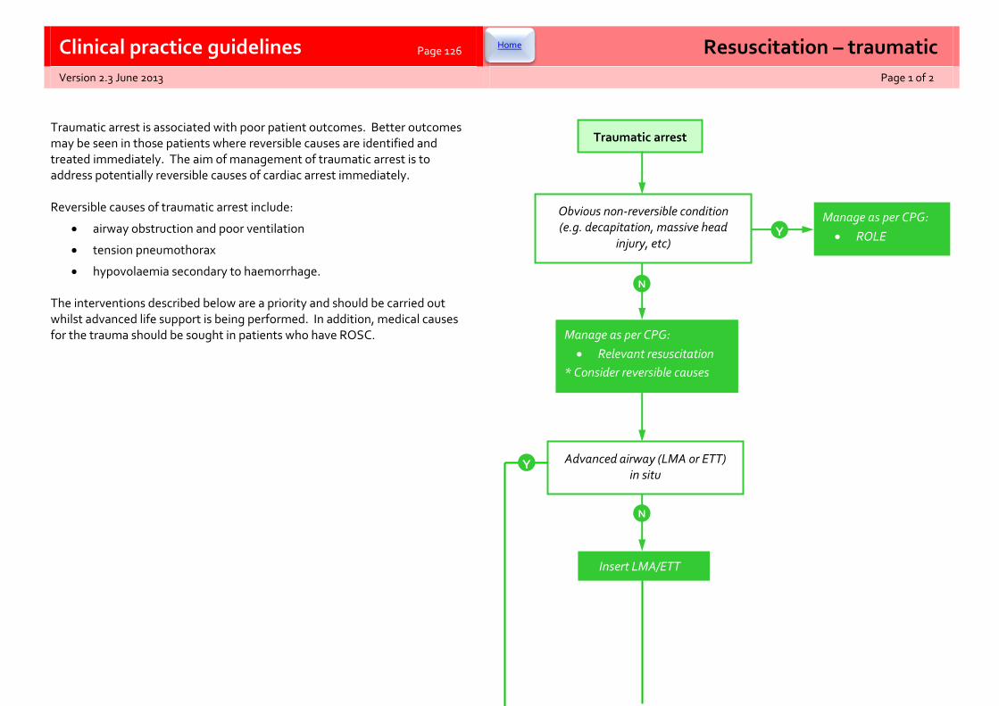
Clinical practice guidelines Resuscitation – traumatic
Version 2.3 June 2013 Page 1 of 2
Page 126
Traumatic arrest is associated with poor patient outcomes. Better outcomes may be seen in those patients where reversible causes are identified and treated immediately. The aim of management of traumatic arrest is to address potentially reversible causes of cardiac arrest immediately. Reversible causes of traumatic arrest include:
airway obstruction and poor ventilation
tension pneumothorax
hypovolaemia secondary to haemorrhage. The interventions described below are a priority and should be carried out whilst advanced life support is being performed. In addition, medical causes for the trauma should be sought in patients who have ROSC.
Traumatic arrest
Obvious non-reversible condition (e.g. decapitation, massive head
injury, etc) Y
Manage as per CPG:
ROLE
N
Manage as per CPG:
Relevant resuscitation
* Consider reversible causes
Advanced airway (LMA or ETT) in situ
N
Insert LMA/ETT
Y
Home

Resusitation - traumatic – Page 2 of 2
Uncontrolled when printed Page 127
Contemporary literature review shows that nearly all survivors from patients with blunt traumatic cardiac arrest upon arrival of paramedics have airway or breathing abnormalities. Therefore airway and breathing interventions are a priority. Patients who have exsanguinated before paramedic arrival are not able to be resuscitated. Adult patients in traumatic cardiac arrest should not be transported from the scene if the airway management, chest decompression and IV access can be established on scene, unless return of spontaneous circulation is achieved. Paediatric patients will be transported on all occasions if resuscitation has been initiated, unless exceptional circumstances are present. All patients who are not trapped and suffer cardiac arrest in the presence of paramedics should be transported immediately once airway and breathing interventions are performed. Massive blood loss in this situation may be amenable to surgical intervention at the receiving hospital. Patients with penetrating traumatic arrest should be immediately transported from the scene if the patient has suffered cardiac arrest < 10 minutes before paramedics arrived, or had vital signs upon arrival of paramedics.
ROSC achieved?
N
Y
Bilateral chest decompression
ROSC achieved?
N
IV fluid (20 mL/kg up to 2 L)
ROSC achieved?
N
Continue resuscitation for:
20 min total CPR OR
1 cycle after completion of above measures (whichever is longer)
Consider:
ROLE
Y
IV fluid
Consider
Whole blood
Manage as per CPGs:
Sedation
ROSC
Y
Note: Officers are only to perform procedures for which they have received specific training and authorisation by SJANT.
Clinical practice guidelines Return of Spontaneous Circulation management
Version 2.3 June 2013 Page 1 of 2
Page 128
There is increasing recognition that systematic post-cardiac arrest care can improve the likelihood of patient survival with a good quality of life. Resuscitation continues after a Return of Spontaneous Circulation (ROSC)
Primary aims of after initial resuscitation care include:
- support circulation, airway and breathing
- maintain cerebral perfusion
- manage cardiac dysrrhythmias.
Determine and manage the cause of the cardiac arrest giving consideration to those that are reversible:
- hypoxia
- hypo/hyperthermia
- hypovolaemia
- hypo/hyperkalaemia
- hydrogen ion (acidosis)
- tension pneumothorax
- tamponade
- toxins
- thrombemboli
Administration of prophylactic antiarrhythmics is not recommended.
All ROSC patients require a 12-Lead ECG.
Following out of hospital cardiac arrest (suspected to be of cardiac aetiology) patients should, where possible, be transported to a pPCI capable facility.
Clinical Features
Patient presents with signs of life post cardiac arrest
Pulse confidently detected within 10 seconds.
Risk Assessment
Not applicable
Home

Return of Spontaneous Circulation management – Page 2 of 2
Uncontrolled when printed Page 129
ECG 3-Lead / 12-Lead
Treat presenting dysrrhythmias
Consider and manage reversible causes
Note: Officers are only to perform procedures for which they have received specific training and authorisation by SJANT.
Standard Cares
Optimise ventilation and oxygenation
Maintain SpO2 94%
Consider advanced airway
Maintain EtCO2 of 35 – 40 mmHg
If no EtCO2 ventilate at rate of 8-12 per minute
Do not hyperventilate
Optimise circulation
Aim for SBP 100 mmHg for adults
Aim for SBP 80 mmHg for children
Consider
Appropriate posturing
Adrenaline
Transport to hospital Pre-notify as appropriate

Page 130
Page intentionally blank

Clinical Practice Guidelines - Toxicology
Uncontrolled when printed Page 131
08 -Toxicology
01 -Alcohol toxidromes 13 -Paraquat
02 -Anticholinergic toxidromes 14 -Psychostimulant emergencies
03 -Benzodiazepines 15 -Seratonin toxicity
04 -Beta blocker toxicity 16 -Snake bite
05 -Calcium channel blocker toxicity 17 -Spider bite
06 -Carbon monoxide 18 -Sympathomimetic toxicity
07 -Corrosives 19 -The poisoned patient
08 -Cyanide 20 -Toxic metals
09 -Gamma-hydroxbutyrate 21 -Tricyclic antidepressant toxicity
10 -Marine envenomation
11 -Opioid toxicity
12 -Organophosphate/cholinergic
Home

Clinical practice guidelines Alcohol toxidrome
Version 2.3 June 2013 Page 1 of 2
Page 132
Approximately half of all Australian adults drink alcohol at levels that put them at higher risk of short-term harm, with about a quarter risking their long-term health from alcohol consumption. Alcohol depresses the brain’s arousal, motor and sensory centres by acting as a GABA receptor agonist, in addition to antagonising NMDA receptors. Among other effects, this impacts coordination, speech, cognition and conscious level, with acute toxicity leading to:
injuries and fractures from falls
disability or death from TRCs
injuries from assaults and rape
STDs and unplanned pregnancies
airway compromise and aspiration risk. Furthermore, prolonged alcohol abuse can have chronic effects, such as
cardiovascular disease
cancers (oral cavity, larynx, GI, liver and breast)
diabetes
nutrition-related conditions
obesity
risks to unborn babies
liver disease
mental health conditions. Alcohol is an addictive drug that can lead to dependence. However, patients attempting to cease their consumption can also suffer alcohol withdrawal syndrome as a result of the CNS having adapted to the prolonged presence of alcohol. The resultant neurophysiologic changes within the brain predispose such patients to potentially lethal seizures and delirium tremens.
Clinical Features
Acute alcohol toxicity may involve:
loss of balance
loss of inhibition and behavioural changes
aggression
nausea and vomiting
dehydration
respiratory depression
loss of consciousness
Signs and symptoms of chronic alcohol toxicity will depend on the related diseases.
Risk Assessment
Other than inducing aggression, alcohol affects the ability to resolve conflict or explain oneself verbally, with intoxicated patients often having to express themselves physically, or violently.
Apparent drunken behaviour can result from causes other than alcohol, such as hypoglycaemia or head injuries. A thorough patient assessment is especially important to rule out other causes.
Other drugs are often taken in conjunction with alcohol and their potential effects must be taken into account.
Excessive alcohol consumption is a major risk factor for self-harm and suicidal behaviour.
Home

Alcohol toxidrome – Page 2 of 2
Uncontrolled when printed Page 133
Signs of life?
Y
N
Consider:
Verbal de-escalation
NTPOL assistance
Physical restraint
SDO
Manage as per CPG:
Sedation
Treat injuries and, if possible, lie patient in recovery position.
Consider
Oxygen
IPPV
IV access
Antiemetic
IV fluid
Note: Officers are only to perform procedures for which they have received specific training and authorisation by SJANT.
Transport to hospital
Pre-notify as appropriate
Standard Cares
N Manage as per CPG:
Relevant resuscitation
Ongoing imminent risk of harm? Y
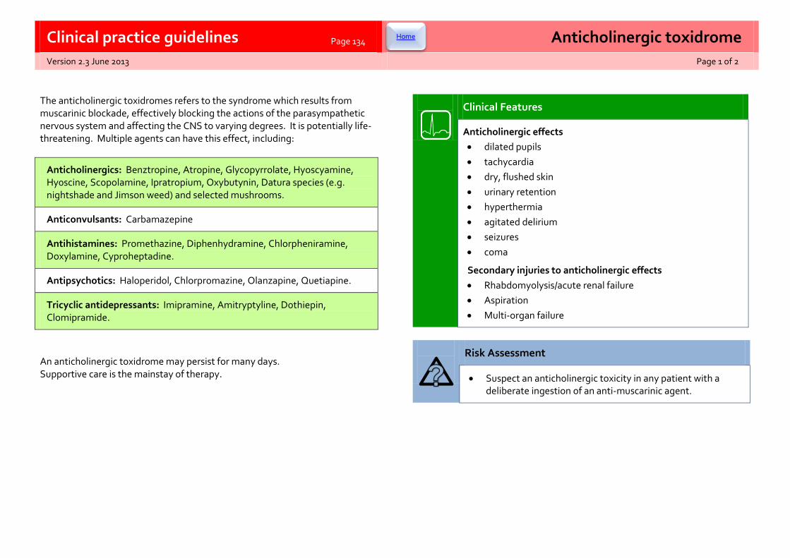
Clinical practice guidelines Anticholinergic toxidrome
Version 2.3 June 2013 Page 1 of 2
Page 134
The anticholinergic toxidromes refers to the syndrome which results from muscarinic blockade, effectively blocking the actions of the parasympathetic nervous system and affecting the CNS to varying degrees. It is potentially life-threatening. Multiple agents can have this effect, including:
Anticholinergics: Benztropine, Atropine, Glycopyrrolate, Hyoscyamine, Hy0scine, Scopolamine, Ipratropium, Oxybutynin, Datura species (e.g. nightshade and Jimson weed) and selected mushrooms.
Anticonvulsants: Carbamazepine
Antihistamines: Promethazine, Diphenhydramine, Chlorpheniramine, Doxylamine, Cyproheptadine.
Antipsychotics: Haloperidol, Chlorpromazine, Olanzapine, Quetiapine.
Tricyclic antidepressants: Imipramine, Amitryptyline, Dothiepin, Clomipramide.
An anticholinergic toxidrome may persist for many days. Supportive care is the mainstay of therapy.
Clinical Features
Anticholinergic effects
dilated pupils
tachycardia
dry, flushed skin
urinary retention
hyperthermia
agitated delirium
seizures
coma
Secondary injuries to anticholinergic effects
Rhabdomyolysis/acute renal failure
Aspiration
Multi-organ failure
Risk Assessment
Suspect an anticholinergic toxicity in any patient with a deliberate ingestion of an anti-muscarinic agent.
Home

Anticholinergic toxidrome – Page 2 of 2
Uncontrolled when printed Page 135
Consider
Oxygen
IV access
IV fluid
Glucose
Cooling
Midazolam
Transport to hospital
Pre-notify as appropriate
Note: Officers are only to perform procedures for which they have received specific training and authorisation by SJANT.
Standard Cares
Ongoing imminent risk of harm?
N
Y
Consider:
NTPOL assistance
Physical restraint
SDO
Manage as per CPG:
Sedation

Clinical practice guidelines Benzodiazepines
Version 2.3 June 2013 Page 1 of 2
Page 136
Benzodiazepines are a diverse group of sedative hypnotic agents, which act by potentiating the effects of the inhibitory neurotransmitter GABA within the CNS. They are one of the most common drugs used in self-poisoning, with the patient usually overdosing on their own prescribed medication. Examples include:
Alprazolam
Clonazepam
Diazepam
Midazolam
Nitrazepam
Oxazepam
Temazepam
Clinical Features
Symptoms usually manifest within two hours
- ataxia
- drowsiness
- slurred speech
- decreased level of consciousness
Hypotension, bradycardia and hypothermia are possible with very large ingestions.
Risk Assessment
For isolated benzodiazepine overdoses that receive supportive management, a full recovery is expected in the majority of patients. However, when combined with other agents, benzodiazepines are involved in a large proportion of drug-related deaths.
Features suggesting higher risk of complications:
- elderly
- cardio respiratory comorbidity
- co-ingestion of other CNS depressants
Additional information
Profound coma suggests a co-ingested agent.
Home

Benzodiazepines – Page 2 of 2
Uncontrolled when printed Page 137
Ongoing imminent risk of harm?
Consider:
Oxygen
IPPV
IV access
IV fluid
Glucose
Y
Transport to hospital
Pre-notify as appropriate
Note: Officers are only to perform procedures for which they have received specific training and authorisation by SJANT.
Standard Cares
Consider:
Verbal de-escalation
NTPOL assistance
Physical restraint
SDO
Manage as per CPG:
Sedation
N

Clinical practice guidelines Beta blocker toxicity
Version 2.3 June 2013 Page 1 of 2
Page 138
Beta blockers act as antagonists at the beta adrenergic receptors and are prescribed widely for the management of cardiac dysrrhythmias, hypertension and following myocardial infarction. Unfortunately, toxicity from accidental or intentional overdose is not uncommon and the subsequent bradycardia is associated with significant mortality. Beta receptors work by influencing myocardial calcium channels and therefore their blockade is similar to calcium channel blocker toxicity. In significant overdoses, both atropine and transcutaneous pacing may have little effect on blood pressure due to the lack of intracellular calcium necessary for effective contractions. Examples:
Propranolol
Sotalol
Atenolol
Bisoprolol
Carvedilol
Metoprolol
Clinical Features
Clinical features typically manifest within four hours following ingestion.
Cardiovascular effects - bradycardia - heart block - hypotension - cardiogenic shock
Systemic effects - hypoglycaemia/hyperglycaemia - hyperkalaemia - bronchospasm - seizures - coma
Risk Assessment
Beta blocker toxicity is potentially life threatening.
High risk populations: - underlying cardio respiratory disease
- elderly
- co-ingestion with calcium channel blockers or digoxin
Additional information:
Propranolol behaves like a TCA in overdose and should be managed in accordance with tricyclic antidepressant CPG.
Sotalol blocks K+ channels leading to QT prolongation and Torsades de Pointes.
Home

Beta blocker toxicity – Page 2 of 2
Uncontrolled when printed Page 139
Y
Standard Cares
Note: Officers are only to perform procedures for which they have received specific training and authorisation by SJANT.
N
Ongoing imminent risk of harm? Consider:
Verbal de-escalation
NTPOL assistance
Physical restraint
SDO
Manage as per CPG:
Sedation
Consider:
Oxygen
IV access
ECG 3-Lead / 12-Lead
Midazolam
Bradycardia or hypotension?
N
ECG changes?
N
Transport to hospital
Pre-notify as appropriate
Consider:
Sodium bicarbonate 8.4% (QRS widening)
Magnesium sulphate (Torsades de Pointes)
Y
Consider:
IV fluid
Atropine
Transcutaneous pacing
Adrenaline
Glucagon
Y

Clinical practice guidelines Calcium channel blocker toxicity
Version 2.3 June 2013 Page 1 of 2
Page 140
Calcium channel blockers (CCBs) are commonly prescribed medications used in the treatment of hypertension, angina pectoris and cardiac arrhythmia. Examples:
Verapamil
Diltiazem
Amlodipine
Nifedipine
Felodipine
Lercanidipine
Nimodipine Calcium gluconate 10% is the antidote for CCB toxicity.
Clinical Features
Cardiovascular effects that predominate and may be irreversible:
bradycardia
heart block
hypotension
myocardial ischaemia
cardiogenic shock
Systemic effects:
seizures
coma
hyperglycaemia
metabolic acidosis
Risk Assessment
CCB toxicity is potentially life threatening, particularly verapamil and diltiazem which are the more cardioselective CCBs.
High risk populations:
underlying cardio respiratory disease
elderly
co-ingestion with beta blockers or digoxin
Additional information:
The onset of clinical features may be delayed up to 16 hours following ingestion of slow release preparations.
Home

Calcium channel blocker toxicity – Page 2 of 2
Uncontrolled when printed Page 141
Ongoing imminent risk of harm?
N
Y
Transport to hospital
Pre-notify as appropriate
Consider:
Oxygen
IV access
ECG 3-Lead / 12-Lead
Midazolam
Note: Officers are only to perform procedures for which they have received specific training and authorisation by SJANT.
Standard Cares
Consider:
Verbal de-escalation
NTPOL assistance
Physical restraint
SDO
Manage as per CPG:
Sedation
Bradycardia or hypotension? Y Consider:
IV fluid
Calcium gluconate 10%
Atropine
Transcutaneous pacing
Adrenaline
N
ECG changes? Y
Consider:
Sodium bicarbonate 8.4% (QRS widening)
N

Clinical practice guidelines Carbon monoxide
Version 2.3 June 2013 Page 1 of 2
Page 142
Carbon monoxide (CO) is a colourless, odourless and tasteless gas produced by the incomplete combustion of hydrocarbon fuels e.g. contained fires, car exhaust, etc.
Compared to oxygen, CO has over 200 times the affinity for haemoglobin, which will bind preferentially to CO forming carboxyhaemoglobin, thereby decreasing the oxygen-carrying capacity of the blood. CO toxicity leads to hypoxia, but patients will not appear cyanosed, nor are they likely to have a cherry red colouration as this is rarely seen.
If there is any suspicion of CO poisoning, the highest concentration of O2 possible should be applied immediately and continued until the patient is assessed by the receiving facility.
In attending this type of incident, paramedics must maintain a high index of suspicion that the area is still contaminated with carbon monoxide and should not enter the scene until the area is declared safe by the NTFS.
Additional information
Oxygen saturation monitors are unable to differentiate between carboxyhaemoglobin and oxyhaemoglobin. Therefore saturation monitors cannot be relied on to guide a patient’s true blood oxygen saturation in the setting of CO toxicity.
Elimination of CO is determined by the oxygen tension in the blood. In room air, the elimination half life is 240 minutes; with 100% oxygen it is 90 minutes; and with 100% oxygen in a hyperbaric chamber, set at three atmospheres, it is 23 minutes.
CO poisoning in pregnancy is particularly dangerous. A foetus is more susceptible to injury as their haemoglobin binds CO more avidly.
Clinical Features
Neurological:
headache, nausea, confusion
ataxia, dizziness
seizures
coma
Cardiovascular:
tachycardia
hypotension
myocardial ischaemia
arrhythmia
Respiratory:
pulmonary oedema
Other:
rhabdomyolysis
metabolic acidosis
disseminated intravascular coagulation
Risk Assessment
Everyone is susceptible to carbon monoxide poisoning, however the following are at an even greater risk, such as:
pregnant women and their unborn children
infants
people who smoke
geriatrics
people with chronic heart disease, respiratory disease or anaemia.
Home

Carbon monoxide – Page 2 of 2
Uncontrolled when printed Page 143
Y
Transport to hospital
Pre-notify as appropriate
Consider:
Oxygen
IPPV
IV access
IV fluid
Adrenaline
Note: Officers are only to perform procedures for which they have received specific training and authorisation by SJANT.
Standard Cares
Consider:
Verbal de-escalation
NTPOL assistance
Physical restraint
SDO
Manage as per CPG:
Sedation
Consider safety issues and remove patient from exposure
Ongoing imminent risk of harm?
N

Clinical practice guidelines Corrosives
Version 2.3 June 2013 Page 1 of 2
Page 144
Corrosive agents cause direct injury to the respiratory and gastrointestinal tract if ingested. Resultant airway compromise can be lethal.
Corrosive agents:
Acids: hydrochloric acid, sulphuric acid, hydrofluoric acid.
Alkali: sodium hydroxide, potassium hydroxide, ammonia, sodium hypochlorite
Other: zinc chloride, mercuric chloride, glyphosate, phenols, potassium permanganate, button batteries.
Clinical Features
Respiratory toxicity:
hoarse voice
dyspnoea
stridor
Gastrointestinal toxicity:
oral burns
drooling
painful swallowing
vomiting
abdominal pain
Risk Assessment
High risk features for airway compromise:
hoarse voice
dyspnoea
stridor
Additional information
The absence of oral burns does not exclude significant gastrointestinal injury.
Do not place a NGT in the pre-hospital setting given the potential for gastrointestinal mucosal injury.
Hydrofluoric acid exposure is life-threatening, even after seemingly minor quantities
o fluoride binds calcium and magnesium, precipitating intractable ventricular arrhythmias and death.
o follow first-aid instructions provided at industrial sites.
o transport without delay and pre-notify.
Home

Corrosives – Page 2 of 2
Uncontrolled when printed Page 145
Standard Cares
Consider safety issues and decontamination
Rinse patient mouth with water
Do not encourage vomiting
Consider:
Oxygen
IPPV
IV access
Analgesia
Antiemetic
IV fluid
SDO
Transport to hospital
Pre-notify as appropriate
High risk features:
stridor
dyspnoea
hoarse voice
hydrofluoric acid exposure/ingestion
Note: Officers are only to perform procedures for which they have received specific training and authorisation by SJANT.

Clinical practice guidelines Cyanide
Version 2.3 June 2013 Page 1 of 2
Page 146
Cyanide toxicity can be lethal. It acts to inhibit normal cellular metabolism. Cyanide compounds are used in industry for multiple purposes. Cyanide is also a product of combustion of natural and synthetic materials and cyanide toxicity is seen in victims trapped in an enclosed fire. Additional information
Industrial sources of cyanide include:
- fumigant in ships, buildings, aeroplanes
- metallurgy and ore extraction
- plastics manufacturing
Clinical Features
Acute inhalation leads to rapid onset of clinical features, whereas symptoms develop in 30 – 60 minutes following ingestion.
nausea and vomiting
headache
dyspnoea
hypertension
tachycardia
agitation
collapse
seizures
confusion
chest tightness
These symptoms can progress to hypotension, bradycardia, respiratory depression and coma.
Risk Assessment
Hydrogen cyanide poses the greatest risk as it occurs as a
colourless gas above 26C and has a faint smell of bitter almonds that can only be detected by certain people (due to a genetic trait).
It is possible for cyanide to be absorbed through intact skin, resulting in systemic toxicity. Therefore, precautions need to be taken to avoid self-contamination.
In cases of ingestion, any emesis should be isolated as it may emit hydrogen cyanide.
Home

Cyanide – Page 2 of 2
Uncontrolled when printed Page 147
Standard Cares
Shock, respiratory distress, seizures or coma? Y Manage as per appropriate CPG
N
Decontamination
Prevent ongoing exposure
Remove clothes and wash skin (with soap and water if available)
Other symptoms of toxicity?
headache, confusion, nausea
dyspnoea, chest tightness
Y
Consider:
Hydroxycobalamin
Oxygen
IV access
IV fluid
Antiemetic
Transport to hospital
Pre-notify as appropriate
Consider:
Oxygen
IV access
Antiemetic
N
Note: Officers are only to perform procedures for which they have received specific training and authorisation by SJANT.

Clinical practice guidelines Gamma-hydroxybutyrate
Version 2.3 June 2013 Page 1 of 2
Page 148
Gamma-hydroxybutyrate (GHB) is a recreational drug which is a colourless, odourless, bitter tasting liquid
Gamma-hydroxybutyrate
Fantasy
Grievous Bodily Harm
Liquid E
Easy Lay
Clinical Features
The duration of toxic effects is short, with recovery expected within six hours.
euphoria
vomiting
sweating
miosis
respiratory depression
coma
bradycardia
In toxic doses there is typically a rapid onset of coma. The patient may be rousable with stimulus only to return to deep unconsciousness once undisturbed. Sudden recovery in approximately two hours is typical.
Risk Assessment
In overdose, GHB can be lethal secondary to respiratory compromise.
Additional information
Myoclonic jerking movements are not uncommon and can mimic seizure.
Coma longer than six hours duration suggests an alternate diagnosis.
Home

Gamma-hydroxybutyrate – Page 2 of 2
Uncontrolled when printed Page 149
Standard Cares
ALOC?
Consider:
Oxygen
IPPV
IV access
IV fluid
Antiemetic
Transport to hospital
Pre-notify as appropriate
Note: Officers are only to perform procedures for which they have received specific training and authorisation by SJANT.

Clinical practice guidelines Marine envenomations
Version 2.3 June 2013 Page 1 of 2
Page 150
Marine stingers:
Box jellyfish (Chironex fleckeri) – antivenom available
Irukandji syndrome (Carukia barnesi)
Blue-bottle (Physalia) Fish:
Stonefish – antivenom available
Puffer fish (Fugu) Molluscs:
Cone shells
Blue-ringed octopus Sea snake – see snake bite CPG
Clinical Features
Box jellyfish:
severe localised pain, increasing when tentacles are wiped off
development of welts
possible dyspnoea, difficulty speaking, hypotension, arrhythmia, ALOC and cardio-respiratory arrest
Irukandji syndrome:
delayed sting and systemic symptoms
massive catecholamine release leads to severe pain, agitation and possible cardiogenic shock
Blue bottles and other stingers:
immediate or delayed pain, welts, nausea and vomiting, sweating, muscle cramps and/or headache.
Clinical Features (continued)
Stonefish:
open wound from spine entry with immediate intense localised pain
venom can cause muscle weakness, paralysis and shock.
Puffer fish:
weakness, numbness, dyspnoea, paralysis and cardio-respiratory arrest
symptoms can be delayed up to 20 hours
Ciguatoxin from tropical fish ingestion:
GIT upset (vomiting, diarrhoea and abdominal pain; neurological symptoms (hot/cold sensation reversal, tingling lips, hands and feet, severe itching)
severe cases progress to paralysis, respiratory cardio-respiratory arrest, bradycardia.
Cone shell:
pain at ingestion site
numbness, weakness, unco-ordination, visual and speech disturbance, respiratory depression.
Blue-ringed octopus:
painless bite, but weakness and numbness within 10 minutes, with dyspnoea, nausea and vomiting
severe cases progress to flaccid paralysis and respiratory arrest.
Home

Marine envenomations – Page 2 of 2
Uncontrolled when printed Page 151
Risk Assessment
Children are at greater risk from marine envenomations due to their smaller body mass and inquisitive nature.
Tetanus and serious infections may occur following marine envenomations.
Box jellyfish envenomations are rare but potentially fatal. If envenomation is fatal, death will occur within 5 – 20 minutes of the sting.
Jellyfish tentacles can still sting even if the jellyfish is no longer present.
Spines on fish barbs can cause injury during attempted removal.
Stonefish venom is released depending on pressure exerted and the number of spines involved.
Additional information
Antivenom is available for:
- box jellyfish
- stonefish
- sea snakes.
There is limited evidence for antivenom use in box jellyfish.
Hot water can assist with the pain from stonefish and blue bottle stings.
Vinegar is used on irukandji and box jellyfish.
Ice may relieve box jellyfish pain.
Pressure immobilisation bandages may be applied in blue-ringed octopus bites.
Standard Cares
Shock or cardio-respiratory arrest?
N
Suspected box jellyfish or irukandji envenomation?
N
Suspected stonefish or blue bottle envenomation?
Suspected sea snake envenomation?
N
Transport to hospital
Pre-notify as appropriate
Consider:
Box jellyfish antivenom
Manage as per CPG:
Resuscitation
Y
Copious flushing with vinegar
Remove tentacles from skin
Analgesia and ice
Consider:
Magnesium sulphate
GTN
Box jellyfish antivenom
Y
Remove spine
Remove tentacles and flush with sea water
Apply heat as tolerated (avoid burn)
Analgesia
Y
Y Manage as per CPG:
Snake bite
Note: Officers are only to perform procedures for which they have received specific training and authorisation by SJANT.

Clinical practice guidelines Opioid toxicity
Version 2.3 June 2013 Page 1 of 2
Page 152
Opioid analgesics are widely available, including over the counter agents. These agents may be fatal in children even in small doses. Opioids:
Heroin
Morphine
Fentanyl
Pethidine
Oxycodone
Codeine
Dextropropoxyphene
Clinical Features
Opioid toxidrome
pin point pupils
sedation
respiratory depression
Complications of prolonged periods of deep sedation
hypothermia, skin necrosis, compartment syndrome
Risk Assessment
Opioid intoxication can be fatal with respiratory depression or airway obstruction, especially in children.
Supportive care (including ventilation) is often all that is required.
Naloxone may be used in severe respiratory depression.
Additional information
The half life of naloxone is approximately 1.5 hours which is shorter than the half life of most opioids available. A rebound effect with relapsing opioid toxicity can occur after the administration of a single dose of naloxone, with subsequent respiratory depression.
Home

Opioid toxicity – Page 2 of 2
Uncontrolled when printed Page 153
Standard Cares
Respiratory compromise? Y
Consider:
IPPV
Oxygen
Naloxone
N
Transport to hospital
Pre-notify as appropriate
Note: Officers are only to perform procedures for which they have received specific training and authorisation by SJANT.

Clinical practice guidelines Organophosphate/cholinergic syndrome
Version 2.3 June 2013 Page 1 of 2
Page 154
Organophosphates are pesticides that inhibit acetylcholinesterase enzymes, increasing the action of the neurotransmitter acetylcholine at parasympathetic and presynaptic sympathetic ganglion receptors. Acetylcholine excess leads to cholinergic syndrome, and may be fatal.
Carbamates are similar to organophosphates in toxicity but the clinical features are typically less severe.
Organophosphates: chlorpyrifos, coumaphos, diazinon, dichlorvos, dimethoate, fenthion, malathion, parathion, trichlorfon
Carbamates: aldicarb, carbendazole, carbendazim, carbazine
Nerve agents: Sarin, Tabun, Soman
Pharmaceuticals: neostigmine, pyridostigmine, edrophonium, pilocarpine, bethanechol, carbachol, acetylcholine
Clinical Features
Clinical features are of cholinergic excess. The onset can be delayed up to 12 hours with some agents.
Muscarinic excess ‘DUMBBELS’
diarrhoea
urination
miosis (constricted pupils)
bronchorrhoea
bronchospasm
emesis
lacrimation
salivation
bradycardia
hypotension
Clinical Features
Nicotinic excess
fasciculations
tremor
muscle weakness
respiratory muscle paralysis
Central effects
agitation
seizures
coma
Risk Assessment
Deliberate self poisoning can be life-threatening
Additional information
Atropine is used to block the muscarinic affects of acetylcholine.
In life threatening toxicity, large cumulative doses of atropine may be required.
A chemical pneumonitis can develop if the hydrocarbon solvent is aspirated.
Universal precautions are sufficient to prevent contamination of others.
Inhalation or dermal exposure is rarely life threatening. The smell of agent does not indicate exposure as it is usually the solvent and poses no toxicity risk to paramedics or bystanders.
Home

Organophosphate/cholinergic syndrome – Page 2 of 2
Uncontrolled when printed Page 155
Note: Officers are only to perform procedures for which they have received specific training and authorisation by SJANT.
Transport to hospital
Pre-notify as appropriate
Standard Cares
Decontamination
Remove clothes and wash skin with soap and water
Evidence of serious toxicity?
hypotension
bronchospasm
paralysis
seizure
coma
Y
Consider:
Atropine
Oxygen
IPPV
IV access
IV fluid
N
Consider:
Oxygen
IV access
Antiemetic

Clinical practice guidelines Paraquat
Version 2.3 June 2013 Page 1 of 2
Page 156
Paraquat is a caustic herbicide. It is usually lethal in overdose. Early haemodialysis to facilitate paraquat elimination may be life saving, but this is the exception.
Clinical Features
Immediate:
gastrointestinal upset
Early (hours):
oral corrosive injury, metabolic acidosis with large ingestions
Delayed (24-48 hours):
progression of acidosis, multi-organ failure.
Late (> 48 hours):
pulmonary fibrosis
Risk Assessment
Deliberate ingestions of more than a mouthful is typically fatal.
Dermal and inhalational exposures are generally insignificant.
Additional information
In children, an ingestion of as little as a teaspoon can be fatal.
Patients with paraquat poisoning may be harmed by supplemental oxygen. Avoid oxygen unless patient is hypoxaemic – target SpO2 88 – 92%.
Home

Paraquat – Page 2 of 2
Uncontrolled when printed Page 157
Standard Cares
Decontamination
Remove clothes and wash skin if topical exposure
Consider:
Oxygen
IPPV
Analgesia
Antiemetic
Transport to hospital
Pre-notify as appropriate
Note: Officers are only to perform procedures for which they have received specific training and authorisation by SJANT.

Clinical practice guidelines Psychostimulant emergencies
Version 2.3 June 2013 Page 1 of 2
Page 158
Psychostimulants are a group of drugs that stimulate the activity of the CNS, with a variety of therapeutic applications. However, they are best known as drugs of abuse, with amphetamines being the second most commonly used illicit drug in Australia after cannabis. They are usually taken intranasally (snorting) or orally (bombing), with IV use usually suggesting a higher level of dependence and being associated with a greater potential for toxicity. In toxic levels they can produce severe agitation and psychotic behaviour, but over-stimulation of the sympathetic nervous system causes serious complications such as myocardial ischaemia, severe hypertension, hyperthermia, coagulopathy and rhabdomyolysis. Psychostimulant medications:
Dexamphetamine - used to treat ADHD and narcolepsy.
Methylphenidate hydrochloride (e.g. Ritalin) - used to treat ADHD.
Diethylpropion hydrochloride and phentermine - appetite suppressants.
Illicit psychostimulants:
Amphetamine and methamphetamine
Cocaine
MDMA (methylenedioxymethamphetamine)
Clinical Features
Clinical feature of acute toxicity:
restlessness, agitation, rapid speech, hyper-vigilance, paranoia
motor agitation or pacing
tachycardia, hypertension, hyperthermia
other features of toxicity will reflect underlying processes (e.g. CVA, AMI, rhabdomyolysis etc).
the usual assessment should take place.
Early (hours):
malnutrition
sores on skin (from delirium and hallucinations of bugs on skin)
evidence of IVDU (e.g. needle marks or thrombophlebitis)
Home
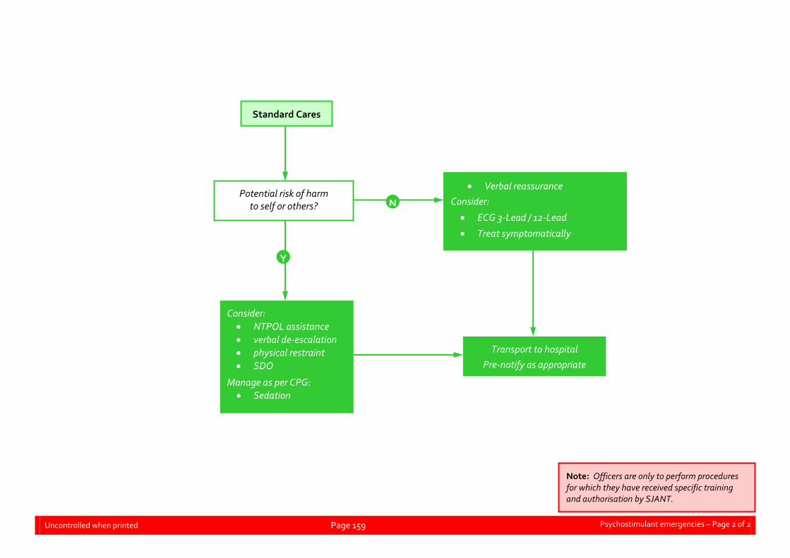
Psychostimulant emergencies – Page 2 of 2
Uncontrolled when printed Page 159
Standard Cares
Potential risk of harm to self or others?
Y
N
Verbal reassurance
Consider:
ECG 3-Lead / 12-Lead
Treat symptomatically
Consider:
NTPOL assistance
verbal de-escalation
physical restraint
SDO
Manage as per CPG:
Sedation
Transport to hospital
Pre-notify as appropriate
Note: Officers are only to perform procedures for which they have received specific training and authorisation by SJANT.

Clinical practice guidelines Serotonin toxicity
Version 2.3 June 2013 Page 1 of 2
Page 160
Serotonin toxicity or syndrome is due to excessive serotonergic neurotransmission. It covers a spectrum from mild self-limiting to severe, potentially life threatening toxicity. It is caused by agents which increase the release, or reduce the uptake or metabolism of serotonin.
Agents associated with serotonin toxicity
Antidepressants/antiepileptic:
- selective serotonin re-uptake inhibitor (SSRI)
- selective noradrenaline re-uptake inhibitor (SNRI) antidepressants
- monoamine oxidase inhibitor (MAOI)
- TCA
- lithium
- sodium valproate
Analgesics:
- tramadol
- fentanyl
- pethidine
Antiemetics:
- metoclopramide
- ondansetron
Clinical Features
Neurological dysfunction
anxiety, agitation
delirium
seizure
coma
Autonomic dysfunction
flushed skin, dilated pupils, diaphoresis
tachycardia, hypertension
hyperthermia
diarrhoea
Neuromuscular dysfunction
tremor
clonus, increased muscle tone
hyperreflexia
Risk Assessment
Serotonin toxicity needs to be considered in cases of deliberate ingestion of multiple serotonergic agents.
Home

Serotonin toxicity – Page 2 of 2
Uncontrolled when printed Page 161
Additional information
SSRI antidepressants include
- citalopram
- escitalopram
- fluvoxamine
- fluoxetine
- paroxetine
- sertraline
SNRI antidepressants include:
- venlafaxine
- duloxetine
MAOI antidepressants include:
- moclobemide
- phenelzine
- selegiline
- tranylcypromie
Citalopram and escitalopram can also prolong the QT interval in overdose.
Standard Cares
Consider:
ECG 3-Lead / 12-Lead
Oxygen
IV access
Midazolam (seizures or severe agitation)
IPPV
Treatment of underlying toxicity (e.g. TCA)
SDO
Transport to hospital
Pre-notify as appropriate
Note: Officers are only to perform procedures for which they have received specific training and authorisation by SJANT.

Clinical practice guidelines Snake bite
Version 2.3 June 2013 Page 1 of 2
Page 162
Australia has some of the most venomous snakes in the world including brown, tiger, black, taipan and death adder snakes. All of these snakes are found in the Northern Territory, in addition to exotic snake species found in zoos. All patients with a history of possible snakebite should be transported to hospital for ongoing assessment and management as life-threatening effects may take from minutes to hours to present. If there is any doubt as to the validity of the bite, treat it as a snake bite.
Clinical Features
Non-specific symptoms present in most cases: - nausea, vomiting, headache, abdominal pain, diarrhoea
and diaphoresis.
Other signs or symptoms that may present: - altered mental status, visual disturbances and seizures - respiratory dysfunction - hypotension, haemorrhage or haematoma at site - paired or single fang marks or scratch marks and
swollen or tender glands of affected limb.
Systemic effects (depending on particular venom):
- Neurotoxicity - drooping of eyelids/drooling are early signs - paralysis (monitor airway patency/protection and
ventilatory rate, rhythm and effort)
- Coagulopathy - bleeding from nose and gums common, major
haemorrhage uncommon
- Myotoxicity - damage to skeletal muscles (muscular pain,
tenderness and weakness)
- Renal impairment/failure
Risk Assessment
Do not attempt to capture or injure the snake.
Most Australian snakebites are associated with minimal local pain and bite marks can be easily missed.
Home

Snake bite – Page 2 of 2
Uncontrolled when printed Page 163
Standard Cares
Arrest, shock or respiratory distress?
N
Y
Manage as per CPG:
Resuscitation
Shock
Consider
Pressure immobilisation bandage
Restrict movement of patient, do not let them walk
Do not wash the bite site
Cover the bite site with a dressing.
If limb involved:
Apply pressure immobilisation bandage and mark bite site
Splint limb
If bite site is on torso/head
Completely immobilise patient
Transport to hospital
Pre-notify as appropriate
Note: Officers are only to perform procedures for which they have received specific training and authorisation by SJANT.

Clinical practice guidelines Spider bite
Version 2.3 June 2013 Page 1 of 2
Page 164
Spider bites are common and fall into three categories:
Large black OR funnel-web spider
- Funnel-web spider bites are potentially lethal.
- All large black spider bites, are to be managed as if they were a funnel-web spider bite.
Redback spider
- Redback spider bites are common, but rarely life-threatening.
- Clinical features can be distressing for the patient and refractory to management.
All other spiders
- These are rarely of clinical significance.
Clinical Features
Large black OR Funnel-web spider
Localised severe pain at bite site.
Puncture/fang marks are often visible.
Signs and symptoms suggesting severe systemic envenomation:
- general: agitation, nausea, vomiting, headache
- neurological: muscular spasm, numbness/tingling in and around the mouth and altered mental status.
- autonomic: diaphoretic, salivation, goose bumps, lacrimation.
- cardiovascular: hypertension or hypotension, tachycardia or bradycardia, and pulmonary oedema.
Clinical Features
The first indication that a young child has been bitten may be sudden severe illness with inconsolable crying, salivation, vomiting, ALOC, or collapse.
Redback spider
Local and regional signs and symptoms:
- bites are not initially painful
- increasing pain
- pain radiating from the bite site to the proximal limb, trunk or local lymph nodes
- localised or regional diaphoresis
- less common: goose bumps, localised redness and puncture/fang marks.
Systemic signs and symptoms:
- nausea, vomiting and headache
- malaise and lethargy
- potentially generalised pain
- less common: hypertension, irritability and agitation (more common in paediatrics), fever, paraesthesia, muscle spasms, priapism.
All other spiders
Pain
Minor non-specific signs and symptoms
Risk Assessment
Do not attempt to capture or injure the spider.
Home
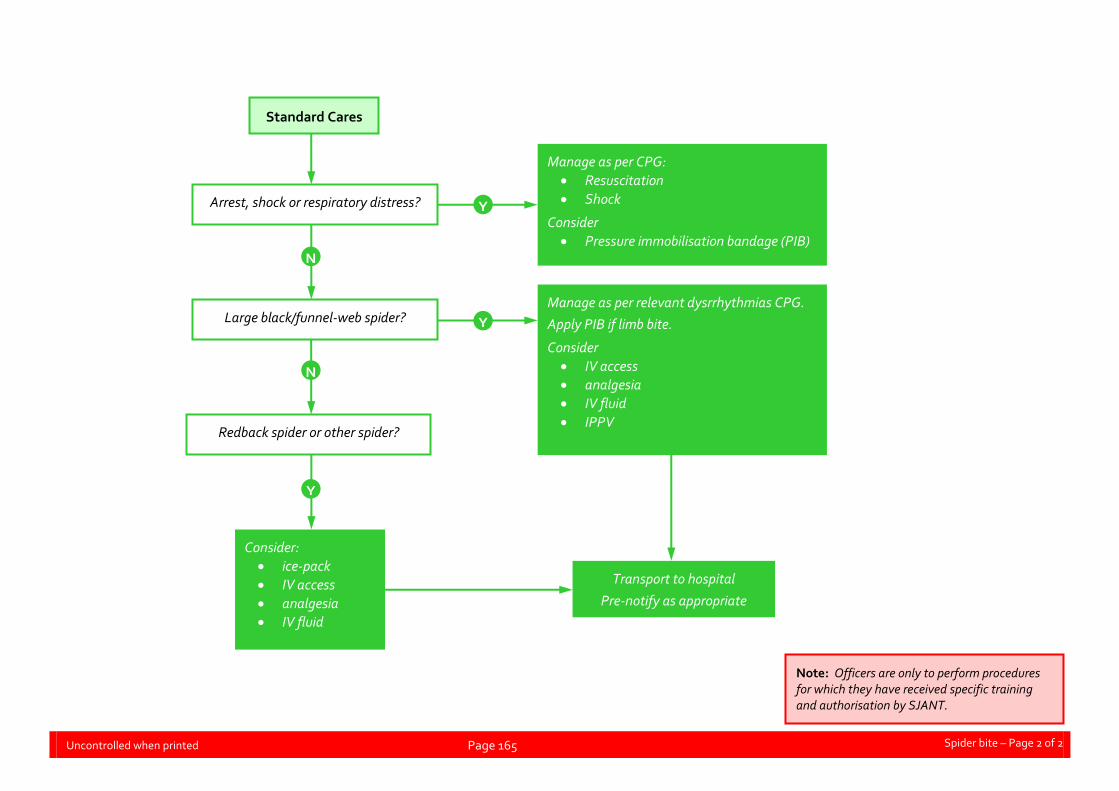
Spider bite – Page 2 of 2
Uncontrolled when printed Page 165
Standard Cares
Arrest, shock or respiratory distress? Y
N
Large black/funnel-web spider?
N
Y
Redback spider or other spider?
Y
Consider:
ice-pack
IV access
analgesia
IV fluid
Manage as per CPG:
Resuscitation
Shock
Consider
Pressure immobilisation bandage (PIB)
Manage as per relevant dysrrhythmias CPG.
Apply PIB if limb bite.
Consider
IV access
analgesia
IV fluid
IPPV
Transport to hospital
Pre-notify as appropriate
Note: Officers are only to perform procedures for which they have received specific training and authorisation by SJANT.

Clinical practice guidelines Sympathomimetic toxicity
Version 2.3 June 2013 Page 1 of 2
Page 166
Sympathomimetic agents act through activating the sympathetic nervous system. In overdose, the sympathomimetic toxidromes can be life-threatening. Sympathomimetic agents:
Dexamphetamine
Methylphenidate hydrochloride
Diethylpropion hydrochloride
Phentermine
Illicit drugs: methamphetamine, MDMA, cocaine
Clinical Features
Sympathetic activation
agitation, dilated pupils, diaphoresis
tremor
tachycardia, hypertension
Cardiovascular
arrhythmia
acute coronary syndrome
aortic dissection
pulmonary oedema
Neurological
intracranial haemorrhage
seizures
Psychiatric
agitation
aggression
psychosis
Clinical Features (continued)
Other
hyperthermia
rhabdomyolysis
hyponatraemia
Risk Assessment
Small doses in some patients can result in significant toxicity.
High risk features:
- headache
- chest pain
- hyperthermia
- focal neurological signs.
Additional information
Sympathetic hyperactivation is best treated with titrated benzodiazepines.
ACS in setting of sympathomimetic toxicity is treated with benzodiazepines, not thrombolysis.
Home

Sympathomimetic toxicity – Page 2 of 2
Uncontrolled when printed Page 167
Standard Cares
High risk features?
Headache, chest pain, hyperthermia, focal neurological signs?
N Y
Manage as per CPG:
ACS (do not give thrombolysis)
seizures
hyperthermia
Consider:
IV access
Midazolam for severe agitation
analgesia
Transport to hospital
Pre-notify as appropriate
Note: Officers are only to perform procedures for which they have received specific training and authorisation by SJANT.

Clinical practice guidelines The poisoned patient
Version 2.3 June 2013 Page 1 of 2
Page 168
Acute poisoning can be unintentional exposures or deliberate ingestions in response to suicidal ideation. These patients can be challenging to manage as heightened distress is often a feature.
Management
A structured approach to management is important in the poisoned patient. Paramedics must consider resuscitative requirements and supportive care in all patients. Decontamination, the delivery of an antidote or promoting elimination, are specific treatment options which may be appropriate, depending on the toxin involved.
Clinical Features
Signs and symptoms develop as a result of the toxin involved.
Classic constellations of clinical features or ‘toxidromes’ are associated with specific toxin ingestions and can guide further management
Toxidromes include:
Cholinergic syndrome Small pupils, sweating, salivation, bronchorrhoea, lacrimation, bradycardia, agitation, fasciculations.
Anticholinergic syndrome
Dilated pupils, fever, agitation, tachycardia, dry mouth, flushed skin
Opioid toxicity Pin point pupils, respiratory depression, sedation
Serotonin toxicity Dilated pupils, fever, agitation, increased tone
Sympathomimetic toxicity
Dilated pupils, fever, agitation, tachycardia, sweating, tremor, aggression
Risk Assessment
Predict the expected clinical course of the exposure by determining:
agent/s ingested
dose/s
timing of ingestion
any symptoms or signs which have developed
important patient factors (e.g. pre-existing coronary heart disease.
Gathering empty pill packets or gaining collateral history from friends and family may be required.
An Emergency Examination Order is necessary if the patient is deemed to be at imminent risk of harm to self or others.
Additional information
Resuscitation takes priority over decontamination.
Poisons Information Centre Hotline: 131 126.
Standard PPE is adequate for the majority of toxic exposures.
The poisoned child is approached similarly, but recognise that much smaller quantities can cause significant toxicity.
Small children rarely ingest more than three tablets or a mouthful of poison.
Some agents are harmful in even small ingestions.
Home

The poisoned patient – Page 2 of 2
Uncontrolled when printed Page 169
Potentially lethal paediatric ingestions
Two pills that kill:
amphetamines
calcium channel blockers
chloroquine/hydroxychloroquine
opioids/dextropropoxyphene
propranolol
sulfonylureas (glibenclamide, gliclazide, glimepiride, glipizide)
theophylline
TCAs
Two mouthfuls that kill:
organophosphates
paraquat
hydrocarbons/solvents
camphor
naphthalene (mothballs)
lead
It is essential to consider child safety issues and to ensure parents or guardians are notified if a child has toxicity.
Standard Cares
Signs of life? Manage as per CPG:
Resuscitation
Y
N
If imminent risk of harm
Consider:
SDO
Specific management for known toxidromes/ingestion
Decontamination
Remove clothes and wash skin with soap and water
Rinse out mouth with water
Antidote
Administer as indicated
Consider:
Oxygen
IPPV
IV access
analgesic
antiemetic
Midazolam
ECG 3-Lead / 12-Lead
Transport to hospital
Pre-notify as appropriate Note: Officers are only to perform procedures for which they have received specific training and authorisation by SJANT.

Clinical practice guidelines Toxic metals
Version 2.3 June 2013 Page 1 of 2
Page 170
Toxic metals are a group of metallic elements that have no normal biological role, but are capable of disrupting essential physiological processes when introduced to the body. Other than radioactive metals, they include:
antimony
arsenic
barium
beryllium
cadmium
lead
mercury
osmium
thallium
vanadium Other essential trace elements can become toxic if they occur in high concentrations (e.g. iron), or are present in the wrong ionic form (e.g. chromium). Acute sources of toxicity are often easily identified, with ingestions leading to gastrointestinal irritation as well as systemic effects, dependant on the compound involved and the level of exposure. Chronic metal poisoning is less well defined, manifesting as general ill health, cancers, or teratogenic malformations. Lithium is used therapeutically as a mood stabiliser bit it is potentially toxic in overdose, particularly with chronic exposure. Other than supportive measures, treatment focuses on the prevention of further absorption, inactivation with chelating agents and elimination of the metal from the body.
Clinical Features
Local gastrointestinal effects:
vomiting, diarrhoea
abdominal pain
gastrointestinal haemorrhage
Systemic effects:
Multi-organ dysfunction/failure
- predominately neurological (lead, lithium, mercury)
- predominately hepatic (iron)
Risk Assessment
Risk of toxicity is proportional to dose ingested.
Very small quantities in children can cause significant toxicity.
Home

Toxic metals – Page 2 of 2
Uncontrolled when printed Page 171
Standard Cares
Immediate, life-threatening symptoms? Y
N
Manage as per relevant CPG
If imminent risk of harm
Consider:
SDO
Consider:
oxygen
IPPV
IV access
IV fluid
analgesic
antiemetic
midazolam
Transport to hospital
Pre-notify as appropriate
Note: Officers are only to perform procedures for which they have received specific training and authorisation by SJANT.

Clinical practice guidelines Tricyclic antidepressant toxicity
Version 2.3 June 2013 Page 1 of 2
Page 172
Tricyclic antidepressants (TCAs) are potentially lethal in overdose. TCA agents act on multiple receptor sites. Their principal antidepressant action is mediated by serotonin and noradrenaline re-uptake inhibition. Myocardial toxicity is via sodium channel blockade. Other toxicity is mediated by the inhibitory action at the muscarinic, histamine and adrenergic receptors. TCAs work by inhibiting serotonin and noradrenaline re-uptake in the CNS. They are potentially lethal in overdose, causing myocardial toxicity (via sodium channel blockade). They also affect muscarinic, histamine and adrenergic receptors, causing other toxic effects.
Additional information
QRS > 0.10 seconds predicts seizure.
QRS > 0.16 seconds predicts VT.
Defibrillation is unlikely to be successful in TCA overdose, due to sodium channel blockade.
Sodium bicarbonate 8.4% is the antidote for TCA toxicity and should be given in all cases with ECG changes, seizure or cardiac arrest.
Clinical Features
Anticholinergic effects
agitation, delirium
dilated pupils
dry, warm, flushed skin
tachycardia
urinary retention
Neurotoxicity
sedation
seizures
coma
Cardio toxicity
tachycardia
hypotension
broad complex arrhythmias
bradycardia (late)
ECG changes
prolonged PR, QRS and QT interval
large terminal R wave in aVR
Risk Assessment
Ingestions of > 10 mg/kg are potentially life-threatening, with severe toxicity expected with ingestions > 30 mg/kg.
Severe toxicity is usually evident within two hours of ingestion, with rapid development of coma, seizures or cardiac arrhythmia.
Home

Tricyclis antidepressant toxicity – Page 2 of 2
Uncontrolled when printed Page 173
Standard Cares
Arrest/shock/respiratory distress? Y
N
Manage as per relevant CPG:
relevant resuscitation
If imminent risk of harm
Consider:
SDO
Consider:
oxygen
IPPV
IV access
IV fluid
ECG 3-Lead / 12-Lead
sodium bicarbonate 8.4%
Midazolam if severe agitation
Transport to hospital
Pre-notify as appropriate Note: Officers are only to perform procedures for which they have received specific training and authorisation by SJANT.

Page 174
Page intentionally blank

Clinical Practice Guidelines - Trauma
Uncontrolled when printed Page 175
09 -Trauma
01 -Abdominal trauma 10 -Pain management
02 -Burns 11 -Pelvic injury
03 -Chest injuries 12 -Post submersion
04 -Crush injuries 13-Spinal injury
05 -Electric shock 14-Taser® incidents
06 -Eye injury 15 -Trauma in pregnancy
07 -Fluid injection injury 16 -Traumatic Brain Injury (TBI)
08 -Hypovolaemic shock
09 -Limb injury
Home

Clinical practice guidelines Abdominal trauma
Version 2.3 June 2013 Page 1 of 2
Page 176
Penetrating and blunt trauma to the abdomen can produce significant and life-threatening injuries. Many abdominal injuries may appear insignificant, making it extremely difficult to predict severity. The close proximity of organs within the torso makes distinguishing between abdomen, chest and pelvic injuries difficult. Associated injuries outside that cavity should be considered in all patients.
Blunt abdominal trauma
Blunt trauma results in compression and shearing force injuries. Compression forces are those that result in abdominal organs and blood vessels being crushed between solid objects. Shearing forces cause tearing and rupture of solid organs and blood vessels at multiple sites.
Penetrating trauma
The extent of vessel and organ damage, including haemorrhage, due to penetrating trauma is dependent on the mechanism (e.g. stab wound vs gunshot wound). Many of these patients will require formal surgical exploration and repair. Apparently small entry wounds may mask significant internal injury.
Children
Due to their physique, children are particularly susceptible to abdominal trauma. In comparison to adults, their relatively large abdomen is poorly protected by lower ribs and pelvis and children may present with few external signs of trauma.
Clinical Features
Features may be obvious but in some presentations unexplained shock may be the only sign of severe abdominal trauma. Signs and symptoms include:
ALOC
Dyspnoea
Abdominal pain/discomfort, guarding and tenderness on palpation
Hypovolaemic shock
Abdominal bruising (Cullen’s sign and Grey Turner’s sign), distension (can be a late sign and difficult to determine)
Shoulder tip pain (Kehr’s sign)
Risk Assessment
Significant abdominal injuries may present with little external evidence of trauma or a trivial pattern of injury and or mechanism.
Fluid resuscitation – minimal fluid therapy to achieve a systolic BP 90 mmHg or perfusion of vital organs.
Refer to specific CPG for abdominal trauma in head-injured or pregnant patient.
Additional information Pattern bruising such as Cullen’s and Grey Turner’s sign may take
hours or days to develop.
Trauma that presents with eviscerated bowel should be covered with moist sterile pads or cling wrap.
Home

Abdominal trauma – Page 2 of 2
Uncontrolled when printed Page 177
Shocked?
Y
Consider:
IV access
IV fluid (maintain a radial pulse)
Pelvic binder
Whole blood
Consider
IV access
IV fluid (SBP 100-120 mmHg)
Pelvic binder
Whole blood
Note: Officers are only to perform procedures for which they have received specific training and authorisation by SJANT.
Transport to hospital
Pre-notify as appropriate
Standard Cares
Shocked with TBI?
Y
N
N
Consider:
IV access
Analgesia
Antiemetic
Maintain normothermia
Manage any other injuries concurrently
Pregnant patient:
Manage as per CPG:
Trauma in pregnancy

Clinical practice guidelines Burns
Version 2.3 June 2013 Page 1 of 4
Page 178
Most burn injuries are a result of flame burns or scalds, with electrical and chemical burns less common. Concurrent blast injuries can accompany explosions and need to be considered when assessing a patient with major burns.
Burns can cause a wide range of injuries. In the acute setting, airway burns and inhalational injury can lead to respiratory compromise. Over a number of hours, fluid and electrolyte abnormalities develop in major burns and can lead to shock.
Fires in enclosed spaces pose further danger from the production of potentially lethal toxic gases (e.g. carbon monoxide and cyanide).
Clinical Features
Depth of burn
Accurate burn-depth assessment can be difficult, as most burns usually have a mixture of different depths. Burn depth assessment has implications in guiding treatment, but lengthy assessment in the pre-hospital setting is not required.
Depth Appearance Sensation
Superficial Erythema, brisk cap refill
Painful
Superficial dermal Moist, reddened with blisters, brisk cap refill
Painful
Deep dermal White slough, reddened and mottled, sluggish or absent cap refill
Painful
Full thickness Dry, charred, whitish. Absent cap refill
No pain
Risk Assessment
Ensure safety for self and bystanders.
Life threats
Respiratory compromise can manifest quickly in airway and inhalation burns. Early endotracheal intubation is required to ensure airway patency.
Consider the possibility of an airway or inhalational burn if there is the presence of:
facial/oral burns
singed nasal hair
carbonaceous sputum
tachypnoea, stridor, hoarseness
Hypovolaemia does not manifest from burns acutely but develops over many hours. The presence of circulatory shock in the early stages of a burn implies an associated injury (e.g. blast injury).
Features of carbon monoxide and cyanide toxicity should be sought if the patient was trapped in an enclosed fire with the potential for significant smoke inhalation.
Circumferential burns to the torso may restrict ventilation, requiring urgent surgical intervention.
Limb threats
Deep dermal and full thickness burns cause inelastic dead tissue, referred to as eschar. Circumferential burns may compromise limb vascular supply leading to ischaemia if untreated. Limbs with circumferential burns are at risk of vascular compromise.
Home

Burns – Page 2 of 4
Uncontrolled when printed Page 179
Estimation of surface area affected
The total area burnt can be estimated with the rule of nines, or the more complicated, but more accurate, Lund Browder burn chart. Do not include skin with just isolated erythema. The area over the patient’s palm can also be used to approximate 1% body surface area. Rule of nines and Lund Browder burn chart estimation charts are below.
13 13

Clinical practice guidelines Burns
Version 2.3 June 2013 Page 3 of 4
Page 180
Additional Information
Burns requiring management in dedicated burns unit
- Partial thickness burns > 20% all ages or > 10% in < 10 year old and > 50 year old
- Full thickness burns > 5%
- Burns involving face, eyes, ears, hands, feet, genitalia, perineum or overlying a major joint
- All inhalation burns
- All significant electrical burns
- Burns in people with significant co-morbidities (e.g. heart failure)
IV fluids should not be administered to patients with significant facial, neck or upper chest burns with high potential for airway or ventilatory compromise before the airway is formally secured at hospital. Large amounts of fluids increase the risk of interstitial oedema and tissue swelling, potentially increasing the difficulty of endotracheal intubation.
Hypothermia must be avoided in major burn injury.
In the paediatric population, consider non-accidental injury as a mechanism for burn injuries.
Escharotomies are surgical incisions through burnt eschar to release tissue pressure in circumferential limb or thoracic burns. They are best performed in hospital by electrocautery as the wounds tend to bleed. They may be necessary in the pre-hospital environment in situations where there is imminent limb or ventilator compromise.

Burns – Page 4 of4
Uncontrolled when printed Page 181
Evidence of airway burn, inhalation injury or circumferential thoracic?
Consider:
Other injury (e.g. blast, cyanide toxicity)
IV fluid
Continue to reassess airway
Y
Transport to hospital
Pre-notify as appropriate
Note: Officers are only to perform procedures for which they have received specific training and authorisation by SJANT.
Standard Cares
Consider:
Oxygen
IPPV
Early airway assessment
N
Haemodynamically unstable? Y
N
Pain? Consider:
Active cooling (first 20 min only)
Analgesia
Midazolam
Ketamine
N
Protect against hypothermia
Consider
Active cooling (first 20 min only)
Longitudinal Cling wrap
Burnaid dressing only if < 10% BSA kids, < 20% BSA adult
Y

Clinical practice guidelines Chest injuries
Version 2.3 June 2013 Page 1 of 2
Page 182
Half of all trauma deaths have some form of chest injury. Although most thoracic trauma in Australia results from blunt forces, penetrating injuries are on the increase. Life threatening injuries may not be initially apparent and the mechanism of injury is important in guiding further investigation (e.g. rib fractures suggest significant force with possible underlying organ damage). Lack of obvious fractures doesn’t exclude injury especially in a paediatric patient.
Clinical Features
Depends on mechanism, injuries sustained
Penetrating trauma: - entry and exit wound - external bleeding may be evident
Blunt trauma: - contusion/abrasion - haematoma - obvious rib fracture
Signs suggesting life-threatening conditions:
unequal air entry and/or crackles
asymmetrical or paradoxical chest wall movement
surgical emphysema
chest hypomobility
bubbling or sucking wounds
extreme tachypnoea
tracheal shift
hypotension
altered conscious state
jugular venous distension
muffled heart sounds
cardiac dysrrhythmias
Risk Assessment
Over-zealous IPPV may precipitate a tension pneumothorax, especially in an intubated patient.
Chest pain in trauma can be due to ischaemia, but blunt trauma to the heart can precipitate ECG changes as seen in myocardial contusion.
Consider the possibility of cardiac arrest after trauma.
Penetrating trauma to thorax may appear minor, but life-threatening injury can be sustained (e.g. aortic or ventricular laceration, pneumo or haemothorax). All wounds are treated as life-threatening no matter the size or perceived depth.
Additional information:
Common features:
pleuritic pain, shallow respirations and postural splinting
reduced or absent breath sounds (pneumothorax), crepitus/subcutaneous emphysema
hypoxia, tachypnoea.
Home

Chest injuries – Page 2 of 2
Uncontrolled when printed Page 183
Y
Standard Cares
Note: Officers are only to perform procedures for which they have received specific training and authorisation by SJANT.
N
Signs of tension pneumothorax?
Thoracic decompression:
Needle
Pneumocath
Thoracostomy
Shock?
N
Transport to hospital
Pre-notify as appropriate
Consider:
Oxygen
IV access
Analgesia
Antiemetic
IV fluid
Stabilise mechanical injuries
Stabilise mechanical injuries
Manage as per CPG:
Shock
Y

Clinical practice guidelines Crush injury
Version 2.3 June 2013 Page 1 of 2
Page 184
Crush injuries include simple crush injury, compartment syndrome and crush syndrome. There are many causes ranging from isolated limb injuries, multisystem trauma, envenomation, drug and toxin exposure, heat stroke, burns and some bacterial/viral infections. Crush injury – localised tissue injury that occurs when a compressive force is applied. Compartment syndrome – compromised perfusion to tissues within an anatomical compartment due to increased pressure within that compartment. Left untreated, this can lead to tissue necrosis, permanent impairment and crush syndrome. Crush syndrome – is a systemic condition that results from injuries sustained by compressive forces sufficient in duration and pressure to cause widespread ischemia and necrosis to soft tissue. Ischaemia of the muscle leads to increased permeability of cell membranes and the release of potassium, enzymes, and myoglobin into the systemic circulation. Crush syndrome is characterised by rhabdomyolsis, lactic acidosis, hyperkalaemia, renal failure, shock, dysrrhythmias and death. The development of crush syndrome is TIME and PRESSURE dependent. Crush syndrome can develop over a short time period where the compressive force is large and, conversely, over long periods where compressive forces are relatively small.
Clinical Features
Common presentations of compartment and crush syndrome:
Fracture (especially tibial), severe soft-tissue injury, prolonged limb compression
Fluid infusion, arterial puncture and haemorrhage
Envenomation
Electric shock/burns
Surgical repair of muscle hernia
Constriction by casts, circumferential dressings, clothing.
Co-morbidities associated with increased risk:
Diabetes
Hypothyroidism
Bleeding disorders/anticoagulation
Compartment syndrome is characterised by:
Palpable tension or swelling of an anatomical compartment
Pain disproportionate to the injury
Pain on passive stretching of muscles within the anatomical compartment
Paraesthesia of skin and paresis of muscles supplied by nerves traversing the compartment
Pallor of skin over the compartment
Pulses diminishing as the condition develops is common, but normal peripheral pulses and capillary filling is not uncommon
Home

Crush syndrome – Page 2 of 2
Uncontrolled when printed Page 185
Clinical Features (continued)
Crush syndrome is characterised by:
Compartment syndrome
Haemodynamic instability
Reperfusion injuries leading to:
- lactic acidosis and hyperkalaemia
- dysrrhythmias
- myoglobinaemia leading to renal failure
Shock
Hypothermia
Risk Assessment
Compressive force removal - anticipate the development of crush syndrome following
removal of compressive force.
- Anticipate hypovolaemic shock post removal of compressive force.
- Chest involvement requires immediate release of compressive force.
Hypothermia is a potential risk for patients suffering crush injuries.
Additional information:
Compartment syndrome is a surgical emergency. If not diagnosed quickly and treated appropriately it is associated with a high morbidity. Management includes surgical decompression of the affected muscle compartment by fasciotomy and, in the case of circumferential burns, escharotomy.
Evidence of hyperkalaemia on ECG?
N
Compressive force in situ?
Transport to hospital
Pre-notify as appropriate
Manage as per CPG:
Hyperkalaemia Y
Control compressive force release
Anticipate reperfusion injuries o hyperkalaemia o dysrrhythmias o shock
Consider in trapped patients: o tourniquet
Consider:
analgesia
IV fluid (20 ml/kg prior to release)
N
Consider:
Analgesia
IV fluids
Elevate limbs Y
Note: Officers are only to perform procedures for which they have received specific training and authorisation by SJANT.
Standard Cares

Clinical practice guidelines Electric shock
Version 2.3 June 2013 Page 1 of 2
Page 186
All electric shocks (including lightning strike) should be managed as per this CPG.
The extent of injury following electric shock depends on (i) the amount of current that passes through the body, (ii) the duration of the current, and (iii) the tissues traversed by the current.
Visible injury is not an indicator of severity. There may be serious internal injury to nerves and vessels as they offer little resistance to electrical energy.
Clinical Features
Electric shock can result in the following:
Neurological injury
- ALOC
- seizures
- amnesia
- dysphasia
- motor dysfunction
- spinal cord damage
Respiratory arrest or dysfunction
Cardiac arrest or dysfunction:
- arrhythmias
- palpitations
- myocardial damage
Pain (including chest pain or tightness)
Vascular damage
Renal failure
Clinical Features
Trauma:
- burns
- fractures
- entry and exit wounds
- secondary injuries due to falls
- compartment syndrome
Risk Assessment
Safety is paramount.
The patient must not be approached until the scene is declared safe by appropriate agency/organisation or personnel.
Additional information
Lightning strikes may cause respiratory and cardiac arrest (usually asystole) with fixed dilated pupils.
Despite this, resuscitation should be initiated, as it is often successful (mortality 30%).
Home

Electric shock – Page 2 of 2
Uncontrolled when printed Page 187
Y
Transport to hospital
Pre-notify as appropriate
Consider:
C-spine injuries
IV access
Analgesia
ECG 3-Lead / 12-Lead
IV fluid
Dysrrhythmia treatments
Burns treatment Note: Officers are only to perform procedures for which they have received specific training and authorisation by SJANT.
Standard Cares
Consider:
C-spine injuries
Manage as per CPG:
Resuscitation
Ensure scene safety
Cardiac arrest?
N
Unconscious? Y Manage as per CPG:
ALOC
N

Clinical practice guidelines Eye injuries
Version 2.3 June 2013 Page 1 of 2
Page 188
Eye injuries are common and may be serious despite a benign appearance.
All patients with suspected eye trauma and patients who have an ALOC should have their eyes assessed and basic eye protection precautions implemented.
General management principles include:
Irrigation with water or saline for chemical or biological fluid exposure, foreign body or thermal burns
Protect eye with shield (cardboard cone or Styrofoam cup)
Antiemetic
Position patient head up.
Clinical Features
Significant eye injury may be present, despite normal vision and minimal symptoms.
If eyelid oedema makes opening of the lids difficult – attempt gentle assessment and document findings.
General symptoms: - pain or sensation of ‘grittiness’ in the eye - redness - copious tears - spasm of the eyelid - impaired or double vision - photophobia - haemorrhage - fluid loss from the eye
Chemical exposure: - sensation of foreign body within the eye - pain - blurred vision, tears - redness.
Clinical Features (continued)
Penetrating eye injury: - abnormally shaped or collapsed globe - obvious laceration or presence of prolapsed tissue - hyphema
Blunt eye injury: - orbital injury - traumatic mydriasis - hyphema - occasional retinal detachment
Retinal detachment: - can occur spontaneously or months after an injury - history of light flashes - presence of floating black specks - curtain-like narrowing of peripheral vision
Flash burns: - history of unprotected exposure to welding flash or
sun lamp - pain develops several hours following exposure - foreign body sensation within the eyes - redness and photophobia.
Risk Assessment
Nil in this setting
Home

Eye injuries – Page 2 of 2
Uncontrolled when printed Page 189
Additional information
With most eye injuries the priority is initial stabilisation of the patient, protection of the eye and transport to an appropriate facility (preferably one with an ophthalmologist).
If possible, patients with eye injuries should have a visual acuity test completed: o Test one eye at a time. o Initially test the patient’s ability to count fingers (question patient
on clarity of vision). o Should the patient be unable to complete this, test for hand
motion, or light perception. o Do not delay initial treatment to perform visual acuity test.
Administration of an antiemetic following penetrating or blunt eye injury is highly recommended. Vomiting significantly increases intraocular pressure and should be avoided. o It is recommended that ondansetron is used in these circumstances
especially if opioid pain relief is given. o It is highly recommended that medications such as maxolon are
avoided, due to the risk of dystonic reactions occurring and perpetuating the injury.
Routine padding of eyes is no longer recommended. If padding is used, it must not place pressure on the globe. Do not pad an eye with a penetrating injury.
Reducing time for irrigation following chemical exposure is beneficial.
Patients transported by air may have special requirements. Consult with receiving facility.
When flushing eyes, place injured/damaged eye down and flush from medial aspect.
Eye injuries associated with capsicum spray should be irrigated until pain subsides.
Preferred positioning for patients with eye injuries is supine with head elevated.
Blunt or penetrating?
N
Chemical exposure?
Leave penetrating item in place and protect the eye with a raised shield (e.g. cardboard cone).
Do not pad the eye.
Y
Irrigate eye with H2O or sodium chloride 0.9% for < 15 minutes.
If foreign body present, attempt removal with a moist cotton bud.
Y
Note: Officers are only to perform procedures for which they have received specific training and authorisation by SJANT.
Standard Cares
Remove contact lens
N
Irrigate the eye and both lids with H2O or sodium
chloride 0.9% for 30 minutes.
If capsicum spray, continue irrigation until pain subsides.
Transport to hospital
Pre-notify as appropriate

Clinical practice guidelines Fluid injection injury
Version 2.3 June 2013 Page 1 of 2
Page 190
The high pressure injection of a fluid such as hydraulic oil, grease, or paint constitutes a medical and surgical emergency. This requires rapid access to appropriate specialist surgical review. The injury is frequently worse than it will initially appear and paramedics must have a high level of suspicion.
After the initial injection, the fluid travels in a stream until resistance (e.g. from muscle or bone) is encountered. The fluid then rapidly disperses in all directions along tissue planes, potentially causing traumatic dissections and compressing neurovascular bundles, leading to vascular spasm, ischaemia and thrombosis. Furthermore, the presence of the fluid and subsequent tissue oedema, can cause a pressure build up, reducing perfusion and resulting in a form of compartment syndrome.
Two additional issues are the chemical composition of the fluid, which can have cytolytic properties, and infection, which can occur during the injection, or subsequent to the tissue damage and ischaemia.
Due to the initially benign symptoms, these injuries are often complicated by a delay to seek medical assistance, often several hours. Without adequate treatment there is a high rate of amputation.
Clinical Features
The point of entry may look very small and may not bleed.
The area of injury will usually be on the working surface of the hand, however it may be located on any body area.
Initially, the patient may not complain of pain but may have a feeling of numbness, or increased pressure within the affected part.
Clinical Features (continued)
Damage in the early stage is normally related to the physical injury as well as damage from the chemicals in the injected material.
The affected body part will progressively become increasingly irritated, with the patient complaining of throbbing pain.
Risk Assessment
If the fluid is a smaller hydrocarbon compound such as white spirit or kerosene, local anaesthetics should be avoided as they will potentiate the effects and therefore must NOT be administered.
With larger sized compounds such as those typically used as hydraulic mineral oils, the higher viscosity usually results in less penetration but is often more difficult to remove.
Additional information
Intact skin can be penetrated by pressures of 7 bar ( 1000 psi), but this requires direct contact. Much higher pressures exist within industrial machinery, such as paint guns and hydraulic lines, where infiltration of subcutaneous tissues can occur when the liquid is fired from a distance.
Dependent upon the entry pressure, injected fluid can travel a significant distance from the initial site of entry, resulting in more widespread tissue damage.
Home

Fluid injection injury – Page 2 of 2
Uncontrolled when printed Page 191
Standard Cares
Provide ongoing appropriate patient assessment
Transport to hospital
Pre-notify as appropriate
Clean and cover wound
Elevate and splint the affected limb
Consider:
Analgesia
Other injuries (e.g. compartment syndrome)
Note: Officers are only to perform procedures for which they have received specific training and authorisation by SJANT.
Note: These patients require urgent transfer to medical facilities with surgical capability.

Clinical practice guidelines Hypovolaemic shock
Version 2.3 June 2013 Page 1 of 2
Page 192
This CPG deals with hypovolaemic shock. Other sources of shock are dealt with in the relevant CPGs.
Acute haemorrhage, secondary to trauma, is the major cause of hypovolaemic shock. However, non-haemorrhagic causes must be considered, i.e. GI losses, environmental exposure and neglect.
Blood loss can be ‘hidden’ and not immediately apparent i.e. pelvic injury, ruptured ectopic pregnancy or GI haemorrhage, or intracranial bleeding in children.
Awareness of the clinical features of shock is of paramount importance, as early recognition of hypovolaemia can be life-saving. Assessment of volume status extends beyond the vital signs and requires a comprehensive review of the patient. ‘Treat the patient, not the vital signs.’
Clinical Features
Blood Loss Signs
15% (750 mL in 70kg)
Minimal or no tachycardia response
Blood pressure changes do not usually occur
15-30% (750 mL – 1500 mL)
Tachycardia
Narrow pulse pressure
Minimal – moderate drop in blood pressure
> 30% (> 1500mL)
Tachycardia
Hypotension
Peripheral hypoperfusion
Altered level of consciousness
> 40% (> 2L)
Haemodynamic compensation at its limit
Decompensation imminent
Clinical Features (continued)
Other clinical features
CVS:
- pale, cool peripheries, with or without being clammy.
- tachycardia > 100 bpm or bradycardia < 60 bpm
- decreased pulses peripherally
- capillary refill > 3 seconds
- SBP < 100 mmHg
Note: Elderly may not be tachycardic. BP may be pseudonormal. Fit/young patients may have normal vital signs and be very volume depleted.
Neurological:
- ALOC
- initially quiet with decreased alertness
- confusion/agitation
- obtundation (mental blunting)
Note: Be cautious interpreting ALOC as being due to substance misuse or alcohol.
Hypotensive trauma patients may not be secondary to haemorrhage – consider other causes (i.e. obstructive shock (tension pneumothorax tamponade) SCI or toxins – see Trauma CPG).
Risk Assessment
Nil in this setting.
Home

Hypovolaemic shock – Page 2 of 2
Uncontrolled when printed Page 193
Standard Cares
Haemorrhagic/traumatic
Control haemorrhage
Oxygen
IV access
IV fluid (SBP 100 – 120 mmHg)
Maintain normothermia
Transport to hospital
Pre-notify as appropriate
Note: Officers are only to perform procedures for which they have received specific training and authorisation by SJANT.
Non-haemorrhagic
Associated traumatic brain
injury?
Control haemorrhage
Oxygen
IV access
IV fluid (Target: palpable radial pulse)
Maintain normothermia
Y N Oxygen
IV access
IV fluid
(SBP 90 mmHg, PR < 100 bpm)

Clinical practice guidelines Limb injury
Version 2.3 June 2013 Page 1 of 2
Page 194
Limb injuries can be very painful and visually distressing for a patient. As such, they can distract the patient and the clinician from more serious injuries in a multitrauma situation. Gaining a good history of the event to assess the mechanism of injury, and completing a thorough primary and secondary survey are always essential.
Clinical Features
A fracture should be suspected if one or more of the following are present:
pain
swelling
bruising
loss of function
deformity
bony crepitus
Where communication is difficult (e.g. young children or dementia patients) the reluctance to move a limb may be the only sign of a fracture.
Note: soft tissue injuries can include all but the latter two presentations.
Suspect neurovascular damage if there is poor distal perfusion, or reduced distal sensation or movement.
Risk Assessment
Appropriate analgesia is very important.
Procedural sedation (ketamine) may be required when managing complicated injuries (e.g. grossly displaced compound, compromised vascular supply).
Limb immobilisation should generally be in near-anatomical position.
Home

Limb injury – Page 2 of 2
Uncontrolled when printed Page 195
Standard Cares
Note: Officers are only to perform procedures for which they have received specific training and authorisation by SJANT.
Standard Cares
Patient shocked?
Limb poorly perfused?
N
Y
Manage as per CPG:
Shock
Consider:
Traction and splinting
Analgesia
Consider:
IV access
IV fluid
Analgesia
Procedural sedation
Re-alignment, traction and immobilisation
Y
N
Consider:
Analgesia
Positioning
Immobilisation
Transport to hospital
Pre-notify as appropriate
Note 1: Open wounds/compound injuries should be washed out with 1-2 litres of normal saline following adequate analgesia.
Note 2: Crush injuries to limbs should be treated as per crush injury CPG.

Clinical practice guidelines Pain management
Version 2.3 June 2013 Page 1 of 2
Page 196
Pain is an unpleasant sensory and emotional experience that is potentially associated with tissue damage. It is individual and subjective and is influenced by factors such as culture, previous experiences, belief, mood and ability to cope. There are therefore no definitive clinical signs of pain. Effective pain management involves the use of an appropriate pain assessment tool, especially when managing patients who are unable to effectively communicate (people with an altered level of consciousness or language barriers, those who are developmentally delayed, children or the elderly, etc). There are two major categories of pain management:
Non-pharmacological techniques (should always be considered)
o appropriately inform patient
o reassurance
o distraction
o posturing
o positioning
o heat or cold therapy
o splinting
Pharmacological
o paracetamol, methoxyflurane, GTN, morphine, fentanyl, ketamine, lignocaine.
o Consider giving combination pharmacological therapy. Pain management will not prevent the diagnosis of conditions or injuries and, therefore, should be implemented for all patients.
Clinical Features
History of potentially painful condition/injury
Self reported pain
Distress
Pallor, muscle tension/guarding, sweating
Dilated pupils
Nausea/vomiting
Increased heart rate, respiratory rate and blood pressure
Signs and symptoms specific to underlying cause.
Additional information
Wong-Baker FACES Pain Rating Scale
Point to each face using the words to describe the pain intensity. Ask the child to choose a face that best describes their own pain and record the appropriate number.
From: Hockenberry MJ, Wilson D: Wong’s essentials of pediatric nursing, ed. 8, St. Louis, 2009, Mosby. Used with permission. Copyright Mosby.
Visual Analogue Score
Home

Pain management – Page 2 of 2
Uncontrolled when printed Page 197
Standard Cares
Consider non-pharmacological pain management techniques:
Splinting
Psychological support
Heat or cold therapy
Consider specific injury/condition management
Consider most appropriate pharmacological strategy:
Paracetamol
Methoxyflurane
Morphine
Fentanyl
Ketamine
Antiemetic
Transport to hospital
Pre-notify as appropriate
Pain alleviated?
Note: Officers are only to perform procedures for which they have received specific training and authorisation by SJANT.
N

Clinical practice guidelines Pelvic injury
Version 2.3 June 2013 Page 1 of 2
Page 198
Pelvic injuries are potentially life threatening and require early identification and management.
The pelvis is extremely vascular with many blood vessels situated close to the pelvic bones. Pelvic fractures may cause disruption of these blood vessels and subsequent internal haemorrhage, shock and death.
The paediatric pelvis is more compliant, making it less likely to fracture, but the force is transmitted to the underlying organs.
In most instances considerable force may be required to fracture the pelvic bones, therefore associated intra-abdominal and pelvic organ injuries should always be considered.
The application of circumferential pelvic binders in patients with suspected pelvic fractures can reduce fractures and stabilise the pelvic ring which will help to decrease active bleeding.
Pelvic trauma should be suspected in all patients with significant mechanism of injury and, in particular, in patients with haemodynamic instability after trauma.
Clinical Features
Common mechanisms of injury resulting in pelvic fracture include:
traffic, pedestrian and motorcyclist collisions
falls from heights
crush
Clinical Features (continued)
Signs and symptoms of pelvic trauma include:
pain
bruising:
- scrotal or vulval bruising
- flanks (retroperitoneal)
bleeding
- urethral meatus (urethral/prostate/bladder injury)
- vaginal (vagina/uterus/bladder injury)
- rectal (bowel perforation)
pelvic asymmetry/shortening of limb
decrease of lower limb pulses
reduced or absent sensation or power in lower limbs
haemodynamic instability and shock.
Ultrasound investigation (FAST scan) may reveal free fluid in the pelvis.
Note: Pelvic springing is not to be performed. Springing of the pelvis may disrupt sacral clots and cause further haemorrhage. In addition to this, clinical assessment of the pelvis has a low sensitivity for diagnosing pelvic fractures.
Risk Assessment
Nil in this setting
Home

Pelvic injury – Page 2 of 2
Uncontrolled when printed Page 199
Note: Officers are only to perform procedures for which they have received specific training and authorisation by StJANT.
Transport to hospital
Pre-notify as appropriate
Standard Cares
Apply pelvic binder
Manage as per CPG:
Shock
Consider:
Other injuries
Analgesia
Evidence of shock/haemodynamic compromise?
Consider:
Other injuries
Analgesia
N
Consider:
Oxygen
IV access
Antiemetic
Y
Note: Officers are only to perform procedures for which they have received specific training and authorisation by SJANT.

Clinical practice guidelines Post submersion
Version 2.3 June 2013 Page 1 of 2
Page 200
Post submersion refers to survival post submersion/immersion where potential or actual respiratory compromise has occurred. Presentations may be asymptomatic, with only a history or mechanism of submersion, to respiratory and cardiovascular compromise deteriorating to cardiac arrest. Secondary complications post submersion includes noncardiogenic pulmonary oedema, aspiration pneumonia and anoxic encephalopathy. A clear history regarding the circumstances surrounding the patient’s presentation should be gained, including time frames, mechanism of trauma or medical conditions such as intoxication, seizure, stroke or ACS that may have precipitated the submersion.
Clinical Features
Neurological:
ALOC
confusion
agitation
Cardiovascular:
dysrrhythmias (usually associated with hypothermia, unless patient has an underlying cardiac condition)
hypotension
cardiac arrest
Respiratory:
dyspnoea
non-cardiogenic pulmonary oedema
ARDS
aspiration pneumonia
respiratory arrest
Clinical Features (continued)
Other clinical features include:
spinal injury
vomiting and nausea
hypothermia
Risk Assessment
All post submersion patients should be transported to hospital for assessment, due to the potential for developing complications such as pulmonary oedema and aspiration pneumonia. These conditions may occur in patients with few initial symptoms or a normal initial vital sign survey.
Hypothermia can occur as a secondary result of submersion or through evaporative heat loss after rescue.
Spinal immobilisation in all patients with significant mechanism of injury or GCS < 15.
Gastric tubes should be considered in all intubated post-submersion patients especially children (to alleviate diaphragmatic splinting and secondary gastric distention).
Additional information
Ensure treatable underlying conditions (e.g. overdose, hypoglycaemia, seizure and/or trauma) are managed concurrently.
If the patient is hypothermic, with no pulse and apnoeic, manage as a hypothermic cardiac arrest.
Home

Post submersion – Page 2 of 2
Uncontrolled when printed Page 201
Standard Cares
Manage as per CPG:
Resuscitation
Consider:
Oxygen
IPPV
IV access
Analgesia
Antiemetic
Maintain normothermia
Transport to hospital
Pre-notify as appropriate
Note: Officers are only to perform procedures for which they have received specific training and authorisation by SJANT.
Cardio/respiratory arrest? Y
N
ALOC and/or SpO2 ≤ 90%?? N Y
Consider:
Oxygen
IPPV
PEEP
IV access
Gastric tube
Analgesia
Antiemetic
Correct reversible causes
Maintain normothermia

Clinical practice guidelines Spinal injury
Version 2.3 June 2013 Page 1 of 4
Page 202
Spinal cord injury (SCI) is injury of the spine with associated motor, sensory and autonomic deficit. SCI can be caused by hyperflexion, hyper-extension, rotation, compression, or penetrating injury of the spinal cord. The leading causes of SCI include RTCs’, falls, acts of violence, sporting injury and recreational water activity. The diagnostic pattern for SCI involves:
mechanism of injury
pain/tenderness over or adjacent to the spinal vertebra
impaired motor or sensory function
impaired autonomic function. The principle of pre-hospital management of SCI is to limit neurological deficit and prevent secondary injury. This is achieved through:
appropriate spinal immobilisation
maintaining a high index of suspicion of SCI
identification and reversal of life threats in the primary survey
cardiovascular and ventilator support
ensuring appropriate thermoregulation.
Clinical Features
Impaired motor function:
diaphragmatic ventilation
paralysis
flaccidity
abnormal posturing.
Impaired sensory function:
local or generalised paraesthesia/anaesthesia
loss of proprioception
Impaired autonomic function:
hypotension
bradycardia
thermo-deregulation
priaprism
Consider neurogenic versus spinal shock.
Risk Assessment
Always consider spinal injury in the unconscious patient (especially cervical spine).
The extrication board is to be used as an immobilisation device only and not a restraint device.
Patients with SCI are high risk for concomitant trauma particularly TBI and haemorrhagic shock.
KED/NEIJ devices are inappropriate where rapid extrication is required for patients with life threatening injury and/or vital sign alteration. In such circumstances, use MILS with all appropriate resources for extrication.
Home

Spinal injury – Page 2 of 4
Uncontrolled when printed Page 203
Additional information Canadian C-Spine Rule (CCR) has been developed for the clinical clearance of the cervical spine, without need for diagnostic imaging, in alert and stable trauma patients. CCR was considered superior to NEXUS Low Risk Criteria with respect to sensitivity and specificity for cervical-spine injury. Studies have found that paramedics can apply the CCR reliably, without missing any important cervical spine injuries. However CCR does not take into account:
painful distracting injury or
intoxication (alert could include intoxication). Clinical judgement should still take precedence. All patients with dangerous mechanisms and distracting injuries are to have C-spine immobilised.
Canadian C-Spine Rule (CCR) Rule does NOT apply if:
non-trauma cases
GCS < 15
unstable vital signs
age < 16
acute paralysis
known vertebral disease
previous C-spine injury
* Definition dangerous mechanism
fall from > 1 metre/5 stairs
axial load to head (diving)
high speed RTC (> 100kph), rollover, ejection
motorised recreational vehicle
bicycle struck/collision ** Simple rear RTC excludes:
pushed into oncoming traffic
hit by bus/large truck
rollover
hit by high speed vehicle *** Delayed
not immediate onset of neck pain
Standard Cares
1. Any high mechanism that mandates radiography?
Age ≥ 65 or
dangerous mechanism* or
paraesthesia in extremities
2. Any low risk factor which allows safe range of motion?
simple rear end RTC**
ambulatory at any time
delayed onset of neck pain***
absence of midline C-spine tenderness
3. Able to actively rotate neck?
45 left and right?
No immobilisation
Immobilise and transport
N
Y
Y
Y
N
N

Clinical practice guidelines Spinal injury
Version 2.3 June 2013 Page 3 of 4
Page 204
Consider neurogenic shock:
IV fluid
Inotropic support
Spinal immobilisation
Standard Cares
Hypotensive and bradycardic
N
Y

Spinal injury – Page 4 of 4
Uncontrolled when printed Page 205
Transport to hospital
Pre-notify as appropriate
Note: Officers are only to perform procedures for which they have received specific training and authorisation by SJANT.
Hypotensive and tachycardic Y
N
Consider:
Analgesia
Antiemetic
Consider:
IV fluids

Clinical practice guidelines Taser® incidents
Version 2.3 June 2013 Page 1 of 2
Page 206
Taser® is a brand name of one of a number of weapons in the general category of ‘conducted energy devices’ and is currently the only one in use by the NTPS. They are a ‘less than lethal’ use of force option that may assist officers resolve incidents involving violent people. The Taser® is a hand held, neuro-muscular disruption device capable of incapacitating a person through the application of an electrical current. The Taser® has two main capabilities:
Propelled wired darts embed in the targeted person, followed by a short duration of high voltage electrical pulse which affects the sensory and motor functions of the nervous system (probe mode).
Direct contact of the Taser® to the body, or clothing of a person (drive-stun mode).
Clinical Features
Tasers® have the potential to cause strong muscle contractions and the potential for serious secondary injury including: - fractures - spinal injuries - head injuries - soft tissue injuries - hyperthermia.
Death immediately following Taser® use has been reported. Follow standard resuscitation guidelines.
Risk Assessment
Ensure safety precautions are taken at all times.
Ensure the wires from the Taser® have been disconnected or cut from the probes.
Probe removal To remove the probes, one hand should be used to stabilise the skin around the probe and the other hand should be used to grasp the probe firmly and pull straight out in a rapid motion. Do not attempt to pull the probes out by the wires as they are very fragile and will easily break. The process is usually painless due to the electrocautery effect on the surrounding tissue. Once removed the probes should be:
inspected to see they are intact, with the straightened barbs still attached to the probe body;
separated, or removed from the copper coated wires. (These wires are thin and can be cut by scissors or will break easily if pressure is applied);
disposed of in a sharps container. Probes should not be removed if embedded in:
eyes
genitals
face or neck. Transport is indicated in patients where:
probes cannot be removed;
the patient requires a psychiatric evaluation;
assessment of injuries (other than probe injuries) is required;
the patient is affected by substances other than alcohol. Manage as per foreign body/penetrating injury and transport to medical facility.
Home

Taser® incidents – Page 2 of 2
Uncontrolled when printed Page 207
Standard Cares
Ensure safety issues are considered
Disconnect probes/wire from device
ECG
BGL
Consider:
SDO
12-Lead ECG
Removal of probes
Manage injuries
Transport to hospital
Pre-notify as appropriate
Note: Officers are only to perform procedures for which they have received specific training and authorisation by SJANT.

Clinical practice guidelines Trauma in pregnancy
Version 2.3 June 2013 Page 1 of 2
Page 208
Trauma affects approximately 7% of pregnancies and can cause premature labour, placental abruption, foeto-maternal haemorrhage and foetal demise. Even seemingly minor injuries can be dangerous. Approximately half of the trauma experienced in pregnancy is secondary to motor vehicle accidents, with falls, assaults and burns occurring less frequently.
Clinical Features
Specific trauma related injuries of pregnancy
Premature labour
Placental abruption:
- Shearing forces from deceleration injuries can separate the placenta from the underlying uterine wall, causing an abruption. It occurs in 1-5% minor trauma in pregnancy and 20-50% major trauma. Importantly it can have a delayed manifestation 24-48 hours after the initial injury.
Uterine rupture:
- Uterine rupture is a rare but devastating traumatic complication. It should be suspected if there is maternal shock, difficulty defining the uterus on palpation or if there are easily palpable foetal parts.
Foeto-maternal haemorrhage:
- Foeto-maternal haemorrhage refers to the spread of foetal blood into the maternal circulation which can lead to Rhesus sensitisation in the mother. This has implications for further pregnancies.
Risk Assessment
Important physiological changes occur in pregnancy which impact on maternal and foetal risk in trauma.
Maternal risk
Blood volume increases approximately 45% approaching term – this relative hypervolaemia can mask haemorrhagic shock. Up to 35% of maternal blood volume can be lost before signs of haemorrhagic shock appear.
The diaphragm rises approximately 4cm in the later stages of pregnancy, with a reduction in residual lung volume. There is a relative reduction in respiratory reserve in these patients.
Delayed gastric emptying and the displacement of intra-abdominal organs by the growing foetus increases the risk of aspiration.
Foetal risk
Maternal hypovolaemia at any stage, can cause foetal harm. Adequate maternal blood volume must be maintained in the traumatised pregnant patient.
In the first trimester the uterus resides within the bony pelvis, protecting it from direct trauma. Beyond the first trimester, there is increased risk to foetus from direct trauma.
Home

Trauma in pregnancy – Page 2 of 2
Uncontrolled when printed Page 209
Standard Cares
Evidence of shock? Y
N
Consider:
Potential for shock (pregnancy may mask signs/symptoms in patient)
Pelvic binder
IV fluid
C-spine immobilisation
Antiemetic
Analgesia
Consider resuscitation
IV fluid
Pelvic binder
Antiemetic
C-spine immobilisation
Analgesia
Transport to hospital
Pre-notify as appropriate
Note: Officers are only to perform procedures for which they have received specific training and authorisation by SJANT.
Position to left lateral tilt
Be prepared for:
Spontaneous delivery
Be mindful of:
Placental abruption
Uterine rupture resulting from traumatic injury
Managing associated traumatic injuries

Clinical practice guidelines Traumatic brain injury
Version 2.3 June 2013 Page 1 of 2
Page 210
Traumatic Brain Injury (TBI) is a significant cause of morbidity and mortality in Australia, with over 22,000 people admitted to hospital per annum. The most common causes of injuries are falls, motor vehicle accidents and assault. The goal of pre-hospital care is to reduce secondary brain injury due to hypoxia, abnormal carbon dioxide levels or hypotension. In areas where it is available, pre-hospital rapid sequence intubation using muscle relaxants may be beneficial. Intubation of patients without muscle relaxants, except in arrest, is harmful to TBI patients, and is not indicated.
Clinical Features
External evidence of head injury:
- scalp abrasion, laceration, haematoma, complaining of headache.
- obvious depressed skull fracture/open head injury
- blood from ears or nose (suggestive of base of skull injury)
- brain matter on view is an extremely poor prognostic sign.
ALOC/focal neurology
- reduced GCS (patients may be agitated and appear intoxicated)
- unilateral weakness, seizure, unequal/unreactive/dilated pupils.
Clinical Features (continued)
Abnormal vital signs
- bradycardia and hypertension (Cushing’s reflex) is a sign of raised ICP and suggests imminent brain herniation.
- hypoxia or hypotension have important prognostic implications and receiving facilities should be informed of this.
- Excluding hypoglycaemia is essential.
Risk Assessment
Any episode of hypoxia or hypotension in the setting of TBI will significantly increase morbidity and mortality.
Hyper or hypoventilation of patients causing abnormal CO2 levels will also impair brain perfusion. Hyperventilation causes hypocapnoeic vasoconstriction; hypoventilation causes hypercapnoeic vasodilation and raised intracranial pressure (ICP).
It is important to avoid raised ICP from impaired venous return by ensuring constricting tapes, ties or collars are loosened from around the neck and the patient is positioned head-up to 30 degrees if possible.
Home

Traumatic brain injury – Page 2 of 2
Uncontrolled when printed Page 211
Management
Pre-hospital
Support oxygenation and ventilation to prevent hypoxia.
Use appropriate IV fluid and control of haemorrhage (e.g. fracture splints/pelvic binders, external pressure) to prevent/treat hypotension.
Additional information
Some areas may participate in trials assessing the impact of hypothermia in TBI. Except in these situations, the goal is to achieve normothermia.
‘Cold’ intubation in the GCS 3 patient is extremely predictive of a poor outcome.
Standard Cares
GCS = 15?
N
Y
Oxygen
IV access
IV fluid (SBP 100-120 mmHg)
Consider:
basic airway adjuncts
C-spine support
analgesia
midazolam
Consider:
C-spine support
oxygen
IV access
Analgesia
IV fluid
Transport to hospital
Pre-notify as appropriate
Note: Officers are only to perform procedures for which they have received specific training and authorisation by SJANT.

Page 212
Page intentionally blank

Clinical Practice Guidelines - Other
Uncontrolled when printed Page 213
10 -Other
01 -Abuse and assault
02 -Agitated patient
03-Non SJANT transportation
04 -Patient refusal of treatment or transport
05 -ROLE
06 -Standard cares
07 -Suicidal patient
08 -Palliative Care Patient
Home

Clinical practice guidelines Abuse and assault
Version 2.3 June 2013 Page 1 of 4
Page 214
If a clinician has any suspicions regarding the safety or welfare of a patient, then those concerns need to be acted upon. They can be based on actions or information that is:
directly witnessed
appears suspicious
stated by the patient
gained from a third party. Of particular concern are:
physical abuse (including domestic and family violence)
sexual abuse
child abuse
neglect. All individuals are potentially at risk, however certain groups within society face a higher likelihood of harm, such as:
children
the elderly
Aboriginal and Torres Strait Islanders
people who are lesbian, gay, bisexual or transgender
ethnic or religious minorities
the intellectually or physically impaired
homeless people
pregnant women
those with drug dependence
individuals with mental illness.
Patients may experience a range of feelings including disbelief, fear, guilt, shame, depression and an inability to trust others. The social stigma attached to many forms of abuse can heighten these feelings and increase the distress experienced. Paramedics should provide ongoing supportive cares and manage the patient with empathy. It is important to assure them that disclosing the information was the right thing to do.
Reporting of suspected child abuse
Paramedics are encouraged to report suspected child abuse to the 'Department of Child Safety (DCS). Seemingly trivial information could allow the DCS or NTPOL to take action. Section 22 of the Child Protection Act 1999 protects a person from breach of confidentiality, and Section 186 of the Act ensures that the identity of anyone who reports a concern is protected.
Home

Abuse and assault – Page 2 of 4
Uncontrolled when printed Page 215
Clinical Features
Clinical features of harm or abuse can be subtle or obvious. Any individual may suffer a combination of physical, emotional or sexually related injuries, or actions that constitute neglect.
Features suggestive of physical abuse:
Pattern of injury:
- old bruising or history of injuries that are difficult to explain;
- bruising of the arms, wrists, throat or chest from a tight grip;
- parallel stripe pattern consistent with being struck with a belt, cord, or stick;
- injuries to forearms and back consistent with blows sustained while in a defensive pose;
- bite marks
- burns and scalds (including cigarette burns);
- severe nappy dermatitis, suggesting neglect;
- injuries in obscure sites – behind the ears, neck, angle of jaw, inside of mouth/tongue, soles of feet, genital region or buttocks;
- poor physical appearance – associated with poor nutrition.
Suspicious circumstances:
- delays in soliciting medial aid for injuries sustained;
- injuries observed are not consistent with the related history of events;
- vague or no explanation is given for the injuries;
Clinical Features (continued)
- continued questioning produces variations in the history of events;
- patient may present with a minor complaint that does not correspond to their psychological state, they may be disproportionately distressed, anxious or fearful;
- obvious lack of empathy and concern or inappropriately defensive behaviour from the alleged perpetrator; and
- different witnesses provide markedly different explanations for how the injuries occurred.
Child abuse
Child abuse can incorporate physical, emotional, sexual abuse as well as acts of neglect.
Emotional abuse or psychological child abuse relates to actions that cause serious behavioural, emotional, or mental disorders in a child in the absence of physical harm.
Suspicious circumstances include:
- reluctance by alleged perpetrator for the child to be examined;
- siblings are blamed for causing the injuries;
- it is inferred that the injury was self inflicted;
- injuries inconsistent with the level of development.

Clinical practice guidelines Abuse and assault
Version 2.3 June 2013 Page 3 of 4
Page 216
Clinical Features (continued)
Sexual abuse
Adult
- disclosure of sexual assault;
- loss of consciousness, episode of amnesia or drug related blackout;
- genital/anal injury;
- patient may present with a minor complaint that does not correspond to their psychological state, they may be disproportionately distressed, anxious or fearful;
- evidence of self harm, history of suicide or eating disorders.
Child
- developmental regressive behaviour;
- history of sleep disturbances;
- old bruising or history of injuries that are difficult to explain;
- abdominal pain;
- urinary, or faecal incontinence;
- phobias;
- sexualised behaviour.
Clinical Features (continued)
Neglect
Neglect can be perpetrated against any member of the community however the elderly, children, and physically/intellectually impaired individuals are at heightened risk.
Neglect is the continued failure to maintain a person’s physical or psychological needs. It may involve failing to:
- provide adequate food, clothing, shelter;
- protect them from physical harm or danger;
- provide appropriate medical care or treatment.
These actions are likely to lead to the impairment of health and development of the individual (especially in children).
Risk Assessment
Personal safety, the safety of other clinicians, the patient and bystanders is paramount.

Abuse and assault – Page 4 of 4
Uncontrolled when printed Page 217
Additional information
Patients should be encouraged to talk using their own language. Ask only enough questions to be clear about what the patient is telling you. Do not over interrogate the patient about the incident or ask leading questions. Ensure the questions are medically pertinent and relevant to the ongoing treatment of the patient.
Do not make any promises to the patient with regard to keeping any disclosed information secret.
Act on the basis that the information disclosed to you is true, leaving the NTPOL and/or DCS to perform the investigation.
Do not advise the alleged perpetrator of the allegations.
Ensure detailed factual notes are made describing what the patient has disclosed to you, any questions asked or comments made and actions taken following the disclosure (i.e. notification of a doctor, NTPOL, or DCS).
With regard to cases involving sexual assault, if possible:
Impress upon the patient the importance of not showering or washing so as to preserve evidence.
Do not destroy, discard, or wash clothing worn by the patient during the alleged assault.
Preserve the potential crime scene and everything within it as best as possible.
Notify the SDO, doctor, NTPOL or DCS.
Transport and notify hospital as required.
Crime scenes
Alleged acts of sexual, physical and child abuse are crimes and as such crime scene preservation is always important. Clinicians should be mindful of making every attempt to minimise disruption of a scene while treating a patient appropriately.

Clinical practice guidelines Agitated patient
Version 2.3 June 2013 Page 1 of 2
Page 218
Management of the agitated patient is common and a thorough understanding of this patient group is vital to ensure appropriate, safe, treatment and transport. The main goals in the overall care of an agitated patient are:
to promote a trusting supportive relationship;
to dissipate any overwhelming stress or emotion;
to ensure the patient is not left in a situation where he or she is likely to bring harm to self or others;
to ensure the patient’s needs, both psychological and medical, are met;
to ensure patient is left with a supportive and caring person.
Additional information
Clues toward a possible organic aetiology include:
> 40 years of age
disorientation/ALOC
altered vital signs
visual hallucinations and delusions
no past history of psychiatric disorders
no history of substance abuse
sudden onset
fluctuating conscious state.
Clinical Features
Agitation can stem from numerous aetiologies although generally there are three broad categories:
Psychiatric disorders
schizophrenia/psychosis
personality disorder
mania
Organic disorders
hypoxia
hypoglycaemia
trauma
sepsis
metabolic disorders
CNS conditions
endocrine conditions
acute intoxication and withdrawal
medications
Situational crisis
grief
overwhelming stress
Risk Assessment
Common factors of aggression:
young male
past history
substance abuse
increased anxiety and tension
verbal abuse
increased hyperactivity
Home

Agitated patient – Page 2 of 2
Uncontrolled when printed Page 219
Is it safe to proceed? N Request urgent NTPOL assistance:
Ensure sufficient physical assistance
Consider:
Organic and other causes and treat appropriately
Standard Cares
Y
Consider:
verbal de-escalation techniques
dissipating overriding stress and emotion
how to gain patient co-operation
Able to manage without restraint or sedation?
Y
N Manage as per CPG:
procedural sedation
Consider:
treatable organic causes
treatable clinical causes
SDO
Transport to hospital
Pre-notify as appropriate
Note: Officers are only to perform procedures for which they have received specific training and authorisation by SJANT.

Clinical practice guidelines Interfacility transfers
Version 2.3 June 2013 Page 1 of 2
Page 220
Interfacility transfers (IFTs) or medically authorised transports (MATs) are a frequent component of ambulance practice and must be viewed as more than simply ‘transportation of a patient’. Both require the same level of professionalism, clinical assessment and intervention as any other case attended by a SJANT clinician.
Patients being transferred between facilities fall into two distinct categories:
medical escort IFT/MAT – medical doctor or nurse (RN/EN) present; or
non-medical escort IFT/MAT – a parent, relative, or other person may be present with the patient.
Medical escort present
In these cases the medical escort assumes responsibility for patient care, however if appropriate, offer assistance as required.
All patient care initiated by clinicians is to be as per SJANT clinical policies. Should an escort doctor be present it is appropriate to perform medical interventions in consultation with the doctor. However SJANT clinicians are to operate within their authorised scope of practice at all times. No medical escort present
In these cases the SJANT clinician assumes full responsibility of patient and therefore it is imperative one clinician remains with the patient at all times. Documentation
Where there is:
a medical escort present AND no SJANT patient care/intervention was required; OR
patient being transported to a non-acute care destination (i.e. dialysis appointment or being discharged home)
may be considered a ‘transport case’ and documented accordingly.
All other transports must be documented in accordance with SJANT documentation standards (i.e. recording of all history, assessment, management, etc.).
Key documentation requirements
These requirements are designed primarily for transfers without a medical escort, however select components are pertinent to all transfers.
Obtain: - patient identification; - documentation and other items required to be transported with
the patient (review as appropriate); - a comprehensive handover, including potential complications/
issues during transfer; and - altitude requests (if applicable).
Check: - airway, breathing and circulation is appropriately managed; - appropriate medical equipment is available, connected to the
patient and operating correctly; - tubes, lines, drains and catheters are appropriate, patent and
secured; - required drug therapies are prepared and labelled correctly; - patient is comfortable and secured appropriately; - all equipment, baggage and personnel (including non-medical
escorts) are secured appropriately; and - receiving facility is notified, exact destination is known and the
receiving facility is notified of pending arrival.
Provide: - ongoing, appropriate patient assessment; - ongoing, appropriate patient care as per relevant clinical policy; - ongoing, appropriate reassurance to the patient and any non-
medical escorts; - ongoing, appropriate communication with the patient; - all documentation and other items requiring to be transported
with the patient to the receiving facility; - appropriate pressure area prevention; and - appropriate handover to medical staff at the receiving facility.

Interfacility transfers – Page 2 of 2
Uncontrolled when printed Page 221
Consider: - anti-emetic. Prophylactic management is cautioned in
paediatrics; - issues/risks associated with transport at altitude and manage as
appropriate; - issues/risks associated with transport by road and manage as
appropriate.
Note: SJANT clinicians are not to take responsibility for patients who are being treated with equipment and/or drug infusions for which they are unauthorised to manage and which have the potential to cause deterioration in patient condition if mishandled. Such patients must be accompanied by an appropriate clinical escort.
This does not apply to ancillary equipment, infusions, or other medication for which a patient has received specific training and/or authorisation to utilise, and where the patient has sufficient capacity (mental and physical) to operate, control or self administer without assistance (i.e. preset infusion devices for palliative care patients). Potential altitude related complications/risks
Patients transported via air have a specific set of potential complications or risks. Clinicians should consider any possible mitigation for each of the following:
reduced oxygen partial pressure
the need for pressurisation to sea level when clinically indicated
risk of rapid depressurisation
expansion of air filled cavities such as endotracheal tube cuff, middle ear, air-filled spaces and airtight dressings etc
limb swelling beneath plaster casts
worsening of air embolism or decompression sickness
danger from agitated patients
limited space, lighting and facilities for interventions
noise and vibration
extremes of temperature and/or humidity
acceleration, deceleration and turbulence
vibration
electromagnetic interference between avionics and monitoring devices
danger from loose, mobile equipment.
NT Health acute resuscitation plans
NT Health has commenced usage of acute resuscitation plans for patients. The form only applies to adult patients and is intended to replace resuscitation orders. The main purpose of the form is to provide patients, families and health care providers with documented advice regarding the extent of resuscitative efforts which should be performed.
The form is completed by NT Health staff in situations where it can reasonably be expected that an adult patient with significant end stage medical disease might suffer an acute deterioration where the consideration of life support measure is required. It should be noted that the acute resuscitation plan differs from a patient’s advanced health directive. An advanced health directive is a legal document formalising the patient’s choices for end of life care.
Clinicians should take into account the contents and recommendations of the acute resuscitation plan when considering the continuation/discontinuation of life support measures.

Clinical practice guidelines Non SJANT transportation
Version 2.3 June 2013 Page 1 of 4
Page 222
There are three possible circumstances in which the SJANT may respond to a patient and, thereafter, provide no ambulance transportation.
These circumstances include:
patient deceased
patient refusal of recommended treatment and/or transport
paramedic decision that ambulance transport is not required (ANR). Patient deceased
It is not the role of the SJANT to transport deceased persons.
In limited circumstances it may be necessary and appropriate for SJANT paramedics to transfer a deceased person from the place at which the death occurred to the closest mortuary.
Refer to CPG ROLE and Management of a deceased person. Patient decision to refuse transportation
Every adult person of sound mind has a right to make decisions regarding health care, including the decision to reject ambulance treatment and/or a decision to reject ambulance transport to a health facility contrary to that which is recommended by the attending paramedic.
When attending a patient who expressly refuses ambulance transportation to a health facility, the paramedic is required to inform the patient of the possible risks associated with their condition and possible consequences of the decision to reject ambulance transport. Thereafter the paramedic is required to conduct an assessment to determine if the decision to refuse recommended ambulance transport is valid.
Refer to CPG Refusal of treatment and transportation.
Paramedic Decision – transportation not required
Following a thorough and detailed clinical assessment the paramedic may form the view that the patient’s condition does not require ambulance transportation to a hospital emergency department or other facility.
Circumstances where this may occur include the following:
where the patient is not suffering from any obvious illness or injury and the assessment findings do not raise any reasonable suspicion that an illness or injury exists; or
where the patient is suffering from a very minor condition which is transient and unlikely to escalate or deteriorate and where urgent attendance at a hospital is not warranted.
The paramedic may consider one of the following options:
no ambulance treatment is required and no subsequent medical assessment or treatment is indicated; or
no ambulance treatment is required but subsequent support services and/or non-urgent medical treatment is indicated; or
ambulance/first aid treatment is required and provided and further medical assessment and treatment is not indicated; or
ambulance/first aid treatment is required and provided and non-urgent medical treatment and/or other support services is indicated.
This does not mean that the patient does not require timely medical assessment and an alternative means of transport is appropriate and available.
Home

Non StJANT transportation – Page 2 of 4
Uncontrolled when printed Page 223
Factors to be considered
The following factors must be considered by the paramedic when determining the most appropriate management option:
clinical assessment
social history and support network
non-urgent medical referral
referral to support services
access to private transport
person’s wishes
age of the patient.
If the patient is a child or young person the paramedic must ascertain if a responsible adult is present and able to provide adequate supervision in the circumstances. Consideration must also be given to the means by which information can be conveyed to the parent/s of the child or young person regarding the ambulance attendance and outcome of that attendance.
Clinical assessment
Before any decision is made regarding ambulance treatment and/or transportation it is essential that the paramedic conducts a clinical assessment that is relevant to the circumstances and is satisfied that the patient’s condition or circumstances do not warrant ambulance transportation to hospital.
The clinical assessment findings must be documented on the ePCR.
Social history/support network
The paramedic should elicit information regarding the patient’s social history and support network to determine if there are any safety issues that would influence a decision regarding ambulance transportation to hospital.
Non-urgent medical referral
If the paramedic considers that non-urgent medical treatment or review is required, possibly by a general medical practitioner, the paramedic must ascertain if the patient has a doctor that he or she attends, and if it is possible for the patient to attend in the recommended timeframe.
Private transport
If the paramedic is to consider an option which may require the patient access non-urgent medical or other services, the paramedic should ascertain if the patient has access to appropriate transportation for this purpose.
Referral to support services
The paramedic should consider if the patient’s circumstances warrant referral to a support service. Reasons for referral may include:
domestic violence
drug and/or alcohol abuse
elder abuse or neglect
suicide prevention
bereavement support
support for young people (12-25 years)
victim of sexual assault
mental health issues
disability support
homeless support
elder not coping at home.
Person’s wishes
The paramedic must discuss with the patient the various options available in the circumstances other than ambulance transportation to a hospital emergency department and thereafter ascertain the patient’s wishes in this regard.
Active and passive referral to a support service
Active referral by a paramedic on behalf of the patient requires the prior consent of the person before the referral can be made.
Passive referral does not require prior consent. Passive referral involves the provision of a SJANT information card, with contact details of various support services to the person, his or her carer, or substitute decision maker.

Clinical practice guidelines Non SJANT transportation
Version 2.3 June 2013 Page 3 of 4
Page 224
Non SJANT transportation
Patient refusal of transport
Paramedic decision Transport NOT required Patient deceased
See relevant CPG See relevant CPG
Clinical assessment
clinical assessment must be relevant to circumstances
provisional ambulance diagnosis must be made
Paramedic determines patient condition does NOT require ambulance transport to definitive care
Inform patient of:
clinical assessment findings
possible/probable condition if relevant
recommended course of action (see below)
CAUTION:
If any doubt exists regarding the patient’s condition, the patient should be transported to definitive care
No ambulance treatment required
No subsequent medical assessment or treatment is indicated
No ambulance treatment required
Non-urgent medical treatment and/or support service is indicated
Minor ambulance /first aid treatment required
No subsequent medical assessment or treatment is indicated
Minor ambulance/first aid treatment required
Non-urgent medical treatment and/or support service is indicated

Non StJANT transportation – Page 4 of 4
Uncontrolled when printed Page 225
Proceed with relevant treatment as per relevant
CPPs and CPGs
Note: Officers are only to perform procedures for which they have received specific training and authorisation by SJANT.
Consider:
support network/appropriate supervision available to patient
need for referral to medical and/or support services
appropriate transportation available to patient (to access medical/support services)
Inform patient of:
Non-urgent medical treatment and/or support service that are indicated and the time frame in which the patient should access these services having regard for all the circumstances
Refer patient to a support service if indicated
Passive referral
provide patient with SJANT information card
Active referral
Patient consent required (other than in cases where reporting is required, e.g. child abuse)
Document
findings of clinical assessment
information provided to patient
ambulance treatment provided
advice provided to patient regarding additional non-urgent medical treatment and/or support services plus recommended timeframe for same.
referral process if used

Clinical practice guidelines Patient refusal of treatment or transport
Version 2.3 June 2013 Page 1 of 6
Page 226
Every adult person has a right to make decisions regarding health care, including the decision to reject that which is recommended by the person’s health care provider. This right of choice is not limited to decisions that others, including family members and health providers, may regard as sensible or even rational.
If the patient has provided a valid refusal, the paramedic must respect the patients’ wishes.
When attending a patient who expressly refuses ambulance treatment and/or transportation to hospital, the paramedic is required to conduct an assessment of the validity of that decision. This assessment is referred to as a VIRCA assessment.
Voluntary: The decision to refuse ambulance treatment and/or transportation must be a voluntary choice free of coercion or influence from another person.
Informed: The patient is to be informed of the risks or possible consequences of the decision to reject ambulance treatment and/or transportation.
Relevant: The refusal must be relevant in that it relates to the treatment that has been recommended.
Capacity: The patient has the requisite capacity or understands the nature and consequence of the decision to refuse.
Advice: If the refusal is deemed to be valid, the patient has been provided with advice or recommendations to promote comfort and safety if the patient is to remain at home.
If the patient is not capable of making a decision to accept or reject treatment and/or transportation, and there is no other authorised person available to do so (e.g. health attorney), the paramedic is to act in the patient’s best interests and ensure that necessary and urgent ambulance treatment is provided and that the patient is safely transported to a hospital or health facility.
Valid decision to refuse treatment or transport The elements of a valid decision to refuse treatment and/or transport include:
Voluntary
Informed
Relevant
Capacity
Home

Patient refusal of treatment or transport – Page 2 of 6
Uncontrolled when printed Page 227
Voluntary The decision to refuse treatment and transportation to hospital must be one that is made voluntarily by the patient, free from any coercion or psychological pressure. The refusal would not be real or valid if it was later found that the decision was made on the basis of fraudulent information or misinformation of a significant kind. The decision to consent or reject treatment may also be invalid if it is provided under duress or influence from another. When considering if there has been any influence exerted by a third party, the paramedic should have regard for the strength of the will of the patient and the relationship between the patient and the person exerting, or suspected of exerting influence. With respect to a patient’s strength of will, the paramedic should be mindful that circumstances such as pain, fatigue, depression or fear can render a person in such a state whereby they could easily be overborne. There are no set guidelines as to what may amount to coercion or influence on the part of another. Whether or not a patient has been influenced or convinced to refuse under duress are matters of fact that will be determined having regard for the circumstances in each case.
Is the decision to refuse treatment and/or transportation a voluntary decision?
Informed If a patient is to make a choice regarding whether or not to proceed with a recommended course of action then it is only logical that the patient must be provided with information in order to make that choice. The paramedic must provide the patient with information about his or her condition, the treatment that is recommended, and the possible risks associated with that condition if the recommended treatment is not provided.
Has the patient been provided with information regarding condition, treatment and potential risks if no treatment is provided?
Relevant The refusal must be relevant in that it must relate specifically to that which is recommended and the risks which the patient is willing to accept.
Is the patient specifically refusing the treatment that is recommended?
Is the patient willing to accept the risks as explained?

Clinical practice guidelines Patient refusal of treatment or transport
Version 2.3 June 2013 Page 3 of 6
Page 228
Capacity The right to make a choice regarding health care presupposes a capacity to do so. Capacity essentially means that the person is capable of understanding the nature and purpose of the treatment that is proposed, and consequences or risks associated with the decision, be it to consent to the treatment, reject it, or choose one rather than another of the treatments that may be available. The patient must demonstrate that he or she is capable of understanding the information that has been provided and is able to arrive at a clear choice. In the case of a young person, the test for capacity requires that the young person is sufficiently intelligent and has the maturity to understand the nature and consequences of the decision. The level of understanding for a young person is higher than that required for an adult. Fluctuations in capacity
Capacity to make decisions is not a fixed state, that is, either present or not. It is a fluid concept that can shift in response to a number of variables. Paramedics would appreciate the practical nature of this statement as it is not uncommon that they will observe fluctuations in a patient’s conscious state, degree of orientation, and level of comprehension in the relatively short period of time that the patient is in the paramedic’s care. These fluctuations are mostly attributable to the patient’s clinical condition or the effects of substances such as alcohol, illicit substances and prescribed pharmacological preparations.
It is important to identify if a patient is suffering from any condition which could impact on the patient’s decision making capacity however, such a finding should not, of itself, result in a conclusion that the patient lacks the capacity to decide. The paramedic would still be required to assess if the patient has the requisite capacity and understands the nature and consequences of the decision at hand. Gravity of the decision
The gravity of the decision that the patient is making, and the potential for serious risk, is another factor which the paramedic is required to consider when assessing a patient’s capacity. In circumstances where there is a potential for serious risk, the patient is required to demonstrate a greater level of capacity.
Is the patient suffering from any condition that may limit their capacity to understand treatment information?
Can the patient retain the information provided regarding treatment and risks?
Does the patient demonstrate that they understand the information provided?
Has the patient weighted up the information provided, arrived at a clear choice and communicated that decision effectively?
Valid decision
If the paramedic forms the view that the patient has provided a valid refusal, the paramedic must respect the patient’s decision and provide advice.

Patient refusal of treatment or transport – Page 4 of 6
Uncontrolled when printed Page 229
Advice The advice to be provided to the patient or carer includes information aimed at promoting comfort and safety and measures that the patient should take if circumstances change and treatment and/or transportation to hospital is desired. Invalid decision
If the paramedic reasonably considers that:
the patient has impaired decision making capacity; and
there is no other person present that is authorised to provide consent on behalf of the patient; and
the patient is suffering from a condition which requires urgent treatment and/or transportation to hospital in order to meet imminent risk to the patient’s life or health; or
the patient is suffering significant pain or distress;
the paramedic should provide treatment in accordance with CPGs and explore options to ensure the patient is transported safely to hospital.

Clinical practice guidelines Patient refusal of treatment or transport
Version 2.3 June 2013 Page 5 of 6
Page 230
Patient refusal of treatment or transport
Clinical assessment
Determine clinical condition and possible risks so that:
patient can be informed of risks; and
level of capacity (understanding) is commensurate to the level of risk.
Proceed with relevant treatment as per relevant
CPPs and CPGs
N
Inform patient of:
clinical assessment findings
possible/probable condition
recommend treatment and/or transportation
possible risks associated with condition if treatment not provided.
Does the patient continue to reject treatment and/or
refuse transport?
Y

Patient refusal of treatment or transport – Page 6 of 6
Uncontrolled when printed Page 231
VIRCA assessment
Assess each element of a valid refusal:
Voluntary
Informed
Relevant
Capacity
Advice
Note: Officers are only to perform procedures for which they have received specific training and authorisation by SJANT.
Has the patient been adequately informed of:
Condition?
Recommended treatment?
Possible risks?
Provide treatment in accordance with
relevant CPGs
Transport patient and notify as appropriate
N Is the patient’s decision to refuse
treatment or transport a valid refusal as per the
VIRCA guidelines?
Y N
Invalid refusal
Is a substitute decision maker available?
health attorney
appointed guardian
parent (in cases involving young persons).
N
Document:
findings of clinical assessment
findings of VIRCA assessment
information provided to patient regarding condition and risks
treatment provided OR
advice provided to patient regarding options, safety and comfort
Obtain consent for treatment/transport from substitute decision maker
Y Y
Valid refusal
Provide advice to the patient regarding:
alternative options such as consulting general medical practitioners
precautions aimed at promoting safety and comfort

Clinical practice guidelines Recognition of life extinct (ROLE) and management of a deceased person
Version 2.3 June 2013 Page 1 of 6
Page 232
Part A of this CPG, Recognition of life extinct, addresses the following areas:
commencement of resuscitation;
criteria for discontinuing resuscitation; and
criteria to determine life extinct.
Part B of this CPG, Management of a deceased person, addresses the following:
recording life extinct;
notification of a death to Police;
reporting of a death to the Coroner;
preservation of evidence where death is reportable under the Coroners Act;
remaining at the scene where a death has occurred;
movement of a deceased person; and
transport of a deceased person.
Part A – Recognition of life extinct (ROLE)
Commencement of resuscitation
In cases of cardio-pulmonary arrest, cardio-pulmonary resuscitation (CPR) is to be commenced unless:
there are obvious signs of death; - decomposition/putrefaction - hypostasis - rigor mortis
performing CPR would endanger the life of the paramedic;
injuries are totally incompatible with life;
- decapitation
- cranial and cerebral destruction
- hemicorporectomy (or similar massive injury)
- incineration
- foetal maceration
a lawful direction to withhold resuscitation has been provided to the paramedic (see below).
Lawful directions to withhold or withdraw resuscitation
A lawful direction to withhold resuscitation or to cease or withdraw resuscitation after it has been commenced, would include one of the following:
a verbal direction issued by a medical practitioner at the time the decision to withhold or withdraw resuscitation is made; or
a written direction issued or authorised by the patient’s treating medical practitioner. The direction should include details of the patient’s diagnosis; prognosis; the patient’s wishes regarding resuscitation, if those wishes are known; and information regarding the measures that should, and should not be implemented in accordance with good medical practice; or
a verbal decision made in consultation with the paramedic and conveyed by the patient’s health attorney, in circumstances where the commencement or continuation of resuscitation would be inconsistent with good medical practice;
a valid advance health directive in which the patient’s wishes regarding the withholding or withdrawing of resuscitation have been recorded; and documentary evidence in the form of a letter from the patient’s medical practitioner confirming that the additional requirements under the Powers of Attorney Act have been satisfied and that the directive may now operate.
Home

ROLE and management of a deceased person – Page 2 of6
Uncontrolled when printed Page 233
An example of a written direction issued or authorised by the patient’s medical practitioner may include a RDH Acute Resuscitation Plan that is valid and intended to apply in the circumstances (the incident giving rise to the resuscitation relates to the person’s underlying condition and not an acute unrelated event).
If any doubt exists, commence resuscitation and contact the patient’s medical practitioner.
Recognition of life extinct
The following criteria must be present before a determination is made that life is extinct:
no palpable carotid pulse;
no heart sounds heard for 30 seconds;
no breath sounds heard for 30 seconds;
fixed dilated pupils; and
no response to centralised stimuli.
Good medical practice
Defined in the Powers of Attorney Act as:
Good medical practice for the medical profession in Australia having regard for recognised standards, procedures and practices.
Health attorney
A health attorney is a person or persons appointed or nominated under the provisions of the Powers of Attorney Act to make decisions about health matters for and on behalf of another person.
Operation of advance health directive – additional requirements
A direction in an advance health directive to withhold or withdraw resuscitation cannot operate unless one of the following exists:
(i) the patient has a terminal illness or incurable condition and, in the opinion of a doctor treating the patient, and one other doctor, the patient may reasonably be expected to die within 1 year;
(ii) the patient is in a persistent vegetative state;
(iii) the patient is permanently unconscious and has brain damage so severe that there is no reasonable prospect of the patient regaining consciousness; or
(iv) the patient has an illness or injury of such severity that there is no reasonable prospect of recovery.
AND
the patient has no reasonable prospect of regaining capacity to make decisions regarding health matters.
Powers of Attorney Act 1998, S 36 (a) and (c).

Clinical practice guidelines Recognition of life extinct (ROLE) and management of a deceased person
Version 2.3 June 2013 Page 3 of 6
Page 234
Discontinuation of resuscitation
General discontinuation criteria CPR may be discontinued after 20 minutes of continuous resuscitation if:
ROLE criteria are satisfied; and
asystole or PEA rate < 10; or
refractory VF
If shockable cardiac rhythm (ventricular fibrillation or ventricular tachycardia) remains after 30 minutes of resuscitation, call the SJANT Medical Director.
Rapid discontinuation criteria
CPR may be discontinued before the expiration of 20 minutes if:
patient is unresponsive and pulseless for at least 10 minutes prior to the arrival of the paramedic;
no CPR was provided during this period;
ROLE criteria are satisfied; and
asystole.
CPR may also be discontinued before the expiration of 20 minutes in circumstances where there is a lawful direction to cease/withdraw resuscitation.
No clinical procedures to be performed following recognition of life extinct
Once it has been determined that life is extinct, all resuscitation must be immediately ceased. It is inappropriate to continue with the resuscitation if the patient is deceased.
It is unacceptable to continue resuscitation, perform any invasive procedure, or implement any form of treatment, if the performance of the procedure, or the implementation of the treatment, is for the sole purpose of affording the paramedic the opportunity to acquire and/or maintain clinical competencies.
Part B – Management of a deceased person
Recording life extinct
Following determination that life is extinct, the paramedic is to complete an ePCR.
Details regarding the criteria relied upon to determine life extinct is to be recorded on the ePCR.
Notification of death to police
The Northern Territory Police Service (NTPOL) is to be notified of all deaths and provided with information that will assist the police to determine if the death is a reportable death.
In circumstances where it is clear that a death is not a reportable death as defined under the Coroners Act, it would be appropriate to convey this information to the NTPOL.
An example of a death that is clearly not reportable is one that is an expected outcome of a diagnosed condition, and the patient’s medical practitioner has indicated that he or she will issue a death certificate.
Note: If the death has occurred in the NT the police service should be notified.

ROLE and management of a deceased person – Page 4 of 6
Uncontrolled when printed Page 235
Reporting a death to the coroner
When the NTPOL is notified of a death the police officer in receipt of the information will determine if the death is one that must be reported to a coroner. The police officer will then report the death in writing to the Office of the Coroner. A reportable death includes:
the death has occurred outside of hospital;
the identity of the person is not known;
the death was violent or unnatural;
the death happened in suspicious circumstances;
the death was a health care related death;
it is unlikely that a death certificate will be issued;
the death was a death in care;
the death was a death in custody; or
the death occurred in the course of, or as a result of, police operations. Death in care involves a death in one of the following circumstances:
the person had a disability as defined in the Disability Services Act 2006 and was living or receiving services in/from a level 3 residential service;
the person was being taken to, or detained in, a place that is an authorised mental health service or was, at the time of death, under an emergency examination order under the Mental Health Act 2000;
the person was a child placed in care under the Child Protection Act 1999; or
the person was under the guardianship of the chief executive under the Adoption of Children Act 1964.
Preservation of medical equipment attached to the deceased
In circumstances where the death is deemed to be a reportable death, it is desirable that items of medical equipment inserted into the body during resuscitation remain in place for the pathologist to examine as part of the autopsy.
If it is essential that these items are removed (for example, to make the body safe to handle during extrication or to minimise distress to relatives) details of each item of equipment and the site at which it was inserted and subsequently removed, should be recorded in the ePCR.
Preservation of evidence at the scene
The first priority is the resuscitation of the patient.
If resuscitation is discontinued and the death is deemed to be a reportable death, care must be taken to preserve vital evidence at the scene of the death.
Any non-medical items that were attached to or upon the body (for example, a noose, knife or item of clothing) and were removed by paramedics during resuscitation must be noted in the ePCR and preserved separately for the NTPOL and pathologist to examine.
Remaining at the scene
In circumstances where the death is deemed to be a reportable death, it would be appropriate for the paramedic to remain at the scene until after the arrival of the NTPOL.
The NTPOL Communications Centre must be consulted and any decision made in this regard, done so in consideration of competing demands for ambulance services.

Clinical practice guidelines Recognition of life extinct (ROLE) and management of a deceased person
Version 2.3 June 2013 Page 5 of 6
Page 236
Movement of a deceased person
In circumstances where the death is a reportable death, the body should not be moved until police have attended.
If movement of the body is deemed to be necessary for safety or compelling practical reasons, the paramedic must consult with police beforehand and comply with any directions that police may issue.
If the death has occurred in a public place, cover the body with a sheet or similar item.
Transportation of a deceased person
It is not the role of the SJANT to transport deceased persons.
In limited circumstances, it may be necessary and appropriate for SJANT paramedics to transfer a deceased person from the place at which the death occurred, to the closest mortuary.
Transfer of a deceased person must not take place without police authorisation.
SJANT paramedics must comply with any direction issued by police as it relates to the transfer of the deceased person.
Death during transport
Road transport
If the death occurs during ambulance transportation, the SDO and SJANT communications centre is to be notified.

ROLE and management of a deceased person – Page 6 of 6
Uncontrolled when printed Page 237
Recognition of life extinct?
N
Signs of life?
Take action to preserve the scene
Notify the communications centre to contact police and advise them of a reportable death
Remain on scene if appropriate
Complete documentation
Y
Y
Note: Officers are only to perform procedures for which they have received specific training and authorisation by SJANT.
Cardiopulmonary arrest
Commence resuscitation
N
Manage as per relevant CPG
Recognition of life extinct
Is the criteria for discontinuation satisfied?
N
Continue resuscitation
Is the general criteria for discontinuation satisfied?
Y
Recognition of life extinct (ROLE)
Is this a reportable death? Y Take action to
preserve the scene
Notify the communications centre to contact police and advise them of a reportable death
Remain on scene if appropriate
Complete documentation
N
Is the GP known? N
Y
Will the GP issue a death certificate?
Y
Notify the communications centre and ask for police to be notified of details
Complete documentation
Offer condolences to relatives (if present)
N Y

Clinical practice guidelines Standard Cares
Version 2.3 June 2013 Page 1 of 2
Page 238
Every patient necessitates a measure of assessment and clinical deliberation to satisfy the paramedic’s duty of care. All CPG flowcharts within the CPM begin with ‘Standard Cares’, which refers to the initial and continual gathering of clinical information, from vital signs and symptoms to a systematic clinical history and the analysis of this information.
Patient assessment
The standard SJANT patient assessment includes a primary survey, an appropriate secondary survey and a well planned clinical history, with review and evaluation of all clinical information. This dynamic assessment should continue until the patient is transported to a suitable medical facility, or no longer requires assistance.
The primary survey is a rapid procedure designed to identify life-threatening conditions that require immediate intervention.
The secondary survey is a comprehensive compilation of clinical signs and symptoms, measured in combination with pertinent medical history, which is the foundation of a detailed patient examination.
Patient assessment is not a singular event, but a continuous process that constantly considers and re-evaluates clinical presentations.
Transport considerations
The transport criticality of every patient must be carefully considered. Decisions around this concept depend on overall patient assessment and the level of pre-hospital clinical intervention available.
Interventions performed on scene must add value both in immediate patient care and to the overall patient care continuum.
Consideration should always be given towards transport to the most appropriate receiving facility depending on the patient’s clinical presentation.
Additional information
Effectual teamwork and role allocation will facilitate assessments and interventions to be conducted simultaneously.
Often a definitive diagnosis can not be established in the pre-hospital setting. A paramedic’s provisional diagnosis provides a treatment plan, which is not fixed and should be altered in accordance with variations or trends in patient presentation.
A paramedic’s clinical judgement and experience, in conjunction with concepts of patient assessment, will determine what is critical in immediate management, and what intervention is required.
Home

Standard cares – Page 2 of 2
Uncontrolled when printed Page 239
Take measures to ensure scene safety
Apply PPE
Signs of life? N Manage as per relevant CPG:
relevant resuscitation
Life-threatening circulatory, airway or breathing compromise?
Y
Manage as per relevant CPG
N
Within normal limits?
Y
Abnormalities found? N
Evaluate vital signs:
GCS
colour
pulse rate
blood pressure
respiratory rate and effort
equality of air entry
pupillary response
ECG
blood glucose level
pulse oximetry
temperature
Record and consider:
patient’s medical history
Note: Officers are only to perform procedures for which they have received specific training and authorisation by SJANT.
Conduct head to toe assessment
Look and feel for:
bleeding
deformities
loss of sensation
motor control
oedema
subcutaneous emphysema
skin turgor
Y
N
Y

Clinical practice guidelines Suicidal patient
Version 2.3 June 2013 Page 1 of 2
Page 240
Patients at risk of suicide may demonstrate obvious signs of distress, have carried out an attempt, or may be expressing intent to self harm. Paramedic interaction with these at-risk patients may have a significant impact on the physical and psychological recovery made. Suicide is the fourteenth leading cause of all deaths in Australia, with males accounting for over 75%. The four primary suicide methods are:
hanging, strangulation and suffocation;
poisoning by solids, liquids and gases;
poisoning by drugs;
firearms and explosives.
Clinical Features
Warning signs of suicidal intent may include:
change in personality, behaviours, sleep patterns and/or eating habits
loss of interest
worries and fears
drug or alcohol abuse
subtle or obvious suicidal statements and plans
finalising affairs.
Verbal clues may exist to which the paramedic should be attentive:
‘tomorrow, there won’t be a tomorrow’
‘sometimes I think I’d be better off dead’
‘I talked to my family last night so everything is taken care of’
On recognising warning signs and verbal clues the paramedic should definitively determine suicidal intent by directly asking ‘Do you want to kill yourself?’
Risk Assessment
Males account for over 75% of all suicides with younger age groups of both sexes comprising a much higher proportion of total deaths than compared with older age groups. Other factors that influence suicide risk are:
- psychiatric disorders
- employment status
- occupation
- past suicide attempt
- stressful life events
- drug and alcohol abuse
- access to lethal means
When assessing the suicide risk in a patient, it may be helpful to establish answers to the following questions:
Means Is the method available?
Method Is there detailed knowledge of the method and how lethal is it?
Plans Has a time, date and place been established, or a plan rehearsed?
Intent Is there intent to carry through the plan and actually die?
Thoughts Anxious turmoil, worthless, hopeless?
Supports Are there friends, family, a case worker or a social network available?
History Have there been previous attempts, associated illnesses, or a family history?
Home

Suicidal patient – Page 2 of 2
Uncontrolled when printed Page 241
Psychiatric Emergencies
For an involuntary assessment order by NT Police, SJANT is to transport a patient to an appropriate facility for further mental health assessment under the direction of NT Police.
NT Police may be used to transport patients against their will if there is significant risk of harm to self or others.
Advising the receiving facility is important as patients sectioned may receive a higher triage category or be moved to a secure part of the department.
Standard Cares
Does the patient have any injury or require clinical
management?
Is it safe to proceed? N Request urgent
Police assistance
Y
One officer should liaise with the patient
employ an empathetic non judgemental attitude
N
Mental state assessment
Consider:
Police if the patient expresses an intent to harm themselves or others
Transport to hospital (with appropriate mental health resources)
Pre-notify as appropriate
Manage per relevant CPGs Y
Note: Officers are only to perform procedures for which they have received specific training and authorisation by SJANT.

Clinical practice guidelines Palliative care patients
Version 2.3 June 2013 Page 1 of 2
Page 242
Palliative Care patients Palliative Care patients have a range of conditions that have no medical means of cure, are usually in the terminal phase of the illness and usually only have an expected survival of weeks to months. The most common reason for palliative care is advanced cancer, usually metastatic, however other conditions include end stage heart failure, end stage respiratory failure (e.g. COPD), renal failure not for dialysis or advanced liver failure. The aim of palliative care is to make this terminal phase as comfortable and dignified as possible. Advance Care Directive and Resuscitation Status Form An Advance Care Directive (ACD) is a legal document that details the level of care that the person desires in the event of an adverse event, usually a cardiac or respiratory arrest Resuscitation Status Form (RSF). This is a Department of Health form that lists either do not attempt resuscitation or resuscitation with certain limits, e.g. not for intubation, not for CPR. This form is signed by the medical team caring for the patient and the patient themselves. It is important to understand that these forms place an upper limit on the amount of resuscitation that the patient themselves want. As such, all health professionals including ambulance officers are not duty bound to resuscitate if it is against a patients expressed valid desires. All patient's' under the care of the Palliative Care team have a “red folder”. This document stays with the patient; it details the illness, medications plan and also in the front is the ACD and/or the RSF There is a Palliative Care Consultant on call at all times. They can be contacted through switch 8922 8888 at RDH for all of the NT.
Situations when Ambulance called
1. Transport of patient from Hospice to home.
These patients are usually being transported home to die. Receiving crews must review the resuscitation status and check the RSF and note this on the AORF/ePCR. This acts as a medical order. Should the patient die in transit then contact the hospice. Usually this will result in returning the body to the hospice
2. Transport of patient from hospital ward to hospice.
These are patients who have been treated on the ward in the hospital and are now under the care of the Palliative Care team. Normally these patients will be transported by the in hospital patient transfer service but sometimes this is not available. The above applies, i.e. crew must review resuscitation status and note this on AORF/ePCR. This acts as a medical order. Should the patient die en route then continue to the hospice.
3. Transport of patient from home to hospice.
This may be for respite or more usually symptom control, e.g. pain or respiratory . The above approach applies.
4. Ambulance called to home.
This can be very difficult. The hospice spends a lot of effort educating patients and their families about when to call and more importantly when not to call the ambulance. It is commonly instigated by a member of the family not familiar with the patient’s condition or not part of the decision making process. It may also be a sign of family disagreement about the care of the patient. In these difficult situations the best approach is to treat the patient according to their wishes and communicate with the palliative care team
Home

Palliative care patients – Page 2 of 2
Uncontrolled when printed Page 243
Treatment Approach BASIC LIFE SUPPORT It is appropriate to institute measure and give treatments that are aimed at comfort and relief of symptoms, e.g. relief of pain, anxiety or shortness of breath. It is inappropriate to institute measures that artificially prolong the process of dying, e.g. CPR, assisted respirations or IV fluids. When possible, follow the patient’s wishes regarding hospital admission (they may wish to die at home), and take into account the views of the family. If transport is required, this should be to the hospice if at all possible, providing this is arranged via phone. Paramedics may administer drugs and may recommend the patient not be transported; providing this is consistent with ongoing symptom control and they make contact with the patient’s palliative care team. Some patients may be issued with medications for injection in the event of distress at the time of sudden deterioration. ICP & paramedics may administer such medications, even if they are outside of their scope of practice, providing all of the criteria are met:
There are clear written instructions from the palliative care team and the patient is in distress, and
No other suitable personnel are immediately available to administer the medications, and
The blue copy of the AORF or the ePCR is sent (along with a note describing the circumstances) to the SO/DOM or OM for review, and
The person administering the treatment is responsible for ensuring this occurs