clinical practice parameters and facility standards€¦ · dr. mark prieditis toronto dr ......
TRANSCRIPT

Independent Health Facilities
Clinical Practice Parameters and Facility Standards
Diagnostic Imaging 4th Edition – July 2012
Revised May 2013

The College of Physicians and Surgeons of Ontario
Vision Statement
Quality Professionals, Healthy System, Public Trust
Our Mandate
Build and maintain an effective system of self-governance.
The profession, through and with the College, has a duty to serve and protect the public interest by regulating the practice of the profession and governing in accordance with the Regulated Health Professions Act.
Our Vision Defined
Quality Professionals, Healthy System, Public Trust.
Our new vision is the framework by which we organize ourselves.
It guides our thinking and actions into the future. It defines not only who we are, but what we stand for, the role we see for ourselves, our critical relationships, in what system we work, and the outcomes we seek.
Each component of our vision is defined below:
Quality Professionals – as a profession and as professionals, we recognize and acknowledge our role and responsibility in attaining at a personal, professional, and at a system-level, the best possible patient outcomes.
We are committed to developing and maintaining professional competencies, taking a leadership position on critical issues that impact the performance of the system, and actively partner to provide tools, resources, measurement, to ensure the optimal performance at all levels of the system.
Healthy System – the trust and confidence of the public and our effectiveness as professionals is influenced by the system within which we operate. Therefore, we, as caring professionals, are actively involved in the design and function of an effective system including:
• accessibility • the interdependence of all involved • measurements and outcomes • continued sustainability
Public Trust – as individual doctors garner the trust of their patients, as a profession we must aim to have the trust of the public by:
• building positive relationships with individuals • acting in the interests of patients and communities • advocating for our patients and a quality system
Our Guiding Principles
Integrity, accountability, leadership and cooperation.
The public, through legislation, has empowered the profession to regulate itself through the College.

Central to the practice of medicine is the physician-patient relationship and the support of healthy communities. As the physician has responsibility to the patient, the profession has the responsibility to serve the public through the health-care system.
To fulfill our vision of quality professionals, healthy system, public trust we will work to enhance the health of the public guided by professional competence and the following principles:
Integrity – in what we do and how we go about fulfilling our core mandate: • Coherent alignment of goals, behaviours and outcomes;
• Steadfast adherence to a high ethical standard.
Accountability to the public and profession – we will achieve this through: • An attitude of service;
• Accepting responsibility;
• Transparency of process;
• Dedicated to improvement.
Leadership – leading by proactively regulating our profession, managing risk and serving the public.
Cooperation – seeking out and working with our partners – other health-care institutions, associations and medical schools, etc. – to ensure collaborative commitment, focus and shared resources for the common good of the profession and public.

Independent Health Facilities
Clinical Practice Parameters and Facility Standards
Diagnostic Imaging 4th Edition – July 2012
Revised May 2013

First Edition, February 1995: Members of the Diagnostic Imaging Task Force: Dr. David Hynes, Chair Toronto Dr. Isadore Czosniak Toronto Dr. Murray Miller Toronto Dr. Tim Richardson Toronto Dr. Shia Salem Toronto Dr. Harvey Shaul Toronto Dr. Harald Stolberg Hamilton Dr. James Davidson** Hamilton
**Dr. James Davidson, one of the original Task Force members, contributed significantly to the initial work of the Task Force in the development of the parameters and standards. He resigned from the Task Force in September 1993.
Second Edition, July 2002: Members of the Diagnostic Imaging Task Force: Dr. Margaret Sabine, Chair October 2000-February 2003
Puslinch
Dr. Colin McIver, Chair October 1993 to October 2000
Owen Sound
Dr. Barry Hobbs London Dr. Roberta Jong Toronto Dr. Donald Lee London Dr. Samuel Rabinovitch Toronto Dr. Giuseppe Tarulli Etobicoke
Third Edition, November 2006 Members of the Diagnostic Imaging Task Force: Dr. Nabil Bechai, Chair Toronto Dr. Pat Garces Timmins Dr. Alex Hartman Toronto Ms Joan Hatcher Niagara-on-the- Lake Dr. Erik Jurriaans Hamilton Dr. Paul O’Brien Kingston Dr. Mark Prieditis Toronto Dr. Lalitha Shankar Toronto Dr. Paul Voorheis Barrie
Fourth Edition, July 2012 Members of the Diagnostic Imaging Task Force: Dr. Paul Voorheis, Chair Barrie Dr. Isadore Czosniak Toronto Ms Heather Gillis Mississauga Ms Joan Hatcher Niagara-on-the-Lake Dr. Mark Prieditis Toronto Dr. Lalitha Shankar Toronto Dr. Ants Toi Toronto NT – Working Group Members

Dr. Nan Okun Toronto Mr. John Lai Mississauga Published and distributed by the College of Physicians and Surgeons of Ontario. For more information about the Independent Health Facilities program, contact:
Wade Hillier
Associate Director, Practice Assessment and Enhancement
The College of Physicians and Surgeons of Ontario
80 College Street
Toronto, Ontario M5G 2E2
Toll free (800) 268-7096
(416) 967-2600 ext. 636
Email: [email protected]

Table of Contents
Preface
Purpose of Clinical Practice Parameters ...................................................................................... i
Role of the College of Physicians and Surgeons .......................................................................... i
Responsibilities of the College .................................................................................................... ii
Updating this Document ............................................................................................................ iii
Radiology Guiding Principles ...................................................................................................... iii
VOLUME 1: FACILITY STANDARDS
Chapter 1 Staffing a Facility ..................................................................................... 1
Overview .................................................................................................................................... 1
Qualifications of Physicians Providing Diagnostic Services ....................................................... 1
Radiologists Involved in Interpreting Nuclear Medicine Reporting .......................................... 2
Continuing Professional Development (CPD) ............................................................................ 2
Quality Advisor ........................................................................................................................... 2
Role of the Quality Advisor..................................................................................................... 2
Responsibilities of the Quality Advisor ................................................................................... 3
Radiation Protection Officer ...................................................................................................... 4
Duties and Responsibilities of the RPO .................................................................................... 4
Medical Radiation Technologists ............................................................................................... 5
Technologists Performing Bone Densitometry....................................................................... 5
Technologists Performing Mammography ............................................................................ 5
Technologists and Fluoroscopy .............................................................................................. 5
Duties and Responsibilities of MRTs .......................................................................................... 5
Continuing Professional Development for MRTs....................................................................... 6
Sonographers ............................................................................................................................. 6
Duties and Responsibilities ........................................................................................................ 7
Continuing Professional Development for Sonographers ......................................................... 8
Chapter 2 Facilities, Equipment and Supplies ........................................................... 9
Overview .................................................................................................................................... 9
Facilities, Equipment and Supplies ............................................................................................ 9
Imaging Equipment Quality Control ........................................................................................ 10
Chapter 3 Policies and Procedures ......................................................................... 13
Overview .................................................................................................................................. 13
Developing Policies and Procedures ........................................................................................ 13
Infection Control ...................................................................................................................... 14
Respiratory Infections .............................................................................................................. 16
PHIPA ........................................................................................................................................ 16
Radiation Safety and Dose Reduction (ALARA Principles) ....................................................... 16
Chapter 4 Requesting and Reporting Mechanisms .................................................. 18
Requesting Procedures ............................................................................................................ 19
Overview .................................................................................................................................. 19
Technologist Worksheets ......................................................................................................... 19
The Diagnostic Imaging Final Written Report .......................................................................... 19
Report Attributes ..................................................................................................................... 21
Body of the Report ................................................................................................................... 21
Procedures and Materials .................................................................................................... 21
Findings ................................................................................................................................ 21
Limitations ............................................................................................................................ 22
Clinical Issues ....................................................................................................................... 22
Comparative Data ................................................................................................................ 22
Preliminary Report ................................................................................................................... 23
Verbal or Other Direct Communication ................................................................................... 23
Retrieval of Films from another IHF/Institution ...................................................................... 24
Chapter 5 Providing Quality Care ........................................................................... 25
Overview .................................................................................................................................. 25
Quality Management Program Goals ...................................................................................... 25
Providing Quality Care ............................................................................................................. 26
Components of a Quality Management Program.................................................................... 26
Monitoring the Program .......................................................................................................... 27
Chapter 6 Nuchal Translucency (NT) ....................................................................... 28
Overview .................................................................................................................................. 28

Physicians Involved in Nuchal Translucency Reporting ........................................................... 28
Ontario Regional Prenatal Screening Laboratories Contact Information ................................ 29
Appendix I Independent Health Facilities Act - Ontario Regulation 57/92 ................. 31
Quality Advisor and Advisory Committee ................................................................................ 31
Standards ................................................................................................................................. 32
Records of Employees .............................................................................................................. 32
Patient Records ........................................................................................................................ 33
Books and Accounts ................................................................................................................. 35
Notices ..................................................................................................................................... 35
Miscellaneous .......................................................................................................................... 36
Appendix II Risk of Serious Infection from Ultrasound and Medical Gels - Notice from Health Canada October 2004 .................................................................. 37
Appendix III Fetal Ultrasound for Non-Medical Reasons – CPSO Policy Statement #4-1039
Bibliography ......................................................................................................................... 41
Appendix IV Sample Emergency Safety Policy ............................................................ 42
Appendix V Sample Patient Survey: Quality of Care .................................................. 44
Appendix VI Sample Referring Physician Survey ......................................................... 46
VOLUME 2: CLINICAL PRACTICE PARAMETERS
Chapter 7 Position Statement from the IHF Diagnostic Imaging Task Force ............. 51
Chapter 8 Routine Chest Radiography in a Primary Care Setting ............................. 54
Source .................................................................................................................................. 54
Abstract ................................................................................................................................ 54
Comment in ......................................................................................................................... 54
VOLUME 3: TELERADIOLOGY
OAR Teleradiology Practice Standard ............................................................................. 57
CAR Standards for Teleradiology .................................................................................... 66
ACR Standards for Teleradiology .................................................................................... 73
CPSO Telemedicine Policy .............................................................................................. 82


Clinical Practice Parameters and Facility Standards for Diagnostic Imaging – July 2012 i
Preface The Independent Health Facilities Act (IHFA), proclaimed in April 1990, amended in 1996 and 1998, gives the College of Physicians and Surgeons of Ontario the primary responsibility for carrying out quality assessments in Independent Health Facilities. These non-hospital facilities may provide some of the following insured services:
• in diagnostic facilities: radiology, ultrasound, magnetic resonance imaging, computed tomography, positron emission tomography (PET), nuclear medicine, pulmonary function, and sleep studies
• in treatment or surgical facilities: one or more of a variety of procedures in peripheral vascular disease, plastic surgery, obstetrics and gynaecology, dermatology, nephrology, ophthalmology, and their related anaesthetic services and perhaps other specialties.
The College of Physicians and Surgeons of Ontario has a legislative mandate under the Act to perform quality assessment and inspection functions. This responsibility, and others set out by agreement with the Ministry of Health and Long-Term Care (MOHLTC, Ministry), contribute to the College achieving its goals as stated in the College’s Mission Statement. An important goal of the College is to promote activities which will improve the level of quality of care by the majority of physicians. The Independent Health Facilities (IHF) program helps reach this goal by developing and implementing explicit clinical practice parameters and facility standards for the delivery of medical services in Ontario, assessing the quality of care provided to patients, and as a result, promotes continuous quality improvement.
Purpose of Clinical Practice Parameters The Independent Health Facilities Clinical Practice Parameters and Facility Standards are designed to assist physicians in their clinical decision-making by providing a framework for assessing and treating clinical conditions commonly cared for by a variety of specialties. The primary purpose of this document is to assist physicians in developing their own quality management program and act as a guide for assessing the quality of patient care provided in the facilities.
Note: The parameters and standards are not intended to either replace a physician’s clinical judgment or to establish a protocol for all patients with a particular condition. It is understood that some patients will not fit the clinical conditions contemplated by certain parameters and that a particular parameter will rarely be the only appropriate approach to a patient’s condition.
In developing these Clinical Practice Parameters, the objective is to create a range of appropriate options for given clinical situations, based on the available research data and the best professional consensus. The product, therefore, should not be thought of as being “cast in stone”, but rather subject to individual, clinically significant patient differences.
Role of the College of Physicians and Surgeons The College adopted the role of a facilitator for the development of these Clinical Practice Parameters and Facility Standards. Representatives of national specialty societies and sections
ii The College of Physicians and Surgeons of Ontario
of the Ontario Medical Association, and individuals with acknowledged skill, experience and expertise formed specialty-specific Task Forces.
All Clinical Practice Parameters and Facility Standards undergo an external review process.
External Reviewers include: Registrars of other regulatory colleges, department heads at relevant academic institutions, relevant national and provincial organizations, independent health facilities, IHF assessors and other stakeholders as determined by the relevant Task Force.
Task Force members ensure that:
• clinical practice parameters are based on the appropriate mix of current, scientifically-reliable information from research literature, clinical experience and professional consensus.
• any parameter-setting exercise are done exclusively from the quality perspective. That may well mean that some of the conclusions reached could add to medical care costs.
• parameters are flexible enough to allow for a range of appropriate options and need to take into account the variations in practice realities from urban to rural areas.
• parameters are developed by consensus and consultation with the profession at large.
• parameters provide support and assistance to physicians without boxing them in with “cookbook formulas.”
• parameters are regularly updated based on appropriate research studies.
• parameters help to reduce uncertainty for physicians and improve their clinical decision-making.
• information on practice parameters is widely distributed to ensure that all physicians benefit from this knowledge.
Responsibilities of the College Responsibilities of the College include:
• assessing the quality of care when requested by the Ministry. The College will maintain a roster of physicians, nurses, technologists and others to serve as inspectors and assessors as required.
• inspecting the illegal charging of facility fees by unlicensed facilities when requested by the Ministry.
• monitoring service results in facilities. The College’s information system will monitor individual and facility outcome performance. This is a unique feature of the IHFA, which for the first time in North America, requires facility operators to establish and maintain a system to ensure the monitoring of the results of the service or services provided in a facility.
• providing education and assisting facilities so that they may continually improve the services they provide to patients. The College will work with and assist physicians in these facilities so that they can develop their own Quality Management Programs

Clinical Practice Parameters and Facility Standards for Diagnostic Imaging – July 2012 iii
based on the parameters and standards, monitor facility performance by conducting quality assessments, work with facilities to continually improve patient services, assist in resolving issues and conducting reassessments as necessary.
Updating this Document These parameters and standards are subject to periodic review, and amendments may be issued from time to time. Such updates will be mailed automatically to all relevant Independent Health Facilities. It is planned to issue new editions of the parameters and standards at intervals not greater than five years. The external review process will be repeated to validate the new parameters as they are developed.
Radiology Guiding Principles Extracted from the first edition (February 1995) of Clinical Practice Parameters and Facility Standards for Diagnostic Imaging, Appendix I: Goals and Objectives.
A diagnostic imaging practice is a consultative physician service rendered by qualified specialists who have completed an accredited residency program in diagnostic radiology which includes using all modalities in the imaging portrayal of human morphology and physiological principles in medical diagnosis.
The elements of a radiologic consultation include:
• pre-examination evaluation by a referring physician.
• a request for radiologic consultation. The request includes pertinent clinical findings, a working diagnosis, and signature of referring physician or other qualified health professional.
• a safe patient environment in which the radiologist supervises qualified staff whose efforts are directed at producing a radiologic examination yielding maximum diagnostic information and consistent with the least possible exposure to radiation.
Diagnostic imaging is a patient care specialty and it is an important function of the radiologist to advise referring physicians about the best sequence of examinations for resolving a clinical problem expeditiously and with the least risk and cost.
It is not possible to establish a “minimum” or “optimum” standard of care. Guiding principles and attributes for appropriate care in diagnostic imaging can be summarized as follows.
• Examinations and procedures are performed with the greatest benefit and least risk to the patient.
• Examinations and procedures are interpreted with the highest degree of competence using all available information including comparison with previous examinations and procedures.
• Examination/procedure findings and conclusions are communicated promptly and expeditiously to the referring physician.
• Referring physicians are consulted in order to select and perform only the most useful examinations/procedures.
iv The College of Physicians and Surgeons of Ontario
• Flow of data including storage, retrieval, and general handling of images and reports are managed efficiently.
• Patient services provided are considerate of the human side of care as well as the purely technical component of care.
• Patient services are managed so that productivity is maintained and optimal use of available resources is assured.
These principles should constitute the basis for the evaluation of desirable and undesirable practice patterns.

Independent Health Facilities: Clinical Practice Parameters and Facility Standards Diagnostic Imaging
Volume 1
Facility Standards

Clinical Practice Parameters and Facility Standards for Diagnostic Imaging – July 2012 1
Chapter 1 Staffing a Facility
Overview Each licensee in consultation with the Quality Advisor (QA) ensures:
• There is a current written plan describing the organization of the facility and its services.
• There are sufficient numbers of qualified physicians, technologists, and clerical personnel available to meet the stated goals and objectives.
• Physicians who are not licensed to practice in Ontario by the CPSO cannot refer to themselves as physicians or doctors in any setting relating to an IHF. Similarly ultrasound technologists not registered with American Registry of Diagnostic Medical Sonographers (ARDMS) or the Canadian Association of Registered Diagnostic Ultrasound Professionals (CARDUP) cannot refer to themselves as technologists in any setting relating to an IHF.
• That the duties and responsibilities of all diagnostic imaging service staff are specified in job descriptions. They are kept up to date and on site.
• Quality Advisors, Physicians, Technologists and Licensees review their legal obligations and may consider obtaining professional liability insurance as there is potential for liability issues in IHFs.
• Staff obtains education in Workplace Hazardous Materials Information System (WHMIS) which is documented and maintained on-site for future review at the time of Ministry of Labour (MOL) inspections.
• At least one staff member with current Basic Cardiac Life Support (BCLS) certification is on site at all times during hours of operation. Documentation regarding BCLS certification is maintained on site. It is expected that the training includes being certified in both theory and hands-on components. To identify training courses contact the Heart and Stroke Foundation of Ontario and/or St. John’s Ambulance.
Qualifications of Physicians Providing Diagnostic Services The physician is a member licensed to practice in Ontario by the College of Physicians and Surgeons.
Diagnostic imaging services are provided by physician(s):
• certified by the Royal College of Physicians of Canada (FRCPC) in Diagnostic Imaging
or
• certified by the Royal College of Physicians and Surgeons of Canada (FRCSC & FRCPC) to conduct ultrasound services within the scope of their practice and demonstrates knowledge, skills and competency to perform these studies. They have active hospital privileges with an equivalent scope of practice and have documentation of their

2 The College of Physicians and Surgeons of Ontario
training that meets the standards set out by the Royal College of Physicians and Surgeons in Diagnostic Imaging.
or
• approved by the Registration Committee of the College of Physicians and Surgeons of Ontario with an independent practice licence.
Radiologists Involved in Interpreting Nuclear Medicine Reporting Radiologists certified by the Royal College of Physicians and Surgeons of Canada (FRCPC) who wish to report nuclear medicine examinations in an IHF setting must apply to the College of Physicians and Surgeons of Ontario to request a change to their scope of practice.
Continuing Professional Development (CPD) All physicians ensure ongoing CPD relevant to the diagnostic imaging services provided, which complies with their Royal College.
Quality Advisor The Quality Advisor (QA) must be a physician licensed to practice in Ontario by the College of Physicians and Surgeons of Ontario and meet the qualifications as outlined above.
The Quality Advisor must submit the Notice of Appointment of Quality Advisor and Quality Advisor Acknowledgement forms to the Director, IHF. These forms are available at http://www.health.gov.on.ca/en/public/programs/ihf/forms.aspx
Role of the Quality Advisor The role of the Quality Advisor is an important one. Quality Advisors play a vital role in the overall operation of the IHF to ensure that the services provided to patients are being conducted appropriately and safely.
Each IHF licensee is responsible for operating the facility and providing services in accordance with the requirements of the IHFA. Pursuant to O. Reg. 57/92 under the Independent Health Facilities Act (see Appendix I), “every licensee is required to appoint a Quality Advisor to advise the licensee with respect to the quality and standards of services provided in the IHF. The Quality Advisor must be a health professional who ordinarily provides insured services in or in connection with the facility and whose training enables him or her to advise the licensee with respect to the quality and standards of services provided in the facility”.
Note: The term “health professional” as referenced in the IHFA, refers to a physician.

Clinical Practice Parameters and Facility Standards for Diagnostic Imaging – July 2012 3
Responsibilities of the Quality Advisor The Quality Advisor is responsible for advising the licensee with respect to the quality and standards of services provided. In order to fulfill this duty the Quality Advisor:
• Shall personally attend the facility at least twice each year, and may attend more frequently, where in the opinion of the Quality Advisor it is necessary based on the volume and types of services provided in the facility. The visits may be coordinated as part of the Quality Advisory Committee (QA Committee) meetings.
• Shall document all visits to the facility made in connection with the Quality Advisor’s role.
• Shall ensure that a qualified physician be available for consultation during the facility’s hours of operation.
• Shall seek advice from other health professionals where in the opinion of the Quality Advisor it is necessary to ensure that all aspects of the services provided in the facility are provided in accordance with generally accepted professional standards and provide such advice to the licensee.
• Shall chair the QA Committee. The QA Committee shall meet at least twice a year if the facility employs more than six full-time staff equivalents including the Quality Advisor; otherwise the QA Committee shall meet at least once a year. Regular agenda items should include: review of cases; policies and procedures; quality control matters on equipment; incidents, medical and technical issues.
• Shall ensure all QA Committee meetings are documented.
• Obtain copies of assessment reports from the licensee/owner/operator. If deficiencies were identified in the assessment, the Quality Advisor shall review same with the QA Committee and document such review. The Quality Advisor’s signature is required on any written plan submitted by the licensee to the College.
The Quality Advisor shall advise the licensee on the implementation of an ongoing Quality Management (QM) Program, which should include, but not be limited to, the following:
• Ensuring ongoing and preventive equipment maintenance.
• Follow-up of interesting cases.
• Follow-up of patient and/or medical and technical staff incidents.
• Continuing education for medical and technical staff.
• Ensuring certificates of registration, BCLS, etc. are current.
• Regular medical and technical staff performance appraisals.
• Patient and referring physician satisfaction surveys.
The Quality Advisor will advise the licensee, and document the provision of such advice, in connection with the following:
4 The College of Physicians and Surgeons of Ontario
• Health professional staff hiring decisions, in order to ensure that potential candidates have the appropriate knowledge, skill and competency required to provide the types of services provided in the facility.
• Continuing education for all health professional staff members employed in the facility, as may be required by their respective regulatory Colleges or associations.
• Appropriate certification for all health professional staff members employed in the facility with the respective regulatory Colleges or associations.
• Leadership, as may be required to address and resolve any care-related disputes that may arise between patients and health professional staff.
• Appropriate resources for health professional staff members employed in the facility.
• Formal performance appraisals for all health professional staff.
• Technology used in the facility, in order to ensure it meets the current standard(s) and is maintained through a service program to deliver optimal performance.
• Establishment and/or updating of medical policies and procedures for the facility, e.g., consultation requests, performance protocols, infection control, and standardized reports, and other issues as may be appropriate.
• Equipment and other purchases as may be related to patient care.
• Issues or concerns identified by any staff member, if related to conditions within the facility that may affect the quality of any aspect of patient care.
• Establishing and/or updating system(s) for monitoring the results of the service(s) provided in the facility.
If the Quality Advisor has reasonable grounds to believe the licensee is not complying with the licensee’s obligation to ensure that services are being provided in accordance with the generally accepted standards and to ensure that the persons who provide services in the facility are qualified to provide those services, the Quality Advisor must inform the Director of Independent Health Facilities forthwith in accordance with the provisions and Regulations under the IHFA.
Radiation Protection Officer According to the HARP Act, a Radiation Protection Officer (RPO) must be designated for the facility. This role may be assumed or designated by the Quality Advisor.
http://www.search.e-laws.gov.on.ca/en/isysquery/474b6425-e893-4119-9bf8-97692fe88216/1/doc/?search=browseStatutes&context=#hit1
Duties and Responsibilities of the RPO The OAR has recently published a paper outlining the roles and responsibilities of the RPO.
http://www.oar.info/pdf/OAR_RPO_DUTIES_2011.pdf

Clinical Practice Parameters and Facility Standards for Diagnostic Imaging – July 2012 5
Medical Radiation Technologists In Ontario, Medical Radiation Technologists (MRTs) are self-regulated professionals. They must practice in accordance with the applicable provincial legislation, the Medical Radiation Technology Act (MRTA) and the College of Medical Radiation Technologists of Ontario (CMRTO) standards of practice.
Medical Radiation Technologists have a current and valid certificate of registration with the College of Medical Radiation Technologists of Ontario (CMRTO). Technologists Performing Bone Densitometry A MRT registered in any of the specialties of the CMRTO is authorized to operate an x-ray bone densitometry machine provided that he/she has sufficient knowledge, skill and judgment to comply with the HARP requirements and to operate the x-ray bone densitometry machine.
MRTs responsible for performing densitometry must obtain certification by the International Society for Clinical Densitometry or any equivalent competency training in BMD.
Technologists Performing Mammography Technologists must have training in mammography either in his or her training curriculum or through special courses and which fulfill CAR Accreditation requirements.
Technologists and Fluoroscopy Under current legislation (Medical Radiation Technology Act, Healing Arts Radiation Protection Act), MRTs are allowed to perform some controlled acts (i.e., administering some contrast agents, applying ionizing radiation) when the controlled act is ordered by a physician. MRTs must provide documentation of successful completion of a recognized training program to be able to perform these controlled acts and procedures. It is ultimately the responsibility of the radiologist to ensure that all fluoroscopic procedures (including but not limited to barium enemas, small bowel follow-through, upper GIs, and barium swallows) are performed correctly and without complication. In order to interpret the procedures, the radiologist must be physically present in the room to review all fluoroscopic imaging.
Duties and Responsibilities of MRTs Technologists are responsible for the day-to-day operation of the facility. These responsibilities include, but are not limited to the following:
• ensuring correct patient identification (e.g., confirmation of patient name, DOB, examination to be performed, and physician authorization is present).
• ensuring that patient examination media contains patient name, ID#, date of examination and type of examination.
• ensuring clinical history is supplemented if not available by the referring physician.
• explaining the procedure to the patient.

6 The College of Physicians and Surgeons of Ontario
• instructing the patient to remove only the clothing and items that will interfere with the procedure, providing the patient with a gown or sheet to cover areas where clothing was removed and explaining to the patient when and where the MRT may touch him/her and why.
• ensuring exposure factors are recorded.
• adhering to infection control policies.
• ensuring the technologist signature/initials are on the requisition or film bag and/ or recorded on the DR/CR system.
• female patients are confirmed “Not Pregnant”.
• records are maintained of unusual occurrences, reactions, etc.
• markers are present in radiation field and correctly placed.
• evidence of collimation.
• correct anatomy is displayed on film/accuracy of positioning.
• film or digital image is correctly marked with correct date, name, ID# to match the requisition.
• adequate contrast and density on exposed imaging media; corrective action is taken if required.
• door to the examination room is closed during radiation exposures.
• film or CR cassettes are not left in the examination room for subsequent radiation exposures.
Technologists are also responsible for:
• performing quality control procedures as per facility policies.
• implementing the facility’s policies and procedures.
• assisting with, and maintaining relationship with Quality Advisor.
Continuing Professional Development for MRTs Medical Radiation Technologists attend and document their attendance at relevant continuing professional development programs, as mandated by the CMRTO.
Sonographers All Sonographers are registered within their designated specialty with the American Registry of Diagnostic Medical Sonographers (ARDMS) or the Canadian Association of Registered Diagnostic Ultrasound Professionals (CARDUP).
CARDUP has three credential categories for sonographers:
• CRGS – Canadian Registered Generalist Sonographer (core and Generalist)

Clinical Practice Parameters and Facility Standards for Diagnostic Imaging – July 2012 7
• CRCS – Canadian Registered Cardiac Sonographer (core and Cardiac)
• CRVS – Canadian Registered Vascular Sonographer (core and Vascular)
Sonographers are recommended to maintain membership with the Canadian Society of Diagnostic Medical Sonographers.
Sonographers Performing Vascular Studies All sonographers conducting vascular ultrasound examinations must obtain RVT certification by January 1, 2014 through either ARDMS or CARDUP. (To be reviewed)
Duties and Responsibilities Sonographers are responsible for the day-to-day operations of the facility. These responsibilities include, but are not limited to the following:
• ensuring correct patient identification (e.g., confirmation of patient name, DOB, examination to be performed and physician authorization is present).
• ensuring patient examination media contains patient name, ID#, referring physician, type and date of examination.
• explaining the procedure to the patient.
• instructing the patient to remove only the clothing and items that will interfere with the procedure, providing the patient with a gown or sheet to cover areas where clothing was removed and explaining to the patient when and where the sonographer may touch him/her and why.
• supplementing clinical history if not provided by the referring physician.
• following facility examination protocols and perform ultrasound studies ordered by the referring physician.
• completing a worksheet on each patient examination and sign or initial it.
• ensuring the examination includes interrogation of all relevant anatomy using appropriate transducers and gain settings.
• providing sufficient images to allow accurate interpretation.
• producing images of diagnostic quality, correctly annotated including accurate measurements.
• adhering to infection control policies.
• maintaining patient privacy at all times.
Sonographers are also responsible for:
• performing quality control procedures as per facility policies.
• implementing the facility’s policies and procedures.
• assisting with, and maintaining relationship with the Quality Advisor.

8 The College of Physicians and Surgeons of Ontario
Continuing Professional Development for Sonographers All Sonographers attend and document their attendance at relevant continuing professional development programs as mandated by ARDMS or CARDUP.
Clinical Practice Parameters and Facility Standards for Diagnostic Imaging – July 2012 9
Chapter 2 Facilities, Equipment and Supplies
Overview The facility must have adequate space, equipment, and supplies for the safe and efficient performance of diagnostic imaging services.
Facilities, Equipment and Supplies Facilities have sufficient space to meet workload requirements and ensure the effective care and privacy of patients.
Appropriate safety precautions are maintained and documented against electrical, mechanical, and radiation hazards as well as against fire and explosion, so that personnel and patients are not endangered.
There is appropriate emergency facilities/equipment for the types of services provided. The following must be available:
• Fire extinguisher
• MSDS information
• First Aid Kit
Pregnancy warning signs are posted in the waiting area, change rooms and examination rooms.
The thermoluminescent dosimeter (TLD) monitoring service of the Personnel Dosimetry Services of Health Canada, Bureau of Radiation and Medical Devices, is used and documented to ensure the safety of personnel. Records are available in the facility for staff information.
Note: According to the Ontario Ministry of Labour, Medical Radiation Technologists that perform mammography exclusively are not required to wear dosimetry due to the relatively low penetrating voltage and resultant scatter emitted by the patient and the engineered requirement of needing to be behind the leaded glass/plexiglass shield in order to operate the x-ray machine. While no longer a requirement, MRTs should be strongly encouraged to continue to wear their TLD badges for their own personal safety.
The facility has alternate materials available for patients with known or suspected latex allergies.
Basic supplies for infection, prevention and control are on site and used appropriately as per current provincial guidelines/policies. Resources are available through the Provincial Infectious Diseases Advisory Committee of Public Health Ontario at http://www.publichealthontario.ca/en/BrowseByTopic/InfectiousDiseases/PIDAC/Pages/Infection-Prevention-and-Control-for-Clinical-Office-Practice.aspx

10 The College of Physicians and Surgeons of Ontario
Eye Wash Stations IHFs must ensure that an emergency eyewash station is available for its employees as per WHMIS requirements. http://hc-sc.gc.ca/ewh-semt/occup-travail/whmis-simdut/index-eng.php
The Ministry of Labour adheres to the American National Standards Institute (ANSI) Standard Z348.1-2004 that emergency eyewash stations (whether plumbed or self-contained) shall be capable of:
• Activating within 1 second or less.
• Flushing both eyes simultaneously.
• Delivering flushing “tepid” temperature fluid to both eyes of no less than 1.5L per min, (0.4 gpm) for 15 minutes.
• Providing hands-free operation.
• A softened water flow so the force does not drive contaminants into the optic system.
Imaging Equipment Quality Control
Radiography All radiation emitting equipment undergoes HARP approved inspections at six month intervals. Written records of preventive maintenance and equipment calibration are maintained.
Appropriate lead protective equipment is available in each radiation examination room.
Doors leading to all radiation examination rooms are self-closing.
Appropriate equipment should be on site for the performance of quality control activities. Equipment should include, but not be limited to:
• densitometer (if processing).
• sensitometer (if processing).
• processor thermometer (if processing).
• splash glasses, protective apron and gloves.
Quality Control activities should include, but not be limited to:
• regular processor cleaning, maintenance and monitoring (if applicable).
• screen contact testing.
• screen cleaning.
• lead protective devices screened on at least an annual basis for cracks, wear and tear.
• repeat/reject analysis.

Clinical Practice Parameters and Facility Standards for Diagnostic Imaging – July 2012 11
Ultrasound Lighting during diagnostic imaging examinations is best controlled by a dimmer switch.
All ultrasound scanners have a regular program of preventive maintenance to ensure optimal operation. Preventive maintenance and inspection of the ultrasound equipment is conducted as per the manufacturer’s recommendations. This will include regular checks using a tissue equivalent phantom as well as checks for adequacy of image recording.
Written records of preventive maintenance and equipment calibration are maintained.
Ultrasound Gels are in use according to Health Canada recommended practices (Health Canada Notice to Hospitals -October 20, 2004) (see Appendix II). Mammography Equipment and Quality Control activities meet the CAR Accreditation. All facilities providing mammography services must be CAR accredited by January 2014. Bone Mineral Densitometry Equipment and Quality Control activities meet Canadian BMD Accreditation Program (CBMD) requirements.
Radiologist Reporting Stations Please refer to Volume 3 Teleradiology. Aging Equipment Modern diagnostic equipment is highly computerized with continuous technical modifications and innovations that enhance patient care. It is therefore expected that equipment will be kept up to date and ultimately replaced when no longer able to meet the standard of practice. In order to provide the optimum quality of care, it is strongly recommended that the age of the diagnostic equipment from its manufactured date should not be older than:
• Ultrasound – 7 years
• BMD - meets Canadian BMD Accreditation Program (CBMD) requirements
• Mammography - meets CAR Accreditation Standards requirements
• Radiography and Fluoroscopy - 20 years
A clear upgrade pathway, defined to keep the technology current must be implemented by the facility. In recognition of changing technology standards, machines need to be upgradeable to future state-of-the-art requirements.
Note: In circumstances where imaging equipment is beyond the recommended age, the facility owner must maintain documentation to demonstrate the equipment continues to meet HARP requirements

12 The College of Physicians and Surgeons of Ontario
and/or has been upgraded to meet current specifications. The onus falls on the facility owner to have specific documentation available to the assessors prior to the assessment.

Clinical Practice Parameters and Facility Standards for Diagnostic Imaging – July 2012 13
Chapter 3 Policies and Procedures
Overview Current written policies and procedures are required to provide staff with clear direction on the scope and limitations of their functions and responsibilities for patient care.
Developing Policies and Procedures The procedure manual is available for consultation by all facility staff. The manual is reviewed annually, revised as necessary, and dated to indicate the time of the last review or revision.
There is documentation to indicate who makes the policies, sets the standards, and who supervises physicians, technologists, and other staff.
Procedures in the manual include, but are not limited to, the following:
Facility
• scope and limitations of diagnostic imaging services provided by the facility.
• patient-booking systems.
• documentation of and method for receiving written and telephone referrals for consultation.
Facility Staff
• delegated acts and medical directives. Refer to CPSO policy on Delegation of Controlled Acts http://www.cpso.on.ca/policies/policies/default.aspx?ID=1554.
• safety training for medical and non-medical staff. (see Appendix IV)
Records and Communication/ Reporting & Privacy Principles
• methods for preliminary interpretations (e.g., verbal reports) and/or telephone calls of reports, and for the subsequent written interpretation of images by qualified diagnostic imaging physicians.
• patient consent, written or verbal, based on the scope of practice in the facility and in accordance with the Health Care Consent Act.
• maintenance of requisitions, imaging media and interpretation reports (see Appendix I, Independent Health Facilities Act- Ontario Regulation 57/92) .
• confidentiality.
Diagnostic Services
• instructions regarding routine preparation of patients.

14 The College of Physicians and Surgeons of Ontario
• appropriate technique charts for all diagnostic imaging services performed in the facility.
• use of protective devices including procedures on proper collimation and shielding.
• performance of additional views and examinations -any additional views or examinations are identified in the imaging report with reasons.
• timing and permission of additional family/friend presence during the performance of any examinations.
Equipment Maintenance
• routine maintenance and calibration of equipment.
Emergency Procedures and Safety Policies
• specific first aid measures to be followed in an adverse health event, including a description of the arrangements for transferring patients to an acute care facility when required.
• latex anaphylaxis.
• Material Safety Data Sheets (MSDS) for all chemicals maintained in the facility.
• infection control. Resources are available through the Provincial Infectious Diseases Advisory Committee of Public Health Ontario at http://www.publichealthontario.ca/en/BrowseByTopic/InfectiousDiseases/PIDAC/Pages/Infection-Prevention-and-Control-for-Clinical-Office-Practice.aspx
Quality Management
• see Chapter 5
Infection Control Routine practices to prevent infection are in keeping with provincial guidelines. Resources are available through the Provincial Infectious Diseases Advisory Committee of Public Health Ontario at http://www.publichealthontario.ca/en/BrowseByTopic/InfectiousDiseases/PIDAC/Pages/Infection-Prevention-and-Control-for-Clinical-Office-Practice.aspx
Equipment
Imaging Equipment
The patient table, stands, x-ray tubes and accessory equipment must be cleaned regularly according to infection control protocols.

Clinical Practice Parameters and Facility Standards for Diagnostic Imaging – July 2012 15
Ultrasound Probe Care
All ultrasound vaginal and transrectal probes or any other probes coming into contact with bodily fluids are covered by a disposable sheath for the examination. Following the examination, the probes must be manually cleaned then soaked in a high level anti-microbial disinfectant solution according to manufacturer and infectious disease recommendations.
High-level disinfectants have much longer contact times (varies dependent on disinfectant but can range from 12 minutes to 120 minutes) than low level disinfectant. Sufficient reprocessing time as per manufacturer’s directions must be given to properly clean and disinfect the endo-vaginal/transrectal transducers between uses.
Hand hygiene must be performed before handling the disinfected transducer to dry the unit and replace back into holder.
Disinfectant solutions must be changed and disposed as per manufacturers’ instructions. When these solutions are changed, the container must be cleaned and disinfected prior to the container being refilled with new disinfectant solution. At Risk Patients The facility must identify and manage patients who have any possibility of transmitting infection at the front desk. Hand Hygiene It is recommended to post the Ministry of Health “Hand Washing Techniques” document for IHF staff and patients in designated areas. (refer to http://www.health.gov.on.ca/english/public/pub/pubhealth/pdf/handwash_tech.pdf
Personal Protective Equipment Gloves, masks, gowns and eye-protection equipment must be used where and when necessary to protect both patient and personnel. Disposal of Sharps Appropriate precautions must be taken to prevent injuries from sharps by following careful drawn protocols such as no recapping of needles and passing needles without injuring each other and disposal in dedicated sharp containers. Needle Safety Under the Occupational Health and Safety Act, the Needle Safety section states, “when a worker is to do work requiring the use of a hollow-bore needle, the employer shall provide the worker with a safety-engineered needle that is appropriate for the work. O. Reg. 474/07, s. 3(1)”. Therefore IHFs shall provide appropriate access to safety-engineered needles as required.

16 The College of Physicians and Surgeons of Ontario
Respiratory Infections Each facility should implement a written protocol to manage all patients with potentially infectious respiratory conditions. These are the following guidelines set for outpatient clinic settings:
Outpatient Settings
• Identify patients who may have infectious respiratory illnesses in outpatient settings, screen patients in the reception area about the presence of fever or respiratory symptoms.
• Offer the patient a surgical face-mask. If possible, provide a separate waiting area where possible for patients or visitors with respiratory symptoms.
• Encourage practice of “respiratory etiquette” for patients and visitors:
o provide surgical masks to individuals coughing, sneezing or with other respiratory symptoms.
o provide hand hygiene products and tissues in waiting area -provide designated containers of disposal of used tissues.
• All personnel should wear surgical masks, or ideally, fit-tested masks when evaluating patients with suspected infectious respiratory illnesses, and practice frequent hand hygiene.
PHIPA The independent health facility is expected to implement the various privacy procedures and policies to maintain patient information confidentiality within the organization. The organization must respect all laws that apply to it, including laws relating to privacy, confidentiality, security of records and access to records, including the Personal Health Information Protection Act, 2004.
Information and Privacy Commissioner/Ontario, Suite 1400, 2 Bloor Street East, Toronto, ON M4W 1A8 www.ipc.on.ca.
Radiation Safety and Dose Reduction (ALARA Principles) The ALARA principle (As Low As Reasonably Achievable) must be considered for all examinations using ionizing radiation to minimize radiation exposure to the patient and staff.
Wherever possible the application of ionizing radiation should be limited to the anatomical area of concern using collimation and specific anatomical shielding should be used when appropriate (e.g., gonadal lead protection)
Policies and procedures should be developed under the direction of the radiation protection officer (RPO) to ensure compliance with the HARP Act and other applicable legislation.
For more information please see the following:

Clinical Practice Parameters and Facility Standards for Diagnostic Imaging – July 2012 17
http://www.acr.org/SecondaryMainMenuCategories/quality_safety/guidelines/med_phys/reference_levels.aspx
http://www.acr.org/SecondaryMainMenuCategories/quality_safety/guidelines/med_phys/management_fluoro_procedures.aspx

18 The College of Physicians and Surgeons of Ontario
Chapter 4 Requesting and Reporting Mechanisms
The content of this chapter has been extracted from the CAR Standard for Communication of Diagnostic Imaging Findings (2010).
Communication is a critical component of the art and science of medicine and is especially important in Diagnostic Imaging. It is incumbent upon radiologists and the facilities in which they work to ensure that the results of diagnostic studies are communicated promptly and accurately in order to optimize patient care.
The final product of any consultation is the submission of a report on the results of the consultation. In addition, the radiologist and the ordering physician have many opportunities to communicate directly with each other during the course of a patient’s case management. Such communication is encouraged because it leads to more effective and appropriate utilization of Diagnostic Imaging services and it can enhance the diagnostic yield of the study in question. From a utilization standpoint, discussions with the referring team will help to focus attention on such concerns as radiation exposure, appropriate imaging studies, clinical efficacy, and cost-effective examinations. The provision of a well-defined clinical question and the overall clinical context can improve interpretation of complex cases and may enable the radiologist to streamline the diagnostic impression into a few likely and relevant differential considerations rather than providing a textbook list of possible differential diagnoses that may be of less utility and of less impact.
These principles apply to all radiology consultations irrespective of the technology used including teleradiology, Picture Archival Computer System (PACS) or an equivalent electronic work station with an archival system, refer to Volume 3: Teleradiology (PACS).
In order to afford optimal care to the patient and enhance the cost-effectiveness of each diagnostic examination, radiological consultations should be provided and images interpreted within a known clinical setting. No screening radiological examination should be performed unless evidence-based or part of an organized population-based screening program.
The Canadian Association of Radiologists supports radiologists who insist on clinical data with each consultation request and the IHF Task Force supports this same principle.
All communication should be performed in a manner that respects patient confidentiality. Medical images and reports constitute confidential patient information and must be treated accordingly. It is incumbent upon IHF staff and all imaging personnel including radiologists to ensure patient privacy. This includes institution of appropriate privacy procedures, and appropriate policies and procedures for release of images or reports from medical images to third parties.

Clinical Practice Parameters and Facility Standards for Diagnostic Imaging – July 2012 19
Requesting Procedures Written requests for radiological consultations are completed for all diagnostic imaging procedures.
Overview An appropriate request for all radiological consultations specifies:
• basic demographic information of the patient such as name, health number, date of birth, and sex.
• name of the ordering physician/healthcare provider and the names of any other physicians who are to receive copies of the report.
Note: If patient information is entered electronically, clinic staff must ensure that the patient demographic information including the requesting physician noted on the requisition is current and correct. Any changes to update the information must be made prior to the performance of the study.
• the type of procedure requested for the patient including any special instructions where applicable.
• pertinent clinical information including indications, pertinent history, and provisional diagnosis.
Note: This is the responsibility of the ordering physician/healthcare provider. If a patient arrives with a requisition containing incomplete information, the diagnostic imaging physician or designated staff member should attempt to contact the ordering physician/healthcare provider or interview the patient to obtain the necessary information prior to conducting the procedure.
When a consultation for a procedure is requested by telephone, the person to whom the consultation was requested writes the procedure(s) requested, the working diagnosis, the name of the ordering physician/healthcare provider, the date and time of the request, and signs the record of the request.
Technologist Worksheets Technologists and Sonographers must initial the film bag, worksheet or equivalent at the time of the examination in order for the interpreting physician to identify the technologist/sonographer performing the examination.
The Diagnostic Imaging Final Written Report The final report is considered to be the definitive means of communicating to the ordering physician or other healthcare professionals the results of an imaging examination or procedure. Additional methods of communication of results are necessary in certain situations.
The final report should be transmitted to the ordering physician or healthcare professional who is responsible for the clinical follow-up. The ordering physician or other healthcare

20 The College of Physicians and Surgeons of Ontario
professional also shares in the responsibility of obtaining the results of imaging studies he or she has ordered.
The timelines of reporting any imaging examination varies with the nature and urgency of the clinical problem. The written final report should be made available to the ordering physician within 24 hours if possible; for mobile services, within 24 to 48 hours.
The final report should be proofread carefully to avoid typographical errors, accidentally deleted words, and confusing or conflicting statements, and should be authenticated by the reporting radiologist, whenever possible.
Note: If this is not possible, a disclaimer statement is stated on the report that the report has not been proofread.
Electronic and rubber-stamp signature devices, instead of a written signature, are acceptable if access to them is secure. In any case, the name of the dictating radiologist must appear as such on the report.
A copy of the diagnostic image is retained as the permanent record for the appropriate length of time as prescribed by regulations.
If there was a significant discrepancy between the preliminary report and the final report, this should be documented and the referring physician notified of the change in cases where the change may alter immediate patient management.
Voice recognition systems are widely employed to facilitate timely reporting. These systems are not foolproof and methods should be in place to allow detection and correction of program generated errors.
Final reports may be transmitted by paper, fax, and email, provided appropriate security measures are in place. Facilities should seriously consider instituting “read receipt” mechanisms to identify any report that has not been picked up by the ordering physician/healthcare provider.
A copy of the final report should be archived by the imaging facility as part of the patient’s medical record (paper or electronic) and be retrievable for future reference. It is of sufficient quality to record permanent findings, to be used for comparison with subsequent examinations, and enable third party radiologists to confirm the diagnosis.
• The IHF must have the ability to retrieve and/or produce a copy of the image(s) stored within one working day of the request as required.
The imaging media and reports are filed using an accepted coding system which allows films and reports to be retrieved by patient identification information.
Unusual and interesting examinations are maintained for educational purposes in accordance with the IHF Regulations.
Previous stored diagnostic images are available for the interpreting physician.
Clinical Practice Parameters and Facility Standards for Diagnostic Imaging – July 2012 21
Report Attributes Reports of the interpretation of imaging procedures include the following:
• name of patient and another identifier, such as gender, birth date, pertinent identification number or office identification number.
• the facility or location where the study was conducted.
• name of the ordering physician.
• name of most responsible physician for patients cared for by multiple clinical services.
o rationale: To provide more accurate routing of the report to one or more locations specified by the ordering physician. Each facility has a policy to ensure proper distribution of the written report to the most responsible physician and/or other physicians/healthcare professionals.
• name or type of examination.
• date of examination.
o whenever possible, the month should be spelled rather than risking the ambiguity of US and international formats (e.g., 03 July 2010 rather than 03/07/10 or 07/03/10).
• dates of dictation.
o rationale: quality control.
Body of the Report The effective transmission of imaging information from the radiologists to the ordering physician/healthcare provider constitutes the main purpose of the report.
The report should be clear and concise. Normal or unequivocally positive reports can be short and precise. Whenever indicated the report includes:
Procedures and Materials A description of the examinations and/or procedures performed and any contrast media (including agent, concentration, volume and route of administration, where applicable), medications, catheters, or devices if not reported elsewhere. Any known significant patient reaction or complication should be recorded.
• Rationale: To ensure accurate communication and availability of the information for future reference.
Findings Use precise anatomical, radiological and pathological terminology to describe the findings accurately. Abbreviations should be avoided to avoid ambiguity and risk of miscommunication, unless initially spelled out.

22 The College of Physicians and Surgeons of Ontario
Limitations Where appropriate, identify factors that can limit the sensitivity and specificity of the examination. Such factors might include technical factors, patient anatomy (e.g., dense breast pattern), and limitations of the technique (e.g., the low sensitivity of a chest X-ray for pulmonary embolism).
Clinical Issues The clinical history, indication or clinical question may be inserted at the beginning of the report. While not mandatory this practice is encouraged.
The report should address or answer any pertinent clinical issues raised in the request for the imaging examination. If there are factors that prevent answering the clinical question, these should be stated.
Note: For example, to rule out pneumothorax, state “there is no evidence of pneumothorax” or to rule out fracture, state “there is no evidence of fracture”. It is not appropriate to use universal disclaimers such as “the mammography examination does not exclude the possibility of cancer” as it is expected that the ordering physician understands that even a well performed diagnostic exam does not necessarily have a 100% sensitivity. Descriptive reporting that offers no opinion, or guidance for resolution of the clinical question should generally be avoided.
Comparative Data Comparisons with previous examinations and reports, when possible, are part of an imaging consultation and report, and should be included in the body of the report and/or conclusion section when appropriate.
Assessment and Recommendations The report should conclude with an interpretive commentary on the data described. The proper terminology for ending the report may include the following terms: conclusion, impression, interpretation, opinion, diagnosis or reading.
Each examination should contain such an interpretive commentary. Exceptions can be made when the study is being compared with other recent studies and no changes have occurred during the interval or the body of the report is very brief and a separate conclusion would be a redundant repetition of the body of the report.
• Give a precise diagnosis whenever possible.
• Give a differential diagnosis when appropriate.
• Recommend follow-up and/or additional diagnostic imaging studies to clarify or confirm the conclusion, only when appropriate.
• Any significant patient reaction should be reported.
Standardized Computer-Generated Template Reports

Clinical Practice Parameters and Facility Standards for Diagnostic Imaging – July 2012 23
Standardized computer-generated template reports (or other structured report formats) that satisfy the above criteria are considered acceptable.
Preliminary Report A preliminary report may precede the final report in certain circumstances and contains limited information relevant to immediate patient management. It may be time sensitive and should not be expected to contain all the imaging findings. It should be generated when a timely communication is necessary in unexpected elective cases where clinical urgency mandates immediate communication of the results. It is acknowledged that not all serious findings require a preliminary report if they are already known or could have been reasonably expected by the referring physician (e.g., bowel cancer on a barium enema) as long as the final report is generated within 24-48 hours.
A preliminary report may not have the benefit of prior imaging studies and/or reports and may be based upon incomplete information due to evolving clinical circumstances which may compromise its accuracy. Preliminary reports may be communicated verbally, in writing or electronically and this communication should be documented. Preliminary communications should be reproduced into a permanent format as soon as practical and appropriately labelled as a preliminary report, distinct from the final report.
Note: Technologists are not permitted to provide preliminary findings of any examination either directly to the patient and/or the ordering physician without first consulting the radiologist. The radiologist must then decide, based on the preliminary findings who will convey the information to the ordering physician.
Verbal or Other Direct Communication Radiologists should attempt to co-ordinate their efforts with those of the ordering physician in order to best serve the patient’s well-being. In some circumstances, such co-ordination may require direct communication of unusual, unexpected or urgent findings to the ordering physician in advance of the formal written report. These include:
• The detection of conditions carrying the risk of acute morbidity and/or mortality which may require immediate case management decisions.
• The detection of disease sufficiently serious that it may require prompt notification of the patient, clinical evaluation or initiation of treatment.
• Detection of life or limb threatening abnormalities which might not have been anticipated by the referring physician.
• Any clinically significant discrepancy between an emergency or preliminary report and the final written report should be promptly reconciled by direct communication to the ordering physician or his/her representative.
In these circumstances, the radiologist or his/ her representative, should attempt to communicate directly (in person or by telephone) with the ordering physician or his/ her representative. Alternative methods including fax, text messaging or email could be used for
24 The College of Physicians and Surgeons of Ontario
these purposes if there is a way of verifying receipt of the report. The timeliness of direct communication should be based upon the immediacy of the clinical situation.
Documentation of actual or attempted direct communication may be a desirable facility policy.
It is incumbent upon ordering physicians to make available a way of communicating results to an alternative provider in circumstances such as holiday, sickness or restricted office hours. Charges for Copying Patient Records (As Per MOHLTC Fact Sheet) http://www.health.gov.on.ca/en/public/programs/ihf/fact_sheets.aspx
If an individual requires a copy of all or any part of his/her patient record, which may include imaging media, for the provision of ongoing care by another health care provider, the IHF must provide a copy of the record(s) at no cost/charge to the patient or health care provider
When the patient attends an IHF to obtain a copy of their images and reports for their ongoing care/treatment the acceptable turnaround time for requests that are received by the IHF for the images and reports to be made available for courier or pick-up is within 3 working days of receiving the request.
Retrieval of Films from another IHF/Institution When previous images and reports are required from another IHF in order to make a comparison, the acceptable turnaround time for requests that are received by the IHF would be for the images and reports to be made available for courier or pickup within 3 working days of receiving the request. Based on the above turnaround time couriered images and reports must be received by the requesting party within a maximum of 5 working days of the IHF receiving the original request.

Clinical Practice Parameters and Facility Standards for Diagnostic Imaging – July 2012 25
Chapter 5 Providing Quality Care
Overview A Quality Advisory Committee (QA Committee) is established as per the IHF Act (see Appendix I). The QA Committee shall consist of health professionals who provide health services in or in connection with the independent health facility and must be chaired by the Quality Advisor. Regular meetings are held and minutes maintained (IHF Act Regulation 57/92).
Note: An exception to this is where the physician is the sole provider of the services, is owner/operator and Quality Advisor, and the services provided are part of his/her office practice.
The requirements for, and responsibilities of the Quality Advisor (QA) are detailed in Chapter 1- Staffing a Facility.
• The QA Committee shall meet at least twice a year if the facility employs more than six full time staff equivalents including the Quality Advisor, otherwise the QA Committee shall meet at least once a year. Regular agenda items may include but not be limited to: review of cases; policies and procedures; QC matters on equipment, incidents, staffing issues.
• All QA Committee meetings shall be documented.
o Records are to be kept of the:
o Minutes of the quality advisory committee.
o Minutes of general staff meeting.
• The Committee is to supervise creation and maintenance of a quality management program adequate to reach the goals detailed below.
• The goals, procedures and protocols for the quality management program of the facility are written and included in the policy and procedure manual.
Quality Management Program Goals The goals of the program include but are not limited to ensuring that:
• The services planned and provided are consistent with the patient’s needs and assure diagnostic reliability and patient safety.
• Services conducted in the facility are safe.
• Services conducted are appropriate to the problem(s) being investigated.
• The performance of diagnostic radiological examinations comply with current Canadian Association of Radiologists (CAR) Guidelines accepted by the College of Physicians and Surgeons of Ontario and in the absence of current standards and guidelines generally accepted medical standards of practice.
26 The College of Physicians and Surgeons of Ontario
Providing Quality Care A diagnostic imaging physician must be available for consultation with the technologist/sonographer on a case-by-case basis. Ideally, the imaging physician should be on-site and available to participate in the examination when required.
Diagnostic imaging procedures are carried out in a manner in which patient privacy is respected.
Components of a Quality Management Program The facility establishes and maintains a system to regularly monitor the results of the services provided.
The facility establishes a Quality Management Program appropriate for its size, volume and types of services provided. It is recognized that Quality Management Programs will vary depending on the facility size, scope of practice, and geographical considerations.
Quality Management Program activities are documented and maintained on-site.
To ensure that the goals of the Quality Management Program are met the Committee’s tasks include but are not limited to:
• Review quality management goals and objectives annually.
• Supervise and document a systematic ongoing review of the facility policy and procedures manual.
• Review safety data on any equipment new to the facility since the last meeting, and ensure that all equipment in the facility meets safety standards.
• Review any incident or accident report since the last meeting and document any such actions to prevent similar incidents or accidents. Provide a report of all such proceedings to the facility’s Quality Advisor.
• Recommendations from other assessing bodies such as the Ministry of Health X-ray Inspection Services and HARP.
• Supervise and document a program of annual performance reviews for all staff who have patient contact, including documentation of action taken to correct any significant deficiencies in performance.
• Ensure registration certificates, BCLS certificates, etc., are current.
• Review the CPD activities of the technical and medical staff.
• Promote the discussion of interesting/challenging cases seen at the facility and disseminate any teaching points to the staff for educational purposes.
• Review results of regular surveys of patient, referring physician and staff satisfaction, documenting actions to address any suggestions, problems or issues raised.
• Scope of practice: services provided including staff qualifications and CPD plans.

Clinical Practice Parameters and Facility Standards for Diagnostic Imaging – July 2012 27
• Compliance with quality assurance protocols as appropriate (such as nuchal translucency NT qualifications).
• Assessing the accuracy of interpretations and the appropriateness of procedures process. The IHF must provide a description of the process of how this is done.
• Staff participation in planning strategies to overcome any deficiencies and to continually improve the services provided to patients.
Monitoring the Program To monitor the program the QA Committee shall be comprised of a minimum of 2 health professionals who provide health services in or in connection with the IHF, including at least one physician and at least one technologist.
Recommendations from the QA Committee shall be circulated to all staff as minutes of the meeting once they are finalized. These recommendations shall be reviewed at a general staff meeting including all health care professionals who provide services in or in connection with the IHF. Quorum for such meetings shall be 2 or 50% of the staff whichever is greater. Staff members who cannot attend are to review and sign off on the minutes of that meeting.
28 The College of Physicians and Surgeons of Ontario
Chapter 6 Nuchal Translucency (NT)
Overview Each year in Ontario, many pregnant women will undertake prenatal screening for Down syndrome. The screening combines measurements from a biochemistry test and a nuchal translucency (NT) ultrasound, which measures the fluid at the back of the fetal neck performed between 11 and 13+6 weeks of pregnancy.
It is critical that this NT measurement be accurate, therefore it must be performed by an accredited Sonographer who has completed the Fetal Medicine Foundation (FMF) certification program for the 11-13 week scan (www.fetalmedicine.com).
The ultrasound examination should be booked specifically as an Integrated Prenatal Screen (IPS) or First Trimester Screening (FTS) ultrasound. The patient should present with a prenatal screening requisition with the top and bottom sections already filled in by the referring healthcare provider. The sonographer will be responsible for the ultrasound information. The patient then takes the form to any Ontario registered clinical laboratory for the blood tests, preferably on the same day as the ultrasound.
Physicians Involved in Nuchal Translucency Reporting It is recommended, physicians involved in NT scan reporting participate in the same certification process as sonographers, but the minimum preparation should include completion of The Fetal Medicine Foundation internet course on the 11-13 weeks Scan (http://www.fetalmedicine.com/fmf).
Sonographers Performing Measurement of Nuchal Translucency With improved methods of prenatal screening available in Ontario and provider requests for NT examinations, sonographers are required to obtain certification (www.fetalmedicine.com) as part of their continuous quality improvement program.
Sonographers performing NT scans must complete the following requirements:
• Completion of The Fetal Medicine Foundation internet course on the 11-13 Weeks Scan. (http://www.fetalmedicine.com/fmf)
• Obtain The Fetal Medicine Foundation 11-13 weeks Scan Certificate of Competence by uploading and submitting 3 satisfactory NT images for image audit.
• Contact one of the 5 provincial prenatal screening laboratories for instructions on how to enrol. Sonographers will be required to submit 15 further NT/CRL paired measurements to their respective regional laboratories for enrolment in the Ontario program.

Clinical Practice Parameters and Facility Standards for Diagnostic Imaging – July 2012 29
• Sonographers currently registered in the Ontario program are strongly encouraged to perform a minimum of 20 NT scans per year to allow accurate ongoing audit of their performance.
• Sonographers currently performing NT scans in Ontario must use ONLY their own unique sonographers ID number.
Ontario Regional Prenatal Screening Laboratories Contact Information
Once the FMF Certificate of competence in the measurement of NT has been obtained, the sonographer will contact one of the Ontario Regional Prenatal Screening laboratories for sonographer enrolment information.
Credit Valley Hospital
(905) 813-4104
Mount Sinai Hospital
(416) 586-8510, option 2
North York General Hospital
(416) 756-5996
London Health Sciences Centre
(519) 685-8500 ext. 77626
Children’s Hospital of Eastern Ontario
(613) 737-7600 ext. 2138
For more information on the performance of NT please visit:
http://www.fetalmedicine.com/fmf/online-education/01-11-136-week-scan/


APPENDICES


Clinical Practice Parameters and Facility Standards Diagnostic Imaging – July 2012 31
Appendix I Independent Health Facilities Act - Ontario Regulation 57/92
Note: Ontario Regulation 57/92 has previously been amended. Those amendments are listed in the Table of Regulations - Legislative History Overview which can be found at www.e-laws.gov.on.ca. Facilities are encouraged to check the Government Website for updates.
Quality Advisor and Advisory Committee 1(1) Every licensee shall appoint a quality advisor to advise the licensee with respect to the quality and standards of services provided in the independent health facility.
(2)If the quality advisor dies or ceases to be the quality advisor, the licensee shall appoint a new quality advisor forthwith.
(3)The quality advisor must be a health professional who ordinarily provides insured services in or in connection with the independent health facility and whose training enables him or her to advise the licensee with respect to the quality and standards of services provided in the facility.
(4)It is a condition of a licence that the quality advisor be a physician if all the insured services provided in the independent health facility that support the facility fees that the licensee may charge are provided by physicians.
(5)In subsection (4), an insured service supports a facility fee if the facility fee is for or in respect of a service or operating cost that supports, assists or is a necessary adjunct to the insured service.
(6)A licensee who is qualified under subsection (3) may appoint himself or herself as the quality advisor only if there is no other health professional who is qualified to be the quality advisor who will consent to be the quality advisor. O. Reg 57/92, s.1.
2(1)Every licencee shall appoint an advisory committee to advise the quality advisor.
(2)The advisory committee shall consist of health professionals who provide health services in or in connection with the independent health facility.
(3)The quality advisor shall be the chair of the advisory committee.
(4)Every licensee shall use his or her best efforts to ensure that there is a representative on the advisory committee from the health profession and each specialty and sub-specialty of medicine, practitioners of which provide health services in or in connection with the independent health facility. O.Reg. 57/92, s.2.
3(1)Every licensee shall give the Director the name of the quality advisor in writing forthwith after the quality advisor is appointed.
(2)If the quality advisor dies or ceases to be the quality advisor, the licensee shall inform the Director in writing forthwith.
(3)Every licensee shall give the Director, on request, the names of the members of the advisory committee in writing. O. Reg. 57/92, s.3.
32 The College of Physicians and Surgeons of Ontario
Standards 4 (1)Every licensee shall ensure that all aspects of the services provided in the independent health facility are provided in accordance with generally accepted professional standards.
(2)Every licensee shall ensure that the persons who provide services in the independent health facility are qualified, according to generally accepted professional standards, to provide those services.
(3)If the quality advisor has reasonable grounds to believe that this section is not being complied with, he or she shall inform the Director forthwith. O.Reg. 57/92, s.4.
5 Every licensee shall keep a system to monitor the results of the services provided in the independent health facility. O. Reg. 57/92, s.5.
6(1)Every licensee shall ensure that all tissues removed from a patient during an operation or curettage performed in an independent health facility are sent to a laboratory for examination and report unless the physician performing the operation or curettage is of the opinion that it is not necessary according to generally accepted medical standards.
(2)The licensee shall ensure that a short history of the case and a statement of the findings of the operation or curettage are sent with the tissues. O. Reg. 57/92, s.6.
Records of Employees 7 (1)Every licensee of an independent health facility shall maintain, for each employee of the facility who is not a physician, an employment record setting out the employee’s qualifications and employment history including a record of any registration with or licensing by the governing body of a health profession.
(2) Every licensee shall retain an employee’s employment record for at least two years after the employee ceases to be an employee. O.Reg. 57/92, s.7.
8 (1)Every licensee of an independent health facility shall maintain a record of qualifications and work history for:
(A) each person the licensee contracts with to manage the facility; and
(B) each person who is not a physician who the licensee contracts with to provide patient-related services in the facility.
(2)The record shall include a record of any registration with or licensing by the governing body of a health profession.
(3) Every licensee shall retain the record for a person the licensee contracts with for at least two years after the licensee ceases to contract with the person. O. Reg. 57/92, s.8.
9 (1)Every licensee shall maintain a declaration of professional standing for each physician who provides professional services in the independent health facility.
(2)A declaration of professional standing must include the following information:
1. The physician’s name
Clinical Practice Parameters and Facility Standards Diagnostic Imaging – July 2012 33
2. The physician’s registration number with the College of Physicians and Surgeons of Ontario
3. The physician’s number registered with the Health Insurance Division of the Ministry of Health.
4. The class of the physician’s licence issued under Part III of the Health Disciplines Act and any terms and conditions attached to it.
5. The physician’s specialty.
(3)Every licensee shall give the Director a copy of each declaration of professional standing, forthwith after the obligation to maintain it begins under subsection (1).
(4)Every licensee shall give the Director a written statement of any change in a declaration of professional standing forthwith after the change.
(5)Subsections (3) and (4) do not apply with respect to physicians providing services on a temporary basis for less than twelve weeks. O.Reg. 57/92, s.9.
Patient Records 10 (1) Every licensee of an independent health facility shall keep, for each person who is or was a patient, a health record relating to the health services provided in the facility.
(2)A patient’s health record must include:
(a) the patient’s name and home address
(b) the patient’s date of birth
(c) the patient’s health number
(d) the name of any attending physician or practitioner and his or her number as registered with the Health Insurance Division of the Ministry of Health
(e) the name of any referring physician or practitioner and his or her number as registered with the Health Insurance Division of the Ministry of Health
(f) a history of the patient
(g) a written record of any orders for examinations, tests, consultations or treatments
(h) particulars of any examination of the patient
(i) any reports of examinations, tests or consultations including any imaging media from examinations and any physicians’ interpretive or operative reports
(j) any reports of treatment including any physicians’ operative reports
(k) any orders for and reports of any discharge of the patient from supervised care
(l) any consents; and
(m) any diagnoses of the patient.
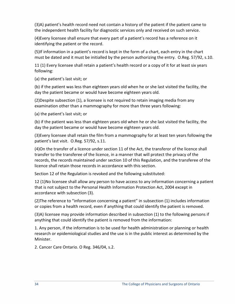
34 The College of Physicians and Surgeons of Ontario
(3)A) patient’s health record need not contain a history of the patient if the patient came to the independent health facility for diagnostic services only and received on such service.
(4)Every licensee shall ensure that every part of a patient’s record has a reference on it identifying the patient or the record.
(5)If information in a patient’s record is kept in the form of a chart, each entry in the chart must be dated and it must be initialled by the person authorizing the entry. O.Reg. 57/92, s.10.
11 (1) Every licensee shall retain a patient’s health record or a copy of it for at least six years following:
(a) the patient’s last visit; or
(b) if the patient was less than eighteen years old when he or she last visited the facility, the day the patient became or would have become eighteen years old.
(2)Despite subsection (1), a licensee is not required to retain imaging media from any examination other than a mammography for more than three years following:
(a) the patient’s last visit; or
(b) if the patient was less than eighteen years old when he or she last visited the facility, the day the patient became or would have become eighteen years old.
(3)Every licensee shall retain the film from a mammography for at least ten years following the patient’s last visit. O.Reg. 57/92, s.11.
(4)On the transfer of a licence under section 11 of the Act, the transferor of the licence shall transfer to the transferee of the licence, in a manner that will protect the privacy of the records, the records maintained under section 10 of this Regulation, and the transferee of the licence shall retain those records in accordance with this section.
Section 12 of the Regulation is revoked and the following substituted:
12 (1)No licensee shall allow any person to have access to any information concerning a patient that is not subject to the Personal Health Information Protection Act, 2004 except in accordance with subsection (3).
(2)The reference to “information concerning a patient” in subsection (1) includes information or copies from a health record, even if anything that could identify the patient is removed.
(3)A) licensee may provide information described in subsection (1) to the following persons if anything that could identify the patient is removed from the information:
1. Any person, if the information is to be used for health administration or planning or health research or epidemiological studies and the use is in the public interest as determined by the Minister.
2. Cancer Care Ontario. O Reg. 346/04, s.2.

Clinical Practice Parameters and Facility Standards Diagnostic Imaging – July 2012 35
Books and Accounts 12.1(1)This section applies to licensees of independent health facilities that are funded under section 24 of the Act, other than independent health facilities whose funding is based solely on the Ministry of Health publication titled “Schedule of Facility Fees”.
(2)Every licensee shall keep the following records in relation to the independent health facility:
1. Current financial records showing:
(i) the amounts paid by the Minister to the licensee under section 24 of the Act.
(ii) the revenue earned by the licensee from facility fees charged by the licensee for or in respect of services or operating costs that support, assist or are a necessary adjunct to the primary insured services set out in the licensee’s licence, and
(iii) the expenditures, assets and liabilities of the facility that relate to the costs paid by the Minister under section 24 of the Act.
2. A reporting record listing each service provided in the facility that is a primary insured service set out in the licensee’s licence and each service provided in the facility that is a funded service under section 24 of the Act and showing how many of each of such services are provided.
3. An annual income and expense statement showing the income received and the expenses incurred by the licensee in connection with the services mentioned in paragraph 2.
4. An annual inventory of the assets of the facility that have an acquisition cost exceeding $3,500 and that relate to the costs paid by the Minister under section 24 of the Act.
(3)Every licensee shall ensure that the records required under section (2):
(a) are kept in the independent health facility; and
(b) are kept in a bound or loose-leaf book or are recorded by a system of mechanical or electronic data processing or any other information storage device.
(4)Every licensee shall ensure that any part of a record required under subsection (2) that relates to a period of time is retained for at least six years following the end of the period.
(5)Every licensee shall ensure that the accounts of the independent health facility are audited by a person licensed under the Public Accountancy Act. O.Reg. 283/94, s.1, part.
12.2 Every licensee of an independent health facility shall furnish such information and accounts as the Director may require. O. Reg. 283/ 94, s.1, part.
Notices 13 Every licensee of an independent health facility,
(a) who decides to cease operating the facility at a future date shall give the Director, as soon as possible, written notice of the date; and
(b) who ceases to operate the facility shall give the Director, within seven days after the date the licensee ceases to operate the facility, written notice of the date. O. Reg. 57/92, s.13.

36 The College of Physicians and Surgeons of Ontario
14 Every licensee of an independent health facility shall give the Director:
(a) if the licensee is a corporation, written notice of any change in the location of the licensee’s head office within ten days after the change; and
(b) written notice of any change in the name under which the licensee carries on business within ten days after the change. O.Reg. 57/ 92, s.14.
Miscellaneous 15 It is a condition of a licence that the licensee post the first page of the licence in a conspicuous place in the independent health facility. O. Reg. 57/92, s.15.
16(1) The fee for a licence is $100.
(2) The fee for the transfer of a licence is $100.
(3) The fee for the renewal of a licence is $100. O. Reg. 57/92, s.16.
17 The administrative charge for the purposes of section 36 of the Act is $50. O. Reg. 57/92, s.17.

Clinical Practice Parameters and Facility Standards Diagnostic Imaging – July 2012 37
Appendix II Risk of Serious Infection from Ultrasound and Medical Gels - Notice from Health Canada October 2004
http://www.hc-sc.gc.ca/dhp-mps/medeff/advisories-avis/prof/_2004/ultrasound_nth-ah-eng.php
Overview
Health Canada is aware of several cases of bacteremia and septicaemia whose source was traced to ultrasound and medical gels used during various procedures. The cause of the problem was a contamination of the gel at the manufacturing facility and appropriate corrective measures were taken with the manufacturers to rectify the problem.
This situation led Health Canada to examine current practices regarding the use of ultrasound and medical gels in clinical settings and revealed significant deficiencies: for example, gel containers placed in a warming device, without a cap and for extended periods of time, refillable squeeze bottles are not cleaned between refills, non-sterile gels labelled for external use only are used on mucous membranes or during invasive procedures like biopsies.
In order to minimize the health risks associated with the use of ultrasound gels, Health Canada is making the following recommended practices.
Sterile Gels
Sterile gels must be used for all invasive procedures that pass a device through a tissue, e.g., needle aspiration, needle localization, and tissue biopsy, for all procedures involving sterile environment or non-intact skin and on neonates.
Sterile gels should be used for procedures performed on intact mucous membranes, e.g., esophageal, gastric, rectal or vaginal and in patients with immunodeficiencies or on immunosuppressive therapy.
Aseptic technique should be followed when using sterile gels.
Non-Sterile Gels
Single use containers are recommended for non-sterile gels.
If reusable containers are used, they must be emptied, washed in soapy water or hospital-grade disinfectant, rinsed thoroughly and dried prior to refilling. Bottles should not be “topped up”. Cracked reusable bottles should be discarded.

38 The College of Physicians and Surgeons of Ontario
When filling a reusable container, ensure that the large bulk container has not passed the expiration date.
Bottles should be filled using a dispensing device on the large bulk container, not by inserting the tip of the refillable bottle into the bulk container to aspirate the contents.
Bottles should be refilled as close as possible to the time of use.
When opening a new gel bottle or a newly refilled bottle, date the bottle and discard unused gel after one month.
Tips of containers or dispensing nozzles must not come in direct contact with a patient, staff, instrumentation or the environment. Gel should be dispensed into a medicine cup or on a clean disposable cloth and then to the patient’s skin.
If a medicine cup or a disposable cloth is not used, wipe the dispensing nozzle clean with an alcohol swab and wipe the outside of the container with a disinfectant between patients.
If a gel is being used on a patient who is in droplet or contact isolation, use a single-use gel container, or leave the reusable container in the room if repeat procedures are necessary and discard the gel when isolation of the patient is discontinued.
For infrequent procedures, use small or single-use containers.
Warming of Gel
Warmed gel should only be used when required.
Bottles should be removed from the warmer as soon as possible and dried immediately.
Gel warmers must be cleaned weekly with low level hospital-approved disinfectant, and immediately if the warmer becomes soiled.
Storage of Ultrasound and Medical Gels
Products must be stored in areas that are dry and protected from potential sources of contamination such as dust, moisture, insects, rodents, etc.
If evidence of contamination is present or package integrity has been breached, the product must be discarded.
Products should be rotated when restocking takes place.

Clinical Practice Parameters and Facility Standards Diagnostic Imaging – July 2012 39
Appendix III Fetal Ultrasound for Non-Medical Reasons – CPSO Policy Statement #4-10
Approved by Council: May 2004 Reviewed and Updated: May 2010 Publication Date: Dialogue, Issue 2, 2010 Key Words: Ultrasound, Diagnostic Imaging, Entertainment, 3D, 4D, Gender
selection Related Topics: Practice Guide Reference Materials: Health Canada; Ontario Association of Radiologists; Canadian
Association of Radiologists; Society of Obstetricians and Gynecologists of Canada; Canadian Society of Diagnostic Medical Sonographers; Food and Drug Administration; American College of Obstetricians and Gynecologists; American Institute of Ultrasound in Medicine.
College Contact: Public and Physician Advisory Service
40 The College of Physicians and Surgeons of Ontario
Purpose Physicians routinely order or perform diagnostic fetal ultrasounds during the course of a patient’s pregnancy. At times, physicians may be asked by expectant mothers and their families to order or perform a fetal ultrasound for non-medical reasons. The purpose of this policy is to outline the College’s expectations of physicians with respect to ordering and performing fetal ultrasounds.
Principles 1. The physician’s responsibility is to act in the best interest of the patient.
2. Acting in the patient’s best interest includes maintaining the medical knowledge and clinical skills necessary to provide quality care to patients.
Background Diagnostic fetal ultrasound is an essential component of prenatal care.1 Fetal ultrasound, in conjunction with other appropriate diagnostic tests, provides important medical information, such as the size, age and state of health of the fetus.2
Fetal ultrasound technology is used by others for non-medical reasons, such as for entertainment or gender identification:
1) Entertainment
As more advanced ultrasound technologies are becoming available such as 3D/4D ultrasound, expectant mothers and their families are requesting fetal keepsake videos and portraits.
2) Gender Identification
Requests are sometimes made for the performance of an ultrasound to determine the sex of the baby for gender selection purposes.
There are a number of organizations that have formal statements that provide that the use of ultrasound for entertainment or fetal gender determination purposes is inappropriate.3 The College of Physicians and Surgeons of Ontario’s Independent Health Facilities Program refers to these statements, along with this policy, when inspections are carried out in facilities where fetal ultrasounds are performed.
1 Experts in Canada recommend that all women have a diagnostic ultrasound when they are pregnant. The Society of Obstetricians and Gynaecologists of Canada has clinical guidelines with respect to diagnostic fetal ultrasound. 2 Health Canada, “Fetal Ultrasound for Keepsake Videos”, November 2003, available at http://www.hc-sc.gc.ca/hl-vs/iyh-vsv/med/ultrasound-eochographie-eng.php. 3 Health Canada, the Ontario Association of Radiologists, the Canadian Association of Radiologists, the Society of Obstetricians and Gynaecologists of Canada, the Canadian Society of Diagnostic Medical Sonographers, the Food and Drug Administration, the American College of Obstetricians and Gynecologists, and the American Institute of Ultrasound in Medicine are just some of the organizations that oppose the use of ultrasound for these purposes.
Clinical Practice Parameters and Facility Standards Diagnostic Imaging – July 2012 41
College Policy
Physicians must ensure that all diagnostic fetal ultrasounds are ordered and conducted for appropriate clinical indications, in accordance with relevant statements and guidelines. .
The purpose of an imaging examination should always be to obtain information relevant to the diagnosis or treatment of a patient. Therefore, when ordering the diagnostic fetal ultrasound, the physician should specify the clinical indications.
If a physician orders or performs a diagnostic fetal ultrasound for medical reasons, they may provide their patients with any picture or video of the fetus that is created as a result of that imaging examination.
However, it is inappropriate and contrary to good medical practice to use ultrasound only to view the fetus to obtain a picture or video of the fetus or to determine gender of the fetus. Bibliography
Canadian Association of Radiologists - CAR Standard for Communication -Diagnostic Radiology. Approved June 1997 Dolinkievich, R., Murphy, K., Holland, R. “Infection Control Standards in Diagnostic Imaging” Journal of Diagnostic Medical Sonography. March/April 1992; 86-88. Guide to the Health Care Consent Act. Members’ Dialogue. May 1996; 24. ICAVL, “Essentials and Standards”, Part 1: Vascular Laboratory Operations—Organization, 1/2000 Maintenance of Certification (MOC) program, The Royal College of Physicians and Surgeons of Canada, March 2001. McIver, C. “Latex Allergy Alert.” Members’ Dialogue. November/ December 1997;30-31. Stolberg HO, Hynes DM, Rainbow AJ, Moran LA. “Requesting Diagnostic Imaging Examinations: A Position Paper of the Canadian Association of Radiologists.” Members’ Dialogue. November/December 1998;13-16. Sussman, G.L., Gold, M. Guidelines for the Management of Latex Allergies and Safe Latex Use in Health Care Facilities. Canadian Healthcare Association 1996. Sussman, G. “Latex Allergies.” Members’ Dialogue. March/April 1998;10-12. The Healing Arts Radiation Protection Act (HARP) and Regulation 543 (X-Ray Safety Code), Government of Ontario, amended version dated: Sept. 28, 2001. The Healing Arts Radiation Protection Guidelines, Ontario, Ministry of Health, June 1987. Wenghofer E, Gold G, Craighead J. “Diagnostic Imaging Services in Independent Health Facilities.” Members’ Dialogue. November/ December 1998;6-12

42 The College of Physicians and Surgeons of Ontario
Appendix IV Sample Emergency Safety Policy Safety Training for all staff should be carried out. In addition, an emergency safety policy should be included in the policies and procedures manual. This appendix has been provided as a sample of what the policy may look like and include. Each policy must be site specific to the facility and may include but is not limited to the following areas: Employer Responsibilities (in all incident cases): Provide first aid in accordance with the regulations. Record first aid attention, adverse effects, incident report. Assist to provide immediate transportation to the hospital, doctor, worker/patient’s home, when/as necessary. Employee Responsibilities: Acute Care Transfer Should a patient, visitor, and/or staff become ill while in the clinic the following is carried out:
1. Immediately, the technologist or clerical staff will alert the attending Radiologist of the problem.
2. In the event that the attending Radiologist is not available, contact a local GP (agreement should be made prior between facility and physician – contact numbers should be available for staff).
3. If the physician is not immediately available, call 911, identify yourself and request transfer to the nearest hospital.
Fire Prevention and Control Plan
1. All staff members employed at the facility is required to know the fire plan. To facilitate this, an annual review of the plan will be carried out and is mandatory for all staff members.
2. The fire plan is site-specific for the facility. Staff members are required to familiarize themselves with the plan for this location.
3. Each employee should have the ability to assess the situation quickly and initiate appropriate measures upon discovering a fire. This may vary from using a fire extinguisher to contain a fire or alerting others, evacuating the building and calling the fire department.
If you discover a fire in your area:
1. Remove patients from rooms and out of danger. 2. Turn off lights, any electrical equipment, gases, and close windows and doors. 3. Pull the alarm located closest to you. 4. Dial 911 and advise the Fire Department of the Emergency. Give them your name, location of
the fire and type of fire to the communications operator (electrical, gas, other). 5. If possible (i.e the fire is contained to a specific area) go back to the room and attempt to put
out the fire using a fire extinguisher.

Clinical Practice Parameters and Facility Standards Diagnostic Imaging – July 2012 43
DO NOT ATTEMPT TO USE THE FIRE HOSE. Everyone should be removed from the office. Have a staff member positioned at the main corridor junction to direct fire fighters. If you hear a fire alarm:
1. Collect all patients, visitors, and staff members in the facility and guide them to the closest exits.
2. DO NOT USE THE ELEVATOR. All staff members along with anyone in the office at the time of the evacuation alarm, must meet at a predetermined assembly point outside of the building.
3. Personnel will be requested to assist with duties such as checking the office before leaving ensuring that everyone is accounted for, turning off lights in the fire area, turning off gases (oxygen), turning off all electrical equipment and closing doors and windows.
The First Aid Box As a minimum the first aid box should contain:
• A current edition of a first aid manual • One card of safety pins • Dressings, consisting of:
o 12 adhesive dressings, individually wrapped o 4 sterile gauze pads, 3 inches square o 2 rolls of gauze bandages, 2 inches wide
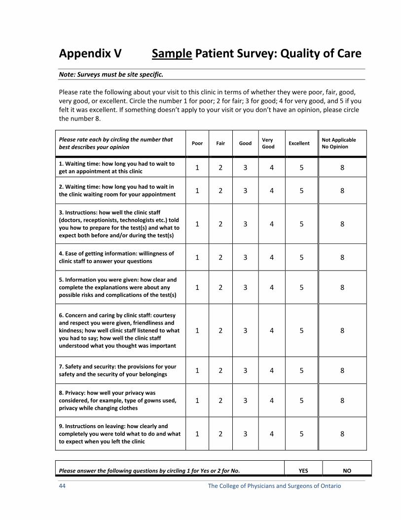
44 The College of Physicians and Surgeons of Ontario
Appendix V Sample Patient Survey: Quality of Care Note: Surveys must be site specific.
Please rate the following about your visit to this clinic in terms of whether they were poor, fair, good, very good, or excellent. Circle the number 1 for poor; 2 for fair; 3 for good; 4 for very good, and 5 if you felt it was excellent. If something doesn’t apply to your visit or you don’t have an opinion, please circle the number 8.
Please rate each by circling the number that best describes your opinion
Poor Fair Good Very Good Excellent Not Applicable
No Opinion
1. Waiting time: how long you had to wait to get an appointment at this clinic 1 2 3 4 5 8
2. Waiting time: how long you had to wait in the clinic waiting room for your appointment 1 2 3 4 5 8
3. Instructions: how well the clinic staff (doctors, receptionists, technologists etc.) told you how to prepare for the test(s) and what to expect both before and/or during the test(s)
1 2 3 4 5 8
4. Ease of getting information: willingness of clinic staff to answer your questions 1 2 3 4 5 8
5. Information you were given: how clear and complete the explanations were about any possible risks and complications of the test(s)
1 2 3 4 5 8
6. Concern and caring by clinic staff: courtesy and respect you were given, friendliness and kindness; how well clinic staff listened to what you had to say; how well the clinic staff understood what you thought was important
1 2 3 4 5 8
7. Safety and security: the provisions for your safety and the security of your belongings 1 2 3 4 5 8
8. Privacy: how well your privacy was considered, for example, type of gowns used, privacy while changing clothes
1 2 3 4 5 8
9. Instructions on leaving: how clearly and completely you were told what to do and what to expect when you left the clinic
1 2 3 4 5 8
Please answer the following questions by circling 1 for Yes or 2 for No. YES NO

Clinical Practice Parameters and Facility Standards Diagnostic Imaging – July 2012 45
10. Were you told to leave the clinic before you felt ready to do so? 1 2
11. Did you have to visit a physician, walk-in clinic, emergency room, urgent care centre or hospital in the days following this service because your health got worse as a result of the service(s) received at the clinic?
1 2
12. Would you recommend the clinic to a friend or family member if they needed services that it provides? 1 2
Please rate this item by circling the number that best describes your opinion
Poor Fair Good Very Good Excellent Not Applicable
No Opinion
13. Overall quality of care: how you evaluate the services you received and the way you were treated
1 2 3 4 5 8
14. If there were some things you could change about this visit to improve it, what would they be?
Thank you for completing this survey. Please double check that you have answered all questions and then place the survey in the envelope provided. Your answers will be kept completely confidential.
Thank you again for your help!

46 The College of Physicians and Surgeons of Ontario
Appendix VI Sample Referring Physician Survey Note: Surveys must be site specific.
Name of facility
Please answer the following questions regarding your experience with the above facility by filling in the blank or circling the number that best describes your answer.
1. How long have you referred patients to this facility?
_______years or _______months
Please base your answers on your contact with the facility in the past 6 months.
2. How satisfied are you with how long it generally takes: (Please rate each item by circling the number that best describes your opinion) Not Applicable Very
Dissatisfied Dissatisfied Neutral Satisfied Very Satisfied
to get an appointment for a patient at this facility?
1 2 3 4 5
to obtain written results (a written consultation) from this facility,
1 2 3 4 5 once your patient is seen?
to get an oral report from this
facility when it is required because of an urgent or emergency 0 1 2 3 4 5 situation, once your patient is seen?
3. How often do you speak to a physician at the IHF regarding the patient’s clinical condition before your patient receives a diagnostic work-up?
Never Rarely Occasionally Sometimes Often Almost all the time
4. Approximately how many patients have you referred to this facility in the past 6 months? ___________(number of patients referred)
5. Do you refer your patients to more than one facility of this type?
A. No (if you circled No, please skip to Question number 7) B. Yes
6. What are the reasons you refer patients to this particular facility? (Please circle all that apply.)
a. Nearer Patient’s home
b. Has specialized equipment needed for test requested
c. Turnaround time to receive the results is shortest

Clinical Practice Parameters and Facility Standards Diagnostic Imaging – July 2012 47
d. Has staff that speak other languages, and thus can better understand my patients
e. Is able to quickly see patients when feedback is urgently required
f. Has convenient hours of operation
g. Quality of the services provided
h. Other, please describe ___________________ Please skip to Question number 8.
7. What are the reasons you refer patients only to this facility? (Please circle all that apply.)
a. Only facility of its type in this community
b. Our group has a service contract with this facility
c. Facility is located near this practice and is thus convenient for patients
d. Has staff that speak other languages and thus can better understand my patients
e. Has specialized equipment needed for tests requested
f. Turn-around time to receive results is short
g. Nearest patients’ homes
h. Is able to quickly see patients when feedback is urgently required
i. Quality of the services provided
j. Has convenient hours of operation
k. Other, please describe____________________
9. Have you been dissatisfied with a consult you received from this facility in the past six months?
a. No b. Yes
10. Please rate each item by circling the number that best describes your experience with the IHF based on your contacts in the last 6 months.
Never Seldom Sometimes Frequently Usually
The waiting period for a test to be done is long. 1 2 3 4 5
Requests for consultation are handled promptly. 1 2 3 4 5
The facility accommodates patients when the test is urgently required. 1 2 3 4 5
The interpreting physician is available to you for consultation. 1 2 3 4 5
This facility meets the needs of my 1 2 3 4 5

48 The College of Physicians and Surgeons of Ontario
patients whose first language is other than English or French.
The recommendations received are useful in patient management. 1 2 3 4 5
The recommendations are clearly stated. 1 2 3 4 5
The reports received are too wordy. 1 2 3 4 5
Reports of results are sent out in a timely fashion. 1 2 3 4 5
The consulting physician orders tests in addition to those you requested.
1 2 3 4 5
When tests are added the resulting recommendations add information important to patient care.
1 2 3 4 5
The interpreting physician’s findings are generally consistent with your clinical findings.
1 2 3 4 5
If 2 (Yes), please explain:
10. Overall, how satisfied are you with the contacts you have had with this facility in the past six months?
a. Very Dissatisfied b. Dissatisfied c. Neutral d. Satisfied e. Very Satisfied
Thank you for participating in this survey. Please return the survey in the envelope provided.
Our address is:

Clinical Practice Parameters and Facility Standards Diagnostic Imaging – July 2012 49
Independent Health Facilities: Clinical Practice Parameters and Facility Standards Diagnostic Imaging
Volume 2
Clinical Practice Parameters


Clinical Practice Parameters and Facility Standards for Diagnostic Imaging – July 2012 51
Chapter 7 Position Statement from the IHF Diagnostic Imaging Task Force
It is the position of the IHF Diagnostic Imaging Task Force that the revised (2012) Clinical Practice Parameters and Facility Standards will contain a list of CAR Standards that are applicable to the services provided in Independent Health Facilities.
To ensure that Radiologists and Facilities are in compliance with current CAR Standards the radiologist and facility staff are responsible for, at least annually, reviewing the Canadian Association of Radiologists website (www.car.ca) to ensure that they have obtained and are in compliance with the most current standards of practice for the profession.
X-ray
Acute Ankle Injury (accessed December 15, 2011)
http://www.ices.on.ca/informed/periodical/subissue/218-ip1190.pdf
Chest Radiography (accessed December 15, 2011)
http://www.car.ca/uploads/standards%20guidelines/chest_radiography.pdf
General (Plain) Radiography (accessed December 15, 2011)
http://www.car.ca/uploads/standards%20guidelines/general_radiography.pdf
Performance of the Cervical Spine in Children and Adults (accessed December 15, 2011)
http://www.car.ca/uploads/standards%20guidelines/cervical_spine.pdf
Performance of the Thoracic (Dorsal) Spine (accessed December 15, 2011)
http://www.car.ca/uploads/standards%20guidelines/thoracic_spine.pdf
Performance of the Lumbosacral Spine (accessed December 15, 2011)
http://www.car.ca/uploads/standards%20guidelines/lumbosacral_spine.pdf

52 The College of Physicians and Surgeons of Ontario
BMD
BMD (accessed December 15, 2011)
http://www.car.ca/uploads/standards%20guidelines/201001_en_car_bmd_technical_standards.pdf
Fluoroscopy
Adult Barium Enema Examinations (accessed December 15, 2011)
http://www.car.ca/uploads/standards%20guidelines/barium_enema.pdf
Contrast Studies of the Adult Upper Gastrointestinal Tract (accessed December 15, 2011)
http://www.car.ca/uploads/standards%20guidelines/upper_gi.pdf
Contrast Studies of the Pharynx and Esophagus in the Adult (accessed December 15, 2011)
http://www.car.ca/uploads/standards%20guidelines/pharynx_oeso.pdf
Contrast Studies for Performance of Contrast examinations of the small bowel in adults (accessed December 15, 2011)
http://www.car.ca/uploads/standards%20guidelines/bowel.pdf
Mammography
Breast Imaging (accessed December 15, 2011)
http://car.ca/uploads/standards%20guidelines/breast_imaging.pdf
Ultrasound
Musculoskeletal Ultrasound (accessed December 15, 2011)
http://www.car.ca/uploads/standards%20guidelines/us_msk.pdf\
Obstetrical Ultrasound (accessed December 15, 2011)
http://www.car.ca/uploads/standards%20guidelines/20101125_en_standard_diagnostic_ob_ultrasound.pdf
Performance of Breast Ultrasound (accessed December 15, 2011)
http://www.car.ca/uploads/standards%20guidelines/breast_ultrasound.pdf
Clinical Practice Parameters and Facility Standards for Diagnostic Imaging – July 2012 53
Thyroid and Parathyroid (accessed December 15, 2011)
http://www.car.ca/uploads/standards%20guidelines/20110428_en_standard_thyroid_parathyroid.pdf
Ultrasound Examination of the Abdomen and Retroperitoneum (accessed December 15, 2011)
http://www.car.ca/uploads/standards%20guidelines/abdoretro.pdf
Ultrasound Examination of the Female Pelvis (accessed December 15, 2011)
http://www.car.ca/uploads/standards%20guidelines/pelvis.pdf
Ultrasound Examination of the Scrotum (accessed December 15, 2011)
http://www.car.ca/uploads/standards%20guidelines/20110428_en_standard_scrotal_ultrasound.pdf
Vascular Ultrasound (accessed December 15, 2011)
http://www.car.ca/uploads/standards%20guidelines/vascular.pdf
Other
Diagnostic Imaging Referral Guidelines – A Guide for Physicians First Edition 2005 http://car.ca/uploads/membership/Guidelines_ENG.pdf
All Clinical Practice Parameters referenced within this document should be read in conjunction with the Facility Standards (Volume 1) developed by the IHF Diagnostic Imaging Task Force. A guiding principle should be that Diagnostic Imaging examinations only be performed for a valid medical reason with the minimum exposure that provides the image quality necessary for an adequate diagnostic examination.

54 The College of Physicians and Surgeons of Ontario
Chapter 8 Routine Chest Radiography in a Primary Care Setting
http://www.ncbi.nlm.nih.gov/pubmed/15516621
Source
Departments of Radiology, Internal Medicine, and Pulmonary Medicine, Emory Clinic, Bldg. A, 1365 Clifton Rd NE, Atlanta, GA 30322, USA. [email protected].
Abstract
PURPOSE:
To determine the frequency, diagnostic yield, outcomes, cost, and rate of false-positive results of routine chest radiography performed in asymptomatic patients in the primary care setting.
MATERIALS AND METHODS:
Radiography reports on all patients who underwent routine or screening poster anterior and lateral chest radiography at a university-affiliated primary care clinic in 2001 were reviewed. Radiographic results were coded as normal or minor findings or as major abnormalities, such as pulmonary nodules, requiring further diagnostic evaluation. Outcomes of patients with major abnormalities were established by using chart reviews or reviewing additional radiographs. Costs were estimated by using 2002 Medicare reimbursement rates. The main measures assessed were frequency, costs, and rate of false-positive results of routine chest radiography.
RESULTS:
Of 3812 radiographs obtained at the primary care clinic, 1282 (34%) were ordered for routine or screening purposes by the referring physician. Nine hundred twenty-two radiographs were obtained in male patients and 360 were obtained in female patients; their mean and median age was 49 years (age range, 4-87 years). Fifteen chest radiographs showed major abnormalities. No patient younger than 40 years had a major abnormality. Fourteen of the 15 findings of major abnormalities proved to be false-positive. No disease requiring treatment was diagnosed as a result of radiographic findings. The total cost for follow-up radiography and computed tomography was US dollar 46,609.49.
CONCLUSION:
Routine chest radiography has low diagnostic yield in asymptomatic primary care patients.
Comment in

Clinical Practice Parameters and Facility Standards for Diagnostic Imaging – July 2012 55
• Radiology. 2005 Jul;236(1):368; author reply 368.
Tigges S, Roberts DL, Vydareny KH, Schulman DA


Independent Health Facilities: Clinical Practice Parameters and Facility Standards Diagnostic Imaging
Volume 3
Teleradiology (PACS)


Clinical Practice Parameters and Facility Standards Diagnostic Imaging – July 2012 57
OAR Teleradiology Practice Standard June 2007
OAR TELERADIOLOGY PRACTICE STANDARD
Definition
Teleradiology in Ontario is the electronic transmission of radiographic images from one geographical location to another for the purposes of interpretation and consultation by diagnostic imaging physicians accredited by the Royal College of Physicians and Surgeons of Canada (or recognized equivalent) and licensed by the College of Physicians and Surgeons of Ontario.
These guidelines and standards have been developed to protect patients and ensure their data is kept confidential. Teleradiology services are to facilitate patient care and are not intended to be a cost-cutting measure, which may jeopardize patient safety and the standards of health care.
Preface
The transmission of images between centres has been going on for a number of years and has proved to be valuable for centres seeking expert opinions on emergency and problem cases. The most common such connections have been with radiologists who work at a site and are now able to offer image interpretations online from other sites within an institution, from their offices, home or elsewhere. More recently radiological images have been transmitted to main centres from smaller community hospitals in areas of low population density where small radiology departments have proven unsustainable. The vastly improved capacity of the internet and the speed of transmission have permitted a much wider use of teleradiology.
Teleradiology has advantages but it must be done properly to ensure that a high quality of care is provided to patients and to maintain the radiologist interaction with their clinical colleagues. It is also important that those radiologists providing the service are properly trained, are registered with the appropriate authorities, and undergo continuing update through Continuing Medical Education (CME). The services provided must be open to audit and the ability to discuss cases with those reporting the studies must be available. This standard has been developed to provide guidance to radiologists, managers of health care facilities, patient’s representatives and governments on appropriate standards for teleradiology services.
Teleradiology has undergone a number of health-technology assessments in different countries with regard to the context of its use, but a great deal of thought and study is still required. Teleradiology clearly has a number of advantages, but it also has the potential to create considerable difficulties for the delivery of a high quality radiological service to patients, unless its role and the legal responsibilities involved are clearly defined.
Role of a Diagnostic Radiologist
The role of a radiologist providing medical services in a diagnostic imaging service is considerably wider than simply issuing a diagnostic interpretation and report. It includes:
• Evaluating the clinical information produced by referring physician clinicians • Deciding which test is appropriate

58 The College of Physicians and Surgeons of Ontario
• Establishing and assuming responsibility for the imaging protocols, quality parameters and a host of other technical factors that are integral to the creation of the diagnostic image and report
• Being responsible for the technical staff/standards involved in the diagnostic imaging facility
• Optimizing the study and assisting the referring physician colleague • Evaluating the study and relating it to the clinical findings • Having knowledge of the practice of referring physicians • Reviewing previous examinations and their interpretations to compare them with the
current study • Identifying further appropriate management including diagnostic investigations
essential to obtain a comprehensive diagnosis and treatment, and reviewing those recommendations with referring physicians
• Reviewing all clinical data in a multi-disciplinary environment • Performing interventional therapeutic and diagnostic procedures • Assuming responsibility for the appropriate management of the patient during the
diagnostic imaging procedure • Contributing radiological expertise to the management of the diagnostic imaging
service to ensure the highest possible quality assurance and quality control • Being responsible for patient safety by ensuring minimal exposure to radiation dose
and other matters that could compromise patient care • Adhering to all provincial and federal regulations, statutes relating to the delivery of
medical services generally and diagnostic imaging services provincially; meeting and exceeding the standard of care in the delivery of diagnostic imaging services in the province; maintaining membership in all of the licensing bodies and fulfilling the requirements of that licensure regime
• Ensuring the selection and use of appropriate and modern equipment, properly trained staff and other elements in the high quality delivery of diagnostic imaging
• Where relevant, teaching radiology residents and fellows according to national training program requirements
• Where relevant, participating in radiology research • Auditing the delivery of radiology services in the sites where the radiologist works • Ensuring timely communication of urgent findings • Maintaining appropriate records/confidentiality as mandated by legislation
In essence, appropriate teleradiology in this era is the same as the whole practice of radiology. The fact that patient data can be moved over a broadband connection does not alter the role or responsibilities of the supervising and interpreting radiologist.
The importance of interaction between the referring clinicians and the radiologist cannot be over-emphasized. There are considerable quality patient care and medical-legal implications when teleradiology services are provided by a radiologist outside the patient’s jurisdiction. Regulatory bodies, licensing and credentialing (including the College of Physicians and Surgeons of Ontario, the Royal College of Physicians and Surgeons of Canada, Health Protection Branch, the Ministry of Health’s Independent Health Facility branch, OHIP, X-ray Inspection branch, and other provincial and federal bodies), are unable to enforce regulations outside their jurisdiction yet have a responsibility to patients with respect to the enforcement of a wide spectrum of regulations and statutes inter-linked to the high quality delivery of radiologists’ services in the province. The requirements of these and other related
Clinical Practice Parameters and Facility Standards Diagnostic Imaging – July 2012 59
bodies are constantly subject to change requiring the radiologist to comply with a new and more stringent degree of responsibility with respect to the delivery of patient care.
Key Principles
1. Diagnostic radiology is an integrated medical service required in every modern health care system.
2. Referring physicians are dependent upon the local availability of diagnostic imaging physicians to assist them to manage the health of their patients.
3. Only fully qualified diagnostic radiologists should provide the teleradiology service. They must be properly accredited, registered, and licenced in Ontario. The radiologist should be subject to licensing and quality assurance requirements of the provincial health authority; legislative and professional requirements of the facility providing the service; the provincial College of Physicians and Surgeons, accreditation and be in good standing with the Royal College of Physicians and Surgeons of Canada.
4. A definitive report is mandatory with the signature of the reporting radiologist. Electronic signatures are acceptable as long as they can be authenticated.
5. In a public hospital the members of the radiology department must be credentialed and be part of the recognized medical staff.
6. The department head via the Medical Advisory Committee (MAC) and Board is responsible for the medical service.
7. In an Independent Health Facility (IHF), the off-site radiologist must be approved by the radiologist Quality Advisor who is legislatively responsible for Quality Control/Quality Assurance (QC/QA) at the IHF.
8. All radiologists providing teleradiology services must be covered by the Canadian Medical Protective Association (CMPA) for medical liability issues and ensure they are compliant with current CMPA guidelines and policies covering diagnostic imaging physicians to safeguard patient interests.
9. Ensure that all radiologists and their staff involved in the delivery of teleradiology services are in full compliance with relevant privacy legislation and facility policies to protect patient confidentiality.
10. Ensure that the information received for a primary read is the full data set and that the reading radiologist should have all of the functionality of the PACS at his/her disposal to do an interpretation.
Key Management Issues
1. Teleradiology services must be organized between the source radiologists and the off-site radiologist provider to guarantee the proper management of the patient. This will ensure that:
a. The clinical evaluation and data is provided with the request for the examination.
b. The requirements of the Healing Arts Radiation Protection Act (HARP) (including justification, appropriate techniques, optimization, and good procedure) are fulfilled.
c. The report of the teleradiology service can be reviewed with clinicians and where applicable, in multi-disciplinary meetings and integrated with patients’ notes and previous studies.
d. The reporting radiologist of the teleradiology service is able to communicate directly with the referring radiology department and clinicians in order to discuss the clinical background and unexpected diagnosis, which may be relevant to the timely management of the patient.
60 The College of Physicians and Surgeons of Ontario
e. Teleradiology services that are developed to meet the needs of rural, remote and small community areas must be linked to the nearest substantive radiology department and the service is managed by that department. The radiologists involved in providing the service must have a close connection and knowledge of referring clinicians, and technologists, and should understand any particular local disease and cultural factors.
2. Equipment used for teleradiology should provide a similar level of resolution and functionality as is available in the radiology department/facility.
3. The American College of Radiology’s (ACR) Technical Standard for Teleradiology for equipment and other supporting technologies used in the delivery of teleradiology is the acknowledged current technical standard. Radiologists delivering teleradiology standards are expected to comply or exceed the ACR Technical Standard for Teleradiology.
Real and Potential Problems
Clinico-Radiological Communication
If reporting of radiographs is taken away from close proximity with the patient, the clinical contact between the referring clinicians and radiologists is substantially reduced. It is imperative that teleradiology facilities have phone links with the hospitals and/or clinics from which images are obtained, and have the ability for direct discussion between a referring clinician and the reporting radiologist on individual cases. Without this, the bond between the patient and the radiologist becomes unclear. If urgent or significant unexpected features are found, the teleradiology service must transmit them directly to the referring clinician. This will be impossible unless there is a clear point of contact for the teleradiology service.
Team Working
The ability to hold multi-disciplinary meetings is much more difficult with teleradiology, even with teleconference links. It is now widely accepted that multi-disciplinary meetings, which are often led by the radiology department, are essential in the management of problematic cases, i.e., cancer care. They maximize the understanding of the clinical problems by radiologists.
External reviews of health care disasters have emphasized the importance of teamwork especially in medicine and the need for enhanced teamwork, involving radiology has been highlighted. Interaction between different members of the hospital team with radiology may be impaired, if radiology is undertaken at the long distance by a teleradiology link.
Communication
It is necessary that there be good communication between referring physicians, radiologists and technologists.
Wording of Report and Clinical Impact
Even if radiologists and referring clinicians have a common first language, it has to be recognized that radiological reporting may be subject to regional variation. Radiological reports often rely on verbal expressions of probability and may contain some regionally used expressions.
Modern imaging commonly demonstrates an abundance of reportable findings, some of which are clinically relevant and some of which are incidental findings/pseudo-disease. Multiple pathologies can exist in the same patient. The clarity and certainty conveyed in the text is particularly important in

Clinical Practice Parameters and Facility Standards Diagnostic Imaging – July 2012 61
converting a report that is merely ‘diagnostically accurate’ into one that has a diagnostic outcome and potentially a therapeutic outcome for the patient. Clinicians are more likely to act on the nuances intended in a report generated by a radiologist with whom they regularly liaise compared with a report generated by a third party teleradiology service from someone they never met. Specific wording of reports for general family doctors may be necessary, which is different from the reports to specialists within their sphere of interest. Familiarity with the referring doctors can make specific reports more appropriate and useful. Knowledge of referring doctors can make specific reports more appropriate. Health care delivery varies between different jurisdictions. Recommendations for further imaging/specialist referral, which might be appropriate in the locale where a teleradiology service is provided, may be inappropriate in the area where the patient is located.
Access to Previous Examinations/Interpretations
The failure to review previous examinations and interpretations has been shown to be a significant cause of errors in both perception and cognition. It is therefore important that previous studies and reports are available to the reporting radiologist where these are relevant. This should be possible if the teleradiology service has access to the referrer’s PACS system. There also has to be access to the hospital information system, so relevant lab data and clinical notes can be reviewed.
Downstream Costs
Teleradiology may generate significant downstream costs. There is potentially increased cost from recommendations by the teleradiology service (which may actually be unnecessary) are required due to the inexperience or insecurity of the reader of the initial study or from clinicians responding to reports describing clinically insignificant radiological findings. There may be variations in the style of practice in different jurisdictions that impact the kind or volume of studies ordered. This problem will be compounded by a potential lack of background clinical knowledge of the case and the clinical expectations of the referring clinician by the teleradiology service. Clinicians who are not confident in a report from a teleradiology service may ask radiologists with whom they work to re-report the images and to advise on case management, thus leading to duplication and poor use of financial resources. For all of these reasons, the importance of close communication between the radiologist and the clinician to minimize inappropriate clinical referrals for imaging cannot be over emphasized.
Quality Control and Quality Assurance
Quality control is paramount with teleradiology in order to prevent errors in radiology. Learning from mistakes through participation in radiological discrepancy/error meetings is established practice. Much informal feedback occurs at clinico-radiological meetings and corridor encounters. Audit is another potent form of radiological quality assurance. All these activities are much more difficult for a teleradiology service which would need a very close link between the radiologists and clinicians at the source hospital/facility. It is difficult for teleradiology services to have a proper feedback of the outcome and undertake satisfactory audit of their reports.
Radiologists providing services may provide advice relating to radiation exposure, image quality, patient positioning, and several other quality assurance and quality control (QA/QC) issues based on images they have received for interpretation. They must communicate directly with technologists, often real time, so as to be able to intervene directly to ensure optimal QA and QC. The Radiation Protection Officer, an on-site radiologist, remains responsible for the overall QA and QC and ensuring safe operation of a facility.
62 The College of Physicians and Surgeons of Ontario
Legal Issues
There are a number of potential legal issues.
a. The registration of the reporting doctors must be accredited by the regulatory body of the local jurisdiction of a hospital/facility or the health authority purchasing the service. This is an essential requirement in order to maintain proper standards of practice. The reporting radiologists must demonstrate that they undergo appropriate CME and are properly trained in the tasks to be undertaken.
b. The providers of the service must abide by the jurisdiction’s health and safety legislation.
c. The use of radiology also creates difficulties in terms of the medico-legal issues and the medico-legal responsibilities of the referring hospital/facility and that of the reporting teleradiology services must be identified. Any radiologist that reviews images has a responsibility. Liability may also reside with the purchasers of the radiology service and/or the employers of the “radiologist”. It must be clear who maintains responsibility for the patient. It is clear that the “radiologist” has a direct responsibility for the patients whose study they interpret. Teleradiology providers would have to comply with any statutory duty of candor to inform the hospital/facility and patient(s) when they become aware of a negligent act or omission. At present, the legal status of teleradiology remains to be clearly established.
d. Consent. It is not clear whether the patients will be required to give explicit consent for their images to be transferred to another country or different provincial jurisdiction for reporting.
e. Jurisdiction. An individual has the right to sue a company providing electronic services within another country and the suit would be heard in the patient’s own country or provincial jurisdiction.
f. Patient confidentiality. The teleradiology service must ensure patient confidentiality and be of adequate technical specification. It must comply with the data protection legislation in the transmitting and receiving provincial jurisdiction.
g. There is increasing awareness of the need to reduce the radiation dose that many patients receive, particularly CT scanning. When creating teleradiology contracts, it must be made clear who has responsibility for defining the protocol of an individual imaging study, e.g. high or low dose depending on clinical indication. Teleradiology providers need to comply with pertinent directives mandated in the provincial jurisdiction.
Guidelines for the Development and Appropriate Use of Teleradiology
1. The principle that the patient is best served by a close liaison between the patient, the clinicians and the clinical radiology department should be paramount.
2. The radiologist’s expected duty of care to the patient must not be compromised, lowered, or altered in any way by the use of teleradiology.
3. Teleradiology referrals should, be in the majority of cases, organized between clinical radiologists and the teleradiology provider. It is important that the radiologists act as practitioners under the statutes, regulations, directives, policies, bulletins, bylaws issued by provincial and local hospital/clinic authorities in order to ensure that appropriate investigations are performed and to justify any further investigations suggested by the reporting radiologist.
4. The full agreement of radiologists should be obtained in order for the development of teleradiology services to be implemented.
Clinical Practice Parameters and Facility Standards Diagnostic Imaging – July 2012 63
5. Teleradiology services developed for rural, remote and/or under-serviced areas should be linked to other facilities in the province of Ontario and the service should be managed by the receiving department/clinic unless there is a radiologist at the originating centre who may elect to assume that responsibility or share it with the receiving centre radiologist. The radiologists involved in providing the service should have close communication with the referring clinicians and patients and should understand any particular local disease and cultural factors.
6. The radiologists providing the service must be properly accredited and registered within the provincial jurisdiction where the patient receives the service. They should also be registered and subject to quality and revalidation requirements, where applicable.
7. Under no circumstances should teleradiology reports be made by radiologists in training without supervision and the implementation of teleradiology should not be to the detriment of the training in the originating centre.
8. The use of subspecialty services should be for the benefit of a second opinion or for the immediate transfer of patients to specialist centres and not for the centralization of subspecialty reporting away from general hospitals/clinics.
9. The reporting radiologist of the teleradiology service must be able to communicate directly with the referring radiology department and clinicians in order to discuss the clinical background and unexpected diagnosis which may be relevant to the timely management of the patient. The equipment used to undertake the whole process of teleradiology must be of a quality and standard that provides diagnostic quality images at all times.
10. Proper audit procedures should be in place in order to check the quality of the teleradiology service, the accuracy of the radiological reports and the overall therapeutic and clinical impact of the service. This must include user/clinician feedback.
11. The teleradiology service must comply with all national and provincial data protection standards. Transfer of images outside the province could pose significant problems of data protection. It is essential that the privacy and the integrity of patient information must be preserved at all times.
12. There needs to be clearly defined agreement with the teleradiology service with regard to confidentiality of the images which should allow retention for comparison, proper defense against litigation or other clinically appropriate reason.
13. The legal arrangements must be clearly defined between the user and the provider so that proper restitution may be made to patients, if errors are made. If the service is less than optimal, patients should not be required to litigate in the foreign country in the event of a complaint unless they have consented formally to the transfer of their rights for local litigation in addition to initial image transfer.
14. At all times the provision of teleradiology must be primarily developed in the best interest of the patient care and not as a cost cutting measure which may jeopardize patient safety and standards of health care.
References 1. HOU SH, BUSHINSKY DA, WISH JB, COHEN JJ, HARRINGTON JT. Hospital-acquired renal insufficiency: a prospective study. Am J Med 1983;74:243-8.
2. NASH K, HAFEEZ A, HOU S. Hospital-acquired renal insufficiency. Am J Kidney Dis 2002; 39:930-6.
3. MCCULLOUGH PA, WOLYN R, ROCHER LL, LEVIN RN, O’NEILL WW. Acute renal failure after coronary intervention: incidence, risk factors, and relationship to mortality. Am J Med 1997;103:368-75.
64 The College of Physicians and Surgeons of Ontario
4. RIHAL CS, TEXTOR SC, GRILL DE, et al. Incidence and prognostic importance of acute renal failure after percutaneous coronary intervention. Circulation 2002;105:2259-64.
5. LEVY EM, VISCOLI CM, HORWITZ RI. The effect of acute renal failure on mortality. A cohort analysis. Jama 1996;275:1489-94.
6. DAWSON P. Patient dose in multislice CT: why is it increasing and does it matter? Br J Radiol 2004;77 Spec No 1:S10-3.
7. MORCOS SK. Contrast medium-induced nephrotoxicity. In: Dawson P, ed. Textbook of Contrast Media. Oxford: Isis Medical Media Ltd., 1999.
8. MORCOS SK, THOMSEN HS, WEBB JA. Contrast-media-induced nephrotoxicity: a consensus report. Contrast Media Safety Committee, European Society of Urogenital Radiology (ESUR). Eur Radiol 1999;9:1602-13.
9. WAYBILL MM, WAYBILL PN. Contrast media-induced nephrotoxicity: identification of patients at risk and algorithms for prevention. J Vasc Interv Radiol 2001;12:3-9.
10. GAULT MH, LONGERICH LL, HARNETT JD, WESOLOWSKI C. Predicting glomerular function from adjusted serum creatinine. Nephron 1992;62:249-56.
11. FOUNDATION NK. K/DOQI Clinical Practice Guidelines for Chronic Kidney Disease: Evaluation, classification, and Stratification.
12. DANGAS G, IAKOVOU I, NIKOLSKY E, et al. Contrast-induced nephropathy after percutaneous coronary interventions in relation to chronic kidney disease and hemodynamic variables. Am J Cardiol 2005;95:13-9.
13. BEST PJ, LENNON R, TING HH, et al. The impact of renal insufficiency on clinical outcomes in patients undergoing percutaneous coronary interventions. J Am Coll Cardiol 2002;39:1113-9.
14. RUDNICK MR, GOLDFARB S, WEXLER L, et al. Nephrotoxicity of ionic and nonionic contrast media in 1196 patients: a randomized trial. The Iohexol Cooperative Study. Kidney Int 1995;47:254-61.
15. MANSKE CL, SPRAFKA JM, STRONY JT, WANG Y. Contrast nephropathy in azotemic diabetic patients undergoing coronary angiography. Am J Med 1990;89:615-20.
16. COCHRAN ST. Determination of serum creatinine level prior to administration of radiographic contrast media. Jama 1997;277:517-8.
17. TIPPINS RB, TORRES WE, BAUMGARTNER BR, BAUMGARTEN DA. Are screening serum creatinine levels necessary prior to outpatient CT examinations? Radiology 2000;216:481-4.
18. BARTHOLOMEW BA, HARJAI KJ, DUKKIPATI S, et al. Impact of nephropathy after percutaneous coronary intervention and a method for risk stratification. Am J Cardiol 2004;93:1515-9.
19. MEHRAN R, AYMONG ED, NIKOLSKY E, et al. A simple risk score for prediction of contrast-induced nephropathy after percutaneous coronary intervention: development and initial validation. J Am Coll Cardiol 2004;44:1393-9.
20. RICH MW, CRECELIUS CA. Incidence, risk factors, and clinical course of acute renal insufficiency after cardiac catheterization in patients 70 years of age or older. A prospective study. Arch Intern Med 1990;150:1237-42.
21. THOMSEN HS, MORCOS SK. In which patients should serum creatinine be measured before iodinated contrast medium administration? Eur Radiol 2005;15:749-54.
22. CHOYKE PL, CADY J, DEPOLLAR SL, AUSTIN H. Determination of serum creatinine prior to iodinated contrast media: is it necessary in all patients? Tech Urol 1998;4:65-9.
23. THOMSEN HS. How to avoid CIN: guidelines from the European Society of Urogenital Radiology. Nephrol Dial Transplant 2005;20 Suppl 1:i18-22.
24. BARRETT BJ, PARFREY PS. Clinical practice. Preventing nephropathy induced by contrast medium. N Engl J Med 2006;354:379-86.
25. OLSEN JC, SALOMON B. Utility of the creatinine prior to intravenous contrast studies in the emergency department. J Emerg Med 1996;14:543-6.
26. BLAUFOX MD, AURELL M, BUBECK B, et al. Report of the Radionuclides in Nephrourology Committee on renal clearance. J Nucl Med 1996;37:1883-90.
27. MORCOS SK, THOMSEN HS. European Society of Urogenital Radiology guidelines on administering contrast media. Abdom Imaging 2003;28:187-90.
28. TRIVEDI HS, MOORE H, NASR S, et al. A randomized prospective trial to assess the role of saline hydration on the development of contrast nephrotoxicity. Nephron Clin Pract 2003;93:C29-34.
29. MUELLER C, BUERKLE G, BUETTNER HJ, et al. Prevention of contrast media-associated nephropathy: randomized comparison of 2 hydration regimens in 1620 patients undergoing coronary angioplasty. Arch Intern Med 2002;162:329-36.
30. MERTEN GJ, BURGESS WP, GRAY LV, et al. Prevention of contrast-induced nephropathy with sodium bicarbonate: a randomized controlled trial. Jama 2004;291:2328-34.

Clinical Practice Parameters and Facility Standards Diagnostic Imaging – July 2012 65
31. MCCULLOUGH PA, SOMAN SS. Contrast-induced nephropathy. Crit Care Clin 2005;21:261-80.
32. TAYLOR AJ, HOTCHKISS D, MORSE RW, MCCABE J. PREPARED: Preparation for Angiography in Renal Dysfunction: a randomized trial of inpatient vs outpatient hydration protocols for cardiac catheterization in mild-to-moderate renal dysfunction. Chest 1998;114:1570-4.
33. FREEMAN RV, O’DONNELL M, SHARE D, et al. Nephropathy requiring dialysis after percutaneous coronary intervention and the critical role of an adjusted contrast dose. Am J Cardiol 2002;90:1068-73.
34. Compendium of Pharmaceuticals and Specialties – The Drug Reference for Health Professionals: Canadian Pharmaceutical Association, 2005.
35. BUSH WA. Update on Metformin (Glucophage®) Therapy and the Risk of Lactic Acidosis: Change in FDA-approved Package Insert. ACR Bulletin 1998;54.
36. THOMSEN HS. Guidelines for contrast media from the European Society of Urogenital Radiology. AJR Am J Roentgenol 2003;181:1463-71.
37. RADIOLOGISTS TRAANZCO. Guidelines for Metformin Hydrochloride and Intravascular Contrast Media, 2003 (vol 2006).
38. MARENZI G, MARANA I, LAURI G, et al. The prevention of radiocontrast-agent-induced nephropathy by hemofiltration. N Engl J Med 2003;349:1333-40.
39. ASPELIN P, AUBRY P, FRANSSON SG, STRASSER R, WILLENBROCK R, BERG KJ. Nephrotoxic effects in high-risk patients undergoing angiography. N Engl J Med 2003;348:491-9.
40. NYMAN U, ELMSTAHL B, LEANDER P, NILSSON M, GOLMAN K, ALMEN T. Are gadolinium-based contrast media really safer than iodinated media for digital subtraction angiography in patients with azotemia? Radiology 2002;223:311-8; discussion 328-9.
41. NALLAMOTHU BK, SHOJANIA KG, SAINT S, et al. Is acetylcysteine effective in preventing contrast-related nephropathy? A meta-analysis. Am J Med 2004;117:938-47.
42. BAKER CS, WRAGG A, KUMAR S, DE PALMA R, BAKER LR, KNIGHT CJ. A rapid protocol for the prevention of contrast-induced renal dysfunction: the RAPPID study. J Am Coll Cardiol 2003;41:2114-8.
43. MCCULLOUGH PA, SANDBERG KR. Epidemiology of contrast-induced nephropathy. Rev Cardiovasc Med 2003;4 Suppl 5:S3-9.
44. BAKER CS, WRAGG A, KUMAR S, DE PALMA R, BAKER LR, KNIGHT CJ. A rapid protocol for the prevention of contrast-induced renal dysfunction: the RAPPID study. J Am Coll Cardiol 2003;41:2114-8.
45. MCCULLOUGH PA, SANDBERG KR. Epidemiology of contrast-induced nephropathy. Rev Cardiovasc Med 2003;4 Suppl 5:S3-9.
46. Solomon R. The role of osmolality in the incidence of contrast-induced nephropathy: A systematic review of agiographic contrast media in high risk patients. Kidney International. 2005;68:2256-2263.

66 The College of Physicians and Surgeons of Ontario
CAR Standards for Teleradiology http://www.car.ca/uploads/standards%20guidelines/Standard_Teleradiology_EN.pdf
Approved: May 2008
These Standards were developed, in collaboration with the Canadian Association of Medical Radiation
Technologists by PACS / Teleradiology Committee members, Benvon Cramer M.D., Gregory Butler M.D., Jean Chalaoui M.D., Kelly Silverthorn M.D., Luigi Lepanto M.D., David Koff M.D.
The standards of the Canadian Association of Radiologists (CAR) are not rules, but are guidelines that attempt to define principles of practice that should generally produce radiological care. The physician and medical physicist may modify an existing standard as determined by the individual patient and available resources. Adherence to CAR standards will not assure a successful outcome in every situation. The standards should not be deemed inclusive of all proper methods of care or exclusive of other methods of care reasonably directed to obtaining the same results. The standards are not intended to establish a legal standard of care or conduct, and deviation from a standard does not, in and of itself, indicate or imply that such medical practice is below an acceptable level of care. The ultimate judgment regarding the propriety of any specific procedure or course of conduct must be made by the physician and medical physicist in light of all circumstances presented by the individual situation.
I. DEFINITION
Teleradiology is the electronic transmission of diagnostic imaging studies from one location to another for the purposes of interpretation and/or consultation.
This definition includes interfacility PACS networks as well as remote teleradiology. An onsite supervising qualified radiologist provides the optimum clinical environment for patients and referring physician providing daily interaction, input and consultation. Where there is difficulty in filling manpower needs, teleradiology will provide support for night, weekend and vacation leave, for excess workload and for interpretation of complex cases.
Teleradiology must be a quality centered, patient focused method of augmenting services. It must never compromise the radiologist responsibility to provide quality professional services.
Teleradiology will also allow more timely and efficient interpretation of radiological images, give greater access to secondary consultations and improve continuing education. To achieve this, appropriate technology must be utilized according to the CAR standards (see below).
It is recommended that teleradiology is directed by the local radiologist if present and provided in all circumstances preferentially at local, regional, and provincial centers respectively prior to being sent nationally.
II. QUALIFICATIONS AND RESPONSIBILITIES OF PERSONNEL
A. Radiologists
A Radiologist is a specialist physician, who uses imaging based modalities and techniques in the practice of medicine for diagnosis and treatment. Teleradiology is one of these imaging based techniques.
Radiologists involved in the performance, supervision and interpretation of teleradiology must have a Fellowship or Certification in Diagnostic Radiology with the Royal College of Physicians and Surgeons of Canada and/or the Collège des médecins du Québec.
Also acceptable are equivalent foreign Radiologist qualifications if the Radiologist is certified by a recognized certifying body, holds a valid Canadian provincial license and is appropriately credentialed in the site where the imaging was performed.

Clinical Practice Parameters and Facility Standards Diagnostic Imaging – July 2012 67
As new imaging modalities and interventional techniques are developed additional clinical training, under supervision and with proper documentation, should be obtained before radiologists interpret or perform such examinations or procedures independently. Such additional training must meet with pertinent provincial/regional regulations. Continuing professional development must meet with the requirements of the Maintenance of Certification Program of the Royal College of Physicians and Surgeons of Canada.
The official interpretation of images must be done by a radiologist with an understanding of the basic technology of Teleradiology including its strengths and limitations. Provision must be made by the reporting radiologist to provide a consultative service. The reporting radiologist has a pivotal role in all aspects of the diagnostic imaging examination. This includes appropriateness screening, supervision of technical standards and procedures, image interpretation and consultation. This safeguard allows teleradiology to be equivalent to on-site radiology in selected instances.
The radiologist workload for teleradiology and on site should be at a level that quality of care and interpretation accuracy are not compromised. The local, or if unavailable, reporting radiologist should therefore be involved in decisions involving teleradiology. If there is no local radiologist, then the reporting radiologist or another radiologist must regularly visit the department for quality control.
B. Technologists
The Medical Radiation technologist must meet the certification requirements for the province in which they are practising. For most provinces, for MRT this would be certification by either the CAMRT or the Ordre des technologues en radiologie du Quebec. For Sonographers, this would be certification by ARDMS or CARDUP.
Under the overall supervision of the radiologist, the technologist will have the responsibility for evaluation and operation of the equipment and the applicable quality assurance program. In remote sites, technologists need ongoing feedback and supervision from the radiologist responsible for the teleradiology system's quality assurance program.
Continuing education of technologists must meet the Provincial regulations. Sonologists performing tele-ultrasound should receive hands on experience, preferably under the guidance of the radiologist supervising the tele-ultrasound facility.
C. Others
Teleradiology services must have access to medical physicists, bioengineers and image communications specialists, or image management system specialists on-site or as consultants on an "as needed" basis.
III. EQUIPMENT STANDARDS
Digital imaging sent by Teleradiology will usually originate from a PACS system. In occasional circumstances, the digital conversion of hard copy or analogue images may be necessary if the transmitting site does not have PACS. The scanner used must not reduce the digital resolution below that considered an acceptable threshold as indicated in the next section.
A. Specific Standards
Specifications for equipment used in teleradiology will vary depending on the individual facility's needs, but in all cases it should provide image quality and availability appropriate to the clinical need. Compliance with the current DICOM and Canadian IHE standard is required for all new equipment acquisitions, and consideration of periodic upgrades incorporating the enhancements recommended in that standard should be part of the continuing quality improvement program.
Equipment guidelines cover two basic categories of teleradiology when used for rendering the official interpretation: small matrix size (e.g., computed tomography [CT], magnetic resonance imaging [MRI], ultrasound, nuclear medicine, digital fluorography, and digital angiography) and large matrix size (e.g., digital radiography and

68 The College of Physicians and Surgeons of Ontario
digitized radiographic films). For small-matrix, the data set should provide a minimum of 512 x 512 matrix size at a minimum 8-bit pixel depth for processing or manipulation with no loss of matrix size or bit depth at display. For large-matrix, the data set should allow a minimum of 2.5 lp/mm spatial resolution at a minimum 10-bit pixel depth.
These pixel depths are the standard in the absence of compression, and will need adjustment if compression is used as per the lossy compression standards when these are implemented.
B. Acquisition or Digitization
Initial image acquisition should be performed in accordance with the appropriate CAR modality or examination guideline or standard.
1. Direct image capture
The entire image data set produced by the digital modality in terms of both image matrix size and pixel bit depth, should be transferred to the PACS / teleradiology system. The DICOM standard must be used.
2. Secondary image capture
a. Small-matrix images: Each image should be digitized to a matrix size as large as or larger than that of the original image by the imaging modality. The images should be digitized to a minimum of 8 bits pixel depth. Film digitization or video frame grab systems conforming to the above specifications are acceptable.
b. Large-matrix images: These images should be digitized to a matrix size corresponding to 2.5 lp/mm or greater, measured in the original detector plane. These images should be digitized to a minimum of 10 bits pixel depth.
These pixel depths are the standard in the absence of compression, and will need adjustment if compression is used as per the lossy compression standards when these are implemented.
C. Mammography and Fluoroscopy and Ultrasound
i) Mammography:
Digital Mammography is evolving rapidly but at this time primary reading is not performed on PACS systems. This standard will be updated as tele-mammography technology matures.
ii) Fluoroscopy:
At present the standard for fluoroscopy is to have a radiologist performing the examination. If physician extenders are to be utilized in the future, it is also recommended that there is a supervising radiologist on-site. There may be exceptions when fluoroscopic images can be transmitted for interpretation via teleradiology.
iii) Tele-Ultrasound
A radiologist must be available for consultation with the sonographer on a case by case basis. Ideally the radiologist should be on-site and available to participate actively in the ultrasound examination when required. It is recognized however that the geographic realities in Canada do not permit the presence of an on-site radiologist in all locations. Adequate documentation of each examination is critical and should include sonographer annotations and if necessary video clips. As with all aspects of teleradiology, the reports must be timely and the radiologist must be available by telephone for consultation with the sonographer and the referring physician. The radiologist should visit the facility on a regular basis to provide on-site review of ultrasound procedures and sonographer supervision.
D. General Standards
1. Image Management
Clinical Practice Parameters and Facility Standards Diagnostic Imaging – July 2012 69
Most teleradiology systems are now PACS systems with network connections with a few remaining point to point systems. All systems shall include an integrity checking mechanism to ensure that all transmitted information from the site of origin is received intact by the reviewing site as well as:
a. Capability for the selection of the image sequence for transmission and display at all the reviewing sites.
b. The patient must be identified accurately and unambiguously. This may include patient name, identification number, date and time of examination, film markers, institution of origin, type of examination, degree of compression (if used) and a brief patient history. This information should be bundled with the image file but may also be transmitted by other secure means e.g. fax.
c. Capacity to obtain prior examinations and reports.
d. The issue of compression is currently under investigation by members of the CAR PACS /Teleradiology committee who hope to define and recommend compression levels for varying modalities. In the interim compression should be used judiciously.
e. Image storage at either the acquisition or reviewing site as well as transmission must be arranged such that patient confidentiality is maintained and that the system is secure.
f. The provider must ensure that the image quality is the same at the acquisition site and reviewing site(s).
E. Transmission of Images and Patient Data
Communications protocols, file formats and compression shall conform to the current DICOM and Canadian IHE standard. There should be provision for the selection of appropriate compression for improved transmission rates and reduced archiving/storage requirements. There must be no reduction in clinically diagnostic image quality. The types and ratios of compression used for different imaging studies transmitted and stored by a system must be selected and periodically reviewed by the responsible physician to ensure appropriate clinical image quality. A more specific recommendation will be provided following the compression study that is currently in progress.
F. Display Capabilities
Display workstations employed for teleradiology / PACS systems must provide the following characteristics:
1 Luminance of the gray-scale monitors of at least 50 foot-lamberts.
2. Display stations must accurately reproduce the original study and must include:
a. brightness and contrast and/or interactive window and level function
b. a magnification function
c. the capability of rotating and flipping the displayed images
d. the capability of accurate linear measurements and CT Hounsfield units
e. the capability of inverting the gray-scale values of the displayed image
f. the capability to display clinically relevant parameters
G. Patient Database
For radiological images transmitted by PACS / Teleradiology, a database must be available that includes.
1. patient name, identification number and date
2. type of examination e.g. Chest
3. modality e.g. CT, MRI etc.

70 The College of Physicians and Surgeons of Ontario
4. number of images
5. image acquisition site
6. date and time of acquisition and availability for review
H. Security
Teleradiology systems must provide network and/or software protocols to protect the confidentiality of the patient's record(s), image(s), interpretation(s) and other data and insure that the system is secure and used only on an as needed basis by those authorized by the patient in accordance to provincial privacy of information legislation and CMA guidelines.
I. Reliability and Redundancy
Quality patient care may depend on timely availability of the image interpretation. There should be an internal redundancy system, backup telecommunication links, and a disaster plan.
IV. STORAGE OF RECORDS
The legal requirements for the storage and retention of images and reports will vary from province to province and the providers of the teleradiology service are responsible for adhering to these requirements.
Images stored at either the acquisition or reviewing site shall meet the jurisdictional requirements of the acquisition site. Images interpreted off-site need not be stored at the reviewing facility provided that they are stored at the acquisition site. The policy on record retention should be in writing and may in part reflect the accreditation requirements of the two facilities involved.
V. DOCUMENTATION
Communication is a critical component of teleradiology. Radiologists interpreting teleradiology examinations shall render reports in accordance with the CAR Standard of Communication.
VI. QUALITY CONTROL FOR TELERADIOLOGY
The interpreting radiologist has to ensure that the quality of the images being reviewed is of acceptable standard.
It must be stressed that the images at the reviewing site can only be as good as the images generated at the acquisition site. It is imperative that a radiologist should visit the acquisition site on a regular basis to ensure that the equipment is functioning properly and that the technologists are adequately supervised and trained.
Both the acquisition and reviewing sites must have documented policies and procedures for monitoring and evaluating the effective management, safety, proper performance of imaging, transmitting, receiving and display equipment.
The quality control program should be designed to minimize patient, personnel and public risks, and to maximize the quality of the diagnostic information. Equipment performance must be monitored at intervals consistent with proper quality control.
Important parameters must be accompanying the transmitted study when used for the official authenticated written interpretation. These will include, at a minimum, the matrix size, bit depth, compression (if used), and what kind of image processing, if any, was used (edge enhancement etc.).
A radiologist must be involved in the selection of imaging systems at both the reviewing and acquisition sites. In this period of fiscal restraint, it is important to ensure that the scarce healthcare resources are used to acquire diagnostically acceptable equipment, which has been approved by a duly qualified diagnostic imager.
Clinical Practice Parameters and Facility Standards Diagnostic Imaging – July 2012 71
VII. QUALITY IMPROVEMENT
The use of teleradiology does not reduce the responsibilities for the management and supervision of diagnostic imaging. Procedures must be systematically monitored and evaluated as part of the overall quality improvement program of the facility. Monitoring shall include the evaluation of the accuracy of the interpretations as well as the appropriateness of the examination. Incidence of complications and adverse events must be reviewed to identify opportunities to improve patient care.
With the increasing use of PACS technology, radiologists should ensure that institutions identify and train PACS administrators (image management specialist). Their responsibilities would include the monitoring of quality and confidentiality of transmitted images and to maintain a viable system.
The increased use of networking also allows for remote auditing and peer review when required.
VIII. LICENSING, CREDENTIALING AND LIABILITY
a) In order to protect the patient, the radiologist must be licensed in the province in which the patient undergoes the examination. The radiologist must also comply with the regulations of the jurisdiction where he or she is physically present during the performance of the interpretation.
b) The radiologist must be appropriately credentialed at the site in which the examination is performed when this is required by that site.
The radiologists who are involved in practicing teleradiology will conduct their practice in a manner consistent with the bylaws, rules, and regulations for patient care at the site in which the patient undergoes the examination.
c) The radiologist must carry appropriate malpractice coverage. This must be valid in the province in which the patient undergoes the examination.
ACR/NEMA - the American College of Radiology and the National Electrical Manufacturers Association
Bit (Binary Digit) - the smallest piece of digital information that- a computing device handles. It represents off or on (O or 1). All data in computing devices are processed as bits or strings of bits.
Canadian IHE – Integrating the Healthcare Enterprise. A national vision of a connected and interoperable healthcare infrastructure
Data Compression - methods to reduce the data volume by encoding it in a more efficient manner, thus reducing the image processing and transmission times and the storage space required.
DICOM (Digital Imaging Communications in Medicine) - a standard for interconnection of medical digital imaging devices, developed by the ACR/NEMA committee.
Digitize - the process by which analog (continuous wave) information is converted into digital (discrete value) information. This process is a necessary function for computer imaging applications because visual information is inherently in analog format and most computers use only digital information.
Gray Scale - the number of different shades or levels of gray that can be stored and displayed by a computer system. The number of gray levels is directly related to the number of bits in each pixel: 6 bits = 64 gray levels, 7 bits = 129 gray levels, 8 bits = 256 gray levels, 10 bits = 1024 gray levels and 12 bits = 4096 gray levels.
K (Kilo) - stands for the number one thousand (1,000). It is used primarily when referring to computer storage and memory capacities. E.g. 1 Kbytes = 1024 bytes.
Lossless - no loss of the original digital information upon reconstruction of the digital image.
Matrix - an image formed by distinct points in both the horizontal and vertical directions. E.g. a 512 matrix is made up of 512 points in one axis and 512 points in the other.
PACS – Picture Archival and Communication System

72 The College of Physicians and Surgeons of Ontario
Resolution - the ability of an imaging system to differentiate between objects.
Sonographer - a technologist approved by the regional licensing body to perform diagnostic ultrasound services.

Clinical Practice Parameters and Facility Standards Diagnostic Imaging – July 2012 73
ACR Standards for Teleradiology
http://imaging.stryker.com/images/ACR_Standards-Teleradiology.pdf
The American College of Radiology, with more than 30,000 members, is the principal organization of radiologists, radiation oncologists, and clinical medical physicists in the United States. The College is a nonprofit professional society whose primary purposes are to advance the science of radiology, improve radiologic services to the patient, study the socioeconomic aspects of the practice of radiology, and encourage continuing education for radiologists, radiation oncologists, medical physicists, and persons practicing in allied professional fields.
The American College of Radiology will periodically define new standards for radiologic practice to help advance the science of radiology and to improve the quality of service to patients throughout the United States. Existing standards will be reviewed for revision or renewal, as appropriate, on their fifth anniversary or sooner, if indicated.
Each standard, representing a policy statement by the College, has undergone a thorough consensus process in which it has been subjected to extensive review, requiring the approval of the Commission on Standards and Accreditation as well as the ACR Board of Chancellors, the ACR Council Steering Committee, and the ACR Council. The standards recognize that the safe and effective use of diagnostic and therapeutic radiology requires specific training, skills, and techniques, as described in each document. Reproduction or modification of the published standard by those entities not providing these services is not authorized.
The standards of the American College of Radiology (ACR) are not rules, but are guidelines that attempt to define principles of practice that should generally produce high-quality radiologic care. The physician and medical physicist may modify an existing standard as determined by the individual patient and available resources. Adherence to ACR standards will not assure a successful outcome in every situation. The standards should not be deemed inclusive of all proper methods of care or exclusive of other methods of care reasonably directed to obtaining the same results. The standards are not intended to establish a legal standard of care or conduct, and deviation from a standard does not, in and of itself, indicate or imply that such medical practice is below an acceptable level of care. The ultimate judgment regarding the propriety of any specific procedure or course of conduct must be made by the physician and medical physicist in light of all circumstances presented by the individual situation.
I. INTRODUCTION AND DEFINITION
Teleradiology is the electronic transmission of radiologic images from one location to another for the purposes of interpretation and/or consultation. Teleradiology may allow more timely interpretation of radiologic images and give greater access to secondary consultations and to improved continuing education.
Users in different locations may simultaneously view images. Appropriately utilized, teleradiology may improve access to radiologic interpretations and thus significantly improve patient care.
Teleradiology is not appropriate if the available teleradiology system does not provide images of sufficient quality to perform the indicated task. When a teleradiology system is used to render the

74 The College of Physicians and Surgeons of Ontario
official interpretation,1 there should not be a clinically significant loss of data from image acquisition through transmission to final image display. For transmission of images for display use only, the image quality should be sufficient to satisfy the needs of the clinical circumstance.
This standard defines goals, qualifications of personnel, equipment guidelines, licensing, credentialing, liability, communication, quality control, and quality improvement for teleradiology. While not all-inclusive, the standard should serve as a model for all physicians and health care workers who utilize teleradiology. A glossary of commonly used terminology and a reference list are included.
II. GOALS
Teleradiology is an evolving technology. New goals will continue to emerge.
The current goals of teleradiology include:
A. Providing consultative and interpretative radiologic services.
1 The ACR Medical Legal Committee defines official interpretation as that written report (and any supplements or amendments thereto) that attach to the patient’s permanent record. In health care facilities with a privilege delineation system, such a written report is prepared only by a qualified physician who has been granted specific delineated clinical privileges for that purpose by the facility’s governing body upon the recommendation of the medical staff.
B. Making radiologic consultations available in medical facilities without on-site radiologic support.
C. Providing timely availability of radiologic images and image interpretation in emergent and nonemergent clinical care areas.
D. Facilitating radiologic interpretations in on-call situations.
E. Providing subspecialty radiologic support as needed.
F. Enhancing educational opportunities for practicing radiologists.
G. Promoting efficiency and quality improvement.
H. Providing interpreted images to referring providers.
I. Supporting telemedicine.
J. Providing supervision of off-site imaging studies.
III. QUALIFICATIONS OF PERSONNEL
The radiologic examination at the transmitting site must be performed by qualified personnel trained in the examination to be performed. In all cases this means a licensed and/or registered radiologic technologist, radiation therapist, nuclear medicine technologist, or sonographer. This technologist must be under the supervision of a qualified licensed physician.
It is desirable to have a Qualified Medical Physicist and/or image management specialist on site or as consultants.
A. Physician
The official interpretation of images must be done by a physician who has:
1. An understanding of the basic technology of teleradiology, its strengths and weaknesses (as well as limitations), and who is knowledgeable in the use of the teleradiology equipment.
Clinical Practice Parameters and Facility Standards Diagnostic Imaging – July 2012 75
2. Demonstrated qualifications as delineated in the appropriate American College of Radiology (ACR) standard for the particular diagnostic modality being transmitted through teleradiology.
B. Radiologic Technologist, Radiation Therapist, Nuclear Medicine Technologist, or Sonographer The technologist, therapist, or sonographer should be:
1. Certified by the appropriate registry and/or possess unrestricted state licensure.
2. Trained to properly operate and supervise the teleradiology system.
C. Qualified Medical Physicist
A Qualified Medical Physicist is an individual who is competent to practice independently in one or more of the subfields in medical physics. The ACR considers that certification and continuing education in the appropriate subfield(s) demonstrate that an individual is competent to practice one or more of the subfields in medical physics and to be a Qualified Medical Physicist. The ACR recommends that the individual be certified in the appropriate subfield(s) by the American Board of Radiology (ABR).
The appropriate subfields of medical physics are: Therapeutic Radiological Physics, Diagnostic Radiological Physics, Medical Nuclear Physics, and Radiological Physics.
The continuing education of a Qualified Medical Physicist should be in accordance with the ACR Standard for Continuing Medical Education (Res. 17, 1996).
D. Image Management Specialist
1. The image management specialist is an individual who is qualified to assess and provide problem-solving input, initiate repair, and coordinate system-wide maintenance programs to assure sustainable high-image quality and system function. This individual would also be directly involved with any system expansion programs.
2. This specialist should be available in a timely manner in case of malfunction to facilitate return to optimal system functionality.
IV. EQUIPMENT SPECIFICATIONS
Specifications for equipment used in teleradiology will vary depending on the individual facility’s needs but in all cases should provide image quality and availability appropriate to the clinical need.
Compliance with the ACR/NEMA (National Electrical Manufacturers Association) Digital Imaging and Communication in Medicine (DICOM) Standard is strongly recommended for all new equipment acquisitions, and consideration of periodic upgrades incorporating the expanding features of that standard should be part of the continuing quality-improvement program.
Equipment guidelines cover two basic categories of teleradiology when used for rendering the official interpretation: small matrix size (e.g., computed tomography [CT], magnetic resonance imaging [MRI], ultrasound, nuclear medicine, digital fluorography, and digital angiography) and large matrix size (e.g., digital radiography and digitized radiographic films).
Small matrix: The data set should provide a minimum of 512 x 512 matrix size at a minimum 8-bit pixel depth for processing or manipulation with no loss of matrix size or bit depth at display.
Large matrix: The data set should allow a minimum of 2.5 lp/mm spatial resolution at a minimum 10-bit pixel depth.
A. Acquisition or Digitization
Initial image acquisition should be performed in accordance with the appropriate ACR modality or examination standard.

76 The College of Physicians and Surgeons of Ontario
1. Direct image capture
The entire image data set produced by the digital modality both in terms of image matrix size and pixel bit depth should be transferred to the teleradiology system. It is recommended that the DICOM standard be used.
2. Secondary image capture
a. Small matrix images. Each individual image should be digitized to a matrix size as large or larger than that of the original image by the imaging modality.
The images should be digitized to a minimum of 8 bits pixel depth. Film digitization or video frame grab systems conforming to the above specifications are acceptable.
b. Large matrix images. These images should be digitized to a matrix size corresponding to 2.5 lp/mm or greater, measured in the original detector plane. These images should be digitized to a minimum of 10 bits pixel depth.
3. General requirements
At the time of acquisition (small or large matrix), the system must include:
Annotation capabilities including patient name, identification number, date and time of examination, name of facility or institution of acquisition, type of examination, patient or anatomic part orientation (e.g., right, left, superior, inferior), and amount and method of data compression. The capability to record a brief patient history is desirable.
B. Compression
Data compression may be used to increase transmission speed and reduce storage requirements. Several methods, including both reversible and irreversible techniques, may be used, under the direction of a qualified physician, with no reduction in clinically significant diagnostic image quality. The types and ratios of compression used for different imaging studies transmitted and stored by the system should be selected and periodically reviewed by the responsible physician to ensure appropriate clinical image quality.
C. Transmission
The type and specifications of the transmission devices used will be dictated by the environment of the studies to be transmitted. In all cases, for official interpretation, the digital data received at the receiving end of any transmission must have no loss of clinically significant information. The transmission system shall have adequate error-checking capability.
D. Display Capabilities
Display workstations used for official interpretation and employed for small matrix and large matrix systems should provide the following characteristics:
1. Luminance of the gray-scale monitors should be at least 50 foot-lamberts.
2. Lighting in the reading room should be controlled to eliminate reflections in the monitor and to lower the ambient lighting level as much as is feasible.
3. Capability for selecting image sequence.
4. Capability of accurately associating the patient and study demographic characterizations with the study images.
5. Capability of window and level adjustment, if those data are available.
6. Capability of pan and zoom functions.

Clinical Practice Parameters and Facility Standards Diagnostic Imaging – July 2012 77
7. Capability of rotating or flipping the images provided correct labeling of patient orientation is preserved.
8. Capability of calculating and displaying accurate linear measurements and pixel value determinations in appropriate values for the modality (e.g., Hounsfield units for CT images), if those data are available.
9. Capability of displaying prior image compression ratio, processing, or cropping.
10. Should have the following elements of display available:
a. Matrix size.
b. Bit depth.
c. Total number of images acquired in the study.
d. Clinically relevant technical parameters.
When the display systems are not used for the official interpretation, they need not meet all the characteristics listed above.
E. Archiving and Retrieval
If electronic archiving is to be employed, the guidelines listed below should be followed:
1. Teleradiology systems should provide storage capacity sufficient to comply with all facility, state, and federal regulations regarding medical record retention. Images stored at either site should meet the jurisdictional requirements of the transmitting site. Images interpreted off-site need not be stored at the receiving facility, provided they are stored at the transmitting site.
However, if the images are retained at the receiving site, the retention period of that jurisdiction must be met as well. The policy on record retention must be in writing.
2. Each examination data file must have an accurate corresponding patient and examination database record, which includes patient name, identification number, examination date, type of examination, and facility at which examination was performed. It is desirable that space be available for a brief clinical history.
3. Prior examinations should be retrievable from archives in a time frame appropriate to the clinical needs of the facility and medical staff.
4. Each facility should have policies and procedures for archiving and storage of digital image data equivalent to the policies for protection of hard-copy storage media to preserve imaging records.
F. Security
Teleradiology systems should provide network and software security protocols to protect the confidentiality of patients’ identification and imaging data consistent with federal and state legal requirements. There should be measures to safeguard the data and to ensure data integrity against intentional or unintentional corruption of the data.
G. Reliability and Redundancy
Quality patient care may depend on timely availability of the image interpretation. Written policies and procedures should be in place to ensure continuity of teleradiology services at a level consistent with those for hard-copy imaging studies and medical records within a facility or institution. This should include internal redundancy systems, backup telecommunication links, and a disaster plan.
V. LICENSING, CREDENTIALING, AND LIABILITY

78 The College of Physicians and Surgeons of Ontario
Physicians who provide the official interpretation of images transmitted by teleradiology should maintain licensure as may be required for provision of radiologic service at both the transmitting and receiving sites. When providing the official interpretation of images from a hospital, the physician should be credentialed and obtain appropriate privileges at that institution. These physicians should consult with their professional liability carrier to ensure coverage in both the sending and receiving sites (state or jurisdiction).
The physician performing the official interpretations is responsible for the quality of the images being reviewed.2
Images stored at either site should meet the jurisdictional requirements of the transmitting site. Images interpreted off-site need not be stored at the receiving facility, provided they are stored at the transmitting site. However, if images are retained at the receiving site, the retention period of that jurisdiction must be met as well. The policy on record retention should be in writing.
The physicians who are involved in practicing teleradiology will conduct their practice in a manner consistent with the bylaws, rules, and regulations for patient care at the transmitting site.
VI. DOCUMENTATION
Communication is a critical component of teleradiology. Physicians interpreting teleradiology examinations should render reports in accordance with the ACR Standard for Communication: Diagnostic Radiology.
VII. QUALITY CONTROL AND IMPROVEMENT, SAFETY, INFECTION CONTROL, AND PATIENT EDUCATION CONCERNS
Policies and procedures related to quality, patient education, infection control, and safety should be developed and implemented in accordance with the ACR Policy on Quality Control and Improvement, Safety, Infection Control, and Patient Education Concerns appearing elsewhere in the ACR Standards Book.
Any facility using a teleradiology system must have documented policies and procedures for monitoring and evaluating the effective management, safety, and proper performance of acquisition, digitization, compression, transmission, archiving, and retrieval functions of the system. The quality-control program should be designed to maximize the quality and accessibility of diagnostic information.
A test image, such as the SMPTE test pattern,3 should be captured, transmitted, archived, retrieved, and displayed at appropriate intervals, but at least monthly, to test the overall operation of the system under conditions that simulate the normal operation of the system. As a spatial resolution test, at least 512 x 512 resolution should be confirmed for small-matrix official interpretation, and 2.5 lp/mm resolutions for large-matrix official interpretation.
As a test of the display, SMPTE pattern data files sized to occupy the full area used to display images on the monitor should be displayed. The overall SMPTE image appearance should be inspected to assure the absence of gross artifacts (e.g., blurring or bleeding of bright display areas into dark areas or aliasing of spatial resolution patterns). Display monitors used for primary interpretation should be tested at least monthly. As a dynamic range test, both the 5% and the 95% areas should be seen as distinct from the respective adjacent 0% and 100% areas.
2 The ACR Rules of Ethics state: “it is proper for a diagnostic radiologist to provide a consultative opinion on radiographs and other images regardless of their origin. A diagnostic radiologist should regularly interpret radiographs and other images only when the radiologist reasonably participates in

Clinical Practice Parameters and Facility Standards Diagnostic Imaging – July 2012 79
the quality of medical imaging, utilization review, and matters of policy which affect the quality of patient care.” 3 SMPTE test pattern RP 133-1991. Grey JF, Lisk KG, Haddick DH, et al. Test pattern for video displays and hard copy cameras. Radiology 1985; 154:519-527.
The use of teleradiology does not reduce the responsibilities for the management and supervision of radiologic medicine.
ACKNOWLEDGEMENTS
This standard was revised according to the process described in the ACR Standards Book by the Committee on Standards of the Commission on General and Pediatric Radiology.
Michael C. Beachley, MD, Chair
Kimberly Applegate, MD
Anthony Bruzzese, MD
Eric N. Faerber, MD
Edmund A. Franken, MD
Sam Kottamasu, MD
William A. McAlister, MD
William R. Reinus, MD
Arvin E. Robinson, MD
Edward Weinberger, MD
J. Bruce Hauser, MD, Chair, Commission
Theron W. Ovitt, MD, CSC
REFERENCES 1. Ackerman LV, Gitlin JN. ACR-NEMA digital imaging communication standard: demonstration at RSNA 1992 InfoRAD. Radiology 1992; 185:394.
2. Ackerman SJ, Gitlin JN, Gayler RW, et al. Receiver operating characteristic analysis of fracture and pneumonia detection: comparison of laser-digitized workstation images and conventional analog radiographs. Radiology 1993; 186:263-268.
3. Averch TD, O’Sullivan D, Breitenbach C, et al. Digital radiographic imaging transfer: comparison with plain radiographs. J Endouro 1997; 11:99-101.
4. Barnes GT, Morin RL, Staab EV. InfoRAD: computers for clinical practice and education in radiology. Teleradiology: fundamental considerations and clinical applications. RadioGraphics 1993; 13:673-681.
5. Batnitzky S, Rosenthal SJ, Siegel EL, et al. Teleradiology: an assessment. Radiology 1990; 177:11-17.
6. Baur HJ, Engelmann U, Saurbier F, et al. How to deal with security issues in teleradiology. Comput Methods Programs Biomed 1997; 53:1-8.
7. Berger SB, Cepelewicz BB. Medical-legal issues in teleradiology. Am J Roentgenol 1996; 166:505-510.
8. Bidgood WD, Horii SC. Introduction to the ACR-NEMA DICOM standard. RadioGraphics 1992; 12:345-355.
9. Bidgood WD, Horii SC. Modular extension of the ACR-NEMA DICOM standard to support new diagnostic imaging modalities and services. J Digital Imaging 1996; 9:67-77.

80 The College of Physicians and Surgeons of Ontario
10. Blaine GJ, Cox JR, Jost RG. Networks for electronic radiology. Radiol
Clin North Am 1996; 34:505-524.
11. Bolle SR, Sund T, Stormer J. Receiver operating characteristic study of image preprocessing for teleradiology and digital workstations. J Digit Imaging 1997; 10:152-157.
12. Braunschweig R, Klose HJ, Neugebauer E, et al. Digital radiography.
Results of a survey (part A) and a consensus conference (part B).
Eur Radiol 1997; 3:S94-S101.
13. Brenner RJ, Westenberg L. Film management and custody: current and future medicolegal issues. AM J Roentgenol 1996; 167:1371- 1375.
14. Brody WR, Johnston GS. Computer applications to assist radiology, SCAR 1992. 11th Symposium for Computer Applications in Radiology.
15. Busch HP. Digital radiography for clinical applications. Eur Radiol 1997; 7:66-72.
16. Cawthon MA, Goeringer F, Telepak RJ, et al. Preliminary assessment of computed tomography and satellite teleradiology from Operation Desert Storm. Invest Radiol 1991; 26:854-857.
17. Deibel SR, Greenes RA. Radiology systems architecture. Radiol Clin North Am 1996; 34:681-696.
18. De Simone DN, Kundel HL, Arenson RL, et al. Effect of a digital imaging network on physician behavior in an intensive care unit. Radiology 1988; 169:41-44.
19. Dwyer SJ III. Imaging system architectures for picture archiving and communication systems. Radiol Clin North Am 1996; 34:495-503.
20. Dwyer SJ III, Templeton AW, Batnitzky S. Teleradiology: costs of hardware and communications. Am J Roentgenol 1991; 156:1279-1282.
21. Franken EA Jr, Berbaum KS. Subspecialty radiology consultation by interactive telemedicine. J Telemed Telecare 1996; 2:35-41.
22. Franken EA Jr, Berbaum KS, Smith WL, et al. Teleradiology for rural hospitals: analysis of a field study. J Telemed Telecare 1995;1:202-208.
23. Franken EA Jr, Harkens KL, Berbaum KS. Teleradiology consultation for a rural hospital: patterns of use. Acad Radiol 1997; 4:492-496.
24. Gitlin JN. Teleradiology. Radiol Clin North Am 1986; 24:55-68.
25. Goldberg MA, Rosenthal DI, Chew FS, et al. New high-resolution teleradiology system: prospective study of diagnostic accuracy in
685 transmitted clinical cases. Radiology 1993; 186:429-434.
26. Goldberg MA. Teleradiology and telemedicine. Radiol Clin North Am 1996; 34:647-665.
27. Gray JE. Use of the SMPTE test pattern in picture archiving and communication systems. J Digital Imaging 1992; 5:54-58.
28. Hassol A, Gaumer G, Irvin C, et al. Rural telemedicine data/image transfer methods and purposes of interactive video sessions. J Am Med Inform Assoc 1997; 4:36-37.
29. Horii SC. Image acquisition: sites, technologies, and approaches. Radiol Clin North Am 1996; 34:469-494.
30. Kamp GH. Medical-legal issues in teleradiology: a commentary. Am J Roentgenol 1996; 166:511-512.
31. Kehler M, Bengtsson PO, Freitag M, et al. Teleradiology by two different concepts. Technical note. Acta Radiol 1997; 38:338-339.
32. Kelsey CA. A guide to teleradiology systems. Reston, Va: American College of Radiology, 1993.
Clinical Practice Parameters and Facility Standards Diagnostic Imaging – July 2012 81
33. Langlotz CP, Seshadri S. Technology assessment methods for radiology systems. Radiol Clin North Am 1996; 34:667-679.
34. Lou SL, Huang HK, Arenson RL. Workstation design. Image manipulation, image set handling, and display issues. Radiol Clin North Am 1996; 34:525-544.
35. Maldjian JA, Liu WC, Hirschorn D, et al. Wavelet transform-based image compression for transmission of MR data. Am J Roentgenol 1997; 169:23-26.
36. Martel J, Jimenez MD, Martin-Santos FJ, et al. Accuracy of teleradiology in skeletal disorders: solitary bone lesions and fractures. J Telemed Telecare 1995; 1:13-18.
37. Pierce JR. An introduction to information theory; symbols, signals, and noise. New York, NY: Dover Publications, 1980.
38. Prokop M, Schaefer-Prokop CM. Digital image processing. Eur Radiol 1997; 7:73-82. 39. Rose A. Vision, human and electronic. New York, NY: Plenum Press,1973.
40. Sargent TA, Kay MG, Sargent RG. A methodology for optimally designing console panels for use by a single operator. Hum Factors 1997; 39:389-409.
41. Stewart. Clinical utilization of grayscale workstations. IEEE Eng Med Biol 1993:86-102.
42. Stormer J, Bolle SR, Sund T, et al. ROC-study of a teleradiology workstation versus film readings. Acta Radiol 1997; 38:176-180.
43. Templeton AW, Dwyer SJ, Rosenthal SJ, et al. A dial-up digital teleradiology system: technical considerations and clinical experience. Am J Roentgenol 1991; 157:1331-1336.
44. Wang J, Langer S. A brief review of human perception factors in digital displays for picture archiving and communications systems. J Digital Imaging 1997; 10:158-168.
45. Whelan LJ. Teleradiology legal issues. J Digital Imaging 1997; 10 (Suppl 1):17-18.
46. Yamamoto LG, Ash KM, Boychuk RB, et al. Personal computer teleradiology interhospital image transmission of neonatal radiographs to facilitate tertiary neonatology telephone consultation and patient transfer. J Perinatol 1996; 16:292-298.
47. Yoo SK, Kim SH, Kim NH, et al. Design of an emergency teleradiology system based on progressive transmission. Yonsei Med J 1995; 36:426-437.

82 The College of Physicians and Surgeons of Ontario
CPSO Telemedicine Policy
http://www.cpso.on.ca/policies/policies/default.aspx?ID=1642
APPROVED BY COUNCIL: April 2007
TO BE REVIEWED BY: April 2012
PUBLICATION DATE: July 2007
KEY WORDS: Telemedicine, Jurisdiction
REFERENCE MATERIALS: John D. Blum, “Telemedicine poses new challenges for the law,” published in the journal Health Law in Canada, August 1999, Volume 20, No. 1.
COLLEGE CONTACT: Physician Advisory Service

Clinical Practice Parameters and Facility Standards Diagnostic Imaging – July 2012 83
TELEMEDICINE
COLLEGE POLICY The College recognizes the value of telemedicine and, in particular, the way in which it enables patients to have greater access to care. ‘Telemedicine’ has been defined as “the use of telecommunications technologies to create audio/visual linkages between physicians and patients in different locations, in actual or stored time.”1
Telemedicine provides physicians with another means to interact with patients but it does not modify any of the practice expectations that apply to a physician-patient relationship. This means the College expects physicians practicing telemedicine to:
• Be in accord with established clinical practice standards;
• Use technology that is of sufficient quality to enable the physician to provide quality care; and
• Ensure that patient information remains confidential (for example, ensure the locations of the physician and patient are secure, and the lines of communication are protected from interference).
One of the ways to ensure that the technology is of sufficient quality and the practice environment is secure is to carry out telemedicine sessions within a facility accredited by the Ontario Telemedicine Network.
The College recognizes that telemedicine enables physicians to deliver health services across provincial/territorial and international borders. In many cases, physicians in Ontario refer patients or provide patients’ information to a specialist located outside of the province. Where this occurs and the physician outside of the province is not registered with the CPSO, the College expects the physician in Ontario to inform the patient of that fact and that any potential complaint would need to be considered outside of the province (for example, in the jurisdiction of the specialist). Providing this information is part of the process for obtaining the patient’s informed consent to the medical consultation.
For Ontario physicians providing care to patients outside of the province via telemedicine, the College suggests that they:
• comply with the licensing requirements of any province/territory/country in which they are providing medical services; and
• in addition, understand that the College maintains jurisdiction over its members wherever they may practice and therefore is required to review any complaint made to it about a member, even if made by a patient located in another jurisdiction. This is based on the principle that patients must be protected from harm and physicians held accountable for the quality of services they perform. Ontario physicians with a certificate of registration in another jurisdiction should also be aware that the College may review concerns arising in the other jurisdiction and may take action with respect to the physician’s certificate of registration in Ontario.
Telemedicine is in a constant state of evolution as technology provides endless opportunities for developing new approaches to the delivery of health services. In recognizing the tremendous potential for growth in this area, the College acknowledges that telemedicine will likely be one of the greatest influences on the way medicine is practiced in the future. For this reason, the College will continue to monitor future developments and provide additional information, in particular, on jurisdictional issues and certificates of registration. It also views telemedicine as an impetus for the future development of a national medical registry.
For questions regarding telemedicine practice, physicians may contact the Physician Advisory Service at the College or the Ontario Telemedicine Network for information. They are also advised to contact a lawyer for any legal advice.

84 The College of Physicians and Surgeons of Ontario
“Telemedicine” as defined in the article “Telemedicine poses new challenges for the law” by John D. Blum, published in the journal Health Law in Canada, August 1999, Volume 20, No. 1, p.115.