clinical relevance of urate nephrolithiasis in bottlenose ... · diseases of aquatic organisms dis...
TRANSCRIPT

DISEASES OF AQUATIC ORGANISMSDis Aquat Org
Vol. 89: 167–177, 2010doi: 10.3354/dao02187
Published March 9
INTRODUCTION
Nephrolithiasis (the presence of renal calculi) hasbeen reported in many animal species, including cats,dogs, humans, and river otters, and may lead toureteral hypertrophy, obstruction, physical renal tissuedamage, hydronephrosis, and renal failure (Osborne etal. 1995, Grove et al. 2003, Moe 2006). Nephrolithiasishas been reported in few marine mammals, including anorthern elephant seal, a harbor seal, a California sealion, and 2 West Indian manatees (Stroud 1979, Denni-son et al. 2007, Keller et al. 2008). We previouslyreported a case of end-stage renal disease associated
with urate nephrolithiasis in an adult male bottlenosedolphin Tursiops truncatus (Venn-Watson et al. 2008).While postmortem cases of nephrolithiasis have beenreported in marine mammals and are known to exist inthe Navy Marine Mammal Program (MMP) dolphinpopulation, the clinical significance of underlyingnephrolithiasis in dolphin populations has not beenassessed. We conducted a case-control study at theMMP comparing 14 dolphins with ultrasonographicevidence of nephrolithiasis to 6 controls, given the nullhypothesis that there would be no significant differ-ences in mean clinicopathologic values, estimatedglomerular filtration rate (eGFR), and clinical urine
© Inter-Research 2010 · www.int-res.com*Email: [email protected]
Clinical relevance of urate nephrolithiasis inbottlenose dolphins Tursiops truncatus
Stephanie Venn-Watson1,*, Cynthia R. Smith1, Shawn Johnson2, Risa Daniels1,Forrest Townsend3
1National Marine Mammal Foundation, San Diego, California 92106, USA2US Navy Marine Mammal Program, San Diego, California 92152, USA3Bayside Hospital for Animals, Fort Walton Beach, Florida 32547, USA
ABSTRACT: Few cases of nephrolithiasis (renal calculi) have been reported in bottlenose dolphinsTursiops truncatus. A case-control study was conducted to compare ultrasonographic images andclinicopathologic serum and urine values among 14 dolphins with nephrolithiasis (mild cases: 1 to19 nephroliths, n = 8; advanced cases: ≥20 nephroliths, n = 6) to 6 controls over an 18 mo period.Archived nephroliths collected postmortem from 7 additional bottlenose dolphins were characterizedusing quantitative analysis. All advanced cases had bilateral nephroliths, and 67% had visible col-lecting ducts. During the study, 2 of the advanced cases developed hydronephrosis, and 1 of thesecases had ureteral obstruction due to a nephrolith. Compared to controls, cases (mild and advanced)were significantly more likely to have anemia (hematocrit [HCT] < 38%), high blood urea nitrogen(>59 mg dl–1), high creatinine (>1.9 mg dl–1), and low estimated glomerular filtration rate (<150 mlmin–1 2.78 m–2). Advanced-case urine samples were more likely to have erythrocytes, occult blood,and lower pH compared to mild cases and controls. Mean serum uric acid among all study groupswas low (0.15 to 0.27 mg dl–1). Urinary uric acid concentrations were highest among mild cases(272 mg g–1 creatinine), but advanced cases had levels lower than that of controls (40 and 127 mg g–1
creatinine, respectively). All nephroliths were characterized as 100% ammonium acid urate. We con-clude that nephrolithiasis is clinically relevant in dolphins and can decrease renal function and HCT.The presence of nephrolithiasis, presumably ammonium acid urate nephrolithiasis, in the face of lowserum uric and relatively low urinary uric acid in advanced cases may indicate a metabolic syndromesimilar to that reported in humans.
KEY WORDS: Nephrolithiasis · Ammonium acid urate · Bottlenose dolphin · Tursiops truncatus
Resale or republication not permitted without written consent of the publisher

Dis Aquat Org 89: 167–177, 2010
values when comparing dolphins with or without evi-dence of nephroliths. Additionally, archived nephro-liths previously collected postmortem from dolphins atthe MMP and other facilities were submitted for chem-ical analysis.
MATERIALS AND METHODS
Animal care and use. The MMP is accredited by theAssociation for Assessment and Accreditation of Labo-ratory Animal Care International and adheres to thenational standards of the United States Public HealthService Policy on the Humane Care and Use of Labora-tory Animals and the Animal Welfare Act. As requiredby the Department of Defense, the MMP’s animal careand use program is routinely reviewed by an Institu-tional Animal Care and Use Committee and theDepartment of Defense Bureau of Medicine.
Retrospective stone evaluation. Archived nephro-liths collected postmortem at the MMP and other facil-ities during 1985 through 2009 were submitted to theMinnesota Urolith Center (University of Minnesota,College of Veterinary Medicine, St. Paul, Minnesota)for quantitative analysis using polarizing light micro-scopy (Ulrich et al. 1996). The reference laboratory alsoobserved the samples for the presence of xanthineusing infrared spectroscopy (Ulrich et al. 1996).
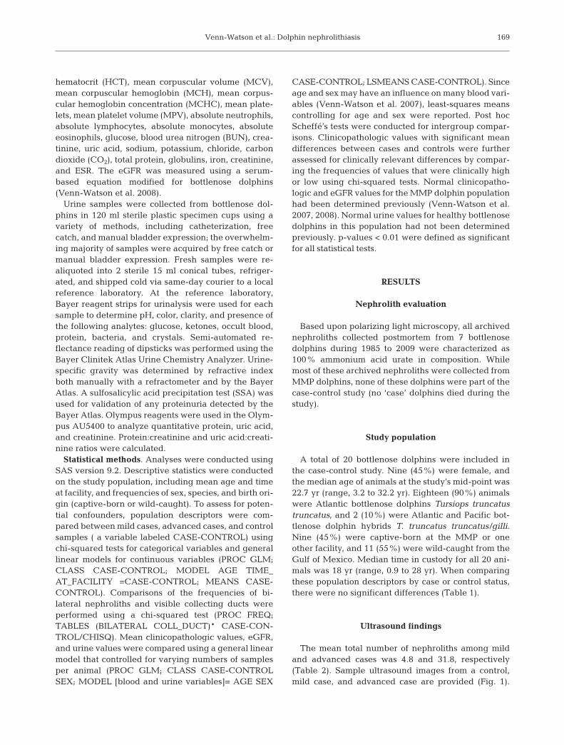
Ultrasound evaluations and study population defin-itions. Both kidneys from 20 dolphins were evaluatedand total nephrolith counts were estimated using atleast 1 renal ultrasound examination during the studyperiod. A Voluson i portable ultrasound machine witha 2 to 5 MHz 4D transducer (RAB2-5-RS; General Elec-tric Healthcare) was used to capture images of the kid-neys. Ultrasound evaluation using B-mode with tissueharmonic imaging included (1) nephrolith identifica-tion and quantification, and (2) measurement of thecollecting duct if it was visible. Collecting-duct mea-surements were only included if color Doppler ultra-sound confirmed that the image was a collecting ductand not a vessel. To evaluate kidneys for the presenceof nephroliths, cine loops of each kidney were cap-tured in both the longitudinal and transverse planesand then reviewed with OsiriX imaging software (opensource; www.osirix-viewer.com). Nephroliths wereidentified as hyperechoic foci within the kidney thatcreated acoustic shadowing and were quantitated for atotal nephrolith count per kidney (see Fig. 1). We didnot use color or power Doppler to look for twinklingartifact as an identifier of nephroliths in our study.Nephrolith measurements were made using the trans-verse plane. Of the population, 6 (30%) had no evi-dence of nephroliths, 8 (40%) had 1 to 19 nephroliths,and 6 (30%) had ≥20 nephroliths. These categories
were defined as controls, mild cases, and advancedcases, respectively. Example ultrasound images fromthe study population are provided to demonstrate acontrol, a mild case, and an advanced case (see Fig. 1).Although not included as a formal part of the presentstudy, 3 of the advanced cases had computer tomogra-phy (CT) scans for medical-management purposes.The number of nephroliths detected by CT in those 3animals closely matched the numbers identified byultrasound.
Sample collection and analyses. Serum and urinesamples were collected from cases and controls over18 mo (January 2006 through June 2007). Blood sam-ples were collected by venipuncture from either theperiarterial venous rete in the caudal peduncle or afluke blade (Dold & Ridgway 2007). These methods forblood collection have been used by the MMP for>30 yr, including for establishing normal referenceranges of complete blood-cell counts and serumchemistries (Venn-Watson et al. 2007). While bloodcollection from the periarterial venous rete has thepotential for arterial or mixed arterial-venous sam-pling, no differences in blood values involved in thepresent study have demonstrated significant differ-ences by sampling site (S. Venn-Watson, C. R. Smith,R. Daniels unpubl. data). Animals were trained to vol-untarily present their tail for sampling, or behavioralconditioning was utilized to collect samples out of thewater on a foam mat during a routine physical exami-nation. Samples were collected using either a 20 or21 gauge 1.5 inch (3.8 cm) Vacutainer® needle (BectonDickinson VACUTAINER Systems) or a 21 gauge0.75 inch (1.9 cm) butterfly needle attached to a Vacu-tainer® Holder. Blood was collected into a Vacu-tainer® serum separator tube (SST) or a Vacutainer®
EDTA (K3) tube for serum chemistries and completeblood counts (CBC), respectively.
Samples were chilled for 30 min and centrifugedwithin 2 h. Centrifugation was performed at 1006 × g at21°C for 10 min. Fibrin clots were removed and serumwas transferred to a 5 ml plastic submission tube.Whole blood was submitted in EDTA Vacutainer®
tubes. All samples were sent on ice via courier to QuestDiagnostic Laboratories in San Diego, California.Automated analyses were used by Quest DiagnosticLaboratories, including the Coulter® LH 1500 Series(Beckman Coulter) for hematology and the Olympus®
AU600 (Olympus America) for serum chemistry analy-sis. Fisherbrand Dispette 2®, correlating with theWestergren method, was used to determine 60 minerythrocyte sedimentation rates (ESR) from 1 ml EDTAwhole blood.
The following hematologic and serum biochemicalvariables were measured and incorporated into theretrospective study: total white blood cell count (WBC),
168
Venn-Watson et al.: Dolphin nephrolithiasis
hematocrit (HCT), mean corpuscular volume (MCV),mean corpuscular hemoglobin (MCH), mean corpus-cular hemoglobin concentration (MCHC), mean plate-lets, mean platelet volume (MPV), absolute neutrophils,absolute lymphocytes, absolute monocytes, absoluteeosinophils, glucose, blood urea nitrogen (BUN), crea-tinine, uric acid, sodium, potassium, chloride, carbondioxide (CO2), total protein, globulins, iron, creatinine,and ESR. The eGFR was measured using a serum-based equation modified for bottlenose dolphins(Venn-Watson et al. 2008).
Urine samples were collected from bottlenose dol-phins in 120 ml sterile plastic specimen cups using avariety of methods, including catheterization, freecatch, and manual bladder expression; the overwhelm-ing majority of samples were acquired by free catch ormanual bladder expression. Fresh samples were re-aliquoted into 2 sterile 15 ml conical tubes, refriger-ated, and shipped cold via same-day courier to a localreference laboratory. At the reference laboratory,Bayer reagent strips for urinalysis were used for eachsample to determine pH, color, clarity, and presence ofthe following analytes: glucose, ketones, occult blood,protein, bacteria, and crystals. Semi-automated re-flectance reading of dipsticks was performed using theBayer Clinitek Atlas Urine Chemistry Analyzer. Urine-specific gravity was determined by refractive indexboth manually with a refractometer and by the BayerAtlas. A sulfosalicylic acid precipitation test (SSA) wasused for validation of any proteinuria detected by theBayer Atlas. Olympus reagents were used in the Olym-pus AU5400 to analyze quantitative protein, uric acid,and creatinine. Protein:creatinine and uric acid:creati-nine ratios were calculated.
Statistical methods. Analyses were conducted usingSAS version 9.2. Descriptive statistics were conductedon the study population, including mean age and timeat facility, and frequencies of sex, species, and birth ori-gin (captive-born or wild-caught). To assess for poten-tial confounders, population descriptors were com-pared between mild cases, advanced cases, and controlsamples ( a variable labeled CASE-CONTROL) usingchi-squared tests for categorical variables and generallinear models for continuous variables (PROC GLM;CLASS CASE-CONTROL; MODEL AGE TIME_AT_FACILITY =CASE-CONTROL; MEANS CASE-CONTROL). Comparisons of the frequencies of bi-lateral nephroliths and visible collecting ducts wereperformed using a chi-squared test (PROC FREQ;TABLES (BILATERAL COLL_DUCT)* CASE-CON-TROL/CHISQ). Mean clinicopathologic values, eGFR,and urine values were compared using a general linearmodel that controlled for varying numbers of samplesper animal (PROC GLM; CLASS CASE-CONTROLSEX; MODEL [blood and urine variables]= AGE SEX
CASE-CONTROL; LSMEANS CASE-CONTROL). Sinceage and sex may have an influence on many blood vari-ables (Venn-Watson et al. 2007), least-squares meanscontrolling for age and sex were reported. Post hocScheffé’s tests were conducted for intergroup compar-isons. Clinicopathologic values with significant meandifferences between cases and controls were furtherassessed for clinically relevant differences by compar-ing the frequencies of values that were clinically highor low using chi-squared tests. Normal clinicopatho-logic and eGFR values for the MMP dolphin populationhad been determined previously (Venn-Watson et al.2007, 2008). Normal urine values for healthy bottlenosedolphins in this population had not been determinedpreviously. p-values < 0.01 were defined as significantfor all statistical tests.
RESULTS
Nephrolith evaluation
Based upon polarizing light microscopy, all archivednephroliths collected postmortem from 7 bottlenosedolphins during 1985 to 2009 were characterized as100% ammonium acid urate in composition. Whilemost of these archived nephroliths were collected fromMMP dolphins, none of these dolphins were part of thecase-control study (no ‘case’ dolphins died during thestudy).
Study population
A total of 20 bottlenose dolphins were included inthe case-control study. Nine (45%) were female, andthe median age of animals at the study’s mid-point was22.7 yr (range, 3.2 to 32.2 yr). Eighteen (90%) animalswere Atlantic bottlenose dolphins Tursiops truncatustruncatus, and 2 (10%) were Atlantic and Pacific bot-tlenose dolphin hybrids T. truncatus truncatus/gilli.Nine (45%) were captive-born at the MMP or oneother facility, and 11 (55%) were wild-caught from theGulf of Mexico. Median time in custody for all 20 ani-mals was 18 yr (range, 0.9 to 28 yr). When comparingthese population descriptors by case or control status,there were no significant differences (Table 1).
Ultrasound findings
The mean total number of nephroliths among mildand advanced cases was 4.8 and 31.8, respectively(Table 2). Sample ultrasound images from a control,mild case, and advanced case are provided (Fig. 1).
169

Dis Aquat Org 89: 167–177, 2010170
Descriptor Control Mild cases Advanced cases p(n = 6) (n = 8) (n = 6)
Percent female 66.7 37.5 33.3 0.44Percent captive born 33.3 50 50 0.79Median age (yr) 22.7 (range 4.2–26.2) 14.7 (range 3.2–32.2) 12.6 (range 0.7–28.4) 0.78Median time at institution (yr) 18.1 (range 4.2–19.3) 11.6 (range 0–28.4) 12.6 (range 0.7–28.4) 0.85
Table 1. Tursiops truncatus. Comparisons of population descriptors by study group. Mild cases: 1 to 19 nephroliths, advancedcases: ≥20 nephroliths
Ultrasound descriptor Control Mild cases Advanced cases p(n = 6) (n = 8) (n = 6)
Mean total nephroliths 0 4.8 31.8 <0.0001Mean nephrolith size (cm) na 0.8 0.9 0.23No. (%) bilateral nephroliths na 4/8 (50) 6/6 (100) 0.02No. (%) left collecting duct visualized 0 0 3/6 (50) <0.0001No. (%) right collecting duct visualized 0 0 2/6 (33) <0.0001Range of size of collecting duct, if visualized (cm) 0 0 0.7–1.7 na
Table 2. Tursiops truncatus. Comparisons of ultrasound descriptors by study group. Mild cases: 1 to 19 nephroliths, advancedcases: ≥20 nephroliths. na = not applicable
Fig. 1. Tursiops truncatus. Representative renal ultrasoundimages (Voluson i portable ultrasound machine with a 2 to5 MHz 4D transducer; RAB2-5-RS; General Electric Health-care) using B-mode with tissue harmonic imaging. (a) A nor-mal control, (b) a mild case of nephrolithiasis demonstratinga single hyperechoic nephrolith with acoustic shadowing,
and (c) an advanced case with multiple nephroliths

Venn-Watson et al.: Dolphin nephrolithiasis
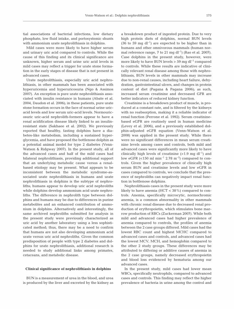
There were no significant differences in meannephrolith size when comparing mild and advancedcases (p = 0.23). The mean and median nephrolith sizesin mild cases were 0.8 ± 0.4 (SD) cm and 0.7 cm (range,0.3 to 1.7 cm), and in advanced cases were 0.9 ± 0.4 cmand 0.7 cm (range, 0.3 to 3.5 cm), respectively. Four(50%) mild cases had bilateral nephroliths, and all(100%) advanced cases had bilateral nephroliths.None of the controls or mild cases had confirmed visi-ble collecting ducts. Three (50%) advanced cases hadvisible collecting ducts with sizes ranging from 0.7 to1.7 cm; 2 of these cases had collecting ducts visualizedbilaterally (Fig. 2). During the study period, 2 of theadvanced cases developed hydronephrosis visualizedby ultrasound, and ureteral obstruction due to anephrolith was confirmed in 1 of these animals by CTscan.
Clinicopathologic tests and eGFR
A summary of mean clinicopathologic values bystudy group is provided in Table 3. Serum BUN wassignificantly higher in advanced cases compared tomild cases, and in mild cases compared to controls.The same trend was detected with BUN:creatinineratios, and there were no significant differences inmean serum creatinine among all 3 groups.
Mild cases were more likely to have lower total WBCcounts and absolute neutrophils compared to ad-vanced cases and controls. Advanced cases had signif-icantly lower serum iron compared to mild cases and
controls. Both mild and advanced cases had lowermean HCT and hemoglobin compared to controls.While advanced cases were more likely to have lowerred blood cell (RBC) distribution width and MCV com-pared to the other 2 study groups, mild cases weremore likely to have higher MCHC and lower RBCcounts. Controls had higher new RBC counts compareto both mild and advanced cases.
Mean platelet count was significantly higher in mildcases compared to advanced cases, and in advancedcases compared to controls. Controls had significantlyhigher total protein and globulins, but not albumin,compared to mild and advanced cases. Mild cases hadhigher serum uric acid compared to advanced casesand controls.
When testing for clinically significant differences inclinicopathologic values among the 3 study groups,both mild and advanced cases were more likely to beanemic, have high BUN, high creatinine, and loweGFR compared to controls (Table 4). Mild cases weremore likely to have thrombocytosis compared toadvanced cases and controls.
Renal health indicators fluctuated over the 18 mostudy period among each of the animals. Examples offluctuations in serum BUN, creatinine, and HCTamong control, mild-case, and advanced-case animalsare provided in Fig. 3.
Urinalyses
Compared to mild and advanced cases, controlswere more likely to have nitrite in urine and higherurine protein concentrations (Tables 5 & 6). Advancedcases were more likely to have hematuria (urine ery-throcytes and occult blood) and lower urinary pH com-pared to mild cases and controls. Mild cases had signif-icantly higher uric acid concentrations in urinecompared to advanced cases and controls. Of 4 ad-vanced cases in which urinary crystals were identified,3 animals had urinary crystals characterized as urate;the fourth advanced case had urinary crystals charac-terized as triple phosphate.
DISCUSSION
Ammonium acid urate nephrolithiasis
We report on a case-control study involving 14 casesof nephrolithiasis in bottlenose dolphins at the MMPwith the purpose of determining the clinical relevanceof this disease and assisting in diagnostic and treat-ment decisions. Nephrolithiasis has been reported inother marine mammal species, including a harbor seal
171
Fig. 2. Tursiops truncatus. Renal ultrasound image (Volusoniportable ultrasound machine with a 2 to 5 MHz 4D trans-ducer; RAB2-5-RS; General Electric Healthcare) of an ad-vanced case of nephrolithiasis. Multiple nephroliths arevisible in the kidney, as well as a dilated collecting duct (CD)and a nephrolith in the collecting duct (arrow) with acoustic
shadowing

Dis Aquat Org 89: 167–177, 2010172
Blood variable Normal reference Least-squares means p Significantrange, if Controls Mild cases Advanced cases comparisons
established (n = 117) (n = 100) (n = 130)
Renal healthBUN (mg dl–1) 34–59 49 53 63 <0.0001 Cont < Mild < AdvCreatinine (mg dl–1) 1.1–1.8 1.3 1.3 1.3 0.39BUN:creatinine ratio 30.9–53.6 39.2 41.6 50.7 <0.0001 Adv > Cont, Mild
InflammationWhite blood cells (cells µl–1) 4895–10935 9203 7427 9311 <0.0001 Mild < ContNeutrophils (cells µl–1) 2737–7570 6227 4927 6141 <0.0001 Mild < Adv, ContLymphocytes (cells µl–1) 312–2731 1358 1252 1300 0.43Monocytes (cells µl–1) 0–576 252 191 232 0.08Eosinophils (cells µl–1) 100–2731 1417 1501 1642 <0.0001 Cont < Mild < AdvGlobulins (g dl–1) 1.9–3.8 2.8 2.6 2.5 <0.0001 Cont > Mild, AdvIron (µg dl–1) 92–332 218 235 161 <0.0001 Adv < Mild, ContAlkaline phosphatase (U l–1) 158–709 340 374 323 0.0460 min ESR (mm h–1) 0–19 10 13 13 0.04
RBC productionHematocrit (%) 38–46 43 41 40 <0.0001 Cont > Mild, AdvHemoglobin (g dl–1) 14.7 14.4 14.0 <0.0001 Cont > Mild, AdvRBCs (× 106 µl–1) 3.3 3.2 3.3 <0.0001 Mild < Adv, ContRBC distribution width (%) 14.4 14.5 13.6 <0.0001 Adv < Mild, ContNew RBCs (100 cells–1) 0.6 0.3 0.2 0.0002 Cont > Mild, AdvMCV (fl) 128 129 121 <0.0001 Adv < Mild, ContMCH (pg) 44 45 42 <0.0001 Adv < Mild, ContMCHC (g dl–1) 34.6 35.0 34.6 <0.0001 Mild > Adv, Cont
Platelets (cells × 103 µl–1) 46–173 94 118 123 <0.0001 Cont < Mild, AdvMPV (fl) 13.1 12.6 12.9 0.03Total protein (g dl–1) 5.9–7.7 7.1 6.9 6.8 <0.0001 Cont > Mild, AdvGlucose (mg dl–1) 80–144 109 104 106 0.05Carbon dioxide (mEq l–1) 17–28 24.8 25.0 24.4 0.13Sodium (mEq l–1) 152–159 154 154 153 0.01 Adv < Mild, ContChloride (mEq l–1) 115–125 118 118 119 0.02Potassium (mEq l–1) 3.4–4.1 3.8 3.8 3.8 0.53Uric acid (mg dl–1) 0.0–0.7 0.17 0.29 0.15 0.0008 Mild > Adv, ContAlbumin (g dl–1) 3.8–4.9 4.4 4.3 4.3 0.43eGFR (ml min–1 2.78 m–2) >150 230 234 234 0.81
Table 3. Tursiops truncatus. Comparisons of mean clinicopathologic and estimated glomerular filtration rate (eGFR) by nephrolithstudy group. Mild cases: 1 to 19 nephroliths, advanced cases: ≥20 nephroliths. Adv: advanced cases; BUN: blood urea nitrogen;Cont: controls; ESR: erythrocyte sedimentation rate; fl: femtoliter; MCH: mean corpuscular hemoglobin; MCHC: mean corpus-
cular hemoglobin concentration; MCV: mean corpuscular volume; MPV: mean platelet volume; RBC: red blood cell
Descriptor Definition No. (%) No. (%) No. (%) pcontrol samples mild cases advanced cases
(n = 116) (n = 99) (n = 128)
Anemia HCT < 38% 1 (0.9) 16 (16.2) 17 (13.3) <0.0001Thrombocytosis Platelets > 140000 cells µl–1 18 (15.5) 32 (32.3) 31 (26.6) 0.01Lymphopenia Lymphocytes < 300 cells µl–1 3 (2.6) 0 (0) 3 (2.3) 0.13High BUN BUN > 59 mg dl–1 10 (8.6) 15 (15.20 73 (56.6) <0.0001High creatinine Creatinine > 1.9 mg dl–1 1 (0.9) 12 (12.1) 7 (5.4) 0.001Hyponaturia Sodium < 152 mEq l–1 13 (11.2) 10 (10.1) 19 (14.7) 0.53Hyperchloremia Chloride > 125 mEq l–1 1 (0.9) 0 (0) 1 (0.8) 0.50Hypoglobulinemia Globulins < 2.0 g dl–1 5 (5.2) 0 (0) 5 (3.9) 0.02Low iron Iron < 100 µg dl–1 3 (2.6) 4 (4.0) 11 (8.5) 0.10Elevated ESR ESR > 20 mm h–1 14 (12.7) 13 (13.7) 19 (15.1) 0.87Low eGFR (<150) eGFR < 150 ml min–1 2.78 m–2 1 (0.9) 12 (12.1) 7 (5.4) 0.001
Table 4. Tursiops truncatus. Comparisons of percent high or low clinicopathologic values by nephrolith study group. Definitionsare based upon previously established normal reference ranges for this animal population (Venn-Watson et al. 2007, 2008). Mildcases: 1 to 19 nephroliths, advanced cases: ≥20 nephroliths. BUN: blood urea nitrogen; eGFR: estimated glomerular filtration rate;
ESR: erythrocyte sedimentation rate; HCT: hematocrit

Venn-Watson et al.: Dolphin nephrolithiasis
(Stroud 1979), northern elephant seal, California sealion (Dennison et al. 2007), and 2 West Indian mana-tees (Keller et al. 2008); the northern elephant seal andCalifornia sea lion had urate nephrolithiasis. We previ-ously reported a case of urate nephrolithiasis associ-
ated with end-stage renal disease in anadult bottlenose dolphin (Venn-Watson etal. 2008).
Urate nephrolithiasis is caused by uricacid, sodium acid urate, or ammonium acidurate calculi. In the present study, allnephroliths tested postmortem from MMPnon-study bottlenose dolphins were char-acterized as being 100% ammonium acidurate. It is assumed, therefore, that the pri-mary type of nephrolithiasis diagnosed byultrasound in our dolphin population isammonium acid urate.
We report significantly lower urinary pHamong advanced cases (pH = 6.0) com-pared to mild cases and controls (pH = 6.4and 6.3, respectively). The foremost riskfactor for urate nephrolithiasis, specificallyuric acid nephrolithiasis, in terrestrial ani-mals is acidic urine (pH < 5.5) (Shekarriz &Stoller 2002). While the present study didassociate lower urinary pH with advancedcases, it also demonstrated that urate neph-rolithiasis may be present in dolphins withurine pH > 5.5 and that urinary pH did notdiffer among mild-nephrolith-formers com-pared to non-nephrolith-formers. Thesefindings are consistent with ammoniumacid urate nephroliths, which can form inacidic, neutral, and even slightly alkalineurine (Berenyi 1972, Klohn et al. 2004). Fur-ther research is needed to better under-stand whether lower urinary pH causesadvanced cases, or if the presence of morenephroliths lowers urinary pH. Addition-ally, if low urinary pH is not the primarycause of urate nephrolithiasis in dolphins,use of alkalinizing treatments may be lim-ited in preventing or treating mild cases.
High-purine diets have been associatedwith urate nephrolithiasis (Ekeruo et al.2004). In most mammals, ingested proteins(purines) are converted to xanthine, whichis subsequently converted to hypoxanthine,xanthine oxidase, uric acid, and, finally, al-lantoin, which is excreted by the kidneys.Exceptions to this purine metabolic path-way include humans and other higher pri-mates, dalmations, birds, reptiles, anddesert mammals, all of which lack the en-
zyme uricase (Vogels & Van Der Drift 1976). For theseanimals, the final product of purine metabolism is uricacid, which can predispose animals to uratenephrolithiasis. While a review of the literature did notconfirm how dolphins metabolize purines, dolphins
173
30
40
50
60
70
80 a
b
c
0 60 120 180 240 300 360 420 480 540
BU
N (m
g d
l–1)
Control Mild case Advanced case
Episode of hydronephrosis in advanced case animal
0.8
1
1.2
1.4
1.6
1.8
2
0
30 60 90
120
150
180
210
240
270
300
330
360
390
420
450
480
510
540
Cre
atin
ine
(mg
dl–1
)
32
36
40
44
0 30 60 90 120
150
180
210
240
270
300
330
360
390
420
450
480
510
540
Days of study
Hem
atoc
rit (%
)
Episode of hydronephrosis in advanced case animal
Fig. 3. Tursiops truncatus. Eighteen month time series of serum (a) bloodurea nitrogen (BUN), (b) creatinine, and (c) hematocrit in a control, a mildcase of nephrolithiasis, and an advanced case. Horizontal dashed line:
normal level of BUN/creatinine/hematocrit

Dis Aquat Org 89: 167–177, 2010
have osmoregulation capabilities resembling that ofsome desert mammals, and as such, may have similarmeans of conserving water and metabolizing purines(Ortiz 2001). It is also possible that cetaceans haveevolved their own process for breaking down purines,independent of the 2 traditional mammalian models, ashas been recently reported in marsupials and mono-tremes (Keebaugh & Thomas 2009). There is a need tostudy purine metabolism, including the presence or ab-sence of uricase in dolphins.
Ammonium urate nephrolithiasis in terrestrial mam-mals has been associated with infections by urealyticbacteria, low dietary and urinary phosphate, low fluidintake, and portosystemic shunts (Osborne et al. 1995,Klohn et al. 2004). The controls in the present studywere significantly more likely to have urinary nitrite,an indicator of bacteria, compared to cases; further,controls had the highest prevalence of urinary bacte-ria. These findings do not support the conclusion thaturinary tract infection or nephritis is the primary causeof urate nephrolithiasis in our population. Further, the
overwhelming majority of urine samples in the presentstudy were collected by free catch, and the presence ofcontaminant bacteria in urine not collected by cathe-terization is not unusual. There were no instances,among case or control animals in the present study,when the presence of bacteria in urine was interpretedas a primary bacterial urinary-tract infection. To bettercharacterize true bacterial urinary-tract infectionsamong this animal population, urine samples wouldneed to be collected consistently with a catheter. Casesand controls were fed similar diets of capelin, herring,mackerel, and squid, decreasing the chance thatdietary differences were present among animals in ourpopulation. Finally, animals in the present study popu-lation are not known to have portosystemic shunts, andcases were not immediately related to each other; dueto the high prevalence of nephrolithiasis in our popula-tion, it is unlikely that nephrolithiasis is caused byportsystemic shunts. There have been no formal evalu-ations, however, for portosystemic shunts in dolphins.Additional studies are needed to better assess poten-
174
Descriptor No. (%) No. (%) No. (%) pcontrol samples mild cases advanced cases
(n = 49) (n = 67) (n = 96)
Glucose — positive 8/49 (16.3) 12/67 (17.9) 18/96 (18.8) 0.80Ketones — positive 2/47 (4.3) 0/67 (0) 2/96 (2.1) 0.47Nitrite — positive 16/49 (32.7) 0 (0) 0 (0) <0.0001Leukocytes — positive 23/49 (46.9) 23/67 (34.3) 40/96 (41.7) 0.006Erythrocytes — positive 11/25 (44.0) 7/40 (17.5) 53/55 (96.4) <0.0001Clarity — cloudy 10/28 (35.7) 1/45 (2.2) 26/67 (38.8) <0.0001Appearance — abnormal 5/20 (25) 1/40 (2.5) 9/63 (14.3) 0.02Occult blood — positive 15/48 (31.3) 3/65 (4.6) 61/94 (64.9) <0.0001Bacteria — positive 6/25 (24) 3/40 (7.5) 7/48 (14.6) 0.36Crystals — positive 3a/25 (12) 2b/40 (5) 6c/55 (10.9) 0.14Hyaline casts — positive 3/49 (6.1) 3/70 (4.3) 8/98 (8.2) 0.59
aThree samples from 3 control animals (1 with urate crystals, 1 with calcium oxalate crystals, 1 with triple phosphate crystals)bTwo samples from 2 mild cases (2 animals with triple phosphate crystals)cSix samples from 4 advanced cases (3 animals with urate crystals, 1 animal with triple phosphate crystals)
Table 5. Tursiops truncatus. Comparisons of urinalysis results (categorical variables) by nephrolith study group. Mild cases: 1to 19 nephroliths; advanced cases: ≥20 nephroliths
Urine variable Control samples Mild cases Advanced cases p Significant group(n = 49) (n = 67) (n = 96) comparisons
pH 6.3 6.4 6.1 0.007 Adv < Cont, MildSpecific gravity 1.026 1.026 1.028 0.33Creatinine (mg dl–1) 97 76 104 0.04Protein (mg mg–1 creatinine) 1025 141 333 <0.0001 Cont > Mild, AdvUric acid (mg mg–1 creatinine) 118 294 69 <0.0001 Mild > Adv, Cont
Table 6. Tursiops truncatus. Comparisons of urine values (continuous variables) by nephrolith study group. Mild cases: 1 to19 nephroliths; advanced cases: ≥20 nephroliths. Adv: advanced cases; Cont: controls
Venn-Watson et al.: Dolphin nephrolithiasis
tial associations of bacterial infections, low dietaryphosphate, low fluid intake, and portosystemic shuntswith ammonium urate nephrolithiasis in dolphins.
Mild cases were more likely to have higher serumand urinary uric acid compared to controls. While thecause of this finding and its clinical significance areunknown, higher serum and urine uric acid levels inmild cases may reflect a trigger for urate stone forma-tion in the early stages of disease that is not present inadvanced cases.
Urate nephrolithiasis, especially uric acid nephro-lithiasis, in other mammals has been associated withhyperuricemia and hyperuricosuria (Ngo & Assimos2007). An exception is pure urate nephrolithiasis asso-ciated with insulin resistance in humans (Abate et al.2004, Daudon et al. 2006); in these patients, pure uratestone formation occurs in the face of normal urine uric-acid levels and low serum uric-acid levels. Normouric-osuric uric-acid nephrolith-formers appear to have arenal acidification disease likely linked to an insulin-resistant state (Sakhaee et al. 2002). We previouslyreported that healthy, fasting dolphins have a dia-betes-like metabolism, including a sustained hyper-glycemia, and have proposed the bottlenose dolphin asa potential animal model for type 2 diabetes (Venn-Watson & Ridgway 2007). In the present study, all ofthe advanced cases and half of the mild cases hadbilateral nephrolithiasis, providing additional supportthat an underlying metabolic cause versus a renal-based etiology may be present. What appears to beinconsistent between the metabolic syndrome-as-sociated urate nephrolithiasis in humans and uratenephrolithiasis in dolphins is the subtype of nephro-liths; humans appear to develop uric acid nephrolithswhile dolphins develop ammonium acid urate nephro-liths. The difference in nephrolith type between dol-phins and humans may be due to differences in purinemetabolites and an enhanced contribution of ammo-nium in dolphins. Alternatively and interestingly, thesame archived nephroliths submitted for analysis inthe present study were previously characterized asuric acid by another laboratory using a less sophisti-cated method; thus, there may be a need to confirmthat humans are not also developing ammonium acidurate versus uric acid nephroliths. Given the commonpredisposition of people with type 2 diabetes and dol-phins for urate nephrolithiasis, additional research isneeded to study additional links among primates,cetaceans, and metabolic disease.
Clinical significance of nephrolithiasis in dolphins
BUN is a measurement of urea in the blood, and ureais produced by the liver and excreted by the kidney as
a breakdown product of ingested protein. Due to veryhigh protein diets of dolphins, normal BUN levels(36 to 59 mg dl–1) are expected to be higher than inhumans and other omnivorous mammals (human nor-mal reference range, 7 to 21 mg dl–1) (Rao et al. 2007).Case dolphins in the present study, however, weremore likely to have BUN levels > 59 mg dl–1 comparedto controls. While these results are indicative of clini-cally relevant renal disease among those with nephro-lithiasis, BUN levels in other mammals may increasedue to non-renal causes, including heart failure, dehy-dration, gastrointestinal ulcers, and changes in proteincontent of diet (Pagana & Pagana 2006); as such,increased serum creatinine and decreased GFR arebetter indicators of reduced kidney function.
Creatinine is a breakdown product of muscle, is pro-duced at a constant rate, and is filtered by the kidneyswith no reabsorption, making it a reliable indicator ofrenal function (Perrone et al. 1992). Serum creatinine-based eGFR are routinely used in human medicine(Levey et al. 2006), and a previously established dol-phin-adjusted eGFR equation (Venn-Watson et al.2008) was applied in the present study. While therewere no significant differences in mean serum creati-nine levels among cases and controls, both mild andadvanced cases were significantly more likely to haveclinically high levels of creatinine (>1.9 mg dl–1) andlow eGFR (<150 ml min–1 2.78 m–2) compared to con-trols. Given the higher prevalence of clinically highserum BUN and creatinine levels and low eGFR incases compared to controls, we conclude that the pres-ence of nephroliths can negatively impact renal func-tion in bottlenose dolphins.
Nephrolithiasis cases in the present study were morelikely to have anemia (HCT < 38%) compared to con-trols. Anemia, specifically microcytic, normochromicanemia, is a common abnormality in other mammalswith chronic renal disease due to decreased renal pro-duction of erythropoietin, which stimulates bone mar-row production of RBCs (Zuckerman 2007). While bothmild and advanced cases had higher prevalence ofanemia compared to controls, the profiles of anemiabetween the 2 case groups differed. Mild cases had thelowest RBC count and highest MCHC compared toadvanced cases and controls, and advanced cases hadthe lowest MCV, MCH, and hemoglobin compared tothe other 2 study groups. These differences may beattributed to differing or additive causes of anemia inthe 2 case groups, namely decreased erythropoietinand blood loss evidenced by hematuria among ouradvanced cases.
In the present study, mild cases had lower meanWBCs, specifically neutrophils, compared to advancedcases and controls. This finding may reflect the higherprevalence of bacteria in urine among the control and
175

Dis Aquat Org 89: 167–177, 2010
advanced case animals (24 and 14.6% of urine sam-ples, respectively) compared to the mild cases (7.4% ofurine samples). These differences in bacteria-positiveurine samples were not statistically significant, how-ever, and most of the samples were susceptible to con-tamination via free catch.
Lacking in our case animals was a significant changein specific gravity, which is expected to change inother species with advanced renal disease. Dolphinsare very good osmoregulators and have a reniculaterenal morphology (Ortiz 2001). Given the challenge ofliving in a saline environment without access to freshwater, dolphins and other marine mammals may havea highly adaptable renal physiology that limits the useof specific gravity for clinical purposes until they nearend-stage renal disease.
Study limitations include the use of ultrasonographicdiagnosis of renal nephrolithiasis, in which the numberof nephroliths counted is less reliable than using CTscanning (Fowler et al. 2002). Three (21%) case dol-phins also had renal CT scans, however, and resultswere comparable with ultrasound analysis in thoseanimals. Case definitions were dependent uponnephrolith counts, and nephrolith sizes varied from3 mm to >2 cm. An alternate means of defining casesand controls may have been volume of nephrolithsrelative to kidney size, but a reliable measuring toolwas not readily accessible for the present study. Inmost of the dolphins, case animals were diagnosedwith nephrolithiasis before the study period (January2006 to June 2007); as part of the MMP’s treatmentplan for dolphins with nephrolithiasis, 6 (43%) caseanimals were on hydration therapy (1 to 4 l tube-fedfresh water d–1), and varying animals were on varyingtreatments, including 2 (14%) cases on allopurinol (900to 2100 mg orally twice a day), 3 (21%) cases on potas-sium citrate (30 to 100 mEq orally twice d–1), and 2(14%) cases on sodium bicarbonate (2600 to 3250 mgorally 3 to 4 times d–1). Hydration therapy, uric acid for-mation inhibitors, and urine alkalinizers may haveinfluenced blood and urine values, masking differ-ences between cases and controls. A preliminaryassessment among cases over 2 yr, however, has notyielded any evidence that these medications have sig-nificantly impacted serum uric acid, urine uric acid, orurine pH over time; and treatment with allopurinol hasbeen discontinued while we investigate whetheradministering allopurinol with a naturally purine-richfish diet could lead to xanthine nephrolith formation.Finally, given the high prevalence of nephrolithiasisand possible presence of predisposing risk factorsamong all animals in the study population, the value ofcomparisons between cases and controls may be lim-ited. Similar comparisons of blood and urine variablesbetween this population and wild dolphins may pro-
vide valuable insight into the risk factors for and etiol-ogy of nephrolithiasis.
Nephroliths in marine mammals can lead to ureteraland urethral obstruction, hydronephrosis, kidney atro-phy, and loss of function (Dennison et al. 2007, Kelleret al. 2008, Venn-Watson et al. 2008), as was also evi-dent in the present study’s population of dolphins. Assuch, the MMP is striving to treat and prevent nephro-lithiasis in dolphins. Invasive treatments used inhumans, including lithotripsy, have been consideredbut are logistically problematic. Urine alkalinizers todate have not appeared to have changed urine pH ornephrolith presence in case dolphins. Currently, caseanimals are provided 1 to 3 l fresh water d–1, and on-going studies are investigating the potential role ofdiet and associated metabolic responses in preventingor facilitating ammonium urate stone formation.
In conclusion, ammonium acid urate nephrolithiasisis present in bottlenose dolphins.
Among dolphins with nephroliths visualized byultrasound, this disease appears to be clinically rele-vant, impacting both renal function and erythropoietinproduction. While the cause of ammonium acid uratenephrolithiasis in dolphins is unknown, the lack ofhyperuricosuria and hyperuremia in case animals,along with a high prevalence of bilateral disease, mayindicate that a metabolic syndrome is present. Betterunderstanding of purine metabolism in dolphins isneeded to target prevention and treatment of nephro-lithiasis in dolphins.
Acknowledgements. We thank the reviewers for their valuedtime, patience, and contributions that improved the qualityand impact of this manuscript. This work was funded by theOffice of Naval Research’s In-house Laboratory IndependentResearch (ILIR) program.
LITERATURE CITED
Abate N, Chandalia M, Cabo-Chan AV, Moe OW, SakahaeeK (2004) The metabolic syndrome and uric acid nephro-lithiasis: novel features of renal manifestation of insulinresistance. Kidney Int 65:386–392
Berenyi M (1972) Models for the formation of uric acid andurate stones. Int Urol Nephrol 4:199–204
Daudon M, Traxer O, Conort P, Lacour B, Jungers P (2006)Type 2 diabetes increases the risk for uric acid stones.J Am Soc Nephrol 17:2026–2033
Dennison S, Gulland F, Haulena M, Morais HE, Colgrove K(2007) Urate nephrolithiasis in a Northern elephant seal(Mirounga angustirostris) and a California sea lion (Zalo-phus californianus). J Zoo Wildl Med 38:114–120
Dold C, Ridgway S (2007) Cetaceans. In: West G, Heard DJ,Caulkett N (eds) Zoo animal and wildlife immobilization andanesthesia. Blackwell Publishers, Ames, IA, p 485–496
Ekeruo W, Tan Y, Young M, Dahm P and others (2004) Meta-bolic risk factors and the impact of medical therapy onthe management of nephrolithiasis in obese patients.J Urol 172:159–163
176

Venn-Watson et al.: Dolphin nephrolithiasis
Fowler KAB, Locken JA, Duchesne JH, Williamson MR (2002)US for detecting renal calculi with nonenhanced CT as areference standard. Radiology 222:109–113
Grove RA, Bildfell R, Henny CJ, Buhler DR (2003) Bilateraluric acid nephrolithiasis and ureteral hypertrophy in afree-ranging river otter (Lontra canadensis). J Wildl Dis39:914–917
Keebaugh AC, Thomas JW (2009) The genomes of the SouthAmerican opossum (Monodelphin domestica) and platy-pus (Ornithorhynchus anatinus) encode a more completepurine catabolic pathway than placental mammals. CompBiochem Physiol D 4:174–178
Keller M, Moliner JL, Vasquez G, Cruz D and others (2008)Nephrolithiasis and pyelonephritis in two West Indianmanatees (Trichechus manatus spp.). J Wildl Dis 44:707–711
Klohn M, Bolle JF, Reverdin NP, Susini A, Baud A, Graber P(2004) Ammonium urate urinary stones. Urol Res 14:315–318
Levey AS, Coresh J, Greene T, Stevens LA and others (2006)Using standardized serum creatinine values in the modifi-cation of diet in renal disease study equation for estimat-ing glomerular filtration rate. Ann Intern Med 145:247–254
Moe O (2006) Kidney stones: pathophysiology and medicalmanagement. Lancet 367:333–344
Ngo TC, Assimos DG (2007) Uric acid nephrolithiasis: recentprogress and future directions. Rev Urol 9:17–27
Ortiz RM (2001) Osmoregulation in marine mammals. J ExpBiol 204:1831–1844
Osborne CA, Lulich JP, Bartges JW, Unger LK and others(1995) Canine and feline urolithiasis: relationship ofetiopathogenesis to treatment and prevention. In: Os-borne CA, Finco DR (eds) Canine and feline urology.Williams & Wilkins, Baltimore, MD, p 798–888
Pagana KD, Pagana TJ (2006) Mosby’s manual of diagnosticand laboratory tests, 3rd edn. Mosby, St. Louis, MO
Perrone RD, Madias NE, Levey AS (1992) Serum creatinine asan index of renal function: new insights into old concepts.Clin Chem 38:1933–1953
Rao DA, Le T, Bhushan V (2007) First aid for the USMLE step1 2008. McGraw-Hill Medical, New York
Sakhaee K, Adams-Huet B, Moe OW, Pak CYC (2002) Patho-physiologic basis for normouricosuric uric acid nephro-lithiasis. Kidney Int 62:971–979
Shekarriz B, Stoller ML (2002) Uric acid nephrolithiasis: cur-rent concepts and controversies. J Urol 168:1307–1314
Stroud RK (1979) Nephrolithiasis in a harbor seal. J Am VetMed Assoc 175:924–925
Ulrich LK, Bird K, Koehler L, Swanson L (1996) Urolith analy-sis: submission, methods, and interpretation. Vet ClinNorth Am Small Anim Pract 26:393–400
Venn-Watson S, Ridgway S (2007) Big brains and blood glu-cose: common ground for diabetes mellitus in humans anddolphins. Comp Med 57:241–246
Venn-Watson S, Jensen ED, Ridgway SH (2007) Effects of ageand sex on clinicopathologic reference ranges in a healthymanaged Atlantic bottlenose dolphin population. J AmVet Med Assoc 231:596–601
Venn-Watson S, Smith CR, Dold C, Ridgway SH (2008) Use ofa serum-based glomerular filtration rate prediction equa-tion to assess renal function by age, sex, fasting, andhealth status in bottlenose dolphins (Tursiops truncatus).Mar Mamm Sci 24:71–80
Vogels GD, Van Der Drift C (1976) Degradation of purinesand pyrimidines by microorganisms. Bacteriol Rev 40:403–468
Zuckerman K (2007) Approach to the anemias. In: Goldman L,Ausiello D (eds) Cecil medicine, 23rd edn. Saunders Else-vier, Philadelphia, PA, p 1179–1186
177
Editorial responsibility: Michael Moore,Woods Hole, Massachusetts, USA
Submitted: August 12, 2009; Accepted: December 1, 2009Proofs received from author(s): March 1, 2010