clinical significance of cerebrovascular complications in ... · cvc was associated with high risk...
TRANSCRIPT

Lee et al. BMC Neurology 2014, 14:30http://www.biomedcentral.com/1471-2377/14/30
RESEARCH ARTICLE Open Access
Clinical significance of cerebrovascularcomplications in patients with acute infectiveendocarditis: a retrospective analysis of a 12-yearsingle-center experienceSeung-Jae Lee1*, Sam-Sae Oh2, Dal-Soo Lim3, Chan-Young Na4 and Jae-Hyun Kim4
Abstract
Background: Cerebrovascular complications (CVCs) frequently occur in patients with acute infective endocarditis(IE). The aim of this study is to describe the clinical findings of CVCs and to evaluate the impact of CVCs onlong-term mortality in patients with IE.
Methods: We retrospectively analyzed 144 patients who fulfilled the modified Duke’s criteria for definite left-sidedIE. CVCs were classified into minor (silent cerebral embolism, TIA and stroke with an initial modified Rankin scale ≤ 2) ormajor (an initial modified Rankin scale ≥ 3) CVCs. Cox proportional hazards model was used for mortality analysis.Hazard ratio (HR) and 95% confidence interval (CI) were obtained.
Results: The mean age of the 144 patients (96 males and 48 females) was 49.1 years (range 6-85 years). A CVCwas found in 37 (25.7%) patients. Of these, 25 were treated with surgical therapy. The patients who underwentearly surgery within 2 weeks after stroke had a statistical trend toward a higher risk of postoperative brainhemorrhage (50% versus 4.8%, P = 0.057 by Fisher exact test). The minor CVC group had a similar risk of death as theno-CVC group (P= 0.803; HR 0.856; CI 0.253-2.894), whereas the major CVC group had a higher mortality (P= 0.013; HR 2.865;CI 1.254-6.548) than the no-CVC group. In the multivariate analysis, major CVC (P = 0.002; HR 3.893; CI 1.649-9.194) was asignificant predictor of mortality in IE patients, together with advanced age (P = 0.005; HR 3.138; CI 1.421-6.930) andprosthetic valve IE (P = 0.008; HR 2.819; CI 1.315-6.044).
Conclusions: IE can give rise to various forms of CVC, most frequently, acute ischemic brain lesions. In our study, majorCVC was associated with high risk of mortality although total CVC was not significantly related to the risk of death inpatients with IE.
Keywords: Infective endocarditis, Cerebrovascular complication, Stroke
BackgroundCerebrovascular complications (CVCs) frequently occurin patients who are in the active stage of infective endocar-ditis (IE), and result from cerebral septic embolization ofan endocardial vegetation. They include stroke, transientischemic attack (TIA) and silent cerebral embolism (SCE).A CVC is generally accepted as a predictor of poor
prognosis with an increased mortality in patients with IE[1-3]. Especially, several studies have recently reported
* Correspondence: [email protected] of Neurology, Sejong General Hospital, Bucheon, South KoreaFull list of author information is available at the end of the article
© 2014 Lee et al.; licensee BioMed Central LtdCommons Attribution License (http://creativecreproduction in any medium, provided the or
that the mortality in IE patients depends on type orseverity of IE-related CVC [4,5].In this study, we attempted to (1) describe the inci-
dence, lesion type and neurologic outcome of CVCs, (2)identify the variables determining the occurrence ofCVCs and (3) elucidate the impact of types of CVCs onlong-term mortality in patients with IE.
MethodsUsing the endocarditis registry of Sejong CardiovascularCenter, the authors of this study ascertained the namesand registry numbers of 282 consecutive patients with
. This is an Open Access article distributed under the terms of the Creativeommons.org/licenses/by/2.0), which permits unrestricted use, distribution, andiginal work is properly credited.

Lee et al. BMC Neurology 2014, 14:30 Page 2 of 9http://www.biomedcentral.com/1471-2377/14/30
suspected IE, who were admitted to the CardiovascularCenter at Sejong General Hospital between January 2000and September 2012. We then retrospectively reviewedtheir medical records. From these 282 patients, 104 pa-tients with right-sided IE, 27 with possible left-sided IE,and 7 with incomplete study were excluded. Patientswith both left- and right-sided IE were classified into theleft-sided group. Finally, we analyzed 144 patients whofulfilled the modified Duke’s criteria [6] for definite left-sided IE, and investigated detailed clinical informationincluding medical history (age, sex, hypertension, dia-betes mellitus, Charlson comorbidity index [7], atrial fib-rillation, current smoking status, congestive heart failure(CHF) and history of IE), operation records, computedtomography (CT), magnetic resonance imaging (MRI),echocardiography and clinical outcome including mor-tality. The follow-up data were obtained from outpatientmedical records. The study protocol was reviewed andapproved by the Institutional Review Board of SejongGeneral Hospital. In addition, we had consent from pa-tients or their legal guardians to publish clinical details.Transthoracic and transesophageal echocardiography
were performed in all cases. Echocardiographic data in-cluded IE-related valve regurgitation, vegetation length,mobility and location. The vegetation length was mea-sured in various planes during the first echocardiographyand follow-up studies. It was determined whether themaximal vegetation length was > 1 cm.Diagnosis of a CVC was based on clinical findings, CT
or MRI. CVCs included stroke (ischemic or hemorrhagic),TIA, and SCE. An ischemic stroke was defined as a focalneurologic deficit of an abrupt onset lasting > 24 hourswith an evidence of new lesions on brain imaging, whereasTIA was defined as a focal neurologic symptom lasting <24 hours with or without brain lesions. Hemorrhagicstroke was defined as a neurologic symptom with the pres-ence of intracranial bleeding on CT or MRI, and includedprimary intracerebral hemorrhage (ICH), hemorrhagic in-farct (HI) and subarachnoid hemorrhage (SAH). A newasymptomatic brain lesion with high signal intensity on dif-fusion MRI was considered as SCE.CVCs were classified into minor or major CVCs. A
minor CVC was diagnosed when the initial neurologicsigns were absent, transient or mild. This category in-cluded SCEs, TIAs and strokes with an initial modifiedRankin scale (mRS) ≤ 2. A major CVC was defined as astroke that initially caused a moderate to severe disability(mRS ≥ 3). In addition, cerebral infarct combined withintracranial hemorrhage or one type of CVCs with add-itional neurologic complications (seizure, meningitis or my-cotic aneurysm) was considered as a complicated stroke.The topography of brain lesions was determined using
the commonly accepted arterial supply templates for theterritorial and border zone areas, as described previously
[8-10]. Multiple infarcts were defined as more than twolesions that were topographically distinct (separated inspace or discrete on contiguous slices) [11]. An uninter-rupted lesion visible in contiguous territories was con-sidered a single lesion.As described in a previous study [12], we classified the
embolic brain lesions related to IE into the following 4patterns: (1) single lesion, (2) multiple closely spaced lesionsin a single arterial territory- “territorial infarct”, (3) multiplepunctate disseminated lesions, and (4) multiple small(< 10 mm) and medium (10-30 mm) or large (> 30 mm)disseminated lesions (Figure 1).Statistical analyses were performed with SPSS soft-
ware, version 18.0 (SPSS Inc., Chicago, IL). Independentt-test or Chi-square test (or Fisher exact test) was usedfor comparing the different groups. Multivariate logisticregression analysis was performed to determine the in-dependent predictors for IE-related CVCs. Odds ratio(OR) and 95% confidence interval (CI) were obtained.Besides, the Kaplan-Meier survival curves were com-puted according to the type of CVC, and comparedusing the log-rank test. Cox proportional hazards modelwas used to perform univariate and multivariate analysesfor long-term mortality. Unadjusted and adjusted hazardsratio (HR) and CI were obtained. P-values < 0.05 wereconsidered statistically significant.
ResultsGeneral characteristics of the study populationThe mean age of 144 patients (96 males and 48 females)included in this study was 49.1 years (range 6-85 years)at admission. There were 134 patients with only left-sided IE and 10 with both-sided IE.General characteristics of the study patients are shown
in Table 1. Streptococci including viridans species werethe most common micro-organisms (42 patients, 29.2%).77 patients (53.5%) underwent brain imaging (CT orMRI) and 37 (25.7%) were confirmed as having acutebrain lesions that were comparable to CVCs. Fourteenof 37 patients with CVCs underwent only CT, while theother 23 patients underwent MRI including diffusion-weighted imaging with or without CT.In addition, 31 peripheral embolic events were identi-
fied, involving the spleen in 10 patients (6.9%), kidneysin 7 patients (4.9%), lower limbs in 7 patients (4.9%),heart in 4 patients (2.8%; left anterior descending arteryin 3 and right coronary artery in 1) and lungs in 3 pa-tients (2.1%) with both-sided IE.When compared with the no-CVC group, the CVC
group had a significantly higher prevalence of staphylo-coccus (S.) aureus infection, large (> 1 cm) and mobilevegetation (P < 0.05); and showed a statistical trend to-ward a higher frequency of smoking, dialysis, mitralvalve involvement, no surgery and in-hospital mortality

Figure 1 Patterns of acute embolic lesions on diffusion-weighted MRI in patients with infective endocarditis. Pattern 1 shown in a63-year-old female with dysarthria and right hemiparesis, pattern 2 in a 43-year-old male with dysarthria and right hemiparesis, pattern 3 in a59-year-old female with dysarthria alone, and pattern 4 in a 47-year-old male with confused mentality, dysarthria and left hemiplegia.
Lee et al. BMC Neurology 2014, 14:30 Page 3 of 9http://www.biomedcentral.com/1471-2377/14/30
(P < 0.2). However, there was no significant difference inage, gender, hypertension, diabetes mellitus, atrial fibril-lation, history of CHF, comorbidity, C-reactive proteinlevel at admission, period from the initial symptom to
diagnosis and warfarin use at admission between the twogroups.Logistic regression analysis (using the variables of mi-
tral valve IE, smoking, S. aureus and mobile vegetation)
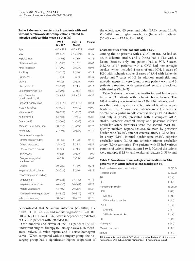
Table 2 Prevalence of neurologic complications in 144patients with acute infective endocarditis: n (%)
Total cerebrovascular complications 37 (25.7)
Ischemic stroke 30 (20.8)
TIA 2 (1.4)
SCE 1 (0.7)
Hemorrhagic stroke 16 (11.1)
ICH 7 (4.9)
ICH only 4 (2.8)
ICH + ischemic stroke 3 (2.1)
SAH 2 (1.4)
SAH only 0 (0)
SAH + ischemic stroke 2 (1.4)
HI 7 (4.9)
Meningitis 1 (0.7)
Mycotic aneurysm 1 (0.7)
Seizure 3 (2.1)
TIA, transient ischemic attack; SCE, silent cerebral embolism; ICH, intracerebralhemorrhage; SAH, subarachnoid hemorrhage; HI, hemorrhagic infarct.
Table 1 General characteristics in patients with andwithout cerebrovascular complications related toinfective endocarditis: mean ± SD, n (%)
CVC (-)N = 107
CVC (+)N = 37
P value
Age 49.5 ± 18.1 48.0 ± 17.1 0.663
Male 69 (64.5) 27 (73.0%) 0.345
Hypertension 18 (16.8) 7 (18.9) 0.772
Diabetes mellitus 21 (19.6) 6 (16.2) 0.647
Atrial fibrillation 31 (29.0) 12 (32.4) 0.692
Smoking 12 (11.2) 8 (21.6) 0.115
History of IE 1 (0.9) 1 (2.7) 0.449
Dialysis 0 (0.0) 2 (5.4) 0.065
History of CHF 32 (29.9) 9 (24.3) 0.517
Comorbidity index >2 22 (20.6) 9 (24.3) 0.631
Initial C-reactiveprotein level, mg/dL
7.4 ± 7.3 8.9 ± 6.3 0.437
Diagnostic delay, days 32.8 ± 35.3 29.9 ± 33.3 0.654
Prosthetic valves 45 (42.1) 16 (43.2) 0.900
Mitral valve IE 78 (72.9) 31 (83.8) 0.183
Aortic valve IE 52 (48.6) 17 (45.9) 0.781
Dual valve IE 22 (20.6) 11 (29.7) 0.253
Warfarin use at admission 38 (35.5) 11 (29.7) 0.522
No surgery 21 (19.6) 12 (32.4) 0.111
Causative microorganism
Streptococcus viridans 18 (16.8) 4 (10.8) 0.441
Other streptococci 15 (14.0) 5 (13.5) 0.939
Staphylococcus aureus 10 (9.3) 9 (24.3) 0.020
Enterococci 6 (5.6) 2 (5.4) 1.000
Coagulase negativestaphylococci
4 (3.7) 2 (5.4) 0.647
Others 30 (28.0) 7 (18.9) 0.274
Negative blood cultures 24 (22.4) 8 (21.6) 0.919
Echocardiographic findings
Vegetations 99 (92.5) 37 (100) 0.113
Vegetation size >1 cm 46 (43.0) 24 (64.9) 0.022
Mobile vegetations 43 (40.2) 29 (78.4) <0.001
IE-related valve regurgitation 88 (82.2) 30 (81.1) 0.874
In-hospital mortality 18 (16.8) 10 (27.0) 0.176
Lee et al. BMC Neurology 2014, 14:30 Page 4 of 9http://www.biomedcentral.com/1471-2377/14/30
demonstrated that S. aureus infection (P = 0.047; OR3.012; CI 1.013-8.962) and mobile vegetation (P = 0.001;OR 4.768; CI 1.952-11.647) were independent predictorsof CVC in patients with left-sided IE.One hundred and eleven of the 144 patients (77.1%)
underwent surgical therapy (53 biologic valves, 36 mech-anical valves, 16 valve repairs and 6 aortic homograftvalves). When compared with the surgery group, the no-surgery group had a significantly higher proportion of
the elderly aged 65 years and older (39.4% versus 14.4%;P = 0.002) and high-comorbidity (index > 2) patients(36.4% versus 17.1%; P = 0.018).
Characteristics of the patients with a CVCAmong the 37 patients with a CVC, 30 (81.1%) had anacute ischemic stroke, and 2 (5.4%) had a TIA with alesion. Besides, only one patient had a SCE. Sixteen(43.2%) of 37 patients with a CVC had hemorrhagicstrokes, which included 4 cases of only ICH, 3 cases ofICH with ischemic stroke, 2 cases of SAH with ischemicstroke and 7 cases of HI. In addition, meningitis andmycotic aneurysm were found in one patient each, and 3patients presented with generalized seizure associatedwith strokes (Table 2).Table 3 shows the vascular territories and lesion pat-
terns in 33 patients with ischemic brain lesions. TheMCA territory was involved in 23 (69.7%) patients, and itwas the most frequently affected arterial territory in pa-tients with IE. Among these patients, most (19 patients,82.6%) had a partial middle cerebral artery (MCA) stroke,and only 4 (17.4%) presented with a complete MCAstroke. Posterior cerebral artery and posterior inferiorcerebellar artery territories were the second most fre-quently involved regions (24.2%), followed by posteriorborder zone (15.2%), anterior cerebral artery (12.1%), basi-lar artery (9.1%), internal border zone (9.1%), superiorcerebellar artery (6.1%) and anterior inferior cerebellarartery (3.0%) territories. The patients with IE had variouspatterns of lesions, from pattern 1 to 4. Most of the lesionswere multiple (84.8%); and territorial (pattern 2, 33%) and

Table 3 Imaging characteristics of 33 patients withischemic brain lesions related to infective endocarditis:n (%)
Vascular territory
Anterior circulation
MCA 23 (69.7)
Complete MCAS 4 (12.1)
Partial MCAS 19 (57.6)
ACA 4 (12.1)
Posterior circulation
PCA 8 (24.2)
BA 3 (9.1)
SCA 2 (6.1)
AICA 1 (3.0)
PICA 8 (24.2)
Border zone
Anterior 0 (0)
Posterior 5 (15.2)
Internal 3 (9.1)
Lesion patterns
Single
Pattern 1 5 (15.2)
Multiple 28 (84.8)
Pattern 2 11 (33.3)
Pattern 3 7 (21.2)
Pattern 4 10 (30.3)
MCAS, middle cerebral artery stroke; ACA, anterior cerebral artery; PCA, posteriorcerebral artery; BA, basilar artery; SCA, superior cerebellar artery; AICA, anteriorinferior cerebellar artery; PICA, posterior inferior cerebellar artery.
Lee et al. BMC Neurology 2014, 14:30 Page 5 of 9http://www.biomedcentral.com/1471-2377/14/30
disseminated small and large lesions (pattern 4, 30.3%)were the prominent lesion patterns.Of the 37 patients with a CVC, 25 were treated with sur-
gical therapy. The mean duration from brain imaging tosurgery was 42 days (range 3-143 days). Only 3 of 25patients treated with surgery expired, while 7 out of 12 pa-tients without surgery died during hospitalization (12.0%versus 58.3%, P = 0.006). Of the 25 patients treated withsurgery, 4 underwent surgical treatment within 2 weeksafter the diagnosis of a minor ischemic CVC (early surgerygroup), whereas the other 21 underwent surgical treatmentafter more than 2 weeks (delayed surgery group). Two of 4patients who underwent early surgery had postoperativeintracranial bleeding (left frontal SAH and intraventricularhemorrhage, respectively), whereas only one of 21 patientswho underwent delayed surgery developed SAH and sub-dural hematoma in the right frontotemporal region post-operatively (50% versus 4.8%, P = 0.057). Among the 4patients who underwent early surgery, the patient whodeveloped intraventricular hemorrhage died from hydro-cephalus and brain herniation, while 2 of 21 patients who
underwent delayed surgery expired due to postoperativebrain complications (the patient mentioned earlier) andseptic shock, respectively (25.0% versus 9.5%, P = 0.422).Thus, the early surgery group had a statistical trend towarda higher risk of postoperative brain complications.
Influence of a CVC on the outcome in patients with IEThe median follow-up period was 51.9 months (range2 - 149 months). Of the 144 patients with IE, 28 diedduring hospitalization. The most common cause of in-hospital death was sepsis with multiple organ failure(18 cases, 64.3%), followed by postoperative ICH (3 cases,10.7%), cerebral infarct progression (2 cases, 7.1%),preoperative ICH (2 cases, 7.1%), sudden cardiac arrest(2 cases, 7.1%) and postoperative bleeding complication(1 case, 3.6%). In addition, two patients expired due topulmonary embolism and sudden cardiac arrest, respect-ively after hospital discharge. Of the 144 patients, 4 with-out a CVC and 2 with a minor CVC (4.2%) were lostduring the follow-up period. These patients were censoredat their last clinic visit.During the follow-up period, the CVC group had a
statistical trend toward a higher mortality, but did notshow a significantly different risk of death from that inthe no-CVC group (P = 0.141; HR 1.747; CI 0.831-3.673).When CVCs were classified according to the severity ofinitial neurologic symptoms, 20 patients and 17 pa-tients belonged to the minor and major CVC groups,respectively. There was a significant difference in thesurvival rate among the no-CVC group, minor CVCgroup and major CVC group (Figure 2A, P = 0.020 bylog rank test). The minor CVC group had a similar riskof death as the no-CVC group (P = 0.803; HR 0.856;CI 0.253-2.894), whereas the major CVC group hada significantly higher mortality (P = 0.013; HR 2.865;CI 1.254-6.548) than the no-CVC group.Of the 20 patients with a minor CVC, 17 (85.0%) had no
disability (mRS 0 or 1) and the other 3 (15.0%) were deadat 6 months. In contrast, only 2 (11.8%) of the 17 patientswith a major CVC had no disability (mRS 0 or 1). In theremaining patients (88.2%), the outcome was disability ordeath at 6 months; 8 patients (47.1%) had a significant dis-ability (mRS 2 or 3 in 7 patients and mRS 5 in 1 patient)and the other 7 (41.2%) were dead at 6 months.Of the 37 patients with a CVC, 16 patients had a com-
plicated stroke, which included 11 cases of ischemicstroke combined with hemorrhage, 1 case of ICH relatedto mycotic aneurysm, 1 case of ischemic stroke withmeningitis, 2 cases of seizures associated with ischemicstroke and 1 case of ischemic stroke with ICH/seizure.There was no significant difference in mortality betweenthe no-CVC group, uncomplicated stroke group andcomplicated stroke group (Figure 2B, P = 0.142). Inaddition, the vascular territory (the involvement of MCA
Figure 2 Long-term survival rate according to the type of CVC. CVC, cerebrovascular complication; MCA, middle cerebral artery.
Lee et al. BMC Neurology 2014, 14:30 Page 6 of 9http://www.biomedcentral.com/1471-2377/14/30
territory) was not related to the risk of death in patientswith IE (Figure 2C, P = 0.210).As shown in Table 4, the univariate Cox regression
model for mortality demonstrated that advanced age(≥ 65), diabetes mellitus, hypertension, atrial fibrillation,history of CHF, comorbidity index >2, prosthetic valveIE, S. aureus infection, no surgery and major CVC havea statistically significant impact on the risk of death.In addition, two levels of multivariate analysis were
performed to evaluate the impact of a major CVC onmortality. In the model 1 where only the age was ad-justed, major CVC (P = 0.002; HR 3.786; CI 1.635-8.771)was significantly associated with high risk of death inpatients with IE. In the model 2, prosthetic valve IE wasadded as a variable in the multivariate analysis. Comor-bidity and S. aureus were excluded despite their statis-tical significance in the univariate analysis because theywere closely related to major CVC (P = 0.011 and 0.012respectively by Chi-square test) thus having a potentialto behave as redundant variables (multicollinearity). Inthis analysis, major CVC (P = 0.002; HR 3.893; CI 1.649-9.194) remained a significant predictor of mortality inIE patients, together with advanced age (P = 0.005; HR3.138; CI 1.421-6.930) and prosthetic valve IE (P = 0.008;HR 2.819; CI 1.315-6.044).
DiscussionA CVC was observed in 25.7% of the patients with IE,which is within the range of 10 to 80% that has been re-ported in previous studies [2,3,5,13-16]. A wide variationin the prevalence among studies may be due to the fail-ure to detect mild or transient neurologic signs in critic-ally ill patients leading to an underestimation of CVC,especially in retrospective studies that used chart re-views. In contrast, the rate of IE-related CVC can beincreased more in patients with S. aureus infection andsevere form of the disease necessitating intensive care
unit admission [2,3]. Additionally, more frequent scan-ning of brain, especially using diffusion-weighted MRI,enhances the detection rate of brain lesions in IE patients.Several prospective studies with a higher frequency of brainimaging showed that the incidence of CVC could reach65-80% although many patients were asymptomatic [15,16].In many previous studies, S. aureus infection has been
found to be associated with a high rate of stroke andmortality in IE patients [3,17,18]. Our results also showedthat S. aureus infection has a significant relationship withthe occurrence of a CVC and death on the univariateanalysis. In addition, multivariate analysis performed inthis study demonstrated that S. aureus infection togetherwith mobile vegetation independently predicted CVC inIE patients.The most common type of neurologic complication re-
lated to IE was acute ischemic stroke (30 cases, 20.8%),followed by hemorrhagic stroke (16 cases, 11.1%), sei-zures (3 cases, 2.1%) and TIA (2 cases, 1.4%). SCE, men-ingitis and mycotic aneurysm were found in only lpatient (0.7%), each. Of these, SCE and meningitis had alower incidence rate (0.7%) in our study than that in pre-vious recent studies, in which SCE was found in 4-30%of patients with IE [3,5,16] and meningitis was found in3-25% of patients with IE [1,4,19]. This could be ex-plained partly by the non-routine use of brain imagingin this study. For example, most of the brain scans seemto have been done only when the patients had a clinicalsymptom suggesting brain complications. In addition,headache, if mild, may not have been investigated formeningitis in patients with serious heart conditions.Acute ischemic lesions in IE patients are mostly mul-
tiple, and mainly located in the MCA territory corre-sponding with the cardioembolic stroke subtype. Inaddition, various patterns of lesions including single, ter-ritorial, disseminated punctate, and disseminated smalland large lesions were seen. These results of this study
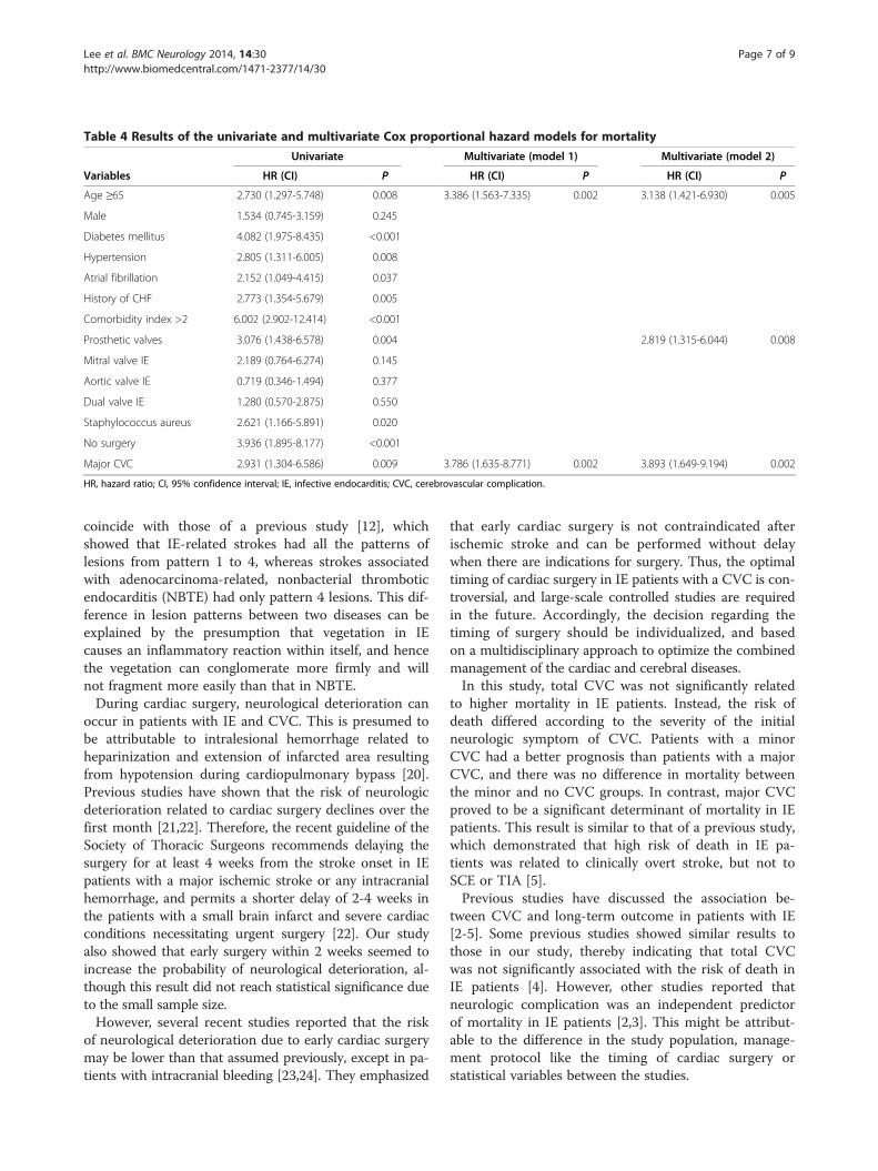
Table 4 Results of the univariate and multivariate Cox proportional hazard models for mortality
Univariate Multivariate (model 1) Multivariate (model 2)
Variables HR (CI) P HR (CI) P HR (CI) P
Age ≥65 2.730 (1.297-5.748) 0.008 3.386 (1.563-7.335) 0.002 3.138 (1.421-6.930) 0.005
Male 1.534 (0.745-3.159) 0.245
Diabetes mellitus 4.082 (1.975-8.435) <0.001
Hypertension 2.805 (1.311-6.005) 0.008
Atrial fibrillation 2.152 (1.049-4.415) 0.037
History of CHF 2.773 (1.354-5.679) 0.005
Comorbidity index >2 6.002 (2.902-12.414) <0.001
Prosthetic valves 3.076 (1.438-6.578) 0.004 2.819 (1.315-6.044) 0.008
Mitral valve IE 2.189 (0.764-6.274) 0.145
Aortic valve IE 0.719 (0.346-1.494) 0.377
Dual valve IE 1.280 (0.570-2.875) 0.550
Staphylococcus aureus 2.621 (1.166-5.891) 0.020
No surgery 3.936 (1.895-8.177) <0.001
Major CVC 2.931 (1.304-6.586) 0.009 3.786 (1.635-8.771) 0.002 3.893 (1.649-9.194) 0.002
HR, hazard ratio; CI, 95% confidence interval; IE, infective endocarditis; CVC, cerebrovascular complication.
Lee et al. BMC Neurology 2014, 14:30 Page 7 of 9http://www.biomedcentral.com/1471-2377/14/30
coincide with those of a previous study [12], whichshowed that IE-related strokes had all the patterns oflesions from pattern 1 to 4, whereas strokes associatedwith adenocarcinoma-related, nonbacterial thromboticendocarditis (NBTE) had only pattern 4 lesions. This dif-ference in lesion patterns between two diseases can beexplained by the presumption that vegetation in IEcauses an inflammatory reaction within itself, and hencethe vegetation can conglomerate more firmly and willnot fragment more easily than that in NBTE.During cardiac surgery, neurological deterioration can
occur in patients with IE and CVC. This is presumed tobe attributable to intralesional hemorrhage related toheparinization and extension of infarcted area resultingfrom hypotension during cardiopulmonary bypass [20].Previous studies have shown that the risk of neurologicdeterioration related to cardiac surgery declines over thefirst month [21,22]. Therefore, the recent guideline of theSociety of Thoracic Surgeons recommends delaying thesurgery for at least 4 weeks from the stroke onset in IEpatients with a major ischemic stroke or any intracranialhemorrhage, and permits a shorter delay of 2-4 weeks inthe patients with a small brain infarct and severe cardiacconditions necessitating urgent surgery [22]. Our studyalso showed that early surgery within 2 weeks seemed toincrease the probability of neurological deterioration, al-though this result did not reach statistical significance dueto the small sample size.However, several recent studies reported that the risk
of neurological deterioration due to early cardiac surgerymay be lower than that assumed previously, except in pa-tients with intracranial bleeding [23,24]. They emphasized
that early cardiac surgery is not contraindicated afterischemic stroke and can be performed without delaywhen there are indications for surgery. Thus, the optimaltiming of cardiac surgery in IE patients with a CVC is con-troversial, and large-scale controlled studies are requiredin the future. Accordingly, the decision regarding thetiming of surgery should be individualized, and basedon a multidisciplinary approach to optimize the combinedmanagement of the cardiac and cerebral diseases.In this study, total CVC was not significantly related
to higher mortality in IE patients. Instead, the risk ofdeath differed according to the severity of the initialneurologic symptom of CVC. Patients with a minorCVC had a better prognosis than patients with a majorCVC, and there was no difference in mortality betweenthe minor and no CVC groups. In contrast, major CVCproved to be a significant determinant of mortality in IEpatients. This result is similar to that of a previous study,which demonstrated that high risk of death in IE pa-tients was related to clinically overt stroke, but not toSCE or TIA [5].Previous studies have discussed the association be-
tween CVC and long-term outcome in patients with IE[2-5]. Some previous studies showed similar results tothose in our study, thereby indicating that total CVCwas not significantly associated with the risk of death inIE patients [4]. However, other studies reported thatneurologic complication was an independent predictorof mortality in IE patients [2,3]. This might be attribut-able to the difference in the study population, manage-ment protocol like the timing of cardiac surgery orstatistical variables between the studies.
Lee et al. BMC Neurology 2014, 14:30 Page 8 of 9http://www.biomedcentral.com/1471-2377/14/30
The present study has several limitations. First of all,the main limitation of this study may be the small sam-ple size based on a single-center experience resulting inthe attenuation of statistical power in the mortality ana-lysis. Second, patients were not managed according to astandardized protocol, and the data were collected retro-spectively. Thus, an arbitrary decision about patientmanagement could have influenced the results of thisstudy. For example, brain imaging was performed in onlyabout half of the study patients based on the decision ofthe attending physician in each situation. Particularly,the critically ill patients did not seem to undergo MRIbecause it could put them at a greater risk. Conse-quently, the rate of asymptomatic brain infarct couldhave been underestimated in comparison with that inthe results of several previous studies.
ConclusionsIE can give rise to various forms of CVC, most fre-quently, acute ischemic brain lesions. In our study,major CVC (initial mRS ≥ 3) was associated with highrisk of mortality although total CVC was not signifi-cantly related to the risk of death in patients with IE.
Competing interestsThe authors declare that they have no competing interests.
Authors’ contributionsAll authors met the criteria for authorship and have approved the contentsof the text. SJL and DSL contributed to study concept and design. SJL didstatistical analysis, wrote the first draft and revised the manuscript. SSO, DSL,CYN and JHK participated in the acquisition, analysis and interpretation of data.
AcknowledgementsThe authors thank the following colleagues for their contributions to dataacquisition in the study: Drs Young-Moo Ro, Suk-Keun Hong, Rak-KyeongChoi, Cheol-Woong Yu and Jin-Sik Park, Department of Cardiology, SejongGeneral Hospital, Bucheon, South Korea.
Author details1Department of Neurology, Sejong General Hospital, Bucheon, South Korea.2Department of Thoracic and Cardiovascular Surgery, Sejong GeneralHospital, Bucheon, South Korea. 3Department of Cardiology, Sejong GeneralHospital, Bucheon, South Korea. 4Department of Thoracic and CardiovascularSurgery, Dong-san Medical Center, Keimyung University, Daegu, South Korea.
Received: 1 July 2013 Accepted: 11 February 2014Published: 15 February 2014
References1. Sonneville R, Mourvillier B, Bouadma L, Wolff M: Management of
neurological complications of infective endocarditis in ICU patients.Ann Intensive Care 2011, 1:10.
2. Mourvillier B, Trouillet JL, Timsit JF, Baudot J, Chastre J, Régnier B, Gibert C,Wolff M: Infective endocarditis in the intensive care unit: clinicalspectrum and prognostic factors in 228 consecutive patients.Intensive Care Med 2004, 30:2046–2052.
3. Sonneville R, Mirabel M, Hajage D, Tubach F, Vignon P, Perez P, Lavoué S,Kouatchet A, Pajot O, Mekontso Dessap A, Tonnelier JM, Bollaert PE, Frat JP,Navellou JC, Hyvernat H, Hssain AA, Tabah A, Trouillet JL, Wolff M,ENDOcardite en REAnimation Study Group: Neurologic complicationsand outcomes of infective endocarditis in critically ill patients: theENDOcardite en REAnimation prospective multicenter study. Crit CareMed 2011, 39:1474–1481.
4. Ruttmann E, Willeit J, Ulmer H, Chevtchik O, Höfer D, Poewe W, Laufer G,Müller LC: Neurological outcome of septic cardioembolic stroke afterinfective endocarditis. Stroke 2006, 37:2094–2099.
5. Thuny F, Avierinos JF, Tribouilloy C, Giorgi R, Casalta JP, Milandre L, BrahimA, Nadji G, Riberi A, Collart F, Renard S, Raoult D, Habib G: Impact ofcerebrovascular complications on mortality and neurologic outcomeduring infective endocarditis: a prospective multicentre study. Eur Heart J2007, 28:1155–1161.
6. Li JS, Sexton DJ, Mick N, Nettles R, Fowler VG Jr, Ryan T, Bashore T, CoreyGR: Proposed modifications to the Duke criteria for the diagnosis ofinfective endocarditis. Clin Infect Dis 2000, 30:633–638.
7. Charlson ME, Pompei P, Ales KL, MacKenzie CR: A new method ofclassifying prognostic comorbidity in longitudinal studies: developmentand validation. J Chronic Dis 1987, 40:373–383.
8. Tatu L, Moulin T, Bogousslavsky J, Duvernoy H: Arterial territories ofthe human brain: brainstem and cerebellum. Neurology 1996,47:1125–1135.
9. Tatu L, Moulin T, Bogousslavsky J, Duvernoy H: Arterial territories of thehuman brain: cerebral hemisphere. Neurology 1998, 50:1699–1708.
10. Ringelstein EB, Stogbauer F: Border zone infarcts. In Stroke Syndromes.2nd edition. Edited by Bogousslavsky J, Caplan L. New York: CambridgeUniversity Press; 2001:564–582.
11. Wong KS, Gao S, Chan YL, Hansberg T, Lam WW, Droste DW, Kay R,Ringelstein EB: Mechanisms of acute cerebral infarctions in patientswith middle cerebral artery stenosis: a diffusion-weighted imaging andmicroemboli monitoring study. Ann Neurol 2002, 52:74–81.
12. Singhal AB, Topcuoglu MA, Buonanno FS: Acute ischemic strokepatterns in infective and nonbacterial thrombotic endocarditis:a diffusion-weighted magnetic resonance imaging study. Stroke 2002,33:1267–1273.
13. Anderson DJ, Goldstein LB, Wilkinson WE, Corey GR, Cabell CH, SandersLL, Sexton DJ: Stroke location, characterization, severity, andoutcome in mitral vs aortic valve endocarditis. Neurology 2003,61:1341–1346.
14. Okazaki S, Yoshioka D, Sakaguchi M, Sawa Y, Mochizuki H, Kitagawa K:Acute ischemic brain lesions in infective endocarditis: incidence, relatedfactors, and postoperative outcome. Cerebrovasc Dis 2013, 35:155–162.
15. Duval X, Iung B, Klein I, Brochet E, Thabut G, Arnoult F, Lepage L, Laissy JP,Wolff M, Leport C, IMAGE (Resonance Magnetic Imaging at the Acute Phaseof Endocarditis) Study Group: Effect of early cerebral magnetic resonanceimaging on clinical decisions in infective endocarditis: a prospectivestudy. Ann Intern Med 2010, 152:497–504.
16. Snygg-Martin U, Gustafsson L, Rosengren L, Alsiö A, Ackerholm P,Andersson R, Olaison L: Cerebrovascular complications in patients withleft-sided infective endocarditis are common: a prospective study usingmagnetic resonance imaging and neurochemical brain damage markers.Clin Infect Dis 2008, 47:23–30.
17. Murdoch DR, Corey GR, Hoen B, Miró JM, Fowler VG Jr, Bayer AS, Karchmer AW,Olaison L, Pappas PA, Moreillon P, Chambers ST, Chu VH, Falcó V, Holland DJ,Jones P, Klein JL, Raymond NJ, Read KM, Tripodi MF, Utili R, Wang A, Woods CW,Cabell CH, International Collaboration on Endocarditis-Prospective Cohort Study(ICE-PCS) Investigators: Clinical presentation, etiology, and outcome ofinfective endocarditis in the 21st century: the International Collaboration onEndocarditis-Prospective Cohort Study. Arch Intern Med 2009, 169:463–473.
18. Fowler VG Jr, Miro JM, Hoen B, Cabell CH, Abrutyn E, Rubinstein E, Corey GR,Spelman D, Bradley SF, Barsic B, Pappas PA, Anstrom KJ, Wray D, Fortes CQ,Anguera I, Athan E, Jones P, van der Meer JT, Elliott TS, Levine DP, Bayer AS,ICE Investigators: Staphylococcus aureus endocarditis: a consequence ofmedical progress. JAMA 2005, 293:3012–3021.
19. Lepur D, Barsić B: Incidence of neurological complications in patientswith native-valve infective endocarditis and cerebral microembolism: anopen cohort study. Scand J Infect Dis 2009, 41:708–713.
20. Derex L, Bonnefoy E, Delahaye F: Impact of stroke on therapeutic decisionmaking in infective endocarditis. J Neurol 2010, 257:315–321.
21. Angstwurm K, Borges AC, Halle E, Schielke E, Einhäupl KM, Weber JR:Timing the valve replacement in infective endocarditis involving thebrain. J Neurol 2004, 251:1220–1226.
22. Byrne JG, Rezai K, Sanchez JA, Bernstein RA, Okum E, Leacche M, Balaguer JM,Prabhakaran S, Bridges CR, Higgins RS: Surgical management of endocarditis:the society of thoracic surgeons clinical practice guideline. Ann Thorac Surg2011, 91:2012–2019.

Lee et al. BMC Neurology 2014, 14:30 Page 9 of 9http://www.biomedcentral.com/1471-2377/14/30
23. Barsic B, Dickerman S, Krajinovic V, Pappas P, Altclas J, Carosi G, Casabé JH,Chu VH, Delahaye F, Edathodu J, Fortes CQ, Olaison L, Pangercic A, Patel M,Rudez I, Tamin SS, Vincelj J, Bayer AS, Wang A, International Collaborationon Endocarditis–Prospective Cohort Study Investigators: Influence of thetiming of cardiac surgery on the outcome of patients with infectiveendocarditis and stroke. Clin Infect Dis 2013, 56:209–217.
24. Yoshioka D, Sakaguchi T, Yamauchi T, Okazaki S, Miyagawa S, Nishi H,Yoshikawa Y, Fukushima S, Saito S, Sawa Y: Impact of early surgicaltreatment on postoperative neurologic outcome for active infectiveendocarditis complicated by cerebral infarction. Ann Thorac Surg 2012,94:489–495.
doi:10.1186/1471-2377-14-30Cite this article as: Lee et al.: Clinical significance of cerebrovascularcomplications in patients with acute infective endocarditis: aretrospective analysis of a 12-year single-center experience. BMCNeurology 2014 14:30.
Submit your next manuscript to BioMed Centraland take full advantage of:
• Convenient online submission
• Thorough peer review
• No space constraints or color figure charges
• Immediate publication on acceptance
• Inclusion in PubMed, CAS, Scopus and Google Scholar
• Research which is freely available for redistribution
Submit your manuscript at www.biomedcentral.com/submit