clinical study the increase of intra-abdominal...
TRANSCRIPT

Clinical StudyThe Increase of Intra-Abdominal Pressure Can AffectIntraocular Pressure
Ilhan Ece,1 Celalettin Vatansev,2 Tevfik Kucukkartallar,3 Ahmet Tekin,3
Adil Kartal,3 and Mehmet Okka4
1Department of Surgery, Selcuk University, Faculty of Medicine, 42075 Konya, Turkey2Department of Surgery, Konya Medicana Hospital, 42100 Konya, Turkey3Department of Surgery, Necmettin Erbakan University, Faculty of Medicine, 42080 Konya, Turkey4Department of Eye Surgery, Necmettin Erbakan University, Faculty of Medicine, 42080 Konya, Turkey
Correspondence should be addressed to Ilhan Ece; [email protected]
Received 8 October 2014; Accepted 20 November 2014
Academic Editor: Subhas Gupta
Copyright © 2015 Ilhan Ece et al.This is an open access article distributed under the Creative CommonsAttribution License, whichpermits unrestricted use, distribution, and reproduction in any medium, provided the original work is properly cited.
Objective. This study aims to explore the usage of intraocular pressure measurements as the early indicator of the increase in intra-abdominal pressure.Methods. In this prospective study, 40 patients undergoing elective surgerywere included. Patientswere dividedinto four groups of 10 patients. The control group (Group C) was not subjected to laparoscopic intervention. Laparoscopic surgerywas, respectively, performed with an intra-abdominal pressure of 9, 12, and 15mmHg in Groups L (low), M (medium), and H (highpressure). Intraocular pressurewasmeasured binocularly in each patient at three different times (before, during, and end of surgery)using a contact tonometer. Results. Patients’ gender, age, body mass index (BMI), American Society of Anesthesiology (ASA) class,and operative times were not different among the groups. No complications occurred with either the surgery or measurement ofintraocular pressure. Intubation was associated with a severe rise in IOP (𝑃 < 0.05). An increase in intraocular pressure was seenin groups M and H (𝑃 < 0.05). Conclusion. Intraocular pressure was increased in the groups with an intra-abdominal pressure of12 mmHg or more. Measuring the intraocular pressure might be a useful method to estimate the intra-abdominal pressure. Thistrial is registered with NCT02319213.
1. Introduction
The adverse effects of intra-abdominal hypertension (IAH)on other systems were first revealed with the recognitionof the relationship between IAH and oliguria. The influenceof IAH on cardiovascular, renal, and the pulmonary systembecame definite through the publications in the first half ofthe 20th century [1].
The normal pressure of the abdominal cavity is almostequal to atmospheric pressure. However, the pressureincreases 5–7mmHg with the respiratory cycle. Pressureswhich constantly develop above 12mmHg are defined as IAH.
Intra-abdominal pressure might be measured invasivelythrough a port placed in the abdomen, the transvesicalroute, the transgastric route, or the inferior vena cava. Thetransvesical method is the most common one as it costs less,and itmight be easily applied and learned [2, 3].Measurementwith transgastric and vena cava ports has not been commonly
used due to the high costs and the infection risk. It has beenreported that intrathoracic pressure, intracerebral pressure[4, 5], and intraocular pressure [6, 7] increase when IAPincreases. Nevertheless, it is known that increased intracra-nial pressure would affect intraocular pressure [8]. However,there have not been adequate studies about the relationshipbetween IAP and IOP.
In our study, we aimed to determine the IAP increase bymeans of measuring the IOP as an alternative to transvesicalmeasurement and to introduce amore practicalmeasurementmethod for clinical use. The advantages of IOP measurementare the absence of infection risk, easy comprehension, and thenonexistence of costs other than tonometry.
2. Methods
After the approval of the institutional Ethics Committee(no. 08-38) and obtaining of informed consent, 40 adult
Hindawi Publishing CorporationBioMed Research InternationalVolume 2015, Article ID 986895, 4 pageshttp://dx.doi.org/10.1155/2015/986895

2 BioMed Research International
Table 1: Demographic data of the patients.
Group C(𝑛 = 10)
Group L(𝑛 = 10)
Group M(𝑛 = 10)
Group H(𝑛 = 10) 𝑃
Age (y) 40.5 ± 12.4 41.0 ± 10.5 43.1 ± 9.6 42.1 ± 10.2 NSGender (M/F) 7/3 6/4 7/3 6/4 NSBMI (kg/m2) 25.8 ± 2.1 25.1 ± 1.9 25.5 ± 2.0 24.8 ± 1.6 NSASA score (I/II) 4/6 5/5 6/4 5/5 NSOperative time (min) 56.4 ± 12.4 58.5 ± 10.2 55.5 ± 11.3 52.3 ± 14.3 NSNS: not significant; BMI: body mass index; ASA: American Society of Anesthesiologists.
patients between 18 and 55 years old with a body mass index(BMI) of 30 kg/m2 or less were included in the study inaccordance with the American Society of Anesthesiologists(ASA) I-II status. Exclusion criteria were preexisting eyedisease, cardiovascular or neuromuscular disease, difficultintubation, and the use of any antihypertensive agents. Acomplete ophthalmologic evaluation of each patient wasperformed before the surgery by the same physician from theOphthalmology Department.
The patients were divided into four groups with 10subjects in each. The control group consisted of the patientswho had undergone a surgery for inguinal hernia. The studygroups were comprised of patients who had undergonelaparoscopic cholecystectomy, which allows direct and themost accurate intra-abdominal pressure monitorization. Inthese cases, the pressure values were monitored from theinsufflator monitor and the abdomen was insufflated with9mmHg (𝑛 = 10), 12mmHg (𝑛 = 10), and 15mmHg (𝑛 =10). IAP in control group patients was accepted as “zero” inthe measurements made without insufflation. Measurementof IOP was performed for both eyes three times: firstly (time1): postanesthesia induction and 30 seconds prior to theintubation, secondly (time 2): 1 minute after the intubation,and lastly (time 3): 45 minutes after the insufflation. IOPwas measured by the same physician from the Ophthalmol-ogy Department using the Perkins Hand-Held ApplanationTonometer (ClementClarke International Limited, England).
2.1. Surgical Technique. In control group, patients underwentinguinal hernia repair. In study groups, intra-abdominalinsufflation was performed with an infraumbilical 1 cm inci-sion, and all laparoscopic cholecystectomies were performedby a single experienced surgeon with four ports.
2.2. Anesthesia Protocol. A standard anesthesia protocol andmechanical ventilation settings were applied to all patientsin order to eliminate the anesthesia-related changes. Ananaesthesia monitor (Infinity Vista XL, Dragerwerk AG &Co, Germany) was used for standard monitoring includingelectrocardiography, pulse oximetry, heart rate, noninvasiveblood pressure, and neuromuscular transmission.
Patients who had received premedication the previousnight and one hour before the operation were administered1mcg/kg Remifentanil, 0.5mg/kg Atracurium, 1-2mg/kgPropofol for induction, and 0.25mcg/kg Remifentanil. Addi-tionally, 1 MAC desflurane was used for maintenance
of anesthesia. Standardization was attempted by keepingthe respiratory rate at 10–12/min, the tidal volume at 8–20mL/kg, PEEP at 3 cmH
2O, and EtCO
2at 35–40mmHg on
mechanical ventilation (Drager Primus, Dragerwerk AG &Co, Germany).
2.3. Statistical Analysis. The data were evaluated by using theSPSS v13.0 program (SSPS, Inc., Chicago, IL, US). A nor-mality analysis was performed. As the data were consistentwith a normal distribution, variance analysis was used for therepeated measurements and the post hoc Tukey test was usedfor multiple comparisons. The Bonferroni correction 𝑡-testwas used in the dependent groups in order to determine thedifference between the measurements. A 𝑃 level of <0.05 wasaccepted as it was statistically significant.
3. Results
The mean age of 40 patients (26 males, 14 females) was41.4 ± 8.2 years (range 19–55). The mean operative time was54.2 ± 11.6 minutes and the ASA scores were I and II. Thebodymass index was 25.3±1.8 (22–30) kg/m2. There was notany statistical difference between the groups in terms of age,sex, and BMI. The demographic data are shown in Table 1.
The control group consisted of patients who had under-gone an operation for inguinal hernia. All the patientsin the study group underwent standard four-port laparo-scopic cholecystectomy. There were no complications fromthe surgery or measurement of IOP. As the IOP values ofthe right and left eyes did not show a statistically significantdifference, analysis was performed on 40 measured values.
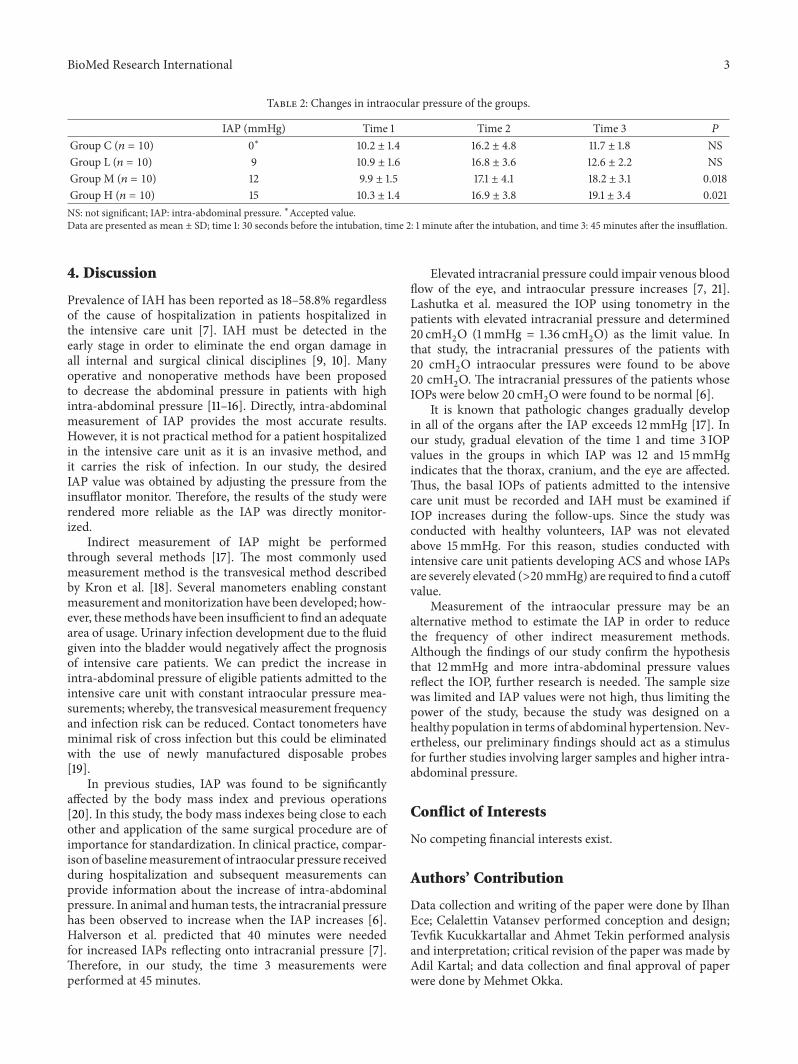
The mean IOP in the four groups showed similar valuesbefore intubation (time 1) and a similar rise after endotra-cheal intubation (time 2) (6.4 ± 3.7mmHg). Furthermore,12mmHg or more pressured (Groups M and H) pneu-moperitoneum induction led to a significant rise in IOPaveraging 8.5 ± 3.4mmHg. The IOP values of medium andhigh pressure group at all the measurement points (times2-3) were higher than the preintubation levels (time 1).However, the IOP values of groups C and L and time 3measurements were, respectively, decreased from 27% to 25%compared with the time 1 measurements (Table 2). Time 1,IOP values were not different between the male and femalepatients. In group C and group L, a mild IOP increase wasobserved at preintubation (time 1) and postinsufflation (time3) measurements.
BioMed Research International 3
Table 2: Changes in intraocular pressure of the groups.
IAP (mmHg) Time 1 Time 2 Time 3 𝑃
Group C (𝑛 = 10) 0∗ 10.2 ± 1.4 16.2 ± 4.8 11.7 ± 1.8 NSGroup L (𝑛 = 10) 9 10.9 ± 1.6 16.8 ± 3.6 12.6 ± 2.2 NSGroup M (𝑛 = 10) 12 9.9 ± 1.5 17.1 ± 4.1 18.2 ± 3.1 0.018Group H (𝑛 = 10) 15 10.3 ± 1.4 16.9 ± 3.8 19.1 ± 3.4 0.021NS: not significant; IAP: intra-abdominal pressure. ∗Accepted value.Data are presented as mean ± SD; time 1: 30 seconds before the intubation, time 2: 1 minute after the intubation, and time 3: 45 minutes after the insufflation.
4. Discussion
Prevalence of IAH has been reported as 18–58.8% regardlessof the cause of hospitalization in patients hospitalized inthe intensive care unit [7]. IAH must be detected in theearly stage in order to eliminate the end organ damage inall internal and surgical clinical disciplines [9, 10]. Manyoperative and nonoperative methods have been proposedto decrease the abdominal pressure in patients with highintra-abdominal pressure [11–16]. Directly, intra-abdominalmeasurement of IAP provides the most accurate results.However, it is not practical method for a patient hospitalizedin the intensive care unit as it is an invasive method, andit carries the risk of infection. In our study, the desiredIAP value was obtained by adjusting the pressure from theinsufflator monitor. Therefore, the results of the study wererendered more reliable as the IAP was directly monitor-ized.
Indirect measurement of IAP might be performedthrough several methods [17]. The most commonly usedmeasurement method is the transvesical method describedby Kron et al. [18]. Several manometers enabling constantmeasurement andmonitorization have been developed; how-ever, thesemethods have been insufficient to find an adequatearea of usage. Urinary infection development due to the fluidgiven into the bladder would negatively affect the prognosisof intensive care patients. We can predict the increase inintra-abdominal pressure of eligible patients admitted to theintensive care unit with constant intraocular pressure mea-surements; whereby, the transvesical measurement frequencyand infection risk can be reduced. Contact tonometers haveminimal risk of cross infection but this could be eliminatedwith the use of newly manufactured disposable probes[19].
In previous studies, IAP was found to be significantlyaffected by the body mass index and previous operations[20]. In this study, the body mass indexes being close to eachother and application of the same surgical procedure are ofimportance for standardization. In clinical practice, compar-ison of baselinemeasurement of intraocular pressure receivedduring hospitalization and subsequent measurements canprovide information about the increase of intra-abdominalpressure. In animal and human tests, the intracranial pressurehas been observed to increase when the IAP increases [6].Halverson et al. predicted that 40 minutes were neededfor increased IAPs reflecting onto intracranial pressure [7].Therefore, in our study, the time 3 measurements wereperformed at 45 minutes.
Elevated intracranial pressure could impair venous bloodflow of the eye, and intraocular pressure increases [7, 21].Lashutka et al. measured the IOP using tonometry in thepatients with elevated intracranial pressure and determined20 cmH
2O (1mmHg = 1.36 cmH
2O) as the limit value. In
that study, the intracranial pressures of the patients with20 cmH
2O intraocular pressures were found to be above
20 cmH2O. The intracranial pressures of the patients whose
IOPs were below 20 cmH2O were found to be normal [6].
It is known that pathologic changes gradually developin all of the organs after the IAP exceeds 12mmHg [17]. Inour study, gradual elevation of the time 1 and time 3 IOPvalues in the groups in which IAP was 12 and 15mmHgindicates that the thorax, cranium, and the eye are affected.Thus, the basal IOPs of patients admitted to the intensivecare unit must be recorded and IAH must be examined ifIOP increases during the follow-ups. Since the study wasconducted with healthy volunteers, IAP was not elevatedabove 15mmHg. For this reason, studies conducted withintensive care unit patients developing ACS and whose IAPsare severely elevated (>20mmHg) are required to find a cutoffvalue.
Measurement of the intraocular pressure may be analternative method to estimate the IAP in order to reducethe frequency of other indirect measurement methods.Although the findings of our study confirm the hypothesisthat 12mmHg and more intra-abdominal pressure valuesreflect the IOP, further research is needed. The sample sizewas limited and IAP values were not high, thus limiting thepower of the study, because the study was designed on ahealthy population in terms of abdominal hypertension.Nev-ertheless, our preliminary findings should act as a stimulusfor further studies involving larger samples and higher intra-abdominal pressure.
Conflict of Interests
No competing financial interests exist.
Authors’ Contribution
Data collection and writing of the paper were done by IlhanEce; Celalettin Vatansev performed conception and design;Tevfik Kucukkartallar and Ahmet Tekin performed analysisand interpretation; critical revision of the paper was made byAdil Kartal; and data collection and final approval of paperwere done by Mehmet Okka.

4 BioMed Research International
References
[1] S. E. Bradley, “The effect of increased intra-abdominal pressureon renal function in man,”The Journal of Clinical Investigation,vol. 26, no. 5, pp. 1010–1022, 1947.
[2] S. Yol, A. Kartal, S. Tavli, and Y. Tatkan, “Is urinary bladderpressure a sensitive indicator of intra-abdominal pressure?”Endoscopy, vol. 30, no. 9, pp. 778–780, 1998.
[3] M. L. Malbrain and D. H. Deeren, “Effect of bladder volumeon measured intravesical pressure: a prospective cohort study,”Critical Care, vol. 10, no. 4, article R98, 2006.
[4] G. L. Bloomfield, P. C. Ridings, C. R. Blocher, A.Marmarou, andH. J. Sugerman, “A proposed relationship between increasedintra-abdominal, intrathoracic, and intracranial pressure,”Crit-ical Care Medicine, vol. 25, no. 3, pp. 496–503, 1997.
[5] D. H. Deeren, H. Dits, and M. L. N. G. Malbrain, “Correlationbetween intra-abdominal and intracranial pressure in nontrau-matic brain injury,” Intensive Care Medicine, vol. 31, no. 11, pp.1577–1581, 2005.
[6] M. K. Lashutka, A. Chandra, H. N. Murray, G. S. Phillips, andB. C. Hiestand, “The relationship of intraocular pressure tointracranial pressure,” Annals of Emergency Medicine, vol. 43,no. 5, pp. 585–591, 2004.
[7] A. Halverson, R. Buchanan, L. Jacobs et al., “Evaluation ofmechanismof increased intracranial pressurewith insufflation,”Surgical Endoscopy, vol. 12, no. 3, pp. 266–269, 1998.
[8] D. Yavin, J. Luu, M. T. James et al., “Diagnostic accuracy ofintraocular pressure measurement for the detection of raisedintracranial pressure: meta-analysis,” Journal of Neurosurgery,vol. 121, no. 3, pp. 680–687, 2014.
[9] J. Kashtan, J. F. Green, E. Q. Parsons, and J.W. Holcroft, “Hemo-dynamic effects of increased abdominal pressure,” Journal ofSurgical Research, vol. 30, no. 3, pp. 249–255, 1981.
[10] J. D. Richardson and J. K. Trinkle, “Hemodynamic and res-piratory alterations with increased intra abdominal pressure,”Journal of Surgical Research, vol. 20, no. 5, pp. 401–404, 1976.
[11] M. L. N. G. Malbrain, M. L. Cheatham, A. Kirkpatrick etal., “Results from the international conference of experts onintra-abdominal hypertension and abdominal compartmentsyndrome. I. Definitions,” Intensive Care Medicine, vol. 32, no.11, pp. 1722–1732, 2006.
[12] M. L. Cheatham, M. L. N. G. Malbrain, A. Kirkpatrick etal., “Results from the international conference of experts onintra-abdominal hypertension and abdominal compartmentsyndrome. II. Recommendations,” Intensive Care Medicine, vol.33, no. 6, pp. 951–962, 2007.
[13] I. de Laet, E. Hoste, E. Verholen, and J. J. de Waele, “The effectof neuromuscular blockers in patients with intra-abdominalhypertension,” Intensive Care Medicine, vol. 33, no. 10, pp. 1811–1814, 2007.
[14] J. J. deWaele, E. A. J. Hoste, andM. L. N. G. Malbrain, “Decom-pressive laparotomy for abdominal compartment syndrome—acritical analysis,” Critical Care, vol. 10, no. 2, article R51, 2006.
[15] I. E. de Laet, M. Ravyts, W. Vidts, J. Valk, J. J. de Waele, andM. L. N. G. Malbrain, “Current insights in intra-abdominalhypertension and abdominal compartment syndrome: open theabdomen and keep it open!,” Langenbeck’s Archives of Surgery,vol. 393, no. 6, pp. 833–847, 2008.
[16] M. L. Cheatham, “Nonoperative management of intraabdom-inal hypertension and abdominal compartment syndrome,”World Journal of Surgery, vol. 33, no. 6, pp. 1116–1122, 2009.
[17] B. H. Saggi, H. J. Sugerman, R. R. Ivatury, and G. L. Bloomfield,“Abdominal compartment syndrome,” Journal of Trauma, vol.45, no. 3, pp. 597–609, 1998.
[18] I. L. Kron, P. K. Harman, and S. P. Nolan, “The measurementof intra-abdominal pressure as a criterion for abdominal re-exploration,” Annals of Surgery, vol. 199, no. 1, pp. 28–30, 1984.
[19] S. Briesen, M. Schulze Schwering, H. Roberts et al., “Minimalcross-infection risk through Icare rebound tonometer probes:a useful tool for IOP-screenings in developing countries,” Eye,vol. 24, no. 7, pp. 1279–1283, 2010.
[20] D. Vianne, I. de Laet, and G. Vermeiren, “Effect off differentbody positions on intra-abdominal pressure estimated with 3different methods via the bladder and stomach,” Acta ClinicaBelgica, vol. 62, article 257, 2007.
[21] H. H. Kimberly, S. Shah, K. Marill, and V. Noble, “Correlationof optic nerve sheath diameter with direct measurement ofintracranial pressure,” Academic Emergency Medicine, vol. 15,no. 2, pp. 201–204, 2008.

Submit your manuscripts athttp://www.hindawi.com
Stem CellsInternational
Hindawi Publishing Corporationhttp://www.hindawi.com Volume 2014
Hindawi Publishing Corporationhttp://www.hindawi.com Volume 2014
MEDIATORSINFLAMMATION
of
Hindawi Publishing Corporationhttp://www.hindawi.com Volume 2014
Behavioural Neurology
EndocrinologyInternational Journal of
Hindawi Publishing Corporationhttp://www.hindawi.com Volume 2014
Hindawi Publishing Corporationhttp://www.hindawi.com Volume 2014
Disease Markers
Hindawi Publishing Corporationhttp://www.hindawi.com Volume 2014
BioMed Research International
OncologyJournal of
Hindawi Publishing Corporationhttp://www.hindawi.com Volume 2014
Hindawi Publishing Corporationhttp://www.hindawi.com Volume 2014
Oxidative Medicine and Cellular Longevity
Hindawi Publishing Corporationhttp://www.hindawi.com Volume 2014
PPAR Research
The Scientific World JournalHindawi Publishing Corporation http://www.hindawi.com Volume 2014
Immunology ResearchHindawi Publishing Corporationhttp://www.hindawi.com Volume 2014
Journal of
ObesityJournal of
Hindawi Publishing Corporationhttp://www.hindawi.com Volume 2014
Hindawi Publishing Corporationhttp://www.hindawi.com Volume 2014
Computational and Mathematical Methods in Medicine
OphthalmologyJournal of
Hindawi Publishing Corporationhttp://www.hindawi.com Volume 2014
Diabetes ResearchJournal of
Hindawi Publishing Corporationhttp://www.hindawi.com Volume 2014
Hindawi Publishing Corporationhttp://www.hindawi.com Volume 2014
Research and TreatmentAIDS
Hindawi Publishing Corporationhttp://www.hindawi.com Volume 2014
Gastroenterology Research and Practice
Hindawi Publishing Corporationhttp://www.hindawi.com Volume 2014
Parkinson’s Disease
Evidence-Based Complementary and Alternative Medicine
Volume 2014Hindawi Publishing Corporationhttp://www.hindawi.com