clinically compare the efficacy of n-butyl 2...
TRANSCRIPT

CLINICALLY COMPARE THE EFFICACY OF
N-BUTYL 2- CYANOACRYLATE AND VICRYL SUTURE
IN INTRA ORAL WOUND CLOSURE: An In Vivo Study
Dissertation submitted to
The Tamil Nadu Dr M.G.R. Medical University
In the partial fulfillment of the degree of
MASTER OF DENTAL SURGERY
BRANCH III
ORAL AND MAXILLOFACIAL SURGERY
2013 - 2016


ACKNOWLEDGEMENT
All praise to Almighty god with whose grace I was able to carry out this thesis
successfully under the direct supervision of my esteemed teachers and mentors.
I thank my guide Dr. Dhineksh kumar for the able moral support given to me
during my post graduate period. Always, with a smiling face to all post graduate
students, both in surgery and in academics. Best part of him being always
approachable and ready to share the vast knowledge and experience he has acquired
in years.
I extend my sincere heartfelt gratitude to my co - guide Dr. Mathew jose,
Professor and HOD. Under his able guidance and encouragement, I had the
opportunity of being taught and explained the surgical techniques, tips and tricks of
being an efficient surgeon.
I take this opportunity to thank our principal Dr. Elizabeth koshi for her help,
support and patient guidance for finishing this work on the time bound limits.
I express my sincere gratitude to my reader Dr. Sajesh for his constant
support and help.
I am thankful to Dr. Rajeev and Dr. Nandagopan for their timely help and
suggestion in this research.
I would like to thank Dr. Sharath Babu, for providing me with his timely
statistical analysis involved in this study.
I would like to thank Dr. Godwin for supporting me through the hard times
and motivating me to study harder.
I would like to thank Dr. Swaminathan and Dr. Shameem Jamal for the
support and enjoyable moments we had during the post graduate life.
I would like to thank Dr. Harinee and Dr. Abirami for their excellent co -
operation towards me and to the entire maxillofacial unit.
I am also grateful to this institution, the staff members, the librarian, co - post
graduates of all departments for providing timely help and support to complete this
research successfully.
Last but not the least I would thank my family members for the moral support
in each and every part of my research period.

SPECIAL ACKNOWLEDGEMENT
I take this opportunity to thank our chairman Dr. C.K. Velayudhan Nair,
M.S., and Dr. Rema. V. Nair, M.D. Director Sree Mookambika Institute of Dental
Sciences, Kulasekharam, Tamil Nadu for giving me an opportunity to utilize the
facilities available in this institution for conducting this study.

TABLE OF CONTENTS
SL.NO
INDEX
PAGE NO
1 LIST OF ABBREVIATIONS i
2 LIST OF FIGURES ii
3 LIST OF GRAPHS iii
4 LIST OF TABLES iv
5 ABSTRACT v-vi
6 INTRODUCTION 1-4
7 AIMS & OBJECTIVES 5
8 REVIEW OF LITERATURE 6-20
9 MATERIALS & METHODS 21-31
10 RESULTS 32-41
11 INTERPRETATION OF RESULTS 42-46
12 DISCUSSION 47-51
13 SUMMARY & CONCLUSION 52-53
14 BIBLIOGRAPHY vii-xiii
15 ANNEXURES
LIST OF ABBREVIATIONS
AIDS - Acquired Immune Deficiency Syndrome
APTT - Activated Partial Thromboplastin Time
OCA - Octyl Cyanoacrylate
NBCA - N Butyl 2 Cyanoacrylate
BUN - Blood Urea Nitrogen
CRE - Creatinine
ALT - Alanine Amino Transferace
AST - Aspartate Amino Transferace
TBI - Total Bilirubin
TP - Total Protein
ALB - Albumin
AML - Amylase
CO2 - Carbon di Oxide
SPSS - Statistical Package for Social Sciences
i
LIST OF FIGURES
SL.NO INDEX PAGE NO
1 Armamentarium 27
2 Immediate Post Extraction of 21 28
3 Application of N Butyl 2 Cyanoacrylate 28
4 Immediate Post Extraction of 11 29
5 Suturing wound with vicryl 29
6 On day 1 30
7 On day 3 30
8 On day 5 31
9 On day 7 31
ii
LIST OF GRAPHS
SL. NO
INDEX
PAGE NO
1 Distribution of patients according to gender 32
2 Number and percentage of patients based on presence of wound dehiscence at different time periods 36
3
Number and percentage of patients based on absence of wound dehiscence at different time periods
36
4
Comparison of pain score between the groups 41
5
Comparison of pain score within the groups 41
iii

LIST OF TABLES
SL. NO INDEX PAGE
NO 1 Demographic data 32
2 Number and percentage of patients based on presence of wound
dehiscence at different time periods 33
3 Number and percentage of patients based on absence of wound
dehiscence at different time periods 33
4 Comparison of wound dehiscence between the groups at day 1 33
5 Comparison of wound dehiscence between the groups at day 3 34
6 Comparison of wound dehiscence between the groups at day 5 34
7 Comparison of wound dehiscence between the groups at day 7 35
8 Comparison of number and percentage of patients based on the
pain score on day 1 37
9 Comparison of number and percentage of patients based on the
pain score on day 3 37
10 Comparison of number and percentage of patients based on the
pain score on day 5 38
11 Comparison of number and percentage of patients based on the
pain score on day 7 38
12 Comparison of number of patients based on the pain score
between the groups at different pain scores on day 1 39
13 Comparison of number of patients based on the pain score
between the groups at different pain scores on day 3 39
14 Comparison of number of patients based on the pain score
between the groups at different pain scores on day 5 40
15 Comparison of number of patients based on the pain score
between the groups at different pain scores on day 7 40
iv

Introduction
Page 1
Wound closure is assisted by usage of appropriate suturing technique and
suture material in intra oral and general surgical procedures and aimed at maintaining
form, function and aesthetics of surgical site.1
Suturing the wound reduced the inflammatory cells accumulation around the
suture material and it leads to faster healing.2 In Diabetic and immunocompromised
patient tissue response produced for the suture material gains importance.3
Healing by primary intension requires proper approximation of wound edges,
complications of healing after surgery may result because of any of the following
reasons or a combination of them,
1. Improper preoperative assessment
2. traumatic surgery
3. poor post operative care
Generally infection or reinfection in the wound hinders the healing process. This
is the most important aspects in healing of intra oral wounds. Increased chance of
intra oral wound infection is due to presence of plaque and food debris in the oral
cavity, so intra oral surgical procedures are more prone to infections. To minimize the
post operative infection in the intra oral wound need a aseptic environment and
proper handling of the soft tissue and hard tissue structures.
Properly assessed and planned surgery need immobilization of healing tissue and
this can be performed by using appropriate suturing technique and suture material or
tissue adhesive. Surgical Wounds are closed with the sutures from the time of

Introduction
Page 2
immemorial. Though advanced suturing techniques and suture materials are present,
fistulations, rail road track scars and suture granulomas remain as disadvantages for
sutures. It also has disadvantages of pricking the normal parenchymal tissue and
inflammatory tissues while suturing of wound. Because of capillary action of suture
materials, there is increased chance of infection or reinfection. Suturing technique
increases the time of surgical procedure and anesthesia. The surgeon should exert
control force during suturing otherwise the excess forces may lead to the tension in
the suture and result in tearing or necrosis at wound margin. Loose suturing causes
gaping between the wound edges, so it may lead to increase the chance of infection
and delay the healing process. While suturing accidental needle prick causes increased
chance of transmission of disease like AIDS and Hepatitis to the surgeon. Because of
the iatrogenic complication of suturing, alternative technique like tissue adhesive
emerged to close the wound margins. Tissue adhesive materials completely eliminate
the needle prick injury and tearing of the wound margin while closing the wound
margins. So tissue adhesives are becoming popular. Because of the increased
necessity of tissue adhesive, the effectiveness, advantages and disadvantages of the
tissue adhesive was compared over traditional method of wound closure of suture.
Property of ideal tissue adhesive for intra oral wound closure:
Stability,
It should undergo complete polymerization in presence of moisture, (saliva,
blood and water)
It should have adequate working time to apply,
It should cover the optimum area,

Introduction
Page 3
During polymerization it should not exert more heat to the tissue,
It should be biodegradable,
It should be easy to use,
It should not be carcinogenic,
Complete Wettability.
Among the materials in tissue adhesive, N Butyl 2 cyanoacrylate fulfills most
of the ideal properties of tissue adhesives.
Adhesive property of cyanoacrylate was discovered in 1959. Initial tissue
adhesives were in alkyl form and ethyl form. These tissue adhesive were discontinued
because of their toxic effects on the tissue. But long molecular chain of N Butyl 2
cyanoacrylate is not toxic and it has the advantages achieving haemostasis,
bacteriostatic properties and exhibit adhesive property with hard and soft tissue
structures. So, it can be used for repair of organs, mucosa, skin, nerves, vessels and
closure of wounds.6,7,8
Clinical use of N Butyl 2 cyanoacrylate was approved at the beginning of
1996. It is becoming a popular method for closure of wound under less tension. It
provides good cosmetic closure than suturing. Moreover it has minimal time to apply
and is a pain free method of closure than the suturing. It has advantages of better
tensile strength and readily polymerize even in contact with moisture. We can
increase the flexibility of the material by adding the plasticizer to the N Butyl 2
cyanoacrylate. It can also be used for the management of arteriovenous
malformations, gastric and oesophageal varices and for embolizations. Nowadays it is
most commonly used for management of intracranial arteriovenous malformations.10

Introduction
Page 4
Absorbable suture material is commonly used in various surgical procedures
like general surgery, gynecological surgery, ophthalmic surgery, neurosurgery and
dermatology. Vicryl is an absorbable, safe and non toxic product. It is available in the
form of coated as well as non coated form.
Vicryl is a synthetic, monofilament/multifilament absorbable suture material.
It is a copolymer of lactide and glycolide coated with polygalactin 370 and calcium
stearate. Tissue reaction of viryl suture is mild. Vicryl 910 or vicryl plus is coated
with triclosan material.11 Triclosan is a broad spectrum antibacterial agent and
effective against the most common pathogens associated with surgical site infections.
In vicryl rapide, it is treated with gamma irradiation and become low molecular
weight than coated vicryl. It loses all its strength between10 to12th days and gets
totally absorbed within 42 days.
To achieve proper wound healing, the incision should be accurate, tissue
handling should be delicate, precise wound reapproximation, closure material should
have ideal working property and aseptic. Various other factors also contributing for
ideal wound healing are systemic health, nutritional status, immune responses of
individual and presence or absence of infection in the wound.
The purpose of present study is clinically comparing the efficacy of N Butyl 2
Cyanoacrylate and vicryl suture in intra oral mucosal incision.

Aim & Objectives
Page 5
AIM:
The aim of the study is to clinically compare the efficacy of n-butyl-2-
cyanoacrylate with vicryl suture in the closure of intra oral mucosal incisions.
OBJECTIVES:
Postoperatively evaluate the
• Pain
• Wound dehiscence.

Review of Literature
Page 6
Bhaskar SN et al in 196625 conducted a study to evaluate the material of N -
butyl cyanoacrylate (a chemical adhesive) for the clinical use of dressing in
periodontal surgery and other common surgical procedures in human. The study was
conducted in 105 patients out of 276 applicators, and result showed N - Butyl
cyanoacrylate had better periodontal dressing property than other dressing agents.
Mode of application to tissue was easy, it was a better hemostatic agent, it was not
bulky so it won’t interfere with wearing of prosthesis. Even application was possible
after single tooth extraction, postoperative pain was less, single application was
satisfactory and not induced any granulation and it promote the healing process. It
produced immediate hemostasis on fresh extraction socket. In case of recurrent
apthous and leukemia, large ulcer present on the intra oral mucosa, application of
cyanoacrylate over the ulcer causes reduction of pain and discomfort for the patient.
Bhaskar SN et al in 196728 conducted a study to determine the effectiveness of N
Butyl cyanoacrylate on the healing process in extraction wounds. In 48 adult rat
extractions of 96 mxillary first molars were performed. Spray of butyl cyanoacrylate
was used to cover the half of the extracted socket while the other half were left
uncovered. When animals were killed from 1 to 21 days postoperatively, it was found
that the cyanoacrylate spray covered wound consistently showed less inflammatory
cell infiltration than the control wounds. Moreover, cyanoacrylate covered wound
showed epithelialization and collagenation better than the control groups. So, he
concluded that N Butyl 2 cyanoarylate was able to prevent the formation of dry socket
after the extraction.
Schmeissner et al in 197148 studied on the histotoxicity of cyanoacrylates. He
concluded that cyanoacrylate had bactericidal effect against 10 test bacilli.

Review of Literature
Page 7
Bessermann M in 197727 evaluated the effect of n-Butyl-cyanoacrylate as an
hemostatic agent. It can produce maximum effect, when it was applied as thin and
elastic film by local application with spray. Spray for the oral cavity was available in
the form of plastic ampule. This cyanoacrylate spray was used for 27 patients to
achieve hemostasis in prolonged bleeding state. Among 27 patients, 18 patients have
different hemorrhagic diatheses and to achieve hemostasis of these patient general
hemostatic procedures has been performed. Among 27 patients, 9 patients were in
without hemorrhagic diathesis, the cyanoacrylate glue spraying replaced more
complicated procedures. Among 27 patients, 24 patients treated successfully with N
Butyl 2 cyanoacrylate as a hemostatic agent. He concluded that N Butyl 2
cyanoacrylate had a better hemostatic property.
Tse et al in 19847 conducted a study to evaluate the adhesive property of
cyanoacrylate. He used cyanoacrylate in orbital surgery to stop cerebrospinal fluid
leaks. His study results showed n butyl 2 cyanoacrylate had bacteriostatic effect
mainly against the gram positive organism.
Javelet et al in 198526 conducted a study to compare the closure of mucosal
incisions in monkey with isobutyl cyanoacrylate and sutures, then evaluated clinically
and histologically. Eight young green vervet monkeys were selected for the study. In
this study he did the bilateral vertical incisions in the maxillary and mandibular labial
mucosa. He did the closure of mucosal incision with the 4-0 black silk on one side; on
other side he used the isobutyl cyanoacrylate for closure of the wound. On
histological examination, the scoring for degree of inflammation was obtained on 1st,
3rd, 10th, and 20th weeks for the mucosa containing the incision. The results showed
that the inflammatory responses for cyanoacrylates and sutures were not similar.
Cyanoacrylate showed a lesser inflammatory reaction than suture.

Review of Literature
Page 8
Dalvi et al in 198633 conducted a comparative study between N Butyl 2
cyanoacrylate and catgut in closure of skin on 30 patients. He concluded that skin
closure done by N Butyl 2 cyanoacrylate resulted in low incidence of infection and
procedure was time saving when compared with catgut suture.
Mehta et al in 198750 conducted a study to evaluate N Butyl cyanoacrylate for the
use of osteosynthesis in mandibular fractures. He concluded that the use of N Butyl 2
cyanoacrylate adhesives was nontoxic, non mutagenic and non carcinogenic. The
surgical treatment of fractures seems satisfactory.
Toriumi DM et al in 199044 conducted a study to evaluate histotoxicity and bone
graft-binding property between the ethyl-2-cyanoacrylate and butyl-2-cyanoacrylate.
In this study bone grafts from the anterior wall of the maxillary sinus were placed in a
subcutaneous pocket glued to auricular cartilage in the rabbit. Ethyl-2-cyanoacrylate
was used in one side and butyl-2-cyanoacrylate was used in opposite ears. At 1, 2, 4,
12, 24, and 48 weeks, examination of specimens was done. Results showed Ethyl-2-
cyanoacrylate demonstrated severe histotoxicity, butyl-2-cyanoacrylate had minimal
histotoxic effect and good bone graft-cartilage binding ability.
Leahey AB et al in 199330 conducted a study to investigate the occular use of N-
butyl cyanoacrylate tissue adhesive. He proposed the indications, result outcome and
complications of the N Butyl 2 cyanoacrylate in ocular use. Author had used N-butyl
cyanoacrylate on 44 patients over a period of 2 years. Author suggested the possible
use of N butyl-2-cyanoacrylate in corneal perforation (19 eyes), descemetoceles (9
eyes), leaking filtering blebs (6 eyes), stromal thinning (5 eyes), wound leaks (4 eyes),
and exposure keratopathy (1 eye). A bandage contact lens was used over the dried
tissue adhesive in 38 of the 44 eyes. Length of glue adherence ranged from 1 to 660
Review of Literature
Page 9
days (mean, 72 days). Outcome was penetrating keratoplasty (19 eyes), no further
intervention (14 eyes), enucleation (4 eyes), surgical revision of a filter (2 eyes),
scleral patch graft (1 eye), conjunctival transplant (1 eye), failed tarsorrhaphy (1 eye),
suturing of wound (1 eye), and a lamellar graft (1 eye). Vision improved in 52%
(23/44) of eyes. The authors stated that it was an effective method of temporary or
permanent closure of an impending or frank perforation.
Howell et al in 199517 conducted a study on 11 male albino guinea pigs weighing
between 650 and 800 g each to compare the effectiveness of suture and cyanoacrylate
tissue adhesive on contaminated lacerations wound with bacteria. 3 cm long four
dorsal lacerations were made parallel to the spine to deep fascia. He used sterile gauze
to achieve hemostasis. Each laceration inoculated with 0.1ml of bacterial inoculums
with a sterile pipette system. The lacerations were inoculated with Staphylococcus
aureus adjusted to a spectrophotometric absorbance of 0.138 to 0.139. Inoculate were
quantified at approximately 108 CFU/ml by standard microbiological methods. In
selected lacerations, the wound edges were reapproximated manually then thin layer
of adhesive was applied along the wound margin with a plastic applicator. Also in
selected wounds, one intradermal stitch of 4-0 braided polyglactin 910 sutures was
placed. He concluded that contaminated wounds closed with cyanoacrylate alone had
significantly lower staphylococcal counts than lacerations containing suture material.
Giray et al in 199719 performed a study in 15 patients. All the patients underwent
root resections of the upper incisors bilaterally. Silk suture was used to close the
incision on one side of the frenum and on the other side n-butyl-2-cyanoacrylate was
used. Clinically comparison of silk suture and N Butyl 2 cyanoacrylate was made on
the 1st, 2nd, 3rd, 7th, 14th and 21st postoperative days. On the seventh postoperative day
small punch biopsy was taken from N-Butyl-2-cyanoacrylate treated side and sutured

Review of Literature
Page 10
sides after the removal of sutures and N Butyl 2 cyanoacrylate. Then the biopsy
specimens were examined with transmission electron microscope. Clinically third and
seventh postoperative days showed epithelialization was better on the sides treated
with n-butyl-2-cyanoacrylate. On the 21st postoperative day, local inflammation was
significant during healing process and scar formation was significant in sutured site.
The tissue specimens were observed under electron microscope which revealed
normal ultrastructural morphology.
Mayraperez et al in 20009 conducted a study on N Butyl-2-cyanoacrylate and
evaluated tissue adhesive nature of it clinically and biologically. Intra oral wound
closure was done with the cyanoacrylate as a non suture method. He did the wound
closure with tissue adhesive for 130 patients, for 30 patients wound closure done with
suture. Apicoectomy, extraction of molars, and mucogingival grafting procedures
were performed. N Butyl 2 cyanoacrylate permitted immediate hemostasis and normal
healing of incisions. Pain relief was observed when Tissuacryl was used to cover the
donor sites and mucosal ulceration.
Kutcher MJ in 200134 evaluated the bioadhesive device for aphthous ulcers
management. He tested the device in two blinded and sham controlled studies. He
selected 200 patients with a complaint of single, painful aphthous ulcers. In the first
trial, tissue adhesive were applied by the investigators to the ulcers. The subjects
themselves were made to apply the tissue adhesive to their ulcer in the second trial.
Reduction in pain and healing times were evaluated. The authors concluded that
2OCA tissue adhesives were safe and pain reduction was satistically significant when
applied by either the investigators or the subjects.

Review of Literature
Page 11
Montanaro L in 200118 conducted a study to evaluate the two cyanoacrylate
glues for surgical use. He evaluated Cytotoxicity, blood compatibility and
antimicrobial activity of two cyanoacrylate glues (Glubran and Glubran 2). Two
cyanoacrylate surgical glues were tested for cytotoxicity, blood compatibility and the
evaluation of antimicrobial activity also performed. The polymerized glues were used
to test the cytotoxicity and biocompatability. The extracts from Glubran and Glubran
2 after polymerization, it was found that non-toxic to L929 cells in the neutral red
uptake test, and this test was performed with diluted 1:10 with culture medium. A
significant decrease of activated partial thromboplastin time (APTT) was induced by
glubran and glubran 2. Thus it was favorable for achieving desired haemostasis and
results in good adhesion of glue. Otherwise, no significant variation of prothrombin
activity, fibrinogen, platelet number, total and differential leukocyte count was
induced by the glues, which, in addition, did not show haemolytic effect. Glubran and
Glubran 2 had no difference in their haemocompatibility. Bacillus subtilis was used
for testing the glues antimicrobial activity for a time period of 3 weeks: the author
concluded that cytotoxicity was severe with the undiluted glues, but was acceptable
when glues were diluted. Blood compatibility was acceptable for the use of the glues.
After polymerization no difference was found between the gluberan and glubran2.
Cantasdemir M et al in 200331 evaluated the effectiveness of N-butyl
cyanoacrylate (NBCA) for selective endovascular embolisation in the treatment of
traumatic intrarenal arterial pseudoaneurysms. He selected five patients (four males
and one female) with massive haematuria problem. Angiographically, five
pseudoaneurysms were detected. Penetrating trauma was the etiology of all the cases.
Size of the pseudoaneurysm ranged between 7 and 30 mm (mean: 13.8 mm).
Embolization was performed using NBCA and Lipiodolmixture were performed after
Review of Literature
Page 12
superselective catheterization with a microcatheter-microguide wire system. All the
pseudoaneurysms were successfully embolized. They were excluded from the
circulation without any other major intrarenal arterial branch occlusion. There were
no major or minor complications related to the embolization procedures. Haematuria
stopped in 1–3 days after the embolization. During the follow-up period no
occurrence of re-bleeding and deterioration of renal function were seen. The
endovascular management for the renal artery branch pseudoaneurysms was
performed by embolization with NBCA. He concluded that it was an effective
therapeutic technique for intrarenal arterial psuedoaneurysm.
Koranyi et al in 200415 performed a comparitive study between the 7/0 vicryl
suture and fibrin glue. They assessed the duration of surgery and patient complaints,
post operatively. The authors concluded after assessing postoperatively that the fibrin
glue group had less discomfort and time taken for the procedure was less.
Chai et al in 200635 evaluated the efficacy of cyanoacrylate in its role in the
healing process of the large perforation of maxillary sinus membrane during sinus
lifting procedures. He conducted a study in six rabbits. cyanoacrylate adhesive was
used to repair of sinus membrane on one side of the maxillary sinus, an identical
laceration on the other side was not repaired. After 2 weeks histologic evaluation was
done. There was newly formed sinus epithelium on the cyanoacrylate applied side.
Sinusitis was present on the other side of the maxillary sinus.
Jehangirnezhad in 200643 conducted a study for treatment of gingival recession
with coronally repositioned flap using tissue adhesive. He did the same procedure on
without adhesive also. The authors concluded that coronally repositioned semilunar

Review of Literature
Page 13
flap alone or with epiglu was an effective method of root coverage in anterior and
premolar teeth. In shallow defects epiglu improves root coverage.
Sametinal et al in 200612 conducted a study on 10 male wistar rats with the
weight of 220 to 270 g. On buccal mucosa straight incisions were made. Wounds
were closed primarily with N-Butyl cyanoacrylate(indermil). Before the surgical
procedure blood samples were taken from the vena cava after 2, 14 21 and 65 days.
The control group was blood sample taken before the procedure and study group
blood specimens were taken on 2, 14, 21, and 65 days after the application of
cyanoacrylate. The stored plasma samples were analyzed for blood urea nitrogen
(BUN), creatinine (CRE), alanine aminotransferase (ALT), aspartate aminotransferase
(AST), total bilirubin (TBI), total protein (TP), albumin (ALB), and amylase (AML).
In addition to biochemical parameters, histopathological examination was also
performed. Blood parameter values of the control and study groups were statistically
compared with the Duncan test. There were no significant differences in the values of
blood urea nitrogen, creatinine, alanine aminotransferase, aspartate aminotransferase,
total bilirubin, total protein, albumin and amylase between the contro group and study
group on 2, 14, 21, and 65th days. This study concluded that N-Butyl-2-Cyanoacrylate
was a suitable adhesive oral surgical procedures.
Alonso FC et al in 200721 evaluated the wound healing after the incisions in the
upper aerodigestivetract. He conducted a study of prospective and blind study in 186
adult rats. He divided these rats into six groups to create the incisions in the tongue.
With a steel scalpel, wound was made in the first three groups. In the first group no
substance was applied over the wound, but in the second group N-butyl-2-
cyanoacrylate was applied, and trichloroacetic acid at 50 percent concentration was
applied in the third group. In the fourth, fifth, and sixth groups, the wounds created

Review of Literature
Page 14
with the cryosurgery, electrocautery, and CO2 laser. In this study parameters were
hemostasis, wound healing and postoperative oral intake were measured. Second
group showed no hemorrhaging in the wound region, faster reepithelialization and
resolution of the inflammatory response in the wound region. He concluded that N-
Butyl-2-cyanoacrylate had a property of hemostasis and reduction of inflammatory
response.
Knott et al in 200742 performed a study with the use of Octyl-2-Cyanoacrylate.
He appreciated the advantage of watertight closure of the Octyl-2-cyanoacrylate tissue
adhesive as it reduced the exposure to nasal secretion which had high bacterial count.
Kulkarni S et al in 200732 conducted a study to evaluate the healing of the
periodontal flaps. After the surgery, wound closure performed by conventional silk
sutures and tissue adhesive of N-butyl cyanoacrylate. He conducted a study on 24
patients who needed flap surgical procedure for elimination of periodontal pocket. He
founded that healing with the cyanoacrylate was associated with less amount of
inflammation during the first week when compared with silk. However, over a period
between 21 days to 6 weeks, there was no significant difference in healing process
between both groups. He concluded that cyanoacrylate promote the initial healing.
Morettineto et al in 200816 conducted a study to evaluate the biocompatibility
property of three different cyanoacrylate based tissue adhesives. He selected Thirty-
six wistar rats for study. He divided thirty-six wistar rats into four groups of 9 animals
each: A (control group) – distilled water, B group – incision closed with
cyanoacrylate ester (Super Bonder), group C – incision closed with N-Butyl-
cyanoacrylate (Histoacryl) and group D – incision closed with alpha-cyanoacrylate
(Three Bond). These materials were carried in sponge of polyvinyl chloride. After the

Review of Literature
Page 15
animals were incised, the sponges were inserted into the subcutaneous tissue and
sutured. Then each group was sub-divided into the time of sacrifice of the animals:
7th, 21st and 45th days. Histologic analysis showed all groups had some degree of
irritability. Alpha cyanoacrylate showed an inflammatory reaction similar to control
group, so alpha cyanoacrylate showed good biocompatibility than the other
cyanoacrylate. Cyanoacrylate ester and N Butyl 2 cyanoacrylate showed more
inflammatory reaction than the control group. He concluded that alpha cyanoacrylate
was the most biocompatible material when compared to the other cyanoacrylate
materials.
Ghoreishian et al in 200939 conducted a controlled clinical trial study to compare
the cyanoacrylate and 3-0 silk suture. He selected sixteen patients with similar type of
bone impaction bilaterally. After the surgical removal of impacted tooth, one side
incision was closed with cyanoarylate on other side incision was closed with 3-0 silk
suture. Based on his study he concluded that cyanoacrylate can achieve hemostasis
better than the silk suture.
Hasan et al in 200940 conducted a study on pediatric population with the
cyanoacrylate material. He concluded that Cyanoacrylate had a advantage of easy
application and time consuming was less. Moreover, it didn’t need removal of stitch
and less nursing care in the follow up period.
Khalil et al in 200946 conducted a study to compare the efficacy of closure of
intra oral surgical incision with the 3-0 silk suture and tissue adhesive of N-butyl-2-
cyanoacrylate. He selected 20 patients and divided them into two equal groups. N-
butyl-2-cyanoacrylate exhibited better initial healing with no gaps compared than the
silk suture. He concluded that N-butyl-2-cyanoacrylate can reduce the patient
Review of Literature
Page 16
discomfort and irritation and it was an easy and effective method of intra oral wound
closure.
Sybelesaska et al in 200945 conducted a study to evaluate the compatibility of the
adhesives of Ethyl cyanoacrylate (Super Bonder) and Butyl-cyanoacrylate
(Histoacryl). He analyzed the healing of incisions in the dorsum of rats with
respective adhesives and suture. Author concluded that Butyl-cyanoacrylate and
Ethylcyanoacrylate promoted healing of incised tissues and without promoting the
inflammatory reaction. Moreover, adhesives reduced the surgical time for closure of
wound than incision closed with suture. These adhesives allowed low inflammatory
reaction in the subcutaneous layer of rats, so prevented the tissue necrosis. He
concluded that cyanoacrylate adhesives can be used for lacerated wound and
cutaneous incision wound.
Shahlakakoei et al in 20101 conducted study to compare histopathological
reaction in closure of wound with four materials. Materials are Silk, Polyglycolic
acid, Catgut and Polyvinylidene fluoride. He conducted a study in albino rabbits.
Based on this study polyvinylidene fluoride suture exhibited less inflammatory
reaction than other materials.1
Taira et al in 201020 conduted a study to compare the wound bursting strength of
three materials, namely N Butyl 2 cyanoacrylate, Octyl cyanoacrylate (Dermabond)
and adhesive tape in the form of steri-strips. For this study he selected 15 Sprague-
Dawley rats. With the use of No 15 surgical blade, he made bilateral standardized
full-thickness incision, then the incision with 1 of the 3 materials being studied.
Failure of wound closure was due to either breakdown of the wound closure material
or loss of adhesion to the surface of the skin. Wound closure done with the octyl or
Review of Literature
Page 17
butyl cyanoacrylate showed adhesive, cohesive or mixed failure breakdowns. Octyl
cyanoacrylate showed statistically significant greater strength than the N Butyl 2
cyanoacrylate. Failure of skin adhesive property was more common in N Butyl 2
cyanoacrylate than the Octyl cyanoacrylate. N Butyl 2 cyanoacrylte showed better
cohesive property than the Octyl cyanoacrylate. He concluded that Octyl
cyanoacrylate showed better wound bursting strength than the N Butyl 2
cyanoacylate, but both performed better than the steri-strips in wound closure.
Cotton et al in 201129 conducted a study to evaluate the usefulness of
preoperative percutaneous injections in the treatment of vertebral hemangioma. Four
patients selected with the complicated vertebral hemangioma. Among the four cases
three cases were spinal cord compression, one case was intermittent spinal
claudication. Initially arterial embolization done in three cases, then percutaneous
injection of methyl methacrylate done on one day later to strengthen it. On surgery N
Butyl 2 cyanoacrylate was injected into the posterior arch to optimize hemostasis.
Finally one day after percutaneous injection, epidural hemangioma excision and
decompressive laminectomy done (if present). The laminectomy procedure was
performed with minimal blood loss. The epidural component in three cases was
excised without any difficulty. Patient reviewed for 20 months, there was no evidence
of vertebral collapse. He concluded that in vertebral hemangioma surgery, injection of
methyl methacrylate and N Butyl 2 cyanoacrylate prior to surgery was useful.
Joshi a et al in 201114 conducted a study to compare the efficacy of wound
closure with the cyanoacrylate adhesive with conventional suture after the surgical
removal of mandibular impacted third molar. He selected thirty patients with
bilaterally impacted third molar. Then he performed a controlled clinical trial. On one
side incision was closed with the conventional suture after the surgical removal of

Review of Literature
Page 18
impacted third molar. On other side incision was closed with the cyanoacrylate
adhesive after removal of third molar. On first and second post operative day bleeding
was significantly less in cyanoacrylate han with the suture site. There was statistically
no significant difference in severity of pain between the incision closed with the
suture and incision closed with the cyanoacrylate. He concluded that efficacy of
wound closure with the cyanoacrylate and conventional suture similar property in the
severity of pain but cyanacrylate showed better hemostasis than conventional suture.
Abhisheksoni et al in 201336 conducted a study to compare the incision in the
maxillofacial region was closed with the octyl 2 cyanoacrylate issue adhesive with the
subcuticular suture to evaluate the duration of closure , wound morbidity and patient
satisfaction between the both groups. This study was a prospective randomized
clinical trial. He conducted a study in 29 patients. Mean difference between the
wound complication and patient satisfaction was good in octyl 2 cyanoacrylate group.
Ankitavastani et al in 201322 conducted a study to compare healing of intraoral
wounds closed with 3-0 silk suture and incision closed with the isoamy 2-
cyanoacrylate glue, clinically as well as histologically. They selected 30 patient for
this study. All the patients underwent alveoloplasty procedure in the anterior
edentulous mandibular region. Length of the incision was same from the midline in all
the cases. Incision was closed with 3-0 silk material on one side, on other side
incision was closed with the isoamyl 2-cyanoacrylate. The surgical sites were
evaluated clinically on first, seventh, fourteenth, and twenty-first postoperative days
for tenderness and erythema on both side. On seventh post operative day, incisional
biopsy was performed on both sutured site and tissue adhesive site for 15 cases. On
fourteenth postoperative day incisional biopsy was performed on both sutured site and
cyanoacrylate site for other 15 cases. All the specimens were examined with the
Review of Literature
Page 19
microscope to detect the inflammatory cell infiltration, vascularity and fibroblastic
activity. On the sutured site erythema and tenderness was more in first, seventh and
fourteenth postoperative days than the incision closed with the cyanoacrylate. But on
twenty-first postoperative day erythema and tenderness was same on both side. On the
seventh postoperative day, the inflammatory cell infiltration and vascularity were
higher on the sutured side than the cyanoacrylate side. But in fourteenth postoperative
day, vascularity was higher on the sutured site than the cyanoacrylate site. Thus he
concluded that isoamyl 2-cyanoacrylate promote initial wound healing than the
suture.
Gumus et al in 201423 conducted a study to evaluate the amount of shrinkage
after the free gingival graft technique with three different method of stabilization. He
conducted a study on 45 patients, for them after free gingival graft, stabilization of
graft achieved with conventional technique, microsurgery and cyanoacrylate adhesive.
For conventional group he used standard 5-0 sutures to stabilize the graft. For
microsurgery group, he used 7-0 sutures and loupe for stabilize the graft. For third
group he used cyanoacrylate to stabilize the free gingival graft. Width of the
keratinized tissue, gingival recession and surface area of the graft were calculated on
1st, 3rd and 6th month by using the specific software on the standard photograph.
Duration of surgery for each group also recorded. Pain was recorded by using visual
analogue scale on first week after the surgery in both recipient site as well as donor
site. Based on the findings, shrinkage of free gingival graft was comparatively less in
cyanoacrylate group than the others. Pain and duration of surgery was also less in
cyanoacrylate group than the conventional group and microsurgery group. He
concluded that cyanoacrylate can be used as a stabilization agent after free gingival
graft alternative to conventional suturing technique.

Review of Literature
Page 20
Howard B et al in 201413 conducted a study for evaluate the efficacy of Octyl –
cyanoacrylate skin adhesive in posterior spinal injury wound closure without
increased risk of wound complication. He conducted a study in three hundred eighty
two patients. All the patients underwent posterior spinal surgery for reason of
degenerative disease, oncologic problem and traumatic injury. He analyzed site
specific complication of cerebrospinal fluid leak, wound infection and wound
dehiscence. Results showed incisions for posterior spinal injury wound closed
successfully with subcuticular monocryl and cyanoacrylate skin adhesive. The
complication of cerebrospinal fluid leak, wound dehiscence and wound infection were
not increased. However incidence of complication was not significant when compared
with the posterior spinal injury incision closed with the conventional wound closure
technique of suture in the established literature. So, he concluded that cyanoacrylate
skin adhesive can be used for posterior spinal injury wound closure without increased
possibility of wound complication even patient undergoing intradural procedure.
Snehasetrya et al in 201537 conducted a study to evaluate the efficacy of
cyanoacrylates, then advantages and disadvantages of cyanoacrylate for suture less
method of wound closure after surgical removal of impacted mandibular third molar.
In this controlled clinical trial, he selected fifty patients. These patients had bilaterally
symmetrical impacted third molar. After surgical removal of impacted third molar, he
did the conventional suture on the controlled side for wound closure and
cyanoacrylate glue on study side for wound closure. On 1st, 2nd and 7th day after the
surgery, patient experienced comparatively less pain on cyanoacrylate glue site than
conventional suture site. On 1st postoperative day, postoperative swelling and
bleeding were less significant on cyanoacrylate glue site than with the conventional
sutured site.
Materials & Methods
Page 21
Study design:
This is a comparative interventional study for comparing the efficacy of N
Butyl 2 cyanoacrylate and vicryl suture in intra oral wound closure.
Study setting:
Patients who reported to the Department of Oral and Maxillofacial surgery,
Sree Mookambika Institute of Dental science, Kulasekharam, K.K district, Tamilnadu
were included in the study. Thirty patients who fulfilled the inclusion criteria formed
the study sample.
Number of group:
Two group.
Description of group:
Thirty patients reporting for intra oral mucosal incision for extraction
procedure were included in this study.
Group I: Incision closed with vicryl suture material.
Group II: Incision closed with N Butyl 2 cyanoacrylate material.
Sample size of each group:
30 patients
Total sample size of the study
60(30 patients)

Materials & Methods
Page 22
Scientific basis of sample size used in study
Sample size is formula used here is P P
Where p = p1+p2
2.
Q = 1-p
P1 = proportion of 1 group
P2 = proportion of 2 group
Zα = 1.96
Zβ = 0.84
Sampling technique
Convenient sampling technique.
SELECTION CRITERIA:
Inclusion criteria;
• Patient in the age group of 18-55 years will be selected irrespective of sex, caste,
religion and socio-economic status.
• Bilaterally symmetrical mucoperiosteal flap with the same length and design for
removal of teeth and alveoloplasty procedures were included.
• Only clean incisions which can be approximated without tension using cyanoacrylate
were included.
• Length of incision should be 1 to 3 centimeter.
• Patients who agreed to follow the study protocol.

Materials & Methods
Page 23
Exclusion criteria:
• Immunodeficiency disease.
• Uncontrolled systemic diseases.
• Patient with anti-coagulant therapy.
• smoker.
• Uncooperative patients; mentally retarded patients.
• Patients, who are likely not to maintain their oral hygiene.
• Flap, which cannot be approximated passively.
• Patients not willing to commit to an appropriate post procedure follow-up.
This study protocol was reviewed then approved by our departmental review
board, research committee, ethical committee and all the patients in this study were
informed of the benefits and possible risks of this procedure.
Parameters to be studied:
Pain-observed based on visual analogue scale.
Wound dehiscence/gaping- observed clinically by visual examination only.
Armamentarium:
• Mouth mirror(sirag surgical)
• Straight probes(sirag surgical)
• Tweezers(sirag surgical)
• Towel clips(sirag surgical)
• Suction cannula(sirag surgical)
• Disposable syringe(2 ml) with needles(24 gauge)(Dispovan).

Materials & Methods
Page 24
• Lignox 2%.(Lignocaine2% with adrenaline 1:80,000-Warren indoco)
• Surgical scalpel No:3(sirag surgical)
• Bard Parker blade no. 15(from Paramount surgimed Ltd)
• Periosteal elevators No -9(sirag surgical)
• Straight elevators(sirag surgical)
• Winters Cryer’s elevator(sirag surgical)
• Bone file(sirag surgical)
• Bone rongeur(sirag surgical)
• Needle holder(sirag surgical)
• Adsons tissue forceps(sirag surgical)
• Scissors(sirag surgical)
• Straight mosquito forceps(sirag surgical)
• 3-0 vicryl(Ethicon)
• Pre-sterilized N-Butyl 2- cyanoacrylate ampule(Reevax life sciences)
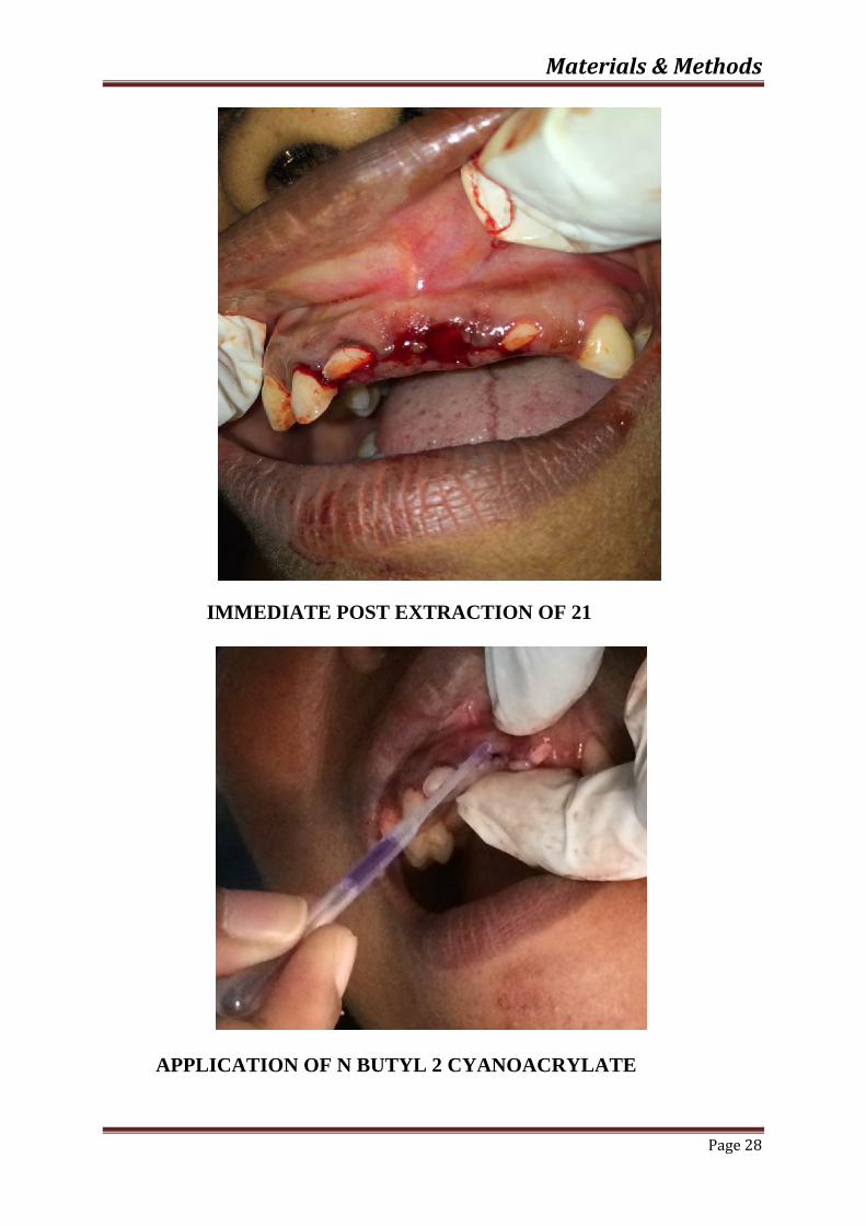
Procedure in detail:
After proper case recording and selecting the patients, the surgical procedure
and also the use of n-butyl cyanoacrylate tissue adhesive for closure of surgical
wounds as an alternative to sutures was explained thoroughly to the patients. Under
aseptic precautions, patient was anaesthetized with 2% lignocaine with adrenaline
1:80000(LIGNOX-2% manufactured by Warren indoco) and prepared for surgery.
The length of incision varied from 1-3 cm depending on the surgical access required
for the procedure. The extraction procedure performed, if required alveolopasty
procedures will be performed. After performing the surgical procedure and achieving
adequate hemostasis, closures was performed on one side with n-butyl cyanoacrylate
tissue adhesive and on the other side with 3-0 vicryl and these sides were randomly

Materials & Methods
Page 25
chosen. The side of the incision where n-butyl cyanoacrylate tissue adhesive was to be
applied, isolated with dry gauze. The incised edges were accurately approximated,
trying not to leave any gap between them. N butyl 2-cyanoacrylate was applied at the
approximated wound margins in the form of drops for closure of the mucoperiosteal
flaps. Same surgical procedure performed on other side also, incision was closed with
3-0 Vicryl suture. The post-operative sites pressure pack was given at the sutured
sites. Post-operative instructions regarding diet, avoid disrupting the wound at glue
site, oral hygiene maintenance and warm saline gargles were given to the patients.
Following medications with their standard dosages were given:-
1. Tab. Fenacplus(Diclofenac sodium 50mg+Paracetamol 500mg) twice a day
for three days
2. Cap. Amox 500mg(Amoxycillin) thrice a day for five days
Follow-up was made at third, fifth and seventh post-operative days. During each
follow up visit, pain was recorded on a visual analogue scale. The pain scale was 4 cm
long subdivided into 4 equal parts, one end corresponding to no pain, the other to
extremely severe pain. It will be recorded at1st day, 3rd day, 5th, and 7th day.
Visual analog scale to evaluate pain: reference values were given to patients
0 No pain patient feels well
1 Mild pain patient is distracted he or she does not feel the pain
2 Severe pain patient is very disturbed but nevertheless can continue
with normal activities
3 Very severe pain patient is forced to abandon normal activities
Materials & Methods
Page 26
Wound dehiscence was observed clinically on 1stday, 3rd day, 5th and 7th post
operative days based on visual examination.
Statistical analysis of the information obtained was performed. The differences with a
P < 0.05 were found to be statistically significant.

Materials & Methods
Page 27
ARMAMENTARIUM
Materials & Methods
Page 28
IMMEDIATE POST EXTRACTION OF 21
APPLICATION OF N BUTYL 2 CYANOACRYLATE
Materials & Methods
Page 29
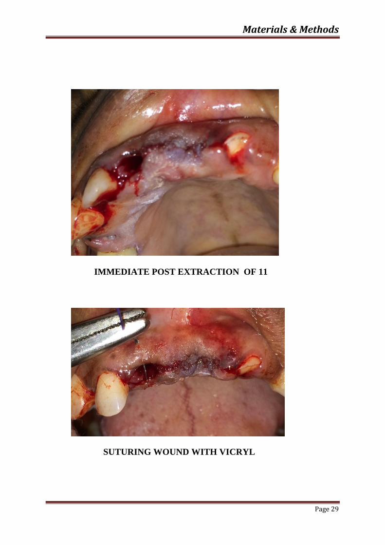
IMMEDIATE POST EXTRACTION OF 11
SUTURING WOUND WITH VICRYL

Materials & Methods
Page 30
On DAY -1
On DAY -3

Materials & Methods
Page 31
On DAY -5
On DAY -7
Dem
G
Statistica
The
16.0) versi
P value les
interval.
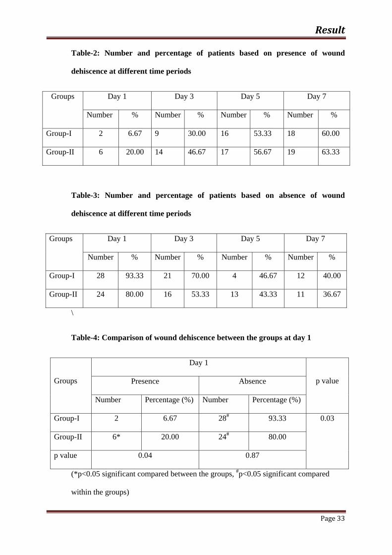
Table-1: D
mographic
data
Groups
Graph-1: D
al analysis
e study was
ion. Chi sq
s than 0.05
Demograph
Age
(MEAN±
D)
38.47±4.5
Distributio
s:
s analyzed
quare test a
(p<0.05) c
ic data
±S
Numb
56 15
n of patien
by Statisti
applied to f
considered s
Male
ber Perc
(
50
nts accordin
ical Analys
find the sig
statistically
Gend
centage
(%)
0.00
ng to gende
is for Soci
gnificant be
significant
der
Nu
Number
15
er
R
ial Sciences
etween the
at 95% con
umber
Percen
(%)
50.0
Result
Page 32
s (SPSS
groups.
nfidence
ntage
)
00
Result
Page 33
Table-2: Number and percentage of patients based on presence of wound
dehiscence at different time periods
Groups Day 1 Day 3 Day 5 Day 7
Number % Number % Number % Number %
Group-I 2 6.67 9 30.00 16 53.33 18 60.00
Group-II 6 20.00 14 46.67 17 56.67 19 63.33
Table-3: Number and percentage of patients based on absence of wound
dehiscence at different time periods
Groups Day 1 Day 3 Day 5 Day 7
Number % Number % Number % Number %
Group-I 28 93.33 21 70.00 4 46.67 12 40.00
Group-II 24 80.00 16 53.33 13 43.33 11 36.67
\
Table-4: Comparison of wound dehiscence between the groups at day 1
Groups
Day 1
p value Presence Absence
Number Percentage (%) Number Percentage (%)
Group-I 2 6.67 28# 93.33 0.03
Group-II 6* 20.00 24# 80.00
p value 0.04 0.87
(*p<0.05 significant compared between the groups, #p<0.05 significant compared
within the groups)

Result
Page 34
Table-5: Comparison of wound dehiscence between the groups at day 3
Groups
Day 3
p value Presence Absence
Number Percentage (%) Number Percentage (%)
Group-I 9 30.00 21# 70.00
0.03 Group-II 14* 46.67 16* 53.33
p value 0.02 0.45
(*p<0.05 significant compared between the groups, #p<0.05 significant compared
within the groups)
Table-6: Comparison of wound dehiscence between the groups at day 5
Groups
Day 5
p value Presence Absence
Number Percentage (%) Number Percentage (%)
Group-I 16 53.33 14# 46.67 0.04
Group-
II
17 56.67 13# 43.33
p value 0.78 0.67
(p>0.05 no significant compared between the groups, #p<0.05 significant compared
within the groups)
Result
Page 35
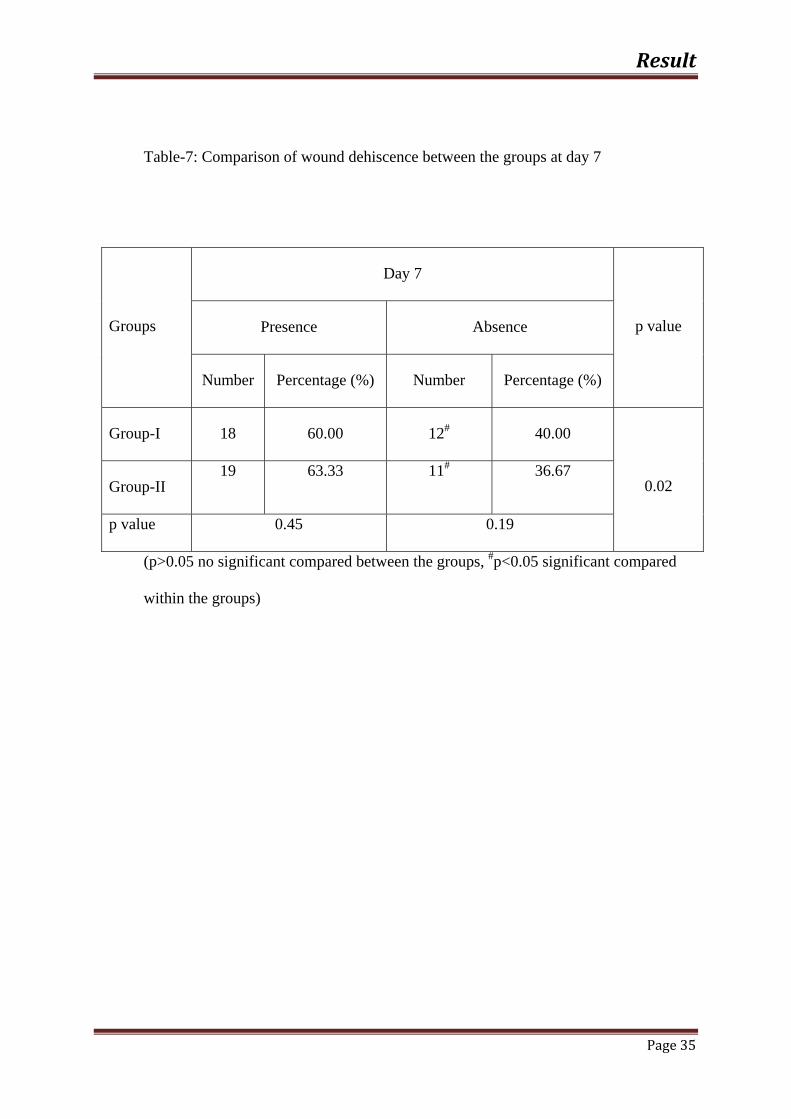
Table-7: Comparison of wound dehiscence between the groups at day 7
Groups
Day 7
p value Presence Absence
Number Percentage (%) Number Percentage (%)
Group-I 18 60.00 12# 40.00
0.02 Group-II 19 63.33 11# 36.67
p value 0.45 0.19
(p>0.05 no significant compared between the groups, #p<0.05 significant compared
within the groups)

Graph-2: N
at different
Graph-3: N
at different
Number and
time period
Number and
time period
d percentage
ds
d percentage
ds
e of patients
e of patient
s based on p
s based on
presence of
absence of
R
f wound deh
f wound deh
Result
Page 36
hiscence
hiscence

Result
Page 37
Table-8: Comparison of number and percentage of patients based on the pain score on
day 1
Pain Score
Group-I Group-II
Number Percentage
(%)
Number Percentage
(%)
Score 0 0 0.00 1 3.33
Score 1 9* 30.00 11* 36.67
Score 2 18*,# 60.00 17*,# 56.67
Score 3 3*,#,$ 10.00 1#,$ 3.33
(*p<0.05 significant compared score 0 with other scores within the groups,
#p<0.05 significant compared score 1 with other scores within the groups,
$p<0.05 significant compared score 2 with other scores within the groups)
Table-9: Comparison of number and percentage of patients based on the pain
score on day 3
Pain Score Group-I Group-II
Number Percentage (%) Number Percentage (%)
Score 0 7 23.33 10 33.33
Score 1 15* 50.00 15* 50.00
Score 2 8# 26.67 5*,# 16.67
Score 3 0*,#,$ 0.00 0*,#,$ 0.00
(*p<0.05 significant compared score 0 with other scores within the groups,
#p<0.05 significant compared score 1 with other scores within the groups,
$p<0.05 significant compared score 2 with other scores within the group

Result
Page 38
Table-10: Comparison of number and percentage of patients based on the pain
score on day 5
Pain Score Group-I Group-II
Number Percentage (%) Number Percentage (%)
Score 0 19 63.33 20 66.67
Score 1 11* 36.67 10* 33.33
Score 2 0*,# 0.00 0*,# 0.00
Score 3 0*,# 0.00 0*,# 0.00
(*p<0.05 significant compared score 0 with other scores within the groups,
#p<0.05 significant compared score 1 with other scores within the groups,
$p<0.05 significant compared score 2 with other scores within the groups)
Table-11: Comparison of number and percentage of patients based on the pain
score on day 7
Pain Score
Group-I Group-II
Number Percentage (%) Number Percentage (%)
Score 0 26 86.67 27 90.00
Score 1 4* 13.33 3* 10.00
Score 2 0*,# 0.00 0*,# 0.00
Score 3 0*,# 0.00 0*,# 0.00
(*p<0.05 significant compared score 0 with other scores within the groups,
#p<0.05 significant compared score 1 with other scores within the groups,
$p<0.05 significant compared score 2 with other scores within the groups)

Result
Page 39
Table-12: Comparison of number of patients based on the pain score between the
groups at different pain scores on day 1
Groups Score 0 Score 1 Score 2 Score 3
Group-I 0 9 18 3
Group-II 1 11* 17 1
p value 0.23 0.04 0.45 0.83
(*p<0.05 significant compared score 1 between the groups)
Table-13: Comparison of number of patients based on the pain score between the
groups at different pain scores on day 3
Groups Score 0 Score 1 Score 2 Score 3
Group-I 7 15 8 0
Group-II 10 15 5 0
p value 0.45 0.67 0.29
(p>0.05 no significant compared between the groups)

Result
Page 40
Table-14: Comparison of number of patients based on the pain score between the
groups at different pain scores on day 5
Groups Score 0 Score 1 Score 2 Score 3
Group-I 19 11 0 0
Group-II 20 10 0 0
p value 0.67 0.19
(p>0.05 no significant compared between the groups)
Table-15: Comparison of number of patients based on the pain score between the
groups at different pain scores on day 7
Groups Score 0 Score 1 Score 2 Score 3
Group-I 26 4 0 0
Group-II 27 3 0 0
p value 0.28 0.56
(p>0.05 no significant compared between the groups)

Graph-4: C
Graph-5: C
Group I- In
Group II- In
Comparison
Comparison
ncision close
ncision clos
of pain sco
of pain sco
ed with vicr
sed with N B
re bwtween
re within th
ryl suture..
Butyl 2 cya
n th groups
he groups
anoacrylate
RResult
Page 41

Interpretation
Page 42
Table 1: Comparison of gender involving this study and the mean age of the
patient.
Table 2: Comparison of presence of wound dehiscence at different time
periods. In group I, 6.67% of wound dehiscence was observed on first day, 30% of
wound dehiscence was observed 3rd day, 53.33% of wound dehiscence was observed
on 5th day, 60% of wound dehiscence was observed on 7th day. In group II, 20% of
wound dehiscence was observed on 1st day, 46.67% of wound dehiscence was
observed on 3rd day, 56.67% of wound dehiscence was observed on 5th day, 63.33%
of wound dehiscence was observed on 7th day.
Table 3: Comparison of absence of wound dehiscence at different time
periods. In group I, wound dehiscence was not observed in 93.33% on 1st day, wound
dehiscence was not observed in 70% on 3rd day, wound dehiscence was not observed
in 46.67% on 5th day, wound dehiscence was not observed in 40% on 7th day. In
group II, wound dehiscence was not observed in 80% on 1st day, wound dehiscence
was not observed in 53.33% on 3rd day, wound dehiscence was not observed in
43.33% on 5th day, wound dehiscence was not observed in 36.67% on 7th day.
Table 4: Comparison of wound dehiscence between group I and group II on
day 1. In group I wound dehiscence was observed in 6.67% on 1st day, in group II
wound dehiscence was observed 20% on 1st day. In group I, wound dehiscence was
not observed in 93.3% on 1st day, in group II wound dehiscence was not observed in
80% on 1st day. There was statistically significant difference between group I and
group II in comparing presence of wound dehiscence.
Table 5: Comparison of wound dehiscence between group I and group II on
day 3. In group I wound dehiscence was observed in 30% on 3rd day, in group II
Interpretation
Page 43
wound dehiscence was observed 46.67% on 3rd day. In group I, wound dehiscence
was not observed in 70% on 3rd day, in group II wound dehiscence was not observed
in 53.33% on 3rd day. There was statistically significant difference between group I
and group II in comparing presence of wound dehiscence.
Table 6: Comparison of wound dehiscence between group I and group II on
day 5. In group I wound dehiscence was observed in 53.33% on 5th day, in group II
wound dehiscence was observed 56.67% on 5th day. In group I, wound dehiscence
was not observed in 46.67% on 5th day, in group II wound dehiscence was not
observed in 43.33% on 5th day. There was statistically no significant difference
between group I and group II
Table 7: Comparison of wound dehiscence between group I and group II on
day 7. In group I wound dehiscence was observed in 60% on 7th day, in group II
wound dehiscence was observed 63.33% on 7th day. In group I, wound dehiscence
was not observed in 40% on 7th day, in group II wound dehiscence was not observed
in 36.67% on 7th day. There was statistically no significant difference between group I
and group II.
Table 8: Comparison of number and percentage of patients based on the pain
score on 1st day. Group I, all patient experienced pain, 30% experienced mild pain
(score 1), 60% experienced severe pain (score2), 10% experienced very severe pain.
Group II, 3.33% patient not experienced pain (score0), 36.67% experienced mild pain
(score 1), 56.67% experienced severe pain(score2), 3.33% experienced very severe
pain. There was statistically no significant difference between group I and group II.
Table 9: Comparison of number and percentage of patients based on the pain
score on 3rd day. Group I, 23.33% experienced no pain(score0), 50% experienced

Interpretation
Page 44
mild pain(score 1), 26.67% experienced severe pain(score2), 0% experienced very
severe pain(score 3).Group II, 33.33% patient not experienced pain(score0), 50%
experienced mild pain(score 1), 16.67% experienced severe pain(score2), 0%
experienced very severe pain(score 3). There was statistically no significant
difference between group I and group II.
Table 10: Comparison of number and percentage of patients based on the pain
score on 5th day. Group I, 63.33% experienced no pain(score0), 36.67% experienced
mild pain(score 1), 0% experienced severe pain(score2), 0% experienced very severe
pain(score 3).Group II, 66.67% patient not experienced pain(score0), 33.33%
experienced mild pain(score 1), 0% experienced severe pain(score2), 0% experienced
very severe pain(score 3). There was statistically no significant difference between
group I and group II.
Table 11: Comparison of number and percentage of patients based on the pain
score on 7th day. Group I, 86.67% experienced no pain(score0), 13.33% experienced
mild pain(score 1), 0% experienced severe pain(score2), 0% experienced very severe
pain(score 3).Group II, 90% patient not experienced pain(score0), 10% experienced
mild pain(score 1), 0% experienced severe pain(score2), 0% experienced very severe
pain(score 3). There was statistically no significant difference between group I and
group II.
Table 12: Comparison between groups based on the pain score on 1st day.
Group I- all experienced pain (score 0), 9 experienced mild pain (score 1), 18
experienced severe pain (score 2), 3 experienced very severe pain (score 3).Group II-
1 experienced no pain (score 0), 11 experienced mild pain (score 1), 17 experienced

Interpretation
Page 45
severe pain (score 2), 1 experienced very severe pain (score 3). p<0.05 There was
statistically significant difference between group I and Group II on score 1.
Table 13: Comparison between the groups based on the pain score on 3rd day.
Group I- 7 experienced no pain (score 0), 15 experienced mild pain (score 1), 8
experienced severe pain (score 2), 0 experienced very severe pain (score 3). Group II-
10 experienced no pain (score 0), 15 experienced mild pain (score 1), 5 experienced
severe pain (score 2), 0 experienced very severe pain (score 3). p>0. There was
statistically no significant difference between group I and group II.
Table 14: Comparison between the groups based on the pain score on 5th day.
Group I- 19 experienced no pain (score 0), 11 experienced mild pain (score 1), 0
experienced severe pain (score 2), 0 experienced very severe pain (score 3). Group II-
20 experienced no pain (score 0), 10 experienced mild pain (score 1), 0 experienced
severe pain (score 2), 0 experienced very severe pain (score 3). p>0.05 There was
statistically no significant difference between group I and group II.
Table 15: Comparison of both groups based on the pain score on 7th day.
Group I- 26 experienced no pain (score 0), 4 experienced mild pain (score 1), 0
experienced severe pain (score 2), 0 experienced very severe pain (score 3). Group II-
27 experienced no pain (score 0), 3 experienced mild pain (score 1), 0 experienced
severe pain (score 2), 0 experienced very severe pain (score 3). p>0.05 There was
statistically no significant difference between group I and group II.
Graph 1: In comparison of distribution of patient according to gender, both
male and female were in equal proportion.

Interpretation
Page 46
Graph 2: comparison of presence of wound dehiscence at different time
periods. On day 1, wound dehiscence was present in 2 patients in group I, 6 patients
in group II. On day 3, wound dehiscence was present in 9 patients in group I, 14
patients in group II. On day 5, wound dehiscence was present in 16 patients in group
I, 17 patients in group II. On day 7, wound dehiscence was present in 18 patients in
group I, 19 patients in group II.
Graph 3: comparison of absence of wound dehiscence at different time
periods. On day 1, wound dehiscence was absent in 28 patient in group I, 24 patient in
group II. On day 3, wound dehiscence was absent in 21 patient in group I, 16 patient
in group II. On day 5, wound dehiscence was absent in 14 patient in group I, 13
patient in group II. On day 7, wound dehiscence was absent in 12 patient in group I,
11 patient in group II.
Graph 4: comparison of pain score between group I and group II at different
time periods. There was statistically no significant difference between two groups.
Graph 5: Comparison of pain score within the group. Statistically significant
difference present within the group at different time period for both groups.

Discussion
Page 42
Wound healing is a reparative process of tissue after injury. Wound healing process is
divided into four phases. Haemostasis is the first phase, Inflammation is the second phase,
proliferation is the third face and maturation is the fourth phase. Immediately after injury,
platelets adhered to the injured site. Then adhered platelets change its shape and release
chemical mediators for clotting. Finally activates fibrin to form a clot. In inflammatory phase,
inflammatory cells are released into wound and engulf the pathogen and dead cells. In
proliferation phase growth of newly formed cells will occur. Angiogenesis, new collagen
formation, epithelial tissue formation, granulation tissue formation and wound contraction
will occur. During maturation period type III collagen is replaced by type I collagen. Wound
healing is affected by local and systemic factors.
Wound closure can be done by primary intention, secondary intention and tertiary
intention. In primary intention wound edges are re-approximated with sutures, staples and
tissue adhesive like N Butyl 2 cyanoacrylate. Advantage of primary intention is to minimize
scarring, faster healing when compared to secondary intention. Usually done in well repaired
laceration, properly reduced bone fractures and healing after flap surgery. In secondary
intention wound is allowed to granulate. Usually healing is slow and more scar tissue. In
tertiary intention, wound is cleaned and debrided for 4 to 5 days before wound closure.
Attainment of ideal wound closure is the important factor for healing at surgical site.
The wound closure material should re-approximate the wound edges properly for sufficient
period for healing to occur. Ideal property of wound closure material is easy to apply, rapid
application, biocompatibility, better tissue tolerance, enough tensile strength to retain the re-
approximated wound edges, free from toxic substances and free from allergic reaction.
Usually intra oral incision is closed with suture material like vicryl and silk suture
materials. Suture material is commonly used for wound closure than staples and tissue

Discussion
Page 43
adhesives. Because of the property like better tensile strength, low dehiscence rate, proper
wound closure. But it has disadvantage like crosshatched marks, needle penetration of normal
tissue on either side of the wound, tissue reactivity, anxiety, and it is a time consuming
procedure. Because of theses disadvantages alternative procedure become developed like
tissue adhesive.
In 1949 Ardis discovered cyanoacrylates. In 1959 cover et al suggested its adhesive
property. Initially it was rejected because of not biocompatibility to the tissue and more
inflammatory reaction. Later in 1964 Tennese Eastman lab developed longer molecular
cyanoacrylate, which one better biocompatibility and produces less inflammatory reaction.
Advantage of N Butyl 2 cyanoacryate over suture material is easy to handle, shorter
duration of application, comfortable for anxiety and fear of patient, better bacteriostatic
property, eliminate the risk of needle prick injury, decreased healing time, haemostatic
property and better esthetic property.
In this comparative interventional study, N Butyl 2 cyanoacrylate and vicryl suture
were compared in intraoral wound closure. Parameters for evaluation in this study was pain
and wound dehiscence.
Pain:
Patient experienced more pain on day1, progressively pain get reduced on day 3, day
5 and day 7 for both groups. On day 1, only one patient experienced no pain in group II, 9
patient experienced mild pain in group I, 11 patient experienced mild pain in group II, 18
patient experienced severe pain in group I, 17 patient experienced severe pain in group II, 3
patient experienced very severe pain group I, 1 patient experienced very severe pain in group
II. Experience of pain between both groups on day 1, score 1 is statistically significant.

Discussion
Page 44
On day 3- 7 patient experienced no pain in group I, 10 patient experienced no pain in
group II, 15 patient experienced mild pain in group I, 15 patient experienced mild pain in
group II, 8 patient experienced severe pain in group I, 5 patient experienced severe pain in
group II, 0 patient experienced very severe pain group I, 0 patient experienced very severe
pain in group II.
On day 5- 19 patient experienced no pain in group I, 20 patient experienced no pain in
group II, 11 patient experienced mild pain in group I, 10 patient experienced mild pain in
group II, 0 patient experienced severe pain in group I, 0 patient experienced severe pain in
group II, 0 patient experienced very severe pain group I, 0 patient experienced very severe
pain in group II.
On day 7- 26 patient experienced no pain in group I, 27 patient experienced no pain in
group II, 4 patient experienced mild pain in group I, 3 patient experienced mild pain in group
II, 0 patient experienced severe pain in group I, 0 patient experienced severe pain in group II,
0 patient experienced very severe pain group I, 0 patient experienced very severe pain in
group II.
Difference in experience of pain between both groups on day1, day3, day5 and day 7
was statistically not significant (p>0.05) except on experience of mild pain between both
groups on day 1 was statistically significant different.
Wound dehiscence:
On 1st day and 3rd day percentage of wound dehiscence was more in group II than
group I. On 5th day and 7th day percentage of wound dehiscence was more or less equal in
group I and group II. In day one, percentage of wound dehiscence in group I was 6.67, for
group II was 20. In day 3, percentage of wound dehiscence in group I was 30, for group II

Discussion
Page 45
was 46.67. In day 5, percentage of wound dehiscence in group I was 53.33, for group II was
56.67. In day 7, percentage of wound dehiscence in group I was 60, for group II was 63.33.
Difference in percentage of wound dehiscence between both groups in day 1 was
statistically significant (p<0.05). Percentage of wound dehiscence was increased on day 3 on
both group. Difference in percentage of wound dehiscence between both groups on day 3 was
statistically significant (p<0.05). Percentage of wound dehiscence was increased on day 5 on
both groups. Difference in percentage of wound dehiscence between both groups on day 5
was statistically not significant (p>0.05). Percentage of wound dehiscence was increased on
day 5 on both group. Difference in percentage of wound dehiscence between both groups on
day 7 was statistically not significant (p>0.05).
N butyl 2 cyanoacrylate has the advantage of bacteriostatic and haemostatic property.
Time consumed for application of N Butyl 2 cyanoacrylate was very low when compared to
vicryl suture. Patient satisfaction was high on N Butyl 2 cyanoacrylate than vicryl suture.
Effects of N Butyl 2 cyanoacrylate and vicryl suture in intra oral wound closure have not
been evaluated on previous studies in cross over basis. In previous studies N Butyl 2
cyanoacrylate compared mainly with silk suture in intra oral wound closure.
Ajit D. Joshi et al clinically compare the efficacy of cyanoacrylate (tissue glue) and
conventional suture after surgical removal of impacted mandibular third molars. He
conducted a study on thirty patients. Based on his study, efficacy of wound closure with
cyanoacrylate and conventional suturing were similar in the severity of pain, but use of
cyanoacrylate showed better haemostasis. The present study showed severity of pain in both
group was statistically no significant at the end of fifth day.
Mohammad Elshall et al conducted study for closure of intra oral incision with tissue
adhesive of N Butyl 2 cyanoacrylate and silk suture. He conducted a study on 20 patients. He

Discussion
Page 46
concluded that difference in pain score between N Butyl 2 cyanoacrylate and silk suture was
not statistically significant. But patient anxiety and psychological stress was reduced with N
Butyl 2 cyanoacrylate. The present study also showed severity of pain in both group was
statistically no significant at the end of third, fifth and seventh day.

Summary & Conclusion
Page 52
Wound closure can be done by suture materials, staples and tissue adhesives.
Purpose of this study is, clinically compare the efficacy of N Butyl 2 cyanoacrylate
with vicryl suture, an in vivo study.
Intra oral mucosal incision was performed in all the patients for the purpose of
tooth extraction due to dental caries and periodontal problems in the same jaw
bilaterally or one in upper jaw and another one in lower arch.
Group I was intra oral mucosal incision was closed with vicryl suture material,
main criteria was flap should be re-approximated passively, before suture. Pain was
recorded by visual analogue scale. Wound dehiscence was recorded by direct visual
examination. Pain and wound dehiscence recorded on 1st day, 3rd day, 5th day and 7th
day.
Group II was intra oral mucosal incision was closed with N Butyl 2
cyanoacrylate, before apply this material after the extraction, the flap was re-
approximated passively. Parameters of pain and wound dehiscence was recorded on
1st day, 3rd day, 5th day and 7th day. Pain was recorded by visual analogue scale.
Wound dehiscence was recorded by direct visual examination.
In this study, statistically significant more score on mild pain present on 1st
day in incision closed with vicryl suture material over incision closed with N Butyl 2
cyanoacrylate. There was statistically no significant on experience of pain between
both groups on 3rd day, 5th day and 7th day.
The measurement of pain and wound dehiscence measured on 1st, 3nd, 5th, 7th
day for all the patients and statistical analysis performed between both groups to find
out the benefit. Based on this analysis, statistically significant score was obtained on

Summary & Conclusion
Page 53
comparing the mild pain between both groups on day one only. In all other score for
pain on day 1, day 3, day, day 5 and day 7 showed statistically no significance
between the both groups. There was statistically significant score was obtained on
presence of wound dehiscence between both groups on day 1and day 3, where
presence of wound dehiscence was more in incision closed with N Butyl 2
cyanoacrylate than incision closed with vicryl suture. But, on 5th, 7th day there was
statistically no significant difference in presence of wound dehiscence between
groups.
N Butyl 2 cyanoacrylate has a minimal role in closure of intra oral mucosal
incision. Further studies are needed to evaluate the tensile strength of material and
long term effects of N Butyl 2 cyanoacrylate.

Bibliography
vii
1. Shahlakakoei, FahimehBaghaei, Shahriardabri, Masoudparirokh and sinakakooei. A
comparative in vivo study of tissue reactions to four suturing materials. Iran Endod J
2010;5(2):69-73.
2. Wallace WR, Maxwell GR, Cavalaris CJ. Comparison of polyglycolic acid suture to
black . J Oral Surg.1970;28:739-46.
3. Katz S, Izhar M, Mirelman d. Bacterial adherence to surgical sutures. A possible
factor in suture induced infection. Ann Surg 1981;194:35-41.
4. kumar MS, Natta S, Shankar G, Krishna SH, Visalakshi D, Seshiah GV. Comparison
between silk sutures and cyanoacrylate adhesive in human mucosa-A clinical and
histological study. J Int Oral Health. 2013;5(5);95-100.
5. Bhaskar SN, Frisch J. Use of cyanoacrylate adhesives in dentistry. J Am Dent Assoc
1968 ;77(4):831-837.
6. Ellis DA, Shaikh A. The ideal tissue adhesive in facial plastic and reconstructive
surgery. J Otolaryngol 1990;19(1):68-72.
7. Tse DT, Panje WR, Anderson RL. Cyanoacrylate adhesive used to stop CSF leaks
during orbital surgery. Arch ophthalmol 1984;102(9):1337-39.
8. Weselis IF, McNeill JI. Applicator for cyanoacrylate tissue adhesive. Ophthalmic
surg. 1989;20(3);211-214.
9. Fernandez PM, Marquez D, Bretana RMG. Use of N-Butyl-2-Cyanoacrylate in oral
surgery: biological and clinical evaluation. Artif Organs. 2000;24:241-3.

Bibliography
viii
10. Idle MR, Monagham AM, Lamin SM, Grant SWJ. N Butyl 2 cyanoacrylate tissue
adhesive as a haemostatic agent in a venous malformation of the mandible. Br J Oral
Maxillofac Surg. 2013;51:565-567.
11. Holzheimer RG. Adverse events of sutures:possible interactions of biomaterials. Eur J
Med Res 2006;10:521-526.
12. Sametinal, NergizYılmaz, CevatNisbet, and TolgaGüvenç, Samsun. Biochemical and
histopathological findings of N-Butyl-2- Cyanoacrylate in oral surgery: an
experimental study. J Oral Surg Oral Med Oral Pathol Oral RadiolEndod. 2006; 102:
e14-e17.
13. HowardBM, Eshraghi SR, Refai D. Octyl-cyanoacrylate skin adhesive is effective for
wound closure in posterior spinal surgery without increased risk of wound
complications. J Cli Neuro. 2014;125:137–142.
14. Joshi AD, Saluja H. Mahindra U. RajshekharHalli. A Comparative Study: Efficacy of
Tissue Glue and Sutures after Impacted Mandibular Third Molar Removal. J
Maxillofac Oral Surg. 2011;10(4):310–315.
15. Koranyi G, Seregard S, Kopp ED. Cut and paste: a no suture, small incision approach
to pterygiumsurgery. Br J Ophthalmol. 2004;88(7):911-914.
16. Morettineto RT, Mello I, Silveiramoretti AB, Carlos Roberto Colombo, Robazza,
Alessandro Antônio Costa, Pereira.In vivo qualitative analysis of the biocompatibility
of different cyanoacrylate-based adhesives. Braz Oral Res. 2008;22(1):43-7.
Bibliography
ix
17. Howell JM, Bresnahan KA, Stair TO, Dhindsa HS and Edwards BA. Comparison of
effects of suture and cyanoacrylate tissue adhesive on bacterial counts in
contaminated lacerations. Antimicrob Agents Chemother. 1995;39(2):559-560.
18. Montanaro L, Arciola CL, CenniE, Ciapetti G, Savioli,F. Filippini, L.A. Barsanti.
Cytotoxicity, blood compatibility and antimicrobial activity of two cyanoacrylate
glues for surgical use. J Biomaterials. 2001;22:59-66.
19. Giray CB, Atasever A, Durgun B, Araz K. Clinical and electron microscope
comparison of silk sutures and n-butyl-2-cyanoacrylate in human mucosa. Australian
Dental J. 1997;42(4):255-8.
20. Taira BR, Singer AJ. An In-Vivo Study of the Wound-Bursting Strengths of Octyl-
Cyanoacrylate, Butyl-Cyanoacrylate, and Surgical Tape in Rats. J \Emerg Med. 2010;
38 (May): 546-550.
21. Alonso FC, Jornet P L. Clinical-pathological study of the healing of wounds provoked
on the dorso-lingual mucosa in 186 albino rats. J Otolaryngol Head Neck Surg
January. 2007;136:1119-124.
22. Ankitavastani, Anishamaria. Healing of Intraoral Wounds Closed Using Silk Sutures
and Isoamyl 2-Cyanoacrylate Glue: A Comparative Clinical. J Oral Maxillofac Surg.
2013;71:241–248.
23. Gumuş, P. and Buduneli, E. Graft stabilization with cyanoacrylate decreases
shrinkage of free gingival grafts. Australian Dental J.2014;59:57–64.

Bibliography
x
24. Bresnahan K.A , John. M. Howell, Joseph Wisoreck -Comparision of tensile strength
of cyanoacrylate inclosure of lacerations V/s suture adhesive. J. Ann Emergency Med
1995 (5); 578-8
25. Bhaskar S N, Frisch J, Leonard F. Application of a new chemical adhesive in
periodontic and oral surgery. J OralSurg Oral Med Oral Pathol.1966;22(4):526–535.
26. Javelet J,Torabinejad M, Danforth R. Isobutyl cyanoacrylate: A clinical and
histologic comparison with sutures in closing mucosal incisions in monkeys. J Oral
Surg Oral Med Oral Pathol. 1985;59(1):91–94.
27. Mogensbessermann. Cyanoacrylate spray in the treatment of prolonged oral bleeding.
Int J Oral surg.1977;6(4):233–240.
28. Bhaskar SN, Frisch J, Duane E. Cutright, Peter Margetis ,Walter Reed.Effect of butyl
cyanoacrylate on the healing of extraction wounds. Oral Surg Oral Med Oral
Pathol.1967;24(5):604–616.
29. Cotton A, H Deramond, B Cortet, J P Lejeune, X Leclerc, P Chastanet and J
Clarisse.Preoperative percutaneous injection of methyl methacrylate and N-butyl
cyanoacrylate in vertebral hemangiomas.Scandinavian J of Surg 2011;100:120-124.
30. Alan B. Leahey, John D. Gottsch, Walter J. Stark.Clinical Experience with N-butyl
Cyanoacryiate (Nexacryl) Tissue Adhesive. Ophthalmology J. 1993 ;100:173-180.
31. Cantasdemir M, Adaletli, D Cebi, F Kantarci, N.D Selcuk, F Numan.Emergency
Endovascular Embolization of Traumatic Intrarenal Arterial Pseudoaneurysms with
N-Butyl Cyanoacrylate. Clinical Radiology.2003;58:560–565.

Bibliography
xi
32. Kulkarni S, Dodwad V, Chava V.Healing of periodontal flaps when closed with silk
sutures and N-butyl cyanoacrylate: A clinical and histological study.Indian J Dent Res
2007;18:72-7.
33. Dalvi A A, Faria M M, Pinto AA. Non-suture closure of wound using cyanoacrylate. J
Postgrad Med. 1986;32:97-100.
34. Kutcher MJ, Ludlow JB, Allen D Samuelson, Tamara Campbel, Susan N Pusek.
Evaluation of bioadhesive device for the management of aphthousulcers. J Am Dent
Assoc. 2001;132:368-376.
35. Chai BH, Kim BY, Huh JY, Lee SH. Cyanoacrylate adhesive for closing sinus
membrane perforations during sinus lifts. J Craniomaxillofac Surg. 2006;34:505-509.
36. Abhisheksoni, Narula R, kumar A, parmar M, sahore M, Mohinderchandel.
Comparing cyanoacrylate tissue adhesive and conventional subcutucular skin sutures
for maxillofacial incision-A prospective randomized trial considering closure time,
wound morbidity and cosmetic outcome. J Oral Maxillofac Surg.
2013;71:2152e1-2152e8.
37. Snehasetrya, Rajshekharhalli, Anandshah, Gauravchhabaria, Tarunsingh.
Comparative evaluation of efficacy of tissue glue and sutures after surgical removal of
impacted mandibular third molars. A prospectivecontrolled clinical study. Oral Surg
Oral Med Oral Pathol. 2015;27:183-188.
38. Cavalris CJ - Comparison of polyglycolicacidsuture to black silk, chromic and plain
catgut inhuman oral tissues. J. OraI surgery. 2006;738-739;970.

Bibliography
xii
39. Ghoreishian M, Gheisari R, Fayazi M. Tissue adhesive and suturing for closure of the
surgical wound after removal of impacted mandibular third molars: A comparative
study. J Oral Surg Oral Med Oral Pathol Oral Radiol Endod 2009;108:e14-e16.
40. Hasan Z, Gangopadhaya AN, Gupta DK, Srivastava P and. Sharma SP. Sutureless
Skin Closure with Isoamyl 2-Cyanoacrylate in Pediatric Day-Care Surgery. Pediatric
Surgery International 2009;25:1123–1125.
41. Knott PD, Zias JE, Banbury J, Djohan R, Yetman RJ, Papay F. A comparison of
dermabond tissue adhesive and suture in primary repair of congenital cleft lip. Annals
of Plastic Surgery. 2007;58(2):121-125.
42. Jehangirnezhad M; Semilunar coronally repositioned flap for the treatment of gingival
recession with and without tissue adhesives: a pilot study, J of dentistry, Tehran
university of medical sciences, Tehran. Iran J. 2006;3(1):25-27.
43. Quin. J.V, Drzewiecki.A, Li.M et al – Arandomized controlled trial comparing
tissueadhesive with suturing in the repair of paediatric facial lacerations. Ann Emerg
Medicine. 1993;22 (7);1130—5.
44. Toriumi DM, Raslan WF, Friedman M, Tardy ME.Histotoxicity of cyanoacrylate
tissue adhesives. A comparative study. Arch Otolaryngol Head and Neck Surg.
1990;116(5):546-50.
45. Sybelesaska, Ana Maria Minarelli Gaspar, Eduardo Hochuli-Vieira. Cyanoacrylate
adhesives for the synthesis of soft tissue. An. Bras. Dermatol. 2009;84:215-220.

Bibliography
xiii
46. Hesham. S. Khalil, Mohammad A. Elshall, Yazeed Al-Harbi, Saeed Al-Asmar,
Mohammad Al-Wadee. Healing of oral surgical wounds using 3/0 silk suture and n-
butyl cyanoacrylate tissue adhesive. Egyptian dental Journal .2009;55:2607.
47. Srinivasan S, Dollin M, McAllum P, Berger Y, Rootman DS, SlomovicAR.Fibrin
glue versus sutures for attaching the conjunctivalautograft in pterygiumsurgery: a
prospective observermasked clinical trial. Br J Ophthalmol2009;93(2):215-218.
48. Schmeissner H, Galil HJ, Lundie MJ, Kaufmann JC. The histotoxicityoc
cyanoacrylates. A systemic review. Neuroradiology. 1985;26(2):279-291.
49. Harper MC and Ralston M, "Isobutyl 2-cyanoacrylate as a osseousadhesive in the
repair of osteochondral fractures," JBiorned. Muter Res. 1983;17:167-177.
50. Mehta MJ, Shah KH, Bhatt RG. Osteosynthesis of mandibular fractures with N butyl
2 cyanoacrylate: a pilot study. J Oral Maxillofac Surg. 1987;45(5):393-396.
51. Rice RM, Hegyeli AF, Gourlay SJ, Wade CWR, Dillion JG, Jaffe H, and Kulkarni
RK, ”Biocompatibility testing of polymers: In vitro studies with in vivo correlation,” J
Biomed Mate. Res. 1978; l2:43-54.
.


CONSENT FORM
PART 1 OF 2 INFORMATION FOR PARTICIPANTS OF THE STUDY
1. Name of the Principal Investigator:
Dr.K.MURUGAN,
Post Graduate student,
Department of Oral and Maxillofacial surgery,
Sree Mookambika Institute of Dental Sciences,
Kulasekharam, Kanyakumari District-629161
2. Name of the Guide: Dr.N.Dhineksh Kumar,
Professor,
Department Of Oral and Maxillofacial Surgery.
Sree Mookambika Institute of Dental Sciences,
Kulasekharam, Kanyakumari District-629161
Dear Volunteers, We welcome you and thank you for your keen interest in participation in this research
project. Before you participate in this study, it is important for you to understand why this research
is being carried out. This form will provide you all the relevant details of this research. It will
explain the nature, the purpose, the benefits, the risks, the discomforts, the precautions and the
information about how this project will be carried out. It is important that you read and understand
the contents of the form carefully. This form may contain certain scientific terms and hence, if you

3. Name of the Co-Guide: Dr. Mathew Jose,
Head of the department,
Department Of Oral and Maxillofacial Surgery,
Sree Mookambika Institute of Dental Sciences,
Kulasekharam, Kanyakumari District-629161
4. Institute:
Sree Mookambika Institute of Dental Sciences,
V.P.M Hospital complex, Padanilam, Kulasekharam,
Kanyakumari – 629161
Tamil Nadu.
5. Title of the study :
“CLINICALLY COMPARE THE EFFICACY OF N-BUTYL 2-
CYANOACRYLATE AND VICRYL SUTURES IN INTRA ORAL WOUND
CLOSURE” - AN IN VIVO STUDY
6. Background information: Conventionally ideal wound closure is achieved by suture material but because of namely
prolonged duration of surgery and anaesthesia, tissue reactivity,risk of needle stick, undesirable
trauma to the intact tissue on either side of wound,permanent suture tracts, early removal which
results in dehiscence, anxiety and pain during removal and inadequate esthetics effect causes
alternate procedures arrived for wound closure ,that is tissue adhesive.The best tissue adhesive
material is N-Butyl 2-cyanoacrylate.Which is easy to apply,reduce the anxiety of the patient.It
also acts as a haemostatic agent and antimicrobial agent.

7. Aim and objectives:
The aim of the study is to compare clinically the efficacy of n-butyl-2-cyanoacrylate with vicryl suture in the closure of oral mucosal incisions. To evaluate
• Pain
• Wound dehiscence.
8. Scientific justification of the study:
Incision is a basic step for surgical procedures,Suitable closure and optimal maintenance
of the surgical area are the most important factors that affect proper wound healing and surgical
success.The conventional method of wound closure causes trauma and many other undesirable
effects .The use of tissue adhesives as an alternative to, or replacement for, sutures in wound
closure has long been an area of interest. A group of these tissue adhesives are - Cyanoacrylates.
They have a wide range of applications in surgery and are supposed to offer some
advantages.Previous study showsn-butyl-2-cyanoacrylate can be used for intra oral wound
closure effectively. The procedure is relatively painless and quick.
9. Procedure for the study:
• Under aseptic precautions, patient was anaesthetized with 2% lignocaine with adrenaline
1:80000 and prepared for surgery.
• In extraction procedures or Alveoloplasties, bilaterally symmetrical crestal incision was
made on the arch.
• After performing the surgical procedure and achieving adequate haemostasis, closures
was performed on one side with n-butyl cyanoacrylate tissue adhesive and on the other
side with 3-0 vicryl suture and these sides were randomly chosen.

• The incised edges were accurately approximated, trying not to leave any gap between
them. After loading the glue in a syringe, it was applied at the approximated wound
margins through the needle in the form of drops for closure of the mucoperiosteal flaps.
• Under same aseptic precautions, anaesthesia, and surgical procedure on the other side,
suturing was done with interrupted braided black 3-0 vicryl suture.The post-operative
sites pressure pack was given at the sutured sites. Post-operative instructions regarding
diet, avoid disrupting the wound at glue site, oral hygiene maintenance and warm saline
gargles were given to the patients.
• Following medications with their standard dosages were given:-
1. Tab. Fenac plus twice a day for three days
2. Cap. Amox500mg thrice a day for five days
• Follow-up was made at third,fifth & seventh postoperative days. During each follow up
visit, pain and wound dehiscence were recorded on a visual analogue scale.
10. Expected risks for the participants:
• When flap closure done under tension ,wound dehiscence may happened.
11. Expected benefits of research for the participants:
• You will not be required to pay for this procedure.
• You can enquire about the outcome of the procedures and your details.
• You will get a better treatment at the end of the procedure.
12. Maintenance of confidentiality:
• You have the right to confidentiality regarding the privacy of your medical
information(Personal details, results of physical examinations, investigations, and your
medical history).
• By signing this document, you will be allowing the research team investigators, other
study Personnel, sponsors, institutional ethics committee and any person or agency
required by law to view your data, if required.
• The results of clinical tests and therapy performed as part of this research may be
included in your medical record.

20
• The information from this study, if published in scientific journals or presented at
scientific meetings, will not reveal your identity.
13. Why have I been chosen to be in this study?
a. Chosen because of grouping under the inclusion and exclusion criteria
b. Need of good sampling size
c. Patient phsycologically fear with suturing,while removal they have pain,risk of
needle prick injury and prolonged duration of surgery and anaesthesia to
overcome this problem tissue adhesive is a better in shorter duration,easy to use
better patient compliance.
d. To improve the aesthetic appearance.
14. How many people will be in the study? 30individuals
15. Agreement of compensation to the participants (In case of a study related
injury): Patient will be taken care in case of complication and medical treatment will be provided in the
institution at the expense of the principle investigator.
16. Anticipated prorated payment, if any, to the participant(s) of the study: For your cooperation to the study, the expenses for the routine blood investigations for which
you originally visited the clinic will be settled by the principal investigator.
17. Can I withdraw from the study at any time during the study period? The participation in this research is purely voluntary and you have the right to withdraw
from this study at any time during the course of the study without giving any reasons.
18. If there is any new findings/information, would I be informed? Yes
19. Expected duration of the participant’s participation in the study: 1 week
20. Any other pertinent information : No other information

21. Whom do I contact for further information?
Place: Signature of Principal Investigator
Date:
Signature of the participant
For any study related queries, you are free to contact:
Dr.K.Murugan
Post Graduate student.
Department of Oral & Maxillofacial Surgery,
Sree Mookambika Institute of Dental Sciences,
Kulasekharam, Kanyakumari District-629161.
Mobile No: 9894269313
CONSENT FORM
PART 2 OF 2
PARTICIPANTS CONSENT FORM
The details of the study have been explained to me in writing and the details have been
fully explained to me. I am aware that the results of the study may not be directly beneficial to
me but will help in the advancement or medical sciences. I confirm that I have understood the
study and had the opportunity to ask questions. I understand that my participation in the study is
voluntary and that I am free to withdraw at any time, without giving any reason, without the
medical care that will normally be provided by the hospital being affected. I agree not to restrict
the use of any data or results that arise from this study provided such a use is only for scientific
purpose(s). I have been given an information sheet giving details of the study. I fully consent to
participate in the study titled
“CLINICALLY COMPARE THE EFFICACY OF N-BUTYL 2-
CYANOACRYLATE AND VICRYL SUTURE IN INTRA ORAL WOUND
CLOSURE”
Serial no / Reference no :
Name of the participant :
Address of the participant :
Contact number of the participant :
Signature / thumb impression of the participant / Legal guardian
Witnesses:
1.
2.
Date: Place:
xg;Gjy; gbtk;
gFjp1-2
Ma;tpy; gq;Nfw;gtUf;fhd gbtk;
kjpg;gpw;Fhpa jd;dhu;tyNu
jhq;fs; ,e;j Ma;tpy; gq;Nfw;f te;jikf;F ed;wp. ,e;j Ma;tpy;
gq;Nfw;gjw;F Kd; ,e;j Ma;tpd; KOtpguq;fisAk; cq;fSf;F njhptpf;Fk;
Ma;tpy; gq;Nfw;gjw;F Kd; ,e;jgbtj;ij KOtJk; gbj;J Ghpe;J nfhz;l gpd;
gq;Nfw;f Ntz;Lk;. ,e;j gbtj;jpy; rpy kUj;Jt kw;Wk; mwptpay; nrhw;fs;
cs;sd. Mjyhy; VNjDk; re;Njfq;fs; Vw;gl;lhy;. ,e;j gbtj;jpd; ,Wjpapy;
Fwpg;gpl;Ls;s eghplk; Ma;tpy; gq;Nfw;gjw;F Kd; njspT ngw Ntz;Lk;.
Ma;thsh;
kU.F. KUfd;
KJfiy gl;ljhhp
tha; kw;Wk; KfrPuikg;G gphpT
= %fhk;gpif gy; kUj;Jtkid fy;Y}hp
FyNrfuk;
fd;dpahFkhp khtl;lk;
topfhl;b
kU.jpNd~;f; Fkhh;
Jiz Nguhrphpah;
tha; kw;Wk; KfrPuikg;G gphpT
= %fhk;gpfs gy; kUj;Jtkid fy;Y}hp
FyNrfuk;
fd;dpahFkhp khtl;lk

Jiztopfhl;b
kU.Nkj;A N[h];
Nguhrphpah; jiyikahsh;
tha; kw;Wk; KfrPuikg;G gphpT
= %fhk;gpfs gy; kUj;Jtkid fy;Y}hp
FyNrfuk;
fd;dpahFkhp khtl;lk;
Ma;T epiyak;
= %fhk;gpfs gy; kUj;Jtkid fy;Y}hp
FyNrfuk;
fd;dpahFkhp khtl;lk;
1. jiyg;G
thapDs; Vw;gLk; fhaq;fis iraNdh mf;fphpNyl; %yKk; itf;hpy; ijay; %yKk; rhp nra;tij xg;gpLjy;.
2. Ma;tpd; gpd;gyk;
thapDs; Vw;gLk; fhaq;fis rhpnra;a ijay; Nghlg;gLk;. Mdhy;
,e;epfo;T NehahspfSf;F gaj;ijAk; Vw;gLj;Jk;. iraNdh mf;hpNyl;
vd;w Ntjpay; nghUis itj;J fhaj;ij rhp nra;tjpd; %yk;
gaj;ijAk; fhyjhkjj;ijAk; jtph;f;fyhk;.
3. Fwpf;Nfhs;
thapDs; Vw;gLk; fhaq;fis itf;hpy; %yk; rhp nra;jy; kw;Wk;
thapDs; Vw;gLk; fhaq;fis iraNdh mf;hpNyl; vd;W Ntjpay; nghUs;
%yk; rhpnra;jy;.
,uz;bw;Fk; ,ilNaahd tpj;jpahrj;ij xg;gpl;L jPh;itf;
fz;Lgpbj;jy;.

4. mwptpay; gpd;gyk;
rpy fhyq;fshf elj;jg;gl;l Ma;Tfspd; %yk; iraNdh
mf;hpNyl; vd;w Ntjpay; nghUSk; itf;hpy; ijaYk; rk msT jd;id
cilaJ.
5. nray;Kiw
• jhq;fs; ,e;j Ma;tpy; gq;F ngw;w gpd; jdp ,yf;f vz;
nfhLf;fg;gLk;.
• rpf;l;irapd; NghJ rijapy; Vw;gl;l fhaj;ij rhpnra;a xU gFjp
itf;hpy; ijay; %yk; rhpnra;ag;gLfpwJ. kWgFjpapy; iraNdh
mf;hpNyl; vd;w Ntjpay; nghUs; %yk; rhpnra;ag; gLfpwJ.
6. vjph;kiw tpisTfs;:,y;iy
7. Vw;gLk; ed;ikfs;
• Ma;tpw;fhf gzk; vJk; ju Njitapy;iy
• Ma;tpd; KbTfis ePq;fs; mwpe;J nfhs;syhk;
8. jftypd; ghJfhg;G
• cq;fSila jfty;fs; midj;Jk; ghJfhf;fg;gLk; ve;j
epiyapYk; ntspaplg;glhJ.
• ePq;fs; ,e;j Ma;tpy; gq;Nfw;wg;gpd; vq;fsJ Ma;thsh;
cq;fSila jfty;fis njhpe;J nfhs;s KbAk;. NkYk; Njit
Vw;gl;lhy; vj;jpf;fy; fkpl;baplKk; fhz;gpf;fg;gLk;.
• Ma;tpd; Kbtpd; cq;fSila kUj;Jt jfty; gbtj;jpy;
(Nfhg;gpy;) gjpT nra;ag;gLk;.
• ,e;j Ma;T KbT ntspaplg;gLk; nghOJ cq;fsJ jfty;fs;
ntspaplg;glhJ.
9. ,e;j Ma;tpy; ePq;fs; Nru;tjw;fhd fhuzk;?
• ePq;fs; ,e;j Ma;tpd; Nrh;g;G kw;Wk; tpLg;G tFg;Gfspd; cs;
mikg;gLtjhy;.

10. Ma;tpd; khjphp vz;zpf;if:60(30 egu;)
11. e~;laPL
Ma;tpy; Vw;gLk; khw;wq;fSf;fhd rpfpl;ir Ma;T epiyaj;jpy;
toq;fg;gLk;.
12. e~;laPL gzk;: gzk; vJTk; toq;fglhJ.
13. Ma;tpy; ,Ue;J ve;j Neuj;jpYk; ve;j fhuzKk; ,d;wp tpyfyhk;
14. Ma;tpd; Kd;Ndw;wj;jpy; Vw;gLk; khw;wq;fs; njhptpf;fg;gl Ntz;Lk;.
15. Ma;tpd; fhy mtfhrk;: xU Mz;L
16. NtW ve;j jfty;fSk; Njit ,y;iy.
17. njhlh;T nfhs;s Ntz;ba egu;
kU. F. KUfd;
KJfiy gl;ljhhp
tha; kw;Wk; KfrPuikg;G gphpT
= %fhk;gpfs gy; kUj;Jtkid fy;Y}hp
FyNrfuk, ; fd;dpahFkhp khtl;lk;
njhiyNgrp vz;:9894269313
kpd; mQ;ry;:[email protected]
,lk;:FyNrfuk; Ma;thsh; ifnahg;gk;
Njjp:
gq;FngWgth; ifnahg;gk;

Xg;Gjy; gbtk;
gFjp-2
gq;FngWgthpd; xg;Gjy; gbtk;
,e;j Ma;tpd; KO tpguq;fSk; vOj;J %ykhfTk; tphpthfTk; vd;dplk;
$wg;gl;lJ. vdf;F ,e;j Ma;tpd; tpisTfs; ve;j gaDk; jutpy;iy
vd;whYk; kUj;Jt mwptpaypd; Nkk;ghl;bw;fhf gad;gLj;jg;gLk; vd;W
mwpNtd;. ehd; ,e;j Ma;tpy; ve;j eph;ge;jKk; ,y;yhky; gq;F
ngWfpNwd;. NkYk; ,e;j Ma;tpd; vy;yh tpjpfs; kw;Wk;
tpjpKiwfisAk; mwpNtd;. ve;j re;Njhfj;jpw;Fk; Nfs;tp Nfl;fNth
my;yJ Ma;tpy; ,Ue;J tpyfNth vy;yh chpikfSk; vdf;F cz;L
vd;W mwpNtd;. ,e;j Ma;tpd; tpisTfs; ve;j mwptpay; gad;ghl;bw;Fk;
gad;gLj;j vdf;F jilapy;iy. vdf;F ,jw;F Kd;ghf Ma;tpd; jfty;
gbtk; nfhLf;fg;gl;lJ. vdf;F ,e;j Ma;tpy; gq;Fngw KO rk;kjk;.
Ma;tpd; ngah;
thapDs; Vw;gLk; fhaq;fis iraNdh mf;fphpNyl; %yKk; itf;hpy; ijay; %yKk; rhp nra;tij xg;gpLjy;.
Fwpg;G vz;:
ngah;:
Kfthp:
njhiyNgrp vz;:
rhl;rp 1: ifnahg;gk;
2.
,lk;:FyNrfuk;
Njjp:

kΩX ]{Xw ˛ `mKw ˛ 1
]T\hpambn klIcn°p∂ hy‡nIfpsS Adnhntebv°v
{]nbs∏´ k∂≤ tkhI≥ / tkhI,
R߃ \nßsf kzmKXw sNøp∂p. AtXmsSm∏w Cu ]T\hpambn klIcn°m\p≈
k∂≤XtbmSv \μn tcJs∏SpØp∂p. \n߃ Cu ]T\Øn¬ ]s¶Sp°p∂Xn\p ap≥]v Cu
]T\w F¥n\mWv \SØs∏Sp∂Xv F∂v Andtb- Xp- v. AXn\m¬ Cu t^mdØn¬ KthjW
]T\Øns‚ hnhcßfpw a‰pw hniZambn tcJs∏SpØnbncn°p∂p. Cu ]T\Øns‚ coXn,
Dt±iw, {]tbmP\w, A]ISkm≤yX, t¢iw, ap≥IcpX¬, Fßs\ Cu ]T\w ap≥t]m´p
sIm- pt]mIp∂p F∂nßs\ F√m hnhcßfpw t^mdØn¬ tcJs∏SpØnbncn°p∂p. kZbw
Cu hnhc߃ hmbn®p a\ nem°phm≥ A`y¿∞n°p∂p. Cu hnhcßfn¬ imkv{X]camb
]Z߃ D≈Xn\m¬ kwib\nhmcWØn\p {][m\ ]T\I¿Ømhnt\mtSm Xmsg tcJs∏SpØn
bncn°p∂ hy‡nItfmtSm t^mdw H∏nSp∂Xn\p ap≥t]m As√¶n¬ Cu ]T\Øns‚ Imemh[n
Xocp∂Xphtctbm kao]n°mhp∂XmWv.
apJy KthjI≥ : tUm. sI. apcpI≥
t]mÃp{KmPpth‰v
Un∏m¿´vsa‚v Hm v̂ amIvkntemt^jy¬ k¿Pdn
{io aqImw_nI C≥Ãn‰yq´v Hm v̂ sU‚¬ kb≥kv,
IpetiJcw ˛ 629 161.
I\ymIpamcn Pn√.
{][m\ am¿§Z¿in : tUm. Znt\jvIv Ipam¿
doU¿ (Akn.s{]m^k¿)
Un∏m¿´vsa‚v Hm v̂ amIvkntemt^jy¬ k¿Pdn
{io aqImw_nI C≥Ãn‰yq´v Hm^v sU‚¬ sk‚¿,
IpetiJcw
kl am¿§ Z¿in : tUm.amXyq tPmkv
slUv Hm^v Zn Un∏m¿´vsa‚v,
s{]m^k¿,
Un∏m¿´vsa‚v Hm v̂ amIvkntemt^jy¬ k¿Pdn
{io aqImw_nI C≥Ãn‰yq´v Hm^v sU‚¬ kb≥kkv,
IpetiJcw.
C≥Ãn‰yq´v : {io. aqImw_nI C≥Ãn‰yq´v Hm^v sU‚¬ kb≥kkv
]S\new, IpetiJcw,
I\ymIpamcn ˛ 629 161.
Xangv\mSv.

1. ]T\Øns‚ io¿jIw : hmbn¬ G¿s∏Sp∂ apdnhpIsf sskt\m A{Inte‰v aptJ\bpw ssh{Ibn¬
Xø¬ aptJ\bpw icnsNøp∂Xns‚ XmcXayw.
2. ]›mØe hnhcw ?
hmbn¬ G¿s∏Sp∂ apdnhpIsf icnbm°m≥ Xø¬ CSp∂XmWv. F∂m¬ Cuapd tcmKnIƒ°v
`bsØbpw ImeXmaksØbpw G¿s∏SpØpw. sskt\m A{Inte‰v F∂ imkv{Xob km[\sØ h®v apdnhns\
icnbm°p∂Xp aptJ\ `bsØbpw, ImeXmaksØbpw Hgnhm°mw.
3. e£yßfpw Dt±ißfpw
hmbv°IØv G¿s∏Sp∂ apdnhpIsf ssh{Ibn¬ aqew icnbm°pI a‰pw
hmbv°IØv G¿s∏Sp∂ apdnhpIsf sskt\m A{Inte‰v F∂ imkv{Xob km[\w aptJ\ icnbm°¬.
c- n\pw CSbnep≈ hyXymksØ XmcXays∏SpØn I- p]nSn°¬
4. KthjWw \SØm\p≈ \ymboIcWw
Ipsd Imeßfmbn \SØs∏´ KthjW߃ aptJ\ sskt\m A{Ite‰pw, ssh{Ibnepw imkv{Xobamb
coXnbn¬ icnbm°p∂Xv hfsc \√XmsW∂v sXfnbn®n´p- v.
5. ]T\ coXn
]T\Øns‚ `mKambn hy‡n°v Hcp \º¿ \¬Ip∂XmWv.
Cu NnIn’bpsS `mKambn NXbn¬ G¿s∏´ apdnhns\ icnbm°m≥ Hcp`mKØv kn¬°v Xø¬ aptJ\
icnbm°p∂p. adp`mKØv sskt\m A{Ite‰v F∂ imkv{Xob km[\w aptJ\ icnbm°p∂p.
6. {]Xo£n°p∂ A]IS km≤yXIƒ ˛ C√
7. Cu ]T\Øn¬ ]s¶Sp°p∂XpsIm- v F\n°v Fs¥¶nepw {]tbmP\apt- m ?
o KthjWØn\v ]Ww H∂pw Xtc- Bhiyan√
o KthjWØns‚ I- p]nSnØw\n߃°v Adnbmhp∂XmWv.
12. Rm≥ Cu ]T\Øn¬ ]s¶Sp°p∂ hnhcw clkyambn hbv°ptam ?
\nßfpsS KthjWØns‚ I- p]nSnØw `{Zambn kq£n°p∂XmWv GsXmcp ImcWhimepw
shfns∏SpØp∂X√.
\n߃ Cu KthjWØn¬ ]s¶SpØXn\p tijw RßfpsS KthjI≥ \nßfpsS
Is- ØepIƒ Adnbm≥ km[n°mhp∂XmWv. taepw Bhiys∏´m¬ FØn°¬ IΩ‰nsb
Adnbn°p∂XmWv.
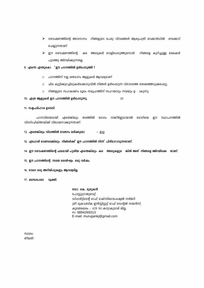
KthjWØns‚ Ahkm\w \nßfpsS t]cp hnhc߃ Bip]{Xn sd°m¿Un¬ sdt°mSv
sNøp∂XmWv.
Cu KthjWØns‚ Is- ØepIƒ shfns∏SpØptºmƒ \nßsf Ipdn®p≈ tcJIƒ
]pdØp Andbn°p∂X√.
9. Fs∂ F¥psIm- v Cu ]T\Øn¬ Dƒs∏SpØn ?
o ]T\Øn\v \√ iXam\w BfpIƒ BhiyamWv
o Nne Iq´n°pd®nepIƒs°mSphn¬ \n߃ Dƒs∏Sp∂ hn`mKsØ sXcs™Sp°s∏´p.
o \nßfpsS klIcWw aqew kaqlØn\v klmbhpw \∑bpw D- mIp∂p.
10. F{X BfpIƒ Cu ]T\Øn¬ Dƒs∏Sp∂p. 30
11. \„]cnlmc DSºSn
]T\hntZbambn GsX¶nepw XcØn¬ tcmKw k¶o¿Æambm¬ tcmKnsb Cu ÿm]\Øn¬
hnZKvZNnIn’bv°v hnt[b\m°p∂XmWv.
12. GsX¶nepw hn[Øn¬ thX\w e`n°ptam ˛ C√
13. Ft∏mƒ thWsa¶nepw \n߃°vv Cu ]T\Øn¬ \n∂v ]n≥amdmhp∂XmWv.
14. Cu KthjWØns‚ ^eambn ]pXnb Fs¥¶nepw Is- ØepIfps- ¶n¬ AXv \nßsf Andbnt°- XmWv.
15. Cu ]T\Øns‚ kab ssZ¿Lyw Hcp h¿jw.
16. thsd Hcp Adnbn∏pIfpw Bhiyan√.
17. _‘s∏tS- hy‡n.
tUm. sI. apcpI≥
t]mÃp{KmPpth‰v
Un∏m¿´vsa‚v Hm v̂ amIvkntemt^jy¬ k¿Pdn
{io aqImw_nI C≥Ãn‰yq´v Hm^v sU‚¬ kb≥kv,
IpetiJcw ˛ 629 161.I\ymIpamcn Pn√. Ph: 9894269313 E-mail: [email protected]
ÿew:
XobXn :

kΩX]{Xw
`mKw ˛ 2
Cu ]T\sØ ]‰nbp≈ F√m Imcyßfpw F\n°v ]d™v a\ nem°n XcnIbpw AXns‚ Hcp ]I¿∏v
F\n°p \¬IpIbpw sNbvXn´p- v. Cu ]T\w KthjWØn\mbn D≈XmsW∂pw F\n°v CXn¬ \n∂v t\cn´v
Hcp ^ehpw D- mIns√∂pw Rm≥ a\ nem°p∂p. Cu ]T\Øns‚ coXnbpw Dt±ihpw F\n°v a\ nem°n
X∂n´p- v. AXp t]mse F\n°v kwib߃ tNmZn°m≥ Ahkc߃ e`n®n´pap- v. CXn¬ ]s¶Sp°m\pw
]s¶Sp°mXncn°m\pw D≈ AhImiw F\n°ps- ∂pw AXpt]mse ]T\Øns‚ GXp L´Ønepw CXn¬
\n∂v ]n≥hßm\p≈ kzmX{¥yhpw F\n°ps- ∂v Rm≥ a\ nem°p∂p. Cu ]T\Øn¬
]s¶Sp°p∂XpsImt- m, ]s¶Sp°mØXpsImt- m Fs‚ a‰p NnIn’Isf _m[n°p∂Xs√∂v Rm≥
Andbp∂p.
“hmbn¬ G¿s∏Sp∂ apdnhpIsf sskt\m A{Inte‰v aptJ\bpw ssh{Ibn¬ Xø¬ aptJ\bpw
icnsNøp∂Xns‚ XmcXayw” F∂ KthjWØn¬ ]s¶Sp°p∂Xn\pw CXns‚ ^e߃ imkv{XteJ\Øn¬
{]kn≤oIcn°p∂Xn\pw F\n°v kΩXamsW∂v Rm≥ CXn\m¬ Andbn®psIm≈p∂p.
kocnb¬ \º¿ / d^d≥kv \º¿ :
]s¶Sp°p∂ Bfns‚ t]cv :
ta¬hnemkw :
t^m¨ \º¿ :
H∏v / hnceSbmfw
km£n :
ÿew :
XobXn

23
SREE MOOKAMBIKA INSTITUTE OF DENTAL SCIENCES
DEPARTMENT OF ORAL AND MAXILLOFACIAL SURGERY
CLINICALLY COMPARE THE EFFICIENCY OF N-BUTYL 2-
CYANOACRYLATE AND VICRYL SUTURE IN INTRA ORAL WOUND
CLOSURE: AN INVIVO STUDY
DATA RECORD SHEET
Patient name : Date:
Age : Op.No:
Sex :
Height :
Weight :
Parameter
Particulars 1st day 3rd day 5th day 7th day
Group1
Group2 Group1 Group2 Group1
Group2 Group1 Group2
Pain
Refernce value based on VAS
Wound dehiscence
Presence Absence
Group1- Vicryl suture. Group2- N-Butyl 2-cyanoacrylate. VAS- visual analogue scale.
0- No pain, 1- Mild pain, 2- severe pain, 3- very severe pain.
Participant’s Signature Investigator’s Signature,
Date: