clinicopathological, hematological and immunological … (53-68).pdffoamy nasal discharge. the death...
TRANSCRIPT
Egypt. J. Comp. Path. & Clinic. Path. Vol. 21 No. 3 (September) 2008; 53 - 68
53
Clinicopathological, hematological and immunological studies on the effect of Nigella sativa-L extract on rabbit hemorrhagic disease vacci-
nated rabbitries in Damietta Governorate By
Dawoud A. S.*and Seham S. Mansy** *Food Inspection Damietta Sea Port Laboratory, Animal Health
Research Institute, Dokki, Giza, Egypt. **Faculty of Science, Damietta, Mansura University, Egypt
SUMMARY
R abbit hemorrhagic disease (RHD) attacked worldwide countries in the last few decades, including Egypt causing severe economic
losses where mortality reached 80-100 %. Vaccination programs failed to control of disease spread completely.
In this study we tried to assure the immunogenic and protective ef-fects of Nigella sativa and utilizing it in improving immune response in vaccinated and infected rabbits. The experiment was designed to include 40 rabbits, divided into 4 groups each of 10 individuals. The 1st group was kept as normal vaccinated control group, the 2nd group was experi-mentally infected with identified RHD virus, the 3rd group was vacci-nated and Nigella sativa chloroform extract and the 4th group was vacci-nated, Nigella sativa treated and experimentally infected with isolated and identified RHD field virus.
Differential leucocytic count studies showed marked lymphopenia and neutropenia in infected group, which were significantly improved by Nigella sativa treatment in groups 3 and 4.
Biochemical studies showed significantly increased liver trans-ferases (AST, ALT and GGT) in experimentally infected group (G2)indicating severe liver damage, these changes returned towards normal levels in Nigella sativa treated groups (G3 and G4) indicating hepatopro-tective effect Total proteins and albumin concentrations showed non sig-nificant change in group 3, while total protein was significantly increased in group 4 accompanied with significant increase in globulin, also globu-lin was significantly increased in group 3 accompanied with decreased albumin/globulin ratio (A/G) , indicating immunostimuating effect of Nigella sativa.
Immunological studies performed using ELISA test results showed both immunostimulating effect of Nigella sativa extract treated groups and immunosuppressive effect of RHDV experimental infection. Cell me-diated immunity, in Nigella sativa treated groups (G3 and G4), was sig-

Egypt. J. Comp. Path. & Clinic. Path. Vol. 21 No. 3 (September) 2008; 53 - 68
54
nificantly increased as showed in increased percentage of rosette shape formation of macrophages engulfing neutral red granules. Pathological studies: Experimentally infected rabbits showed acute signs of the disease 2 days post infection, and on necropsy showed pale liver, congested spleen and kidneys with petichial hemorrhage and accu-mulation of intra-abdominal blood clots. Histopathological studies showed acute necrotic hepatitis accompanied with disseminated in-travascular coagulopathy (DIC), which is a pathognomonic process.
I n conclusion, at a practical level it is recommended to use the natural plant extract of Nigella sativa for immunostimulation for better rabbit
farming.
Referred byReferred by Prof. Dr. Rawhia Dogium Professor of Pathology, Fac. Vet. Med.,
Cairo University Prof. Dr. Mahmoud Samy Professor of Pathology, Fac. Vet. Med.,
Cairo University
INTRODUCTION
R abbit Hemorrhagic Disease (RHD) caused by rabbit hem-
orrhagic disease virus, a calicivirus, was first recorded in 1984 when a major epidemic syn-drome in domestic rabbits in China. Rabbit calicivirus is a highly contagious virus, transmit-ted by direct contact with infected rabbits or indirectly by contact with objects contaminated with vi-rus. Morbidity is often near 100% and mortality 60-90%. Infection results in a peracute febrile disease causing hepatic necrosis, enteritis, and lymphoid necrosis followed by massive coagulopathy resulting in hemorrhages in a variety of organs
(Xu et al. 1986 and Xu and Chan 1988).
RHD damages the liver, intes-tines, and lymphatic tissue and causes terminal massive blood clots. The incubation period is about 24 to 48 hours. Predomi-nantly, young adult and adult rab-bits die suddenly within 6 to 24 hours of the onset of fever with few clinical signs. Fever may be as high as 105 %F (40.5 %C) but of-ten is not detected until rabbits show terminal clinical signs. Most animals appear depressed or reluc-tant to move in the final hours and may show a variety of neurologic signs, including excitement, inco-ordination, paddling, and opist-
Egypt. J. Comp. Path. & Clinic. Path. Vol. 21 No. 3 (September) 2008; 53 - 68
55
hotonos (abnormal position of the head due to spasms of the muscles at the top and back of the neck). Some affected rabbits may have a foamy nasal discharge. The death rate for RHD ranges from 50 to 100 percent (Xu and Chan 1988).
Necropsy revealed the marked hypertrophy of the thymus with numerous petechiae, hemor-rhagic pneumo-tracheitis as well as hypertrophy and degeneration of the liver. Histopathology mainly showed lesions of necrotic hepati-tis. Nigella sativa [NS], or 'black cumin', an annual herb belonging to the family Ranunculaceae, has strong immunomodulatory and in-terferon-like activity.
The diagnosis of RHD is mainly based on observation of ne-cropsy findings and identification of the virus in the liver by hemag-glutination with human type O erythrocytes and ELISA test (Liu et al., 1984).
In Egypt many outbreaks were recorded in several provinces with severe economic losses and mortality rate reached 100% (Gha-nem and Ismail, 1991).
Nigella sativa L. (Ranuncula-ceae) is a grassy plant with green to blue flowers and small black seeds, which grows in temperate and cold climate areas. Seeds con-tain 1.5% volatile oil, while 37.5% Non volatile oil. In addition to this
Albumen, Sugar, Organic acids, Glucoside Melanthin, Metarbin. Also thymoquinone, dithymo-quinone carvacol and anethole 4-terpinole are found (Worthen et al., 1998; Bruits and Bucar, 2000). It improved the T-cells ac-tivity (El-Kadi and Kandil, 1987) and also prevents the decrease in hemoglobin level and leukocytes counts caused by cisplatin (Nair et al., 1990). Thymoquinine had shown antioxidant and immuno-modulator effects on macrophage, Th1 and Th2 (Salem, 2005).
MATERIAL AND METHODS 1-Experimental design: A total of 40, clinically healthy, Bouscat rab-bits with age range about 4 months were divided into 4 groups, each of 10 individuals, under strict hygi-enic and isolation conditions, as follows: Group 1 (G1): kept as normal RHD, intramuscularly, vaccinated control group. Group 2 (G2): kept as non vacci-nated group and experimentally infected with isolated RHD field virus (strict isolation measures were taken to avoid spread of the infection). Group 3 (G3): kept as intramus-cularly RHD vaccinated group and intramuscularly injected with Nigella sativa L seed extract. Group 4 (G4): kept as RHD vac-

Egypt. J. Comp. Path. & Clinic. Path. Vol. 21 No. 3 (September) 2008; 53 - 68
56
cinated group and intramuscularly injected with Nigella sativa L seed extract followed by experimental infection with isolated RHD field virus, one week post last vaccina-tion (strict isolation measures were taken to avoid spread of the infec-tion). 2-Vaccination: Sera of all groups of apparently healthy rabbits were checked with hem agglutination inhibition test to assure seronega-tivity for RHD infection (HI <10) and then groups 1, 3 and 4 were vaccinated with a dose of 0.5 ml injected into the fore back, subcu-taneously, with of oil adjuvant in-activated RHD virus (211 HAU per dose). The vaccine was produced by LABORATORIOS HIPRA, S. A.,SPAIN, under a commercial name CUNIPARVAC-RHD. The 2nd group was kept as non vacci-nated experimentally infected group.
3-Nigella sativa L seed extract: 600 g of ground black seed in 1500 ml chloroform were incubated in 25°C for one week, during which vibration was carried out up to 5 times a day. The resultant solution was filtered. In order to obtain a completely dry extract, the resul-tant extracts were transferred to glass dishes and were left in a 50°C oven for 24 hour. Then, they were left at 40°C until assessment of their antimicrobiological activi-ties. (Mashhadian and Rakhsha-
ndeh, 2005). 4-Preparation of samples: From field, RHD suspected to be in-fected rabbits, of about 4 months age, submitted to Damietta Veteri-nary research Laboratory for ex-amination, with symptoms of mucoid bloody nasal discharge and mucoid diarrhea and post mortem lesions of congested internal or-gans with clotted blood in the ab-dominal cavity.
5-Virus isolation and identifica-tion: 20 grams of liver tissue sam-ples were homogenized with phos-phate buffer saline solution (pH 7.2) for 15 minuets, then clarified by centrifugation at 4000 x g for 15 minuets, the supernatant was filtered through 0.22 µm filter, an-tibiotic was added and the super-natant was kept at-70 °C. A- Hemagglutination test: The suspected virus in the supernatant was identified as an antigen of RHD virus by hemagglutination against human erythrocytes type (O) at a titer of 1024 (Chasey et al.,1995). B- ELISA test: Hyperimmune se-rum was prepared by vaccination of healthy rabbits with alive RHD vaccine (Cunical, Rhone-Merieux, France) intramuscularly and boostered twice at 2 weeks inter-vals The serum was collected and stored at – 7. The prepared hy-perimmune serum of RHDV is

Egypt. J. Comp. Path. & Clinic. Path. Vol. 21 No. 3 (September) 2008; 53 - 68
57
used as positive control. ELISA test procedure was conducted ac-cording to Cluet et al. (1995). 6-Experimental infection: Rab-bits were inoculated subcutane-ously and intranasaly each with 0.5 ml of the prepared virus super-natant filtrate, one week post last vaccination (Chasey et al., 1995). 7-Sampling: Whole blood with EDTA anticoagulant, sera, perito-neal exudates and livers were col-lected for hematological, bio-chemical, immunological and hist-opathological studies, respectively, three days post last infection in ex-perimentally infected group and one week post, vaccination and Nigella sativa extract, treatment. 8-Hematological studies: Blood smears were prepared and stained with May- Grünwald-Giemsa sta-in. Differential leucocytic count was done using four field- mean-der method according to Schalm et al. (1991). 9-Serum biochemistry: All serum samples were examined for gam-ma glutamyle transferase (GGT) activity according to (Henry, 1974), Aspartate aminotransferase (AST) and Alanine aminotrans-fease (ALT) were estimated ac-cording to Reitman and Frankel (1957). Total protein (g/dl) accord-ing to Peter (1968). Serum albu-min (g/dl) according to Drupt (1974). Serum globulin (g/dl) was estimated by the difference be-
tween total protein and albumin while albumin/globulin ratio (A/G) ratio was calculated mathemati-cally according to Kaneko (1996). Parameters were estimated using standard kits and procedures ac-cording to instruction supplied by Bio-merieux, France.
10-Phagocytic cells percentage: The macrophage cell mediated im-munity was studied for examining the ability of living macrophage to engulf neutral red particles form-ing a characteristic rosette of red granules (Nelson, 1969). A- Reagents: Neutral red The stain was formulated as 1% neutral red (Adwic – Prolabo, France) in normal saline, used for staining and counting macrophage in PE-cells. B- Procedure: 1- one part of 1 % neutral red stain in normal saline was added to 9 parts of peritoneal exudates cell suspension. 2- The mixture was left for 10 min to 15 min, and then the number of macrophage was counted. The per-centage of phagocytic cells was determined depending on the ro-sette shape using lens 40 (Cohn and Wiener, 1963).
11-Pathological studies: Rabbits were sacrificed and
gross pathological lesions were re-corded. Livers were preserved in buffered formalin saline (10%), paraffin wax blocks were proc-

Egypt. J. Comp. Path. & Clinic. Path. Vol. 21 No. 3 (September) 2008; 53 - 68
58
essed, sectioned and stained with Haematoxylin & Eosine stain and examined microscopically (Carlt-on and McGalvin, 1995). 12-Statistical analysis: Data ob-tained were statistically analyzed by analysis of variance (ANOVA) using SPSS computer software, version 6.0.
RESULTS
S tudied experimentally infected group (G2), differential leuco-
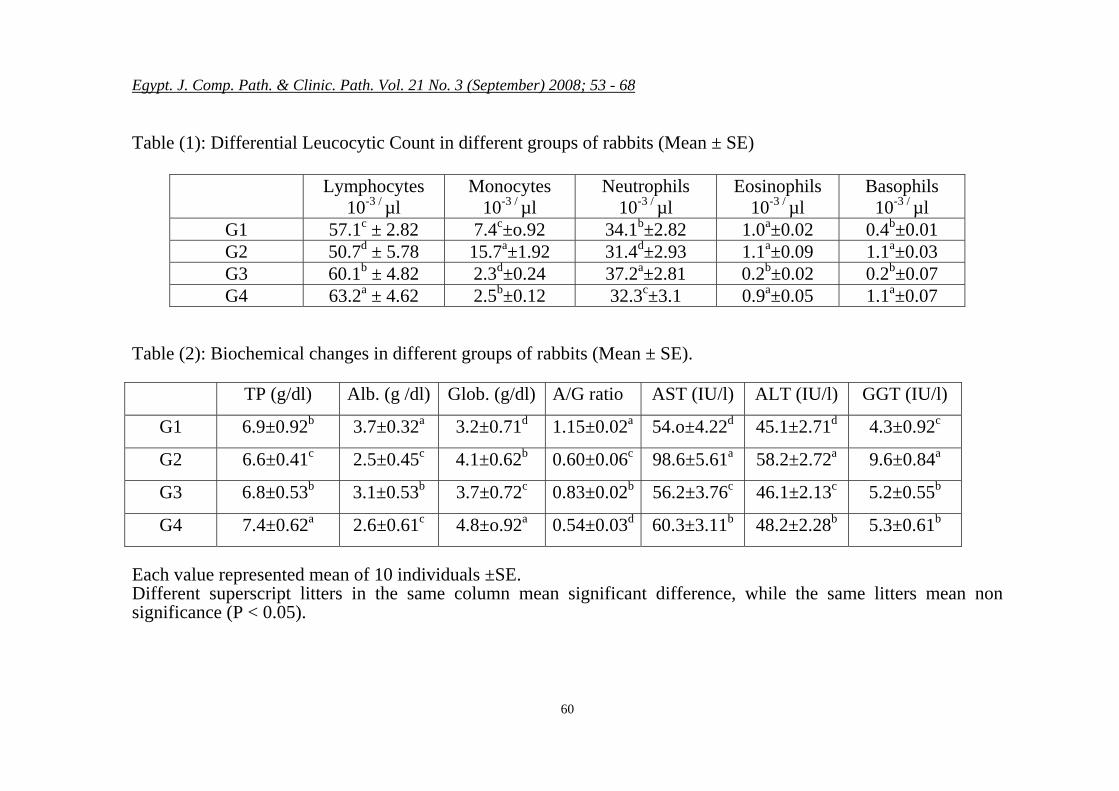
cytic count of examined rabbits re-vealed lymphocytopenia and neu-tropenia, (50.7 ± 5.78 and 31.4 ± 2.93 respectively) these decreases were significant when compared with the other experimental groups, G1 (57.1 ± 2.82 and 34.1 ± 2.82), G3 (60.1 ± 4.82 and 37.2 ± 2.81) and G4 (63.2 ± 4.62 and 32.3 ± 3.1). Also infected group showed significant increase in monocytic percentage (15.7 ± 1.92) compared with the other test groups G1 (7.4 ± 0.92), G3 (2.3 ± 0.24) and G4 (2.5b ± 0.12).
Vaccinated Nigella sativa treated group (G3) and vaccinated, Nigella sativa treated and experi-mentally infected group (G4) both showed significant increase in both lymphocytic and neutrophilic per-centages (60.1 ± 4.82 and 37.2 ± 2.81) and (63.2 ± 4.62 and 32.3 ± 3.1), respectively, in comparison with all test groups, this increase, in both groups, was accompanied
with significant monocytopenia. (3 ± 0.24 and 2.5 ± 0.12), respec-tively, in comparison with all other test groups. Eosinophilic count showed non significant change in G2 (1.1 ± 0.09) and G4 (0.9 ± 0.05) compared with normal vacci-nated non infected group G1 (1.0 ± 0.02), while G3 showed significant decrease (0.2 ± 0.02), in compari-son with all test groups. Basophilic count in G2 and G4 showed sig-nificant increase (1.1 ± 0.03 and 1.1 ± 0.07), respectively, compared with G1 and G3 (0.4 ± 0.01 and 0.2 ± 0.07), respectively. All dif-ferential leucocytic count percent-ages were shown in table (1).
Biochemical studies of the ex-perimental groups showed signifi-cant increase in total protein in group 4 (7.4 ± 0.62) compared with groups 1, 2 and 3 (6.9 ± 0.92, 6.6 ± 0.41 and 6.8 ± 0.53) respec-tively. Non significant difference was observed between group 1 and group 3 (6.9 ± 0.92 and 6.8 ± 0.53) respectively, while group 2 showed significant decrease (6.6 ± 0.41) as compared with other test groups. Albumin concentration in group 2 and group 4 showed significant de-crease (2.5 ± 0.45 and 2.6 ± 0.61) respectively, as compared with group 1 and 3 (3.7 ± 0.32 and 3.1 ± 0.53). Globulin levels in groups 2, 3 and 4 were significantly in-creased (4.1 ± 0.62, 3.7 ± 0.72 and 3.7 ± 0.72) respectively, compared with normal vaccinated control

Egypt. J. Comp. Path. & Clinic. Path. Vol. 21 No. 3 (September) 2008; 53 - 68
59
group (3.2 ± 0.71). Albumin globulin ratio (A/G) was signifi-cantly decreased in groups 2, 3 and 4 (0.60 ± 0.06, 0.83 ± 0.02 and 0.54 ± 0.03) respectively, com-pared with normal vaccinated con-trol group G1 (1.15 ± 0.02).
Liver transferase enzymes As-partate Aminotransferase, Alanine Aminotransferase and Gamma Glutamyle Transferase (AST, ALT, and GGT) showed, AST level, sig-nificant increase in group 2 (98.6 ± 5.61 ) as compared with groups 1, 3 and 4 (54.0 ± 4.22, 56.2 ± 3.76 and 60.3 ± 3.11 ) respectively.
Serum level of ALT showed significant increase in group 2 (58.2±2.72) as compared with groups 1, 3 and 4 (45.1 ± 2.71, 46.1 ± 2.13 and 48.2 ± 2.28) re-spectively.
GGT level showed significant increase in group 2 (9.6 ± 0.84) in comparison with groups 1, 3 and 4 (4.3 ± 0.92, 5.2 ± 0.55 and 5.3 ± 0.61), respectively. All biochemi-cal results were shown in table 2.
ELISA test showed significant increase in group 3 (0.465 ± 0.02) and group 4 (0.412 ± 0.03) com-pared with groups 1 and 2 (0.432 ± 0.02 and 0.387 ± 0.01), while in-fected group (G2) showed signifi-cant decrease compared with all previously mentioned test groups. Phagocytic activity of peritoneal exudates macrophages showed sig-nificant increase in group 3 (38.1 ±
1.21) as compared with groups 1, 2 and 4 (36.8 ± 1.58, 34.1 ± 0.92 and 36.2 ± 1.12) respectively, as shown in table 3. Pathological studies: Experimen-tally infected rabbits showed acute signs of the disease 2 days post in-fection, and on necropsy showed pale liver, congested spleen and kidneys with petichial hemorrhage and accumulation of intra-abdominal blood clots. Histopa-thological studies showed coagula-tive necrotic hepatitis Fig. (1), ac-companied with disseminated in-travascular coagulopathy (DIC), which is a pathognomonic process, as shown in Fig. (2).
Egypt. J. Comp. Path. & Clinic. Path. Vol. 21 No. 3 (September) 2008; 53 - 68
60
Table (1): Differential Leucocytic Count in different groups of rabbits (Mean ± SE)
Lymphocytes 10-3 / µl
Monocytes 10-3 / µl
Neutrophils 10-3 / µl
Eosinophils 10-3 / µl
Basophils 10-3 / µl
G1 57.1c ± 2.82 7.4c±o.92 34.1b±2.82 1.0a±0.02 0.4b±0.01 G2 50.7d ± 5.78 15.7a±1.92 31.4d±2.93 1.1a±0.09 1.1a±0.03 G3 60.1b ± 4.82 2.3d±0.24 37.2a±2.81 0.2b±0.02 0.2b±0.07 G4 63.2a ± 4.62 2.5b±0.12 32.3c±3.1 0.9a±0.05 1.1a±0.07
Table (2): Biochemical changes in different groups of rabbits (Mean ± SE).
TP (g/dl) Alb. (g /dl) Glob. (g/dl) A/G ratio AST (IU/l) ALT (IU/l) GGT (IU/l)
G1 6.9±0.92b 3.7±0.32a 3.2±0.71d 1.15±0.02a 54.o±4.22d 45.1±2.71d 4.3±0.92c
G2 6.6±0.41c 2.5±0.45c 4.1±0.62b 0.60±0.06c 98.6±5.61a 58.2±2.72a 9.6±0.84a
G3 6.8±0.53b 3.1±0.53b 3.7±0.72c 0.83±0.02b 56.2±3.76c 46.1±2.13c 5.2±0.55b
G4 7.4±0.62a 2.6±0.61c 4.8±o.92a 0.54±0.03d 60.3±3.11b 48.2±2.28b 5.3±0.61b
Each value represented mean of 10 individuals ±SE. Different superscript litters in the same column mean significant difference, while the same litters mean non significance (P < 0.05).

Egypt. J. Comp. Path. & Clinic. Path. Vol. 21 No. 3 (September) 2008; 53 - 68
61
Table (3): Immune parameters in different rabbit experimental groups (Mean ± SE).
Phagocytic cells (%) ELISA (Optical density) 36 .8 ±1.58 b 0.432a±0.02b G1
34.1 ±0.92 c 0.387± 0.01d G2
38.1±1.21 a 0.465±0.02a G3
36.2 ±1.12 b 0.412±0.03c G4
Each value represented mean of 10 individuals ± SE. Different superscript litters in the same column mean significant differ-ence, while the same litters mean non significance (P < 0.05).
Fig.(1): Showing hepatic peripheral lobular coagulative necrosis (H&E. X 300)
Fig.(2): Showing disseminated in-travascular Coagulopathy (thrombosis) (H&E. X 300)

Egypt. J. Comp. Path. & Clinic. Path. Vol. 21 No. 3 (September) 2008; 53 - 68
62
DISCUSSION
D ifferential leucocytic count in this study showed significant
decrease in both lymphocytic and neutrophilic counts in experimen-tally infected group (G2). This de-crease can be explained as an im-munosuppressive effect of the vi-rus, which was in accordance with Plassirat et al. (1992) and Guelfi et al. (1993). Nigella sativa L seed chloroform extract showed restora-tion of accompanied immunosup-pression due to infection as was indicated in our results by the in-crease in both lymphocytes and neutrophils. This result was in agreement with the results ob-tained by Afifi and Daghash (1999) and (Kanter et al., 2005). Nigella sativa extract inhibit eico-sanoid generation in leukocytes and membrane lipid peroxidation (El-Dakhakhny et al., 2002). Nig-ella sativa is rich in fatty acid, (oleic, linoleic and linolenic acid) and carotene (AL-Jassir, 1992). NS acts as antioxidant in cells, and contains eight essential amino ac-ids, which improves the natural immune system activity (Omar et al., 1999). Monocytic count show-ed significant increase in experi-mentally infected group (G2) which was considered as a direct response to induced viral infection on the expense of the decrease of both lymphocytes and neutrophils. Also this group (G2) showed sig-nificant increase in basophilic
count as compared with normal vaccinated group (G1). Nigella sa-tiva treatment resulted in signifi-cant decrease in eosinophilic count in both groups 3 and 4, while baso-philic count was decreased signifi-cantly in both groups 3 and 4, these results were in agreement with Bamosa and Sowayan (2000). The significant increase in lymphocytic count in Nigella sa-tiva treated groups G 3 and G 4 was in accordance with Salem (2005) who attributed this increase to thymoquinine which had shown antioxidant and immunomodulator effects on macrophage, Th1 and Th2.
Biochemical studies of serum proteins parameters showed sig-nificant decrease in total proteins in infected group (G2) which was attributed to the significant de-crease in albumin which can be re-ferred to liver damage as a result of infection Ghanem and Ismail (1999), while the significant in-crease in globulin level is ex-plained as a direct immune re-sponse to infection. Nigella sativa treated and infected group (G4) showed significant increase in total protein and both albumin and globulin showed improved liver and immune response illustrated in decreased albumin / globulin ratio (A/G), which is attributed to in-creased globulin as a direct immu-nomodulating effect of Nigella sa-
Egypt. J. Comp. Path. & Clinic. Path. Vol. 21 No. 3 (September) 2008; 53 - 68
63
tiva and this result was in agree-ment with El Kady and Kandil (1986); Wang et al. (1996), Salem (2005) and Gali et al. (2006). Studies on liver transferases as-sured liver damage in experimen-tally infected group (G2) as indi-cated by elevated serum levels of AST, ALT and GGT, which re-turned towards normal levels in Nigella sativa treated groups (G3 and G4), this effect was explained by the liver protective effect of Nigella sativa and its immu-nostimulation impact, which was in agreement with Nair et al. (1991) and El Naggar and Deib (1992).
ELISA test results showed the immunostimulating effect of Nige-lla sativa extract on its treated gr-oups (G3 and G4) compared with both normal vaccinated (G1) and experimentally infected (G2) grou-ps, this immunostimulating effect was in accordance with Tizard (1996). The obtained results were in parallel with those obtained in our study in differential leucocytic count where lymphocytosis was detected in Nigella sativa treated groups, while lymphopenia was obvious in experimentally infected group.
Cell mediated immunity wh-ich was evaluated by phagocytic activity of macrophages in the peritoneal exudates which showed significant decrease in infected
group (G2) owing to cell mediated immunosuppression in this group and showed increased immune re-sponse of these cells in samples collected from Nigella sativa treat-ed groups (G3 andG4) these results were in agreement with El kadi et al. (1989) and El Naggar and Deib (1992). The increase in pha-gocytic activity might be attributed to in vivo activation of macro-phages by production of lymphoki-nes by T helper cells after their stimulation (Tizard, 1996). Also antioxidant effect of Nigella sativa (thymoquinone) appears to en-hance host defenses against infec-tion by improving phagocytic cells function through protecting them from oxidative damage due to its high proportion of polyunsaturated fatty acids in their cell membrane and also the surrounding tissues from oxidative attack by free radi-cal produced from activated macrophage during phagocytosis (Hughes, 1999). A decrease in ni-tric oxide (NO) activity in Nigella sativa treated groups may be attrib-uted to high content of this seed with antioxidant compound (thym-oquinone) which had an important role in scavenging harmful free radicals. Nigella sativa seeds caus-ed a dose dependent decrease in Nitric oxide production from peri-toneal macrophage when activated with lipopolysaccharide (LPS) of E. coli without affecting on cell viability (Victor et al., 2003).

Egypt. J. Comp. Path. & Clinic. Path. Vol. 21 No. 3 (September) 2008; 53 - 68
64
Pathological studies of exam-ined infected rabbits revealed se-vere internal hemorrhage with ma-rked hemorrhagic pneumotrachei-tis as well as pale hypertrophied liver. These observations were in accordance with those recorded by Kavaliski, (1998). The main cause of death was attributed to the se-vere internal hemorrhage (Xu and Chan, 1982). Histopathologically, acute necrotizing hepatitis with slight inflammatory cell infiltration was recorded and was in agree-ment with results observed by Fuchs and Weissenboeck (1992). Disseminated intravascular coagu-lopathy (DIC), recoded in this study, agreed with the results ob-tained by Plassirat et al. (1992).
I n conclusion, at a practical level it is recommended to use the
natural plant extract of Nigella sa-tiva for immunostimulation for better rabbit farming.
REFERENCES Al-Jassir, M.S. (1992): "Chemical
composition and microflora of black cumin (Nigella sativa L.) seeds growing in Saudi Arabia." Food Chem., 45: 239– 242.
Afifi, O.S. and Daghash, H.A. (1999): “Reproductive per-formance of California Doe rabbits as affected by feeding freshly crushed Nigella sativa seeds." Alex. J. Vet. Sci., 15
(5): 995-1006. Bamosa, A. and Ali, B.A. and So-
wayan, S. (2000): "Effect of oral ignition of Nigella sativa on some blood parameters." Saudi Pharmacol. J.,
Burits, M. and Bucar, F. (2000): "Antioxidant activity of Nigella sativa essential oils." Phytother. Res., 14: 323-328.
Chasey, D.; Lucas, M.H.; West-cott, D.G.; Sharp, G.; Kitch-ing, A. and Hughes, S.K. (1995): "Development of di-agnostic approach to the iden-tification of rabbit hemor-rhagic disease." Vet. Rec., 137: 158-160.
Cluet, N.R.; Blanchard, D.; An-dre-Fortaine, G.; Song, B. and Ganiere, J.P. (1995): "Detection of antibodies to rabbit hemorrhagic disease virus: An immunoblotting method using virus coated hu-man erythrocyte membranes." J. Vet. Med. B, 42: 197-204.
Cohn, Z.A. and Wiener, E. (1963-a): "The paticulate hy-drolase of macrophages: I. Comparative enzymology iso-lation and properties." J. Exp. Med., 118: 991.
Drupt, F. (1974): "Colorimetric method for determination of albumin." Pharm. Biol., 9: 777- 779.
El-Dakhakhny, M.; Madi, N.J.; Lembert, N. and Ammon,

Egypt. J. Comp. Path. & Clinic. Path. Vol. 21 No. 3 (September) 2008; 53 - 68
65
H.P. (2002): "Nigella sativa oil, nigellone and derived thy-moquinone inhibit synthesis of 5-lipoxygenase products in po-lymorphonuclear leukocytes from rats." J Ethnopharmacol., 81: 161– 164.
El-Kadi, A. and Kandil O.I. (1987): "The black seed (N. sativa) and immunity, its effect on human T. cell subset." Fed-eration proceedings, 46 (4): 1222.
El-Kadi, A.; Kandil, O. and Tabuni, A.M. (1989): "Nigella sativa and cell mediated im-mubnity." Arch. AIDS Res., 1: 232-233.
El Naggar A. M. and Deib A. M.(1992): A study on some bio-logical activities of Nigella sa-tiva (Black seed) “Habat El Ba-raka” J. Egyp. Sosc. Pharma-col. Exp. Ther., 11: 781-799.
Fuchs A. and Weiessenboeck H. (1992): "Comparative Histopa-thological study of rabbit hem-orrhagic disease (RHD) and European brown hare syn-drome (EBHS)." J. Comp. Pathm., 107 (1): 103-113.
Gali – Mohtasib; Rossener A. and Schneider–Stock R. (2006): "Thymoquinone a promising anti-cancer drug of natural sources." Int. Biochem. Cell Biol. 38 (8): 1249-1253.
Ghanem, T.A. and Ismail, A. (1991): "Occurrence of rabbit
hemrhagic disease in Sharkia province Egyptian-German." Vet. Med. Conf. Nov., 12-15.
Guelfi, J.F.; Ganiere J.P.; Andre-Fontain, G. and Debailleul, M. (1993): "Recueil De Medi-cine Veterinaire." De L, Ecole D, Alfort., 169 (2): 93-99.
Henry, R. J. (1974): "Clinical Prin-ciples and Techniques." 2nd ed., Harper, p. 525.
Hughes, D.A. (1999): "Effect of dietary antioxidants on the im-mune function of middle- aged adults." Proc. Nutr. Soc., 58: 79-88.
Kaneko, J.J. (1996): ''Clinical Bio-chemistry of Domestic Ani-mal.'' 4th ed., Academic Press, N.Y; London, Toronto, Sydney San Francisco.
Kanter, M.; Demir, H.; Kaya, C. and Ozbek, D. (2005 ) : "Gastroprotective activity of Nigella sativa L oil and its con-stituent, thymoquinone against acute alcohol-induced gastric mucosal injury in rats." World J. Gastroenterol., 11: 6662-6666.
Kavaliski, J. (1998): "Monitoring the spread of rabbit hemor-rhagic disease virus as a new biological agent for control of wild European rabbits in Aus-tralia." J. Wild Dis., 34 (3): 421-428.
Liu, S.J.; Xue, H.P.; Pu, B.Q. and Qian, N.H. (1984): "A new vi-

Egypt. J. Comp. Path. & Clinic. Path. Vol. 21 No. 3 (September) 2008; 53 - 68
66
ral disease in rabbits." Animal Husbandry and Veterinary medicine,
Mashhadian, N.V. and Rakhsh-andeh, H. (2005): "Antibac-terial and antifungal effects of Nigella sativa extracts against S. aureus, P. aeuroginosa and C. albicans." Pak. J. Med. B, 42: 197-204.
Nair, S.C.; Salomi, M.J.; Panik-kar, B. and Panikkar, K.R. (1991): "Modulatory effects of crocus sativus and Nigella sativa extracts on cisplatin-induced toxicity in mice." J. Ethenopharmacol., 31: 75-83.
Nelson, D.S. (1969): "Macropha-ges and immunity." North- holand Publishing Co., Am-sterdam.
Omar, A.; Ghosheh, S.; Abdul-ghani, A.; Houdi, A.; and Crookscor, P. A. (1999): "High performance liquid chromatographic analysis of the pharmacologically active quinones and related com-pounds in the oil of the black seed (Nigella sativa L)." J. Pharm. Biomed. Anal., 19: 757– 762.
Peter T. (1968): "Colorimetric de-termination of total protein." Clin. Chem., 14: 11-13.
Plassirat, G; Guelfi, J. F; Ga-niere, J. P.;Wang, B.; An-dre-Fontain, G. and Wyers, M. (1992): "Hematological
parameters and Visceral le-sions relationships in rabbit viral hemorrhagic disease." J Vet. Med. Series B, 39 (6): 443-453.
Ratlif, C.R. and Hall, F. (1973): "Laboratory Manual of Clini-cal Biochemistry.", SCOH and White Memorial Hospital Publication office, Temple TX.
Rchid, H; Nmila, R.; Bessière, J. M.; Sauvaire, Y. and Cho-kaïri, M. (2004): "Volatile Components of Nigella dam-ascena L. and Nigella sativa L. Seeds." Journal of Essen-tial Oil Research: JEOR, Nov/Dec 2004.
Reitman, S. and Frankel, S. (1957): “A colorimetric deter-mination of serum glutamic oxaloacetic and glutamic py-ruvic transaminase.” Am. J. Clin. Path., 28: 56-58.
Salem, M.L. (2005): "Immunomo-dulatory and therapeutic prop-erties of the Nigella sativa seed." Int. Immunopharmaco., 12. (5): 13-14: 1749-70.
Schalm, O. W; Jain, N.C and Carrol, E. (1991): Vet. He-matol. 3rd ed. Lea Fabiger, Philadelphia, USA.
Tizard, I. R. (1996): "Veterinary immunology. An Introduc-tion." 5th Ed., WB Saunders Company, A division of Har-court Brace Company, Phila-

