cloning, expression, and chromosomal localization · pdf filecloning, expression, and...
TRANSCRIPT

Cloning, expression, and chromosomal localization of mouse liver bile acid CoA:amino acid N -acy It ransf erase
Charles N. Falany,'!* Hank Fortinberry," Edward H. kiter: and Stephen Barnes*,+ Department of Pharmacology and Toxicology,* and Comprehensive Cancer Center Mass Spectrometry Shared Facility,' University of Alabama at Birmingham, Birmingham, AL 35294, and The Jackson Laboratory,s Bar Harbor, ME 04609
Abstract A mouse liver LZap XR cDNA library was screened using the coding region of human bile acid CoAamino acid N-acyltransferase (BAT) cDNA as a probe. Ten positive clones were isolated and purified, two of which apparently possessed complete open reading frames for BAT based on sequence analysis of the ends of the cDNAs. One clone (mBAT#9) was selected for sequence analysis and characterization. mBAT#9 is 1869 basepairs in length and the full-length cDNA possesses a 189 basepair 5'-nontranslated region, an open-reading frame of 1260 b airs, and a 404 basepair 3'-nontranslated region fol lowedva poly(A) tail. The open-reading frame codes for a 420 amino acid protein with a calculated molecu- lar mass of 46,525 daltons. The structural gene for mBAT was mapped to mouse Chromosome 4. The amino acid sequence of mBAT is 69% identical and 84% similar to that of hBAT, and 86% identical and 95% similar to that of kan-1, a putative rat liver BAT. Enzymatically active mBAT was expressed in E. coli using the bacterial expression vector pKK233-2. Immu- noblot analysis of expressed mBAT with rabbit anti-human BAT polyclonal antibodies detected a single protein with a molecular mass of approximately 45,000 daltons. Cytosol from cells transformed with mBAT#9 / pKK233-2 possessed signifi- cant amounts of BAT-catalyzed conjugating activity with tau- rine as substrate but the expressed enzyme did not use glycine or fluoro-palanine as substrates. The K,n value for taurine was 1.9 mM -t 0.1 mM in reactions with cholyl CoA as a cosub- strate. The specificity of mBAT for taurine as a substrate was confirmed by the demonstration, using HPLGelectrospray ionization mass spectrometry, that mouse gallbladder bile contained only taurine conjugates of bile acids. The identifi- cation of the types of amino acid conjugates of bile acids pres- ent in mouse bile had not been previously rep0rted.M These results indicate that a taurine-specific form of BAT has been cloned and expressed from mouse liver.-Falany, C. N., H. Fortinberry, E. H. Leiter, and S. Barnes. Cloning, expression, and chromosomal localization of mouse liver bile acid CoA amino acid N-acyltransferase. J. Lzpid Res. 1997. 38: 1139- 1148.
Supplementary key words conjugation
bile acid bile acid amidates taurine
Bile acids are the major solutes in bile. Greater than 99% of the bile acids secreted by the liver into the bile
in mammals are conjugated with amino acids. Amino acid conjugation of bile acids to form N-acyl amidates increases the amphipathic nature of bile acids and hence their detergent properties (1). It also prevents their precipitation in the acidic milieu of the upper small intestine. In addition, amino acid conjugation de- creases the formation of relatively insoluble complexes of bile acids with calcium (2). Bile acids form mixed micelles with phospholipids which serve to increase the aqueous solubility of cholesterol by a factor of lo6. In the intestines, they are responsible for the solubilization and absorption of fats, vitamins, and fat- soluble com- pounds (1).
With the exception of the manatee (3), elephant and hyrax (4, 5 ) which secrete bile alcohol sulfates, mam- mals secrete bile acids conjugated with taurine and/or glycine. Some species such as dogs and cats synthesize only taurine conjugates, whereas other species such as rats and humans synthesize both taurine- and glycine- conjugated bile acids (4).
Amidation of bile acids in the liver requires the se- quential action of two separate enzymes, bile acid CoA synthetase (BAS) and bile acid CoAamino acid N-acyl- transferase (BAT). In the first reaction, BAS catalyzes the formation of bile acid CoA thioesters. In the second reaction, BAT catalyzes the reaction between the bile acid CoA thioester and either taurine or glycine.
Our laboratory has recently reported the molecular cloning of human liver BAT (hBAT) and expression of the enzymatically active enzyme in E. coli (6). Expressed and purified hBAT utilized both taurine and glycine as substrates. The only other known substrate for hBAT is fluoro-palanine, a metabolite of the chemotherapeutic
Abbreviations: BAT, bile acid CoAamino acid N- acyltransferase;
'To whom correspondence should be addressed. BAS, bile acid CoA synthetase.
Journal of Lipid Research Volume 38, 1997 1139
by guest, on May 12, 2018
ww
w.jlr.org
Dow
nloaded from

agent 5-fluorouracil (7, 8). Substrate specificity studies have indicated that BAT is very selective in the amino acids with which it conjugates bile acids (9).
As part of a project to better understand the mecha- nism for the formation and physiological properties of bile acid conjugates and to develop a mouse knockout model of BAT, we have isolated and expressed the cDNA for mouse liver BAT in bacteria. This report de- scribes the kinetic properties of the expressed mouse BAT (mBAT) as well as the chromosomal localization and sequence similarity to other mammalian BATS. Ex- pressed mBAT uses taurine but not glycine as a s u b strate for the conjugation of bile acids and analysis of the composition of mouse bile detected only the pres- ence of taurine conjugates of bile acids.
MATERIALS AND METHODS
Materials
The mouse liver hZap cDNA library was obtained from Stratagene (La Jolla, CA) . Restriction enzymes and other DNAmodifylng enzymes were purchased from New England BioLabs (Beverly, MA) and Promega (Mad- ison, WI). [ cx -~~SI~ATP (3000 Ci/mmol) and [a- 32P]dCTP (800 Ci/mmol) were purchased from DuPont- NEN (Beverly, MA). pKK233-2 was obtained from Phar- macia Biotech Inc. (Piscataway, NJ). The PCR nucleo- tides and Taq DNApolymerase were purchased from Pro- mega. Sequenase Version 2.0 sequencing kits were obtained from United States Biochemical Corporation (Cleveland, OH). Nitrocellulose membranes were pur- chased from Micron Separation Inc. (Westborough, MA). All other reagents were molecular biology grade.
Isolation of mBAT cDNA
A mouse liver hZap XR cDNA library was screened using the coding region of the human BAT cDNA as a probe (6). E. coli XL-1 Blue cells were infected with ali- quots of the mouse liver cDNA library (approximately 30,000 pfu/150 mm petri plate) and incubated over- night at 37°C. To screen the plates, nitrocellulose filters were soaked in 1 M NaCl and then placed on the plates and marked for orientation. After 5 min, the filters were removed and placed in a denaturing solution (0.5 M
NaOH, 1.5 M NaCl) for 10 min. The filters were then neutralized in 0.5 M Tris-HC1, pH 7.0, with 1.5 M NaCl. The phage DNA was covalently bound to the filters by exposure to W light using a Bio-Rad Gene Linker appa- ratus. The filters were pre-hybridized for 15 min in 6X SSC, 5X Denhardt's solution, 0.5% SDS, and 0.2 pg/ ml sonicated salmon sperm DNA (10). hBAT cDNA was radiolabeled using the Prime-A-Gene procedure (Pro-
mega) and ["PIdCTP (3000 Ci/mM) to a specific ra- dioactivity of 10' cpm/pg DNA. Radiolabeled hBAT cDNA was then added to the filters in fresh hybridiza- tion solution at a concentration of 200,000 cpm/ ml and allowed to incubate overnight at 65°C. The next day the filters were washed at 65°C two times with 2X SSC/ 0.1% SDS for 15 min and one time with 2 X SSC for 15 min. The filters were then dried and exposed to autora- diography film with an intensifjmg screen overnight at -70°C. Positive plaques were purified by repeated cy- cles of dilution and rescreening until a single pfu could be isolated. This procedure resulted in the isolation of' ten pure phage clones from the screening of approxi- mately 300,000 pfu of the cDNA library. After isolation of the phage, the cDNAs were recovered in Bluescript phagemids by coinfection with helper phage (R408) as per the manufacturer's instructions (Stratagene).
Nucleotide sequence analysis of &AT cDNAs
The mBAT cDNAs were subjected to double-stranded sequencing by the dideoxynucleotide chain termina- tion method using Sequenase 2.0 and [cx-~'SS]~ATP to label newly synthesized strands. The '' Slabeled prod- ucts were resolved on 6% polyacrylamide-urea gels us- ing a buffer gradient of 0.5X to 2.5X TBE (TBE = 89 mM Tris-borate, 2 mM EDTA). The complete cDNA se- quence of mBAT was obtained by subcloning restriction fragments into pBluescript for sequencing with T3 and T7 primers or by sequencing short fragments generated by Sau 3A or Hae 111 digestion and subcloned into pBluescript. Oligonucleotide primers synthesized to in- ternal sequences of mBAT were also used to sequence specific regions. Sequence gels were read manually and analyzed using MacVector sequence analysis software (Kodak) .
Generation of &AT bacterial expression vector
In order to express enzymatically active mBAT in E. coli, the mBAT cDNA was subcloned into the Nco I- Pst I sites of the bacterial expression vector pKK233-2 (Clontech) in a three- step procedure. An internal 685 basepair Nco I-Pst I fragment was isolated from the BAT cDNA and subcloned into the Nco I and Pst I sites of pKK233-2. The 3'-780 basepair Pst I-Pst I fragment was then subcloned into the Pst I site of this plasmid and the orientation of the Pst I fragment was established by restriction mapping. In order to complete the BAT cDNA and insert the codon for the initial methionine into the Nco I site adjacent to the ribosome binding site, a 220 basepair Nco I fragment with the initial me- thionine codon incorporated into an Nco I site was syn- thesized. PCR was used to generate a Nco I site incorpo- rating the initial methionine codon. A 19-nucleotide primer was synthesized to bases 181-200 in which the two adenosines prior to the ATG were changed to cyto-
1140 Journal of Lipid Research Volume 38, 1997
by guest, on May 12, 2018
ww
w.jlr.org
Dow
nloaded from

sines (underlined), (5'-CTGCAAAC&4TGGCCC- 3'). The template for PCR was the mBAT#S/pBlues- cript vector. Twenty cycles of denaturation, 1 min at 95"C, annealing for 1 min at 59"C, and extension for 40 sec at 72°C were performed. Then, T3 primer was added and the reaction was subjected to 30 cycles of denaturation for 1 min at 95"C, annealing for 1 min at 46"C, and extension for 40 sec at 72°C. PCR products were extracted with chloroform, ethanol precipitated, digested with Nco I, and purified after agarose gel elec- trophoresis using a PCR Cleanup kit (Promega). The 220 basepair Nco I-Nco I fragment was subcloned into the pKK233-2 vector containing the 3'-portion of the mBAT cDNA. The correct orientation of the Nco I frag- ment was determined by restriction mapping and se- quence analysis.
Bacterial expression of mBAT
The pKK233-2/mBAT#9 expression vector was trans- formed into E. coli DH5a cells made competent using a CaC1, procedure (1 1). mBAT activity was induced and isolated as described previously for hBAT (6). The mBAT activity in the induced bacterial cytosol was par- tially purified by DEAE anion-exchange chromatogra- phy as previously described (1 2). The column fractions were assayed for BAT activity and by immunoblot analy- sis using the rabbit anti-human BAT antibody.
BAT assay
mBAT enzyme activity was determined using a ra- dioassay as described previously using [ 3H] taurine and [ "Hlglycine as substrates (6, 8). Briefly, [ 3H] taurine was used for conjugation to unlabeled cholyl CoA to form ['HI taurine conjugates. The sensitivity of the assay will allow for the detection of 0.1 nmol cholyl CoA conjugated in 30 min. Cholyl CoA was chemically syn- thesized as described previously (8), using a modifica- tion of the method of Shah and Staple (13). Protein concentrations were determined using the Bio-Rad pro- tein assay with gamma globulin as a standard. Assays uti- lizing fluoro-palanine as a substrate were performed as described previously except the reaction mixtures were analyzed by mass spectrometry using a PE-Sciex (Con- cord, Ontario, Canada) API 111 triple quadrupole in- strument in the Mass Spectrometry Core Facility of the UAB Comprehensive Cancer Center.
Analysis of mouse and rat bile
Bile was recovered from the gallbladder of a normal AJ male mouse and from the common bile duct of a female Sprague-Dawley rat under ether anesthesia. The biles were diluted in 1 ml of water and passed over an activated CIS Sep-Pak cartridge. Retained bile acids were eluted from the cartridge with 2 ml of methanol. Ali- quots were injected onto a 10 cm X 2.1 mm i.d. Cs re-
versed-phase column using a linear gradient of 30- 100% acetonitrile in 0.1% acetic acid. The flow rate was 0.2 ml/min; the column eluate was split 1:1, with 0.1 ml/min being passed into the IonSprayTM interface of the PE-Sciex API I11 triple quadrupole mass spectrome- ter operating in the negative ion mode, with an orifice potential of -6OV. Spectra were recorded from 300- 800 m/z at 1.5-sec intervals. The operation of the mass spectrometer and analysis of data were carried out using two Macintosh Quadra 950 computers interfaced with an Ethernet link.
Northern blot analysis of mBAT expression
The analysis of the expression of mBAT message in different tissues was carried out using a commercial mouse multiple tissue Northern blot (Clontech). The blot contained 2 pg of poly A+ RNA from eight differ- ent mouse tissues. The membrane was hybridized with the mBAT cDNA labeled with [ 32P] dCTP by random- priming using a Pharmacia Oligolabeling Kit. Hybrid- ization was carried out for 2 h at 65°C in Quickhyb con- taining 1.25 X lo6 cpm/ml of ["€']DNA. The blot was washed under high stringency conditions and autoradi- ography was then performed at -70°C with an intensi- fylng screen.
Immunoblot analysis of mBAT
For immunoblot analysis of expressed mBAT, cyto- solic proteins prepared from the E. coli expressing mBAT were resolved by SDSPAGE in a 12.5% gel, elec- trophoretically transferred to nitrocellulose mem- branes, and analyzed for immunoreactivity using spe- cific rabbit anti-hBAT polyclonal antibodies at a 1/ 1,500 dilution (6, 14). Immunoconjugates were visual- ized by chemiluminescence using the Lumiglo method W L ) .
Chromosomal localization of mBAT gene
The chromosomal localization of the mBAT gene was carried out using the backcross panel of DNAs from the Jackson Laboratory Backcross DNA Panel Mapping Re- source (15). The probe for these studies was a 1533 ba- sepair fragment of the mBAT cDNA containing the complete coding region of the cDNA. The differential presence of a 5 kb Pst I restriction fragment variant in the B6 versus a 6 kb restriction fragment in M. spetus was used to type the (C57B46JEi X SPRET/Ei)Fl X SPRET/Ei DNA panel (DNA from 94 first backcross segregants) by Southern blot analysis.
RESULTS
Molecular characterization of mBAT cDNAs A mouse liver hZap XR cDNA library was screened
under moderate stringency conditions using the human
Falany et al. Cloning and expression of mouse liver BAT 1141
by guest, on May 12, 2018
ww
w.jlr.org
Dow
nloaded from
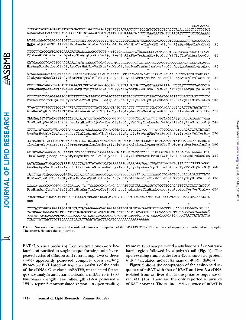
CGAGAACTC TTCCATTATCTACAGTGTTGTCAGAGCCTTGGTTTGAGAGAGTCTCTGAGAAGTCCTGGGCATCTGTGCTGACCGACAGGGCCTCCTTCTCT AGAGCACACCACGTTCCTGAGGGTTGCTGT~CTACTGTTTTGGTG~TATTCCTG~GAA~GTCC~GATTCCCTCTGC~
ATGGCCAAGCTGACAGCTGTTCCTCTCAGTGCTCTTGTTGATGAGCCTGTGCACATCCAGGTCACAGGCCTGGCCCCCTTTCAGGTGGTG ~~aLysLeuThrAlaVa~ProLeuSerA~aLeuValAspGluProValH~sIleGlnValThrGlyLeuAlaProPheGlnValVal
TGCCTTCAGGCATCACTGAATGAGAGGAAACCTGTTAGTTCTCAGGCCTTCTACAGGGCCAGTG~GTGGGTGAGGTAGATCTGGAG CysLeuG~nAlaSerLeuLysAspGluArgLysProValSerSerGl~laPheTyrArgAlaSerGluValGlyGluValAspLeuGlu
CATGACCCCTCACTTGGAGGAGACTATATGGGGGTCCACCCCATGGGCCTTTTCTGGTCCTTGAAACCTGAAAAGCTATTGGGTAGATTG HisAspProSerLeuGlyGlyAsp~rMetGlyValHisProMetGlyLeuPheTrpSerLeuLysProGluLysLeuLeuGlyArgLeu
ATAAAAAGAGATGTGATAAATAGCCCCTACCAAATCCACACAT~GCTTGCCATCCATACTTTCCATTACAAGACCTAGTCGTCAGTCCT IleLysArgAspValIleAsnSerProTyrGlnIleHisIleLysAlaCysHisProTyrPheProLeuGlnAspLeuValValSerPro
CCCTTGGATAGCCTGACTCTGGAAAGGTGGTGGTATGTGGCACCTGGGGTCAAGAGGATCCAGGTAAAGGAAAGCCGCATCCGGGGAGCCCTG ProLeuAspSerLeuThrLeuGluArgTrpTyrValAlaProGlyValLysArgIleGlnValLysGluSerArgIleArgGlyAlaLeu
TTTCTGCCTCCAGGAGAAGGTCCTrrTCCAGGGGTCAGGGGTCATTGACTTGTTTGGAGGTGCTGGTGGATTGATGGAGTTCCGAGCCAGTCTTCTG PheLeuProProGlyGluGlyProPheProGlyValIleAspLeuPheGlyGlyAlaGlyGlyLeuMetGluPheArgAlaSerLeuLeu
GCAAGTCGTGGCTTTGCCACCTTAGCTCTGGCTTACTGGAACTATGATGACCTGCC~CTCGACTGGAGAAGGTAGATCTAGAATATTTT AlaSerArgGlyPheAlaThrLeuAlaLeuAlaTyrTrpAsnTyrAspAspLeuProSerArgLeuGluLysValAspLeuGluTyrPhe
GAAGAAGGTGTAGAGTTTCTCCTGAGACATCCTAAGGTCCTCGGCCCAGGTGTTGGCATCCTTTCTGTATGCATTGGAGCAGAGATTGGA GluGluGlyValGluPheLeuLeuArgHisProLysValLeuGlyProGlyValGlyIleLeuSerValCysIleGlyAlaGluIleGly
CTTTCTATGGCTATTAACCTAAAACAAATAAGAGCCACTGTACTTATCAATGGGCCTAAT~TGT~CTCAAAGTCCACATGTATATCAT LeuSerMetAlaIleAsnLeuLysGlnIleArgAlaThrValLeuIleAsnGlyProAsnPheValSerGlnSerProHisValTyrH~s
GGTCAGGTCTACCCACCTGTACCCAGTAATGAAGAGTTTTGTAGTCACCAATGCCTTGGGACTTGTAGAATTCTATCGAACCTTTCAGGAA GlyGlnValTyrProProValProSerAsnGluGluPheValValThrAs~laLeuGlyLeuValGluPheTyrAr~hrPheGlnGlu
ACTGCAGATAAGGACAGCAAATATTGTTTTCCCATTG~GCTCATGGACATTTCCTTTTTGTGGTTGGAGAAGATGAT~TCTC ThrAlaAspLysAspSerLysTyrCysPheProIleGluLysAlaHisGlyHisPl~eLeuPheValValGlyGluAspAspLysAsnLeu
AACAGCAAAGTGCATGCTAATCAAGCCATAGCACAGCTGATGAAAAATGGAAAGAAGAATTGGACTCTGCTGTCTTACCCTGGGGCAGGT AsnSerLysValHisAlaAsnGlnAlaIleAlaGlnLeuMetLysAsnGlyLysLysAsnTrpThrLeuLeuSerTyrProGlyAlaGl~
CACCTGATTGAGCCTCCCTATACTCCACTGTGCCAAGCCTC~GGATGCCCATTTTGATCCC~GCCTCAGCTGGGGAGGAGAGGTTATC HisLeuIleGluProProTyrThrProLeuCysGlnAlaSerArgMetProIleLeuIleProSerLeuSerTrpGlyGlyGlyGluValIle
CCCCATAGCCAAGCTGCACAGGAGCATTCTTGGAAGGAGATACAG~TTTCTCAAGCAGCATCTCCTTCCAGATTTGAGCAGTCAGCTC ProHisSerGlnAlaAlaGlnGluHisSerTrpLysGluIleGlnLysPheLeuLysGlnHisLeuLeuProAspLeuSerSerGlnLeu
* * * * * * * * * TGAGTGGACTTGATTATATTCCTGGAAAGTGGAGCTGGGCATCTCCTGGCCAGCACCACTCCTCACTTCCATAGAGGAATGTCTTTGATC
* * * * * *
* * * * * * * * *
* * * * *
* * * * * * * * *
* * * * * * *
* * * * * * * *
* * * * * * * *
* * * * * * * *
* * * * * * * * *
* * * * * * * *
* * * * * * * *
* * * * * * * * *
* * * * * * * * *
* * * * * * * * *
* * * * * * * TCTTATCTGGCAAGGAAGGAGAGTACCACAAG~TACAGGAGGATGGAGAGTGATAACGTCTTGAATTTGGAAGGGGAAACATGTTTT CATGGAATGAAAGGTTGTCATGCATGTGAGAGCCCTATATCTACATGAAT~TCGTAGGCC~TCCT~TGTTCAACATCATAGCAACT TTCTGTTATGATAATTATCAGGGAAATTATCAGTGATATCAGTGATAAACCACAGAATACTTTTGTTTAT~GAAACATG~TAATTATATATTA TCACTTATTAATWCTTGAAACTCACATTAAATATACTTAGATC-
30
6 0
90
120
150
180
210
240
270
300
330
360
390
4 2 0
Fig. 1. The asterisk denotes the stop codon.
Nucleotide sequence and translated amino acid sequence of the mBAT#9 cDNA. The amino acid sequence is numbered on the right.
BAT cDNA as a probe (6). Ten positive clones were iso- lated and purified to single plaque forming units by re- peated cycles of dilution and rescreening. Two of these clones apparently possessed complete open reading frames for BAT based on sequence analysis of the ends of the cDNAs. One clone, mBAT#9, was selected for se- quence analysis and characterization. mBAT #9 is 1869 basepairs in length. The full-length cDNA possessed a 189 basepair 5'-nontranslated region, an open-reading
frame of 1260 basepairs and a 404 basepair 3'- nontrans- lated region followed by a poly(A) tail (Fig. 1 ) . The open-reading frame codes for a 420 amino acid protein with a calculated molecular mass of 46,525 daltons.
Figure 2 shows the comparison of the amino acid se- quence of mBAT with that of hBAT and kan-1, a cDNA isolated from rat liver that is the putative sequence of rat BAT (16). These are the only reported sequences of BAT enzymes. The amino acid sequence of mBAT is
1142 Journal of Lipid Research Volume 38, 1997
by guest, on May 12, 2018
ww
w.jlr.org
Dow
nloaded from

mBAT rBAT hBAT
mBAT rBAT hBAT
mBAT rBAT hBAT
mBAT rBAT hBAT
mBAT rBAT hBAT
mBAT rBAT hBAT
mBAT rBAT hBAT
MAKLTAVPLSALVDEPVHIQVTGLAPFQWCLQASLKDER-KPVSSQAF'YRASEVGEVDL 59 R T DKGNLFN 60
IQ T V RA I M S F E NGDMFY H N F 60
EHDPSLGGDYMGVHPMGLFWSLKPEKLLGRLIKRDVINSPYQIHIKACHPYFPLQDLWS 119 R S M T V M R H K V L VEGK I 120 N AS T L M R F VQV LYDLELIVNNK A 120
PPLDSLTLERWYVAPGVKRIQVKESRIRGALFLPPGEGPFPGVIDLFGGAGGLMEFRASL 179 ss I M T H G F 180 A K A T K R G L L L L 180
L A S R G F A T L A L A Y W N Y D D L P S R L E K V D L E Y F E E G V E F L L R I 239 H G 240
S H E RKP VT AAN F S W Q VQ 240
GLSMAINLKQIRATVLINGPNFVSQSPHVYHGQVYPPVPSNEEFGLVEFYRTFQ 299 T SN R K FQ T CS T E 300
Y V T T PFGI Q IHQ L HSAQLIS L L E 300
ETADKDSKYCFPIEKAHGHFLFVVGEDDKNLNSKVHANQAIAQLMKNGKKNWTLLSYPGA 359 K S 360
T Q V G A Q L E Q Q I G TI A E G KRH N 360
GHLIEPPYTPLCQASRMPILIPSLSWGGEVIPHSQAAQEHSWKEIQKFLKQHLLPDLSSL 420 s s FV IN -A N GFN 420 S C TTHDR-- H -A A R RK I VT 418
Fig. 2. Comparison of the translated amino acid sequences of mBAT, kan-1 and hBAT cDNAs. The amino acid sequences were aligned using the MacVector Sequence Analysis programs. The differences in the sequences of kan-I, a putative form of rBAT (16) and hBAT (6) are shown as compared to the sequence of mBAT. Dashes indicate the presence of gaps inserted to optimize sequence alignments. The xxx denotes the stop codon.
69% identical and 84% similar to that of hBAT, and 86% identical and 95% similar to that of kan-1. The re- gions of dissimilarity between the three amino acid se- quences are spread fairly uniformly along the lengths of the proteins. The most extensive region of dissimilar- ity was from residues 88-123.
Expression of mBAT
mBAT was expressed in E. coli using the bacterial ex- pression vector pKK233-2, which had previously been used for the expression of hBAT (6). Expressed mBAT migrated with a molecular mass of approximately 45,000 daltons during SDSpolyacrylamide gel electro- phoresis and reacted with the rabbit anti-hBAT poly- clonal antibodies during immunoblot analysis. No im- munoreactive protein was observed in cytosol prepared from bacteria transformed with the pKK233-2 vector alone. Cytosol prepared from E. coli DH5a cells trans- formed with the bacterial expression vector pKK233-2 alone did not possess detectable levels of BAT activity with cholyl-CoA and either glycine or taurine as co- substrates. Figure 3 shows that cytosol from cells trans- formed with mBAT#9/pKK233-2 possessed significant amounts of cholyl- CoA conjugating activity with taurine and the BAT activity coeluted with immunoreactive
mBAT during DEAE-Sepharose C1-6B chromatography. These results demonstrate that mBAT#9 encodes an en- zymatically active protein. No conjugating activity was detected with glycine as a co-substrate or with fluoro-p alanine as a substrate.
For kinetics studies of mBAT activity, DEAE-Sepha- rose C1-6B chromatography was carried out to resolve the mBAT activity from the cholyl-CoA hydrolyzing ac- tivity. The taurine conjugating activity of the partially purified mBAT was linear with respect to incubation time and protein concentration (data not shown). The apparent K , value for taurine was 1.9 ? 0.1 mM. No activity was detected using either ['Hlglycine or fluoro- palanine as substrates. Addition of concentrations of glycine up to 5 mM to the taurine conjugation reactions did not cause any detectable inhibition of the rate of taurocholate formation.
Northern blot analysis of mouse tissues
Figure 4 shows that the mBAT cDNA hybridized to a single major RNA species with an approximate length of 1900 nucleotides in mouse liver poly A+ RNA. No reactive RNA bands were detected in RNA isolated from the other tissues tested. The length of the mBAT#9 cDNA is 1869 bp (Fig. 1) which correlates well with the size of the poly A+ RNA detected in mouse liver.
Falany et al. Cloning and expression of moue liver BAT 1143
by guest, on May 12, 2018
ww
w.jlr.org
Dow
nloaded from

2ooo1 9 10 11 12 13 14 15 16 17 hBAT
A 0 5 10 15 20 25
Fraction B
Elg. 3. Immunoblot analpis of mBAT exprrssed in E. coli. The mBAT cDNA was inserted into the bacterial expression vector pKK283-2 and exprrssed in E. cot5 DH5a cells as described in Methods. Cytosol was prepared from the E. cdi DH5a cells and applied to a DEAESepharose CldB column equilibrated in 10 mM uiethanolamine, pH 7.4. containing 1 mM dithiothreitol. The column was then eluted with a 0-300 mM NaCl gradient in the same buffer and the fractions were assayed for BAT activity. Panel A shows the immunoblot analysis of the column fractions using rabbit anti-hBAT IgG (14). Panel B shows the elution of BAT activity in the same fractions using cholyl &A and taurine as substrates.
Chromosomal localization The structural gene encoding mouse BAT has been
assigned the symbol Bud. Using the Jadeson BSS back- cross (15), the structural gene for mBAT was mapped to Chromosome 4. The strain.distribution pattern re- vealed linkage to markers on proximal Chromosome 4 (Fig. 5, top), with 1 /94 recombinants with D4Bir-12 ( a p proximately 1 cM proximal to B a d ) and 5/94 recombi- nants with D4Bir-14 (approximately 5.3 cM distal). The localization of Bud on Chromosome 4 is summarized in Fig. 5, bottom.
HPLCMS of bile acid conjugates in mouse and rat bile
To investigate the forms of bile acid amidates present in mouse and rat bile, extracts of bile from both species were analyzed by HPLGMS. Selected ion chromato- grams prepared from the total ion current revealed that only taurine-conjugated bile acids were present in ex- tracts of mouse bile (Fig. 6). Two major peaks (514 m/ z) detected in the samples of mouse bile were tauro$- muricholic acid and taurocholic acid. No peaks were detected for glycine-conjugated bile acids ( m / z of 464 for trihydroxy bile acids, m/z 448 for dihydroxy bile acids). In contrast, HPLGMS analysis of rat bile re- vealed that it contained bothglycine and taurine dihy- droxy and trihydroxy bile acids (Fig. 7).
DISCUSSION
A full-length cDNA encoding mouse BAT was isolated from a mouse liver cDNA library and the enzymatically
active enzyme was expressed in E. coli DH5a cells. Analy- sis of the kinetic properties of expressed mBAT shows that the enzyme is a taurine-specific conjugating form of BAT. This was confirmed by the lack of glycine-conju- gated bile acids in mouse gallbladder bile as demon- strated by HPLGmass spectromeq. To the author's knowledge, this is the first report of the types of amino acid conjugates of bile acids formed in mice.
Different animal species are capable of conjugating the COA derivatives of bile acids with taurine, or both taurine and glycine (4). The evolutionary pattern of taurine-specific conjugation of bile acids is generally as- sociated with a carnivorous diet. Species such as cats and seals tend to be taurine-specific conjugators possi- bly because meat is a major source of taurine and is therefore readily available in their diets. Herbivores and omnivores are generally associated with an ability to conjugate bile acids with both taurine and glycine al- though these differences may only represent trends within different families and genera (4).
Cloned and expressed hBAT has been previously re- ported to conjugate cholyl-CoA with taurine, glycine, and fluoro$-alanine (6). Expressed mBAT has been shown in the present study to utilize only taurine as a substrate. This observation was confirmed by analysis of mouse bile by HPLGESI-MS. Only taurine conjugates of bile acids were identified in mouse bile. Although the bile acids in the mouse have been studied by several other investigators, the nature of their conjugates has not been previously reported. The failure to detect gly- cine conjugates in mouse bile was not due to the elec- trospray ionization method as glycine conjugates form ions almost as efficiently as taurine conjugates (1 7). Rat bile collected by bile duct cannulation, in contrast to
1144 Journal of Lipid Resear& Volume 38,1997
by guest, on May 12, 2018
ww
w.jlr.org
Dow
nloaded from

1 2 3 4 5 6 7 8 c h l m ” 4
9.5- g 7 E - l .u-
4.4-
2.4-
1.35-
Fig. 4. Northern blot analysis of the expression of mBAT message in mouse tissues. A commercial multiple tissue Northern blot (Clon- tech) containing RNA isolated from 8 mouse tissues was probed with [=P]mBAT cDNA. The lanes contained 2 pg poly A+ RNA isolated from ( 1 ) heart; (2) brain; (3) spleen;.(4) lung: (5) liver; (6) skeletal muscle; (7) kidney; and (8) testis.
mouse bile, contained both taurine and glycine bile acid conjugates in a ratio of under 2 : 1, as noted previ- ously (18). Zhang, Barnes, and Diasio (18) have r e ported that glycine and taurine conjugates are formed at approximately the same rates in rat liver; however, the glycine conjugates are more rapidly deconjugated during enterohepatic circulation which gives rise to the ratio of taurine to glycine conjugates of about 8:l ob served in normal bile.
The similarity between the amino acid sequences of mBAT, hBAT and kan-I (rBAT) did not correlate with the ability of the enzymes to utilize taurine or taurine and glycine as substrates. The amino acid sequence of mBAT is 84% similar to the amino acid sequence of human BAT. Kan-I, a putative BAT sequence isolated from a rat liver cDNA library, was more closely related to mBAT to which it is 95% similar. The rabbit anti- hBAT polyclonal antibody js capable of recognizing both mBAT (6) and rBAT (19). Although the BAT se- quences from mouse and rat are more similar in se quence than either sequence is to hBAT, rats and hu- mans are capable of forming both taurine and glycine conjugates of bile acids whereas only taurine conjugates have been detected in the mouse. mBAT, which does not use glycine as a substrate, is also not inhibited by glycine, indicating that the amino acid differences in
Fig. 5. Localization and linkage map. of the mBAT s u u c t u d gene (Boat) on mouse chromosome 4. The upper panel shows the linkage pattern of the &rot gene to markers on proximal Chromosome 4. The lower panel summarizes the strain dismbution pattern of the linkage of Boaf to the markers used to localize the B a d gene. The filled boxes denote the presence of Bs alleles, while the open boxes denote the SPRET allele. The numbers at the bottom of each column denote the total number of individuals with the given haplotype. The recombina- tion frequency (R) -+ S.E.M. between the linked markers as shown.
the proteins prevent glycine from binding at the active site. These observations suggest that the evolutionary changes in the sequence of the BAT enzyme that deter- mine its substrate selectivity probably involve specific amino acids.
The catalytic mechanism of BAT is proposed to in- volve the transient formation of a bile acid thioester with a cysteine in the active site of BAT (20). The se quence of hBAT contains only three cysteine residues, one at position 235 and a doublet at 372/373. In con- trast, the sequence of mBAT has five cysteines, at posi- tions 31, 107,234, 309, and 371, and the sequence of kan-I possesses six cysteines, at 31, 108, 235, 280, 310, and 372. The cysteines correspond to positions 235 and 372 in hBAT and are therefore conserved in the se- quence of each of the BAT sequences. As one of the cysteines in the cysteine doublet in hBAT is not con- served in mBAT, this would suggest that the cysteine at position 235 in hBAT and at 234 in mBAT is involved in the formation of a bile acid thioester at the active
Falany el aL Cloning and expression of mouse k BAT 1145
by guest, on May 12, 2018
ww
w.jlr.org
Dow
nloaded from

514/514
1 oc
75.
E
379,404
0)
4w498 187,841
I
- 50
'O0:
75 B 50-
25-
ol A L 00 2.5 5.0 7.5 100 12.5 1 145 291 436 579 722
gated dihydroxy bile acids ( m / z 498), (C) glycineson- jugated trihydroxy bile acids (m/z 464), and (D) gly-
Tlme (minyScan
654.504
0 0 3.5 6.9 10 4 ScanKim (min)
Fig. 6. Selected ion chromatograms from HPLC-ESI-MS analysis of mouse bile. The bile acids were separated using a OH reversed-phase column at a flow rate of 0.2 ml/min as described in Methods. Part of the column eluate (0.1 ml/min) was diverted to an electrospray ionization interface of the mass spectrometer. Negative ion spectra were recorded from 300-800 amu. Reconstructed ion chroinatograins are presented for (A) taurine-conjugated trihydroxy bile acids ( m / z 514) and (B) taurine-conjugated dihydroxy bile acids ( m / r 498). Ntr ions were detected for glycine-conjugated trihydroxy (m/z 464) or dihydroxy bile acids ( m / z 448). An inn chromatogram (C) for the inass range ni/z 400-550 indicates that only the taurine- conjugated bile acids could be detected in mniise bile.
site of BAT. This has been confirmed by recent experi- ments in which the CYS-235 in hBAT was mutated to an ALA and the mutant protein was expressed in E. coli using the bacterial expression vector pKK233-2. The ALA-235 hBAT mutant-did not express any BAT activity, even though the mutant protein was expressed nor- mally (21).
Northern blot analysis of mBAT message in different mouse tissues only detected mBAT message in liver poly
A+ RNA. No mBAT message was detected in poly A+ RNA isolated from heart, brain, spleen, lung, skeletal muscle, kidney, or testis tissue. Kwakye et al. (22, 23) have reported the presence of both BAT and BAS activ- ity and immunoreactivity in rat kidney, suggesting that bile acid conjugation can occur in kidney tissue. How- ever, the formation of bile acid conjugates by rat kidney tissues has not been demonstrated.
The structural gene for mBAT has been assigned the
5141514 432,427
4QW498 735.378
Fig. 7. Selected ion chromatograms from HPLESl- MS analysis of rat bile. HPLC conditions are the same (u
1 121 242 362 481 600 Time (min)/Scan
1146 Journal of Lipid Research Volume 38, 1997
by guest, on May 12, 2018
ww
w.jlr.org
Dow
nloaded from

symbol Baal and was mapped to the proximal arm of mouse Chromosome 4. These results predict that the human homologue of Baat would be found on Chromo- some 9q22 as this region contains the xeroderma pig- mentosum, complementation group A locus (XPAC). The mouse Xpa locus is approximately 1 cM proximal to Baat in a region exhibiting extensive homology to human Chromosome 9q (24, 25). Similarly, the rat ho- mologue of the Baat gene would be expected to be on rat Chromosome 5 based on comparative mapping data (26). The molecular cloning studies also suggest that BAT in each of these species may be the product of a single gene.
Analysis of the cDNAs for BAT in humans, rats, and mice suggests that a single BAT gene encodes an en- zyme in a given species capable of utilizing taurine or both taurine and glycine to conjugate bile acids. Char- acterization of BAT in more species is needed to con- firm whether this is a consistent feature of bile acid me- tabolism. Minor changes in the amino acid sequence of these enzymes is apparently sufficient to generate the changes in the substrate selectivity. Comparison of the structure of BAT in different species will also aid in un- derstanding the evolutionary trends towards the use of the different amino acids in bile acid conjugation in mammalian species.l
The authors would like to acknowledge the expert technical assistance of Ms. Margot Bridgett and to thank Ms. Lucy Rowe for kindly providing the resources of The Jackson Laboratory Backcross DNA Panel Mapping Resource. These studies were supported by NIH grant 1 R01 DK-46390 to S.B. and DK-36175 and DK-27722 to E.H.L. The mass spectrometer was pur- chased by funds from a NIH Instrumentation Grant (SlORR06487) and from this institution. Operation of the Shared Facility has been supported in part by a NCI Core Re- search Support Grant to the UAB Comprehensive Cancer Center (P30 CA13148). Manusm'pt recezved 21 November 1996 and an reuised fm 6 March 1997.
REFERENCES
Hofmann, A. F. 1989. Enterohepatic circulation of bile acids. In Handbook of Physiology-The Gastrointestinal System 111. S. G. Schultz, editor. American Physiological Society, Bethesda, MD. 567-596. Gu, J. J., A. F. Hofmann, H-T. Ton-Nu, C. D. Schteingart, and K. J. Mysels. 1992. Solubility of calcium salts of uncon- jugated and conjugated natural bile acids.J. Lipid Res. 33:
Kuroki, S., C. D. Schteingart, L. R. Hagey, B. I. Cohen, E. H. Mosbach, S. S. Rossi, A. F. Hofmann, N. Matoba, M. Une, and T. Hoshita. 1988. Bile salts of the West In- dian manatee, Trichechus manatus latirostrir: novel bile al- cohol sulfates and absence of bile acids. J. Lipid Res. 29:
Hagey, L. R. 1992. Bile Acid Biodiversity in Vertebrates
635-646.
509-522.
and Its Chemistry and Evolutionary Implications. Univer- sity of California at San Diego, San Diego.
5. Hagey, L. R., C. D. Schteingart, H-T. Ton-Nu, S. S. Rossi, and A. F. Hofmann. 1993. Unique bile alcohols (3,6,7,25,27- pentahydroxycholestanes) and absence of bile acids are a common feature of three ancient mam- mals. Hepatology. 18: 177A.
6. Falany, C. N., M. Johnson, S. Barnes, and R. B. Diasio 1994. Molecular cloning and expression of human liver bile acid CoAamino acid:N-acyltransferase. J. Biol. Chem.
7. Sweeny, D. J., S. Barnes, G. D. Heggie, and R. B. Diasio. 1987. Metabolism of 5-fluorouracil to an Ncholyl-2-flu- oro-palanine conjugate: previously unrecognized role for bile acids in drug conjugation. Prof. Natl. Acad. Sei. USA. 84: 5439-5443.
8. Johnson, M., S. Barnes, and R. B. Diasio. 1989. A ra- dioassay of bile acid coenzyme A glycine/taurine:N-acyl- transferase using n-butanol solvent extraction procedure. Anal. Biochem. 182: 360-365.
9. Killenberg, P. G., and J. T. Jordan. 1978. Purification and characterization of bile acid-CoAamino acid:N-acyltrans ferase from rat liver. J. Biol. Chem. 253 1005-1010.
10. Maniatis, T., E. F. Fritsch, and J. Sambrook. 1982. Molecu- lar Cloning: a Laboratory Manual. Cold Spring Harbor Laboratory, Cold Spring Harbor, NY. 387-389.
11. Davis, L. G., M. D. Dobner, and J. F. Battery. 1986. Basic Methods in Molecular Biology. Elsevier Press, New York,
12. Falany, C. N., J. Wheeler, T. S. Oh, and J. L. Falany, 1994. Steroid sulfation by expressed human cytosolic sulfotrans- ferases. J. Steroid Biochem. Mol. Biol. 48: 369-375.
13. Shah, P. P., and E. Staple. 1968. Synthesis of coenzyme A esters of some bile acids. Steroids. 12: 571-576.
14. Johnson, M., S. Barnes, J. Kwakye, and R. B. Diasio. 1991. Purification of human liver cholyl CoAamino acid N- acyl- transferase. J. Biol. Chem. 262: 10227-10233.
15. Rowe, L., J. Nadeau, R. Turner, W. Frankel, V. Letts, J. Eppig, M. KO, S. Thurston, and E. Birkenmeier. 1994. Maps from two interspecific backcross DNA panels avail- able as a community mapping resource. Mammal. Genome. 5: 253-274.
16. Furutani, M. S., S. Arii, H. Higashitsuji, M. Mise, M. Fuku- moto, S. Takano, H. Nakayama, M. Imamura, and J. Fuj- ita. 1995. Reduced expression of kan-1 (encoding puta- tive bile acid-CoA-amino acid N-acyltransferase) mRNA in livers of rats after partial heptectomy and during sepsis. Biochem. J 31 1: 203-208.
17. Barnes, S., L. Coward, M. Kirk, and J. M. Crawford. 1995. Electrospray ionization-mass spectrometry (ESI-MS) of bile salts (BS) in physiological fluids. Hepatology. 22: 321A.
18. Zhang, R., S. Barnes, and R. B. Diasio. 1992. Differential intestinal deconjugation of taurine and glycine bile acid N-acyl amidates in rats. Am. J. Physiol. 262 G351-G358.
19. Kwakye, J. B., M. R. Johnson, S. Barnes, and R. B. Diasio. 1991. A comparative study of bile acid CoA:amino acid: N-acyltransferase (BAT) from four mammalian species. Comp. Biochem. Physiol. 100: 13 1 - 136.
20. Czuba, B., and D. A. Vessey. 1986. Structural characteriza- tion of cholyl coenzyme Aglycine-taurine N-acyltransfer- ase and a covalent substrate intermediate. J. Biol. C h m .
21. Barnes, S., L. Coward, M. Smith, H. Fortinberry, and C. N. Falany. 1996. Bile acid CoAamino acid N-acyltransferase (BAT) activity is dependent on a single cysteine residue. Htpatology. 24: 370A.
266: 19375-19379.
NY. 90-92.
261: 6260-6263.
Falany et al. Cloning and expression of mouse liver BAT 1147
by guest, on May 12, 2018
ww
w.jlr.org
Dow
nloaded from

22. Kwakye, J. B., M. R. Johnson, S. Barnes, W. E. Grizzle, and R. B. Diasio. 1991. Identification ofbileacid-CoA:amino acid N- acyltransferase in rat kidney. Biochem. J. 280: 821-824.
23. Kwakye, J. B., S. Barnes, and R. B. Diasio. 1993. Identifica- tion of bile acid coenzyme A synthetase in rat kidney. ,}. Lipid Res. 3 4 95-99.
24. Abbott, C. M., R. Blank, J. J. Rppig. F. T. Fiedorek, W. Frankel, J. M. Friedman, K. E. Huppi, I. Jackson, K. Steel,
B. Mock, J. Stoye, and R. W. Seman. 1994. Mouse chromo- some 4. Mammal. Genome. 5: S51-S64.
25. Pilz, A,, K. Woodward, S. Povey, and C. Abbott. 1995. Comparative mapping of 50 human chromosome 9 loci in the laboratory mouse. Cknomirs. 25: 139-149.
26. Yamada, J., T. Kuramoto, and T. Serikawa. 1994. A rat linkage map and comparative maps for mouse or human homologous rat genes. Mammal. Genome. 5: 63-83.
1148 Journal of Lipid Research Volume 38, 1997
by guest, on May 12, 2018
ww
w.jlr.org
Dow
nloaded from