closed spinal dysraphism: analysis findings in 104 ... · of the neural placode and nerve roots...
TRANSCRIPT
James H. Scatliff1
Brian E. Kendall2
Derek P. E. Kingsley2
Juliet Britton3
D. Norman Grant4
Richard D. Hayward4
This article appears in the March/April 1989 issue of AJNR and the May 1989 issue of AJR.
Received February 3, 1988; accepted after revision September 12, 1988.
This work was supported in part by the William R. Kenan, Jr., Charitable Trust , Kenan Center, University of North Carolina.
' Department of Radiology, University of North Carolina School of Medicine, Chapel Hill , NC 27599 . Address reprint requests to J. H. Scatliff.
2 Lysholm Radiological Department, The National Hospital , Queen Square, WC1 N 3BG, London , UK.
3 Department of Radiology, Atkinson Morley's Hospital , Wimbledon, London, SW20 ONE, UK.
' Department of Neurosurgery, Hospital for Sick Children, Great Ormond St., London WC1 N 3JH , UK.
AJNR 10:269-277, March/April 1989 0195-6108/89/ 1002-0269 © American Society of Neuroradiology
269
Closed Spinal Dysraphism: Analysis of Clinical, Radiological, and Surgical Findings in 104 Consecutive Patients
We reviewed 104 consecutive cases of closed dysraphism in patients seen at one institution between December 1984 and June 1987. All patients had myelographic studies, and 43 had associated CT examinations. Clinical and surgical findings (64 patients) were correlated with myelographic information. Twenty-three patients (22%) with clinical or plain film findings compatible with dysraphism had normal-appearing cords on conventional myelography, movement between supine and prone positions, and no lesions in the spinal canal. Cerebellar tonsillar ectopia (majority of tonsils between foramen magnum and C1) was found in 17 patients (16%). Six patients (6%) exhibited varying degrees of hydromyelia. In the supine position, CT-myelography of meningoceles, meningomyeloceles, or lipomeningomyeloceles may limit demonstration of the neural placode and nerve roots because of compression of the CSF-containing sac. In the decubitus position, CT scans improved demonstration of neural tissue-CSF space relationships. CT scans were useful in demonstrating anomalous paraspinal bones, diastematomyelia spurs, and spinal and sacral bone deficiency. Axial CTmyelography of intradural lipomas showed apparent neural tissue extension into the lipomas.
We reviewed 1 04 cases of suspected or proved spinal dysraphism seen at the Hospital for Sick Children, Great Ormond Street, London , from December 1984 through June 1987. Surgical and pathologic data were correlated with clinical and diagnostic imaging findings. Although a considerable number of radiologic reports describing various expressions of spina bifida occulta have appeared in the last 20 years, the assessment of a large number of cases from one hospital is somewhat unique [1-5]. There were 39 postoperative myelograms in 32 patients. (The findings in these studies and their evaluation will be presented in another article.)
Materials and Methods
In most cases, imaging studies were performed while the patients were under general anesthesia. Only five patients over the age of 1 0 were given a local lumbar anesthetic. Plain films were made before or at the beginning of the myelographic procedure. Lumbar punctures with a 22-gauge needle were done off the midline to avoid a low cord or suspected intraspinal component of a lipoma. lohexol,* 240 mg/1 ml , was injected in volumes of 5-15 mi. The great majority of injections were initial ly subarachnoid, although several mixed (subarachnoidsubdural) injections were made. There was one inadvertant injection into a lumbodorsal hydromyelia. Posteroanterior and lateral films made at right angles during the myelogram showed the needle to be within 2 or 3 mm of the cord or hemicord in most of the studies. In several patients the needle appeared to penetrate or distort the cord . In no study was there evidence of hematomyelia, and there was never any subsequent increase in neurologic findings.
After prone filming and withdrawal of the needle, supine anteroposterior and horizontalbeam lateral films were made in the lumbar region to assess cord movement. In the supine position it was assumed that the cord was not tethered when it floated anteriorly 5 mm or more in the contrast material. Supine filming produced more complete filling of the caudal
• Nyegaard, Oslo, Norway.
270 SCATLIFF ET AL. AJNR:10, MarchfApril 1989
sac. Contrast material was advanced to the dorsal region by turning the patient into a decubitus position and then into a supine position. Anteroposterior and lateral films were made of the thoracic and cervical spine areas, the latter to look for tonsillar ectopia.
Conventional tomograms were made in conjunction with some myelograms; however, the majority of CT studies were made with a Toshiba TCT60A CT unit immediately after the myelogram and with maintenance of anesthesia. Delayed scans were done at 5- and 24-hr intervals in patients in whom hydromyelia was suspected. The value of preoperative CT coupled with myelography has been stressed [6-8]. No MR studies were done. Sonography was used in three infants as a screening procedure prior to myelography.
Results
Table 1 shows the types and prevalence of dysraphic pathology observed in the radiographic studies, as well as the frequency of cerebellar tonsillar ectopia and observed hydromyelia. Assignment of the dysraphism to a particular category was made to emphasize the predominant lesion observed in each group. Low cords were found in the majority of abnormal cases, but the associated pathology of tethered cords, lipomas, lipomyelomeningoceles, meningomyeloceles, meningoceles, and dermoids serves as the basis for classification. In two cases focal thinning of the cord was the only abnormality. Although atypical for dysraphic pathology, these patients presented with clinical signs of dysraphism and were therefore placed in the survey. The films of five patients were unavailable for review, and information on these patients was taken from formal reports . Surgical reports were available in 64 patients: 48 had surgery during the review period and 16 before the review period . Clinical notes were not available in four of the surgery cases, and surgery was pending for four other patients.
Normal Myelogram
Five patients had skin dimples or pits in the intergluteal area; one was cervical. Two patients had lumbar nevi or hemangioma; two had displayed abnormal tufts of hair on the
lower back; and two had probable midline lipomas. Plain films showed minimal congenital bone abnormality in five patients and marked bone change in three. One patient had a neuropathic bladder with reflux, and one had bowel incontinence. Three patients had disparate leg lengths, and three others had talipes equinovarus in one or both feet. The majority of patients had only one of these findings ; however, four patients had two of them.
Twenty-three patients suspected of dysraphism exhibited normal movement of the conus between prone and supine positions. In all except two of the myelograms the conus was between T12 and L3. Barson [9] states that the normal conus medullaris is found between T12 and the L2/L3 interspace at birth . In the two exceptions, the conus was at the L3/L4 interspace; there were cutaneous stigmata of dysraphism, but there was no evidence of cord tethering .
Of interest was moderate widening of the spinal canal and theca in two patients; normal untethered coni were found at T12 and L2, respectively. Both these patients had skin findings seen in dysraphism. It has been suggested [1 0] that spinal cord pathology is less likely when dysraphic skin lesions are not associated with bone defects. This was true in the present series although, conversely, three cases exhibited prominent bone anomalies without evidence of cord pathology.
One patient had rapid onset of limping after upper respiratory infection; however, the child 's gait returned to normal, and in all likelihood there had been a transient viral myelitis. The child's lumbar nevus had prompted myelography. Another child presented with microcephaly, tip-toe walking, and incontinence. Spina bifida at L5 raised the question of more extensive dysraphic pathology, but none was found .
With the exception of one child with bilateral club feet, it was the various stigmata of dysraphism (i .e., skin lesion, feet and legs of unequal size, and congenital bone anomalies) that prompted myelography. The normal or nearly normal position and appearances of the cord , even in the presence of congenital bone anomalies, emphasize that the cord may be normal in myelographic outline and movement in nearly onefifth of patients examined for suspected dysraphism.
TABLE 1: Diagnostic Studies for Spinal Dysraphism Between December 1984-June 1987 (n = 104)
Finding
Normal Tethered cord Lipoma Lipomyelomeningocele Meningomyelocele Meningocele Atretic meningocele Diastematomyelia:
Bone spur Fibrous spur No spur
Dermoid Dermal sinus Local atrophy
Total
Initial Myelograms
23 14 10 16 7 3 2
18 2 5 1 1 2
104
Preoperative Myelograms-CT
1 3 8
10 3 2 2
10 1 1
43
Postoperative Myelograms
4 7 6 4 2
11 1 3
39
Postoperative Myelograms-CT
3 2 2 1
1 2 1
12
Tonsillar Ectopia
6 6 1 1
17
Hydromyelia
1 3
2
6
AJNR :10, March/April1989 SPINAL DYSRAPHISM 271
In all probability, specific neuropathologic changes in the cord were not seen. Only one myelographic-CT study was done in this group. It is possible that some cord clefts or other focal changes were not defined by conventional myelography. It is also uncertain that the demonstration of cord movement between supine and prone positions excludes myelodysplasia. Of interest is the finding in two patients included in the series that local atrophy, or hypoplasia, of the cord found on myelography was responsible for lower extremity changes. One patient had disparity of leg lengths from birth, and the other patient had an inverted foot.
Tethered Cord
Fourteen patients had tethered cord. Eight of these had lumbar skin abnormalities characteristic of dysraphia, and six had foot or leg inequality andjor deformity. There were no club feet. Bilateral hydronephrosis was seen in one patient with a conus in the upper sacrum. A neuropathic bladder with bilateral reflux was noted in a patient with sacral dysgenesis.
Twelve of these patients exhibited an abnormally low conal position; nine had varying degrees of filum thickening. The transverse diameter of the filum at LS ranged from 1 .5 to 4.0 mm. In three of these the cord continued without narrowing
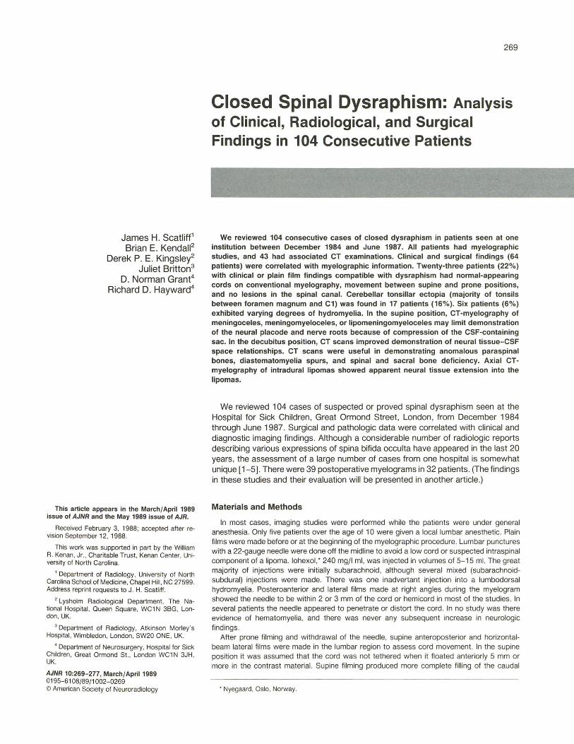
Fig. 1.-9-year-old girl with wasted and weak right leg.
A, Conus extends to L5. Filum is thickened and tethered (arrow).
B, Supine lateral cervical myelogram shows cerebellar tonsillar ectopia to C1 .
A
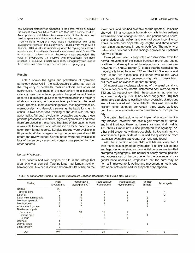
down to the point of tethering . Hydromyelia was seen in a postoperative myelogram in one of three patients in whom CT was done. A tethered cord was found in another patient with an occipital encephalocele and lacunar skull . Tonsillar ectopia to C1 was present in a patient whose cord extended to S1 (Fig . 1 ). Asymmetric hemicords close together were noted only by CT in a patient with typical myelographic findings for a tethered cord (Fig . 2).
All the patients showed decreased movement of the cord as myelographic position changed from prone to supine. This was also true in the two patients whose conus was at a normal level. In one of these patients a thickened filum continued from the conus. In the other the conus was tethered posteriorly by a band evident in the myelogram. The tethering lesions seen at myelography and on CT in all the patients in this category were small lipomas, meningoceles, or fibrous bands .
Lipoma
Ten children had evident or palpable lipomas. Eight of these had urinary or fecal incontinence and six had disparity in leg length as well as foot deformities.
Eight patients had preoperative myelograms, which
B
272 SCATLIFF ET AL. AJNR:10, March/April1989
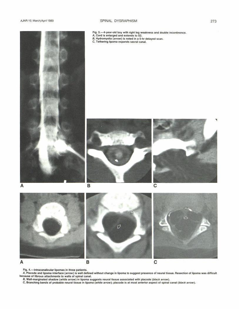
showed significant intradural lipomas intimately involving the conus andjor nerve roots . The lipomas were in either the lumbar or sacral canal and extended to the subcutaneous tissues of the back. In all these patients the spinal cord was abnormally low, and two of these eight had hydromyelia (Fig. 3). Two of the 1 0 patients had had surgery in infancy for their lesions, and they were examined for new or continuing symptoms . One of these was found to have a lumbodorsal hydromyelia and the other a neuroenteric cyst adjacent to the upper lumbar cord.
In three patients the spinal cord was displaced anteriorly by the lipoma; in another three the cord was in a central position in the spinal canal as it fused with the lipoma, and in two patients the cord was displaced posteriorly. The initial preoperative studies in two patients were not available. Transverse CT sections in three patients with lipomas are shown in Figure 4. The patient in Figure 4A appeared to have a welldefined interface between the neural placode and lipoma. However, the neurosurgeon found the lipoma contained considerable disorganized tissue, including fibrous elements,
A 8
which made the placode difficult to identify. The lipoma was also attached by thick bands of fat and fascia to the walls of the spinal canal, and its dissection was difficult.
The CT sections of patients shown in Figures 4B and 4C had well-defined strands of tissue that could be followed from the cord into the lipoma. At surgery, neural tissue was found in the tumors. Direct correlation of this observation with the CT scans of the lipomas could not be made. In three other patients CT showed relatively homogeneous fat density in the lipomas. The surgical dissection in these patients was difficult, however, because of nerve roots at the margins of the lipomas. These could not be identified in retrospect in the CT studies.
Lipomyelomeningocele
Sixteen patients exhibited varying degrees of lipomatous material closely associated with myelomeningoceles. One patient had previous surgery. All the patients had palpable lumbar or sacral masses, some quite large. Seven patients
Fig. 2.-8-year-old boy with pain in both lower limbs, urinary frequency, and fecal incontinence.
A, Conus is at L3 (black arrow). White arrow shows filum tethered by small meningocele.
B, CT at level of conus shows hemicords not seen on conventional myelogram. The small right hemicord (arrow) could be correlated with child's inability to hop or stand on the right foot.
AJNR :10, March/April1989
A 8
A 8
Fig. 4.-lntracanalicular lipomas in three patients.
SPINAL DYSRAPHISM
Fig. 3.-4-year-old boy with right leg weakness and double incontinence. A, Cord is enlarged and extends to S2. B, Hydromyelia (arrow) is noted in a 5-hr delayed scan. C, Tethering lipoma expands sacral canal.
c
c
273
A, Placode and lipoma interface (arrow) is well defined without change in lipoma to suggest presence of neural tissue. Resection of lipoma was difficult because of fibrous attachments to walls of spinal canal.
B, Well-marginated shadow (white arrow) in lipoma suggests neural tissue associated with placode (black arrow). C, Branching bands of probable neural tissue in lipoma (white arrow); placode is at most anterior aspect of spinal canal (black arrow).
274 SCATLIFF ET AL. AJNR:10, March1April1989
had foot or leg abnormalities, including two with unilateral club feet and three with feet and legs of different size and length. Five patients had either urinary incontinence, infection, or reflux. Two patients with urinary tract signs were less than 6 months old . One 8-year-old had fecal incontinence.
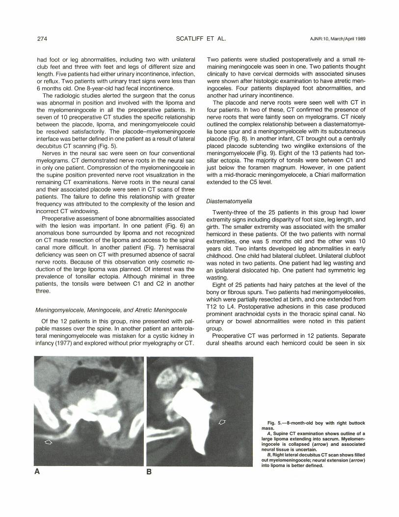
The radiologic studies alerted the surgeon that the conus was abnormal in position and involved with the lipoma and the myelomeningocele in all the preoperative patients. In seven of 1 0 preoperative CT studies the specific relationship between the placode, lipoma, and meningomyelocele could be resolved satisfactorily. The placode-myelomeningocele interface was better defined in one patient as a result of lateral decubitus CT scanning (Fig. 5).
Nerves in the neural sac were seen on four conventional myelograms. CT demonstrated nerve roots in the neural sac in only one patient. Compression of the myelomeningocele in the supine position prevented nerve root visualization in the remaining CT examinations. Nerve roots in the neural canal and their associated placode were seen in CT scans of three patients. The failure to define this relationship with greater frequency was attributed to the complexity of the lesion and incorrect CT windowing .
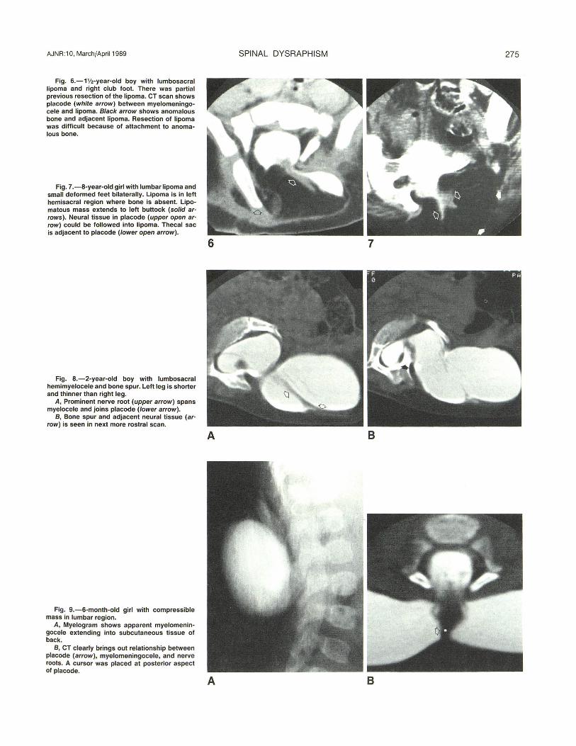
Preoperative assessment of bone abnormalities associated with the lesion was important. In one patient (Fig. 6) an anomalous bone surrounded by lipoma and not recognized on CT made resection of the lipoma and access to the spinal canal more difficult. In another patient (Fig. 7) hemisacral deficiency was seen on CT with presumed absence of sacral nerve roots. Because of this observation only cosmetic reduction of the large lipoma was planned. Of interest was the prevalence of tonsillar ectopia. Although minimal in three patients, the tonsils were between C1 and C2 in another three.
Meningomyelocele, Meningocele, and Atretic Meningocele
Of the 12 patients in this group, nine presented with palpable masses over the spine. In another patient an anterolateral meningomyelocele was mistaken for a cystic kidney in infancy (1977) and explored without prior myelography or CT.
A B
Two patients were studied postoperatively and a small remaining meningocele was seen in one. Two patients thought clinically to have cervical dermoids with associated sinuses were shown after histologic examination to have atretic meningoceles. Four patients displayed foot abnormalities, and another had urinary incontinence.
The placode and nerve roots were seen well with CT in four patients. In two of these, CT confirmed the presence of nerve roots that were faintly seen on myelograms. CT nicely outlined the complex relationship between a diastematomyelia bone spur and a meningomyelocele with its subcutaneous placode (Fig. 8). In another infant, CT brought out a centrally placed placode subtending two winglike extensions of the meningomyelocele (Fig . 9). Eight of the 13 patients had tonsillar ectopia. The majority of tonsils were between C1 and just below the foramen magnum. However, in one patient with a mid-thoracic meningomyelocele, a Chiari malformation extended to the C5 level.
Diastematomyelia
Twenty-three of the 25 patients in this group had lower extremity signs including disparity of foot size, leg length , and girth . The smaller extremity was associated with the smaller hemicord in these patients. Of the two patients with normal extremities, one was 5 months old and the other was 1 0 years old . Two infants developed leg abnormalities in early childhood. One child had bilateral clubfeet. Unilateral clubfoot was noted in two patients. One patient had leg wasting and an ipsilateral dislocated hip. One patient had symmetric leg wasting.
Eight of 25 patients had hairy patches at the level of the bony or fibrous spurs. Two patients had meningomyeloceles, which were partially resected at birth, and one extended from T12 to L4. Postoperative adhesions in this case produced prominent arachnoidal cysts in the thoracic spinal canal. No urinary or bowel abnormalities were noted in this patient group.
Preoperative CT was performed in 12 patients. Separate dural sheaths around each hemicord could be seen in six
Fig. 5.-8-month-old boy with right buttock mass.
A, Supine CT examination shows outline of a large lipoma extending into sacrum. Myelomeningocele is collapsed (arrow) and associated neural tissue is uncertain.
B, Right lateral decubitus CT scan shows filled out myelomeningocele; neural extension (arrow) into lipoma is better defined.
AJNR :10, March/April1989
Fig. 6.-1V2-year-old boy with lumbosacral lipoma and right club foot. There was partial previous resection of the lipoma. CT scan shows placode (white arrow) between myelomeningocele and lipoma. Black arrow shows anomalous bone and adjacent lipoma. Resection of lipoma was difficult because of attachment to anomalous bone.
Fig. 7.-8-year-old girl with lumbar lipoma and small deformed feet bilaterally. Lipoma is in left hemisacral region where bone is absent. Lipomatous mass extends to left buttock (solid arrows). Neural tissue in placode (upper open arrow) could be followed into lipoma. Thecal sac is adjacent to placode (lower open arrow).
Fig. 8.-2-year-old boy with lumbosacral hemimyelocele and bone spur. Left leg is shorter and thinner than right leg.
A, Prominent nerve root (upper arrow) spans myelocele and joins placode (lower arrow).
B, Bone spur and adjacent neural tissue (ar· row) is seen in next more rostral scan.
Fig. 9.-6-month-old girl with compressible mass in lumbar region.
A, Myelogram shows apparent myelomeningocele extending into subcutaneous tissue of back.
B, CT clearly brings out relationship between placode (arrow), myelomeningocele, and nerve roots. A cursor was placed at posterior aspect of placode.
SPINAL DYSRAPHISM 275
6 7
A 8
A 8
276 SCATLIFF ET AL. AJNR :10, March/April1989
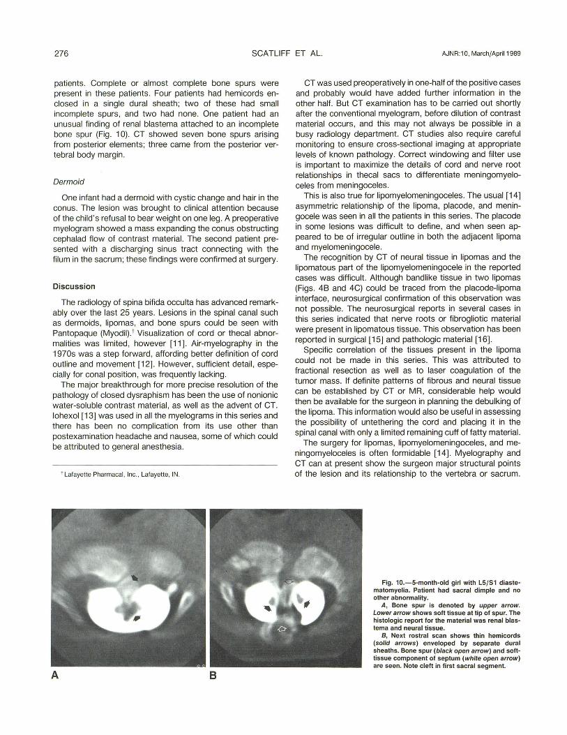
patients. Complete or almost complete bone spurs were present in these patients. Four patients had hemicords enclosed in a single dural sheath; two of these had small incomplete spurs, and two had none. One patient had an unusual finding of renal blastema attached to an incomplete bone spur (Fig . 1 0). CT showed seven bone spurs arising from posterior elements; three came from the posterior vertebral body margin .
Dermoid
One infant had a dermoid with cystic change and hair in the conus. The lesion was brought to clinical attention because of the child's refusal to bear weight on one leg. A preoperative myelogram showed a mass expanding the conus obstructing cephalad flow of contrast material. The second patient presented with a discharging sinus tract connecting with the filum in the sacrum; these findings were confirmed at surgery.
Discussion
The radiology of spina bifida occulta has advanced remarkably over the last 25 years. Lesions in the spinal canal such as dermoids , lipomas, and bone spurs could be seen with Pantopaque (Myodil).t Visualization of cord or thecal abnormalities was limited, however [11]. Air-myelography in the 1970s was a step forward , affording better definition of cord outline and movement [12]. However, sufficient detail , especially for conal position , was frequently lacking.
The major breakthrough for more precise resolution of the pathology of closed dysraphism has been the use of nonionic water-soluble contrast material , as well as the advent of CT. lohexol [13] was used in all the myelograms in this series and there has been no complication from its use other than postexamination headache and nausea, some of which could be attributed to general anesthesia.
1 Lafayette Pharmacal, Inc., Lafayette, IN .
A B
CT was used preoperatively in one-half of the positive cases and probably would have added further information in the other half. But CT examination has to be carried out shortly after the conventional myelogram, before dilution of contrast material occurs, and this may not always be possible in a busy radiology department. CT studies also require careful monitoring to ensure cross-sectional imaging at appropriate levels of known pathology. Correct windowing and filter use is important to maximize the details of cord and nerve root relationships in thecal sacs to differentiate meningomyeloceles from meningoceles.
This is also true for lipomyelomeningoceles. The usual [14] asymmetric relationship of the lipoma, placode, and meningocele was seen in all the patients in this series. The placode in some lesions was difficult to define, and when seen appeared to be of irregular outline in both the adjacent lipoma and myelomeningocele.
The recognition by CT of neural tissue in lipomas and the lipomatous part of the lipomyelomeningocele in the reported cases was difficult. Although bandlike tissue in two lipomas (Figs. 4B and 4C) could be traced from the placode-lipoma interface, neurosurgical confirmation of this observation was not possible. The neurosurgical reports in several cases in this series indicated that nerve roots or fibrogliotic material were present in lipomatous tissue. This observation has been reported in surgical [15] and pathologic material [16].
Specific correlation of the tissues present in the lipoma could not be made in this series. This was attributed to fractional resection as well as to laser coagulation of the tumor mass. If definite patterns of fibrous and neural tissue can be established by CT or MR, considerable help would then be available for the surgeon in planning the debulking of the lipoma. This information would also be useful in assessing the possibility of untethering the cord and placing it in the spinal canal with only a limited remaining cuff of fatty material.
The surgery for lipomas, lipomyelomeningoceles, and meningomyeloceles is often formidable [14]. Myelography and CT can at present show the surgeon major structural points of the lesion and its relationship to the vertebra or sacrum.
Fig. 10.-5-month-old girl with L5/S1 diastematomyelia. Patient had sacral dimple and no other abnormality.
A, Bone spur is denoted by upper arrow. Lower arrow shows soft tissue at tip of spur. The histologic report for the material was renal blastema and neural tissue.
B, Next rostral scan shows thin hemicords (solid arrows) enveloped by separate dural sheaths. Bone spur (black open arrow) and softtissue component of septum (white open arrow) are seen. Note cleft in first sacral segment.
AJNR:10, March/April 1989 SPINAL OYSRAPHISM 277
The same is true for various expressions of diastematomyelia. Finer details-such as exact position and number of nerve roots in relation to the placode-lipoma, the presence of fibrous bands or arachnoidal adhesions , or prominent blood vessels-were discovered only at the time of surgery in this series.
An important point in our study is the visualization in plain films and CT of anomalous or deficient bone formation associated with dysraphic lesions. Naidich et al. [14] and others [17, 18) have emphasized recognition of posterior bone masses. Surgery can be facilitated with the foreknowledge that these may need removal for better access to the dysraphic pathology. Noting that a hemisacrum is largely absent and that sacral nerves are probably absent or abnormal may obviate surgery. CT-based 30 reconstruction of bone has now advanced to a level where the skeletal framework of dysraphic lesions can be shown to good advantage. The pathology of diastematomyelia can be shown very well by CT-myelography, as documented by Naidich and HarwoodNash [5). Abnormal bones and soft tissues depicted in 30 would be helpful in presurgical assessment. For informative display, however, the increased number of thin CT sections would be time-consuming and could have prohibitive radiation dosage.
The clinical findings in this survey have been enumerated not only to catalogue their prevalence but also to emphasize what dysraphism pathology means to patients, their parents, and their primary physicians. The imaging focus has been strongly directed toward revealing the pathology, with the surgeon operating in the belief that "untethering" the cord will arrest the progress of clinical signs. Unfortunately, this does not always occur. It is evident also from the study that nearly a quarter of patients with suspected dysraphism have a normal-appearing cord without restriction of movement.
ACKNOWLEDGMENT
We thank Patricia A. Dunlop for preparing the manuscript.
REFERENCES
1. James CCM , Lassman LP . Spinal dysraphism. London : Butterworth , 1972 2. Burrows FGO. Some aspects of occult spinal dysraphism: a study of 90
cases. Br J Radio/ 1968;41 :496-507 3. HoHman JH, Taechalarn C, Hendrick EB, Humphreys RP. Management of
lipomyelomeningoceles. J Neurosurg 1985;62 : 1- 9 4. Hilal SK, Marton D, Pollack E. Diastematomyelia in children. Radiographic
study of 34 cases. Radiology 1974;112 :609-621 5. Naidich TP, Harwood-Nash DC. Diastematomyelia hemicord and meningeal
sheaths: single and double arachnoid and dural tubes. AJNR 1983:4 : 633-636
6. Rasjo IM, Harwood-Nash DC, Fitz CR , Chuang S. Computed tomographic metrizimide myelography in spinal dysraphism in infants and children. J Comput Assist Tomgr 1978;2:549- 558
7. Ethier R, King DG, Menancon D, Gelange G, Taylor S, Thompson C. Development of high resolution computed tomography of the spinal cord . J Comput Assist Tomogr 1979;3 :433- 438
8. Scotti G, Musgrave MA, Harwood-Nash DC , Fitz CR , Chuang SH. Diastematomyelia in children: metrizimide and CT metrizamide myelography. AJNR 1980;1 :403-410
9. Barson AJ. The vertebral level of termination of the spinal cord during normal and abnormal development. J Anat 1970;106:489-497
10. Hall DE, Udvarhelyi GB, Altman J. Lumbosacral skin lesions as markers of occult spinal dysraphism. JAMA 1981 ;22:2606-2608
11 . Gryspeerdt GL. Myelographic assessment of occult forms of spinal dysraphism. Acta Radio/ 1963;1 :702-717
12. Strand RD . Spinal dysraphism in certain occult forms examined by air myelography without tomography. Ann Radio/ (Paris) 1969;12:393- 399
13. Eldevik OP, Nakstad P, Kendall BE, Hindmarsh T. lohexol in lumbar myelography: preliminary results from an open noncomparative multicenter clinical study. AJNR 1983;4:299-301
14. Naidich TP, Melone DG, Mutluer S. A new understanding of dorsal dysraphism with lipoma (lipomyeloschisis): radiologic evaluation and surgical correction. AJNR 1983;4:1 03-116
15. Dubowitz V, Lorber J, Zachary RB. Lipoma of the cauda equina. Arch Dis Child 1965;40:207-213
16. Emery JL, Lendon RG. Lipomas of the cauda equina and other fatty tumors related to neurospinal dysraphism. Dev Med Child Neurol1969 20 :62- 70
17. McAlister WH, Siegel MJ , Shackelford GD. A congenital iliac anomaly often associated with sacral lipoma and ipsilateral lower extremity weakness. Skef Radiol1 978;3: 161-1 66
18. Cohen JY , Lebatard-Sart RER, Lajat Y, Mitard D, David A. Sacral intraspinal lipoma associated with genital iliac anomaly. Childs Brain 1981;8:181-188