cls® spotorno® hip stem – surgical technique
TRANSCRIPT

CLS® Spotorno® Hip Stem
Surgical Technique


Surgical Technique CLS Spotorno Stem
Table of Contents
CLS Spotorno Stem 4
Indications for the CLS Spotorno Stem 5
Preoperative Planning 10
Surgical Technique 12
Case Study 21
Ordering Information 22

4 CLS® Spotorno® Hip Stem – Surgical Technique
The tapered shape of the stem enables excellent primary stability and ensures that this stability is maintained thanks t o the self-stabilizing properties of the implant. In addition, the structure of the bone surrounding the prosthesis shows that the tapered shape of the implant favours a largely proximal transfer of load.
CLS Spotorno Stem
145°
135°
125°
As the CCD angle is not a constant value, and shows broad variability in individuals, the CLS stem offers three different CCD angles. This provides a wide range of offset options, which allows an adequate restoration of the biomechanical para-meters, such as the center of rotation, the CCD angle as well as leg length and soft tissue balancing.
For implantation of the CLS 145°, the CLS 135° and the CLS 125°, the same instruments and the same surgical technique are used. The modular rasps and trial necks serve the intraoperative trial reduction.

5CLS® Spotorno® Hip Stem – Surgical Technique
The Question: Cemented or Uncemented?The decision between a cemented and an uncemented hip stem is a choice that must be made by the surgeon. Based on the personal experience by the design ing surgeon implanting over 9,000 CLS stems, it was possible to carry out extensive research into the indications for this stem as well as to define its limitations.
* www.jru.orthop.gu.se / Müller L. et al., “Seventeen-year survival of the cementless CLS Spotorno Stem”, 2010
Indications for the CLS Spotorno Stem
The results obtained with the CLS stem since 1984 are excellent*. Even though excellent results have been achieved (even under critical conditions), it is still recommended that surgeons use a set of standard, reliable criteria to help make the decision between cemented and uncemented stems.
6 CLS® Spotorno® Hip Stem – Surgical Technique
Parameter No. 3: OsteoporosisSevere osteoporosis represents a major disadvantage in regard to the primary stability of the implant, or requires the use of an over-dimensioned stem with anchorage in the lower metaphyseal and diaphyseal region. This, in turn, has a negative effect on the blood supply to the bone. Radiological methods such as computer tomography and densio-metry are available for the assessment of the severity of the osteoporosis. A suitable method for a conclusive assessment is the modified analysis of the trabeculae in the neck of the femur according to Singh – a process that is easy to carry out.
Femoral Neck Index On the basis of these assessments, four degrees of severity of osteoporosis can be defined:
Severe (Singh 1–2): 4 points
Moderate (Singh 3–4): 2 points
Slight (Singh 5–6): 1 point
Physiological (Singh 7): 0 points
(Singh M. et al. Changes in Trabecular Pattern of the Upper End of the Femur as an Index of Osteoporosis. Journal of Bone and Joint Surgery 52A: 456, 1970).
Parameter No. 1: Age As far as skeletal changes are con- cerned, it is generally known that age cannot be considered as a purely chronological parameter, but has to be assessed from a biological point of view. In simple terms, it can be said that for patients under the age of 50 years, a cementless stem is the routine solution, while after the age of 70, a cemented stem is generally preferred.
Points Allocated
. 70 years: 4 points
61–70 years: 2 points
51–60 years: 1 point
, 50 years: 0 points
Parameter No. 2: GenderDue to the increasing osteoporosis resulting from the hormonal changes occurring during menopause, older women generally have poorer bone quality.
Points Allocated
Women: 1 point
Men: 0 points
In 1985, an indication protocol was established that is based on the assess-ment of four clinical and radiological parameters in the patient investigated. Each parameter is given a point score. The final value obtained from the sum of the points establishes the indication and thus, provides a valuable guideline for the surgeon. The parameters are age, gender, severity of the osteoporosis and the anatomical characteristics of the femur.
Reliable Decision-Making Through Methodology

7CLS® Spotorno® Hip Stem – Surgical Technique
The bundle of bow-shaped trabeculae is missing. The pres-sure trajectories in the head of the femur have partly disap-peared.
The bow-shaped trajectories have almost completely disappeared.
Partial disappearance of the bow-shaped trabecular system.
The accessory trabecular systems have completely disappeared.
Empty Ward’s triangle. The accessory trabecular systems have partly disappeared.
The picture of the upper triangle of the femoral neck appears to be bound by the bow-shaped bundle of trajectories of the head of the femur and the trochanter.
Normal small, dense trabecu-lae fill the neck of the femur. Trajectories are not visible.
Stage 1 Stage 2
Stage 3 Stage 4
Stage 5
Stage 7
Stage 6
Parameter No. 4: The Anatomy of the Femur
Morpho-Cortical Index Experience shows that this index provides more information parameters. It comprises two variables, which do not always corre -late with one another, in one single value: • The morphology of the femur • The thickness of the cortex
In regard to morphology, it is possible to differentiate between three types of femurs: • Trumpet shape • Cylinder shape • Dysplastic femur
Because of its morphology, the trum pet- shaped femur provides the optimal conditions for an uncemented implant. The cylindrical femur requires an adequate cut in the subtrochanteric region and the removal of metaphyseal bone during the rasping process. The mechanically support-ive cancellous bone and the cortex of the isthmus of the calcar, which forms the basis for the anchorage of the stem, have to be partly removed. The morpho-cortical index (MCI), is defined on a standard X ray picture. It is calculated from the correlation of the extracortical diameter of the femur, measured at the medial tip of the lesser trochanter, to the intracortical diameter, measured 7 cm further in the distal direction.
trumpet-shapedcylindricaldysplastic(Singh M. et al. Changes in Trabecular Pattern of the Upper End of the Femur as an Index of Osteoporosis. Journal of Bone and Joint Surgery 52A:456, 1970).

8 CLS® Spotorno® Hip Stem – Surgical Technique
The MCI is calculated using the following formula: MCI =
CD = Distance between the outer boundaries of the lateral and the medial cortex. The measurement is made at the level of the tip of the trochanter, vertical to the axis of the femur. AB = Diameter of the medullary cavity. The measurement is made 7 cm distal from the CD line, vertical to the axis of the femur.
The MCI in this absolute form can only be used if it was calculated in a stand- ard X ray picture with the legs in the normal 0 position and with rectilineal a-p irradiation.
The pointscores of the MCI
MCI # 2.2: 4 points
MCI . 2.3: 2 points
MCI . 2.7: 1 point
MCI $ 3.0: 0 points
Final AssessmentIn cases where long-term cortisone therapy is envisioned, for example in rheumatoid arthritis, one point must be added as an additional risk factor.
0–4 points: cementless stem
5 points: questionable indication
$ 6 points: cemented stem
CDAB
C D
A B
7 cm
CDCalculation MCI =
AB

9CLS® Spotorno® Hip Stem – Surgical Technique
The uncemented stem in general, and the CLS stem in particular, have definiti-vely proven their worth. In comparison with the cemented stem, the uncemented solution is far more bone conserving. This becomes an important factor if revi-sion surgery is necessary.
The insertion of the uncemented stem is less invasive and takes the biomechanics of the femur into account. The bone and the prosthesis combine to form a unit. As a result, the blood supply and vitality of the bone are maintained.
In principle, the uncemented stem is preferably used in younger patients. It is, however, not contraindicated in the elderly – especially for patients in poor general health. It can be implanted more rapidly and there is no thermal damage to the tissue due to the cement.
The use of a cemented stem is never-theless justified in elderly patients for a number of reasons: The life expectancy of the patient is often shorter than the average survival time of a cemented stem. In the presence of poor bone quality, the cement allows unproblematic correction of defects. With cementing, faster restoration of the ability to walk is also to be expected, since – at least within certain limits – it does not involve the reparative phase, which, in the case of the cementless stem, leads to the definitive anchorage of the bone. Finally, the economic factor plays a role that is not to be underestimated.
It can generally be said that if one adheres to the classical indications – while still allowing the possibility of further indica-tions in the future – good and very good results can be obtained. In this respect, the use of the MCI is of fundamental importance because it follows the basic biomechanical ideas used in the design of the CLS prosthesis. With a cylindrical medullary cavity, it is normally necessary to use a more invasive prosthesis, which has an unfavourable bone-prosthesis interaction.
Comparison has shown that the MCI and the cortical index (CI) after Gruen are good indicators for the quality of the bone. There seems to be a connection both between the CI and the MCI and between the MCI and the measurement of the mineral content of the bone by the Dexa method.
Conclusion: There is a Broad Indication for Uncemented Anchorage

10 CLS® Spotorno® Hip Stem – Surgical Technique
Determination of the Size and Position of the Cup The center of rotation of the joint to be operated on is determined by transposing the two lines that have been drawn on the opposite side. The cup template is then placed on the side that is to be operated on. The position of the acetabu-lar components is determined by the outline of the cup, the center of rotation that was determined, the level of the “teardrop” and the required abduction angle of 40°–45°.
Within the framework of the preoperative planning, the stem size, the optimal anchorage of the stem in the medullary cavity and the correct position of the acetabular and femoral components are determined in order to ensure equal leg length. At the start of the preoperative planning, three lines are drawn on the X ray picture: The tangent of the two ischia forms the base line. A second line is drawn through the floors of the two acetabulae, and a third between the lesser trochanters. On the side that is not to be operated on, the center of rotation of the joint is deter-mined. Then the distances between the joint, baseline and “teardrop” are drawn. In addition, the longitudinal axis of the pelvis is also drawn.
Equal leg length: all lines are parallel
Drawing in of Pelvis and CupThe tracing paper is placed on the X ray picture and the template. The longitudi-nal edge must run parallel to the vertical axis of the pelvis. The pelvis and the cup are drawn in and then the tracing paper removed from the X ray.
Preoperative Planning Systematic Preparation with Suitable Methods of Measurement and Practical Planning Benefit Correct Implantation
The Planning Steps, with an Example of Unilateral Coxarthrosis

11CLS® Spotorno® Hip Stem – Surgical Technique
Height of the Pelvis The femoral template is to be left in place. The drawing of the pelvis is placed with the acetabular components on the X ray picture. If lengthening of a leg is neces-sary, the drawing of the pelvis lies higher than the pelvis on the X ray picture, by the difference in the length to be corrected. In the case of planned shortening of a leg, the drawing must be correspondingly lower by the distance to be corrected. Stem size and length must be selected so that the differences correspond to the measurements that are to be corrected.
Final ResultThe outlines of the femur and the cortex and the selected implant/femoral head combination are drawn on the transparent paper. The distance between the proximal end of the taper of the stem and the lesser trochanter is measured and entered. The line from the shoulder of the stem to the greater trochanter is extended and measured. The line between the tip of the greater trochanter and the center of rotation is drawn in.
Determination of the CCD Angle, the Size and Position of the Stem First, the most adapted CCD angle of the CLS stem (125°, 135° or 145°) to fit into the proximal part of the femur is selected. The selected stem template is placed on the femur so that the stem fits into the medullary canal displaying the correct type of anchorage. The size of the stem must be selected so that at least 3/4 of the proximal ribbed structure is anchored in the cancellous bone. Ideally, one of the four lines from the rotation centers touches the tip of the greater trochanter.

12 CLS® Spotorno® Hip Stem – Surgical Technique
Positioning of the Patient: Placement in the Lateral Position*
The patient is positioned on the operating table with one pressure pad on the pubic bone and one on the sacrum. In the subsequent positioning, it is important that the pelvis is not lowered, either side-ways or in the caudal direction, and that it is fixed securely. The leg on the opposite side is bent 45° at the hip and 90° at the knee, which helps to stabilize the position of the patient.
* Different surgical approaches are possible depending on the surgeon’s preference..
Surgical Technique

13CLS® Spotorno® Hip Stem – Surgical Technique
Surgical Approach to the Hip: Incision The posterolateral approach is recom-mended. The joint is bent at an angle between 30° and 40°. A rectilinear incision is made to the tip of the greater trochanter and is then continued for about 6 cm on the diaphysis. After transection of the subcutis, the fascia lata is exposed.
The Approach to the Deeper LayersThe fascia lata is incised and dissociated of the fibers of the M. gluteus maximus and the Charnley wound retractor is placed directly on the fascia.
In this way, the plane of the external rotator muscles and the tendon attachment of the M. gluteus maximus are exposed on the linea aspera of the femur. The tendon attachment is partly released in order to relax the soft parts. This favours the displacement of the femur in the ventral direction and also its internal rotation.
Transection of the External Rotator Muscles and Dislocation of the HipAfter inserting a bent Hohmann retractor under the M. gluteus medius, the tendon of the M. piriformis is located and transected, as are some of the tendons of the external rotator muscles. The joint capsule is then opened from the dorsocra-nial direction. With a combined flexion, adduction and internal rotation move-ment, the head of the femur can now be dislocated from the acetabulum.

14 CLS® Spotorno® Hip Stem – Surgical Technique
Osteotomy of the Neck of the FemurThe lesser trochanter serves as reference point for the osteotomy plane on the neck of the femur, which was already included in the preoperative planning. The level of the osteotomy is influenced by the ante- torsion of the neck of the femur: the greater the antetorsion, the lower the level of the osteotomy. Normally, it proves an advantage to retain 1 to 1.5 cm of the neck of the femur. This creates a sheath into which the proximal, ribbed part of the stem can fit. The next step is the osteotomy with the reciprocating saw. Starting from the medial mark, the upper edge of the neck of the femur is reached at the point where it rises from the mass of the trochanter. It may be necessary to continue the osteotomy with a cut continued further upwards, parallel to the axis of the femur.
Preparation of the Medullary Cavity of the FemurThe leg is turned inwards, by internal rotation of up to 90°, which is combined with bending and adduction of the hip. The lower leg is bent at 90° to the thigh. This provides a spatial reference point, in order to be able to establish the ante- torsion of the femoral component of the prosthesis and its position parallel to the cortex. In order to facilitate the exposure of the mass of the trochanter, a Hohmann retrac-tor is placed under the lesser trochanter as a lever, taking care to ensure that the iliopsoas tendon is not injured. A second lever is placed at the tip of the greater trochanter in order to move the muscle to the side of the diaphysis and to expose the remaining lateral portion of the neck of the femur. This has to be removed for the correct alignment of the stem.
The remaining portion of the neck of the femur and a small part of the greater trochanter can be removed with the instru-ment provided for this purpose, or directly with the saw.

15CLS® Spotorno® Hip Stem – Surgical Technique
Use of the Awl and Rasping TechniqueThe proximal notches on the awl mark the height of the shoulder of the implant. The awl has to be inserted laterally and slightly dorsal. The attachment of the M. piriformis provides a reference point for this. As a rule, this corresponds to the point at which, in the preoperative planning, the tangent of the endosteal edge of the outer cortex meets the greater trochanter. It provides an accurate, measurable conception of the obstacle to the prosthesis. After the medullary space has been prepared in this way, the awl is inserted deep, for centering in the canal, taking care to ensure that it is pressed in the direction of the greater trochanter. The aim is to follow the predetermined line towards the lateral cortex, parallel to the axis of the femur and to avoid a varus deformity. The bed for the stem is now prepared, using rasps of increasing size, until the highest possible degree of stability is obtained. The preoperatively measured distance between the proximal shoulder of the prosthesis and the greater trochanter serves as orientation.

16 CLS® Spotorno® Hip Stem – Surgical Technique
The desired stability is based on the concept of a press-fit in the cortex and cancellous bone. This is why the rasps have smooth zones for compression of the cancellous bone and cutting zones for rasping of parts of the cortex.*
After the surgeon has established the size of the prosthesis during the planning, the definite size is determined by progressive, stepwise rasping, starting with 3 to 4 smaller rasp sizes. In this way, using the increasing dimensions, the cancellous bone is compressed. Where necessary, the cortex has to be reamed. The rasps are inserted with small, precise hammer blows, and then withdrawn.
The final, definitive reaming must be carried out only with the rasp planned for this. The last rasp should be operated manually.
* The rasp is designed to lead to a defined smaller dimen-sion in relation to the bony structure of the femur. Generally, it is underdimensioned in the proximal portion of the rasp – that is, the portion with which cancellous bone is to be compressed.
At the distal end, the rasp is slightly oversized, in order to prevent stress peaks on the end of the prosthesis.

17CLS® Spotorno® Hip Stem – Surgical Technique
Rasping TechniqueWith the first rasp, care must be taken to ensure a correct antetorsion (10–15°). This can be monitored by means of the handle of the rasp. The neck of the femur may display a pathological antetorsion, which the surgeon has to take into account and possibly rectify. This can be done, for example, by a lower osteotomy of the neck of the femur or by controlled splitting. According to the safety margin concept, the sum of the antetorsion of the femur and the anteversion of the acetabulum must be 25 ± 7°. One should, however, try to keep the angle between the two compo-nents of the prosthesis as close as possi-ble to the physiological angle.
The reference parameter for the correct alignment of the rasp is the plane running through the axis of the diaphysis and parallel to the condyles of the femur. Keeping the angle of the bend of the lower leg at 90°, vertical to the floor, the rasp is turned 15° downwards. The ideal angle, which is formed from the lower leg and the long bar of the rasp, can be checked visually.
10–15°

18 CLS® Spotorno® Hip Stem – Surgical Technique
Trial Repositioning with Trial NeckTrial reduction of the CLS stem may be accomplished with the rasp.
This is done by removing the rasp handle and leaving the rasp in the femoral canal. A trial neck is then inserted into the rasp hole. Once the trial neck is selected, any of the trial heads may be used in conjunction with the trial neck.
For the CLS stem, different trial necks have to be used in order to correctly reproduce the different offset options.
Once the trial neck and the appropriate trial head are selected, the hip is reduced. Leg length and offset are checked. This procedure is repeated as necessary, using trial heads of different lengths, until optimal offset, leg length and stability are established. A trial reduction should not allow significant push-pull or “shuck” of the joint in full extension. The range of motion is checked to avoid bony impinge-ment and instability.
Insertion of the StemAfter removal of the rasp, a prosthesis of the appropriate size is inserted and driven in until it is completely stable. In this process, it is important to proceed with the necessary light touch. This is learned with experience. It should be remembered that because of the wedge mechanism, an excessive load may be exerted that can bear on the trochanter to the point that it might cause a fracture.
It is important to adjust the force of the hammer blows, according to the quality of the bone and to stop the hammer blows immediately if a change in the sound of the blows, from dull (cancellous bone) to sharp (cortex) is perceived.

19CLS® Spotorno® Hip Stem – Surgical Technique
Assembly of the Femoral Head and Repositioning After insertion of the prosthesis, the definitive femoral head is established on the basis of the measurements laid down in the preoperative planning and by trial repositioning using trial heads. The last step in the assembly is the fitting of the previously defined head onto the stem. After thoroughly cleaning and drying the taper, the femoral head is mounted with a light rotational movement and rotated further with axial force until it is firmly seated. The femoral head is seated with one light hammer blow on the head impactor in axial direction.
Upon reducing the joint, the surgeon checks the range of motion and stability of the joint. This should be checked with both internal and external rotation.
With the placing of drains and suturing of the different layers, the operation is now complete.
Intraoperative Extraction of the StemIf the stem needs to be removed intra-operatively only the specific extraction instrument has to be used, which protects the neck and the cone of the stem. Slide the extraction instrument over the stem taper, until the plastic jaws reach the neck of the stem. It is very important that the plastic jaws touch only the neck and not the taper. Afterwards tighten the plastic jaws with the ball hex screwdriver, until the extraction instrument is firmly fixed on the stem. Remove the stem by hammering on the extraction instrument.
Important: The extraction instrument can be used only for intraoperative stem extraction. It is not suitable for revision cases. The plastic jaws can be exchanged if necessary.
Intraoperative extraction of the stem

20 CLS® Spotorno® Hip Stem – Surgical Technique
Unlock screw with screwdriver for extraction instrument
Remove screw from plastic jaw
Extract fixed plastic jaw with the impactor according to the illustration
The disassembled instrument can now be cleaned and sterilized
Note: Damaged plastic jaws have to be replaced!
Disassembly of the Extraction InstrumentFor cleaning and sterilization as well as for the exchange of the plastic jaws

21CLS® Spotorno® Hip Stem – Surgical Technique
Case Study
CLS Spotorno stem 145° and CLS Spotorno expansion cup
preoperative postoperative 15.8 years
22 CLS® Spotorno® Hip Stem – Surgical Technique
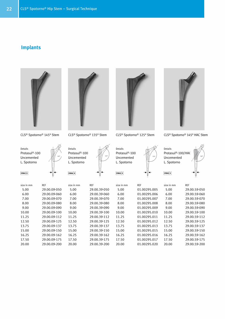
CLS® Spotorno® 125° Stem
Details
Protasul®-100UncementedL. Spotorno
size in mm REF
5.00 01.00295.005 6.00 01.00295.006 7.00 01.00295.007 8.00 01.00295.008 9.00 01.00295.00910.00 01.00295.01011.25 01.00295.01112.50 01.00295.01213.75 01.00295.01315.00 01.00295.01516.25 01.00295.01617.50 01.00295.01720.00 01.00295.020
Implants
CLS® Spotorno® 135° Stem
Details
Protasul®-100UncementedL. Spotorno
size in mm REF
5.00 29.00.39-050 6.00 29.00.39-060 7.00 29.00.39-070 8.00 29.00.39-080 9.00 29.00.39-09010.00 29.00.39-10011.25 29.00.39-11212.50 29.00.39-12513.75 29.00.39-13715.00 29.00.39-15016.25 29.00.39-16217.50 29.00.39-17520.00 29.00.39-200
135°
CLS® Spotorno® 145° Stem
Details
Protasul®-100UncementedL. Spotorno
size in mm REF
5.00 29.00.09-050 6.00 29.00.09-060 7.00 29.00.09-070 8.00 29.00.09-080 9.00 29.00.09-09010.00 29.00.09-10011.25 29.00.09-11212.50 29.00.09-125 13.75 29.00.09-13715.00 29.00.09-15016.25 29.00.09-16217.50 29.00.09-17520.00 29.00.09-200
STERILE RSTERILE R STERILE R
CLS® Spotorno® 145° HAC Stem
Details
Protasul®-100/HACUncementedL. Spotorno
size in mm REF
5.00 29.00.59-050 6.00 29.00.59-060 7.00 29.00.59-070 8.00 29.00.59-080 9.00 29.00.59-09010.00 29.00.59-10011.25 29.00.59-11212.50 29.00.59-12513.75 29.00.59-13715.00 29.00.59-15016.25 29.00.59-16217.50 29.00.59-17520.00 29.00.59-200
STERILE R
145° 125° 145°

23CLS® Spotorno® Hip Stem – Surgical Technique
Instruments
Handle for modular rasp REF
01.00299.100
Long bar REF
70.00.01
Rasp modularsize in mm REF
5.00 01.00299.105 6.00 01.00299.106 7.00 01.00299.107 8.00 01.00299.108 9.00 01.00299.10910.00 01.00299.11011.25 01.00299.11112.50 01.00299.11213.75 01.00299.11315.00 01.00299.11516.25 01.00299.11617.50 01.00299.11720.00 01.00299.120
Tray (complete) REF
ZS 01.00298.110
Standard container cover REF
01.00029.031
Tray (empty) REF
01.00298.110
Insert for tray (empty) REF
01.00298.102

24 CLS® Spotorno® Hip Stem – Surgical Technique
Trial neckCLS REF
125° 01.00299.125135° 01.00299.135145° 01.00299.145
Trial headsx 28 REF
12/14 S 01.01559.12812/14 M 01.01559.22812/14 L 01.01559.32812/14 XL 01.01559.428
x 32 REF
12/14 S 01.01559.13212/14 M 01.01559.23212/14 L 01.01559.33212/14 XL 01.01559.432
Repositioning lever REF
75.11.00-02
Repositioning topsmm REF
28 78.00.38-2832 78.00.38-32
Double-curved gaugemm REF
9 75.09.15
Boxed chisel REF
75.13.02-10
Awl REF
70.08.89
Impactor REF
75.00.36
Rulermm REF
20 95.00.03
Extraction instrument for stem REF
01.00529.102
Screwdriver for extraction instrument REF
01.00529.101

25CLS® Spotorno® Hip Stem – Surgical Technique
Trial headsx 36 REF
12/14 S 01.01559.13612/14 M 01.01559.23612/14 L 01.01559.33612/14 XL 01.01559.436
Handle for modular rasp with IMT REF
01.00299.101
Repositioning topsmm REF
36 78.00.38-36
Plastic jaws for 01.00529.102 REF
01.00529.103
Upon Request


Contact your Zimmer representative or visit us at www.zimmer.com
Copy
righ
t 200
8–20
11 b
y Zi
mm
er G
mbH
Pr
inte
d in
Sw
itze
rland
Su
bjec
t to
chan
ge w
itho
ut n
otic
e
Lit. No. 06.01297.012 – Ed. 2011-01 ZHUB
+H84406012970121/$110101A11Q
Disclaimer
This documentation is intended exclusively for physicians and is not intended for laypersons. Information on the products and procedures contained in this document is of a general nature and does not represent and does not constitute medical advice or recommendations. Because this information does not purport to constitute any diagnostic or therapeutic statement with regard to any individual medical case, each patient must be examined and advised individually, and this document does not replace the need for such examination and/or advice in whole or in part.
Please refer to the package inserts for important product information, including, but not limited to, contraindications, warnings, precautions, and adverse effects.