cns of goat
TRANSCRIPT
CENTRAL NERVOUS SYSTEM OF GOAT:PRESENTED BY:
• SYED MOHAMMAD UMAR SHAH• TALHA HASSNAIN• SOHAIB KHAN• SANA SHAUQAT
PRESENTED TO: Dr. ZEESHAN AKBAR
Central Nervous System: The central nervous system consists of:
Meninges
Brain
Spinal cord
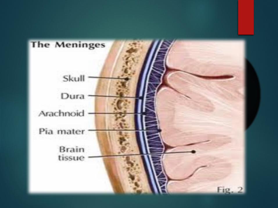
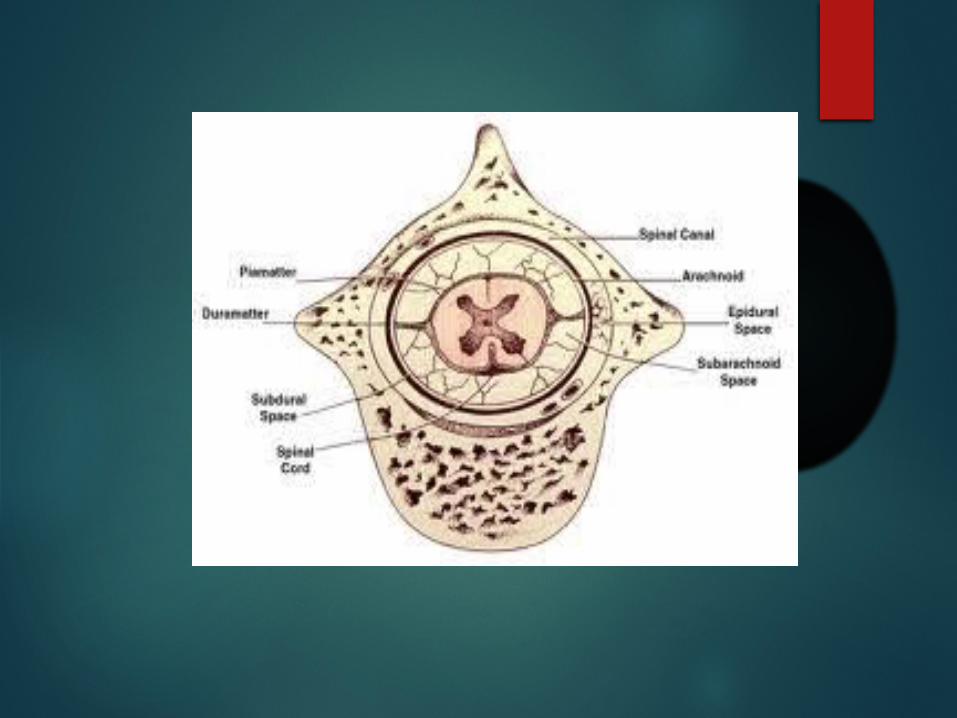
Meninges:The outermost covering of brain and spinal cord is the meninges. It includes three layers from deep to superficial.
Pia Matter
Arachnoid Matter
Dura Matter
1) Pia Matter:
The pia mater, the deepest of the meninges, is a delicate and very vascular membrane that invests the brain and spinal cord, following the grooves and depressions closely. The pia mater forms a sheath around the blood vessels and follows them into the substance of the CNS.
2) Arachnoid Matter:
The arachnoid is a very delicate and transparent membrane which is situated between the dura and pia mater. Together, the pia mater and arachnoid constitute the leptomeninges (from the Latin word lepto; delicate), reflecting their fine, delicate nature.
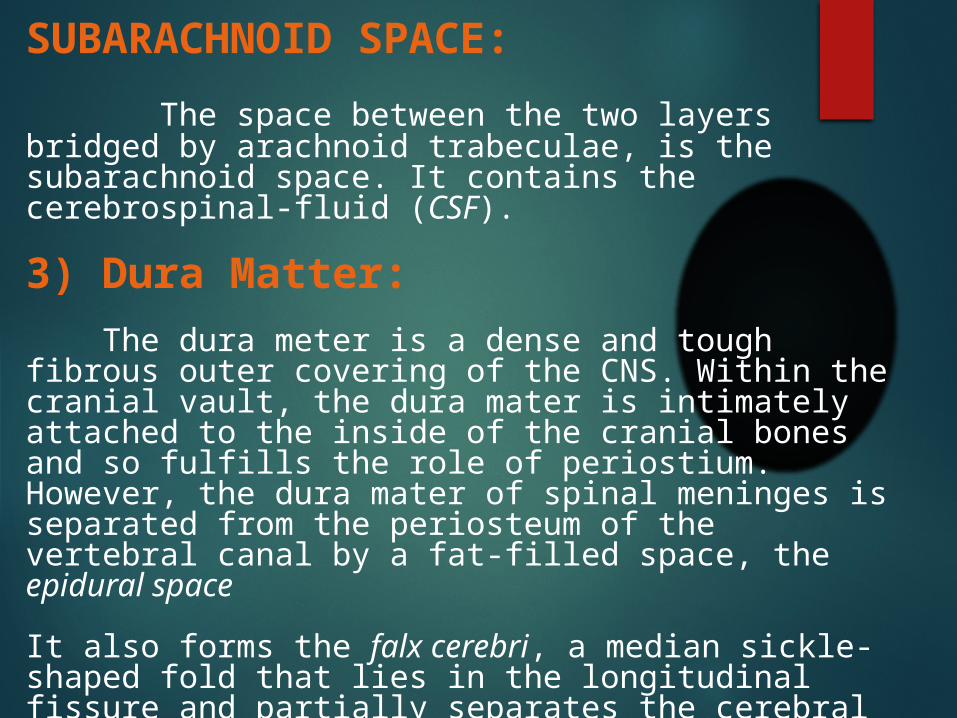
SUBARACHNOID SPACE:
The space between the two layers bridged by arachnoid trabeculae, is the subarachnoid space. It contains the cerebrospinal-fluid (CSF).
3) Dura Matter:
The dura meter is a dense and tough fibrous outer covering of the CNS. Within the cranial vault, the dura mater is intimately attached to the inside of the cranial bones and so fulfills the role of periostium. However, the dura mater of spinal meninges is separated from the periosteum of the vertebral canal by a fat-filled space, the epidural space
It also forms the falx cerebri, a median sickle-shaped fold that lies in the longitudinal fissure and partially separates the cerebral hemispheres. Another fold of dura mater.
The tentorium cerebelli, runs transversally between the cerebellum and the cerebrum.

Brain The brain is central part of the central nervous system that
is situated in the cranial cavity. The gross sub-divisions of the
adult brain include;
a) Cerebrum
b) Cerebellum
c) Brain stem
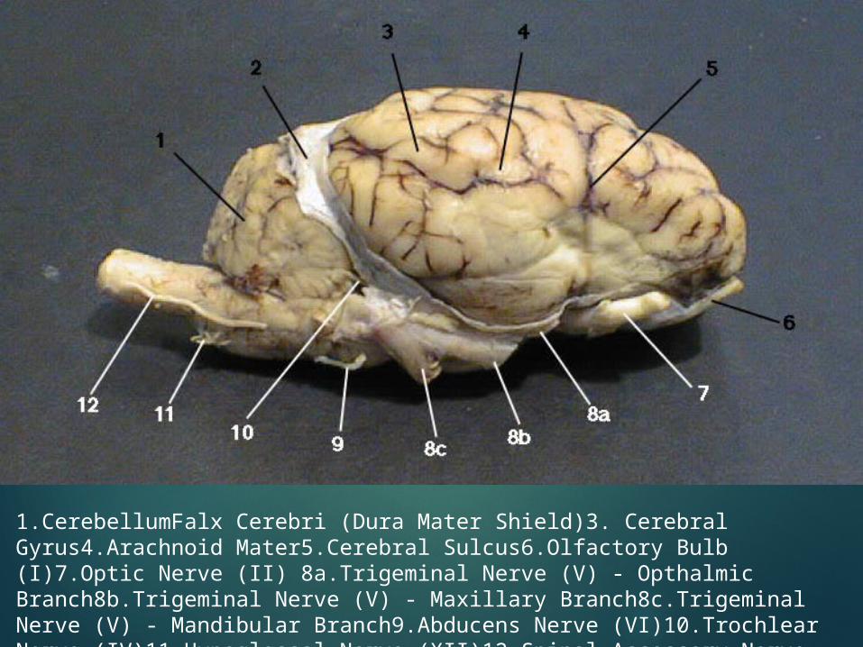
1.CerebellumFalx Cerebri (Dura Mater Shield)3. Cerebral Gyrus4.Arachnoid Mater5.Cerebral Sulcus6.Olfactory Bulb (I)7.Optic Nerve (II) 8a.Trigeminal Nerve (V) - Opthalmic Branch8b.Trigeminal Nerve (V) - Maxillary Branch8c.Trigeminal Nerve (V) - Mandibular Branch9.Abducens Nerve (VI)10.Trochlear Nerve (IV)11.Hypoglossal Nerve (XII)12.Spinal Accessory Nerve (XI)
Different Brain Regions:
Telencephalon (End Brain) Rhinencephalon Diencephalon (Inter Brain) Mesencephalon (Mid Brain) Metencephalon Myelencephalon Ventricular system
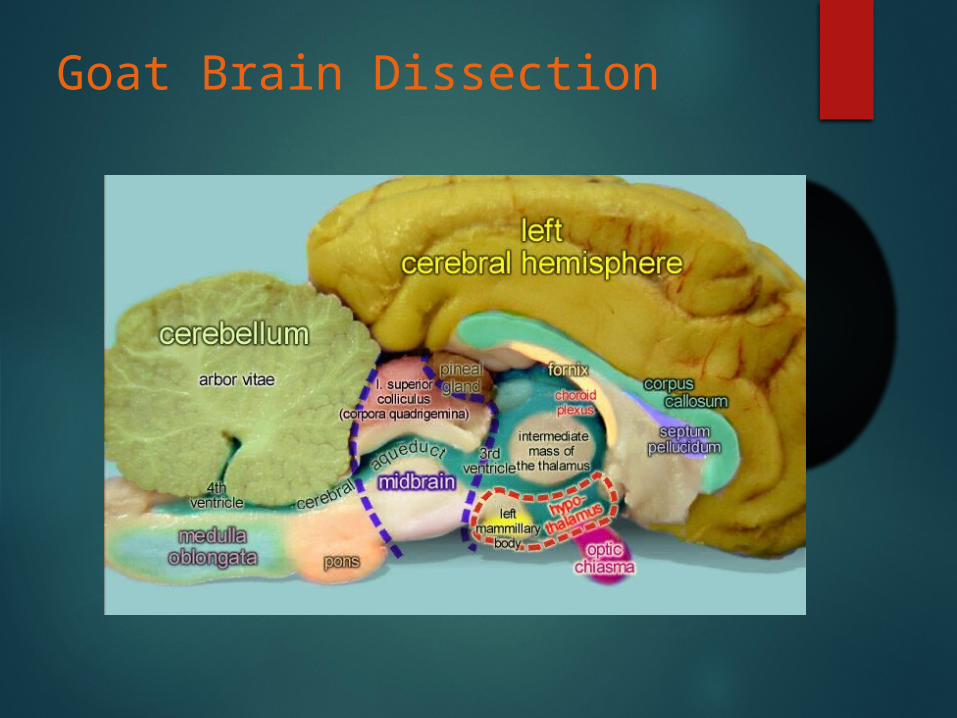
Goat Brain Dissection
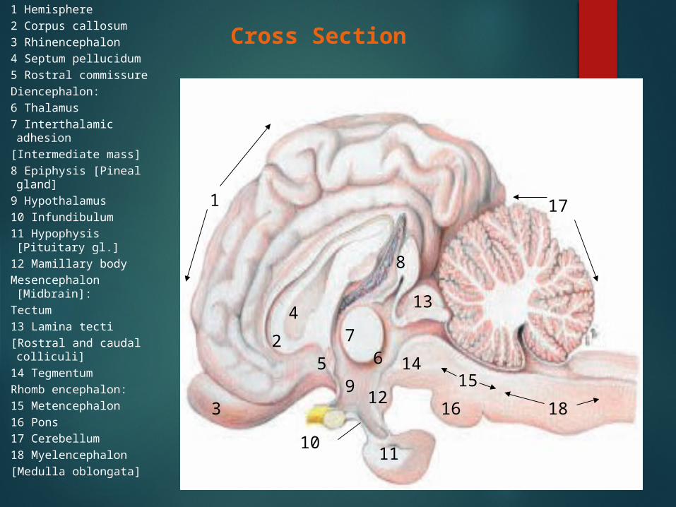
Cross Section
Cerebrum:
1 Hemisphere2 Corpus callosum3 Rhinencephalon4 Septum pellucidum5 Rostral commissureDiencephalon:6 Thalamus7 Interthalamic adhesion[Intermediate mass]8 Epiphysis [Pineal gland]9 Hypothalamus10 Infundibulum11 Hypophysis [Pituitary
gl.]12 Mamillary bodyMesencephalon
[Midbrain]:Tectum13 Lamina tecti[Rostral and caudal
colliculi]14 TegmentumRhomb encephalon:15 Metencephalon16 Pons17 Cerebellum18 Myelencephalon[Medulla oblongata]
3
2
4
1
5 67
8
9
1011
12
13
1415
16 18
17
1)Telencephalon (End Brain)
The telencephalon, or end brain comprises two principal parts, the cerebral hemispheres and the optic part of the hypothalamus (rhinencephalon).
CEREBRAL HEMISPHERES: The cerebral hemispheres form the greater part of the fully developed brain. Viewed from above, they form an ovoid mass, of which the broader is posterior, and the greatest transverse diameter is a little behind the middle.
Telencephalon (End Brain)
LONGITUDINAL FISSURE:
The two hemispheres are separated by a deep longitudinal fissure of the cerebrum, which is occupied by a sickle-shaped fold of dura materTRANSVERSE FISSUERE:
The transverse fissure separates the hemisphere from the cerebellum.EXTERNAL FEATURES:The surface area of the cerebrum is increased by numerous foldings to form convex ridges, called gyri (singular gyrus), which are separated by furrows called fissures or sulci. A particularly prominent fissure, the longitudinal fissure, lies on the median plane and separates the cerebrum into its right and left hemispheres.
INTERNAL STRUCTURE:Unlike the spinal cord, the in the cerebrum the gray maters are the on the exterior. This layer of cerebral grey matter is called cerebral cortex. It is the site at which voluntary movements are initiated, and higher functions, such as reasoning and planning, take place.
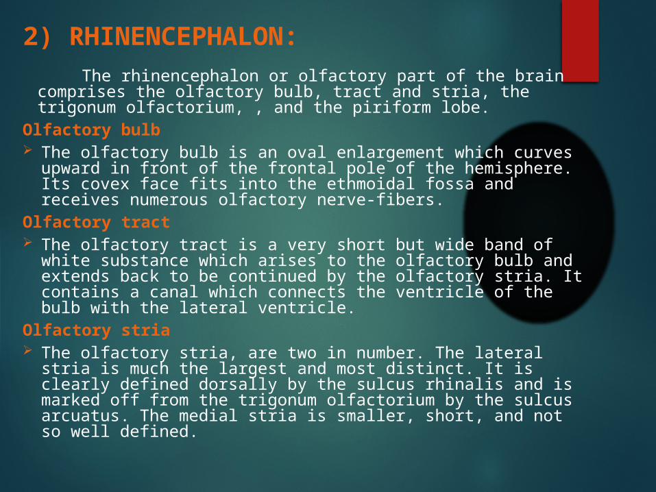
2) RHINENCEPHALON:
The rhinencephalon or olfactory part of the brain comprises the olfactory bulb, tract and stria, the trigonum olfactorium, , and the piriform lobe.
Olfactory bulb The olfactory bulb is an oval enlargement which curves upward in
front of the frontal pole of the hemisphere. Its covex face fits into the ethmoidal fossa and receives numerous olfactory nerve-fibers.
Olfactory tract The olfactory tract is a very short but wide band of white substance
which arises to the olfactory bulb and extends back to be continued by the olfactory stria. It contains a canal which connects the ventricle of the bulb with the lateral ventricle.
Olfactory stria The olfactory stria, are two in number. The lateral stria is much the
largest and most distinct. It is clearly defined dorsally by the sulcus rhinalis and is marked off from the trigonum olfactorium by the sulcus arcuatus. The medial stria is smaller, short, and not so well defined.
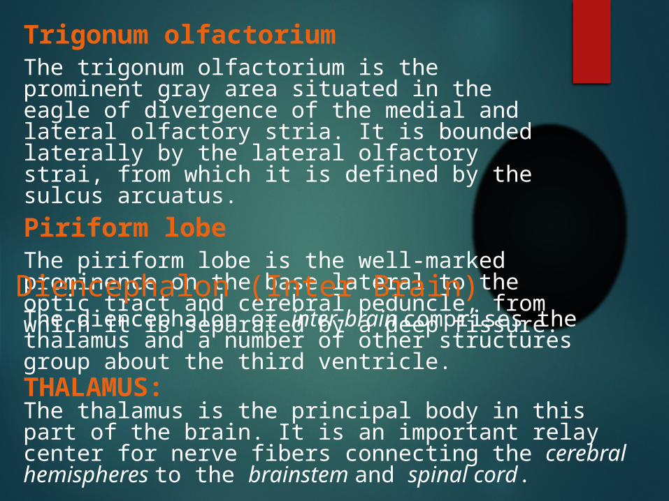
Trigonum olfactoriumThe trigonum olfactorium is the prominent gray area situated in the eagle of divergence of the medial and lateral olfactory stria. It is bounded laterally by the lateral olfactory strai, from which it is defined by the sulcus arcuatus.
Piriform lobeThe piriform lobe is the well-marked prominence on the base lateral to the optic tract and cerebral peduncle, from which it is separated by a deep fissure.
3) Diencephalon (Inter Brain)The diencephalon or inter-brain comprises the thalamus and a number of other structures group about the third ventricle.
THALAMUS:The thalamus is the principal body in this part of the brain. It is an important relay center for nerve fibers connecting the cerebral hemispheres to the brainstem and spinal cord.
Cont..
It is a large, ovoid mass placed obliquely across the dorsal face of the each cerebral peduncle, so that the long axes of the two thalami would meet in front about at right angle.
PINEAL BODY: The pineal body (epiphysis cerebri) is a small ovoid or fusiform red brown mass situated in a deep central depression between the thalami and corpora quadrigemina.
MAMMILARY BODY: The mammilary body is a white, round elevation a little larger than a pea which projects ventrally at the anterior end of the median furrow of the interpeduncular fossa or near infundibulum.
PITUITARY BODY:The pituitary body (hypophysis cerebri) is one of the most important endocrine glands. It was mentioned as covering part of the interpeduncular fossa. It is oval in outline, flattened dors0- ventrally. It is attached by a delicate tubular stalk, the infundibulum, to the tuber cinereum, a single gray prominence situated between the optic chiasm in front and the mammilary body behind.
OPTIC CHIASM AND TRACTSThe optic chiasm and tracts form the anterior boundary of the interpeduncular fossa. The optic chiasm is formed by the union of the both, right and left optic tracts.
4) Mesencephalon (Mid Brain)The mesencephalon or mid-brain, lies between the diencephalon rostrally and the pons caudally. It consists of a dorsal part, the corpora quadrigemina, and a larger ventral part, the cerebral peduncles.
CORPORA QUADRIGEMINA:
The corpora quadrigemina are four rounded eminences which lie under the posterior part of the cerebral hemispheres. They consist of two pairs, separated by a transverse groove. The anterior pair is larger and much higher than the posterior pair.
CEREBRAL PEDUNCLES:
The cerebral peduncles appear on the base of the brain as two large, rope-like stalks which emerge from the pons close together and diverge as they pass forward to enter the cerebrum. These peduncles consist of both sensory and motor fiber tracts.
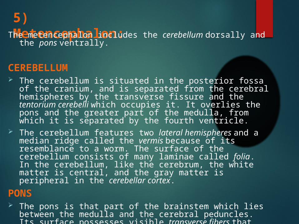
5) Metencephalon:The metencephalon includes the cerebellum dorsally and the pons ventrally.
CEREBELLUM The cerebellum is situated in the posterior fossa of the cranium, and is
separated from the cerebral hemispheres by the transverse fissure and the tentorium cerebelli which occupies it. It overlies the pons and the greater part of the medulla, from which it is separated by the fourth ventricle.
The cerebellum features two lateral hemispheres and a median ridge called the vermis because of its resemblance to a worm. The surface of the cerebellum consists of many laminae called folia. In the cerebellum, like the cerebrum, the white matter is central, and the gray matter is peripheral in the cerebellar cortex.
PONS The pons is that part of the brainstem which lies between the medulla and
the cerebral peduncles. Its surface possesses visible transverse fibers that form a bridge from one hemisphere of the cerebellum to the other.
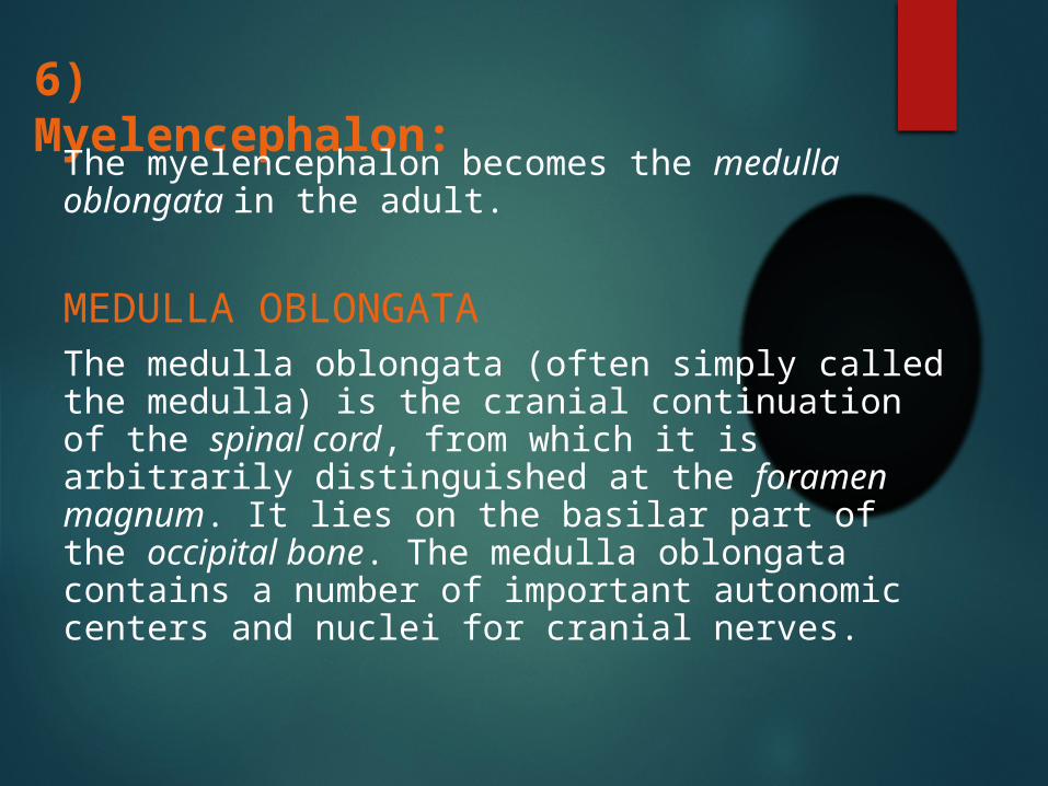
6) Myelencephalon:
The myelencephalon becomes the medulla oblongata in the adult.
MEDULLA OBLONGATAThe medulla oblongata (often simply called the medulla) is the cranial continuation of the spinal cord, from which it is arbitrarily distinguished at the foramen magnum. It lies on the basilar part of the occipital bone. The medulla oblongata contains a number of important autonomic centers and nuclei for cranial nerves.
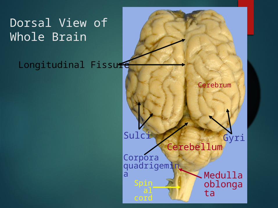
Dorsal View of Whole Brain
Cerebrum
Longitudinal Fissure
GyriSulci
Corpora quadrigemina
Cerebellum
Medulla oblongata
Spinal cord
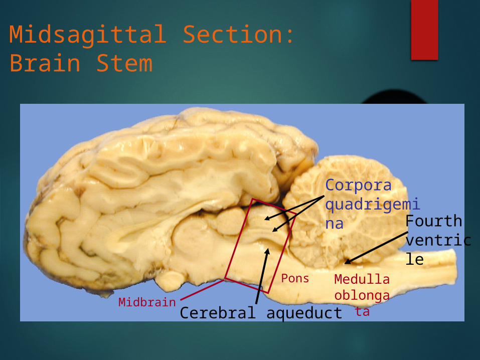
Midsagittal Section: Brain Stem
MidbrainCerebral aqueduct
Corpora quadrigemina
Pons Medulla oblonga
ta
Fourth ventricle
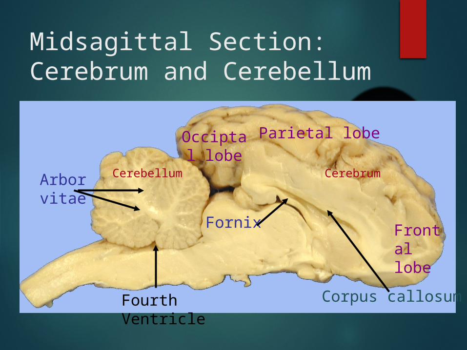
Midsagittal Section: Cerebrum and Cerebellum
CerebellumArbor vitae
Cerebrum
Corpus callosum
Occiptal lobe
Fornix
Parietal lobe
Frontal lobe
Fourth Ventricle
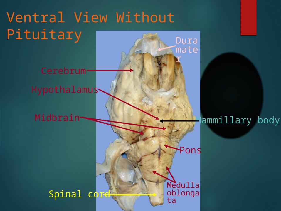
Ventral View Without Pituitary
Pons
Medulla oblongata
Cerebrum
Midbrain
Hypothalamus
Dura mater
Mammillary body
Spinal cord

Ventral View Without Pituitary
Pons
Medulla oblongata
Cerebrum
Midbrain
Hypothalamus
Dura mater
Mammillary body
Spinal cord

Spinal Cord:
Basically, the spinal cord is made up of:
White Matter
Grey Matter
WHITE MATTER:
It forms the dorsal column, ventral column and lateral columns.
GREY MATTER:
It forms dorsal horn, ventral horn and the central canal.
Division:
According to the attachment of spinal nerves, spinal cord is divided into five regions:
Cervical Thoracic Lumber Sacral Coccygeal
Spinal Cord Gross anatomically spinal cord has three main parts:
• Gray matter
• White matter
• Central Canal
The demarcation between the gray and white matter in many places is indistinct
Gray Matter
Resembles to roughly a capital H.Each lateral part is considered as consisting of dorsal and ventral gray columns (Horns)

Dorsal column or horn is elongated and narrow
Substantia gelatinosa: tip of the lateral horn consists of light gray matter
Nucleus dorsalis : is a medial projection on the ventral part of the dorsal column
Ventral column or horn is short, thick, and rounded.
White Matter Divided into three pairs of columns:
• Dorsal columns
• Ventral columns
• Lateral columns
The amounts of gray and white matter vary greatly in different parts of the cord

Thanks….!