co-producing esbl and ampc beta pratibha j and williamson... · int.j.curr.microbiol.app.sci (2015)...
TRANSCRIPT
Int.J.Curr.Microbiol.App.Sci (2015) 4(4): 107-117
107
Original Research Article
Antibacterial and Synergistic activity of Calendula officinalis Methanolic Petal Extract on Klebsiella pneumoniae Co-producing ESBL and AmpC Beta
Lactamase
Shah Pratibha J* and Williamson Manita T
Department of Microbiology, Topiwala National Medical College and B.Y.L. Nair Charitable Hospital, Mumbai- 400008, India
*Corresponding author
A B S T R A C T
Introduction
The rapid global dissemination of resistance towards beta lactam antibiotics, which is often mediated by beta lactamases, is a clinical threat. Infections caused by these pathogens pose a therapeutic challenge due to resistance mediated by different types of beta lactamases (Rawat et al., 2013, Upadhyay et al., 2010). Extended Spectrum
Beta Lactamases (ESBLs) are typically plasmid mediated, clavulanate susceptible enzymes that hydrolyze penicillins, expanded-spectrum cephalosporins, monobactams and are commonly inhibited by beta lactamase inhibitors such as clavulanic acid, sulbactam, and tazobactam (Moland et al., 2002). These plasmids also
ISSN: 2319-7706 Volume 4 Number 4 (2015) pp. 107-117 http://www.ijcmas.com
Co-production of Extended Spectrum Beta Lactamase (ESBL) and AmpC beta lactamase are of increasing clinical concern, especially in Klebsiella pneumoniae, making it highly resistant to most of the antibiotics used. Herbs are being considered as an important source for discovery of new agents for treating ailments related to resistant bacteria. Calendula officinalis Linn. (Asteraceae), a medicinal plant, has been traditionally used in healing wound and skin infections. The aim of the present study was to evaluate the antimicrobial activity of Calendula officinalis methanolic petal extract against 10 Klebsiella pneumoniae isolates co-producing ESBL and AmpC Beta lactamase. The isolates were confirmed for ESBL and AmpC Beta lactamase production by phenotypic confirmatory disc diffusion test and E-test. The screening for antibacterial activity of C. officinalis methanolic extract (CMe) was performed using disc diffusion technique. The minimum inhibitory concentration (MIC) of Cefotaxime and CMe was determined using agar dilution technique and was estimated to be in the range of 350-400 µg/ml and 1-7% (10 - 70 mg/ml) respectively against the selected isolates. Checker board assay was used to determine synergistic interaction between Cefotaxime and CMe. Fractional inhibitory concentration (FIC) index was calculated and synergistic interaction was observed in 40% Klebsiella pneumoniae isolates.
K e y w o r d s
Calendula officinalis, ESBL, AmpC, E-test, MIC, FIC, Synergy
Int.J.Curr.Microbiol.App.Sci (2015) 4(4): 107-117
108
carry resistance genes to other antibiotic groups including aminoglycosides, chloramphenicol, sulfonamides, trimethoprim, and tetracycline. Thus, microbes containing these plasmids generally become multidrug-resistant (Jacoby and Sutton, 1991). AmpC class beta lactamases are cephalosporinases that are poorly inhibited by clavulanic acid. They can be differentiated from ESBLs by their ability to hydrolyse cephamycins as well as other extended-spectrum cephalosporins. The current Clinical and Laboratory Standards Institute (CLSI) guidelines does not describe any method for detection of isolates producing AmpC beta lactamases making its identification difficult in routine diagnosis (Coudron et al., 2000).
It has been observed recently that co-production of ESBL and AmpC beta lactamase is a common phenomenon which is seen in many gram-negative isolates and may result in therapeutic failure with most of the antibiotics used, if not detected (Singla et al., 2014, Moland et al., 2007). When ESBL and AmpC beta lactamase co-exist their detection is difficult because they mask each other and cause an increase in the minimum inhibitory concentrations for -lactamase antibiotics (Sundin et al., 2009). Isolates of Klebsiella pneumoniae have been reported to show multiple beta lactamases, up to 8 different beta lactamases, with a special reference to ESBL and AmpC beta lactamase (Moland et al., 2007). This necessitates the detection and control of these organisms to avoid therapeutic failure.
Herbs are known to produce different secondary metabolites which are toxic to bacteria and thus can be used as an alternative therapeutic agent for resistant organisms. Natural products, either as pure compounds or as standardized herbal extracts, provide wide variety of
opportunities for new drug leads because of their vast chemical diversity (Rojas et al., 2003). Calendula is a traditional herb which can be explored for new antimicrobial molecules. Calendula officinalis L., a member of the Asteraceae family, is an annual plant with yellow to orange flowers; it is also known as pot marigold and has been cultivated as a food and medicinal plant since the Middle Ages. C.officinalis extract possess multiple pharmacological activities; including anti-inflammatory, antioxidant, anticancer, wound healing, antibacterial and antifungal activity (Khalid et al., 2012). Phytochemical studies have demonstrated the presence of several classes of chemical compounds like carotenoids, flavonoids, terpenoids, coumarins, quinines, amino acids, carbohydrates, lipids and other constituents in calendula flower petals (Muley et al., 2009). Previous studies indicate that the methanolic extracts of the C. officinalis petals possessed good antimicrobial and antifungal activity (Efstratiou et al., 2012, Mathur and Goyal 2011, Soni et al., 2012).
Herbal extracts can be combined with antibiotics to have an effective therapy, especially against resistant pathogens. The aim of combination therapy is to enhance antimicrobial activity through synergistic interaction and minimize resistance development for infections. In an earlier study, combination of antibiotics and flavonoids from selected plant extracts had shown enhanced antibacterial activities (Lin, et al., 2005).
Although considerable work has been done on C. officinalis extracts, no report is available on in vitro antimicrobial and synergistic studies of C. officinalis extracts against co-producers of ESBL and AmpC beta lactamase. The aim of the present study
Int.J.Curr.Microbiol.App.Sci (2015) 4(4): 107-117
109
was to evaluate the antimicrobial activity of C. officinalis methanolic petal extracts on Klebsiella pneumoniae, co-producing ESBL and AmpC beta lactamase, isolated from superficial wound infection and to investigate the synergistic effect of the extract and cefotaxime on the isolates.
Materials and Methods
Bacterial strains
Isolates from superficial wound infections of patients from a tertiary care hospital in Mumbai were identified by using standard Microbiological methods. Isolates identified as Klebsiella pneumoniae were selected and used further in the study.
Plant Extract
Fresh Calendula officinalis flowers were purchased from local market. The petals were separated, washed and dried in shade. Ten grams of dried and grounded petals were transferred into a flask containing 150 ml of the solvent methanol. Extract of Calendula officinalis petals was obtained by maceration in methanol for 1 week. The macerate was filtered and the solvent was evaporated. The dried powder obtained after solvent evaporation was dissolved in 50% Dimethyl sulfoxide (DMSO) to obtain a concentration of 500 mg/ml (Bissa and Bohra 2011). The prepared extract was stored at 4°C for further use in the study. Sterility testing of the extract was carried out by inoculating a loopful of the extract on Nutrient Agar and Sabouraud s Agar plates, and checking for growth of bacterial and fungal contaminants respectively after 1 week of incubation at room temperature.
Antimicrobial susceptibility Test (AST)
The antimicrobial susceptibility was determined by Kirby-Bauer disk diffusion
method in accordance with CLSI guidelines using commercially available antimicrobial discs (HiMedia, Mumbai). The following antibiotics were used- Ampicillin
(10 g),
Amikacin (10 g), Ceftriaxone (30 g), Ciprofloxacin (5 g), Gentamicin (10 g), Amoxyclav (30 g), Ceftazidime (30 g), Cefoxitin (30 g), Imipenem (10 g), Meropenem (10 g), Piperacillin (10 g), Piperacillin-Tazobactam (100/10). Susceptibility testing was performed on Mueller-Hinton agar using culture suspensions having turbidity equivalent to McFarland 0.5 standard.
If the diameter of inhibition zone for ceftazidime (CAZ 30µg) was 22mm, ESBL production was indicated. If the diameter of inhibition zone for cefoxitin (CX 30µg) was <18 mm, AmpC beta lactamase production was indicated (Coudran et al., 2000). ESBL and AmpC beta lactamase production was further confirmed by phenotypic confirmatory tests as per CLSI guidelines.
Confirmatory Phenotypic Disc diffusion test
Confirmatory phenotypic disc diffusion test was performed on Mueller-Hinton agar using culture suspensions having turbidity equivalent to McFarland 0.5 standard. Antibiotic discs of Ceftazidime (30 g) and Cefoxitin (CX 30µg) along with Ceftazidime/Clavulanic acid (30/10) and Cefoxitin/Cloxacillin (30/200) were placed on the seeded agar. The Klebsiella strains were phenotypically confirmed as ESBL producers if there was >
5 mm increase in the inhibition zone diameter of ceftazidime in combination with clavulanic acid versus ceftazidime alone, as per CLSI (2011) guidelines. The Klebsiella strains were phenotypically confirmed as AmpC beta lactamase producers if there was >
4 mm
Int.J.Curr.Microbiol.App.Sci (2015) 4(4): 107-117
110
increase in the inhibition zone diameter of cefoxitin in combination with cloxacillin acid versus cefoxitin alone (Tan et al., 2009).
E-test
ESBL and AmpC detection Ezy MICTM
strips (EM081, HiMedia, Mumbai) are drug-impregnated strips in which upper half contains a concentration gradient of 4 antibiotics; Ceftazidime, Cefotaxime, Cefepime and Cloxacillin plus Clavulanic acid and Tazobactam (MIX + Clav-TZ) and lower half contains of Ceftazidime, Cefotaxime, Cefepime and Cloxacillin in a concentration gradient in a reverse direction. The isolates were reported and confirmed as ESBL and AmpC beta lactamase producer as per the application sheet supplied by the manufacturer. These strips are to be used along with pure ESBL detection strips (EM079 HiMedia, Mumbai) to avoid false positive results. E test was performed on Mueller-Hinton agar using culture suspensions having turbidity equivalent to McFarland 0.5 standard.
A standard reference strain of Escherichia coli (ATCC 25922), susceptible to all antimicrobial drugs tested, and positive control strain Klebsiella pneumoniae ATCC 700603 were used as a quality control for Antimicrobial susceptibility test, Confirmatory phenotypic disc diffusion test and the E-test, as per CLSI (2011) guidelines.
Antibacterial activity of Calendula
Antibacterial activity of Calendula officinalis methanolic petal extract (CMe) was carried out by Disc Diffusion method using Mueller Hinton agar. The turbidity of the culture was adjusted to 0.5 McFarland standards. Culture suspensions were
inoculated on the medium so as to obtain a lawn culture. Sterile paper discs (6 mm, HiMedia, Mumbai) were impregnated with 20 l of the 500mg/ml plant extracts and placed on the inoculated agar. For the positive control, a disc of Imipenem (10 g) and for negative control, disc impregnated with 50% DMSO were placed on the inoculated Mueller Hinton agar. The plate was incubated at 37°C for 24 hours. After the incubation period, the zone of inhibition was measured in mm. The experiment was performed in triplicate (Efstratiou et al., 2012).
Minimum Inhibitory Concentration (MIC)
The MIC of CMe and Cefotaxime was determined by Agar dilution method. For MIC of CMe, dilutions were prepared by mixing CMe with sterile Mueller Hinton Agar to get final concentrations ranging between 2.5 mg/ml (0.25%) -70 mg/ml (50%). For MIC of Cefotaxime, dilutions were prepared by mixing Cefotaxime with sterile Mueller Hinton Agar to get final concentrations ranging between 10µg/ml
500µg/ml. A plate of Mueller Hinton agar with DMSO served as a control. These plates were seeded with bacterial suspensions using a loop and were incubated at 37°C for 24 hr. The MIC was recorded as the lowest concentration of CMe and Cefotaxime at which visible bacterial growth was completely inhibited. The experiment was performed in triplicate (Ward et al., 1984).
Determination of synergistic activity by checkerboard assay using agar dilution method
Checker board assay by agar dilution method was used to determine the synergistic interaction of various
Int.J.Curr.Microbiol.App.Sci (2015) 4(4): 107-117
111
concentrations of Cefotaxime and CMe. Two-fold serial dilution agar plates were prepared by mixing CMe in the range of 0.25% 2% along with 10 200 g/ml
of
cefotaxime. The results obtained were used to calculate Fractional inhibitory concentration (FIC) indices. The FIC index ( FIC) was calculated as follows:
FIC= FIC A+ FIC B, where
FIC A = MIC of Cefotaxime in combination/ MIC of Cefotaxime alone FIC B = MIC of CMe in combination/ MIC OF CMe alone.
A minimum FIC index of <0.5 indicates synergy, while a FIC index >2 indicates antagonism. If the minimum FIC index was >0.5 and <1, the effect of the combination was classified as additive. If the minimum FIC index was >1 and <2, the effect of the combination was classified as indifference (Nakamura et al., 2000, Vishwanatha et al., 2012).
Result and Discussion
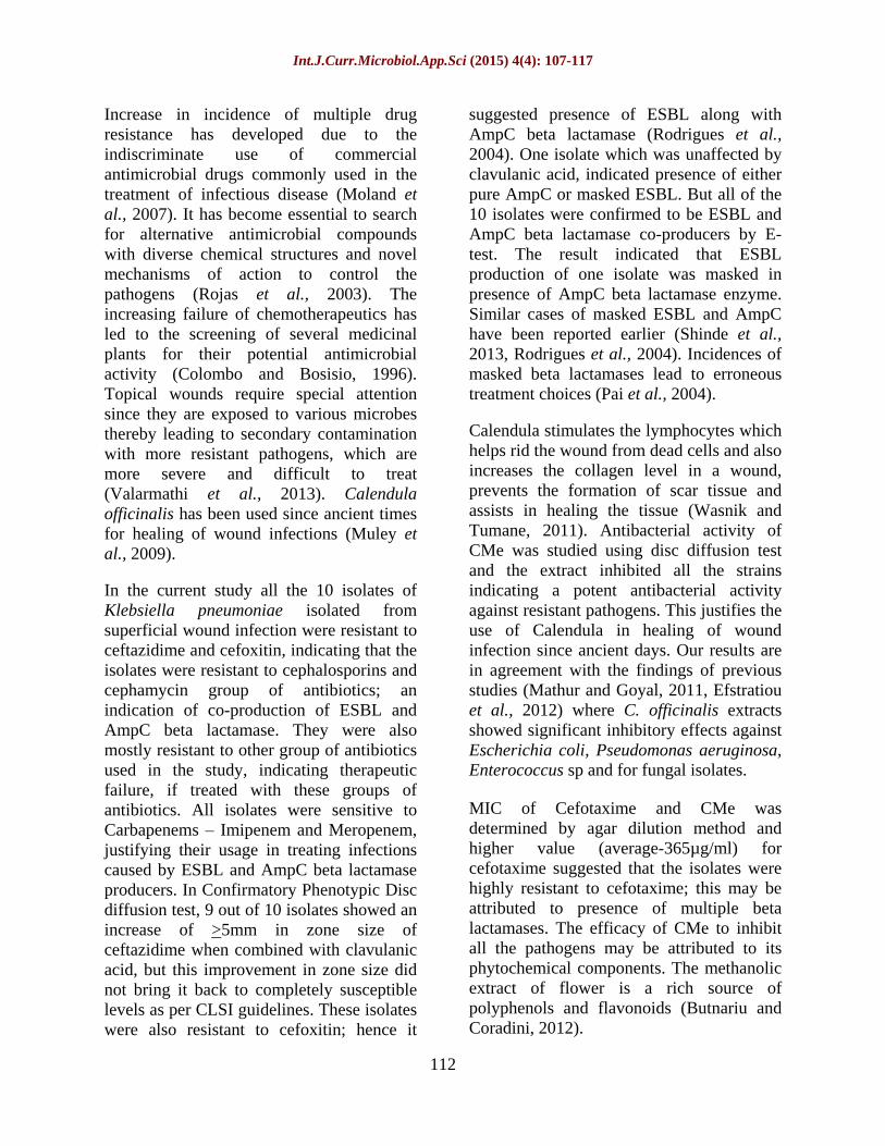
In this study 10 Klebsiella pneumoniae isolates were identified to be co-producing ESBL and AmpC beta lactamase. The AST studies revealed that all the isolates were 100% (10/10) resistant to ampicillin, amoxyclav, ceftazidime, ceftriaxone and cefoxitin, 90% (9/10) to were resistant to Ciprofloxacin, 40% (4/10) of the isolates were resistant to amikacin and gentamicin, whereas 60%(6/10) were resistant to Piperacillin-tazobactam. All the isolates showed sensitivity towards Imipenem and Meropenem (Figure 1).
In vitro resistance of isolates to Ceftazidime and Cefoxitin indicated the production of ESBL and AmpC beta lactamase. This was confirmed by Phenotypic confirmatory disc test and E test. 9 of the 10 isolates showed
an increase in inhibition zone diameter by >
5 mm for Ceftazidime/Clavulanic acid as compared to the usage of Ceftazidime alone, 1 isolate did not show the difference of >
5
mm. An increase by > 4 mm was seen for all 10 strains with Cefoxitin/Cloxacillin when compared to cefoxitin alone.
Hence E- test using Ezy-MICTM strips became significant for confirmation of ESBL and AmpC beta lactamase production. All 10 isolates were confirmed by E-test to be ESBL and AmpC beta lactamase producers.
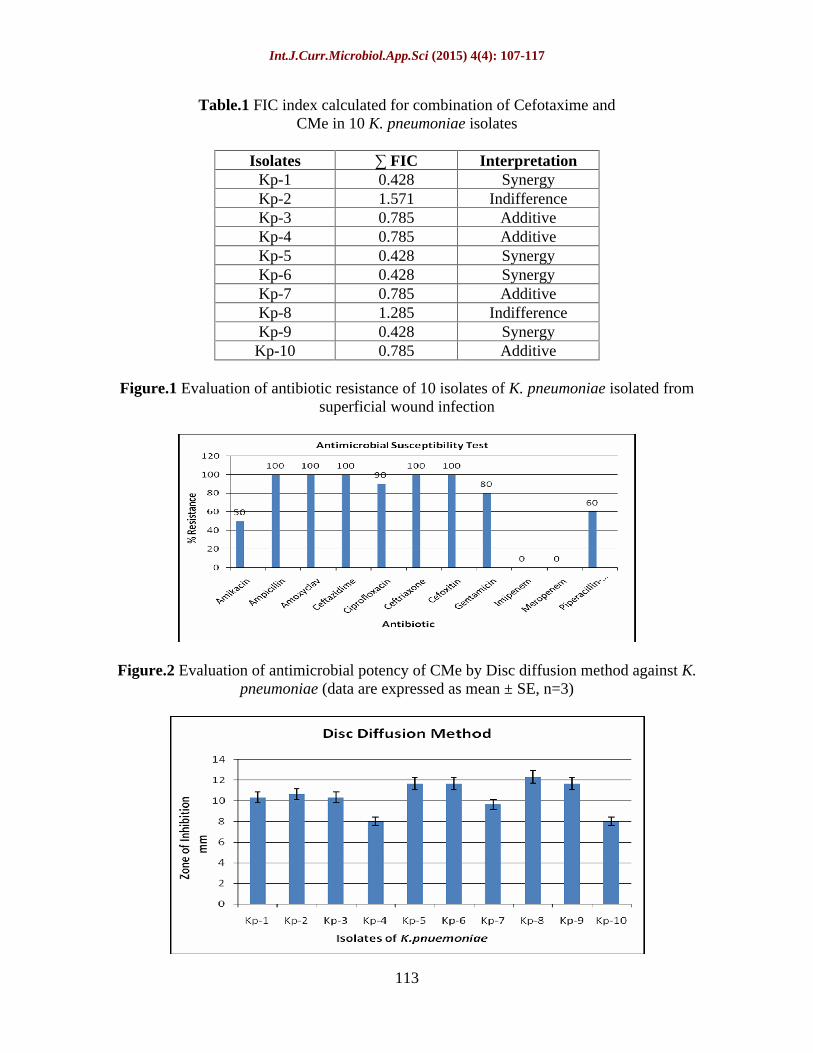
Primary screening for in vitro antibacterial activity of CMe was carried out by Disc diffusion method. The extract showed activity against all 10 Klebsiella pneumoniae isolates co-producing ESBL and AmpC beta lactamase. The average size of inhibition zones was in the range of 8-13mm and mean zone diameter was found to be 10.43 mm (Figure 2). No zone of inhibition was seen for 50% DMSO used as control. MIC of CMe and Cefotaxime was evaluated by Agar dilution method. MIC for CMe was obtained in the range of 1-7 % and average MIC of CMe was determined to be 5.3% (53mg/ml). MIC of Cefotaxime was obtained in the range 350 400µg/ml and average MIC for cefotaxime was determined to be 365µg/ml (Figure 3 and 4). Growth of all isolates was observed on DMSO control plates.
The antimicrobial synergistic activity of cefotaxime and CMe was evaluated in terms of FIC index obtained from Checkerboard assay by agar dilution method (Table1). In the current study, 40% (4/10) isolates exhibited synergistic association as their FIC index was >
0.5, whereas 40% (4/10) isolates exhibited additive association and 20 % (2/10) isolates exhibited indifference (Figure 3 and 4).
Int.J.Curr.Microbiol.App.Sci (2015) 4(4): 107-117
112
Increase in incidence of multiple drug resistance has developed due to the indiscriminate use of commercial antimicrobial drugs commonly used in the treatment of infectious disease (Moland et al., 2007). It has become essential to search for alternative antimicrobial compounds with diverse chemical structures and novel mechanisms of action to control the pathogens (Rojas et al., 2003). The increasing failure of chemotherapeutics has led to the screening of several medicinal plants for their potential antimicrobial activity (Colombo and Bosisio, 1996). Topical wounds require special attention since they are exposed to various microbes thereby leading to secondary contamination with more resistant pathogens, which are more severe and difficult to treat (Valarmathi et al., 2013). Calendula officinalis has been used since ancient times for healing of wound infections (Muley et al., 2009).
In the current study all the 10 isolates of Klebsiella pneumoniae isolated from superficial wound infection were resistant to ceftazidime and cefoxitin, indicating that the isolates were resistant to cephalosporins and cephamycin group of antibiotics; an indication of co-production of ESBL and AmpC beta lactamase. They were also mostly resistant to other group of antibiotics used in the study, indicating therapeutic failure, if treated with these groups of antibiotics. All isolates were sensitive to Carbapenems
Imipenem and Meropenem, justifying their usage in treating infections caused by ESBL and AmpC beta lactamase producers. In Confirmatory Phenotypic Disc diffusion test, 9 out of 10 isolates showed an increase of >5mm in zone size of ceftazidime when combined with clavulanic acid, but this improvement in zone size did not bring it back to completely susceptible levels as per CLSI guidelines. These isolates were also resistant to cefoxitin; hence it
suggested presence of ESBL along with AmpC beta lactamase (Rodrigues et al., 2004). One isolate which was unaffected by clavulanic acid, indicated presence of either pure AmpC or masked ESBL. But all of the 10 isolates were confirmed to be ESBL and AmpC beta lactamase co-producers by E-test. The result indicated that ESBL production of one isolate was masked in presence of AmpC beta lactamase enzyme. Similar cases of masked ESBL and AmpC have been reported earlier (Shinde et al., 2013, Rodrigues et al., 2004). Incidences of masked beta lactamases lead to erroneous treatment choices (Pai et al., 2004).
Calendula stimulates the lymphocytes which helps rid the wound from dead cells and also increases the collagen level in a wound, prevents the formation of scar tissue and assists in healing the tissue (Wasnik and Tumane, 2011). Antibacterial activity of CMe was studied using disc diffusion test and the extract inhibited all the strains indicating a potent antibacterial activity against resistant pathogens. This justifies the use of Calendula in healing of wound infection since ancient days. Our results are in agreement with the findings of previous studies (Mathur and Goyal, 2011, Efstratiou et al., 2012) where C. officinalis extracts showed significant inhibitory effects against Escherichia coli, Pseudomonas aeruginosa, Enterococcus sp and for fungal isolates.
MIC of Cefotaxime and CMe was determined by agar dilution method and higher value (average-365µg/ml) for cefotaxime suggested that the isolates were highly resistant to cefotaxime; this may be attributed to presence of multiple beta lactamases. The efficacy of CMe to inhibit all the pathogens may be attributed to its phytochemical components. The methanolic extract of flower is a rich source of polyphenols and flavonoids (Butnariu and Coradini, 2012).
Int.J.Curr.Microbiol.App.Sci (2015) 4(4): 107-117
113
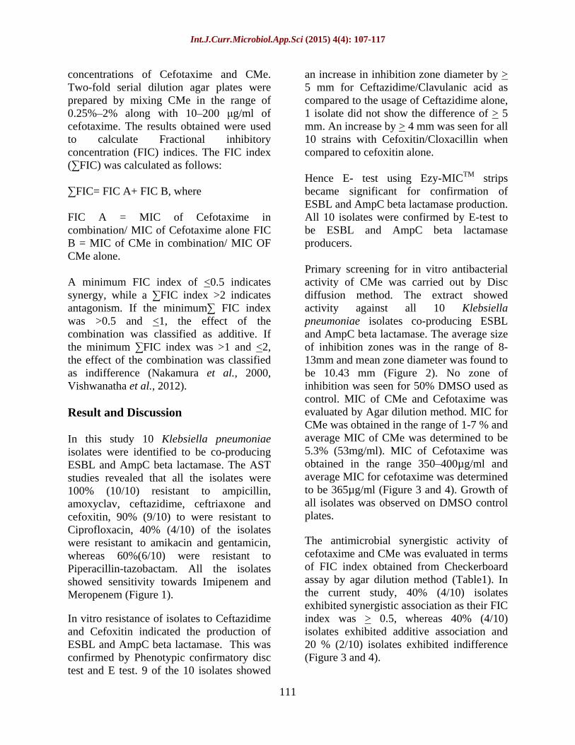
Table.1 FIC index calculated for combination of Cefotaxime and
CMe in 10 K. pneumoniae isolates
Isolates FIC Interpretation Kp-1 0.428 Synergy Kp-2 1.571 Indifference Kp-3 0.785 Additive Kp-4 0.785 Additive Kp-5 0.428 Synergy Kp-6 0.428 Synergy Kp-7 0.785 Additive Kp-8 1.285 Indifference Kp-9 0.428 Synergy
Kp-10 0.785 Additive
Figure.1 Evaluation of antibiotic resistance of 10 isolates of K. pneumoniae isolated from superficial wound infection
Figure.2 Evaluation of antimicrobial potency of CMe by Disc diffusion method against K. pneumoniae (data are expressed as mean ± SE, n=3)

Int.J.Curr.Microbiol.App.Sci (2015) 4(4): 107-117
114
Figure.3 MIC of Cefotaxime alone and in combination with CMe against K. pneumoniae
Isolate 1, 5, 6 and 9 show synergy; 3, 4, 7 and 10 show additive effect; 2 and 8 show indifference
Figure.4 MIC of CMe alone and in combination with Cefotaxime against K. pneumoniae Isolate 1, 5, 6 and 9 show synergy; 3, 4, 7 and 10 show additive effect; 2 and 8 show indifference
The mode of antimicrobial action may be related to their ability to inactivate microbial enzymes, inhibition of DNA gyrase, inhibit cytoplasmic membrane function (Vidal et al., 2006, Cushnie et al., 2006). Synergistic interaction was determined by calculating FIC index. For an interaction to be synergistic, a four-fold reduction in MIC should occur for the drugs, chosen in the combination. In the present study, 40% (4/10) isolates had four-fold or more reduction in MIC of Cefotaxime and CMe; in combination and FIC value was less than 0.5, indicating synergistic interaction. Another 40% (4/10) isolates had two-fold
reduction in their MIC, indicative of additive effect. Thus, overall 80% (8/10) isolates had at least two-fold reduction in MIC of Cefotaxime and CMe. In 20% (2/10) isolates, Cefotaxime and CMe were indifferent to each other. Cefotaxime is the most preferable antibiotic given during severe infections (Badar and Navale, 2012). However, the development of resistance against beta-lactam antibiotics including cefotaxime, limits its use as a therapeutic agent. If combination of CMe and cefotaxime can cause reversal of cefotaxime resistance or lower its dosage in therapy then this combination could potentially
Int.J.Curr.Microbiol.App.Sci (2015) 4(4): 107-117
115
improve the outcome for patients with severe infections.
Synergy is one of the well-established indications for successful combination antimicrobial therapy. To the best of our knowledge research on synergistic interaction between an antibiotic and Calendula officinalis petals has not been reported earlier. Few studies have reported synergy between antibiotics and other herbal extracts (Deepak et al., 2010, Dey et al., 2012). These results suggest that herbal extracts can aid in reducing the dosage of antibiotics in therapeutics, if used in combination. Screening of various herbs and identification of their active agents will help in prediction of lead molecules crucial in drug discovery and development.
Based on our results, it can be concluded that C. officinalis methanolic petal extract has a great potential as an antimicrobial compound against ESBL and AmpC Beta lactamase co-producing Klebsiella pneumoniae isolates. Further scope exists for research with phytochemical analysis, efficacy in in-vivo studies, safety and efficacy in patients (clinical trials). This kind of study could generate development of novel antimicrobial therapies and facilitate use of herbs in combination with antibiotics to treat many more infections.
Acknowledgement
The authors are thankful to Head of the Microbiology department and the Bacteriology section in charge of T.N.M.C. and B.Y.L. Nair Charitable Hospital, Mumbai.
References
Badar, V.A., Navale, S.B. 2012. Study of prescribing pattern of antimicrobial
agents in medicine intensive care unit of a teaching hospital in central India. JAPI, 60: 20 23.
Bissa, S., Bohra, A. 2011. Antibacterial potential of pot marigold. J. Microbiol. Antimicrob., 3(3): 51 54.
Butnariu, M., Coradini, C.Z. 2012. Evaluation of biologically active compounds from Calendula officinalis flowers using spectrophotometry. Chem. Cent. J., 6: 35 42.
CLSI. 2011. Performance standards for antimicrobial disc susceptibility tests, Vol. 31, No. 1. Document M100-S21.
Colombo, M.L., Bosisio, E. 1996. Pharmacological activities of Chelidonium majus L. (Papaveraceae). Pharmacol. Res., 33(2): 127 34.
Coudran, P.E., Moland, E.S., Thomson, K.S. 2000. Occurrence and detection of AmpC beta- lactamases among Escherichia coli, Klebsiella pneumoniae and Proteus mirabilis isolates at a Vetrans Medical Center. J. Clin. Microbial., 38: 1791 6.
Cushnie, T.P., Lamb, A.J. 2005. Antimicrobial activity of flavonoids Int. J. Antimicrob. Agents, 26(5): 343 56.
Deepak, S., Kamat, S.D., Kamat, D.V. 2010. Effect of aqueous extract of Terminalia chebula on Metallobetalactamase. Int. J. Pharm. Pharm. Sci., 2(4): 172 75.
Dey, D., Debnath, S., Hazra, S., Ghosh, S., Ray, R., Hazra, B. 2012. Pomegranate pericarp extract enhances the antibacterial activity of ciprofloxacin against extended-spectrum Beta-lactamase (ESBL) and metallo-beta-lactamase (MBL) producing Gram-negative bacilli.

Int.J.Curr.Microbiol.App.Sci (2015) 4(4): 107-117
116
Food Chem. Toxicol., 50: 43024309.
Efstratiou, E., Hussain, A., Nigam, P., Moore, J., Ayub, M.A., Rao, J.R. 2012. Antimicrobial activity of Calendula officinalis petal extracts against fungi, as well as Gram-negative and Gram-positive clinical pathogens. Complement. Ther. Clin. Pract., 18: 173 176.
Jacoby, G.A., Sutton, L. 1991. Properties of plasmids responsible for production of extended-spectrum beta-lactamases. Antimicrob. Agents Chemother., 35: 164 9.
Khalid, A.K., Silva, J.A. 2012. Biology of Calendula officinalis Linn.-focus on pharmacology, biological activities and agronomic practices. Med. Aromatic Plant Sci. Biotechnol., 6(1): 12 27.
Lin, R.D., Chin, Y.P., Lee, M.H. 2005. Antimicrobial activity of antibiotics in combination with natural flavonoids against clinical extended spectrum beta lactamase (ESBL) producing Klebsiella pneumoniae. Phytother. Res., 19(7): 612 617.
Mathur, R., Goyal, M. 2011.Antimicrobial and phytochemical estimation of Calendula officinalis against human pathogenic. Int. J. Innovat. Bio Sci., 1: 1 10.
Moland, S.E., Black, J.A., Ourada, J., Reisbig, M.D., Hanson, N.D., Thomson, K.S. 2002. Occurrence of newer -lactamases in Klebsiella pneumoniae isolates from 24 U.S. hospitals. Antimicrob. Agents Chemother., 46: 3837 42.
Moland, S.E., Hong, G., Thomson, K.S., Larone, D.H., Hanson, N.D. 2007. Klebsiella pneumoniae isolate producing at least eight different beta lactamases, including AmpC and
KPC beta-lactamases. Antimicrob. Agents Chemother., 51: 800 801.
Muley, B.P., Khadabadi, S.S., Banarase, N.B. 2009. Phytochemical constituents and pharmacological activities of Calendula officinalis Linn (Asteraceae): A review. Trop. J. Pharm. Res., 8(5): 455 465.
Nakamura, A., et al. 2000. Combined effects of meropenem and aminoglycosides on Pseudomonas aeruginosa in vitro J. Antimicrob. Chemother., 46: 901904.
Pai, H., et al. 2004. Epidemiology and clinical features of bloodstream infections caused by AmpC-type beta Lactamase-producing Klebsiella pneumonia. Antimicrob. Agents Chemother., 48(10): 3720 3728.
Rawat, V., Singhai, M., Verma, P.K. 2013. Detection of different -lactamases and their co-existence by using various discs combination methods in clinical isolates of Enterobacteriaceae and Pseudomonas spp. J. Lab. Physicians, 5(1): 21 25.
Rodrigues, C., Joshi, P., Jani, S.H., Alphonse, M., Radhakrishnan, R., Mehta, A. 2004. Detection of beta-lactamases in nosocomial gram negative clinical isolates. Indian J. Med. Microbiol., 22: 247 50.
Rojas, R., Bustamante, B., Bauer, J. 2003. Antimicrobial activity of selected Peruvian medicinal plants, J. Ethnopharmacol., 88: 199 204.
Shinde, S.S., Natraj, G., Mehta, P.R. 2013. Multiple beta lactamase resistance in clinical isolates of Escherichia coli and Klebsiella pneumoniae. Bombay Hosp. J., 55(1): 32 39.
Singla, P., Sikka, R., Deeep, A., Gagneja, D., Chaudhary, U. 2004. Co-production of ESBL and AmpC -lactamases in clinical isolates of A.
Int.J.Curr.Microbiol.App.Sci (2015) 4(4): 107-117
117
baumannii and A. lwoffii in a tertiary care hospital from Northern India. J. Clin. Diagn. Res., 8(4): DC16DC19.
Soni, H., Singhai, A.K. 2012. A recent update of botanicals for wound healing activity. Int. Res. J. Pharm., 3(7): 1 7.
Sundin, D.R. 2009. Hidden beta-lactamases in the Enterobacteriaceae
dropping the extra disks for detection, Part II. Clin. Microboil. Newslett., 31(7): 47 52.
Tan, T.Y., et al. 2009. Evaluation of screening methods to detect plasmid-mediated AmpC in Escherichia coli, Klebsiella pneumoniae, and Proteus mirabilis. Antimicrob. Agents Chemother., 53(1): 146 149.
Upadhyay, S., Sen, M.R., Bhattacharjee, A. 2010. Presence of different beta-lactamase classes among clinical isolates of Pseudomonas aeruginosa expressing AmpC beta-lactamase enzyme. J. Infect. Dev. Ctries., 4: 239 42.
Valarmathi, S., Rajasekara, M.P., Senthilkumar, B. 2013. Incidence and screening of wound infection causing microorganisms. J. Acad. Indus. Res., 1(8): 508 10.
Vidal, O.E., et al. 1989. Flavonol glycosides from Calendula officinalis flowers. Planta Med., 55(1): 73 4.
Vishwanatha, T. 2012. Evaluation of in vitro synergy between Amipicillin and Kanamycin against Staphylococcus aureus. J. Drug Deliv. Ther., 2(4): 144 146.
Ward, P.B., Carson, M., Dodd, J.S., Pavillard, E.R. 1984. Prediction of sulfamethoxazole trimethoprim synergistic action against members of the family Enterobacteriaceae with a two-plate agar dilution
breakpoint mic system. J. Clin. Microbiol., 19(6): 899 901.
Wasnik, D.D., Tumane, P.M. 2011. Comparative antibacterial activity of Tridax procumbens, Calotropis gigantea and Calendula officinalis leaf extracts against clinical isolates from wound infection. As. J. Biotechnol. Resour., 2(6): 781 786.