coalescence of the sites of cowpea mosaic virus rna replication
TRANSCRIPT

JOURNAL OF VIROLOGY, June 2002, p. 6235–6243 Vol. 76, No. 120022-538X/02/$04.00�0 DOI: 10.1128/JVI.76.12.6235–6243.2002Copyright © 2002, American Society for Microbiology. All Rights Reserved.
Coalescence of the Sites of Cowpea Mosaic Virus RNA Replicationinto a Cytopathic Structure
Jan E. Carette,† Kerstin Gühl, Joan Wellink,* and Ab Van KammenLaboratory of Molecular Biology, Wageningen University, 6703 HA Wageningen, The Netherlands
Received 27 November 2001/Accepted 13 March 2002
Cowpea mosaic virus (CPMV) replication induces an extensive proliferation of endoplasmic reticulum (ER)membranes, leading to the formation of small membranous vesicles where viral RNA replication takes place.Using fluorescent in situ hybridization, we found that early in the infection of cowpea protoplasts, CPMVplus-strand RNA accumulates at numerous distinct subcellular sites distributed randomly throughout thecytoplasm which rapidly coalesce into a large body located in the center of the cell, often near the nucleus. Thecombined use of immunostaining and a green fluorescent protein ER marker revealed that during the courseof an infection, CPMV RNA colocalizes with the 110-kDa viral polymerase and other replication proteins andis always found in close association with proliferated ER membranes, indicating that these sites correspond tothe membranous site of viral replication. Experiments with the cytoskeleton inhibitors oryzalin and latrunculinB point to a role of actin and not tubulin in establishing the large central structure. The induction of ERmembrane proliferations in CPMV-infected protoplasts did not coincide with increased levels of BiP mRNA,indicating that the unfolded-protein response is not involved in this process.
Infection with positive-stranded RNA viruses often causesextensive membrane rearrangements in the host cell, establish-ing a distinct compartment where viral RNA synthesis occurs.The viral replication complexes are associated with these mem-branes, which can originate from different intracellular mem-branes including the late and early endomembrane system (21,22, 27, 29, 30, 33). Despite the central role of such a virus-induced membranous compartment in the replicative cycle, thecellular components involved in the formation of this compart-ment are largely unknown.
Cowpea mosaic virus (CPMV), a bipartite positive-strandedRNA virus, is the type member of the comoviruses, which bearstrong resemblance to animal picornaviruses in both the geneorganization and the amino acid sequence of replication pro-teins (1, 14). Both RNA1 and RNA2 are translated into largepolyproteins, which are proteolytically cleaved into the differ-ent cleavage products by the 24-kDa proteinase (24K) (Fig. 1).The proteins encoded by RNA1 are necessary and sufficient forreplication, whereas RNA2 codes for the capsid proteins andthe movement protein. The RNA1-encoded 87-kDa protein(87K) contains a domain specific to RNA-dependent RNApolymerases; however, a 110-kDa protein (110K; 87K plus24K) is the only viral protein present in highly purified, RNA-dependent RNA polymerase preparations capable of elongat-ing nascent RNA chains, suggesting that fusion to 24K is re-quired for replicase activity (13).
Upon infection of cowpea plants with CPMV, a typical cy-topathic structure is formed, often adjacent to the nucleus,consisting of an amorphous matrix of electron-dense material
that is traversed by rays of small membranous vesicles (12).Autoradiography in conjunction with electron microscopy onsections of CPMV-infected leaves treated with [3H]uridine re-vealed that CPMV RNA replication was closely associatedwith the membranous vesicles (12). Additional support for thatview came from analyzing different fractions of homogenatesof CPMV-infected leaves in which the double-stranded, repli-cative form of CPMV RNA was present mainly in the micro-somal fraction (2). Also, the viral RNA-dependent RNA poly-merase activity was found in the crude membrane fraction ofCPMV-infected leaves (13, 39). However, as observed by elec-tron microscopy, the bulk of the replication proteins in CPMV-infected cells were immunolocalized not to the vesicles but tothe adjacent electron-dense structures, suggesting that only asmall part is present in active replication complexes (37).
The membranous vesicles induced upon CPMV infectionmay originate from the endoplasmic reticulum (ER). Throughthe use of transgenic Nicotiana benthamiana plants expressingthe green fluorescent protein (GFP) targeted to the lumen ofthe ER, it was demonstrated that CPMV infection leads to astrong proliferation of ER membranes and that these mem-branes are associated with the viral cytopathic structure (9).For poliovirus, the ER has also been suggested to serve assource for virally induced membranous vesicles, although im-munoisolated vesicles were found to contain marker proteinsof both the ER and the late endomembrane system (29). It hasbeen proposed that the small membranous vesicles in CPMV-infected cells are the result of the unfolded-protein response(9). This response can occur after overcrowding of ER mem-branes and results in a proliferation of ER membranes and theupregulation of ER chaperones like protein disulphide isomer-ase and the lumenal binding protein (BiP) (for a review, seereference 16).
In this study, the intracellular distribution of CPMV RNAduring virus infection was visualized by fluorescent in situ hy-bridization (FISH). The combined use of FISH and immuno-
* Corresponding author. Mailing address: Wageningen University,Laboratory of Molecular Biology, Dreijenlaan 3, 6703 HA Wagenin-gen, The Netherlands. Phone: 31-317483266. Fax: 31-317483584. E-mail: [email protected].
† Present address: VU University Medical Center, Department ofMedical Oncology, Division of Gene Therapy, Amsterdam, The Neth-erlands.
6235
on March 25, 2018 by guest
http://jvi.asm.org/
Dow
nloaded from
fluorescence detection of viral proteins allowed us to deter-mine the spatial relationship of CPMV RNA accumulation andthe accumulation of CPMV proteins involved in replicationand encapsidation and to establish the role of cellular compo-nents in the formation of the cytopathic structure. Further-more, we tested whether the unfolded-protein response wasinvolved in the proliferation of ER membranes in CPMV-infected cells by monitoring the level of BiP mRNA accumu-lation.
MATERIALS AND METHODS
Plasmids. Plasmid pTB1552(�) was used as a template to produce the fluo-rescein-labeled probes used in the in situ hybridization experiments. To createpTB1552(�), the SstI-BamHI fragment of the coding region of RNA1 (compris-ing nucleotides [nt] 2305 to 3857) was released from pTB1G and subcloned intothe plasmid vector Bluescript SK(�) (Stratagene, Inc.). The construction ofpUC-mGFP5-ER, which contains the plant-optimized GFP5 with an N-terminalArabidopsis thaliana basic chitinase signal sequence and a C-terminal HDEL ERretention signal under the control of a CaMV35S promoter, was describedpreviously (9). pMON talin-YFP (generously provided by Gerard van der Krogt,Wageningen University, Wageningen, The Netherlands) contains the actin-bind-ing domain of Dictyostelium talin fused to yellow fluorescent protein (YFP)(27a). The construction of a fusion of GFP with the tubulin-binding part ofmammalian MAP4 (GFP-MBD) (generously provided by Richard Cyr, ThePennsylvania State University, University Park, Pa.) has been described previ-ously (23). To determine BiP mRNA levels, plasmid pBLP2 (generously pro-vided by Jürgen Denecke, University of Leeds, Leeds, United Kingdom) wasused, which contains tobacco BiP (10).
Transfection of cowpea protoplasts and immunofluorescent labeling. Cowpea(Vigna unguiculata L.) mesophyll protoplasts were prepared and transfected bypolyethylene glycol-mediated transformation as described previously (32). Pro-toplasts were harvested at different time points postinfection for immunofluo-rescent staining. One volume of fixing solution (4% paraformaldehyde, 0.1%glutaraldehyde, 0.25 M mannitol, 50 mM sodium phosphate; pH 6) was added tothe protoplast suspension. After incubation for 15 min, the liquid was removed,replaced with fixing solution, and allowed to incubate for another 30 min. Thecells were washed three times with phosphate-buffered saline (PBS) and spottedon polylysine-coated microscope slides. The protoplasts were permeabilized witha 0.5% Triton X-100 solution in PBS for 10 min. In the case that the protoplastswere used for in situ hybridization, the slides were immersed in cold methanol for10 min to reduce background staining of the chlorophyll. This step was omittedwhen the GFP organellar markers were used, since this step abolishes GFPfluorescence. Nonspecific antibody binding was reduced by incubation for 10 minin blocking solution (5% bovine serum albumin in PBS). Subsequently, theprotoplasts were incubated for 1 h with dilutions of the primary anti-48K/58K(38), anti-CPMV (37), anti-32K (15), or anti-110K (31) serum in blocking solu-tion. After three washes with PBS, the protoplasts were incubated with goat
antirabbit antibodies conjugated to Cy3 (Sigma) for another hour. After twowashes with PBS, the cells were either mounted with cover slides by usingCitifluor or prepared for in situ hybridization.
In situ hybridization. In situ hybridization was performed essentially as de-scribed previously (24), with a minor modification. After acetylation, the dehy-dration step was omitted. The fluorescent probe that recognizes CPMV plus-strand RNA was obtained by in vitro transcription of pTB1552(�) linearizedwith SpeI in the presence of fluorescein-12-UTP (Roche Diagnostics GmbH)according to the manufacturer’s recommendation.
Fluorescence microscopy. A Zeiss LSM 510 confocal microscope was used toobtain images. Optical sections were made at 1-�m intervals, and projections ofserial optical sections were obtained by using the software provided by themanufacturer. GFP and fluorescein fluorescence were observed with standardsettings (excitation wavelength, 488 nm; emission band pass filter, 505 to 550nm). Cy3 fluorescence was detected with the following settings: excitation wave-length, 543 nm; emission band pass filter, 560 to 615 nm. In experiments of duallocalization, both fluorophores were scanned independently to reduce the pos-sibility of crossover between the cannels. Furthermore, single immunodetectioncontrols verified the absence of fluorescence crossover.
Northern blotting. At various times postinfection, protoplasts were harvestedand centrifuged at low speed (600 � g). Total RNA was isolated from the cellpellet by using Trizol reagent (Gibco BRL) according to the instructions of themanufacturer. Standard procedures were followed for denaturing the RNA withglyoxal, electrophoresing in a 1% agarose gel, and blotting to a GeneScreenmembrane (NEN Research Products). The blots were hybridized with a32P-labeled probe prepared by random-primer labeling of an EcoRI fragment ofpBLP2 to detect BiP mRNA.
RESULTS
Localization of CPMV RNA in infected protoplasts. Theintracellular accumulation of CPMV RNA during the infectionof cowpea protoplasts was visualized by FISH by using a plus-strand-specific probe corresponding to nt 2894 to 3857 ofCPMV RNA1. The probe was labeled by in vitro transcriptionin the presence of fluorescein isothiocyanate-UTP. The earliesttime at which viral proteins and infectious viral particles can bedetected in cowpea protoplasts by immunofluorescence and bya local lesion assay, respectively, is 12 h postinfection (hpi).Between 12 and 24 hpi, the number of infected cells and theproduction of infectious particles increase rapidly, reaching amaximum at 36 hpi (18). This implies that viral RNA synthesispeaks between 12 and 24 hpi in these cells. Cowpea protoplastswere infected with CPMV RNA1 and RNA2, collected at 16hpi, and hybridized with the probe. The fluorescent signalswere measured with a focal depth of 1 �m by confocal micros-copy. The majority of the plus-strand RNA was localized in alarge, irregularly shaped body that was often located near thenucleus (Fig. 2C). Similar localization patterns were observedwith protoplasts harvested at 24 hpi (data not shown) and 36hpi (Fig. 3B). To assess background staining of the fluorescentprobe, mock-infected protoplasts were subjected to FISH andthe fluorescent signals were measured with settings of theconfocal microscope identical to those used for the CPMV-infected sample at 16 hpi. No significant background stainingwas observed (Fig. 2D).
To determine the localization of plus-strand RNA early ininfection, protoplasts were harvested at 12 hpi. At this time,the plus-strand RNA labeling was still weak and signals higherthan the background signal could be observed only in a smallpercentage of the protoplasts (typically 5%). Approximatelyhalf of these protoplasts displayed the accumulation of plus-strand RNA in a large irregular body located near the nucleus,as was observed at later times. In the other half, the fluores-cence pattern differed markedly and plus-strand RNA was
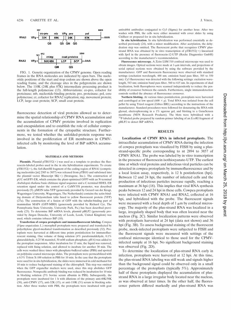
FIG. 1. Genetic organization of the CPMV genome. Open readingframes in the RNA molecules are indicated by open bars. The nucle-otide positions of the start and stop codons are shown above the openreading frame, and the cleavage sites in the polyproteins are shownbelow. The 110K (24K plus 87K) intermediate processing product isthe full-length polymerase (13). Abbreviations: co-pro, cofactor forproteinase; ntb, nucleotide-binding protein; pro, proteinase; pol, corepolymerase; cr, cofactor for RNA2 replication; mp, movement protein;LCP, large coat protein; SCP, small coat protein.
6236 CARETTE ET AL. J. VIROL.
on March 25, 2018 by guest
http://jvi.asm.org/
Dow
nloaded from
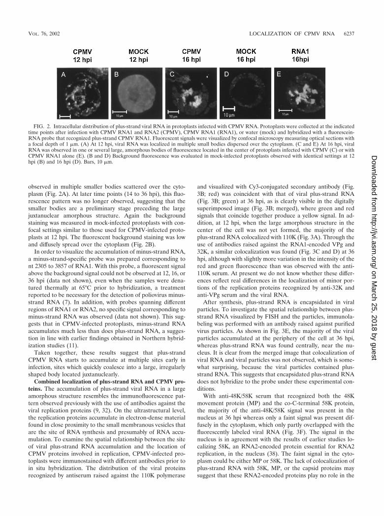
observed in multiple smaller bodies scattered over the cyto-plasm (Fig. 2A). At later time points (14 to 36 hpi), this fluo-rescence pattern was no longer observed, suggesting that thesmaller bodies are a preliminary stage preceding the largejuxtanuclear amorphous structure. Again the backgroundstaining was measured in mock-infected protoplasts with con-focal settings similar to those used for CPMV-infected proto-plasts at 12 hpi. The fluorescent background staining was lowand diffusely spread over the cytoplasm (Fig. 2B).
In order to visualize the accumulation of minus-strand RNA,a minus-strand-specific probe was prepared corresponding tont 2305 to 3857 of RNA1. With this probe, a fluorescent signalabove the background signal could not be observed at 12, 16, or36 hpi (data not shown), even when the samples were dena-tured thermally at 65°C prior to hybridization, a treatmentreported to be necessary for the detection of poliovirus minus-strand RNA (7). In addition, with probes spanning differentregions of RNA1 or RNA2, no specific signal corresponding tominus-strand RNA was observed (data not shown). This sug-gests that in CPMV-infected protoplasts, minus-strand RNAaccumulates much less than does plus-strand RNA, a sugges-tion in line with earlier findings obtained in Northern hybrid-ization studies (11).
Taken together, these results suggest that plus-strandCPMV RNA starts to accumulate at multiple sites early ininfection, sites which quickly coalesce into a large, irregularlyshaped body located juxtanuclearly.
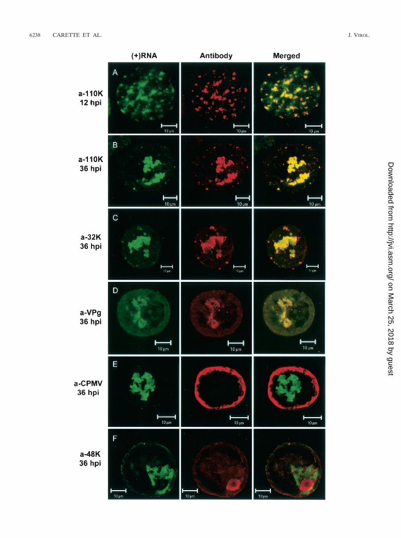
Combined localization of plus-strand RNA and CPMV pro-teins. The accumulation of plus-strand viral RNA in a largeamorphous structure resembles the immunofluorescence pat-tern observed previously with the use of antibodies against theviral replication proteins (9, 32). On the ultrastructural level,the replication proteins accumulate in electron-dense materialfound in close proximity to the small membranous vesicles thatare the site of RNA synthesis and presumably of RNA accu-mulation. To examine the spatial relationship between the siteof viral plus-strand RNA accumulation and the location ofCPMV proteins involved in replication, CPMV-infected pro-toplasts were immunostained with different antibodies prior toin situ hybridization. The distribution of the viral proteinsrecognized by antiserum raised against the 110K polymerase
and visualized with Cy3-conjugated secondary antibody (Fig.3B; red) was coincident with that of viral plus-strand RNA(Fig. 3B; green) at 36 hpi, as is clearly visible in the digitallysuperimposed image (Fig. 3B; merged), where green and redsignals that coincide together produce a yellow signal. In ad-dition, at 12 hpi, when the large amorphous structure in thecenter of the cell was not yet formed, the majority of theplus-strand RNA colocalized with 110K (Fig. 3A). Through theuse of antibodies raised against the RNA1-encoded VPg and32K, a similar colocalization was found (Fig. 3C and D) at 36hpi, although with slightly more variation in the intensity of thered and green fluorescence than was observed with the anti-110K serum. At present we do not know whether these differ-ences reflect real differences in the localization of minor por-tions of the replication proteins recognized by anti-32K andanti-VPg serum and the viral RNA.
After synthesis, plus-strand RNA is encapsidated in viralparticles. To investigate the spatial relationship between plus-strand RNA visualized by FISH and the particles, immunola-beling was performed with an antibody raised against purifiedvirus particles. As shown in Fig. 3E, the majority of the viralparticles accumulated at the periphery of the cell at 36 hpi,whereas plus-strand RNA was found centrally, near the nu-cleus. It is clear from the merged image that colocalization ofviral RNA and viral particles was not observed, which is some-what surprising, because the viral particles contained plus-strand RNA. This suggests that encapsidated plus-strand RNAdoes not hybridize to the probe under these experimental con-ditions.
With anti-48K/58K serum that recognized both the 48Kmovement protein (MP) and the co-C-terminal 58K protein,the majority of the anti-48K/58K signal was present in thenucleus at 36 hpi whereas only a faint signal was present dif-fusely in the cytoplasm, which only partly overlapped with thefluorescently labeled viral RNA (Fig. 3F). The signal in thenucleus is in agreement with the results of earlier studies lo-calizing 58K, an RNA2-encoded protein essential for RNA2replication, in the nucleus (38). The faint signal in the cyto-plasm could be either MP or 58K. The lack of colocalization ofplus-strand RNA with 58K, MP, or the capsid proteins maysuggest that these RNA2-encoded proteins play no role in the
FIG. 2. Intracellular distribution of plus-strand viral RNA in protoplasts infected with CPMV RNA. Protoplasts were collected at the indicatedtime points after infection with CPMV RNA1 and RNA2 (CPMV), CPMV RNA1 (RNA1), or water (mock) and hybridized with a fluorescein-RNA probe that recognized plus-strand CPMV RNA1. Fluorescent signals were visualized by confocal microscopy measuring optical sections witha focal depth of 1 �m. (A) At 12 hpi, viral RNA was localized in multiple small bodies dispersed over the cytoplasm. (C and E) At 16 hpi, viralRNA was observed in one or several large, amorphous bodies of fluorescence located in the center of protoplasts infected with CPMV (C) or withCPMV RNA1 alone (E). (B and D) Background fluorescence was evaluated in mock-infected protoplasts observed with identical settings at 12hpi (B) and 16 hpi (D). Bars, 10 �m.
VOL. 76, 2002 LOCALIZATION OF CPMV RNA 6237
on March 25, 2018 by guest
http://jvi.asm.org/
Dow
nloaded from
6238 CARETTE ET AL. J. VIROL.
on March 25, 2018 by guest
http://jvi.asm.org/
Dow
nloaded from

establishment of the sites of viral RNA accumulation. Thissuggestion was strengthened by the observation that the local-ization of viral RNA in cowpea protoplasts infected withRNA1 alone was similar to the localization observed in cellsinfected with both RNA1 and RNA2 (Fig. 2E).
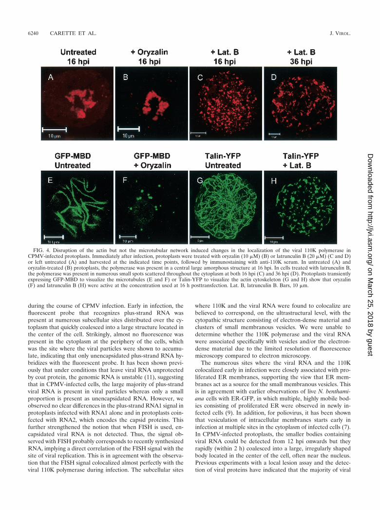
Effects of cytoskeletal inhibitors on the establishment of thesite of replication. As shown above, both viral RNA and the110K polymerase colocalize in numerous small bodies early ininfection, which later in infection coalesce into a large, irreg-ularly shaped body. The possible involvement of the plantcytoskeleton in the formation of this structure was tested byusing the cytoskeletal inhibitors latrunculin B and oryzalin.The highly specific toxin latrunculin B, isolated from a red seasponge, has been shown to effectively depolymerize actin fila-ments in eukaryotic cells (reference 3 and references therein).Oryzalin is an herbicide which strongly binds to plant tubulinmonomers, thereby stimulating the complete disassembly ofthe microtubular network (reference 4 and references therein).Protoplasts were infected with CPMV and then divided intothree equal portions. One sample was left untreated, whereaseither oryzalin (10 �M) or latrunculin B (20 �M) was added tothe incubation medium of the other two samples directly afterinfection. The protoplasts were collected at 16 and 36 hpi andprepared for immunofluorescent staining with antibodiesraised against the 110K polymerase. The percentage of in-fected protoplasts did not differ in the treated and untreatedsamples. In the untreated and oryzalin-treated protoplasts, thepolymerase was present in the large juxtanuclear body at 16 hpi(Fig. 4A and B) and at 36 hpi (data not shown). The pattern ofdistribution of the polymerase in latrunculin B-treated proto-plasts was strikingly different. Fluorescence was present innumerous small bodies that were randomly scattered through-out the cytoplasm (Fig. 4C). A similar pattern was observed at36 hpi (Fig. 4D). The pattern of multiple smaller bodies re-sembled that of polymerase distribution observed early in in-fection, suggesting that latrunculin B arrests the movement ofthese bodies to a juxtanuclear position.
In parallel experiments, it was verified that both inhibitorswere active at the concentrations used. For this purpose, thecowpea protoplasts were transfected with either a plant expres-sion vector encoding GFP-MBD (23) or an expression vectorencoding a fusion of the YFP with the actin-binding part of theDictyostelium talin gene (Talin-YFP) (20). In protoplaststreated with oryzalin, the typical microtubular network labeledby GFP-MBD was rigorously disturbed, as was observed in liveprotoplasts 16 and 36 h posttransfection by confocal micros-copy (Fig. 4E and F and data not shown). In addition, the actincytoskeleton labeled with Talin-YFP was severely disturbedupon treatment with latrunculin B, and only very small fluo-
rescent strands that were not interconnected remained (Fig.4G and H).
Taken together, these data indicate that integrity of the actinfilaments but not of the microtubular cytoskeleton is requiredfor the formation of the juxtanuclear large shapeless body.
CPMV-induced membrane proliferation. CPMV infectionresulted in the formation of proliferated ER membranes thatsurrounded and traversed the site of RNA replication stainedwith anti-110K serum at 36 hpi (Fig. 5, bottom row) (9). Todetermine whether the proliferated ER membranes were al-ready formed early in infection, when the replication com-plexes are present and dispersed over the cytoplasm, cowpeaprotoplasts were transfected with CPMV RNA together withpUC-mGFP5-ER, a plant expression vector that encodes theGFP targeted to the lumen of the ER (17). The protoplastswere harvested at 12 hpi and prepared for immunostainingwith anti-110K serum. It should be noted that mGFP5-ERremains fluorescent during immunostaining procedures andthat the GFP fluorescence could be separated from the fluo-rescence of Cy3, which was used for staining the viral polymer-ase. As shown in Fig. 5 (top row), the smaller bodies stainedwith anti-110K serum were closely associated with proliferatedER membranes.
Previously it has been suggested that the ER membraneproliferation might occur as a result of the unfolded-proteinresponse triggered by CPMV infection (9). In the presentstudy, we tested this hypothesis in cowpea protoplasts by mon-itoring the mRNA levels of the ER lumenal binding proteinBiP, which has been reported to be strongly upregulated inyeast, mammalian, and plant cells during the unfolded-proteinresponse (16, 19). Northern blot analysis with a tobacco BiPprobe revealed that the level of BiP mRNA in CPMV-infectedprotoplasts was not elevated compared to that in uninfectedprotoplasts at 36 hpi, when the CPMV-induced membraneproliferation was at its maximum (Fig. 6). Similar results wereobtained at earlier time points (data not shown). As a controlto ensure that the unfolded-protein response could be inducedin our cowpea protoplast system, tunicamycin, a drug thatinhibits N-glycosylation of ER membrane proteins and po-tently elicits the unfolded-protein response, was used. Treat-ment of the protoplasts with this drug led to a sharp increasein BiP mRNA levels (Fig. 6). These results suggest that the ERmembrane proliferations associated with CPMV replicationare probably not due to the unfolded-protein response.
DISCUSSION
The use of FISH and immunolocalization allowed us tobetter define the subcellular sites of viral RNA replication
FIG. 3. Localization of plus-strand viral RNA with respect to different viral proteins in CPMV-infected protoplasts. Protoplasts were collectedat the indicated time points after infection with CPMV RNA, hybridized with a fluorescein-RNA probe that recognized plus-strand CPMV RNA1(green signal), and immunostained with the indicated antisera (red signal). (A and B) At 12 and 36 hpi, the 110K polymerase was localized almostexclusively in sites where viral RNA accumulated. Colocalization of the two signals is shown in the merged images as yellow. (C) The 32K cofactorfor the proteinase also colocalized substantially with viral RNA. (D) The majority of the proteins recognized by anti-VPg (a-VPg) serumcolocalized with viral RNA. (E) Viral particles localized in the periphery of the cell and did not colocalize with the sites of viral RNA accumulation.(F) The 58K protein recognized by the anti-48K/58K serum localized mainly in the nucleus and did not colocalize with viral RNA. Bars, 10 �m.
VOL. 76, 2002 LOCALIZATION OF CPMV RNA 6239
on March 25, 2018 by guest
http://jvi.asm.org/
Dow
nloaded from
during the course of CPMV infection. Early in infection, thefluorescent probe that recognizes plus-strand RNA waspresent at numerous subcellular sites distributed over the cy-toplasm that quickly coalesced into a large structure located inthe center of the cell. Strikingly, almost no fluorescence waspresent in the cytoplasm at the periphery of the cells, whichwas the site where the viral particles were shown to accumu-late, indicating that only unencapsidated plus-strand RNA hy-bridizes with the fluorescent probe. It has been shown previ-ously that under conditions that leave viral RNA unprotectedby coat protein, the genomic RNA is unstable (11), suggestingthat in CPMV-infected cells, the large majority of plus-strandviral RNA is present in viral particles whereas only a smallproportion is present as unencapsidated RNA. However, weobserved no clear differences in the plus-strand RNA1 signal inprotoplasts infected with RNA1 alone and in protoplasts coin-fected with RNA2, which encodes the capsid proteins. Thisfurther strengthened the notion that when FISH is used, en-capsidated viral RNA is not detected. Thus, the signal ob-served with FISH probably corresponds to recently synthesizedRNA, implying a direct correlation of the FISH signal with thesite of viral replication. This is in agreement with the observa-tion that the FISH signal colocalized almost perfectly with theviral 110K polymerase during infection. The subcellular sites
where 110K and the viral RNA were found to colocalize arebelieved to correspond, on the ultrastructural level, with thecytopathic structure consisting of electron-dense material andclusters of small membranous vesicles. We were unable todetermine whether the 110K polymerase and the viral RNAwere associated specifically with vesicles and/or the electron-dense material due to the limited resolution of fluorescencemicroscopy compared to electron microscopy.
The numerous sites where the viral RNA and the 110Kcolocalized early in infection were closely associated with pro-liferated ER membranes, supporting the view that ER mem-branes act as a source for the small membranous vesicles. Thisis in agreement with earlier observations of live N. benthami-ana cells with ER-GFP, in which multiple, highly mobile bod-ies consisting of proliferated ER were observed in newly in-fected cells (9). In addition, for poliovirus, it has been shownthat vesiculation of intracellular membranes starts early ininfection at multiple sites in the cytoplasm of infected cells (7).In CPMV-infected protoplasts, the smaller bodies containingviral RNA could be detected from 12 hpi onwards but theyrapidly (within 2 h) coalesced into a large, irregularly shapedbody located in the center of the cell, often near the nucleus.Previous experiments with a local lesion assay and the detec-tion of viral proteins have indicated that the majority of viral
FIG. 4. Disruption of the actin but not the microtubular network induced changes in the localization of the viral 110K polymerase inCPMV-infected protoplasts. Immediately after infection, protoplasts were treated with oryzalin (10 �M) (B) or latrunculin B (20 �M) (C and D)or left untreated (A) and harvested at the indicated time points, followed by immunostaining with anti-110K serum. In untreated (A) andoryzalin-treated (B) protoplasts, the polymerase was present in a central large amorphous structure at 16 hpi. In cells treated with latrunculin B,the polymerase was present in numerous small spots scattered throughout the cytoplasm at both 16 hpi (C) and 36 hpi (D). Protoplasts transientlyexpressing GFP-MBD to visualize the microtubules (E and F) or Talin-YFP to visualize the actin cytoskeleton (G and H) show that oryzalin(F) and latrunculin B (H) were active at the concentration used at 16 h posttransfection. Lat. B, latrunculin B. Bars, 10 �m.
6240 CARETTE ET AL. J. VIROL.
on March 25, 2018 by guest
http://jvi.asm.org/
Dow
nloaded from

RNA synthesis occurs between 12 and 24 hpi (18), which sug-gests that the formation of this cytopathic structure early ininfection is important to promote efficient RNA replication.Consistent with this are findings that in cells infected withpoliovirus or tobacco mosaic virus (TMV), the formation of alarge juxtanuclear structure containing viral RNA, nonstruc-tural proteins, and ER membranes precedes bulk RNA syn-thesis (7, 24). Many positive-stranded RNA viruses induce theformation of a membranous compartment in the cytoplasmwhere viral replication occurs, and it has been proposed thatsuch compartmentalization increases the local concentrationsof virus-encoded proteins and viral RNA (8).
Based on the effects of latrunculin B and oryzalin on thedistribution of the viral polymerase, we suggest that in CPMV-infected protoplasts, intracellular trafficking of replicationcomplexes to the large juxtanuclear structure occurs via asso-ciation with the actin cytoskeleton and not the microtubularnetwork. Actin-based movement in plant cells has also beenreported recently for individual Golgi stacks in live plant cellsby using GFP constructs specifically targeted to the Golgi ap-paratus (6, 26). In contrast to CPMV, for the movement ofTMV replication complexes to a juxtanuclear position early ininfection, the integrity of both the actin and the microtubularcytoskeleton is required, as was demonstrated by using cy-tochalasin D and oryzalin (24).
After the formation of the large cytopathic structure, unen-capsidated viral RNA remained in the center of the cell andwas not redistributed to the periphery of the cell. In contrast,viral particles did accumulate in the periphery of the cell,presumably to be transported to the neighboring cell via plas-modesmata. The observation that almost no capsid material
was found in the central structure suggests that after encapsi-dation, the viral particles rapidly spread to the periphery of thecell for intercellular transport. The 48K MP did not colocalizewith viral RNA, and the distribution of viral RNA was similarin protoplasts infected with either RNA1 and RNA2 or RNA1alone, suggesting that the 48K MP does not influence the
FIG. 5. Dual localization of ER-targeted GFP (ER-GFP) and the viral 110K polymerase (a-110K) in CPMV-infected protoplasts. Cells werefixed at 12 hpi (top row) and 36 hpi (bottom row) and processed for immunofluorescence by using antibodies raised against the 110K polymerase.ER-GFP retained its fluorescence throughout the procedure. Proliferated ER membranes surrounded and traversed the sites of 110K accumu-lation both early (12 hpi) and late (36 hpi) in infection. Bars, 10 �m.
FIG. 6. Effect of CPMV infection on BiP mRNA levels in cowpeaprotoplasts. Cowpea protoplasts were infected with CPMV (CPMV)or uninfected (H2O and Tunicamycin). Prior to the harvesting of theprotoplasts (36 hpi), the sample designated Tunicamycin was treatedwith tunicamycin (20 �g/ml) for 2.5 h. Total RNA was extracted,separated on an agarose gel, and blotted on a nylon membrane. Theblots were probed for BiP, and an actin probe (Actin) was used as acontrol for loading differences. CPMV infection (CPMV) did not leadto an increase in BiP mRNA levels compared to those of the unin-fected protoplasts (H2O). Treatment with tunicamycin (Tunicamycin)resulted in upregulation of BiP expression.
VOL. 76, 2002 LOCALIZATION OF CPMV RNA 6241
on March 25, 2018 by guest
http://jvi.asm.org/
Dow
nloaded from
distribution of viral RNA. In contrast, the TMV 30K MPcolocalized with viral RNA at all points during infection andwas essential for the establishment of the large body near thenucleus observed in the middle stages of infection (24). Fur-thermore, at late stages of infection, viral RNA was dispersedover the cytoplasm and at the periphery of the cell (24). Thesedifferences between CPMV and TMV might reflect the differ-ent mechanisms these viruses use for their cell-to-cell move-ment, since TMV RNA spreads from cell to cell as a ribonu-cleoprotein complex of TMV RNA and 30K MP (for a review,see reference 5) whereas CPMV RNA is encapsidated beforeintercellular transport and moves from cell to cell as viralparticles through tubules formed by the MP in plasmodesmata(34–36).
CPMV infection induces an extensive rearrangement of in-tracellular membranes, but the cellular mechanism underlyingthis vesiculation is unclear. Based on the observation that po-liovirus-induced vesicles share cytological characteristics (adouble membrane and cytosolic content) with autophagicvacuoles that are formed in noninfected cells in response tonitrogen or amino acid starvation, autophagy has been pro-posed as a mechanism of induction of the vesicles (29, 30). Onthe other hand, CPMV-induced vesicles do not possess similarfeatures, which makes it unlikely that autophagy is involved forCPMV. Another mechanism that has been proposed for po-liovirus suggests that poliovirus infection interferes with ER-to-Golgi transport, leading to the accumulation of membra-nous vesicles (25). This view is supported by a recent study thatshowed that the poliovirus-induced vesicles bud from the ERand colocalize with the COPII components Sec13 and Sec31,suggesting that poliovirus-induced vesicles are homologous tothe vesicles of the anterograde membrane transport pathway(28). The extensive ER proliferation observed in CPMV-in-fected cells has not been reported for poliovirus-infected cells,which may suggest that CPMV uses a different cellular mech-anism to induce vesiculation. CPMV infection did not result inan increase of the level of BiP mRNA in protoplasts, which isa marker for the unfolded-protein response. The reliability ofthis marker was tested with tunicamycin, which elicited a clearresponse in protoplasts. Since in yeast, animal, and plant cellsupregulation of BiP is a hallmark in the unfolded-protein re-sponse (16, 19), it is unlikely that this stress response is re-sponsible for the ER membrane proliferations. Further studieswill be necessary to unravel the molecular pathways leading toCPMV-induced ER proliferation and vesiculation.
ACKNOWLEDGMENTS
We thank Jürgen Denecke, Richard Cyr, and Gerard van der Krogtfor generously providing the biological material indicated in Materialsand Methods. We gratefully acknowledge Jeroen Pouwels for usefulcomments and technical assistance.
This work was supported by The Netherlands Foundation of Chem-ical Research with financial aid from The Netherlands Organizationfor Scientific Research.
REFERENCES
1. Argos, P., G. Kamer, M. J. Nicklin, and E. Wimmer. 1984. Similarity in geneorganization and homology between proteins of animal picornaviruses and aplant comovirus suggest common ancestry of these virus families. NucleicAcids Res. 12:7251–7267.
2. Assink, A. M., H. Swaans, and A. Van Kammen. 1973. The localization ofvirus-specific double-stranded RNA of cowpea mosaic virus in subcellularfractions of infected Vigna leaves. Virology 53:384–391.
3. Baluska, F., J. Jasik, H. G. Edelmann, T. Salajova, and D. Volkmann. 2001.Latrunculin B-induced plant dwarfism: plant cell elongation is F-actin-de-pendent. Dev. Biol. 231:113–124.
4. Baskin, T. I., J. E. Wilson, A. Cork, and R. E. Williamson. 1994. Morphologyand microtubule organization in Arabidopsis roots exposed to oryzalin ortaxol. Plant Cell Physiol. 35:935–942.
5. Beachy, R. N., and M. Heinlein. 2000. Role of P30 in replication and spreadof TMV. Traffic 1:540–544.
6. Boevink, P., K. Oparka, S. Santa Cruz, B. Martin, A. Betteridge, and C.Hawes. 1998. Stacks on tracks: the plant Golgi apparatus traffics on anactin/ER network. Plant J. 15:441–447.
7. Bolten, R., D. Egger, R. Gosert, G. Schaub, L. Landmann, and K. Bienz.1998. Intracellular localization of poliovirus plus- and minus-strand RNAvisualized by strand-specific fluorescent in situ hybridization. J. Virol. 72:8578–8585.
8. Buck, K. W. 1999. Replication of tobacco mosaic virus RNA. Philos. Trans.R. Soc. Lond. B Biol. Sci. 354:613–627.
9. Carette, J. E., M. Stuiver, J. Van Lent, J. Wellink, and A. Van Kammen.2000. Cowpea mosaic virus infection induces a massive proliferation ofendoplasmic reticulum but not Golgi membranes and is dependent on denovo membrane synthesis. J. Virol. 74:6556–6563.
10. Denecke, J., M. H. Goldman, J. Demolder, J. Seurinck, and J. Botterman.1991. The tobacco luminal binding protein is encoded by a multigene family.Plant Cell 3:1025–1035.
11. De Varennes, A., and A. J. Maule. 1985. Independent replication of cowpeamosaic virus bottom component RNA: in vivo instability of the viral RNAs.Virology 144:495–501.
12. De Zoeten, G. A., A. M. Assink, and A. Van Kammen. 1974. Association ofcowpea mosaic virus-induced double-stranded RNA with a cytopathologicalstructure in infected cells. Virology 59:341–355.
13. Eggen, R., A. Kaan, R. Goldbach, and A. Van Kammen. 1988. Cowpeamosaic virus RNA replication in crude membrane fractions from infectedcowpea and Chenopodium armaranticolor. J. Gen. Virol. 69:2711–2720.
14. Franssen, H., J. Leunissen, R. Goldbach, G. Lomonossoff, and D. Zimmern.1984. Homologous sequences in non-structural proteins from cowpea mosaicvirus and picornaviruses. EMBO J. 3:855–861.
15. Franssen, H., M. Moerman, G. Rezelman, and R. Goldbach. 1984. Evidencethat the 32,000-Dalton protein encoded by bottom-component RNA of cow-pea mosaic virus is a proteolytic processing enzyme. J. Virol. 50:183–190.
16. Hampton, R. Y. 2000. ER stress response: getting the UPR hand on mis-folded proteins. Curr. Biol. 10:R518–R521.
17. Haseloff, J., K. R. Siemering, D. C. Prasher, and S. Hodge. 1997. Removal ofa cryptic intron and subcellular localization of green fluorescent protein arerequired to mark transgenic Arabidopsis plants brightly. Proc. Natl. Acad.Sci. USA 94:2122–2127.
18. Hibi, T., G. Rezelman, and A. Van Kammen. 1975. Infection of cowpeamesophyll protoplasts with cowpea mosaic virus. Virology 64:308–318.
19. Jelitto-Van Dooren, E. P., S. Vidal, and J. Denecke. 1999. Anticipatingendoplasmic reticulum stress. A novel early response before pathogenesis-related gene induction. Plant Cell 11:1935–1944.
20. Kost, B., P. Spielhofer, and N. H. Chua. 1998. A GFP-mouse talin fusionprotein labels plant actin filaments in vivo and visualizes the actin cytoskel-eton in growing pollen tubes. Plant J. 16:393–401.
21. Kujala, P., A. Ikaheimonen, N. Ehsani, H. Vihinen, P. Auvinen, and L.Kaariainen. 2001. Biogenesis of the Semliki Forest virus RNA replicationcomplex. J. Virol. 75:3873–3884.
22. Mackenzie, J. M., M. K. Jones, and E. G. Westaway. 1999. Markers fortrans-Golgi membranes and the intermediate compartment localize to in-duced membranes with distinct replication functions in flavivirus-infectedcells. J. Virol. 73:9555–9567.
23. Marc, J., C. L. Granger, J. Brincat, D. D. Fisher, T. Kao, A. G. McCubbin,and R. J. Cyr. 1998. A GFP-MAP4 reporter gene for visualizing corticalmicrotubule rearrangements in living epidermal cells. Plant Cell 10:1927–1940.
24. Mas, P., and R. N. Beachy. 1999. Replication of tobacco mosaic virus onendoplasmic reticulum and role of the cytoskeleton and virus movementprotein in intracellular distribution of viral RNA. J. Cell Biol. 147:945–958.
25. Maynell, L. A., K. Kirkegaard, and M. W. Klymkowsky. 1992. Inhibition ofpoliovirus RNA synthesis by brefeldin A. J. Virol. 66:1985–1994.
26. Nebenfuhr, A., L. A. Gallagher, T. G. Dunahay, J. A. Frohlick, A. M. Ma-zurkiewicz, J. B. Meehl, and L. A. Staehelin. 1999. Stop-and-go movementsof plant Golgi stacks are mediated by the acto-myosin system. Plant Physiol.121:1127–1142.
27. Pedersen, K. W., Y. van der Meer, N. Roos, and E. J. Snijder. 1999. Openreading frame 1a-encoded subunits of the arterivirus replicase induce endo-plasmic reticulum-derived double-membrane vesicles which carry the viralreplication complex. J. Virol. 73:2016–2026.
27a. Pouwels, J., G. N. van der Krogt, J. van Lent, T. Bisseling, and J. Wellink.The cytoskeleton and the secretory pathway are not involved in targetingthe cowpea mosaic virus movement protein to the cell periphery. Virology,in press.
28. Rust, R. C., L. Landmann, R. Gosert, B. L. Tang, W. Hong, H. P. Hauri, D.
6242 CARETTE ET AL. J. VIROL.
on March 25, 2018 by guest
http://jvi.asm.org/
Dow
nloaded from

Egger, and K. Bienz. 2001. Cellular COPII proteins are involved in produc-tion of the vesicles that form the poliovirus replication complex. J. Virol.75:9808–9818.
29. Schlegel, A., T. H. Giddings, Jr., M. S. Ladinsky, and K. Kirkegaard. 1996.Cellular origin and ultrastructure of membranes induced during poliovirusinfection. J. Virol. 70:6576–6588.
30. Suhy, D. A., T. H. Giddings, Jr., and K. Kirkegaard. 2000. Remodeling theendoplasmic reticulum by poliovirus infection and by individual viral pro-teins: an autophagy-like origin for virus-induced vesicles. J. Virol. 74:8953–8965.
31. van Bokhoven, H., J. W. van Lent, R. Custers, J. M. Vlak, J. Wellink, and A.van Kammen. 1992. Synthesis of the complete 200K polyprotein encoded bycowpea mosaic virus B-RNA in insect cells. J. Gen. Virol. 73:2775–2784.
32. van Bokhoven, H., J. Verver, J. Wellink, and A. van Kammen. 1993. Proto-plasts transiently expressing the 200K coding sequence of cowpea mosaicvirus B-RNA support replication of M-RNA. J. Gen. Virol. 74:2233–2241.
33. van der Meer, Y., E. J. Snijder, J. C. Dobbe, S. Schleich, M. R. Denison, W. J.Spaan, and J. K. Locker. 1999. Localization of mouse hepatitis virus non-structural proteins and RNA synthesis indicates a role for late endosomes inviral replication. J. Virol. 73:7641–7657.
34. Van Lent, J., J. Wellink, and R. Goldbach. 1990. Evidence for the involve-ment of the 58K and 48K proteins in intracellular movement of cowpeamosaic virus. J. Gen. Virol. 71:219–223.
35. Verver, J., J. Wellink, J. Van Lent, K. Gopinath, and A. Van Kammen. 1998.Studies on the movement of cowpea mosaic virus using the jellyfish greenfluorescent protein. Virology 242:22–27.
36. Wellink, J., and A. van Kammen. 1989. Cell-to-cell transport of cowpeamosaic virus requires both the 58K/48K proteins and the capsid proteins.J. Gen. Virol. 70:2279–2286.
37. Wellink, J., J. Van Lent, and R. Goldbach. 1988. Detection of viral proteinsin cytopathic structures in cowpea protoplasts infected with cowpea mosaicvirus. J. Gen. Virol. 69:751–755.
38. Wellink, J., J. W. van Lent, J. Verver, T. Sijen, R. W. Goldbach, and A. vanKammen. 1993. The cowpea mosaic virus M RNA-encoded 48-kilodaltonprotein is responsible for induction of tubular structures in protoplasts.J. Virol. 67:3660–3664.
39. Zabel, P., H. Weenen-Swaans, and A. van Kammen. 1974. In vitro replicationof cowpea mosaic virus RNA: I. Isolation and properties of the membrane-bound replicase J. Virol. 14:1049–1055.
VOL. 76, 2002 LOCALIZATION OF CPMV RNA 6243
on March 25, 2018 by guest
http://jvi.asm.org/
Dow
nloaded from