cobalt, titanium and pmma bone cement debris influence on
TRANSCRIPT

RSC Advances
PAPER
Ope
n A
cces
s A
rtic
le. P
ublis
hed
on 2
5 Se
ptem
ber
2015
. Dow
nloa
ded
on 2
/26/
2022
1:3
5:52
AM
. T
his
artic
le is
lice
nsed
und
er a
Cre
ativ
e C
omm
ons
Attr
ibut
ion
3.0
Unp
orte
d L
icen
ce.
View Article OnlineView Journal | View Issue
Cobalt, titanium
aSchool of Pharmacy and Pharmaceutical S
E-mail: [email protected]; Fax: +
75820bDepartment of Biological Engineering, M
Cambridge, USA
Cite this: RSC Adv., 2015, 5, 83885
Received 2nd August 2015Accepted 22nd September 2015
DOI: 10.1039/c5ra15390e
www.rsc.org/advances
This journal is © The Royal Society of C
and PMMA bone cement debrisinfluence onmouse osteoblast cell elasticity, springconstant and calcium production activity
Emily Callard Preedy,a Stefano Perniab and Polina Prokopovich*ab
Periprosthetic osteolysis and implant loosening are the outcomes of wear debris generation in total joint
replacements. Wear debris formed from the implanted materials consisting of metals, polymers, ceramic
and bone cement initiate the immune system response. Often osteoblasts, the principal cell type in bone
tissue adjacent to the prostheses, are directly impacted. In this study, the influence of cobalt, titanium
and PMMA bone cement particles of different sizes, charges and compositions on mouse osteoblast
adhesion, nanomechanics (elasticity and spring constant) and metabolic activity were investigated. These
studies were accompanied by osteoblast mineralisation experiments and cell uptake after exposure to
particles at defined time points. Our results demonstrate that alteration of the nanomechanical
properties are mainly dependent on the metal type rather than nanoparticles size and concentration.
Moreover, despite uptake increasing over exposure time, the cell characteristics exhibit changes
predominately after the first 24 hours, highlighting that the cell responses to nanoparticle exposure are
not cumulative. Understanding these processes is critical to expanding our knowledge of implant
loosening and elucidating the nature of prosthetic joint failure.
1 Introduction
Other than the onset of an infection, deterioration of thematerial components of the implanted biomedical device is amajor complication in total joint arthroplasty (TJA), whichcoincides with a biological response to the particles releasedfrom the damaged device. As people are generally living longer,then it is necessary for biomedical implant longevity to also risewith the aging population. Yet, aseptic loosening has beenreported to mature within 15–20 years post total joint replace-ment surgery,1 and is inevitable.
Aseptic loosening is debilitating to patients2 and resultsfrom the subtle wear of the articulating surfaces from everyday movements experienced during simple activities such aswalking. The major implication of the slow destruction of thematerials is that signs and symptoms of the damage do notpresent clinically until late stages of implant failure. Further-more, the biological response, or the destruction of theperiprosthetic tissue i.e. osteolysis, is the result of an inam-matory response due to the defence mechanisms of theimmune system responding to the wear debris.3,4 Initially, theforeign particles are phagocytosed by macrophages which
ciences, Cardiff University, Cardiff, UK.
44 (0)29 208 74149; Tel: +44 (0)29 208
assachusetts Institute of Technology,
hemistry 2015
governs the release of proinammatory cytokines;5 as theproinammatory cascade continues the cellular mediators,cytokines, activate osteoclasts, precursor cells of hemopoieticlineage leading to differentiation, and maturation.5–9 In turnthese defence cells inhibit and suppress differentiation andproliferation of osteoprogenitor cells, and therefore the func-tion of osteoblast cells, even causing apoptosis.5
These biochemical interactions have been widely investi-gated2,10–12 and found dependent on the type, size, andnumber of wear particles.13 Simultaneous to these biologicalresponses, cells may also undergo mechanotransduction andadhesive changes.14,15 These changes will naturally take placewithin cells as they are living entities that possess structural,as well as physical properties; these are characteristics thataid cells survival enabling them to withstand physiologicalchanges, such as mechanical stimuli that may occur bothinternally and externally.16 Any disruptions to the usualcharacteristics of the cell will potentially upset its integrityand biological function.14,16 For example, osteoprogenitorcells can sense the rigidity of their surroundings regulatingtheir shape, internal cytoskeletal tension, stiffness andthereby proliferation.14 Yet these physical characteristics havebeen sidelined to biological responses for many years, andonly recently have been thought as contributing factors ofpathologies, with the notion that mechanical signals even-tually are converted into biological and chemical stimuligoverning the growth, differentiation, migration, andapoptosis of cells.16
RSC Adv., 2015, 5, 83885–83898 | 83885

RSC Advances Paper
Ope
n A
cces
s A
rtic
le. P
ublis
hed
on 2
5 Se
ptem
ber
2015
. Dow
nloa
ded
on 2
/26/
2022
1:3
5:52
AM
. T
his
artic
le is
lice
nsed
und
er a
Cre
ativ
e C
omm
ons
Attr
ibut
ion
3.0
Unp
orte
d L
icen
ce.
View Article Online
With enhanced microscopy techniques such as the atomicforce microscope (AFM), detailed quantitative analysis of thenanomechanical and adhesive characteristics can now beeninvestigated.14,15 As cells do not only respond to external forcesbut internal ones too, some responses may even overcomechemical stimuli which in turn could have similar outcomes tothe function, proliferation, differentiation and apoptosis ofcells.14,15,17 The function and integrity of cells is governed bytheir cytoskeleton which is responsible for their structural andmechanical properties.15,18,19 For instance, following a TJA boneremodelling cells also adapt not only to the biological changesbut to an altered mechanical environment,20 which is the directeffect of the redistribution of load, and inherently stress,especially when the femoral head is replaced in a hipreplacement.20
Bone cells such as osteoblasts are essential in bone remod-elling and osseointegration of biomedical implants.21–23 Adhe-sive properties of cells are therefore required to establishsuccessful integration, as well as the biocompatibility of theimplanted device material which is essentially the substrate forthese osteoblast cells.21 Cells tend to adhere and spread betterto rigid substrates15 but spreading also increases cortical stiff-ness.15,24 Internal tension control allows cells to nely tune theirown stiffness and is determined by the actin cytoskeleton.24 It istherefore important to gain further understanding in themechanics andmechanotransduction characteristics24 and howwear debris inuence such properties.
This study aims to premier mouse osteoblasts adhesiveforces, nanomechanical properties and metabolic activity postexposure to metal and bone cement particles simulating weardebris generated by joint replacement devices of various sizes,charges, compositions and concentrations in order to gaininsight on the biological responses of bone cells to stimuli thatcould lead to implant failure.
2 Methods and materials2.1. Particles
Commercially available nanoparticles were obtained of varioussizes and compositions from Sigma-Aldrich, UK. For cobalt (Co)nanoparticles (NPs), two samples were employed:
� Co elemental, 30 nm diameter (referred as ‘Co 30 nm’
throughout the text).� Co(II,III) oxide, 50 nm diameter (referred as ‘Co 50 nm’
throughout the text).Three samples of titanium (Ti) were employed:� Ti elemental, 30 nm diameter.� Ti(IV) oxide anatase, 25 nm diameter.�Mixture of Ti(IV) oxide rutile and anatase, 100 nm diameter.They are referred to as ‘Ti 30 nm’, ‘Ti 25 nm’ and ‘Ti 100 nm’,
respectively, throughout the text.Polymethyl-methacrylate (PMMA) bone cement particles
were generated using a single station pin on plate in-house builtwear simulator under constant applied load in lubricatedconditions as detailed.25 PMMA pins for the wear process wereprepared from PMMA bone cement obtained by manuallymixing the powder phase consisting of poly-methyl-
83886 | RSC Adv., 2015, 5, 83885–83898
methacrylate (4.1 g), barium sulphate (0.46 g) and benzoylperoxide (0.1 g) with the liquid phase consisting of methyl-methacrylate (1.96 g) and N-N dimethyl-p-toluidine (0.04 g)under constant stirring until the powder was fully wetted.26,27
The mixture was subsequently inserted into the mould at anapproximate dough time of 1 minute. The lled mould waspressed between two glass plates for 1 hour. Aer the cementhad hardened, it was pulled out from the mould and storedunder dark, sterile conditions at room temperature. Prior towear experimentation bone cement specimens were condi-tioned at 37 �C for 24 hours.
2.2. Cell culture
Murine MC3T3-E1 osteoblasts cells (Sigma, UK) were routinelycultured in a-MEM (Life Technologies), supplemented with10% (v/v) FBS, 1% (v/v) of solution penicillin (5000 U ml�1) andstreptomycin (5 mg ml�1) (Gibco Invitrogen). Trypsin (GibcoInvitrogen) was used when cells were about 70% conuent inorder to passage and count. The cells were incubated at 37 �C ina humidied atmosphere containing 5% CO2.
Cells were seeded in 24-well plates at a density of 6000 cellsper well and cultured for 24 hours on sterilised polystyreneslides placed inside the well. For each type of particles a stocksolution of the particles suspended in culture media wasprepared at 5 mg ml�1 and the appropriate amount was addedto each well to reach nal concentrations of 5, 12.5, 25 and50 mg ml�1 for Co and Ti nanoparticles and 5, 25, 50, 100 and200 mg ml�1 for PMMA bone cement particles; cells were thenincubated with the nanoparticles for 24 h, 48 h and 72 h.Control samples consisting of cells not exposed to particles andcultured in the same conditions were used for comparison withtreated cells.
2.3. Metabolic activity assay
MTT (3-(4,5-dimethythiazol-2yl)-2,5-diphenyltetrazoliumbromide) assay was used to determine the effects of themetal and PMMA bone cement particles on mouse osteoblast(MC3T3) viability. It is a colorimetric assay and depends on themetabolic activity of the cell as rapidly dividing cells exhibithigh rates of MTT reduction. Cells were initially cultured andexposed to particles as stated above; aer the chosen exposuretime, the media was replaced with phenol red-free mediumand 80 ml of MTT stock solution (5 mg ml�1) was added to eachwell and incubated at 37 �C in humidied atmosphere con-taining 5% CO2 for 2 hour. The metabolised MTT, formazan,was re-suspended with 800 ml of dimethylsulfoxide (DMSO).200 ml were transferred to a 96-well plate and absorbance at 560nm was read using a spectrophotometer (ELISA Reader Lab-tech LT-5000MS). All experiments were performed intriplicates.
2.4. Osteoblast mineralisation
Mouse osteoblast (MC3T3-E1) cells were grown with metal (Coor Ti) nanoparticles or PMMA bone cement particles for theperiod of 21 days. Particles andmedium were changed every 3rdor 4th day. On day 21 all of the medium was removed from the
This journal is © The Royal Society of Chemistry 2015

Paper RSC Advances
Ope
n A
cces
s A
rtic
le. P
ublis
hed
on 2
5 Se
ptem
ber
2015
. Dow
nloa
ded
on 2
/26/
2022
1:3
5:52
AM
. T
his
artic
le is
lice
nsed
und
er a
Cre
ativ
e C
omm
ons
Attr
ibut
ion
3.0
Unp
orte
d L
icen
ce.
View Article Online
wells and replaced with 100 ml of glutaraldehyde (Sigma-Aldrich, UK) 10% (v/v) in PBS followed by incubation for10 min. The glutaraldehyde was then removed and each wellwas washed three times using 100 ml of PBS. 100 ml of alizarinred staining (ARS) (Sigma-Aldrich, UK) 1% (w/v) was pipettedinto each well and incubated for 20 min. The ARS wascompletely removed and each well was washed with Milli-Qwater. 100 ml of acetic acid 10% (v/v) was added to each welland incubated for 30 min. 50 ml samples from each well weretaken and transferred into a fresh 96 well plate. The absorbanceat 405 nm was measured with Labtech-LT5000MS ELISA.28
All experiments were performed in triplicates.
2.5. Zeta potential and size of particles measurements
Particles size was measured through dynamic light scattering(DLS), zeta potential of nanoparticles was measured using laserdoppler micro-electrophoresis which calculates the electro-phoretic mobility to evaluate the zeta potential. Both charac-terisations were carried out using Malvern Zetasizer Nano ZSNano series (Malvern, UK). All measurements were performedon Ti or Co nanoparticles or PMMA bone cement particlessuspensions at 5 mgml�1 prepared from a stock solution of 5 mgml�1.
PMMA debris were imaged aer gold coating for 15 secondsusing a sputter coater from Agar (Model 109A, Stansted, Essex,UK), with a mixture of gold and palladium (80% and 20%,respectively) in argon. Once coated, SEM (XB1540, Carl Zeiss,Germany) was used to obtain images. Size determination wasperformed using ImageJ soware determining the diameter of acircle with the same projected area of the particles.
2.6. Cell nanomechanical properties measurements
All AFM force measurements were conducted in an open liquidcell as described in,29 using PBS as the aqueous phase. Atriangular tipless cantilevers (Bruker, UK) with a nominal springconstants (Kcantilever) of 0.1 N m�1 was used; the actual springconstant of the AFM cantilever was determined using the Sadermethod.30,31 Borosilicate glass beads (10 mm in diameter) wereglued onto the cantilever and served as cell indentor. In order toprevent indentations depth greater than 400–500 nm, themaximum applied load was set, aer preliminary tests, to 1 nNor 2 nN depending on the samples. At least 15 cells were ana-lysed for each sample, at each concentration of particles and ateach time point (24, 48, and 72 hours). Cells were rst locatedand then at least 20 approaching and retracting z-piezo coor-dinates vs. deection curves were extracted from randomlyselected points on the surface of each cell avoiding the peri-nuclear region. Experiments were performed in triplicates.
2.6.1. Cell elasticity and spring constant determination.The approaching part (trace) of the AFM curves was used tocalculate the nanomechanical properties of the cells. The Youngmodulus of the cell surface location under investigation wasdetermined tting the Hertz model (eqn (1)) to the rst part ofthe indentation vs. force curve aer contact between AFM tipand cell surface.
This journal is © The Royal Society of Chemistry 2015
F ¼ 4
3
E
ð1� n2Þffiffiffiffi
Rp
d2=3 (1)
where: F ¼ force recorded by AFM, E ¼ Young modulus, R ¼radius of the spherical indentor (5 mm), n ¼ Poisson ratio(assumed 0.5), d ¼ indentation depth.
The spring constant of the cell surface in the location probedwas determined through the slope of the curve aer the Hert-zian regime according to:
F ¼ Kbd (2)
where: F ¼ force recorded by AFM, Kb ¼ spring constant of thecell, d ¼ indentation depth.
Both models require the determination of the separationbetween cell surface and AFM tip (d), this was calculated fromthe coordinates (z-piezo) of the trace curve assuming that thepoint of contact corresponded to the local minimum of force;from this:
d ¼ |z � z0| � dcant (3)
where: z0 ¼ z-piezo value of the minimum of the trace curve, z¼z-piezo value of the trace curve, dcant ¼ cantilever deection, d¼indentation depth and
F ¼ KCantileverdcant (4)
Both eqn (1) and (2) were tted to the data using the leastsquares method through an in-house written FORTRAN code.
Overall surface heterogeneity of nanomechanical propertieswas studied through the spatial distribution of E and Kb.
2.6.2. Cell adhesion force. The adhesion forces between acell and AFM tip were determined as the minimum value of theretracting (retrace) part of the AFM curve.
2.7. Cell metal (Co or Ti) and Ba uptake quantication
Quantication of the cells uptake of the metal (Co and Ti)nanoparticles and PMMA bone cement (through Ba determi-nation) particles was achieved by using inductively coupledplasma-mass spectroscopy (ICP-MS). The cells were grown andexposed to the particles according to procedure described. Atthe chosen period, all media was removed from each well, cellswere washed twice with sterile PBS, and 500 ml of sub-boilednitric acid (1 : 1) was added to each well. The 24-well platewas then placed in an incubator for 24 hours at 60 �C in order todigest the cells. Aer 24 hours in the incubator, from each well400 ml of the solution was transferred into a 15ml polypropylenetube and lled to a total volume of 8 ml with Milli-Q water. ICP-MS analysis was carried out at sample rate of 1.5 ml min�1 andat characteristic wavelengths of 288.616 nm, 334.940 nm and233.527 nm for cobalt, titanium and barium ion determination,respectively, on Optima 2100DV OES (Perkin-Elmer, Waltham,MA, USA) against the Primar 28 element standard.
All experiments were performed independently at least 3times, and each experiment comprised 3 parallel samples.Results are given as mean � standard deviation.
RSC Adv., 2015, 5, 83885–83898 | 83887

RSC Advances Paper
Ope
n A
cces
s A
rtic
le. P
ublis
hed
on 2
5 Se
ptem
ber
2015
. Dow
nloa
ded
on 2
/26/
2022
1:3
5:52
AM
. T
his
artic
le is
lice
nsed
und
er a
Cre
ativ
e C
omm
ons
Attr
ibut
ion
3.0
Unp
orte
d L
icen
ce.
View Article Online
2.8. Statistical analysis
Comparison of the effect of Ti, Co and PMMA bone cementparticles on mechanical properties of mouse osteoblast(MC3T3-E1) cells was performed through ANOVA test followedpost hoc by Tukey's test for individual pairs of data sets (p <0.05). Adhesion forces were compared using the Kruskal–Wallistest followed post hoc with a Dunn's test for individual pairs ofdata sets. Statistical analysis was performed using SPSS.
3 Results3.1. Size and charge of particles
The zeta potentials of all nanoparticles and cells are given Table1. All particles, all metals and PMMA, had negative zetapotentials. Both compositions of cobalt nanoparticles hadnegative potentials of around �20 mV, whilst 30 nm titaniumnanoparticles displayed the lowest overall negative charge at�44 mV. The smallest sized (25 nm) and largest titaniumnanoparticles (100 nm) had similar values at around �27 mV.PMMA particles also had a negative potential of �12 mV. Anexample of PMMA wear particles image is shown in Fig. 1, theshape did not appear spherical and the equivalent diameter wasabout 5 microns.
3.2. Metabolic activity
MTT was used to assess the metabolic activity of mouseosteoblasts. The metabolic activity gradually declined withtime over a 72 hours period with cobalt nanoparticles
Table 1 Zeta potential of particles employed and pH of MC3T3-E1media solution containing them
Particles pH Zeta potential (mV) Size (nm)
Co 30 nm 8.33 �19.4 � 1.0 27.3 � 2.1CoO2 50 nm 7.19 �20.4 � 0.8 49.3 � 0.6Ti 30 nm 7.07 �44.7 � 1.9 27.7 � 3.5TiO2 100 nm 7.15 �28.9 � 1.7 99.0 � 2.6TiO2 25 nm 7.16 �26.8 � 0.5 27.7 � 2.3PMMA bone cement 7.17 �12.1 � 0.8 5.2 � 1.8 � 103
Fig. 1 Examples of SEM image of PMMA wear debris. Bar correspondto 1 microns.
83888 | RSC Adv., 2015, 5, 83885–83898
compared to control samples (Fig. 2). Results for the controlsample over the 3 days remained consistent at around 1.8 OD;for Co 30 nm, there is a deep decline in metabolic activitywith the lowest concentration of only 5 mg ml�1 for all timepoints (p < 0.05), reducing to around 1.3 OD, for Co 50 nm at24 hours the decrease in metabolic activity was not as
Fig. 2 MTT results of MC3T3-E1 cells exposed to cobalt nanoparticlesfor (a) 24 h, (b) 48 h and (c) 72 h. Control, Co 30 nm and Co50 nm. *Represent samples statistically different from samples notexposed to particles.
This journal is © The Royal Society of Chemistry 2015

Paper RSC Advances
Ope
n A
cces
s A
rtic
le. P
ublis
hed
on 2
5 Se
ptem
ber
2015
. Dow
nloa
ded
on 2
/26/
2022
1:3
5:52
AM
. T
his
artic
le is
lice
nsed
und
er a
Cre
ativ
e C
omm
ons
Attr
ibut
ion
3.0
Unp
orte
d L
icen
ce.
View Article Online
pronounced as for Co 30 nm and was not different fromcontrol samples (p > 0.05); however, aer 48 hours and 72hours metabolic activity for Co 50 nm resembled that of Co 30nm (p > 0.05), thus lower than control samples (p < 0.05). No
Fig. 3 MTT results of MC3T3-E1 cells exposed to titanium nano-particles for (a) 24 h, (b) 48 h and (c) 72 h. Control, Ti 30 nm,Ti 25 nm and Ti 100 nm. *Represent samples statistically differentfrom samples not exposed to particles.
This journal is © The Royal Society of Chemistry 2015
effect of concentration was noticed for both types of Conanoparticles (p > 0.05).
Similarly with titanium nanoparticles exposure (Fig. 3); aer24 hours a general decline in metabolic activity was observedwith Ti 30 nm nanoparticles (p < 0.05) without effect ofconcentration (p > 0.05). Initially aer 24 hours the smallesttitanium particle of 25 nm caused a decrease in metabolicactivity to 1.2 OD with concentration of particles at >5 mg ml�1
(p < 0.05). Ti 100 nm demonstrated a reduction compared tocontrol at concentrations of 25 and 50 mg ml�1 aer 24 hours (p< 0.05). No effect was observed aer 48 hours for Ti 30 nm witha similar metabolic activity to the control (p > 0.05); yet both Ti25 nm and Ti 100 nm reduced the metabolic activity to 1.4 ODregardless of the concentration (p > 0.05). Aer 72 hours thesame pattern was demonstrated i.e. a slight reduction was
Fig. 4 MTT results of MC3T3-E1 cells exposed to PMMA particles for( ) 24 h, ( ) 48 h and ( ) 72 h. *Represent samples statisticallydifferent from samples not exposed to particles.
Fig. 5 Osteoblast mineralisation ability after exposure to particles.Control, Ti 30 nm, Ti 25 nm, Ti 100 nm, Co 25 nm, Co50 nm and PMMA. *Represent samples statistically different fromsamples not exposed to particles.
RSC Adv., 2015, 5, 83885–83898 | 83889
RSC Advances Paper
Ope
n A
cces
s A
rtic
le. P
ublis
hed
on 2
5 Se
ptem
ber
2015
. Dow
nloa
ded
on 2
/26/
2022
1:3
5:52
AM
. T
his
artic
le is
lice
nsed
und
er a
Cre
ativ
e C
omm
ons
Attr
ibut
ion
3.0
Unp
orte
d L
icen
ce.
View Article Online
observed from the control cells for all Ti nanoparticlesconcentrations.
Metabolic activity aer exposure to PMMA particles isshown in Fig. 4. A general decrease in metabolic activity isobserved with concentration of particles above 25 mg ml�1
compared to control samples aer each of the three exposuretime (p < 0.05).
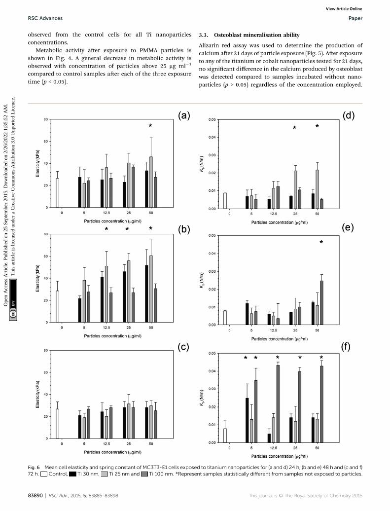
Fig. 6 Mean cell elasticity and spring constant of MC3T3-E1 cells expose72 h. Control, Ti 30 nm, Ti 25 nm and Ti 100 nm. *Represe
83890 | RSC Adv., 2015, 5, 83885–83898
3.3. Osteoblast mineralisation ability
Alizarin red assay was used to determine the production ofcalcium aer 21 days of particle exposure (Fig. 5). Aer exposureto any of the titanium or cobalt nanoparticles tested for 21 days,no signicant difference in the calcium produced by osteoblastwas detected compared to samples incubated without nano-particles (p > 0.05) regardless of the concentration employed.
d to titanium nanoparticles for (a and d) 24 h, (b and e) 48 h and (c and f)nt samples statistically different from samples not exposed to particles.
This journal is © The Royal Society of Chemistry 2015

Paper RSC Advances
Ope
n A
cces
s A
rtic
le. P
ublis
hed
on 2
5 Se
ptem
ber
2015
. Dow
nloa
ded
on 2
/26/
2022
1:3
5:52
AM
. T
his
artic
le is
lice
nsed
und
er a
Cre
ativ
e C
omm
ons
Attr
ibut
ion
3.0
Unp
orte
d L
icen
ce.
View Article Online
For PMMA wear debris, no difference compared to control wasdetermined in calcium production for concentrations up to25 mg ml�1 (p > 0.05), above this concentration an increase wasrecorded (p < 0.05) and statistical difference was found amongthe concentrations tested above 25 mg ml�1 (p > 0.05).
3.4. Nanomechanical properties
Aer 24 hours, cells exposed to titanium (Fig. 6) had the sameelasticity as control samples i.e. cells not exposed to any
Fig. 7 Mean cell elasticity and spring constant of MC3T3-E1 cells exposedh. Control, Co 30 nm and Co 50 nm. *Represent samples sta
This journal is © The Royal Society of Chemistry 2015
particles (p > 0.05) apart for the highest concentration of Ti25 nm. The elasticity of control cells remained constant over the3 day period with an elastic value of around 28 kPa (p > 0.05).Over time, at 48 hours, an increase in elasticity is clearlydemonstrated for Ti 25 nm, however no change was seen withthe largest particles of 100 nm and for Ti elemental (30 nmparticles) (p > 0.05). For Ti 25 nm a general increasewith increasing concentration is observed from 5 mg ml�1 to12.5 mg ml�1 (p < 0.05), aer this no effect of nanoparticles
to cobalt nanoparticles for (a and d) 24 h, (b and e) 48 h and (c and f) 72tistically different from samples not exposed to particles.
RSC Adv., 2015, 5, 83885–83898 | 83891

RSC Advances Paper
Ope
n A
cces
s A
rtic
le. P
ublis
hed
on 2
5 Se
ptem
ber
2015
. Dow
nloa
ded
on 2
/26/
2022
1:3
5:52
AM
. T
his
artic
le is
lice
nsed
und
er a
Cre
ativ
e C
omm
ons
Attr
ibut
ion
3.0
Unp
orte
d L
icen
ce.
View Article Online
concentration was detected (p > 0.05). Aer 72 hours of exposureno effect on cell elasticity was detected for any nanoparticles atany concentration (p > 0.05).
Elasticity results for cobalt nanoparticles are given in Fig. 7.Over all three time points: 24, 48, and 72 hours, the elasticity ofosteoblast cells did not change compared to control samplesregardless of the nanoparticles type and concentration (p >0.05).
The inuence of PMMA bone cement debris on osteoblastnanomechanical properties is shown in Fig. 8; aer 24 hoursexposure the elasticity of cell was reduced to about 14 kPa forconcentrations greater than 20 mg ml�1; no statistical differ-ences were determined for concentrations of PMMA debris inthe range 25 to 200 mgml�1 (p > 0.05), this was not detected aer48 hours, but with increasing exposure time (72 hours) thetrend was the same as aer 24 hours.
For titanium particles the spring constant values are given inFig. 6. Similarly to the elasticity values aer exposure to tita-nium, the spring constant values also did not increase withincreasing concentration. Again the control values of springconstant were consistent over time at around 0.01 N m�1 (p >0.05). Aer exposure to Ti 30 nm and Ti 100 nm, for 24 hours,the spring constant of osteoblast cells did not change with
Fig. 8 Mean cell elasticity (a), spring constant (b) of MC3T3-E1 cellsexposed to PMMA particles for ( ) 24 h, ( ) 48 h and ( ) 72 h.
83892 | RSC Adv., 2015, 5, 83885–83898
increasing concentration with a value of around 0.08 given foreach concentration (p > 0.05); Ti 25 nm however, had the sameinitial value as Ti elemental at 0.08 N m�1 at 5 mg ml�1, thisincreased to 0.012 N m�1 at 12.5 mg ml�1, a large jump in thespring constant was observed at 25 mg ml�1 with a value of 0.022N m�1 which slightly increased to 0.024 N m�1 at 50 mg ml�1.
Aer 48 hours of exposure to the titanium nanoparticles, thespring constant values were not different from the controls forall samples but the highest concentration of Ti 100 nm (p <0.05).
Aer 72 hours of exposure, the largest titanium particles,100 nm, had a dramatic increase in the spring constant even atthe lowest concentration of 5 mg ml�1, no differences withnanoparticles concentration were detected (p > 0.05). In Fig. 7,the spring constant of osteoblast cells is shown for all timepoints for both cobalt nanoparticles treatments. Aer 24 hoursCo 30 nm did not affect the spring constant for concentrationssmaller than 12.5 mg ml�1; the spring constant then dramati-cally increase for the higher concentrations 25 mg ml�1 and 50mg ml�1 at around 0.015 and 0.017 N m�1. For the largerparticles of Co 50 nm an increase in spring constant wasrecorded compared to the control for all the concentrationstested (p < 0.05) without any difference caused by the concen-tration value (p > 0.05).
Aer 48 hours and 72 hours, the spring constant valuesincreased with exposure time. No difference was recordedbetween types of Co nanoparticles and as result of theconcentration (p > 0.05).
The spring constant of osteoblast cells exposed to PMMAdebris is presented in Fig. 8 and revealed that only concentra-tions higher than 100 mg ml�1 for 72 hours were able to asignicant increase of such parameter.
3.5. Metal uptake
Uptake of nanoparticles increased with increasing concentra-tion (Fig. 9); an increase in exposure time also increased theuptake of nanoparticles. Greater uptake was noted for Co 50 nmcompared to Co 30 nm by almost three fold for the higherconcentration of 50 mg ml�1 aer 72 hours of exposure.
For titanium nanoparticles, overall uptake was far less thanfor cobalt nanoparticles, and very little to no uptake wasrecorded aer the initial 24 and 48 hours of exposure for Ti25 nm at the lower concentrations, with only a small uptake at50 mg ml�1. For Ti 30 nm, uptake was observed at all concen-trations, this increased similarly to cobalt nanoparticles withincreasing concentration and increasing time. Yet, for Ti100 nm, only aer 72 hours was any uptake recorded but stillfollowed the same pattern as other particles i.e. increaseduptake with increasing concentration.
PMMA uptake was measured using concentration of barium,as barium is the only metal in the PMMA bone cementcomposition. No uptake was recorded at any time point for thelowest concentration of 5 mg ml�1. Although, uptake increasedwith increasing concentration with increasing time pointssimilarly to both cobalt and titanium nanoparticles.
This journal is © The Royal Society of Chemistry 2015

Fig. 9 Metal uptake of MC3T3-E1 cells exposed to nanoparticles at different concentrations for 24 h, 48 h and 72 h. (a) Ti 25 nm,(b) Ti 30 nm, (c) Ti 100 nm, (d) Co 30 nm, (e) Co 50 nm and (f) PMMA.
Paper RSC Advances
Ope
n A
cces
s A
rtic
le. P
ublis
hed
on 2
5 Se
ptem
ber
2015
. Dow
nloa
ded
on 2
/26/
2022
1:3
5:52
AM
. T
his
artic
le is
lice
nsed
und
er a
Cre
ativ
e C
omm
ons
Attr
ibut
ion
3.0
Unp
orte
d L
icen
ce.
View Article Online
3.6. Cell adhesion forces
Both treated and non-treated cells i.e. not exposed to particlesdemonstrated spatial distributions of adhesion forces on thecell surface and were not normally distributed Fig. 10 and 11,for titanium and cobalt nanoparticles, respectively. The controlsample median did not vary with increasing time with aconsistent value of around 2 nN (p > 0.05).
Adhesion forces for cobalt nanoparticles post exposure didnot differ from each other and there was no change in adhesionaer exposure to either cobalt nanoparticles with increasingconcentration (p > 0.05).
This journal is © The Royal Society of Chemistry 2015
Again, little to no difference in adhesion forces was observedfor titanium aer all exposure duration regardless of composi-tion and size of the particles (p > 0.05) (Fig. 10). For PMMA,(Fig. 12) a general decline in cell adhesion forces were observedfor cells exposed for 24 hours (p < 0.05). No effect of particlesconcentration was detected (p > 0.05).
4 Discussion
Total joint replacement (TJR) is ever increasing in a number ofcases1 yet the leading cause of failure remains the aseptic
RSC Adv., 2015, 5, 83885–83898 | 83893
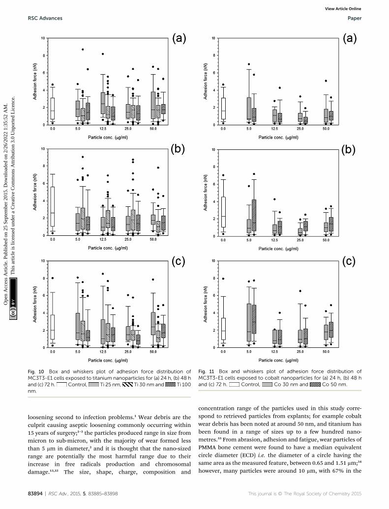
Fig. 10 Box and whiskers plot of adhesion force distribution ofMC3T3-E1 cells exposed to titanium nanoparticles for (a) 24 h, (b) 48 hand (c) 72 h. Control, Ti 25 nm, Ti 30 nm and Ti 100nm.
Fig. 11 Box and whiskers plot of adhesion force distribution ofMC3T3-E1 cells exposed to cobalt nanoparticles for (a) 24 h, (b) 48 hand (c) 72 h. Control, Co 30 nm and Co 50 nm.
RSC Advances Paper
Ope
n A
cces
s A
rtic
le. P
ublis
hed
on 2
5 Se
ptem
ber
2015
. Dow
nloa
ded
on 2
/26/
2022
1:3
5:52
AM
. T
his
artic
le is
lice
nsed
und
er a
Cre
ativ
e C
omm
ons
Attr
ibut
ion
3.0
Unp
orte
d L
icen
ce.
View Article Online
loosening second to infection problems.1 Wear debris are theculprit causing aseptic loosening commonly occurring within15 years of surgery;1–3 the particles produced range in size frommicron to sub-micron, with the majority of wear formed lessthan 5 mm in diameter,2 and it is thought that the nano-sizedrange are potentially the most harmful range due to theirincrease in free radicals production and chromosomaldamage.13,32 The size, shape, charge, composition and
83894 | RSC Adv., 2015, 5, 83885–83898
concentration range of the particles used in this study corre-spond to retrieved particles from explants; for example cobaltwear debris has been noted at around 50 nm, and titanium hasbeen found in a range of sizes up to a few hundred nano-metres.33 From abrasion, adhesion and fatigue, wear particles ofPMMA bone cement were found to have a median equivalentcircle diameter (ECD) i.e. the diameter of a circle having thesame area as the measured feature, between 0.65 and 1.51 mm;34
however, many particles were around 10 mm, with 67% in the
This journal is © The Royal Society of Chemistry 2015

Fig. 12 Box and whiskers plot of adhesion force distribution ofMC3T3-E1 cells exposed to PMMA nanoparticles for ( ) 24 h, ( )48 h and ( ) 72 h.
Paper RSC Advances
Ope
n A
cces
s A
rtic
le. P
ublis
hed
on 2
5 Se
ptem
ber
2015
. Dow
nloa
ded
on 2
/26/
2022
1:3
5:52
AM
. T
his
artic
le is
lice
nsed
und
er a
Cre
ativ
e C
omm
ons
Attr
ibut
ion
3.0
Unp
orte
d L
icen
ce.
View Article Online
submicron range. In order to differentiate between the effect ofboth size and composition (element or oxide) of the metalnanoparticles we employed commercial nanoparticles ofappropriate characteristics.
Generally, the rst stages of an adverse effect is the phago-cytosis of the particles by cells; this has been visually demon-strated using polystyrene-based uorescent particles, such asuoresbrite, that were internalised within 24 hours of in vitroexposure, with cells being saturated with around 40–60 particlesper cell.37 These results are in agreement with our ndings thatreveal changes in the cell properties aer 24 hours of exposureto nanoparticles.
Many techniques21,38–41 have been used to measure themechanical properties of single cells which includes micropi-pette aspiration, cytoindentation, magnetic bead rheometry,optical traps and AFM.38 For mesenchymal stem cells, Darlinget al. (2008),38 tested whether elastic and viscoelastic propertiescould indicate the cells phenotype, using AFM with a borosili-cate functionalised tip at a spring constant of around0.04 N m�1 similar to that we used in our study. Likewise,Bhadriraju and Hansen (2002)42 used AFM tips with springconstant of 0.06 N m�1 to investigate the stiffness andspreading changes of cells using AFM.42 In both these works,the elastic modulus was modelled using Hertz model ofcontact,38,42 as employed for our investigation. Interestingly,Darling et al. (2008)38 also found the elastic data to have a notnormal distribution and that the osteoblasts demonstrated thelargest elastic moduli and no difference in lineage was notedfrom the viscoelastic properties.
Cell elasticity is closely linked to the cytoskeleton structure,39
and in this study it was observed that titanium resulted in anincrease in elasticity suggesting interactions with the networkof stress bres as explained by Thoumine et al. (1999),39 anddisruptions to the membrane integrins of cells, formingcomplex disturbances to the original form which contributes tothe cells rigidity.39,44 This could explain the changes observed in
This journal is © The Royal Society of Chemistry 2015
the elasticity modulus of the osteoblast cells, aer PMMAparticles exposure; there was a general increase in the elasticmodulus with time demonstrating that cells became stiffer. It isexplained45 that disruptions to the cytoskeleton organisationimpacts on the expression of transcription factors andosteoblast-specic genes in osteoblasts,45 as well as inuencingcell behaviour. Similar values in stiffness were observed42 whenchanges in the actin and myosin activity were investigated inrelation to the changes in shape of the cell at around 20 kPa.42
The changes in stiffness are due to the adaptation of the cells tothe stress stimuli introduced by the environment when the cellsare exposed to the metal nanoparticles.46 Cells respond to anumber of stress factors such as uid shear stress, strainstimuli and vibration stress; such mechanosensing allows cellsto detect adhesion of metal nanoparticles to the outermembrane of the cells.46 It had also been suggested that thecytoskeleton is the key aspect responsible for sensing thecellular mechanical changes.46,47
It is thought that the cytoskeleton of a cell, especially thelamentous actin determines the cells ability to maintain astable shape, maintaining the function of the cell.24 It wasproposed that the interactions between these actin-lamentbundles of the osteoblasts cells within the cytoskeleton couldcause a loss of adhesive function and, therefore, changes to theadhesion characteristics of the cell could potentially alter thecell's function.24 A further explanation for the changes inadhesion properties results from the decrease in cell cyto-plasmic spreading and attachment area, stemming from theparticles integrating to the actin laments forming densernetworks within the structure.48 The addition of titaniumparticles to the cell stress bres reduces the spreading of thecell, this reduction in spreading area of the cell meansdecreased contact, thereby reducing the overall adhesion. Thesechanges could explain the pathogenesis of peri-prostheticosteolysis secondary to implant wear debris, with Kwon et al.(2001) showing a decrease in adhesion of around 40–60% inspreading attachment.44
Some of the above studies11,24,35,36,37,40,41,43,44,48 have demon-strated that orthopaedic wear debris affects the viability of cells,including their proliferation, differentiation, and function. Yet,many of these studies do not coincide, featuring manydiscrepancies such as the choice of cell, cell population,culturing conditions, not to mention the wear debris composi-tions variances such as the size, composition, dose and aggre-gation. However, the studies agree that particles and debris ofless than 5 mm tend to interact with cells and undergo uptakevia phagocytosis especially in regards to human, rat, and mousebone cells. Even though results have been inconsistent for theviability assays, the biological effects are dose-dependent, andthe majority of results demonstrate that the higher theconcentration or dose the more adverse the effects. Non-toxicparticles activate osteoblasts from the up-regulation of pro-inammatory and bone-resorbing factors, with the down-regulation of bone forming variables. Interestingly, debrisfrom alumina and polystyrene origins are less harmful incomparison to metals and polymers commonly used in ortho-paedic devices.6,49–51 Our results generally do not indicate a
RSC Adv., 2015, 5, 83885–83898 | 83895

RSC Advances Paper
Ope
n A
cces
s A
rtic
le. P
ublis
hed
on 2
5 Se
ptem
ber
2015
. Dow
nloa
ded
on 2
/26/
2022
1:3
5:52
AM
. T
his
artic
le is
lice
nsed
und
er a
Cre
ativ
e C
omm
ons
Attr
ibut
ion
3.0
Unp
orte
d L
icen
ce.
View Article Online
concentration depending effect on cells properties or a spatialvariation on the cell surface indicating that these nanoparticles– cells interactions are initiated at low nanoparticles concen-tration, possibly the lowest concentration employed in ourstudy is already enough to saturate all the possible interactionsor alternatively the responses are due to biological signallinginvolved in the cell detecting the presence of the particles.
From a mechanistic stand point, the presence of foreignparticles in direct contact with cells causes an uptake of theseparticles. The complete mechanism for damage from thisuptake is still unknown; however, once phagocytosis is observedit is unclear whether the physical contact or the uptake or thepresence of these particles initiates a cellular response viamembrane mediated association.
Our results also indicate that cells response is not completelylinked to metal uptake as the concentration of ions inside thecell monotonically increased with exposure time whilst thenanomechanical changes were observed, particularly for thespring constant, already aer the rst 24 hours of contact.
Spring constant is linked to cell turgid pressure, therefore,its increase with exposure time found in our work could belinked to the metal uptake as a mechanism to prevent furtherion accumulation.
As literature demonstrates that the bone adjacent to animplanted TJA has a substantial bone resorption surface.52 Ithas been assumed that wear debris contact with bone-formingcells located around loosening implants. If this contact causesan inhibition or suppression of the function of these bone-forming cells, for example damage to the cells wall, disrup-tions to the normal viability, proliferation, and differentiation,could lead to further loosening of the implant due to a decreasein bone renewal and formation.
It is has been reported that osteoblasts exposed to PMMAparticles signicantly increased their production of calcium,53,54
this was also true for our study. Moreover, both titaniumelemental and cobalt particles did not increase the calciumformation with increasing concentration.
5 Conclusion
This study aimed at observing the inuence of wear particles onbone cells through an alternative perspective (cells nano-mechanical properties) to the usual biological responsespreviously investigated. Titanium increased the elasticity morethan cobalt nanoparticles even though titanium demonstratedless cytotoxicity; the smaller nanoparticles had a greater impacton the viability of the cells as well as on the adhesion forcesexhibited by the exposed cells. These mechanical changes arethe result of alterations to the cytoskeleton as reported inliterature. Also, the smaller nanoparticles had a higher uptakewhich may be due to phagocytosis for such smaller particles.
The results lend themselves to a novel idea of the importanceof understanding the mechanical changes and their impact onnormal cell function which have previously been under-estimated. These results suggest that physical stimulus canalter the normal function of a cell through potential changes inthe cytoskeleton of the cell in a similar manner to that of
83896 | RSC Adv., 2015, 5, 83885–83898
biological responses; therefore this study points out theimportance of an holistically cell analysis i.e. not only from abiological stand point but also through mechanicalmechanisms.
Acknowledgements
The authors would like to acknowledge Arthritis Research UK(ARUK: 18461) and School of Pharmacy, Cardiff University forfunding a PhD scholarship.
References
1 V. Sansone, D. Pagani and M. Melato, The effects on bonecells of metal ions released from orthopaedic implants, areview, Clinical Cases in Mineral and Bone Metabolism, 2013,10(1), 34–40.
2 Y. Abu-Amer, I. Darwech and J. C. Clohisy, Aseptic looseningof total joint replacements: mechanisms underlyingosteolysis and potential therapies, Arthritis Res. Ther., 2007,9(suppl. 1), S6.
3 C. Kowandy, H. Mazouz and C. Richard, Isolation andanalysis of articular joints wear debris generated in vitro,Wear, 2006, 261(9), 966–970.
4 P. Prokopovich, S. Perni, R. M. Hall and J. Fisher, Spatialvariation of wear on Charite lumbar discs, Acta Biomater.,2011, 7(11), 3914–3926.
5 K. Ren, A. Dusad, Y. Zhang and D. Wang, Therapeuticintervention for wear debris-induced aseptic implantloosening, Acta Pharm. Sin. B, 2013, 3(2), 76–85.
6 S. B. Goodman, P. Huie, Y. Song, D. Schurman, W. Maloney,S. Woolson and R. Sibley, Cellular prole and cytokineproduction at prosthetic interfaces. Study of tissuesretrieved from revised hip and knee replacements, J. BoneJt. Surg., Br. Vol., 1998, 80(3), 531–539.
7 H. W. Fang, C. B. Yang, C. H. Chang, C. H. Huang, H. L. Liuand S. B. Fang, The potential role of phagocytic capacity inthe osteolytic process induced by polyethylene wearparticles, J. Int. Med. Res., 2006, 34(6), 655–664.
8 C. Vermes, T. T. Glant, N. J. Hallab, E. A. Fritz, K. A. Roebuckand J. J. Jacobs, The potential role of the osteoblast in thedevelopment of periprosthetic osteolysis: review of in vitroosteoblast responses to wear debris, corrosion products,and cytokines and growth factors, J. Arthroplasty, 2001,16(suppl. 1 and 8), 95–100.
9 C. H. Lohmann, Z. Schwartz, G. Koster, U. Jahn,G. H. Buchhorn, M. J. MacDougall, D. Casasola, Y. Liu,V. L. Sylvia, D. D. Dean and B. D. Boyan, Phagocytosis ofwear debris by osteoblasts affects differentiation and localfactor production in a manner dependent on particlecomposition, Biomaterials, 2000, 21(6), 551–561.
10 M. Bahraminasab, B. B. Sahari, K. L. Edwards,F. Farahmand, M. Arumugam and T. S. Hong, Asepticloosening of femoral components – a review of current andfuture trends in materials used, Mater. Des., 2012, 42(0),459–470.
This journal is © The Royal Society of Chemistry 2015

Paper RSC Advances
Ope
n A
cces
s A
rtic
le. P
ublis
hed
on 2
5 Se
ptem
ber
2015
. Dow
nloa
ded
on 2
/26/
2022
1:3
5:52
AM
. T
his
artic
le is
lice
nsed
und
er a
Cre
ativ
e C
omm
ons
Attr
ibut
ion
3.0
Unp
orte
d L
icen
ce.
View Article Online
11 B. Behl, I. Papageorgiou, C. Brown, R. Hall, J. L. Tipper,J. Fisher and E. Ingham, Biological effects of cobalt-chromium nanoparticles and ions on dural broblasts anddural epithelial cells, Biomaterials, 2013, 34(14), 3547–3558.
12 R. Chiu, T. Ma, R. L. Smith and S. B. Goodman, Ultrahighmolecular weight polyethylene wear debris inhibitsosteoprogenitor proliferation and differentiation in vitro,J. Biomed. Mater. Res., Part A, 2009, 89(1), 242–247.
13 R. Chiu, T. Ma, R. L. Smith and S. B. Goodman,Polymethylmethacrylate particles inhibit osteoblasticdifferentiation of MC3T3-E1 osteoprogenitor cells,J. Orthop. Res., 2008, 26(7), 932–936.
14 S.-Y. Tee, A. R. Bausch and P. A. Janmey, The mechanicalcell, Curr. Biol., 2009, 19(17), R745–R748.
15 S.-Y. Tee, J. Fu, C. S. Chen and P. A. Janmey, Cell Shape andSubstrate Rigidity Both Regulate Cell Stiffness, Biophys. J.,2011, 100(5), L25–L27.
16 C. T. Lim, E. H. Zhou and S. T. Quek, Mechanical models forliving cells—a review, J. Biomech., 2006, 39(2), 195–216.
17 C. Schmidt, A. A. Ignatius and L. E. Claes, Proliferation anddifferentiation parameters of human osteoblasts ontitanium and steel surfaces, J. Biomed. Mater. Res., 2001,54(2), 209–215.
18 J. Helenius, C. P. Heisenberg, H. E. Gaub and D. J. Muller,Single-cell force spectroscopy, J. Cell Sci., 2008, 121(11),1785–1791.
19 X. Cai, X. Xing, J. Cai, Q. Chen, S. Wu and F. Huang,Connection between biomechanics and cytoskeletonstructure of lymphocyte and Jurkat cells: an AFM study,Micron, 2010, 41(3), 257–262.
20 R. Dattani, Femoral osteolysis following total hipreplacement, Postgrad. Med. J., 2007, 83(979), 312–316.
21 K. Anselme, Osteoblast adhesion on biomaterials,Biomaterials, 2000, 21(7), 667–681.
22 K. Anselme, P. Davidson, A. M. Popa, M. Giazzon, M. Lileyand L. Ploux, The interaction of cells and bacteria withsurfaces structured at the nanometre scale, Acta Biomater.,2010, 6(10), 3824–3846.
23 A. Hunter, C. W. Archer, P. S. Walker and G. W. Blunn,Attachment and proliferation of osteoblasts and broblastson biomaterials for orthopaedic use, Biomaterials, 1995,16(4), 287–295.
24 K. E. Kasza, F. Nakamura, S. Hu, P. Kollmannsberger,N. Bonakdar, B. Fabry, T. P. Stossel, N. Wang andD. A. Weitz, Filamin A is essential for active cell stiffeningbut not passive stiffening under external force, Biophys. J.,2009, 96(10), 4326–4335.
25 S. Perni, M. G. Kong and P. Prokopovich, Cold atmosphericpressure gas plasma enhances the wear performance ofultra-high molecular weight polyethylene, Acta Biomater.,2012, 8(3), 1357–1365.
26 P. Prokopovich, R. Leech, C. J. Carmalt, I. P. Parkin andS. Perni, A novel bone cement impregnated with silver-tiopronin nanoparticles: its antimicrobial, cytotoxic, andmechanical properties, Int. J. Nanomed., 2013, 8, 2227–2237.
This journal is © The Royal Society of Chemistry 2015
27 P. Prokopovich, M. Kobrick, E. Brousseau and S. Perni,Potent antimicrobial activity of bone cement encapsulatingsilver nanoparticles capped with oleic acid, J. Biomed.Mater. Res., Part B, 2015, 103(2), 273–281.
28 C. A. Gregory, W. G. Gunn, A. Peister and D. J. Prockop, AnAlizarin red-based assay of mineralization by adherentcells in culture: comparison with cetylpyridinium chlorideextraction, Anal. Biochem., 2004, 329(1), 77–84.
29 E. C. Preedy, E. Brousseau, S. Evans, S. Perni andP. Prokopovich, Adhesive forces and surface properties ofcold gas plasma treated UHMWPE, Colloids Surf., A, 2014,460, 83–89.
30 J. E. Sader, J. A. Sanelli, B. D. Adamson, J. P. Monty, X. Wei,S. A. Crawford, J. R. Friend, I. van Marusic, P. Mulvaney andE. J. Bieske, Spring constant calibration of atomic forcemicroscope cantilevers of arbitrary shape, Rev. Sci.Instrum., 2012, 83(10), 103705.
31 J. E. Sader, I. Larson, P. Mulvaney and R. L. White, Methodfor the calibration of atomic force microscope cantilevers,Rev. Sci. Instrum., 1995, 66(7), 3789–3798.
32 E. Ingham and J. Fisher, Biological reactions to wear debrisin total joint replacement, Proc. Inst. Mech. Eng., Part H,2000, 214(1), 21–37.
33 P. Prokopovich, Interactions between mammalian cells andnano- or micro-sized wear particles: physico-chemical viewsagainst biological approaches, Adv. Colloid Interface Sci.,2014, 213, 36–47.
34 J. A. Wimhurst, R. A. Brooks and N. Rushton, The effects ofparticulate bone cements at the bone-implant interface,J. Bone Jt. Surg., Br. Vol., 2001, 83, 588–592.
35 E. L. S. da Rosa, Kinetic effects of TiO(2) ne particles andnanoparticles aggregates on the nanomechanicalproperties of human neutrophils assessed by forcespectroscopy, BMC Biophys., 2013, 6, 11.
36 V. H. Grassian, A. Adamcakova-Dodd, J. M. Pettibone,P. I. O'shaughnessy and P. S. Thorne, Inammatoryresponse of mice to manufactured titanium dioxidenanoparticles: comparison of size effects through differentexposure routes, Nanotoxicology, 2007, 1(3), 211–226.
37 R. Chiu and S. B. Goodman, Biological Response ofOsteoblasts and Osteoprogenitors to Orthopaedic WearDebris, in Osteogenesis - Biochemistry, Genetics andMolecular Biology, ed. L. Yunfeng, 2012, CC BY.
38 E. M. Darling, M. Topel, S. Zauscher, T. P. Vail and F. Guilak,Viscoelastic properties of human mesenchymally-derivedstem cells and primary osteoblasts, chondrocytes, andadipocytes, J. Biomech., 2008, 41(2), 454–464.
39 O. Thoumine, O. Cardoso and J. J. Meister, Changes in themechanical properties of broblasts during spreading: amicromanipulation study, Eur. Biophys. J., 1999, 28(3), 222–234.
40 J. Domke, S. Dannohl, W. J. Parak, O. Muller, W. K. Aicherand M. Radmacher, Substrate dependent differences inmorphology and elasticity of living osteoblasts investigatedby atomic force microscopy, Colloids Surf., B, 2000, 19(4),367–379.
RSC Adv., 2015, 5, 83885–83898 | 83897

RSC Advances Paper
Ope
n A
cces
s A
rtic
le. P
ublis
hed
on 2
5 Se
ptem
ber
2015
. Dow
nloa
ded
on 2
/26/
2022
1:3
5:52
AM
. T
his
artic
le is
lice
nsed
und
er a
Cre
ativ
e C
omm
ons
Attr
ibut
ion
3.0
Unp
orte
d L
icen
ce.
View Article Online
41 D. Docheva, D. Padula, C. Popov, W. Mutschler, H. Clausen-Schaumann and M. Schieker, Researching into the cellularshape, volume and elasticity of mesenchymal stem cells,osteoblasts and osteosarcoma cells by atomic forcemicroscopy, J. Cell. Mol. Med., 2008, 12(2), 537–552.
42 K. Bhadriraju and L. K. Hansen, Extracellular Matrix- andCytoskeleton-Dependent Changes in Cell Shape andStiffness, Exp. Cell Res., 2002, 278(1), 92–100.
43 A. G. Moutzouri and G. M. Athanassiou, Insights into theAlteration of Osteoblast Mechanical Properties uponAdhesion on Chitosan, BioMed Res. Int., 2014, 2014, 8.
44 S. Y. Kwon, T. Lin, H. Takei, Q. Ma, D. J. Wood, D. O'Connorand K. L. Sung, Alterations in the adhesion behavior ofosteoblasts by titanium particle loading: inhibition of cellfunction and gene expression, Biorheology, 2001, 38(2–3),161–183.
45 R. D. A. M. Alves, Osteoblast Differentiation and Bone: Relevantproteins, regulatory processes and the vascular connection, inDepartment of Internal Medicine, 2012, Erasmus UniversityRotterdam, Ipskamp Drukkers, p. 166.
46 R. G. Bacabac, D. Mizuno, C. F. Schmidt, F. C. MacKintosh,J. J. van Loon, J. Klein-Nulend and T. H. Smit, Round versusat: bone cell morphology, elasticity, and mechanosensing,J. Biomech., 2008, 41(7), 1590–1598.
47 R. K. Assoian and E. A. Klein, Growth control by intracellulartension and extracellular stiffness, Trends Cell Biol., 2008,18(7), 347–352.
83898 | RSC Adv., 2015, 5, 83885–83898
48 S. Y. Kwon, H. Takei, D. P. Pioletti, T. Lin, Q. J. Ma,W. H. Akeson, D. J. Wood and K. L. Sung, Titaniumparticles inhibit osteoblast adhesion to bronectin-coatedsubstrates, J. Orthop. Res., 2000, 18(2), 203–211.
49 S. B. Goodman, T. Ma, R. Chiu, R. Ramachandran andR. L. Smith, Effects of orthopaedic wear particles onosteoprogenitor cells, Biomaterials, 2006, 27(36), 6096–6101.
50 S. B. Goodman and T. Ma, Cellular chemotaxis induced bywear particles from joint replacements, Biomaterials, 2010,31(19), 5045–5050.
51 G. M. Keegan, I. D. Learmonth and C. P. Case, Orthopaedicmetals and their potential toxicity in the arthroplastypatient: a review of current knowledge and futurestrategies, J. Bone Jt. Surg., Br. Vol., 2007, 89(5), 567–573.
52 Y. Kadoya, P. A. Revell, N. al-Saffar, A. Kobayashi, G. Scottand M. A. Freeman, Bone formation and bone resorptionin failed total joint arthroplasties: histomorphometricanalysis with histochemical and immunohistochemicaltechnique, J. Orthop. Res., 1996, 14(3), 473–482.
53 Anonymous, Bioactivity of Osteoblasts to Wear DebrisGenerated From Orthopedic Devices, 1999, available from:http://biomed.brown.edu/Courses/BI108/BI108_1999_Groups/THRdebris_Team/gordon.html.
54 C. H. Lohmann, D. D. Dean, G. Koster, D. Casasola,G. H. Buchhorn, U. Fink, Z. Schwartz and B. D. Boyan,Ceramic and PMMA particles differentially affectosteoblast phenotype, Biomaterials, 2002, 23(8), 1855–1863.
This journal is © The Royal Society of Chemistry 2015