cochlea and auditory pathways
TRANSCRIPT
88
Cochlea and Auditory PathwaysAnatomical Considerations
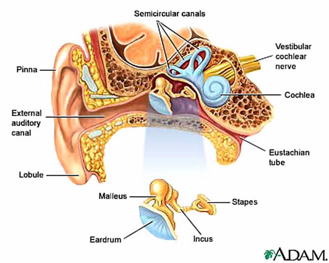
External ear: The external auditory meatus (canal) is formed by auricular and annular cartilages, plus a short contribution
from the temporal bone.
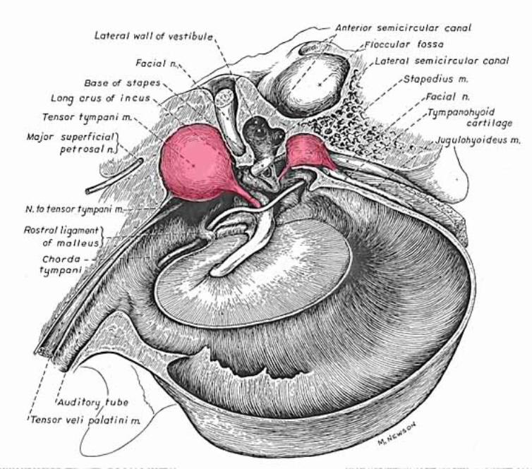
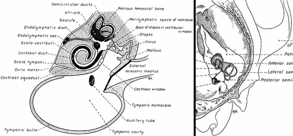
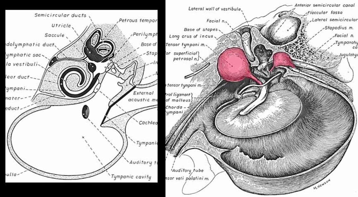
Middle ear: Air-filled tympanic cavity (including a ventrally expanded bulla) that features: — four openings (three sealed by membranes): • auditory tube opening (not sealed) connects middle ear to the nasopharynx; • tympanic membrane (ear drum) separates tympanic cavity from external auditory meatus; • oval (vestibular) window separates the tympanic cavity from perilymph in the vestibule; • round (cochlear) window separates tympanic cavity from perilymph in the scala tympani; — three ossicles, malleus, incus, and stapes, transmit tympanic membrane movements to the membrane of the oval window; — two muscles reflexly dampen ossicle movement, to suppress forceful low frequencies: • stapedius muscle, innervated by facial nerve, pulls the stapes away from oval window; • tensor tympani muscle, innervated by trigeminal nerve, pulls malleus thus tensing the tympanic membrane.
Function of Middle Ear To increases the efficiency of sound transmission. (Pressure oscillations in air (sound waves) are very inefficiently converted to pressure waves in fluid, only 0.1% of the force is normally
transmitted.) Middle ear components convert large amplitude, low force input into low amplitude, high force output. (The middle ear matches low impedance input to high impedance output.) Note: The tympanic membrane occupies a large area and undergoes a large excur-sion, offering low resistance to being vibrated (by air pressure waves). The membrane of the oval window has a small area and makes small excursions against high resistance load (it must push against incompressible perilymph fluid and the round window. The ossicles form a lever system that collects energy from the large tympanic membrane and focuses it on the small oval window membrane (yielding a 60-fold force gain in the cat).
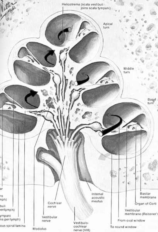
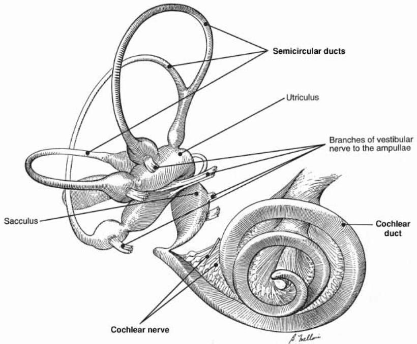
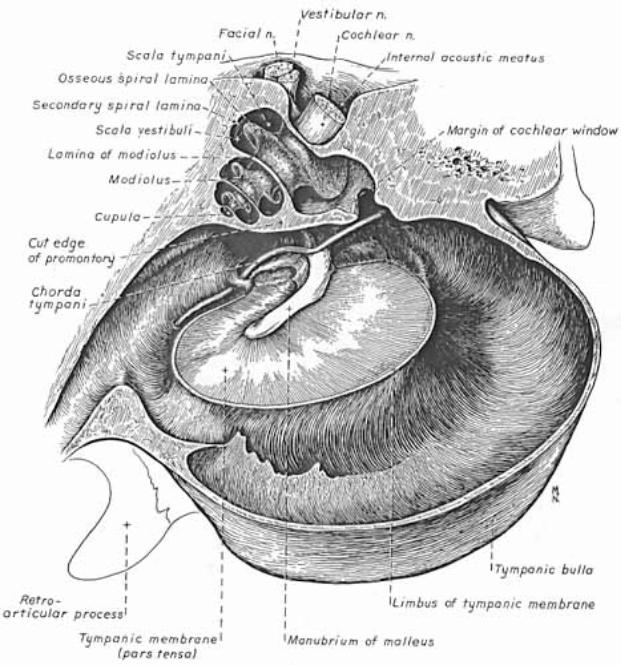
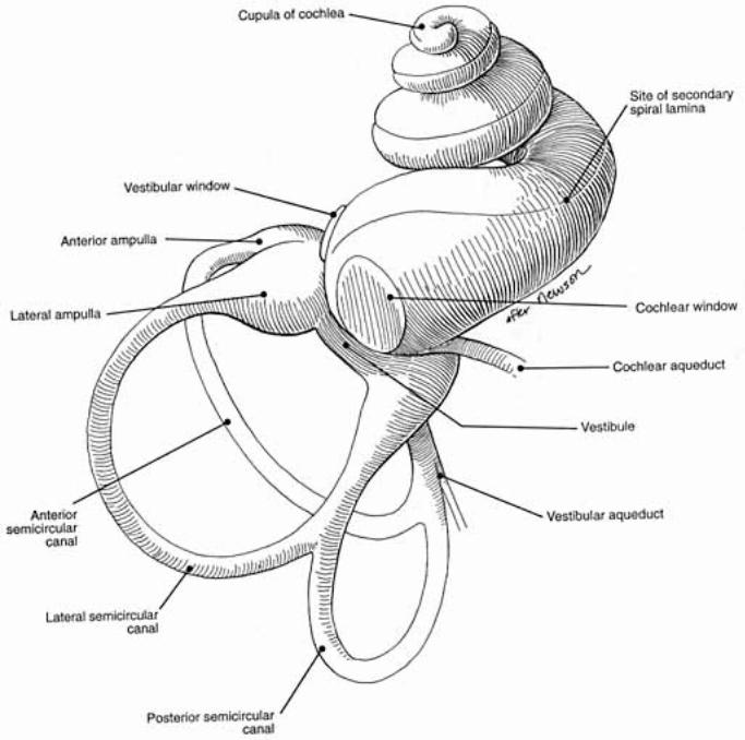
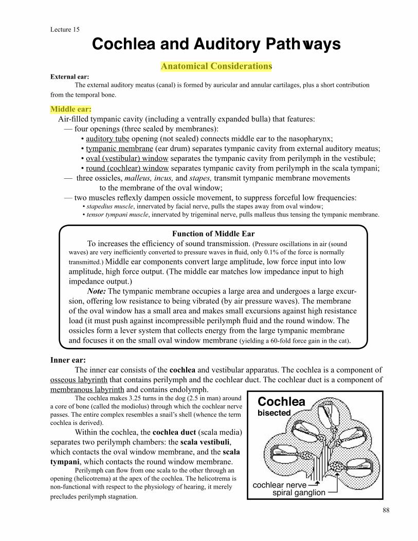
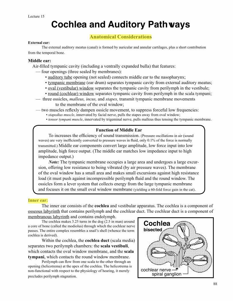
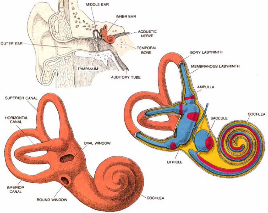
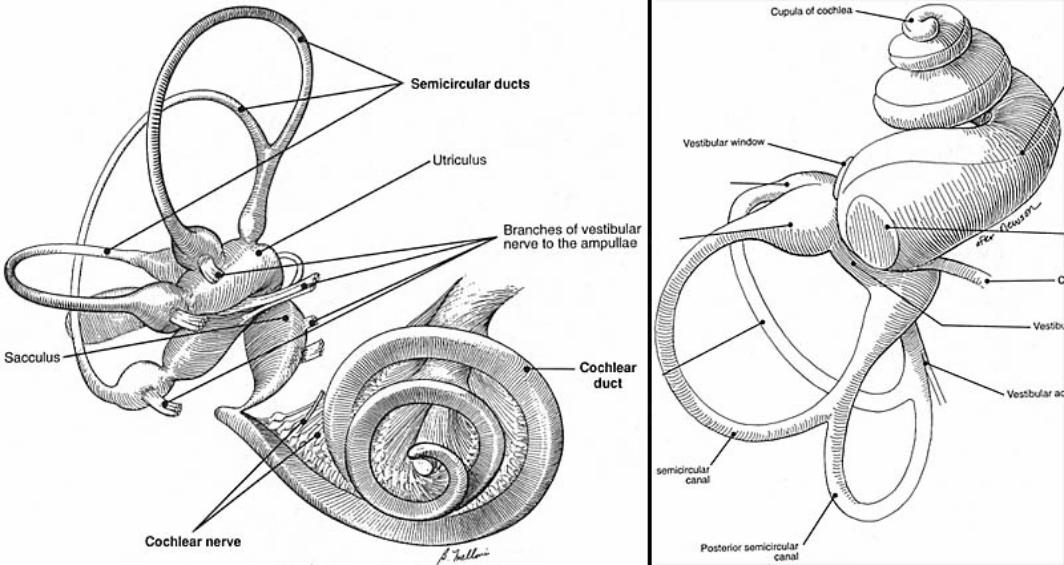
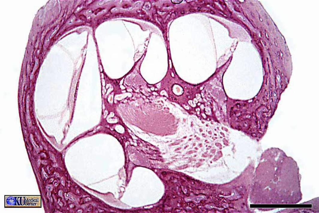
Inner ear: The inner ear consists of the cochlea and vestibular apparatus. The cochlea is a component of osseous labyrinth that contains perilymph and the cochlear duct. The cochlear duct is a component of membranous labyrinth and contains endolymph. The cochlea makes 3.25 turns in the dog (2.5 in man) around a core of bone (called the modiolus) through which the cochlear nerve passes. The entire complex resembles a snail’s shell (whence the term cochlea is derived).
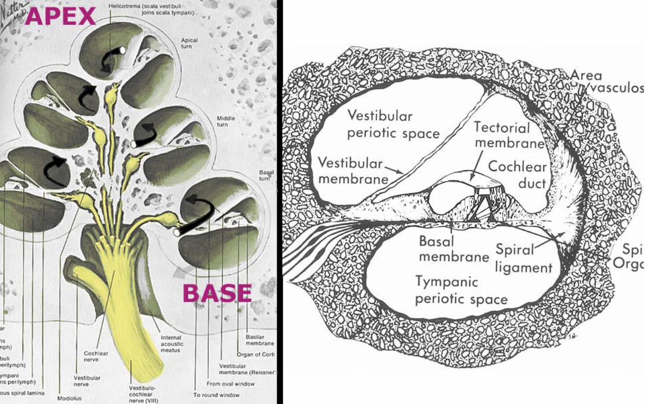
Within the cochlea, the cochlea duct (scala media) separates two perilymph chambers: the scala vestibuli, which contacts the oval window membrane, and the scala tympani, which contacts the round window membrane. Perilymph can flow from one scala to the other through an opening (helicotrema) at the apex of the cochlea. The helicotrema is non-functional with respect to the physiology of hearing, it merely
precludes perilymph stagnation.
Lecture 15
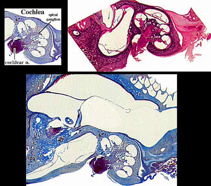
Cochleabisected
cochlear nervespiral ganglion
88
Cochlea and Auditory PathwaysAnatomical Considerations
External ear: The external auditory meatus (canal) is formed by auricular and annular cartilages, plus a short contribution
from the temporal bone.
Middle ear: Air-filled tympanic cavity (including a ventrally expanded bulla) that features: — four openings (three sealed by membranes): • auditory tube opening (not sealed) connects middle ear to the nasopharynx; • tympanic membrane (ear drum) separates tympanic cavity from external auditory meatus; • oval (vestibular) window separates the tympanic cavity from perilymph in the vestibule; • round (cochlear) window separates tympanic cavity from perilymph in the scala tympani; — three ossicles, malleus, incus, and stapes, transmit tympanic membrane movements to the membrane of the oval window; — two muscles reflexly dampen ossicle movement, to suppress forceful low frequencies: • stapedius muscle, innervated by facial nerve, pulls the stapes away from oval window; • tensor tympani muscle, innervated by trigeminal nerve, pulls malleus thus tensing the tympanic membrane.
Function of Middle Ear To increases the efficiency of sound transmission. (Pressure oscillations in air (sound waves) are very inefficiently converted to pressure waves in fluid, only 0.1% of the force is normally
transmitted.) Middle ear components convert large amplitude, low force input into low amplitude, high force output. (The middle ear matches low impedance input to high impedance output.) Note: The tympanic membrane occupies a large area and undergoes a large excur-sion, offering low resistance to being vibrated (by air pressure waves). The membrane of the oval window has a small area and makes small excursions against high resistance load (it must push against incompressible perilymph fluid and the round window. The ossicles form a lever system that collects energy from the large tympanic membrane and focuses it on the small oval window membrane (yielding a 60-fold force gain in the cat).
Inner ear: The inner ear consists of the cochlea and vestibular apparatus. The cochlea is a component of osseous labyrinth that contains perilymph and the cochlear duct. The cochlear duct is a component of membranous labyrinth and contains endolymph. The cochlea makes 3.25 turns in the dog (2.5 in man) around a core of bone (called the modiolus) through which the cochlear nerve passes. The entire complex resembles a snail’s shell (whence the term cochlea is derived).
Within the cochlea, the cochlea duct (scala media) separates two perilymph chambers: the scala vestibuli, which contacts the oval window membrane, and the scala tympani, which contacts the round window membrane. Perilymph can flow from one scala to the other through an opening (helicotrema) at the apex of the cochlea. The helicotrema is non-functional with respect to the physiology of hearing, it merely
precludes perilymph stagnation.
Lecture 15
Cochleabisected
cochlear nervespiral ganglion

89
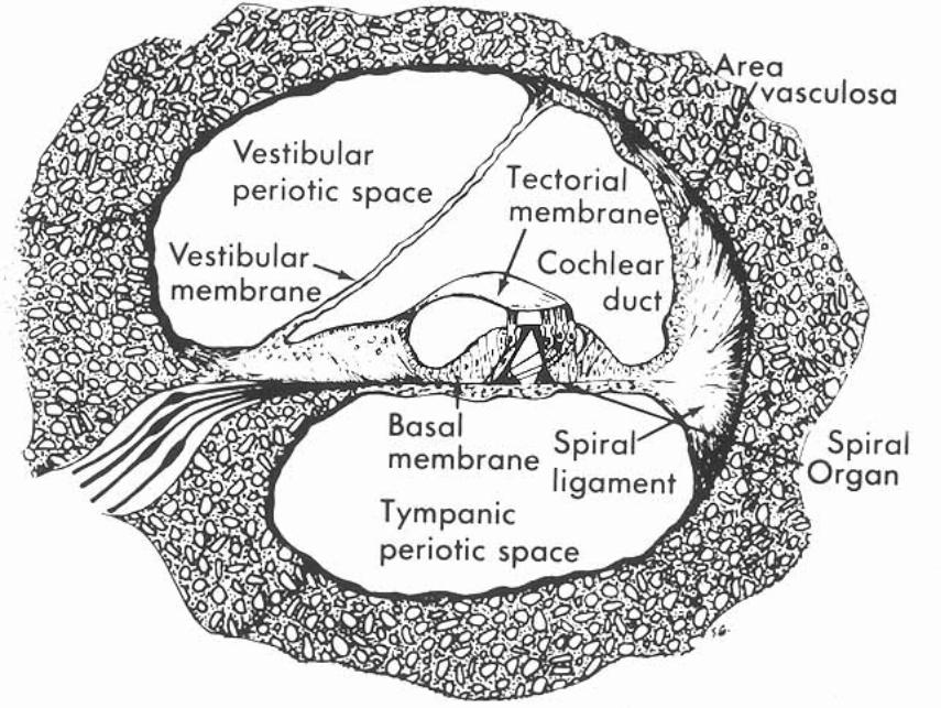
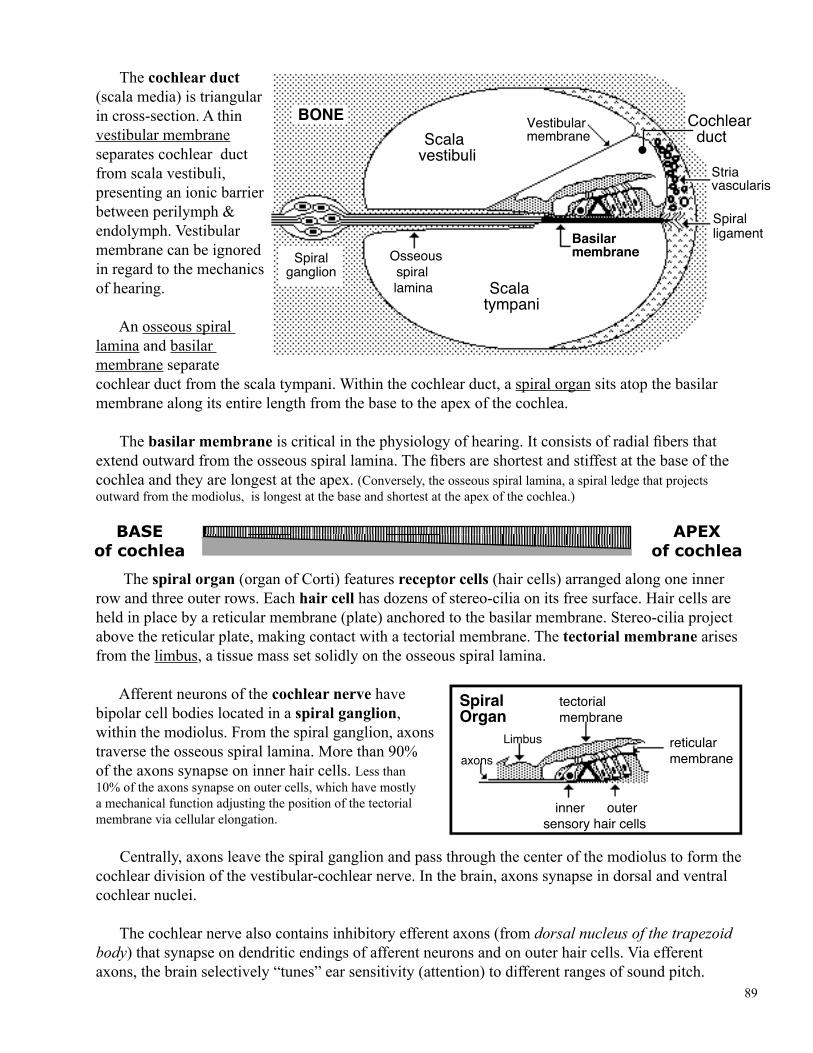
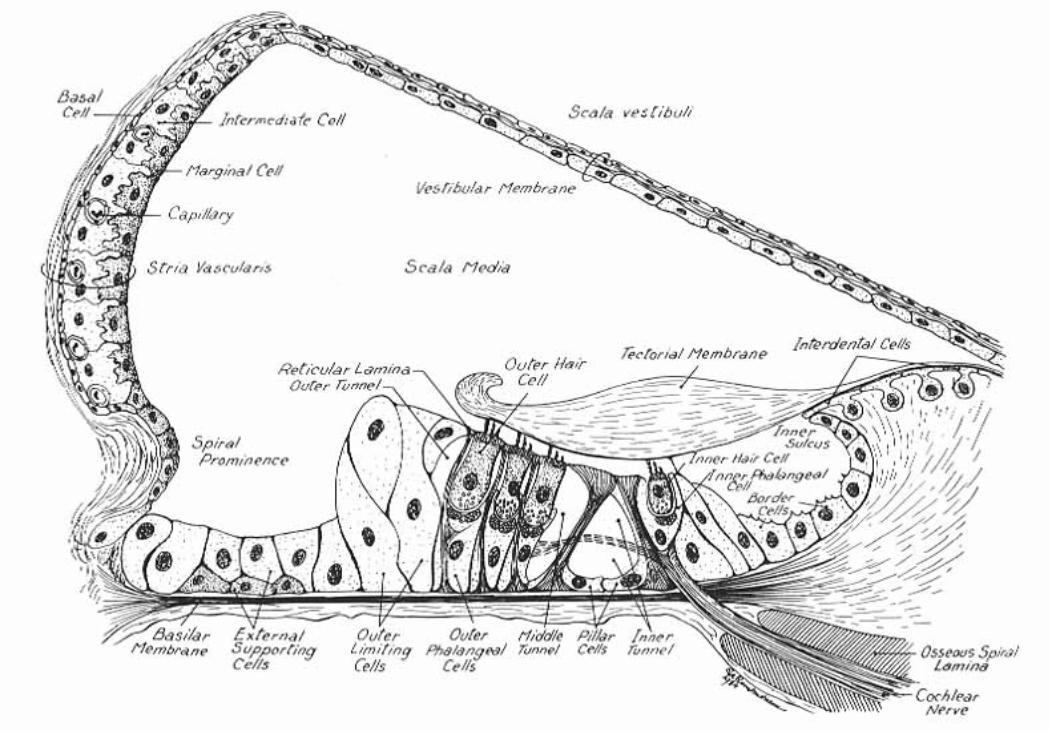
The cochlear duct (scala media) is triangular in cross-section. A thin vestibular membrane separates cochlear duct from scala vestibuli, presenting an ionic barrier between perilymph & endolymph. Vestibular membrane can be ignored in regard to the mechanics of hearing.
An osseous spiral lamina and basilar membrane separate cochlear duct from the scala tympani. Within the cochlear duct, a spiral organ sits atop the basilar membrane along its entire length from the base to the apex of the cochlea.
The basilar membrane is critical in the physiology of hearing. It consists of radial fibers that extend outward from the osseous spiral lamina. The fibers are shortest and stiffest at the base of the cochlea and they are longest at the apex. (Conversely, the osseous spiral lamina, a spiral ledge that projects outward from the modiolus, is longest at the base and shortest at the apex of the cochlea.)
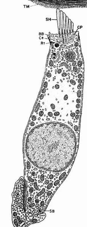
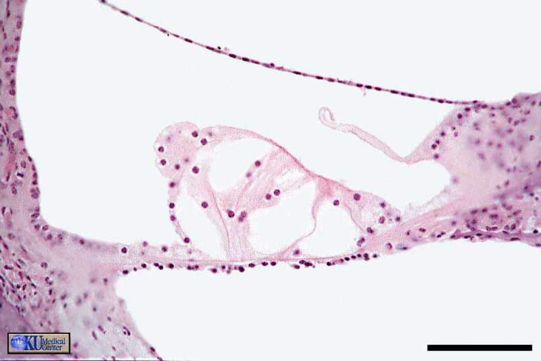
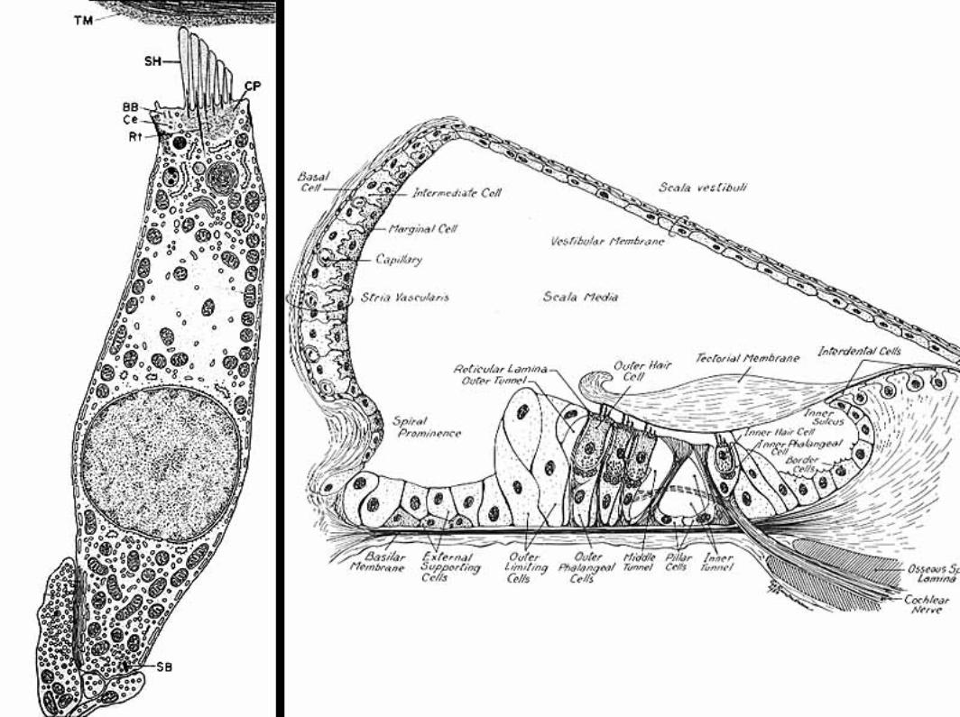
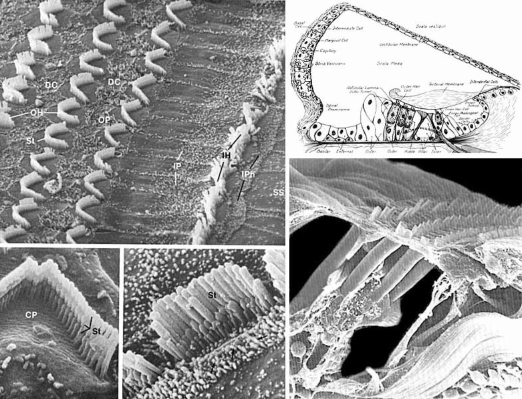
The spiral organ (organ of Corti) features receptor cells (hair cells) arranged along one inner row and three outer rows. Each hair cell has dozens of stereo-cilia on its free surface. Hair cells are held in place by a reticular membrane (plate) anchored to the basilar membrane. Stereo-cilia project above the reticular plate, making contact with a tectorial membrane. The tectorial membrane arises from the limbus, a tissue mass set solidly on the osseous spiral lamina.
Afferent neurons of the cochlear nerve have bipolar cell bodies located in a spiral ganglion, within the modiolus. From the spiral ganglion, axons traverse the osseous spiral lamina. More than 90% of the axons synapse on inner hair cells. Less than 10% of the axons synapse on outer cells, which have mostly a mechanical function adjusting the position of the tectorial membrane via cellular elongation.
Centrally, axons leave the spiral ganglion and pass through the center of the modiolus to form the cochlear division of the vestibular-cochlear nerve. In the brain, axons synapse in dorsal and ventral cochlear nuclei.
The cochlear nerve also contains inhibitory efferent axons (from dorsal nucleus of the trapezoid body) that synapse on dendritic endings of afferent neurons and on outer hair cells. Via efferent axons, the brain selectively “tunes” ear sensitivity (attention) to different ranges of sound pitch.
Scalavestibuli
Scalatympani
Cochlearduct
BasilarmembraneSpiral
ganglionOsseousspirallamina
Spiralligament
Striavascularis
Vestibularmembrane
BONE
tectorialmembrane
reticularmembrane
inner outersensory hair cells
Limbus
axons
SpiralOrgan
BASEof cochlea
APEXof cochlea
90
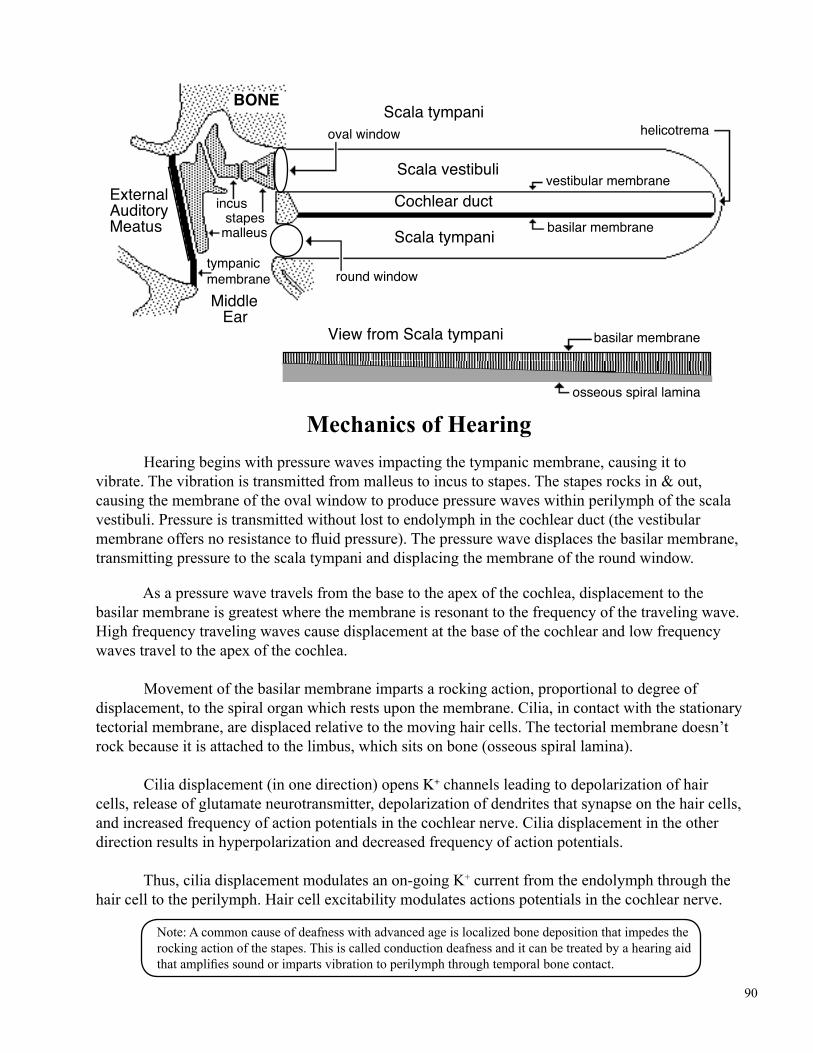
Mechanics of Hearing
Hearing begins with pressure waves impacting the tympanic membrane, causing it to vibrate. The vibration is transmitted from malleus to incus to stapes. The stapes rocks in & out, causing the membrane of the oval window to produce pressure waves within perilymph of the scala vestibuli. Pressure is transmitted without lost to endolymph in the cochlear duct (the vestibular membrane offers no resistance to fluid pressure). The pressure wave displaces the basilar membrane, transmitting pressure to the scala tympani and displacing the membrane of the round window.
As a pressure wave travels from the base to the apex of the cochlea, displacement to the basilar membrane is greatest where the membrane is resonant to the frequency of the traveling wave. High frequency traveling waves cause displacement at the base of the cochlear and low frequency waves travel to the apex of the cochlea.
Movement of the basilar membrane imparts a rocking action, proportional to degree of displacement, to the spiral organ which rests upon the membrane. Cilia, in contact with the stationary tectorial membrane, are displaced relative to the moving hair cells. The tectorial membrane doesn’t rock because it is attached to the limbus, which sits on bone (osseous spiral lamina).
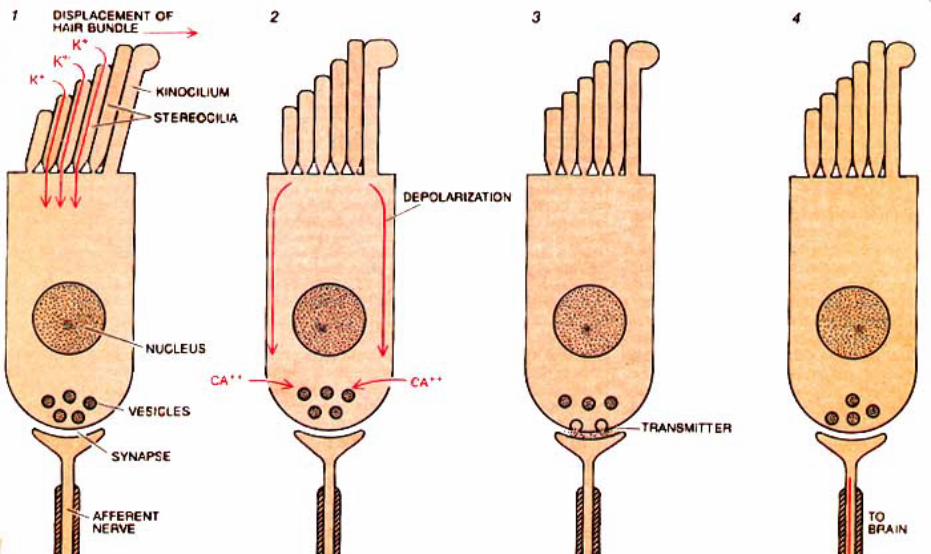
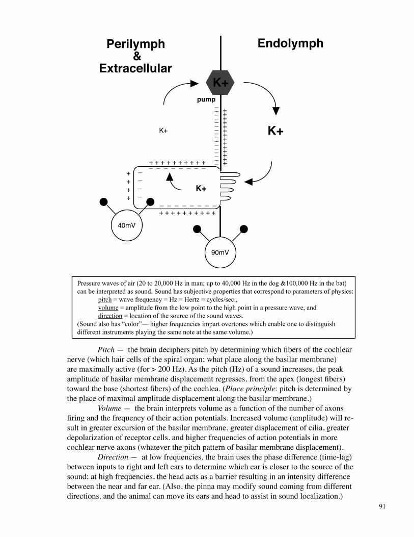
Cilia displacement (in one direction) opens K+ channels leading to depolarization of hair cells, release of glutamate neurotransmitter, depolarization of dendrites that synapse on the hair cells, and increased frequency of action potentials in the cochlear nerve. Cilia displacement in the other direction results in hyperpolarization and decreased frequency of action potentials.
Thus, cilia displacement modulates an on-going K+ current from the endolymph through the hair cell to the perilymph. Hair cell excitability modulates actions potentials in the cochlear nerve.
Note: A common cause of deafness with advanced age is localized bone deposition that impedes the rocking action of the stapes. This is called conduction deafness and it can be treated by a hearing aid that amplifies sound or imparts vibration to perilymph through temporal bone contact.
Scala vestibuli
Scala tympani
Cochlear ductExternalAuditoryMeatus
MiddleEar
View from Scala tympani
oval window
vestibular membrane
basilar membrane
osseous spiral lamina
tympanicmembrane
BONE
incusstapesmalleus
round window
helicotrema
basilar membrane
Scala tympani
91
Pressure waves of air (20 to 20,000 Hz in man; up to 40,000 Hz in the dog &100,000 Hz in the bat) can be interpreted as sound. Sound has subjective properties that correspond to parameters of physics: pitch = wave frequency = Hz = Hertz = cycles/sec., volume = amplitude from the low point to the high point in a pressure wave, and direction = location of the source of the sound waves. (Sound also has “color”— higher frequencies impart overtones which enable one to distinguish different instruments playing the same note at the same volume.)
Pitch — the brain deciphers pitch by determining which fibers of the cochlear nerve (which hair cells of the spiral organ; what place along the basilar membrane) are maximally active (for > 200 Hz). As the pitch (Hz) of a sound increases, the peak amplitude of basilar membrane displacement regresses, from the apex (longest fibers) toward the base (shortest fibers) of the cochlea. (Place principle: pitch is determined by the place of maximal amplitude displacement along the basilar membrane.) Volume — the brain interprets volume as a function of the number of axons firing and the frequency of their action potentials. Increased volume (amplitude) will re-sult in greater excursion of the basilar membrane, greater displacement of cilia, greater depolarization of receptor cells, and higher frequencies of action potentials in more cochlear nerve axons (whatever the pitch pattern of basilar membrane displacement). Direction — at low frequencies, the brain uses the phase difference (time-lag) between inputs to right and left ears to determine which ear is closer to the source of the sound; at high frequencies, the head acts as a barrier resulting in an intensity difference between the near and far ear. (Also, the pinna may modify sound coming from different directions, and the animal can move its ears and head to assist in sound localization.)
Perilymph&
Extracellular
K+
Endolymph
K+
K+
K+pump
+ + + + + + + + + +_ _ _ _ _ _ _ _
+++++++
+++++++
________
________
90mV
40mV
_ _ _ _ _ _ _ _+ + + + + + + + + +
++++
____
92
Auditory Pathway
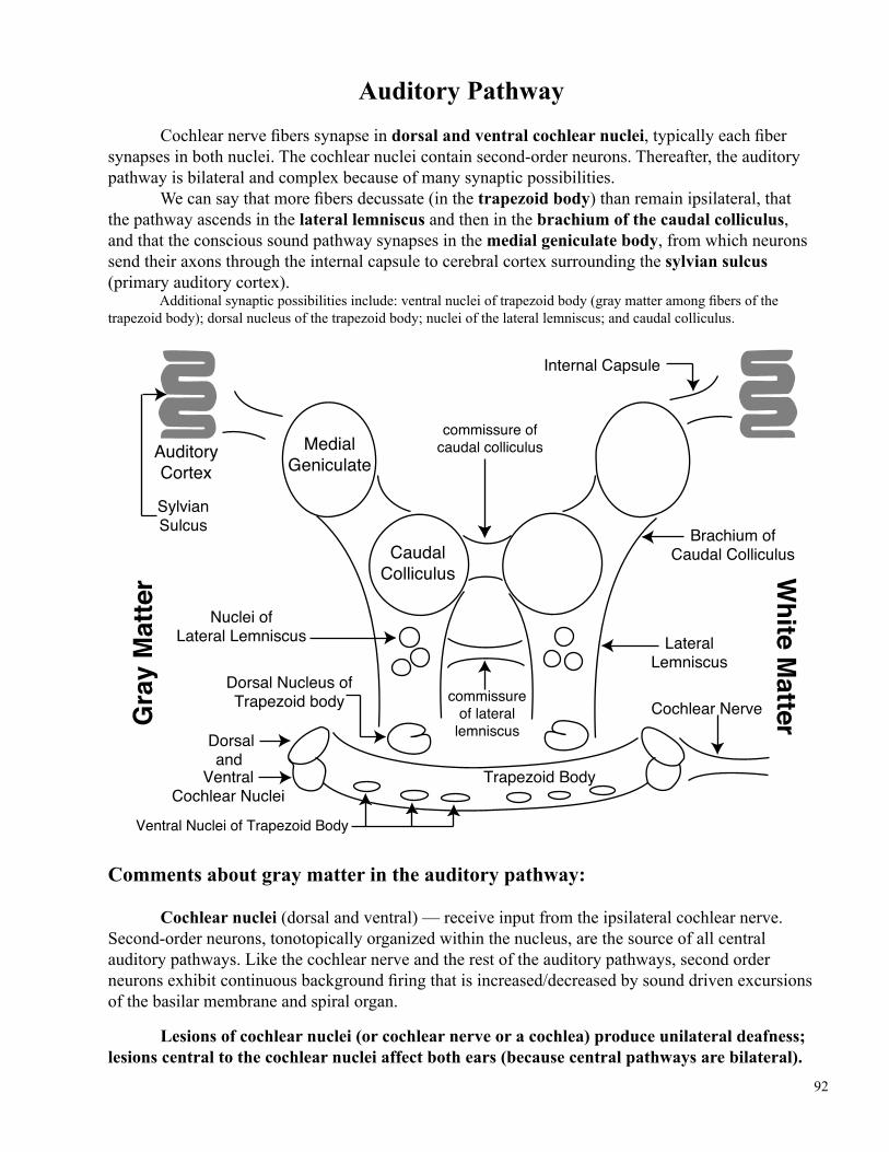
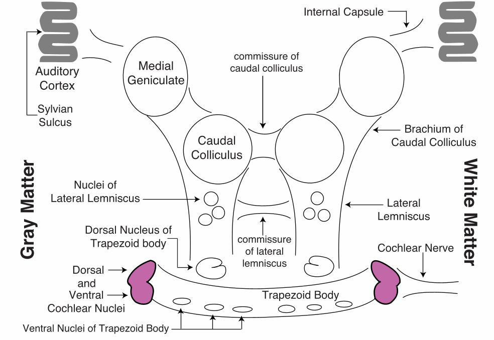
Cochlear nerve fibers synapse in dorsal and ventral cochlear nuclei, typically each fiber synapses in both nuclei. The cochlear nuclei contain second-order neurons. Thereafter, the auditory pathway is bilateral and complex because of many synaptic possibilities. We can say that more fibers decussate (in the trapezoid body) than remain ipsilateral, that the pathway ascends in the lateral lemniscus and then in the brachium of the caudal colliculus, and that the conscious sound pathway synapses in the medial geniculate body, from which neurons send their axons through the internal capsule to cerebral cortex surrounding the sylvian sulcus (primary auditory cortex). Additional synaptic possibilities include: ventral nuclei of trapezoid body (gray matter among fibers of the trapezoid body); dorsal nucleus of the trapezoid body; nuclei of the lateral lemniscus; and caudal colliculus.
Comments about gray matter in the auditory pathway:
Cochlear nuclei (dorsal and ventral) — receive input from the ipsilateral cochlear nerve. Second-order neurons, tonotopically organized within the nucleus, are the source of all central auditory pathways. Like the cochlear nerve and the rest of the auditory pathways, second order neurons exhibit continuous background firing that is increased/decreased by sound driven excursions of the basilar membrane and spiral organ.
Lesions of cochlear nuclei (or cochlear nerve or a cochlea) produce unilateral deafness; lesions central to the cochlear nuclei affect both ears (because central pathways are bilateral).
CaudalColliculus
MedialGeniculate
AuditoryCortex
Trapezoid Body
Cochlear Nerve
LateralLemniscus
Brachium ofCaudal Colliculus
Internal Capsule
Nuclei ofLateral Lemniscus
Dorsal Nucleus ofTrapezoid body
Dorsaland
VentralCochlear Nuclei
commissure ofcaudal colliculus
commissureof laterallemniscus
SylvianSulcus
Ventral Nuclei of Trapezoid Body
White
MatterG
rayMatter
CaudalColliculus
MedialGeniculate
AuditoryCortex
Trapezoid Body
Cochlear Nerve
LateralLemniscus
Brachium ofCaudal Colliculus
Internal Capsule
Nuclei ofLateral Lemniscus
Dorsal Nucleus ofTrapezoid body
Dorsaland
VentralCochlear Nuclei
commissure ofcaudal colliculus
commissureof lateral
lemniscus
SylvianSulcus
Ventral Nuclei of Trapezoid Body
White M
atterGra
y M
atte
r

93
Dorsal nucleus of trapezoid body — each nucleus receives input from right and left ears (via cochlear nuclei). The nucleus functions in sound localization, i.e., detecting phase and intensity differences between the two ears. (Different neurons respond to the different time lags between the two ears. Other neurons respond to different intensity differences between the two ears.) The nucleus sends output to cranial nerves V and VII for reflex contraction of tensor tympani and stapedius muscles to dampen loud sound. The nucleus is the source of efferent axons which selectively “tune” the spiral organ for frequency discrimination (e.g., listening to the play of one instrument within an orchestra). (Efferent innervation affects the length of outer hair cells which changes the position of the tectorial membrane which adjusts the sensitivity of inner hair cells.)
Caudal colliculus — receives input via the lateral lemniscus. The colliculus contains neurons that are sensitive to phase and intensity differences between the ears. Also, caudal colliculus neurons that project to the
medial geniculate are part of a conscious auditory pathway.
Via tectospinal/tectobulbar tracts, output from the caudal colliculus produces reflex turning of the head, ears and eyes toward a sudden sound stimulus. (Collateral branches of auditory pathway axons go to the reticular formation to alert the whole brain to a loud sound stimulation.)
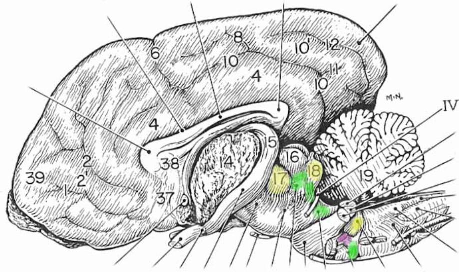
Medial geniculate — receives input via the brachium of the caudal colliculus. Imprecise sound consciousness takes place at the medial geniculate level. Geniculate neurons project their axons through the internal capsule to the primary auditory cortex. (Note: The geniculate body functions for sound like the thalamus functions for tactile sense.)
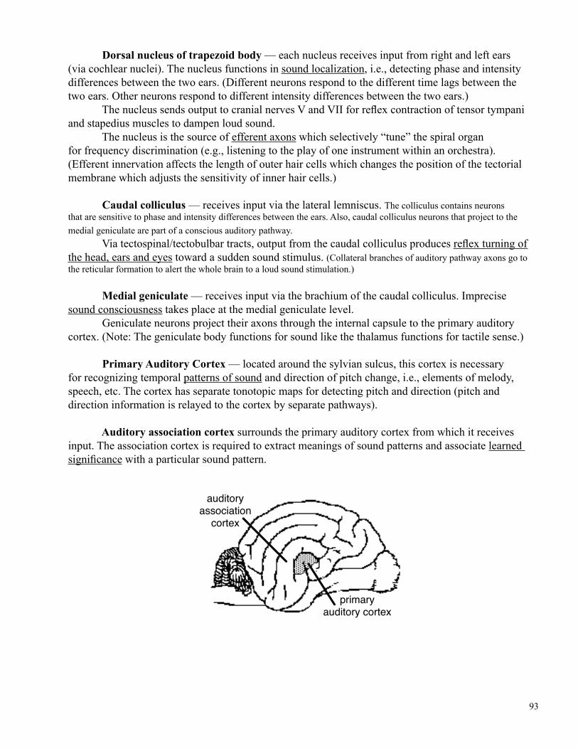
Primary Auditory Cortex — located around the sylvian sulcus, this cortex is necessary for recognizing temporal patterns of sound and direction of pitch change, i.e., elements of melody, speech, etc. The cortex has separate tonotopic maps for detecting pitch and direction (pitch and direction information is relayed to the cortex by separate pathways).
Auditory association cortex surrounds the primary auditory cortex from which it receives input. The association cortex is required to extract meanings of sound patterns and associate learned significance with a particular sound pattern.
auditoryassociationcortex
primaryauditory cortex