coexistence of both gyroid chiralities in individual...
TRANSCRIPT
Coexistence of both gyroid chiralities in individualbutterfly wing scales of Callophrys rubiBenjamin Wintera, Benjamin Butza, Christel Diekera, Gerd E. Schröder-Turkb, Klaus Meckec, and Erdmann Spieckera,1
aInstitute of Micro- and Nanostructure Research and Center for Nanoanalysis and Electron Microscopy, Department of Materials Science and Engineering,Friedrich-Alexander-Universität Erlangen-Nürnberg, 91058 Erlangen, Germany; bSchool of Engineering and Information Technology, Mathematics &Statistics, Murdoch University, Murdoch, 6150 WA, Australia; and cTheoretische Physik, Friedrich-Alexander-Universität Erlangen-Nürnberg, 91058 Erlangen,Germany
Edited by Eli Yablonovitch, University of California, Berkeley, CA, and approved August 31, 2015 (received for review June 22, 2015)
The wing scales of the Green Hairstreak butterfly Callophrys rubiconsist of crystalline domains with sizes of a few micrometers,which exhibit a congenitally handed porous chitin microstructureidentified as the chiral triply periodic single-gyroid structure. Here,the chirality and crystallographic texture of these domains are in-vestigated by means of electron tomography. The tomograms un-ambiguously reveal the coexistence of the two enantiomeric formsof opposite handedness: the left- and right-handed gyroids. Thesetwo enantiomers appear with nonequal probabilities, implying thatmolecularly chiral constituents of the biological formation processpresumably invoke a chiral symmetry break, resulting in a preferredenantiomeric form of the gyroid structure. Assuming validity ofthe formation model proposed by Ghiradella H (1989) J Morphol202(1):69–88 and Saranathan V, et al. (2010) Proc Natl Acad Sci USA107(26):11676–11681, where the two enantiomeric labyrinthinedomains of the gyroid are connected to the extracellular andintra-SER spaces, our findings imply that the structural chirality ofthe single gyroid is, however, not caused by the molecular chirality ofchitin. Furthermore, the wing scales are found to be highly textured,with a substantial fraction of domains exhibiting the<001> directionsof the gyroid crystal aligned parallel to the scale surface normal. Bothfindings are needed to completely understand the photonic purposeof the single gyroid in gyroid-forming butterflies. More importantly,they show the level of control that morphogenesis exerts oversecondary features of biological nanostructures, such as chirality orcrystallographic texture, providing inspiration for biomimetic repli-cation strategies for synthetic self-assembly mechanisms.
electron tomography | chirality | crystallographic texture |photonic crystal | butterfly wing scales
Although the formation of chiral structures is fascinating,their occurrence can often be rationalized by simple energy
or free energy considerations without a need to resort to theirpossible biological origin. For example, handed structures are ob-served in the simplest models of phyllotaxis (1) and self-assembly ofbiological fibers (2). In such models, there is no energetic distinctionbetween and hence, a balance of the two enantiomers [that is, theright-handed (RH) and left-handed (LH) versions of the chiralstructure]. Chiral symmetry breaking, the process by which oneenantiomer occurs exclusively or with prevalence, is commonlyobserved in biological materials on a range of scales from mo-lecular dimensions and the structure of DNA to the macroscopicsize of snails (3). The dominance of one enantiomer is driven bythe presence of a force or molecular building block that favorsone enantiomer over the other; constituent chiral molecules (4),genetically controlled molecular pathways (3), and biological gen-eration of torque (5) are possible causes.Here, we investigate the chiral symmetry breaking of the single-
gyroid structure, a complex network-like ordered nanostructureobserved in the wing scales of various butterfly species, includingCallophrys rubi (6–9), and other arthropod species (10). The gyroidgeometry, which serves as biophotonic crystals, has cubic symmetry(I4132) and is characterized by the topologically particularly simplesrs-net (the label for the chiral degree-three network modeled on
SrSi2) (11). As a chiral structure, it can be realized as one of twoenantiomers (related by mirror symmetry) that are here calledRH and LH. [By convention, we refer to the enantiomer of thesingle gyroid as LH, which has a screw axis along the <111>direction centered within the void domain that is an LH helix(compare with Fig. 3A, row 4). An RH helix is defined by (x, y, z)(t) =(cos t, sin t, t) for t = [0,4π] in a conventional RH coordinatesystem. For the so-defined LH single-gyroid enantiomer, thisconvention implies that the parallel screw axis along the samedirection but centered within the solid domain is RH, that the 41screw axis along the <100> direction centered within the voiddomain is RH (compare with Fig. 3A, row 3), and that the screwaxis along the <100> direction centered within the solid domainis LH. The enantiomer displayed in figure 2 in ref. 12 is LH.] Thechirality of the single gyroid is complicated by the presence ofthe distinct screw axis of opposite handedness (12). Our resultsprovide a clear indication that nature’s morphogenesis of thiscomplex nanostructure exerts control over secondary features ofthe formed complex nanostructure, leading to specific enantio-meric form and specific crystallographic texture (meaning theoccurrence of preferential crystal orientation of the micrometer-sized gyroid domains with regard to the wing scale surface).Within the wing scales, the single gyroid is realized at a largelength scale (lattice parameters around 300 nm), well beyond thesize of the constituent molecular components. This length scaleleads to structural coloration effects caused by photonic crystalproperties of the gyroid in the visible or near-UV spectrum. Thechiral geometry suggests possible circular polarization effects(13), which have been observed in nanofabricated replica and
Significance
Arthropod biophotonic nanostructures provide a plethora ofcomplex geometries. Although the variety of geometric formsobserved reflects those found in amphiphilic self-assembly, thebiological formation principles are more complex. This paperaddresses the chiral single gyroid in the Green Hairstreakbutterfly Callophrys rubi, robustly showing that the formationprocess produces both the left- and right-handed enantiomersbut with distinctly different likelihood. An interpretation ex-cludes the molecular chirality of chitin as the determining featureof the enantiomeric type, emphasizing the need to identify otherchirality-specific factors within the membrane-based biologicalformation model. These findings contribute to an understandingof nature’s ability to control secondary features of the structureformation, such as enantiomeric type and crystallographic tex-ture, informing bioinspired self-assembly strategies.
Author contributions: B.W., B.B., G.E.S.-T., K.M., and E.S. designed research; B.W., B.B.,and C.D. performed research; B.W., B.B., G.E.S.-T., and E.S. analyzed data; and B.W., B.B.,G.E.S.-T., and E.S. wrote the paper.
The authors declare no conflict of interest.
This article is a PNAS Direct Submission.1To whom correspondence should be addressed. Email: [email protected].
This article contains supporting information online at www.pnas.org/lookup/suppl/doi:10.1073/pnas.1511354112/-/DCSupplemental.
www.pnas.org/cgi/doi/10.1073/pnas.1511354112 PNAS | October 20, 2015 | vol. 112 | no. 42 | 12911–12916
APP
LIED
PHYS
ICAL
SCIENCE
SEV
OLU
TION
used for proposed biomimetic nanoengineered devices (14).Given the biological precedence of circular polarization-sensitivevision [mantis shrimp (15)] and the range of strongly circularlypolarizing biological materials (16–19) (based on Bouligand struc-tures reminiscent of cholesteric liquid crystal phases), circular po-larization effects in the butterfly wing scales would seem to be apossibility, but direct measurements of macroscopic reflectionsfail to detect a circular polarization signal (12, 20).In light of their role for circular polarization effects, it has
been questioned by Saranathan et al. (6), Mille et al. (21), andSaba et al. (12) whether both single-gyroid enantiomers occurwithin the wing scales of C. rubi (racemic mixtures with equalproportions of RH and LH enantiomers would clearly annihilateany circular polarization signal). Saranathan et al. (6) proposedthe existence of both chiralities from SEM images of the scale
surface. Mille et al. (21) reported that the RH and LH gyroidnetworks occur with a ratio of 1:7, which was as well derivedfrom SEM images. Because reliable chirality information is hardto obtain only from single projections or surface topographyimages, single-image transmission electron microscopy (TEM) orSEM techniques make it difficult to identify the enantiomerictype of a given single-gyroid structure (22). Small-angle X-rayscattering, which has been successfully used to identify the geo-metric forms observed across a large sweep of arthropod species,cannot be used to identify the enantiomeric type.A second important aspect is the relative orientation of the
nanostructured crystallites (that is, the degree of crystallographictexture). It is an essential determinant of color uniformity, becausethe photonic properties of the single gyroid strongly depend on thecrystal orientation (12, 23). Yoshioka et al. (9) observed a highlytextured crystal orientation within scales of the butterfly Paridessesostris. Although the scales of P. sesostris exhibit a multicrystal-line structure on the micrometer scale, they appear to be ratheruniform in color, which is linked to the preferential orientation ofthe nonchiral <110> direction of the individual crystallites parallelto the surface normal of the scales. This kind of crystallographictexture is not expected for C. rubi, because the crystallites differ incolor and light intensity, which is clearly visible in the light mi-croscopy images (Figs. 1 A and B and 2A).The level of structural detail described by our study is only
accessible because of advances in 3D imaging technology forbiological tissue and materials. Systematic application of scan-ning TEM (STEM) electron tomography (ET) here enables us toobtain sufficiently detailed and reliable real-space spatial data toassert firmly the chirality of the crystallites and the orientationrelation of crystallites within single butterfly wing scales ofC. rubi. Although individual tomograms have been analyzed forspecific biological tissue [including bluebird feathers (24), thebutterfly Teinopalpus imperialis (25), and also, C. rubi (8)], ourstudy clearly shows the potential that systematic application ofET holds for understanding the finer but important secondarycharacteristics, such as chirality or crystallographic texture, forbiological matter.
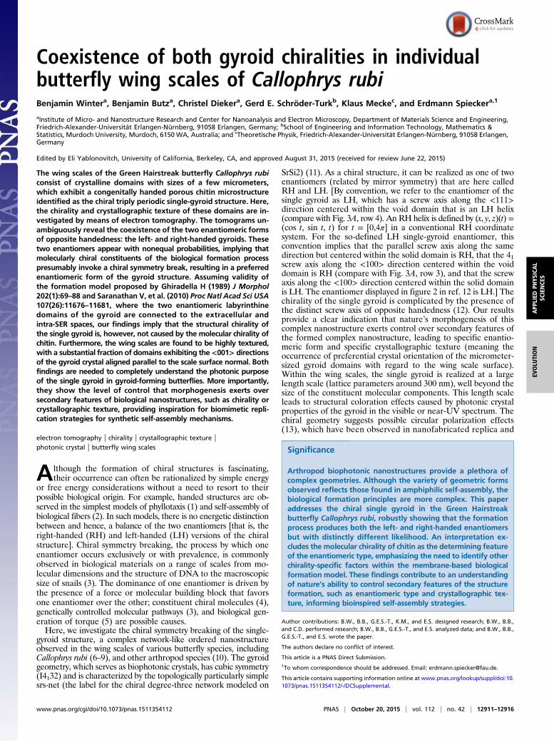
ResultsA photograph of a butterfly of the investigated species C. rubi isshown in Fig. 1A. The ventral side of its wings is covered byapproximately rectangular scales (compare Fig. 1B with Fig. 2A),which are composed of a self-supporting chitin structure with athickness of a few micrometers containing differently orientedinterconnected nanostructured crystallites with a diameter of up
Fig. 1. Butterfly C. rubi and its wing scales microstructure. (A) Photo of theventral side of C. rubi. (B) Light microscopic image (reflection mode) of itshindwing ventral surface showing hundreds of scales attached to the wing—the shimmering, mostly green scales contain the gyroid structure (imaged re-gion marked by the black rectangle in A). (C) SEM image (top view) of theventral scale surface indicating the periodic gyroid structure. (D) Model of theexpected gyroid structure (2 × 2 × 2 unit cells), with a chitin volume fraction of0.3 along (Left) the oppositely handed fourfold screw axis <001> and (Right)the threefold screw axis <111> (a formal description of the gyroid is in SI Text).
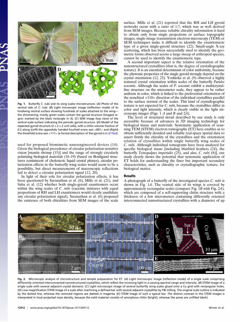
Fig. 2. Microscopic analysis of microstructure and sample preparation for ET. (A) Light microscopic image (reflection mode) of a single scale comprisingdifferently oriented interconnected nanostructured crystallites, which reflect the incoming light in a varying spectral range and intensity. (B) STEM image of asingle scale with several adjacent crystal domains. (C) Light microscopic image of several butterfly wing scales glued onto a Cu grid with rectangular holes.(D) Low-magnification STEM image of a scale after machining a defined bar with several adjacent crystallites by FIB milling. The original scale outline is indicatedby the dotted line, whereas the removed regions are dashed in magenta. (E) STEM image of such a typical bar. The distinct contrast in the STEM images isinterpreted in local projected mass density, because the solid material consists of amorphous chitin (bright), whereas the pores are unfilled (dark).
12912 | www.pnas.org/cgi/doi/10.1073/pnas.1511354112 Winter et al.
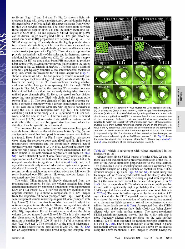
to 10 μm (Figs. 1C and 2 A and B). Fig. 2A shows a light mi-croscopic image with these nanostructured crystal domains beingdistinguishable by reflecting light (in a spectral range from yellowto blue with varying intensities). The clear correlation betweenthose separated regions (Fig. 2A) and the highly crystalline do-mains in SEM (Fig. 1C) and especially, STEM imaging (Fig. 2B)can be drawn. Single scales glued onto a TEM grid before fo-cused ion beam (FIB) preparation are depicted in Fig. 2C. TheSTEM image in Fig. 2B clearly shows the highly periodic struc-ture of several crystallites, which cover the whole scales and areconnected to parallel arranged ribs (bright horizontal line contrast)and cross-ribs (compare with Fig. 1C). Those ribs are supposed toprovide mechanical stability of the scales and furthermore, serve asadditional interference reflectors (26). To optimize the samplegeometry for ET, we used a dual-beam FIB instrument to producea bar geometry by systematically removing material from the scalesas shown in Fig. 2D (details in Methods). The bars with a width ofaround 2 μm typically comprise a few interconnected crystallites(Fig. 2E), which are accessible for tilt-series acquisition (Fig. S1shows a scheme of ET). The bar geometry assures minimal pro-jected sample thickness at high tilt angles, which drastically en-hances the quality of the 3D reconstructions, enabling reliableevaluation of the chiral gyroid morphology. Like the single STEMimages in Figs. 2B, 3, and 4, the resulting 3D reconstructions ex-hibit chitin-filled space that can be clearly distinguished from theunfilled pore channels (Fig. 3). Here, the gyroid surface dividesspace into one subvolume filled with chitin and the empty poresystem (Figs. 1–3). The pore channels of this gyroid structure ex-hibit a threefold symmetry with a certain handedness along the<111> axis and a fourfold symmetry with opposite handednessalong the <001> axis (compare with Fig. 1D). The networkexhibiting an LH screw along <111> is defined as LH srs-net-work, and the one with an RH screw along <111> is namedRH srs-net (13, 22). All reconstructed crystallites contain severalunit cells of the expected cubic gyroid structure, from which thechirality of each crystallite is determined.The tomographic reconstructions of two exemplary gyroid
crystals from different scales of the same butterfly (Fig. 3) un-ambiguously reveal that both possible mirror symmetric chiralitiesare found. Rows 3 and 4 in Fig. 3 show magnified perspectiveviews along the respective zone axes ([001] and [111]) into thereconstructed tomograms and the theoretically expected gyroidsurfaces (volume fraction of 0.3). In total, 12 crystallites from fourdifferent wing scales of one butterfly were characterized. Ten ofthem exhibit LH srs-nets, whereas only two are RH crystals (TableS1). From a statistical point of view, this strongly indicates (with asignificance level <5%) that both chiral networks appear but withunequal probabilities (a significance test is in SI Text). Both RHcrystallites were directly situated adjacent to crystals with oppositechirality (LH srs-net). In one measurement, we were even able toreconstruct three neighboring crystallites, where two LH ones di-rectly bordered one RH crystal. However, another longer barcontained only five LH crystals in a row.The chitin filling fraction is directly determined from the
reconstructed volumes. So far, the volume fraction had beendetermined indirectly by comparing simulations with experimentalSEM or TEM images (7, 21). For two exemplary crystallites withopposite chirality, Fig. 3 shows a comparison of STEM imagesfrom the respective tilt series (compare with Fig. 3, row 1) andsemitransparent volume renderings in parallel view (compare withFig. 3, row 2) of the reconstructions, which are used to adjust theoptimum segmentation threshold. These two crystallites exhibitchitin filling fractions of 0.31 ± 0.05 (Fig. 3A) and 0.29 ± 0.03(Fig. 3B), respectively. For all investigated crystallites, the chitinvolume fraction ranges from 0.28 to 0.36. This is in the range ofthe values reported in the literature, with a spread of the volumefraction of smaller [0.15–0.70 (8) and 0.17 (7)] or comparablesize [0.25–0.35 (21)]. The periodicity of the gyroid microstruc-ture of the reconstructed crystallites is 230–390 nm (SI Texthas an explanation of this quite broad range and compare with
Table S1), which is agreement with values mentioned in theliterature (8, 21).Already from single STEM images of scales (Figs. 2B and 4),
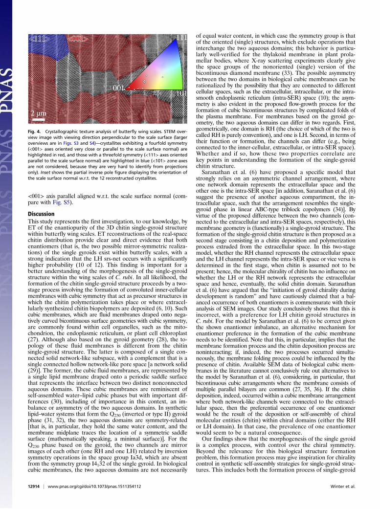
there is a clear indication for a preferred orientation of the <001>axes of the gyroid crystallites perpendicular to the scale surface.This pronounced <001> crystallographic texture is confirmed by asystematic analysis of a large number of crystallites from STEMoverview images (Fig. 4 and Figs. S3 and S4). In total, using thistechnique, 248 of 763 analyzed crystals could be clearly identifiedto have the <001> axis exactly or closely aligned with respect to(w.r.t.) the scale surface normal (a detailed description is in SIText). This corresponds to 32.5%, which indicates a crystallographictexture with a significantly higher probability than the value of1.14% expected for a random isotropic orientation (calculation isin SI Text). The result is further supported by the orientation of 12crystallites studied by ET. The partial inverse pole figure in Fig. 4,Inset shows the relative orientation of each scale surface normalw.r.t. the nearest highly symmetric axes of the reconstructed crys-tallites (more details are in SI Text). As expected from the STEMimages, most of the crystals show their <001> axis being closelyaligned with the scale surface normal. The above-mentionedSTEM analysis furthermore showed that the <111> axis also ismore frequently aligned along (or close to) the scale surfacenormal (12.5%) than expected for random isotropic distribution(1.52%) (SI Text). There is no indication of a preferred in-plane(azimuthal) crystal orientation, which was shown by an analysisusing the above-mentioned STEM images of crystals having the
Fig. 3. Exemplary ET datasets of two crystallites with opposite chirality.(A) LH srs-net and (B) RH srs-net. In row 1, STEM images from the respectivetilt series (horizontal tilt axis) of the investigated crystallites are shown withdirect view along the fourfold [001] zone axes. Row 2 shows representationsof the tomograms (volume rendering; parallel view and visualizationadapted to match the respective STEM projections in row 1) of the respectivecrystals; Insets show surface renderings of smaller parts of the crystallites. Inrows 3 and 4, perspective views along the pore channels of the tomogramsand the respective views in the theoretical gyroid structure are shown(compare with Fig. 1D). The directions of the channels within the respectivecrystallites are indicated by circles ([001]) and arrows ([111]). Fig. S2 showsthe respective views in the tomograms along the [111] zone axes. Movies S1and S2 show animations of the tomograms from A and B.
Winter et al. PNAS | October 20, 2015 | vol. 112 | no. 42 | 12913
APP
LIED
PHYS
ICAL
SCIENCE
SEV
OLU
TION
<001> axis parallel aligned w.r.t. the scale surface normal (com-pare with Fig. S5).
DiscussionThis study represents the first investigation, to our knowledge, byET of the enantiopurity of the 3D chitin single-gyroid structurewithin butterfly wing scales. ET reconstructions of the real-spacechitin distribution provide clear and direct evidence that bothenantiomers (that is, the two possible mirror-symmetric realiza-tions) of the single gyroids exist within butterfly scales, with astrong indication that the LH srs-net occurs with a significantlyhigher probability (10 of 12). This finding is important for abetter understanding of the morphogenesis of the single-gyroidstructure within the wing scales of C. rubi. In all likelihood, theformation of the chitin single-gyroid structure proceeds by a two-stage process involving the formation of convoluted inner-cellularmembranes with cubic symmetry that act as precursor structures inwhich the chitin polymerization takes place or where extracel-lularly synthesized chitin biopolymers are deposited (6, 10). Suchcubic membranes, which are fluid membranes draped onto nega-tively curved bicontinuous surface geometries with cubic symmetry,are commonly found within cell organelles, such as the mito-chondrion, the endoplasmic reticulum, or plant cell chloroplast(27). Although also based on the gyroid geometry (28), the to-pology of these fluid membranes is different from the chitinsingle-gyroid structure. The latter is composed of a single con-nected solid network-like subspace, with a complement that is asingle connected hollow network-like pore space [a network solid(29)]. The former, the cubic fluid membranes, are represented bya single lipid membrane draped onto a periodic saddle surfacethat represents the interface between two distinct nonconnectedaqueous domains. These cubic membranes are reminiscent ofself-assembled water–lipid cubic phases but with important dif-ferences (30), including of importance in this context, an im-balance or asymmetry of the two aqueous domains. In syntheticlipid–water systems that form the Q230 (inverted or type II) gyroidphase (31, 32), the two aqueous domains are symmetry-related[that is, in particular, they hold the same water content, and themembrane midplane traces the location of a symmetric saddlesurface (mathematically speaking, a minimal surface)]. For theQ230 phase based on the gyroid, the two channels are mirrorimages of each other (one RH and one LH) related by inversionsymmetry operations in the space group Ia3d, which are absentfrom the symmetry group I4132 of the single gyroid. In biologicalcubic membranes, the two aqueous domains are not necessarily
of equal water content, in which case the symmetry group is thatof the oriented (single) structures, which exclude operations thatinterchange the two aqueous domains; this behavior is particu-larly well-verified for the thylakoid membrane in plant prola-mellar bodies, where X-ray scattering experiments clearly givethe space groups of the nonoriented (single) version of thebicontinuous diamond membrane (33). The possible asymmetrybetween the two domains in biological cubic membranes can berationalized by the possibility that they are connected to differentcellular spaces, such as the extracellular, intracellular, or the intra-smooth endoplasmic reticulum (intra-SER) space (10); the asym-metry is also evident in the proposed flow-growth process for theformation of cubic bicontinuous structures by complicated folds ofthe plasma membrane. For membranes based on the gyroid ge-ometry, the two aqueous domains can differ in two regards. First,geometrically, one domain is RH (the choice of which of the two iscalled RH is purely convention), and one is LH. Second, in terms oftheir function or formation, the channels can differ (e.g., beingconnected to the inner-cellular, extracellular, or intra-SER space).Whether and if so, how these two properties correlate arekey points in understanding the formation of the single-gyroidchitin structure.Saranathan et al. (6) have proposed a specific model that
strongly relies on an asymmetric channel arrangement, whereone network domain represents the extracellular space and theother one is the intra-SER space [in addition, Saranathan et al. (6)suggest the presence of another aqueous compartment, the in-tracellular space, such that the arrangement resembles the single-gyroid phase in linear ABC-type triblock copolymers (34)]. Byvirtue of the proposed difference between the two channels (con-nected to the extracellular and intra-SER spaces, respectively), thismembrane geometry is (functionally) a single-gyroid structure. Theformation of the single-gyroid chitin structure is then proposed as asecond stage consisting in a chitin deposition and polymerizationprocess extruded from the extracellular space. In this two-stagemodel, whether the RH channel represents the extracellular spaceand the LH channel represents the intra-SER space or vice versa isdetermined in the first stage, when chitin is assumed not to bepresent; hence, the molecular chirality of chitin has no influence onwhether the LH or the RH network represents the extracellularspace and hence, eventually, the solid chitin domain. Saranathanet al. (6) have argued that the “initiation of gyroid chirality duringdevelopment is random” and have cautiously claimed that a bal-anced occurrence of both enantiomers is commensurate with theiranalysis of SEM images. Our study conclusively shows that this isincorrect, with a preference for LH chitin gyroid structures inC. rubi. For the model by Saranathan et al. (6) to be correct giventhe shown enantiomer imbalance, an alternative mechanism forenantiomer preference in the formation of the cubic membraneneeds to be identified. Note that this, in particular, implies that themembrane formation process and the chitin deposition process arenoninteracting; if, indeed, the two processes occurred simulta-neously, the membrane folding process could be influenced by thepresence of chitin. Available SEM data of biological cubic mem-branes in the literature cannot conclusively rule out alternatives tothe model by Saranathan et al. (6), considering, in particular, thatbicontinuous cubic arrangements where the membrane consists ofmultiple parallel bilayers are common (27, 35, 36). If the chitindeposition, indeed, occurred within a cubic membrane arrangementwhere both network-like channels were connected to the extracel-lular space, then the preferential occurrence of one enantiomerwould be the result of the deposition or self-assembly of chiralmolecular entities (chitin) within chiral domains (either the RHor LH domain). In that case, the prevalence of one enantiomerwould seem to be a natural consequence.Our findings show that the morphogenesis of the single gyroid
is a complex process, with control over the chiral symmetry.Beyond the relevance for this biological structure formationproblem, this formation process may give inspiration for chiralitycontrol in synthetic self-assembly strategies for single-gyroid struc-tures. This includes both the formation process of single-gyroid
Fig. 4. Crystallographic texture analysis of butterfly wing scales. STEM over-view image with viewing direction perpendicular to the scale surface (largeroverviews are in Figs. S3 and S4)—crystallites exhibiting a fourfold symmetry(<001> axes oriented very close or parallel to the scale surface normal) arehighlighted in red, and those with a threefold symmetry (<111> axes orientedparallel to the scale surface normal) are highlighted in blue (<101> zone axesare not considered, because they are very hard to identify from projectionsonly). Inset shows the partial inverse pole figure displaying the orientation ofthe scale surface normal w.r.t. the 12 reconstructed crystallites.
12914 | www.pnas.org/cgi/doi/10.1073/pnas.1511354112 Winter et al.
mesoporous silicates, where the formation of single gyroidshas been shown presumably with a racemic (balanced) mix ofenantiomers (37), and platinum and gold replicas from co-polymeric templates (38, 39). Considering the recent interestin gyroid-like structures for photonic and plasmonic applica-tions (reviewed in ref. 40) as well as their potential for chiralsieves, gaining control over the chirality is an important goal. Theoutcome of our study emphasizes the need for substantially morecomprehensive investigations of cubic bicontinous inner-cellularmembranes—a proposition with a scope that is significantlybroader than the enigma of the formation of the butterfly singlegyroid. Despite their ubiquity in biological tissue in essentialfunctional units—including the mitochondrion as the cell’s powerhouse and the thylakoid membrane involved in photosynthesis—our understanding of their formation is limited to an incompleteanalogy to synthetic cubic phases and an appreciation of theirlikely biological role largely absent from mainstream cell biology.Our identification of a preferred enantiomer within the wing
scales of C. rubi renews the question of the purpose, if any, of awell-defined chirality in this system. The single-gyroid material(of a given fixed handedness) has been clearly shown to dis-criminate between LH and RH circular polarized light (13, 14,38, 41, 42). Given the prevalence of the LH enantiomer of thesingle gyroid and the frequent occurrence of the <001> in-clination [being the sole direction with strong circular dichroism(12, 23)], the wing scales of C. rubi fulfill the prerequisites for theexistence of an optical circular polarization signal. However,spectral measurements on the wings of C. rubi (and T. imperialis,another gyroid-forming butterfly species) have failed to recordany discrimination between left and right circularly polarizedlight (12, 20). Given these observations and given the lack ofphysiological evidence of circular polarization sensitivity in in-sects, it seems that the structural chirality does not relate to bi-ologically relevant circular polarization. The possibility of otherfunctional purposes of the chiral structure cannot be eliminated,although the structure size is almost certainly too large for it toact as a chirality-selective molecular sieve [noting that, e.g., manypheromones are chiral molecules (43)]. Evidently, it is possiblethat the chirality of the single gyroid and the prevalence of oneenantiomer within the butterfly system are solely a byproduct ofthe evolutionary optimization w.r.t. nonpolarized optical prop-erties (44, 45). In this context, the intimate relationship of thegyroid to the closely related single-diamond structure is note-worthy; this latter achiral structure, which has been observed andrelated to optical function in weevils (10, 45–47), is likely to emergeby a similar process to the single gyroid [indeed, Saranathan et al.(10) report the coexistence of a single gyroid and a single-diamondstructure within Lamprocyphus weevil]. Considering, in particular,the similar properties w.r.t. nonpolarized light (e.g., figure 5 in ref.45), photonic properties do not provide the need for differentiationbetween these two bicontinuous forms. In the context of the enan-tiomeric composition and the question if the molecular chiralityof chitin contributes to the selection of the enantiomeric type,it is useful to consider a structurally unrelated chiral manifesta-tion of chitin: the Bouligand structure observed in a variety ofbeetles (17, 19, 48); in these beetles, which are strongly circularlypolarizing (19, 49, 50) and where the chirality is widely believed toresult from a process analogous to a cholesteric phase formation,only the LH enantiomer is observed. Interestingly, in the anal-ogous phase found in the cellulose structure of a particular plant,Pollia condensata, both enantiomers are observed (18).In addition to the question of enantiomeric type, our analysis
emphasizes another aspect of nature’s ability to control sec-ondary features of morphogenesis, which is manifest in thecrystallographic texture of the polycrystalline arrangement. Thepolycrystalline arrangement of the single-gyroid chitin structureshows a preferred alignment of certain crystallographic directionsw.r.t the scale’s outer surface normal, named crystallographictexture. Our findings of 12.5% and 32.5% of crystallites orientedalong the <111> and <100> crystallographic directions, respec-tively, are in stark contrast to the observation in another gyroid-
forming butterfly, P. sesostris, where a strong prevalence of the<110> direction is observed (9). This difference in the degree ofcrystallographic texture between these two species is in accordancewith the optical appearance of the wing scales, which show a ratheruniform color in the case of P. sesostris and spatial variations ofreflectivity for C. rubi. We can hypothesize that these differencesare related to the biological function of the coloration, which forC. rubi, is almost certainly camouflage and for P. sesostris, is morelikely aposematism (10, 51). It is also interesting to note that thescales of P. sesostris are covered with a highly efficient UV filter ontheir top surface (51); any reflections from crystallites with <100>or <111> direction [which reflect predominantly in the near-UV(12, 23)] would be strongly dampened by this filter, and it seemslogical that these inclinations do not occur in P. sesostris. By con-trast, C. rubi (44, 51) does not exhibit a frontally covering ab-sorbing filter covering the photonic structures [note the existenceof colocalized pigment within the gyroidal structure in C. rubi(12)]; it is conceivable that the occurrence of UV-reflecting in-clinations contributes to the camouflage effect. The presence ofdifferent preferred inclinations in P. sesostris and C. rubi hints atevolutionary control over this secondary feature of the spatialstructure (44). Like for the enantiomer specificity, a deeper un-derstanding of how the butterfly structure formation controls thecrystallographic texture could lead to useful strategies to gain controlover crystallographic texture in synthetic self-assembly processes ofcubic bicontinuous phases.In conclusion, ET has been used here to show the high level of
control that morphogenetic processes in butterfly wing scalesexert over secondary features of the nanostructure. In particular,a preferred enantiomeric form (chirality) has been identifiedalong with a crystallographic texture that deviates from that ofthe same nanostructural forms in other butterfly species. Moreinvestigation of the precursor inner-cellular cubic membranes isneeded to fully resolve the formation process of this intriguingchiral structure, with a view to replication in chirality-specificsynthetic self-assembly strategies as well.
MethodsSite-Specific FIB Preparation. Individual scales were carefully detached fromthe butterfly wings (C. rubi specimens were obtained from The InsectCompany; www.insectcompany.com) and glued onto copper TEM grids (400 ×100 mesh; glue M-Bond 610) with the long side of the scales being perpen-dicularly aligned w.r.t. with the bars of the grid (Fig. 2C). To minimize re-construction artifacts in ET caused by a drastic increase of projected massthickness at high tilt angles, we fabricated narrow bars (around 2 μmwide and10 to 15 μm long) by removing material from both sides using an FIB in-strument (30 kV, 30 pA; FEI Strata 235) as indicated in Fig. 2D. The bars werechosen to be parallel to the long axis of the scales and therefore, also parallelto the supporting ridges of the scale (bright horizontal lines in Fig. 2B). Toavoid any damage of the sensitive chitin structure, we conducted FIB millingwithout imaging or irradiating the regions of interest directly with the Ga-ionbeam. The locations of the bars were randomly selected. Each bar containedseveral directly neighboring crystallites. No further thinning of the scales wasapplied, because their thickness is usually lower than 2 μm, providing sufficientelectron transparency. In total, we analyzed 12 crystallites chosen from fourdifferent scales of one butterfly. Sample precharacterization was performedusing a Zeiss Merlin Scanning Electron Microscope (Carl Zeiss AG).
Electron Tomography. Electron tomography (ET) was performed using an FEITitan3 80–300 Transmission Electron Microscope operated at an accelerationvoltage of 300 kV. We used annular dark-field STEM imaging because of theadvantages of high depth of field and mass–thickness contrast (Rutherfordscattering). To image the quite thick chitin specimens (1–2 μm) with a suffi-cient depth of field, we reduced the semiconvergence angle α of the electronprobe to α = 1.2 mrad (μProbe STEM imaging mode) (additional details are inSI Text) (52). Tilt series covering a large tilt-angle range of ±75° with tilt in-crements of 1°–2° were obtained with an ultrathin single-tilt tomographyholder (model 2020; Fischione). The 3D reconstructions were computed usingthe FEI Inspect 3D software and applying the simultaneous iterative re-construction technique algorithm (53) with 50 iterations. We performed the3D analysis and visualization of the datasets using VSG Amira ResolveRTsoftware and ImageJ (54). A detailed description of the tomogram analysiscan be found in SI Text.
Winter et al. PNAS | October 20, 2015 | vol. 112 | no. 42 | 12915
APP
LIED
PHYS
ICAL
SCIENCE
SEV
OLU
TION
ACKNOWLEDGMENTS. We thank Bodo Wilts and Anna-Lena Robisch forfruitful discussions. Heiner Jaksch from Carl Zeiss AG is acknowledged for theacquisition of the SEM image. This research was financially supported by
German Research Foundation (DFG) Cluster of Excellence EXC 315 and DFGPriority Program SPP 1570. B.W. acknowledges support from DFG ResearchTraining Group GRK 1161.
1. Mughal A, Chan HK, Weaire D (2011) Phyllotactic description of hard sphere packingin cylindrical channels. Phys Rev Lett 106(11):115704.
2. Prybytak P, Frith WJ, Cleaver DJ (2012) Hierarchical self-assembly of chiral fibres fromachiral particles. Interface Focus 2(5):651–657.
3. Grande C, Patel NH (2009) Nodal signalling is involved in left-right asymmetry insnails. Nature 457(7232):1007–1011.
4. Giraud-Guille M-M, Belamie E, Mosser G (2004) Organic and mineral networks incarapaces, bones and biomimetic materials. C R Palevol 3(6–7):503–513.
5. Naganathan SR, Fürthauer S, Nishikawa M, Jülicher F, Grill SW (2014) Active torquegeneration by the actomyosin cell cortex drives left-right symmetry breaking. eLife3:e04165.
6. Saranathan V, et al. (2010) Structure, function, and self-assembly of single networkgyroid (I4132) photonic crystals in butterfly wing scales. Proc Natl Acad Sci USA107(26):11676–11681.
7. Michielsen K, Stavenga DG (2008) Gyroid cuticular structures in butterfly wing scales:Biological photonic crystals. J R Soc Interface 5(18):85–94.
8. Schröder-Turk GE, et al. (2011) The chiral structure of porous chitin within the wing-scales of Callophrys rubi. J Struct Biol 174(2):290–295.
9. Yoshioka S, Fujita H, Kinoshita S, Matsuhana B (2014) Alignment of crystal orienta-tions of the multi-domain photonic crystals in Parides sesostris wing scales. J R SocInterface 11(92):20131029.
10. Saranathan V, et al. (2015) Structural diversity of arthropod biophotonic nano-structures spans amphiphilic phase-space. Nano Lett 15(6):3735–3742.
11. de Campo L, Delgado-Friedrichs O, Hyde ST, O’Keeffe M (2013) Minimal nets andminimal surfaces. Acta Crystallogr A Found Adv 69(5):483–489.
12. Saba M, Wilts BD, Hielscher J, Schröder-Turk GE (2014) Absence of circular polar-isation in reflections of butterfly wing scales with chiral gyroid structure. MaterialsToday: Proceedings 1(Suppl):193–208.
13. Saba M, et al. (2011) Circular dichroism in biological photonic crystals and cubic chiralnets. Phys Rev Lett 106(10):103902.
14. Turner MD, et al. (2013) Miniature chiral beamsplitter based on gyroid photoniccrystals. Nat Photonics 7(10):801–805.
15. Chiou T-H, et al. (2008) Circular polarization vision in a stomatopod crustacean. CurrBiol 18(6):429–434.
16. Wilts BD, Whitney HM, Glover BJ, Steiner U, Vignolini S (2014) Natural helicoidalstructures: Morphology, self-assembly and optical properties.Materials Today: Proceedings1(Suppl):177–185.
17. Jewell SA, Vukusic P, Roberts NW (2007) Circularly polarized colour reflection fromhelicoidal structures in the beetle Plusiotis boucardi. New J Phys 9(99):1–10.
18. Vignolini S, et al. (2012) Pointillist structural color in Pollia fruit. Proc Natl Acad SciUSA 109(39):15712–15715.
19. Sharma V, Crne M, Park JO, Srinivasarao M (2009) Structural origin of circularly po-larized iridescence in jeweled beetles. Science 325(5939):449–451.
20. Pye JD (2014) Butterflies seem not to reflect circularly polarised light. Antenna 38(4):208–211.
21. Mille C, Tyrode EC, Corkery RW (2013) 3D titania photonic crystals replicated fromgyroid structures in butterfly wing scales: Approaching full band gaps at visiblewavelengths. RSC Adv 3(9):3109–3117.
22. Chin J, Coveney PV (2006) Chirality and domain growth in the gyroid mesophase. ProcMath Phys Eng Sci 462(2076):3575–3600.
23. Saba M, Schröder-Turk GE (2015) Bloch modes and evanescent modes of photoniccrystals: Weak form solutions based on accurate interface triangulation. Crystals 5(1):14–44.
24. Shawkey MD, et al. (2009) Electron tomography, three-dimensional Fourier analysisand colour prediction of a three-dimensional amorphous biophotonic nanostructure.J R Soc Interface 6(Suppl 2):S213–S220.
25. Argyros A, et al. (2002) Electron tomography and computer visualisation of a three-dimensional ‘photonic’ crystal in a butterfly wing-scale. Micron 33(5):483–487.
26. Ghiradella H (1989) Structure and development of iridescent butterfly scales: Latticesand laminae. J Morphol 202(1):69–88.
27. Almsherqi ZA, Landh T, Kohlwein SD, Deng Y (2009) Chapter 6: Cubic membranes themissing dimension of cell membrane organization. Int Rev Cell Mol Biol 274:275–342.
28. Schoen AH (1970) Infinite periodic minimal surfaces without self-intersections. NASATechnical Note TN D-5541 (NASA, Washington, DC), pp 1–92.
29. Kapfer SC, Hyde ST, Mecke K, Arns CH, Schröder-Turk GE (2011) Minimal surfacescaffold designs for tissue engineering. Biomaterials 32(29):6875–6882.
30. Bouligand Y (1990) Comparative geometry of cytomenbranes and water-lipid sys-tems. Journal de Physique Colloques 51(C7):35–52.
31. Luzzati V, et al. (1997) Chapter 1: The cubic phases of lipids. Lipid Polymorphism andMembrane Properties: Volume 44 of Current Topics in Membranes, eds Fambrough DM,Benos DJ, Epand R (Academic Press, San Diego), pp 3–24.
32. Shearman GC, Ces O, Templer RH, Seddon JM (2006) Inverse lyotropic phases of lipidsand membrane curvature. J Phys Condens Matter 18(28):S1105–S1124.
33. Selstam E, Schelin J, Williams WP, Brain APR (2007) Structural organisation of prola-mellar bodies (PLB) isolated from Zea mays. Parallel TEM, SAXS and absorptionspectra measurements on samples subjected to freeze-thaw, reduced pH and high-salt perturbation. Biochim Biophys Acta 1768(9):2235–2245.
34. Shefelbine TA, et al. (1999) Core−shell gyroid morphology in a poly(isoprene-block-styrene-block-dimethylsiloxane) triblock copolymer. J Am Chem Soc 121(37):8457–8465.
35. Luzzati V (1997) Biological significance of lipid polymorphism: The cubic phases. CurrOpin Struct Biol 7(5):661–668.
36. Landh T (1995) From entangled membranes to eclectic morphologies: Cubic mem-branes as subcellular space organizers. FEBS Lett 369(1):13–17.
37. Ryoo R, Joo SH, Jun S (1999) Synthesis of highly ordered carbon molecular sieves viatemplate-mediated structural transformation. J Phys Chem B 103(37):7743–7746.
38. Hur K, et al. (2011) Three-dimensionally isotropic negative refractive index materialsfrom block copolymer self-assembled chiral gyroid networks. Angew Chem Int EdEngl 50(50):11985–11989.
39. Farah P, et al. (2014) Ultrafast nonlinear response of gold gyroid three-dimensionalmetamaterials. Phys Rev Appl 2(4):044002.
40. Dolan JA, et al. (2015) Optical properties of gyroid structured materials: From pho-tonic crystals to metamaterials. Adv Opt Mater 3(1):12–32.
41. Turner MD, Schröder-Turk GE, Gu M (2011) Fabrication and characterization of three-dimensional biomimetic chiral composites. Opt Express 19(10):10001–10008.
42. Oh SS, Demetriadou A, Wuestner S, Hess O (2013) On the origin of chirality innanoplasmonic gyroid metamaterials. Adv Mater 25(4):612–617.
43. Mori K (2007) Significance of chirality in pheromone science. Bioorg Med Chem15(24):7505–7523.
44. Wilts BD, Michielsen K, De Raedt H, Stavenga DG (2012) Iridescence and spectral fil-tering of the gyroid-type photonic crystals in Parides sesostris wing scales. InterfaceFocus 2(5):681–687.
45. Wilts BD, Michielsen K, Kuipers J, De Raedt H, Stavenga DG (2012) Brilliant camou-flage: photonic crystals in the diamond weevil, Entimus imperialis. Proc Biol Sci 279(1738):2524–2530.
46. Galusha JW, Richey LR, Gardner JS, Cha JN, Bartl MH (2008) Discovery of a diamond-based photonic crystal structure in beetle scales. Phys Rev E Stat Nonlin Soft MatterPhys 77(5 Pt 1):050904.
47. Pouya C, Stavenga DG, Vukusic P (2011) Discovery of ordered and quasi-orderedphotonic crystal structures in the scales of the beetle Eupholus magnificus. OptExpress 19(12):11355–11364.
48. Sharma V, Crne M, Park JO, Srinivasarao M (2014) Bouligand structures underlie circu-larly polarized iridescence of scarab beetles: A closer view.Materials Today: Proceedings1(Suppl):161–171.
49. Caveney S (1971) Cuticle reflectivity and optical activity in scarab beetles: The rôle ofuric acid. Proc R Soc Lond B Biol Sci 178(1051):205–225.
50. Pace A, Jr (1972) Cholesteric liquid crystal-like structure of the cuticle of Plusiotisgloriosa. Science 176(4035):678–680.
51. Wilts BD, IJbema N, Stavenga DG (2014) Pigmentary and photonic coloration mech-anisms reveal taxonomic relationships of the Cattlehearts (Lepidoptera: Papilionidae:Parides). BMC Evol Biol 14:160.
52. Biskupek J, Leschner J, Walther P, Kaiser U (2010) Optimization of STEM tomographyacquisition–a comparison of convergent beam and parallel beam STEM tomography.Ultramicroscopy 110(9):1231–1237.
53. Gilbert P (1972) Iterative methods for the three-dimensional reconstruction of anobject from projections. J Theor Biol 36(1):105–117.
54. Schneider CA, Rasband WS, Eliceiri KW (2012) NIH Image to ImageJ: 25 years of imageanalysis. Nat Methods 9(7):671–675.
55. Lambert CA, Radzilowski LH, Thomas EL (1996) Triply periodic level surfaces as modelsfor cubic tricontinuous block copolymer morphologies. Philos Trans A Math Phys EngSci 354(1715):2009–2023.
56. Otsu N (1979) A threshold selection method from gray-level histograms. IEEE TransSyst Man Cybern 9(1):62–66.
57. Midgley PA, Dunin-Borkowski RE (2009) Electron tomography and holography inmaterials science. Nat Mater 8(4):271–280.
12916 | www.pnas.org/cgi/doi/10.1073/pnas.1511354112 Winter et al.