cola beverages accelerate growth of the atherosclerotic ... · cola beverages accelerate growth of...
TRANSCRIPT
ORIGINAL ARTICLE
Cola Beverages Accelerate Growth of the Atherosclerotic Plaque in ApoE-/- Mice
Received: 11/26/2013Accepted: 02/17/2014
Address for reprints:Dr. José Milei(ININCA) Instituto de Investigaciones CardiológicasUBA - CONICETMarcelo T. de Alvear 2270(C1122AAJ) CABA - ArgentinaTel./Fax +54 +11 4508-3836e-mail: [email protected]
ABSTRACT
Cardiological Research Institute. Universidad de Buenos Aires. National Scientific and Technological Research Council (ININCA.UBA.CONICET). Buenos Aires, Argentina“Prof. Dr. Alberto C. Taquini”MTSAC Full Member of the Argentine Society of CardiologyThis study was supported by the Florencio Fiorini Foundation and funds from the Cardiological Research Institute. Universidad de Buenos Aires. National Scientific and Technological Research Council (ININCA.UBA.CONICET)
enriQUetA m. serAFini, mAtilDe e. otero-losADA, gABriel cAo, gAstÓn roDrÍgUeZ-grAnillo; JimenA AgUilerA, AngÉlicA mÜller, grAcielA ottAViAno, FrAncisco AZZAto, JosÉ mileimtsAc,
ObjectivesUnhealthy eating habits during childhood and youth have been suggested as predis-posing factors for atherosclerotic complications later in life. The growing consump-tion of cola beverages in recent decades has been associated with the development of obesity and increased incidence of atherosclerosis and cardiovascular disease. We also know that there is a correspondence between the consumption of these bever-ages and the different stages of life, being higher in children, adolescents and young adults.
ObjectiveThis study evaluates the effect of cola beverage consumption on atherosclerosis.
MethodsApoE-/- mice (8 weeks old) were randomized into 3 groups according to free access to water (W), sucrose sweetened carbonated cola beverage (C) or aspartame-acesulfame K sweetened carbonated ‘light’ cola beverage (L). At 8 weeks, cola beverages were switched to water. Mice were sequentially euthanized: before treatment (8 week old mice) and after treatment discontinuation (16, 20, 24, and 30 week old mice). The ascending aorta and the liver were removed. The ratio between the aortic plaque area and the media layer thickness (plaque/media-ratio) was calculated. Hepatic in-flammation was assessed according to the NASH scale.
ResultsPlaque/media-ratio varied according to the type of beverage treatment (F2,54 = 3.433, p < 0.04) and age (F4,54 = 5.009, p < 0.03), and was higher in the C and L groups (p < 0.05 at 16 and 20 weeks, p < 0.01 at 24 and 30 weeks). Hepatic paren-chymal inflammation (F2,9 = 13.29, p < 0.002) and portal inflammation (F2,9 = 6.30, p < 0.02) increased fivefold and twofold in contrast to steatosis and hepatocel-lular damage which remained unchanged throughout the study. The W group (natu-ral evolution of atherosclerosis) evidenced acceleration of plaque growth in parallel with a rapid increase in hepatic inflammation around week 20 of age.
ConclusionsCola beverage consumption in 8-16 week old ApoE-/- mice accelerated atherosclero-sis progression Data suggest that, in this murine model, sustained cola consumption at early stages may aggravate atherosclerosis progression later in life. REV ARGENT CARDIOL 2014;82:124-128. http://dx.doi.org/10.7775/rac.v82.i1.4027
Key words > Atherosclerosis - Carbonated Beverages - Apolipoprotein E
INTRODUCTIONAtherosclerosis is the leading cause of death world-wide (1) and its risk increases with age (2). Unhealthy
eating habits during childhood and youth have been suggested as predisposing factors for atherosclerotic complications later in life (3, 4). Several longitudinal

125colA BeVerAges AnD AtHerosclerosis in mice / enriqueta m. serafini et al.
studies have shown increased cardiovascular risk in the adulthood of obese children (5.6), added to the fact that exposure to cardiovascular risk factors at early ages can contribute to the development of atheroscle-rosis (7). The growing consumption of cola beverages in recent decades has been associated with the devel-opment of obesity and increased incidence of athero-sclerosis and cardiovascular disease. At the same time we know that there is a correspondence between the consumption of these beverages and different stages of life, being higher in children, adolescents and young adults. We have recently observed the development of metabolic syndrome after prolonged consumption of cola beverages (i.e. treatment) in rats (8, 9). These studies showed the development of hypertension, hy-perglycemia, weight gain, dyslipidemia and echocardi-ographic alterations, whereas histopathological find-ings were not consistent and were more related to the aging process than to treatment (8, 9). We also noted that ApoE-/- C57BL/6J mice are particularly sensitive to damage derived from cola beverage consumption. Sucrose sweetened cola beverages (C) caused arterial disease associated with hyperglycemia (10). Consump-tion of C or L indistinctly produced increased plaque area (28 % C, 50 % L) and aortic stenosis (38 % C, 57% L) (10). Paradoxically, after discontinuing cola beverage consumption, lesions worsened (plaque area increased by 43 % in C and 68 % in L and stenosis by 71 % in C and 46 % in L). A likely explanation is that the recovery period was insufficient to allow the observation of any reversal of arterial damage consid-ering that the determinations were carried out only at the end of treatment, and the timing sequence of the posterior evolution was not assessed. Age was associ-ated with increased atherosclerotic lesions (56%).
The aim of this study was to explore the impact of cola beverage consumption (i.e. treatment) in the progression of arterial damage in ApoE-/- mice (mouse model of atherosclerosis) (11) at different times after treatment discontinuation. The hypothesis tested was that consumption of cola beverages early in life can affect the development and progression of atheroscle-rosis in adulthood.
Since the literature suggests the involvement of hepatic inflammatory processes in the development and progression of atherosclerosis, it was of interest to assess possible changes in the liver in response to cola beverage consumption in this mouse model. Spe-cifically, the temporal association between hepatic damage and atherosclerotic lesions was assessed. Ex-perimental evidence shows that ApoE-/- deficiency is related to the hepatic expression of proinflammatory mediators (12) and to aging accelerators (13).
Despite the widespread use of ApoE-/- mice for multiple purposes, the relationship between athero-sclerotic and hepatic damage is not yet clear (14).
METHODS The tests described in this study were approved by the Insti-
tutional Animal Care and Use Committee of the Universidad de Buenos Aires (IACUC) and were conducted in accordance with the recommendations of the Weatherall report (15).
A batch of sixty C57BL/6J ApoE-deficient mice (ApoE-/-), (Jackson Laboratory, Bar Harbor , Maine) were fed ad li-bitum with commercial standard rodent chow (16-18 % pro-tein, 0.2 g % sodium, Cooperación, Buenos Aires, Argentina) and were housed in a vivarium with a 12/12-hour light-dark cycle. At eight weeks of age ApoE-/-mice were randomized into 3 groups (n = 20 each). Each group had free access to one of the following beverages at room temperature: water (W), common cola beverage (C) (sucrose -sweetened carbon-ated cola beverage, Coca -Cola™, Argentina), and light cola beverage (L) (aspartame-acesulfame K sweetened carbon-ated light cola beverage, Coca -Cola Light™, Argentina) for 8 weeks. Carbon dioxide was removed by vigorous shaking until its total elimination.
After 8 weeks, colas were replaced by water in the C and L groups. Four mice per group were sequentially euthanized under anesthesia with sodium pentobarbital - diphenylhy-dantoin sodium solution (Euthanyl™): before treatment (8 weeks old: W8, C8 and L8) at the end of treatment (16 weeks old: W16, C16 and L16) and after discontinuation (20 weeks old: W20, C20 and L20, 24 weeks old: W24, C24 and L24, and 30 weeks old: W30, C30 and L30).
Tissue was removed from the ascending aorta and liv-er, dissected and immersed in buffered 10% formaldehyde solution (10% formalin buffer solution, pH = 7.0) at room temperature for a period of 24 hours. After dehydration (so-lutions of increasing concentration of ethyl alcohol at 50%, 70 %, 100%), tissues were included in paraffin blocks. Six 5 μm serial transverse sections were obtained from the aor-ta at the origin of the aortic valve leaflets and throughout the entire aortic sinus and stained with hematoxylin-eosin, Masson trichrome and orcein for elastic fiber identification. Each of the sections was evaluated using a Nikon Eclipse E400 microscope coupled to a program (Image Pro plus for Windows, v3) to analyze and average data. The plaque area, the intima layer and the media thickness were measured. The ratio between plaque area and media layer thickness (plaque/media-ratio) was calculated to estimate the degree of arterial remodeling (15). Liver sections 4 μm thick were processed for microscopy and the degree of parenchymal in-flammation was determined according to the non-alcoholic steatohepatitis scale (NASH) (16), whose score ranges from 0 to 4 (0 = lowest, 4 = highest) and includes steatosis, paren-chymal inflammation, hepatocellular injury, portal inflam-mation and fibrosis.
The data obtained were subjected to multivariate analy-sis of variance (MANOVA). The factorial ANOVA model was used to identify the factors responsible for result variations. Subsequently, post hoc Dunnett’s test allowed evaluating differences among experimental groups of the same age throughout the study. The limit of statistical significance was conventionally set at p < 0.05 (SPSS™ software version 17.0).
RESULTSFrom a qualitative point of view, at the time of au-topsy focal accumulations of lipid-laden macrophages were observed in 16 week old mice (Figure 1). Twen-ty and 24 week old mice showed globular clusters of grouped macrophages covered by a thin fibrous layer (Figure 2). At week 30, large acellular necrotic xan-thomas forming a fibro - fatty nodule extending from

ARGENTINE JOURNAL OF CARDIOLOGY / Vol 82 nº 2 / April 2014126
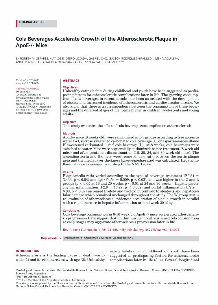
the lumen to the internal elastic lamina were found. The luminal caliber was greatly reduced due to the thinning and loss of the fibrous layer. Interruption of the internal elastic lamina with extensive atrophy of the media layer, which was replaced by plaque com-ponents (Figure 3), was observed. The plaque/media ratio varied with treatment (F2,54 = 3.433, p < 0.04) and mice age (F4,54 = 5.009, p < 0.03) and was higher in the C and L groups compared with mice of the same age in the W group (p < 0.05 in 16 and 20 week old mice, p < 0.01 in 24 and 30 week old mice) (Figure 4). The ApoE-/- mice that never consumed cola bever-ages (group W) spontaneously developed accelerated changes in the growth of atherosclerotic plaque in
parallel with a rapid increase in hepatic inflammation at around 20 weeks of age (Figure 5). Hepatic paren-chymal inflammation (F2,9 = 13.29, p < 0.002) and portal inflammation (F2,9 = 6.30, p < 0.02) varied with time (i.e. mice age) increasing fivefold and two-fold, respectively (p < 0.01 and p < 0.03 ) between weeks 20 and 30, in contrast with steatosis and hepa-tocellular damage that remained unchanged through-out the study (Figure 5).DISCUSSIONAccelerated growth of the atherosclerotic plaque was higher in C and L groups compared with the W group throughout the study. At an earlier stage of this re-search we observed the paradoxical worsening of ath-erosclerosis after discontinuation of cola beverage
Fig. 1. Histological image corresponding to the aortic wall of a 16 week old mouse. An accumulation of macrophages containing intracytoplasmic lipid microvacuoles (foam cells) is observed immediately below the vascular endothelium (between arrows). H & E, 20 ×.
Fig. 2. Histological image corresponding to the aortic wall of a 20-24 week old mouse. It shows cholesterol crystal deposits (asterisk) included in fibrocellular tissue that expand the vascular intima compressing the muscle layer (arrow). H & E, 10 ×.
Fig. 4. Effect of cola consumption on plaque/media ratio in ApoE-/- mice during the study period. Ordinate: Plaque/media ratio. Abscissa: age (weeks).* p < 0.05, # p <0.01 compared with the same age W group.
Fig. 3. Representative histological image of an aortic intimal injury corresponding to a 30 week old mouse, characterized by large acellular, lipidic accumulations (asterisks) with xanthomatous appearance, contained in fibrous tissue (arrows), in dense areas, constituting nodular formations protruding into the lumen. Masson trichrome, 10 ×.
*
*
**
A
C
L
Plaq
ue/
med
ia-r
atio
2.5
2.0
1.5
1.0
0.5
ApoE-/- mice age (weeks)8 1 6 2 4 3 2
Treatment Wash-out period
A
C
L
127colA BeVerAges AnD AtHerosclerosis in mice / enriqueta m. serafini et al.
consumption (10). However, interpretation of these results could have been challenged considering that the determinations made at a single time point after end of treatment and recovery period might have been insufficient to observe damage reversal. The results found in the present study allow us to rule out this al-ternative explanation and to confirm the rapid growth of atherosclerotic lesions as a function of time after cessation of cola beverage consumption. The rapid growth of atherosclerotic lesions observed in treated animals (i.e., who consumed cola beverages at an ear-ly age) exceeded the growth associated to the natural evolution of atherosclerosis observed in animals that never consumed cola beverages. In recent studies, it has been reported that safrole -2‘, 3’ - oxide (SFO), the main metabolite of safrole (sassafras essential oil and a minor component of nutmeg essential oil), is found in cola beverages and could aggravate atherosclerotic lesions in ApoE-/- mice (17).
In the L group, an accelerated progression of ath-erosclerosis was observed in comparison with W and C groups of the same age at the different studied times. Recently, an increase in the activity of hepatic transaminases (2.8 times) together with hyperuremia (74 %) and hypercreatininemia (2.5 times) was report-ed after consumption of “light” cola beverage in this mouse model (10). In this context of knowledge, the findings of our study suggest the association of func-tional alterations in liver, kidney, and/or muscle as a mechanism involved in the acceleration of atheroscle-rosis in ApoE-/-mice after cessation of cola beverage consumption, and provide evidence reaffirming that ApoE-/-mice are idiosyncratically sensitive to arterial damage in response to cola beverage consumption. In this instance, to discuss the real possible mechanisms responsible for these findings would be speculative, since we have not made any determinations in this regard. In any case it should be noted that a deregula-tion in the complex crosstalk among mediators of in-flammation, coagulation and pro-peroxidation mecha-nisms, to name a few, and the vascular system, would
be involved in the current findings (18-20).ApoE-/-mice that did not consume cola bever-
ages (i.e. the W Group) showed accelerated changes in atherosclerotic plaque growth, which would be ac-companied by a rapid increase in hepatic inflamma-tion around 20 weeks of age. These results concern-ing the natural history of atherosclerotic lesions in ApoE-/-mice are consistent with those found by Wat-son et al. who reported spontaneous acceleration of atherosclerotic lesions at about 20 weeks of age in this mouse model (21). As recently reported, the chrono-logical parallelism observed between increased aortic plaque area and hepatic inflammation in this study may reflect the peripheral expression of alterations at the genetic level. (22) Taken together, published re-searches using this mouse model emphasize the key involvement of the liver in the process of atherogen-esis. Experimental evidence confirms the existence of liver-artery interactions, thus illustrating remote or-gan crosstalk in atherosclerosis. (23).
CONCLUSIONSCola beverage consumption, regardless of the sugar content, increased the rate of atherosclerosis progres-sion in ApoE-/- mice, favoring the growth of aortic plaque (internal remodeling) on the thin media layer. The effects of treatment with cola beverages in 8 and 16 week old mice did not reverse even after prolonged discontinuation (30 week old mice). The data suggest that sustained consumption of cola beverages during the early stages of life can accelerate the aggravation of atherosclerotic damage in later stages of life, in a genetically favorable scenario, as is the case of athero-sclerosis prone ApoE-/- mice.
Área de placar= 0.822
Inflamaciónd el parénquima hepáticor= 0.762
Plaq
ue
area
(µ
m2
x 10
.1)
30
20
10
0
Plaque area Hepatic parenchymal inflammation
Hep
atic paren
chym
al infl
amm
ation
(NA
SH in
dex)
Age (weeks)
Fig. 5. Plaque area and hepatic parenchymal inflammation in ApoE-/- mice after cola beverage discontinuation. Ordinate: plaque area (µm2 × 103). Abscissa: age (weeks).
RESUMEN
Las bebidas cola aceleran el crecimiento de la placa ate-rosclerótica en ratones ApoE-/-
IntroducciónLos hábitos de alimentación poco saludables durante la in-fancia y la juventud se han sugerido como favorecedores de las complicaciones ateroscleróticas en edades más avanza-das. El creciente consumo de bebidas cola en las últimas décadas se ha asociado con el desarrollo de obesidad e incre-mento en la incidencia de aterosclerosis y enfermedades car-diovasculares. A su vez, se sabe que existe correspondencia entre el consumo de estas bebidas y etapas de la vida, el cual es mayor en los niños, los adolescentes y los adultos jóvenes.
ObjetivoEvaluar el efecto del consumo de bebidas cola sobre la ate-rosclerosis.
Material y métodosSe distribuyeron ratones ApoE-/- (8 semanas de edad) en tres grupos según el consumo libre de agua (A), bebida cola azucarada (C) y bebida cola edulcorada light (L). Al cabo de 8 semanas las bebidas cola se reemplazaron por agua. Los ratones fueron sacrificados secuencialmente: antes del tra-

ARGENTINE JOURNAL OF CARDIOLOGY / Vol 82 nº 2 / April 2014128
REFERENCES
1. Vasquez EC, Peotta VA, Gava AL, Pereira TM, Meyrelles SS. Cardiac and vascular phenotypes in the apolipoprotein E-deficient mouse. J BiomedSci 2012;19:22. http://doi.org/rkk2. Reddick RL, Zhang SH, Maeda N. Aortic atherosclerotic plaque injury in apolipoprotein E deficient mice. Atherosclerosis 1998;140:297-305. http://doi.org/fmcm7f3. Mahe G, Carsin M, Zeeny M, De Bosschere JP. Dietary pattern, a modifiable risk factor that can be easily assessed for atherosclerosis vascular disease prevention in clinical practice. Public Health Nutr 2011;14:319-26. http://doi.org/d7974r4. Räsänen M, Lehtinen JC, Niinikoski H, Keskinen S, Ruottinen S, Salminen M, et al. Dietary patterns and nutrient intakes of 7-year-old children taking part in an atherosclerosis prevention project in Finland. J Am Diet Assoc 2002;102:518-24. http://doi.org/fmvxzp 5. Cornier MA, Marshall JA, Hill JO, Maahs DM, Eckel RH. Preven-tion of overweight/obesity as a strategy to optimize cardiovascular health. Circulation 2011;124:840-50. http://doi.org/fsx5676. Li S, Chen W, Srinivasan SR, Bond MG, Tang R, Urbina EM, et al.
tamiento (8 semanas de edad) y luego de su interrupción (16, 20, 24 y 30 semanas de edad). Se extrajeron la aorta ascen-dente y el hígado. Se calculó la relación entre el área de la placa aórtica y el espesor de la capa media (relación placa/media). Se evaluó la inflamación del parénquima hepático según la escala de NASH.
ResultadosLa relación placa/media varió según la bebida (F2,54 = 3,433, p < 0,04) y la edad (F4,54 = 5,009, p < 0,03) y fue ma-yor en los grupos C y L (p < 0,05 a las 16 y 20 semanas, p < 0,01 a las 24 y 30 semanas). La inflamación del parénquima hepático (F2,9 = 13,29, p < 0,002) y portal (F2,9 = 6,30, p < 0,02) aumentó cinco y dos veces, respectivamente, en fun-ción del tiempo (p < 0,01 y p < 0,03) entre las semanas 20 y 30, en contraste con la esteatosis y el daño hepatocelular, que no se modificaron. El grupo A (evolución natural de la ate-rosclerosis) se caracterizó por la aceleración del crecimiento del área de placa en paralelo con un rápido aumento de la inflamación hepática alrededor de la semana 20.
ConclusionesEl consumo de bebidas cola en ratones ApoE-/- entre las se-manas 8 y 16 de edad aumentó la tasa de progresión de la aterosclerosis. Los datos sugieren que, en este modelo mu-rino, el consumo sostenido de bebidas cola durante las eta-pas tempranas de la vida puede acelerar el agravamiento del daño aterosclerótico en etapas más tardías de la vida.
Palabras clave > Aterosclerosis - Bebidas gaseosas - Apolipoproteína E
Conflicts of interestNone declared.
AcknowledgementsThe authors thank the Florenci Fiorini Foundation and the Cardiological Research Institute. Universidad de Buenos Ai-res. National Scientific and Technological Research Council (ININCA. UBA. CONICET) for supporting this study
Childhood cardiovascular risk factors and carotid vascular changes in adulthood: the Bogalusa Heart Study. JAMA 2003;290:2271-6. http://doi.org/djfkzf7. Raitakari OT, Juonala M, Kähönen M, Taittonen L, Laitinen T, Mäki-Torkko N, et al. Cardiovascular risk factors in childhood and carotid artery intima-media thickness in adulthood: the Cardiovas-cular Risk in Young Finns Study. JAMA 2003;290:2277-83. http://doi.org/cn2hbd8. Otero-Losada ME, Grana DR, Müller A, Ottaviano G, Ambrosio G, Milei J. Lipid profile and plasma antioxidant status in sweet car-bonated beverage-induced metabolic syndrome in rat. Int J Cardiol 2011;146:106-9. http://doi.org/fcsvx79. Milei J, Otero-Losada M, Gómez Llambí H, Grana DR, Suárez D, Azzato F, et al. Chronic cola drinking induces metabolic and cardiac alterations in rats. World J Cardiol 2011;3:111-6. http://doi.org/d432bf10. Otero-Losada ME, Loughlin SM, Rodríguez-Granillo G, Müller A, Ottaviano G, Moriondo M, et al. Metabolic disturbances and wors-ening of atherosclerotic lesions in ApoE-/- mice after cola beverages drinking. Cardiovasc Diabetol 2013;12:57. http://doi.org/rkm11. Meyrelles SS, Peotta VA, Pereira TM, Vasquez EC. Endothe-lial dysfunction in the apolipoprotein E-deficient mouse: insights into the influence of diet, gender and aging. Lipids Health Dis 2011;10:211. http://doi.org/fxck4z12. Yin M, Zhang L, Sun XM, Mao LF, Pan J. Lack of apoE causes alteration of cytokines expression in young mice liver. Mol Biol Rep 2010;37:2049 e54. http://doi.org/b4373z13. Bonomini F, Filippini F, Hayek T, Aviram M, Keidar S, Rodella LF, et al. Apolipoprotein E and its role in aging and survival. Exp Gerontol 2010;45:149 e57. http://doi.org/cqqh9c14. Alkhouri N, Tamimi TA, Yerian L, Lopez R, Zein NN, Feldstein AE. The inflamed liver and atherosclerosis: a link between histologic severity of nonalcoholic fatty liver disease and increased cardiovas-cular risk. Dig Dis Sci 2010;55:2644 e50. http://doi.org/dpp7qq15. Report of Sir David Weatherall’s working group. The use of non-human primates in research. 2006. 147 p.16. Collier J. Non-alcoholic fatty liver disease. Medicine 2006;35:86-8. http://doi.org/c8wg9b17. Su L, Zhang H, Zhao J, Zhang S, Zhang Y, Zhao B, et al. Saf-role-20,30-oxide induces atherosclerotic plaque vulnerability in apo-lipoprotein E-knockout mice. Toxicol Lett 2013;217:129-36. http://doi.org/rkn18. Kressel G, Trunz B, Bub A, Hülsmann O, Wolters M, Lichting-hagen R, et al. Systemic and vascular markers of inflammation in relation to metabolic syndrome and insulin resistance in adults with elevated atherosclerosis risk. Atherosclerosis 2009;202:263-71. http://doi.org/bbv2hb19. Green D, Foiles N, Chan C, Schreiner PJ, Liu K. Elevated fibrino-gen levels and subsequent subclinical atherosclerosis: the CARDIA Study. Atherosclerosis 2009;202:623-31. http://doi.org/czfqrh20. Auclair S, Milenkovic D, Besson C, Chauvet S, Gueux E, Mo-rand C, et al. Catechin reduces atherosclerotic lesion development in apo E-deficient mice: a transcriptomic study. Atherosclerosis 2009;204:e21-7. http://doi.org/bsdxms21. Watson AM, Soro-Paavonen A, Sheehy K, Li J, Calkin AC, Koitka A, et al. Delayed intervention with AGE inhibitors attenuates the progression of diabetes-accelerated atherosclerosis in diabetic apo-lipoprotein E knockout mice. Diabetologia 2011;54:681-9. http://doi.org/brpwfw22. Xu Z, Azordegan N, Zhao Z, Le K, Othman RA, Moghadasian MH. Pro-atherogenic effects of probucol in apo E-KO mice may be medi-ated through alterations in immune system: Parallel alterations in gene expression in the aorta and liver. Atherosclerosis 2009;206:427-33. http://doi.org/btjctk23. Iwata H, Aikawa M. Liver-artery interactions via the plasmin-ogen-CD36 axis in macrophage foam cell formation: new evidence for the role of remote organ crosstalk in atherosclerosis. Circulation 2013;127:1173-6. http://doi.org/rkp